Embed Size (px)
Citation preview

PATHOLOGY OF RENAL SYSTEM


BVs

Renal cortex


Tubules

Lectures Outline• Congenital anomalies & cystic diseases
• Glomerular diseases The nephrotic & nephritic syndromes
• Tubulointerstitial disease Tubulointerstitial nephritis & ATN
Urinary outflow obstruction
• Vascular disease
• Urinary tract infections
• Neoplastic disease

Function of the kidney
• The kidney is a structurally complex organ that has evolved to carry out a number of important functions.
▫ Excretion of the waste products.
▫ Regulation of body water and salt.
▫ Maintenance of appropriate acid balance.
▫ Secretion of a variety of hormones.

Renal Diseases
• NOT a major cause of death▫ RENAL -------------- 70,000 deaths / year (USA)▫ HEART -------------- 700,000 = = (USA)▫ CANCER ------------ 550,000 = = (USA)▫ STROKES -----------170,000 = = (USA)
• BUT responsible for great deal ofmorbidity▫ UTI▫ Stones▫ Obstructive uropathy▫ Dialysis & transplant▫ Many deaths occur in the young

Classification of kidney diseases
1) Glomerular diseases.
2) Tubulointerstitial diseases.
3) Vascular diseases.
• EARLY stages in the above could be separatedon clinical and morphological grounds.
• LATER all components are involved leading toEnd Stage Renal Disease (ESRD)
▫ Small contracted kidney. Obsolete glomeruli,tubular & vascular changes.
▫ Clinical --- CRF

Definitions
• Azotemia
Biochemical term linked to ↑BUN (N 7-18 mg/dL)& creatinine levels (N0.6-1.2 mg/dL).
Usually due to decrease in GFR.
Azotemia may be prerenal, renal, postrenal.
• Uremia
When azotemia progresses to clinicalmanifestations and systemic biochemicalabnormalities due to failure of renal excretoryfunction

Clinical syndromes (manifestations) of renal diseases1- Nephrotic syndrome:- Results from glomerular injury.
- Heavy proteinuria (excretion of >3.5 gm of protein/dayin adults), hypoalbuminemia, severe edema,hyperlipidemia, and lipiduria (lipid in the urine).
2- Acute nephritic syndrome:- Results from glomerular injury.
- Acute onset of usually grossly visible hematuria (redblood cells in urine), mild to moderate proteinuria,azotemia, edema, and hypertension.
-it is the classic presentation of acute poststreptococcalglomerulonephritis.


3- Asymptomatic hematuria &/or
non-nephrotic proteinuria:- Results from mild glomerular injury.
4-Rapidly progressive GN.- Results from severe glomerular injury leading to loss of
renal function in a few days or weeks and is manifested bymicroscopic hematuria, dysmorphic red blood cells andred blood cell casts in the urine sediment, and mild-to-moderate proteinuria.
5- Acute renal failure
- Oliguria or anuria (no urine flow), with recentonset of azotemia. Has many forms:Pre-renal: due to ↓ renal blood flow.
Renal: due to glomerular (as crescenticglomerulonephritis), tubular (ATN), interstitial or vascularinjury (such as thrombotic microangiopathy).
Post-renal: due to obstruction.
6- Chronic renal failure:
- Results from progressive scarring in the kidneyfrom any cause.
- Characterized by prolonged symptoms and signsof uremia.
- May lead to end-stage kidney disease (ESTD).

7. Urinary tract infection:
- May be symptomatic or asymptomatic.
- Ass. with bacteriuria & pyuria (bacteria and leukocytes in the urine).
- e.g. pyelonephritis, cystitis …
8. Nephrolithiasis:
- Manifested by renal colic, hematuria (without red cell casts), and recurrent stone formation.
9. Others:
- Obstruction, tumors …


Congenital anomaliesof the kidney
• Renal Dysgenesis:
▫ May be associated with other congenital anomalies.
▫ May lead to CRF in childhood.
1. Agenesis:
Unilateral OR
Bilateral (incompatible with life oligohydrominos &potter’s sequence).
2. Hypoplasia:
Failure to develop the normal size.
Reduced lobes and pyramids (< 6) with NO signs ofscarring.


Congenital anomaliesof the kidney
• Ectopic kidney :
▫ Usually located lower than normal (often atpelvic brim or within pelvis).
• Horseshoe kidney:
▫ Due to fusion of kidneys (90% lower poles),anterior to aorta & IVC.
▫ Main complications are obstructive uropathy &stones.
• Congenital cystic diseases


Cystic diseases of the kidney
• Heterogeneous group of hereditary,developmental & acquired disordersthat include:▫ Cystic renal dysplasia
▫ Simple renal cyst
▫ Acquired (dialysis associated) cysticdisease
▫ Polycystic kidney disease (adult &childhood types)
▫ Medullary cystic diseases.

Why they are important?
• Reasonably common.
• Some forms (as APCD) are major cause ofchronic renal failure.
• Present diagnostic problems forclinicians, radiologists, and pathologists.
• Occasionally can be confused withmalignant tumors.

1. Cystic Renal Dysplasia:
• Due to abnormalmetanephrotic differentiation.
• Gross:
▫ Enlarged cystic kidney Unilateral or bilateral(worse).
• Microscopic:
▫ Abnormal lobar organization with the presence oflarge cysts surrounded by (cartilage,undifferentiated mesenchyme, and immaturecollecting ducts).


2. Simple renal cyst(s):• A common post-mortem finding.
• Gross:▫ Single or multiple.▫ Usually small & cortical.▫ Translucent & filled with clear fluid.
• Microscopic:▫ Cysts lined by a single epithelial layer.
• Clinical:▫ NO clinical significance▫ Rarely may bleed into it distends & cause
pain.▫ Main importance is to differentiate them fromrenal tumors.


3. Acquired (dialysis-associated) renal cysts:
• Numerous cortical & medullary cysts inpatients with CRF who have undergone longterm dialysis.
• Usually asymptomatic but sometimespatients have hematuria.
• Main complication is development of renalcell carcinoma* in cyst walls (7% over 10years).


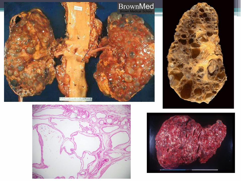
4. ADULT polycystic kidney disease:
• Multiple expanding cysts in BOTH kidneys thateventually destroy intervening parenchyma.
• Common AD disease (1 per 500-1000).
• Responsible for about 10% of CRF.
• Presentation is delayed up to 4th decade.
• Genetics: Mutations in 3 separate genes maycause disease:
▫ PKD1 (Chr 16) that encodes for POLYCSYTIN 1(90%).
▫ PKD2* (Chr 4) that encodes for POLYCYSTIN 2(10%).
▫ PKD3 (in few cases).


• Gross & microscopic features:▫ Bilateral markedly enlarged kidneys (up to 4
Kg!).
▫ Numerous cysts up to 4cm in diameter,containing clear or hemorrhagic fluid.
• Associated lesions:
▫ Polycystic liver disease - 40%.
▫ Berry aneurysms* – 10-30%.
▫ Mitral valve prolapse – ~25%.

• Clinical features:▫ Flank pain –most common presentation.
▫ Abdominal mass or dragging sensation.
▫ Intermittent gross hematuria.
• Complications:
▫ Hypertension (in 75%).
▫ UTI.
▫ Gradual onset of CRF (ESRD at 50 yrs).

5. CHILDHOOD polycystic kidney disease
• AR disease mutations in the gene encodingFIBROCYSTIN (on chr. 6).
• Presents very early & eventually causes CRF.
• Smooth kidney surface with numerous small cystsas well as dilated channels perpendicular tosurface.
• Nearly all cases have associated liver cysts.
▫ Older children who have milder disease may developcongenital hepatic fibrosis.


6. Medullary cystic diseases
A. Medullary sponge kidney.
B. Nephronophthisis–medullary cystic disease complex: is
Increasingly recognized cause CRD in childrenand young adults.
AR disease mutations in NEPHROCYSTINS(imp. in ciliary function)
Kidneys are contracted and contain multiple smallcysts typically at the corticomedullaryjunction