Embed Size (px)
Citation preview

SNA Research Conference Vol. 59 2014
Pathology and Nematology
137
Pathology and
Nematology
Alan Windham
Section Editor

SNA Research Conference Vol. 59 2014
Pathology and Nematology
138
Preliminary Evaluation of Daylily Cultivars for Rust Resistance in a Landscape Setting
Eugene K. Blythe1, Cecil Pounders2, and Michael Anderson1
1Coastal Research and Extension Center, Mississippi State University, South Mississippi Branch Experiment Station, Poplarville, MS 39470
2USDA-ARS, Thad Cochran Southern Horticultural Laboratory, Poplarville, MS 39470
[email protected] Index Words: daylily rust, Hemerocallis, Puccinia hemerocallidis. Significance to Industry: Daylilies (Hemerocallis sp.) are popular perennials in the Southern U.S. because they thrive in full sun, heat, humidity, and periods of dry weather. Daylilies are generally considered to be pest-free. However, a rust disease (Puccinia hemerocallidis), introduced into the U.S. on imported plants in 2000, has become a prevalent problem on daylilies in the lower South. Through the cooperation of a daylily specialist in Southern Mississippi, we evaluated a large, established landscape collection of 575 newer cultivars which had not been sprayed with fungicides to prevent infection by daylily rust during 2013. The warm, damp summer of 2013 was ideal for spread of daylily rust. Plants were rated on a scale of 1 (no or little visual sign of infections) to 3 (severe infection). A total of 119 cultivars received a median rating of 1 or 1.5, but some of these cultivars may be more susceptible to the disease than a single-clump rating might reveal. Planting rust-resistant daylily cultivars can reduce costs and the need to repeatedly spray plants to prevent daylily rust. Nature of Work: Daylilies (Hemerocallis sp.) are popular perennials in the landscape due to their ability to grow in a wide range of climates, soils, and light conditions, along with featuring a wide range of flower colors, shapes, and sizes. Over 17,000 cultivars are registered with the American Hemerocallis Society. Daylilies generally have few insect and disease pests in the landscape; however, daylily rust, caused by the fungus Puccinia hemerocallidis, was introduced into the U.S. on imported plants in 2000 and spread quickly through the U.S. (1). Symptoms of daylily rust include yellow to brown streaks on the leaves and small yellow spots on the upper leaf surface (2). The disease has become a ubiquitous problem on daylilies in the Southern U.S. Soon after the introduction of the disease into the U.S., daylily cultivars in existence at that time were assessed for resistance to daylily rust (3,4). Since that time, information on rust resistance or susceptibility of more recently developed cultivars has been limited. Through the cooperation of a daylily specialist in Southern Mississippi, we were able to evaluate a large, established landscape planting of 575 newer cultivars which, due to planned removal of the planting, had not been sprayed with fungicides to prevent infection by daylily rust during 2013. The warm, damp summer of 2013 was ideal for spread of daylily rust. In mid-August 2013, cultivars received ratings of 1 (showing no or

SNA Research Conference Vol. 59 2014
Pathology and Nematology
139
little visual sign of infection), 2 (moderate infection), or 3 (severe infection). The majority of the cultivars were represented by single clumps of plants in the landscape planting. Results and Discussion: A total of 119 of the 575 cultivars received a median rating of 1 or 1.5 (Table 1), indicating the possibility of rust resistance; however, some of these cultivars may be more susceptible to the disease than a single-clump rating might reveal. These cultivars are worthy of continued evaluation for resistance to daylily rust. Use of rust-resistant daylily cultivars in the landscape can help to reduce the time and expense involved in repeatedly spraying daylilies to prevent rust. Literature Cited: 1. Buck, J.W. and Y. Ono. 2012. Daylily rust. The Plant Health Instructor. DOI:
10.1094/PHI-I-2012-0516-01. 2. Harmon, P.F., C.L. Harmon, AJ. Palmateer, and S.H. Brown. 2008. Rusts on
ornamentals in Florida. University of Florida, IFAS Extension, PP256. 3. Mueller, D. S., J.L. Williams-Woodward, and J.W. Buck. 2003. Resistance of daylily
cultivars to the daylily rust pathogen, Puccinia hemerocallidis. HortScience 38:1137-1140.
4. Robbins, J.A. and S. Vann. 2012. Susceptibility of daylily to daylily rust in Arkansas. In: J.R. Clark and M.R. Evans (eds.) Horticultural Studies 2001. Arkansas Agricultural Experiment Station, Research Series 494, Fayetteville, Arkansas.

SNA Research Conference Vol. 59 2014
Pathology and Nematology
140
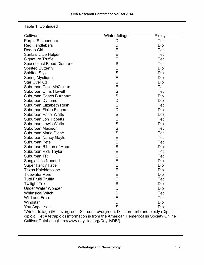
Table 1. Hemerocallis cultivars exhibiting little or no infection by daylily rust in a large landscape planting in southern Mississippi. Established plants had not been sprayed during 2013 and were rated for visual disease symptoms in August 2013. The majority of the cultivars were represented by single clumps of plants in the landscape planting; therefore, further assessment of these cultivars for rust resistance is warranted. Cultivar Winter foliagez Ploidyz Abilene Green Jeans S Tet Adorable Perfection E Dip Alabama Sweet Tee S Dip All About Eve E Tet Apricot Cream Truffle E Tet Baubles and Beads S Tet Big Ross S Dip Bluebird Butterfly E Tet Burgundy Twister E Tet Carpathian Mystic E Dip Celestial Flight E Dip Child of Atlantis S Dip Circle of Whimsy E Dip Circle Upon Circle E Dip Circles in Time S Dip Crackling Fire D Dip Creation S Dip Cricket Call S Dip Dainty is the Word S Dip Debary Canary E Tet Desert Icicle S Dip Dixieland Five D Dip Early Morning Blessing E Dip Easy on the Eyes S Tet Eloquently Edged E Dip Evelyn Gates E Dip Everybody Loves Earnest D Dip Firefly Frenzy E Dip Frilly Bliss E Dip Garden Butterfly D Dip Got the Blues S Dip Green Rainbow D Dip Green Treat S Dip Guadalajara S Tet Heavenly Realms E Dip Heaven's Artwork S Tet Hog Heaven S Tet Home Run King E Dip Hunters Quest D Tet

SNA Research Conference Vol. 59 2014
Pathology and Nematology
141
Table 1. Continued. Cultivar Winter foliagez Ploidyz Irish Icon E Dip J.T. Davis E Tet Jan's Marvel E Dip Joy of Life E Tet Kaleidoscope Jungle Cat E Dip Kaleidoscope Puzzle E Dip Kaleidoscopic Intrigue E Dip Late Afternoon Sunshine D Tet Laughing Skies E Tet Lavender Handlebars D Dip Leonardo's Perspective of Colour S Dip Lillian's Jack Temple S Dip Little Blue Belle S Dip Little Gold Nugget D Dip Little Pink Cloud S Dip Little Raven S Dip Little Red Dumples D Dip Little Trooper S Dip Little Wizard S Dip Look Here Mary S Dip Lynnstar D Dip Make Your Point E Dip Mark's Bouquet E Dip Marseilles Watercolor E Tet Maude Reese E Dip Merlin's Prophecy S Dip Mix and Match D Tet Moon Magic D Dip Morning All Day D Dip My Friend Floyd S Dip Our Friend Tom Wilson E Dip Out of the Blue E Dip Over the Line E Dip Painted Pattern S Dip Pattern Pleaser E Dip Peach Cupcake D Dip Peggy Imrie E Dip Pink Enchilada D Dip Pinwheel Princess E Dip Pixie Pinwheel Party E Dip Planet Max S Dip Plum Cupcake S Dip Pumpkin Mime S Dip
SNA Research Conference Vol. 59 2014
Pathology and Nematology
142
Table 1. Continued
Cultivar Winter foliagez Ploidyz Purple Suspenders D Tet Red Handlebars D Dip Rodeo Girl E Tet Santa's Little Helper E Tet Signature Truffle E Tet Spacecoast Blood Diamond S Tet Spirited Butterfly E Dip Spirited Style S Dip Spring Mystique E Dip Star Over Oz S Dip Suburban Cecil McClellan E Tet Suburban Chris Howell S Tet Suburban Coach Burnham S Dip Suburban Dynamo D Dip Suburban Elizabeth Rush E Tet Suburban Fickle Fingers D Dip Suburban Hazel Watts S Dip Suburban Jon Tibbetts E Tet Suburban Lewis Watts S Dip Suburban Madison S Tet Suburban Maria Diane S Tet Suburban Nancy Gayle E Tet Suburban Pete E Tet Suburban Ribbon of Hope S Dip Suburban Rick Taylor E Tet Suburban TR S Tet Sunglasses Needed E Dip Super Fancy Face E Dip Texas Kaleidoscope E Dip Tidewater Pixie E Dip Tutti Fruiti Truffle E Tet Twilight Text S Dip Under Water Wonder D Dip Whimsical Witch D Tet Wild and Free E Tet Windstar D Dip You Angel You S Dip zWinter foliage (E = evergreen; S = semi-evergreen; D = dormant) and ploidy (Dip = diploid; Tet = tetraploid) information is from the American Hemerocallis Society Online Cultivar Database (http://www.daylilies.org/DaylilyDB/).

SNA Research Conference Vol. 59 2014
Pathology and Nematology
143
Rose Rosette: Identification and Management
Mark Windham1, Alan Windham2, Frank Hale2 and Walter Hitch3
1Department of Entomology and Plant Pathology, University of Tennessee, 37996-4560 2UT Extension Soil, Plant Pest Center, Nashville, TN 37211-5112
3Plateau Research and Education Center, Crossville, TN 38571-2133
Index Words: rose, Rosa, rose rosette, Rose Rosette Virus, Phyllocoptes fructiphilus Significance to Industry: Rose rosette is a viral disease of cultivated roses. Early detection of rose rosette and removal of symptomatic plants from production or landscape plantings are critical since populations of viruliferous (carry the virus) mites may increase rapidly on infected plants. Initial symptoms rose rosette include abnormally long shoots with increased thorniness, red, strapped (thin, elongated) leaves, flattening of stems (fasciation) and formation of witches’ brooms (dense abnormally large bunching of shoots). Not all symptoms may be apparent when symptoms first become apparent and this may make diagnosis difficult. Green barriers of nonhost plants planted to the windward side of rose plantings aid in reducing incidence of rose rosette in rose plantings. Nature of Work: Rose Rosette Virus (RRV) has become widespread in regions of north-central, south-central and southeast U.S (3). Incidence of rose rosette has grown exponentially in cultivated roses in the Mid-South U.S. due to increased use of mass plantings of shrub roses in residential and commercial landscapes (4). The virus is transmitted by the eriophyid mite Phyllocoptes fructiphilus (1) which is wingless and drifts in air currents similarly as do dust particles. Landing of a rose is strictly by chance. This type of movement is known as ‘ballooning’. Rose rosette symptoms are complex and variable as plants of the same cultivar may have different symptoms at the same or different location(s). Whether this is due to variable genetics within the virus population, environmental influences including the time of season when a plant becomes infected or plant age at time of infection is unknown. Because of the variation in symptoms, RRV can be difficult to diagnoses in the field and may be confused with herbicide damage. Symptoms of rose rosette include increased thorniness, deformed leaves, deformed flower buds, proliferation of redden shoots (witches’ broom) and plant death (2). Plants that are symptomatic for rose rosette are less likely to overwinter successfully than healthy plants (4). The Beall Family Rose Garden was created within the University of Tennessee Gardens (Knoxville) in 2009. Since its inception, plants have been monitored several times weekly for symptoms of rose rosette. Initial symptoms for rose rosette were recorded and plants were then rogued by bagging the plants, cutting the plant off at soil-line, digging the root ball and bagging it as well. Bagging plants was performed before cutting, digging or moving plant parts through the garden to reduce the possibility of

SNA Research Conference Vol. 59 2014
Pathology and Nematology
144
mite vectors dropping off the plants. The number of plants destroyed each year and location of plants within the garden were recorded. In a separate experiment located at the Plateau Research and Education Center in Crossville, TN, beds of Knock Out® roses (n=16) were surrounded by Miscanthus sinensis (Andersson, Chinese or Japanese silver grass) to serve as a protective screen or left without screens (controls) (Fig. 2A). Each treatment was replicated 8 times and treatments were arranged in a completely random design. A linear bed of plants symptomatic for rose rosette (confirmed to be infected with RRV using the test described by Laney et al (3)) was established to the windward side of the plots enabling viruliferous mites to be able to balloon into test plots. Plants symptomatic for rose rosette were detected over a two year period in each plot and their location in each plot was recorded. Results and Discussion: Initial symptoms of rose rosette included fast growing erect shoots with distorted red leaves (Figure 1A), flatten stems and increased thorniness (Figure 1A, B and C). The formation of witches’ brooms, which is commonly associated with rose rosette (Figure 1D), was not observed because plants were rogued before this symptom developed. In years 2010-2013, the Beall Family Rose Garden (bushes = 200) lost 2-4% of its roses to rose rosette annually. This level of rogued plants was considered acceptable by garden management since the garden’s plans called for replacing up to 5% of roses annually to keep the garden ‘fresh’. To date, no rose adjacent to a rouge rose has become symptomatic for rose rosette. Therefore, detecting symptoms of rose rosette soon after they form and rogueing symptomatic plants is an effective way to manage rose rosette in a public garden. Barriers of M. sinensis were effective in reducing incidence of rose rosette symptomatic plants (Figure 2B) and treatments differed significantly (Chi Square Test, p=0.01). These data demonstrated that eriophyid mites that ‘balloon’ in air currents, similar to dust particles, can be intercepted by barriers placed on the windward side of a garden. In landscape situations this strategy could be used with fencing or green barriers, dependent of landscaper preference. Literature Cited 1. Amrine, J.W., Zhao,S., 1998, Research on aerial dispersal of Phyllocoptes
fructiphilus (Acari:Eriophyidae), vector of rose rosette disease. Am. Rose 3:28-29. 2. Jacobi, J. 2010. Rose Rosette Disease. http://www.aces.edu/home-garden/lawn-
garden/pests/documents/RoseRosetteDisease-Jacobi.pdf 3. Laney, A., Keller, K., Martin, R., and Tzanetakis, J. 2011. A discovery 70 years in the
making: characterization of the Rose rosette virus. J. Gen. Virol. 92:1727-1732. 4. Windham, M., Windham, A., F. Hale, and J. Amrine, Jr. 2014. Observations on rose
rosette. American Rose. May/June: 56-62.

SNA Research Conference Vol. 59 2014
Pathology and Nematology
145
Figure1. A) Drift rose that has recently become symptomatic for rose rosette. Note how the shoot stands erect above the other foliage and the leaves are redden and strapped. B) Reddening of a stem infected with rose rosette; note the thin, elongated leaves and the unusually thickened cane (stem) with increased number of thorns (pickers). C) Increased thorniness is common in many plants symptomatic for rose rosette and may be accompanied with flattened stems (fasciation). D) Masses of shoot proliferation (witches’ brooms) are often associated with plants that are very susceptible or have been symptomatic for more than one year. These witches’ brooms may harbor large numbers of viruliferous (carry the virus) eriophyid mites.

SNA Research Conference Vol. 59 2014
Pathology and Nematology
146
Figure 2. A) Research using rose plots with a barrier of Miscanthus sinensis between a reservoir of RRV infected roses that harbor large populations of eriophyid mites and RRV have demonstrated that barriers are useful in reducing incidence of RRV in rose plantings. These rose plots are located at University of Tennessee’s Plateau Research and Education Center near Crossville, TN. The rose plot is the foreground is not protected by a grass barrier whereas the rose plot in the background is protected by a barrier of Miscanthus sinensis. Note the plant in the unprotected plot with a witches’ brooms associated with infection by RRV (arrow). B) Percentage of plants in plots with or without barriers of M. sinensis two years after planting. Bars with different letters differ significantly according to a Chi Square test (p=0.01).

SNA Research Conference Vol. 59 2014
Pathology and Nematology
147
Microbial Diversity in Dogwood Seeds and Their Role as Pathogens and as Biocontrol Agents
M.T.Mmbaga1, T. Simmons2, L. Mackasmiel2 and J.O. Joshua1
1 Department of Agricultural and Environmental Sciences
Tennessee State University 3500 John A. Merritt Blvd., Nashville, TN 37209
2 Otis L. Floyd Nursery Research Center
Tennessee State University 472 Cadillac Lane, McMinnville, TN 37110
Index words: Seed-borne microorganisms, pathogens, biological control, woody ornamentals Significance to Industry: Seed quality is vital in producing healthy seedlings and healthy plants. Locally collected seeds provide a cheap source of propagation material for growers to produce plants for sale. However, seeds are capable of perpetuating seed-borne pathogens (3) subsequently leading to disease constraints in tree production and losses in net income from plant sales. Seed may also be a source of beneficial microorganisms that are capable of protecting plants against pathogens or enhance plant fitness to tolerate adverse environments and grow better. By analyzing microbial diversity in germinating seeds, we can identify seed-borne pathogens and develop management strategies to eliminate the pathogens before they impact plant growth; we may also identify beneficial microorganisms that can be utilized as biological agents that provide plants' protection against diseases or/and enhance plant defense system. Nature of Work: The objective of this study was to identify microorganisms that colonize dogwood seed and determine their potential economic importance. Dogwood seeds were collected from landscape trees at four locations in Middle Tennessee; Irving College (Warren County), Tennessee State University (TSU) Nursery Research Center (Warren County), TSU main campus in Nashville (Davidson County), and Murfreesboro (Rutherford County). Seeds were processed using seed cleaning machine, and dried for 5 days at room temperature. The dry seeds were disinfected in 10% Clorox™ bleach for 1 minute and rinsed in sterile water; they were then vernalized in sterile moist Morton’s Grow Mix™ #2 soil, at 5°C for three months. Upon germination, the germinants/germlings were surface disinfected using 70% Ethanol, rinsed twice in sterile water, blotted on sterile tissue paper and plated on Potato Dextrose Agar (PDA), to isolate fungi and bacteria. A total of 16 germinants/germlings from each of the three collection sites at Irving College/Community, TSU main campus, and Murfreesboro, and 32 germinants/germlings from TSU Nursery Research Center, were plated on PDA. Isolates of fungi and bacteria were then quantified in terms of frequency of isolation and

SNA Research Conference Vol. 59 2014
Pathology and Nematology
148
further sub-cultured into pure cultures. Fungi were grown on PDA and bacteria on nutrient agar (NA). All isolates were characterized using morphological features observed under a compound microscope and by DNA sequence analysis. Microbial DNA was extracted and analyzed following standard protocols and DNA sequence analysis was done at Davis Sequencing (Davis, CA). Blast search was done using closest similarity matches with all sequences available in the GeneBank; to refine the isolate identification, DNA sequence alignment was conducted for similarities that had 99-100. All isolates were tested for pathogenicity on flowering dogwood leaves using detached leaf technique and also tested for biological activity against known pathogens using dual cultures in vitro technique. Results and Discussion: A total of 22 isolates comprised of 18 fungi and 4 bacteria were obtained from 80 seeds (Table 1, Fig 1). Isolates of fungi were more abundant than those of bacteria with the highest number of fungal isolates from seed collected at TSU-Nashville main campus, followed by Murfreesboro seed. The TSU NRC had the highest number of bacterial isolates (Fig. 1). The most common fungus was Geomyces species (sp.) that was detected from all 4 locations (36%). Geomyces sp., are known to form ericoid mycorrhizae with the roots of alpine ericales and other perennial hosts and help these plants adapt to low-nutrient environments, its presence in dogwood seed would suggest a need to look at its role as a mycorrhizal partner in dogwood. Unidentified Geomyces spp. have also been reported to secrete a number of asterric acid derivatives with antibacterial or antifungal activity; thus suggesting that this fungus may have a biocontrol activity. Geomyces spp. are known to degrade hairs and nails hence it is used in bio-decomposition in waste management systems (Saxena et al. 2005). Geomyces destructans has been associated with the White Nose Syndrome and the decline of bat populations (4,8,9). Among the isolates that colonized dogwood seed were two known plant pathogens, Alternaria alternata, and Cylindrocapron magnusianum (Table 1). However, these two fungi are not known to cause disease in dogwood and in our pathogenicity tests, these isolates were non-pathogenic to dogwood.
Out of the four bacteria isolates, three were identified as Pseudomonas fluorescens isolated from two locations, Irving College/Community and TSU Research Center. Isolates of P. fluorescens have been reported to secrete antibiotics and hydrogen cyanide that are lethal to plant pathogens and beneficial to plants. Several studies have reported P. fluorescens as a plant growth promoting bacteria by suppressing pathogens in root zones and also by helping nutrient assimilation, while some isolates form colonies on plant surfaces and provide epiphytic fitness to the host. Isolates of P. fluorescens have been reported as biological control agents for soil borne pathogens (6,7), but we have not yet studied its potential against dogwood root pathogens.
Overall, our results show that dogwood seed carry a range of microorganisms including some that have been reported as beneficial biocontrol agents. Evaluation of the biocontrol activity produced mixed results in the in vitro study using dual cultures (data

SNA Research Conference Vol. 59 2014
Pathology and Nematology
149
not shown). Testing of the isolates that are known to have antimicrobial activity (Table 1) on a larger number of pathogens including powdery mildew and root pathogens will help identify those that have biocontrol activity in dogwood. Since these organisms colonize seed, they have great potential for application as seed treatments, for protecting young seedlings that tend to be more vulnerable to diseases. Literature Cited: 1. Bonner, F. T. 2008. Seed biology. In: USDA, The woody plant seed manual.
Agriculture Handbook 727. Washington, DC: USDA Forest Service, pp 17-51. 2. Brinkman, K. A., and Vankus, V. 2008. Seed biology. In: USDA, The woody plant
seed manual. Agriculture Handbook 727. Washington, DC: USDA Forest Service, pp 428-433.
3. Britton, K. O., and Redlin, C. 1995. Damping-off of flowering dogwood seedlings caused by Collectotrichum acutatum and Fusarium oxysporum. Plant Disease 79, 1188.
4. Gargas, A., Trest, M. T., and Christensen, M. 2009. Geomyces destructans sp. nov., associated with bat white-nose syndrome. Mycotaxon 108:147-154.
5. Gragton, W. N. 2012. "Dogwood." e-WV: The West Virginia Encyclopedia. 17 July 2012. Web., (retrieved June 2014).
6. McSpadden, G. B. B. 2007. Diversity and ecology of biocontrol Pseudomonas spp. in agricultural systems. Phytopathology 97:227-226.
7. Saxena P, Kumar A, Shrivastava JN. (2005). Keratinophilic fungi: A microbial way to manage poultry waste feathers. Indian Journal of Microbiology 45(2): 151-154.
8. Stockwell, V.O., and Stack J.P. 2007. Using Pseudomonas spp. for integrated biological control. Phytopathology 97:244-249.
9. National Speleological Society. July 19, 2013. Caving News. http://cavingnews.com/20130719-geomyces-destructans-reclassified-pseudogymnoascus-destructans, (retrieved June 2014).
10. National Speleological Society. March 20, 2012.Caving News. http://cavingnews.com/20120320-geomyces-destructans-confirmed-in-tennessee-great-smoky-mountains-national-park-white-nose-syndrome-wns, (retrieved June 2014).

SNA Research Conference Vol. 59 2014
Pathology and Nematology
150
Table 1: Isolates obtained from vernalized dogwood seeds, the locality from which the seeds were collected in Middle Tennessee and reported economic importance of the isolates.
Isolate
Seed Collection Location
Economic Importance
Alternaria alternata
TSU Nashville
Plant pathogen (leaf spots, rots and blights), Asthma (human)
Auerobasadium pullulans
TSU Research Center
Biological Control Agent (storage diseases)
Cylindrocapron magnusianum
TSU Nashville
Plant pathogen (root rot in alfalfa and red clover)
Epicoccum species
Irving College, TN
Heterocyclic compounds with thiodiketopiperzine backbone (epicoccins, difenylalazines or epicorazines, the latter having antibacterial properties)
Geomyces pannorum
TSU Nashville, TSU Research Center, Irving College & Murfreesboro
Biodegradation of soil buried polyester polyurethane in landfill
Geomyces species
TSU Research Center
Ericoid mycorrhizae in roots (alpine, and other perennials)
Humicola grisea
TSU Nashville
Production of hemicelulase activity (b-xylanse, b-mannanase and a-arabinofuranosidase
Ophiostoma nigrocapum TSU Research Center Saprophyte Pantoea vagans
TSU Nashville
Plant epiphyte. Strain c9-1 biocontrol agent of fire blight
Penicillum species TSU Nashville Food and drug production Pseudomonas fluorescens
TSU Research Center Irving College, TN
Biocontrol agent (Fusarium or Pythium)
Umbelopsis isabellina
Murfreesboro
Oleaginous, high intracellular lipid accumulation in plants
Umbelopsis ramaniana Irving College, TN Oleaginous (produces oils)

SNA Research Conference Vol. 59 2014
Pathology and Nematology
151
Figure 1: Seed-borne fungi and bacteria isolated from vernalized seeds collected from flowering dogwood in Warren County-Community (Co), Murfreesboro (MB) in Rutherford County, Nursery Research Center (NRC & R14 at NRC) in McMinnville, Warren County, and Tennessee State University, Nashville (TSU) in Davidson County.
0
1
2
3
4
5
6
Co TSU NRC MB R14
Num
ber
(Iso
late
)
Seed Source
Fungi Bacteria

SNA Research Conference Vol. 59 2014
Pathology and Nematology
152
Sources of Phytophthora Species and Other Pathogens in Middle Tennessee Nurseries
L. A Mackasmiel 1 and M.T Mmbaga2
1 Otis L. Floyd Nursery Research Center
Tennessee State University 472 Cadillac Lane, McMinnville, TN 37110
2 Department of Agricultural and Environmental Sciences
Tennessee State University 3500 John A Merritt Blvd, Nashville, TN 37209
Index words: Irrigation water, field survey, soil-borne pathogens, woody ornamentals Significance to Industry: The genus Phytophthora consists of destructive species of phytopathogens that cause root rot, root collar rot, blights and cankers in different ornamental plants, as well as in forestry and food crops (2). These pathogens impact nursery production in middle Tennessee; they can survive for short periods as zoospores and sporangia and often survive for many years as chlamydospores and oospores. Wet conditions are most favorable for Phytophthora diseases and they spread very easily in water, soil and plant materials. If not controlled, Phytophthora diseases cause problems over long periods of time (2). A recent report on Phytophthora in Virginia and Mississippi, identified new species associated with irrigation water, these include P. hydropathica and P. mississippiae, respectively; soil and vegetative materials also harbor the pathogen (3,6). Some species of Phytophthora may spread as air-borne sporangia and lead to infection over a wide geographical area. This study was conducted to (i) identify species of phytophthora, (ii) determine the trend of plant infection(s) by Phytophthora spp. and identify other soil-borne pathogens that impact nursery production in middle Tennessee. Nature of Work: This study was conducted during the 2013 growing season to survey for Phytophthora in three creeks and eight nurseries across middle Tennessee, and document any other soil-or water-borne phytopathogens of significance. The three creeks were at least 10 miles apart and they were selected indiscriminately among creeks whose water is used for irrigation. Baiting for Phytophthora was done twice at different periods, using leaves of Rhododendron, Pieris sp. pine (Pinus sp) needles that are known to have high susceptibility to Phytophthora. Samples from irrigation water, soil (field and container), and plant material (roots and leaves) were collected simultaneously from symptomatic nursery plants in 8 nurseries and evaluated for the presence of Phytophthora.
SNA Research Conference Vol. 59 2014
Pathology and Nematology
153
Clean baits consisting of 6 leaves from each bait plant were placed in breathable (pollination) bags and floated on creek water for 3 days. The leaves were then collected, cleaned using sterile water, blotted dry using sterile tissue paper, then cut into 100 mm diameter discs; these were plated on phytophthora specialized medium (PARPH-V8) for 48 hr. Colonies of Phytophthora spp. were sub-cultured in V8 agar using hyphal tips tand pure cultures were generated. Water samples from nursery irrigation ponds were also collected and placed in sterile 100 x 25 mm Petri dishes to a depth of 15 mm; leaf baits consisting of 100 mm diameter leaf discs from Rhododendron, and Pieris; and pine needles were then placed on water surface for up to 72 h in a darkened laminar flow hood, at 12ºC temperature. The leaf baits were then plated in PARPH-V8 agar for 48 h. Fungal growths around the baits were carefully lifted and plated on V8 agar and grown into pure cultures. In the case of soil, the samples were first thoroughly mixed in a clean tray, then filled in 21.0 cm x 13.5 cm x 4.0 cm (L x W x H) sterilized plastic lunch boxes to a depth of 1.5 cm. Sterile water was then added up to a depth of 1.0 cm above the soil line and leaf discs were floated on the water to bait Phytophthora species. A total of 6 leaf baits with two leaves of Rhododendron, Pieris and Pinus were used per sample and replicated three times. After placing leaf baits on the water, the samples were maintained in the dark in a laminar flow hood at 12ºC, as described above. Samples of plant materials for the isolation of Phytophthora and other pathogens were collected from leaves and roots of symptomatic plants, washed to remove soil particles and disinfected in 70% ethanol. They were then rinsed in sterile water and blotted dry on sterile tissue paper before plating on PARPH-V8 for 3 days as described above. Culturing and sub-culturing of fungal samples on V8 agar, was done as described above. A total of four pieces of plant material of roots or leaves per plate for each sample was replicated three times. Fungi isolated were identified using morphological characteristics, and DNA sequence analyses of the internal transcribed spacer (ITS) region, including the intervening 5.8S rRNA gene as described by White et al. (1990). Qiagen DNeasy Plant mini Kit was used for DNA extraction of fungal isolates and DNA amplification was done using universal primer pairs ITS1 and ITS4. Each PCR reaction included 10X PCR buffer, 25 mM of MgCl2, 10mM of dNTPs, Taq polymerase, and sterile double distilled water (ddH2O) in 50-μL reaction mixes. The DNA was amplified using a Techne thermocycler and the PCR products were analyzed by electrophoresis in a 1.5% agarose gel in 0.5X TBE (Tris-Borate-EDTA) buffer. The gels were stained with ethidium bromide (0.5 μg/ml) and DNA was visualized using UV light. The PCR products were purified using the Quiagen PCR purification kit and DNA sequencing was done at Davis Sequencing Inc. (http://www.davissequencing.com). The sequences were compared with information available in the GenBank using Blast search; information was also compared with what is documented in the literature. Results and Discussion: Overall, Pieris and Rhodendron leaf baits had more Phytophthora growth compared to Pinus needles. Individual nurseries showed variations in the number of positive samples baited from soil, and plant material (Fig. 2). More than 75% of water samples collected tested positive for Phytophthora spp., and other fungi, while less than 25% of soil and plant samples had Phytophthora spp or

SNA Research Conference Vol. 59 2014
Pathology and Nematology
154
other fungi (Fig. 2). Water samples had 27.6% positive results, soil showed 10.0%, while plant material had only 0.5% from roots and 0.3% from leaves (Fig 1-3). These results show similar trends to previous surveys (2, 3) with water samples showing higher proportion (%) of Phytophthora spp., and other fungi than soil and plant material (Figs, 2-3). This suggests that using such contaminated water for irrigation may transmit Phytophthora and other pathogens to susceptible plants including nursery plants, or other plants in the field. Although all Phytophthora isolates baited from water may not be pathogenic on nursery plants, a previous evaluation of 10 uncultured Phytophthora spp., from water, tested on nursery plants at the center, showed that all of them were pathogenic on flowering dogwood leaves while some were pathogenic on leaves of several other ornamental plants (4, 5). Among Phytophthora spp. isolated in this study, 50% were P. hydropathica, 10% were P. parsiana while P. irrigata, P. palmivora and P. drechsleri were 3% each (Fig 4). Also detected at 3%, each, were uncultured fungus clones, Halophytophthora polymorphica, and Phytopythium litorale. Although phytophthora-specialized media are expected to limit growth of Pythium, several species of Pythium were isolated; the dominant ones were P. condricola (30%), P. pophyrae (28%), P. catenulatum (17%) and P. litorale (8%). Other fungi detected included Fusarium solani, Verticillium nubilum, and Arthrinium phaeospermum (Fig 5). These fungi may not have any reported pathogenicity on woody ornamentals, but they had significant presence (Fig. 5). Thus, it is important to conduct pathogenicity tests on diverse nursery plants, on both roots and leaves, in order to determine their association/interactions with nursery plants. Acknowledgements: The authors thank Terry Kirby and Terri Simmons for their technical support during the season. Literature Cited: 1. Drench,. A., and Sendall, B. 2001. Practical guide to detection and identification of
Phytophthora. CRC -Tropical Plant Protection, Brisbane, Australia. 2. Erwin, D.C. and O.K. Ribeiro. 1996. Phytophthora diseases worldwide. St. Paul,
Minnesota: APS Press. 3. Hong, C. X., Gallegly, M. E., Richardson, P. A., Kong, P., Moorman, G. W., Lea-
Cox,J. D., and Ross, D. S. 2010. Phytophthora hydropathica, a new pathogen identified from irrigation water, Rhododenon catawbiense and Kalmia latifolia. Plant pathol., 59: 913-921. Doi: 10.1111/j.1365-3059.2010.02323.x.
4. Santamaria, L., and Mmbaga, M. T., et al. 2008. Plant pathogenic Phytophthora species found in Middle Tennessee commercial nurseries. SNA bulletin.
5. Santamaria, L., and Mmbaga, M. T. 2007. A survey for Phytophthora diseases in Middle Tennessee nurseries: Identification and characterization. SNA bulletin.
6. Yang, X., Copes, W. E., and Hong, C. 2013. Phytophthora mississippiae sp. nov., a new species recovered from irrigation reservoirs at a plant nursery in Mississippi. J. Plant Microbiol., 4:180 doi:10.4172/2157-7471.1000180.

SNA Research Conference Vol. 59 2014
Pathology and Nematology
155
Figure 1. Percentage of all samples from water, soil and plant materials that showed positive results for phytopathogens.
Figure 2. Comparison of mean level of all positive results from each nursery sampled
during the survey.
78.86
19.77
0.78 0.590
20
40
60
80
100
Water Soil Roots Leaves
Per
cent
age
(%)
Sample/Specimen Tested
0.00
0.05
0.10
0.15
0.20
0.25
BDN SN GVN MCN MN BFN HHN PGN
Mea
n S
ampl
e (P
ositi
ve)
Nursery (Code Name)

SNA Research Conference Vol. 59 2014
Pathology and Nematology
156
Figure 3. Comparison of creeks showing mean recovery of Phytophthora species and other plant pathogens from creek water. The creeks were baited for pathogens two months apart during 2013 growing season between May and October.
Figure 4: The overall diversity of Phytophphora species and other fungi detected during the survey and their percentage of representation.
Figure 5. Proportion (%) of Pythium species that have been analyzed and confirmed using standard DNA protocols.
Phytophthora hydropathica50%
Phytophthoraparsiana10%
Phytophthora irrigata3%
Phytophthorapalmivora3%
Phytophthoradrechsleri3%
Halophytophthora polymorphica3%
Phytopythium helicoides16%
Phytopythium litorale3%
Uncultured fungus3%
Uncultured Fungus clone3%
Fungal Endophyte3%
Pythium adhaerens
5%Pythium
catenulatum17%
Pythium condricola
30%
Pythium dissotocum
2%
Pythium helicoides
2%Pythium litorale8%
Pythium middletonii
5%
Pythium multisporum
3%
Pythium porphyrae
28%

SNA Research Conference Vol. 59 2014
Pathology and Nematology
157
A New Sclerotinia Disease on Container Grown Abelia and Nandina in Alabama
J. Olive1, K. Conner2 1Auburn University OHRC, Mobile, AL 36689
2Auburn University, Auburn, AL, 36849
Index words: Sclerotinia sclerotiorum, Abelia grandiflora, Nandina domestica
Significance to Industry: Abelia sp. and Nandina sp. are both popular landscape plants that make up a significant portion of sales for nurseries in the southeastern U.S. The warm season disease called southern blight or white mold, caused by Sclerotium rolfsii is common in the Deep South and is fairly easy to diagnose. A disease caused by a similar fungal pathogen, Sclerotinia sp., was identified in two nurseries in 2014 on Abelia and Nandina. This is typically a cool season pathogen commonly found on vegetables. Sclerotinia sp. is not commonly identified on woody ornamentals in the Southeast and may be misdiagnosed. This disease is difficult to control and requires specific fungicides and management practices. It is important for growers to be aware of this disease and to recognize the symptoms to separate it from similar diseases. Nature of the Work: In the early spring of 2014, Abelia x grandiflora ‘Kaleidoscope’ was found at a commercial nursery in south Alabama with browning out and twig dieback. The leaves on individual branches turned straw brown and dropped off, leaving bare twigs and areas of the canopy bare. Common Abelia pathogens were not observed. Plants were collected and symptomatic tissue was placed in a moist chamber. Within 48 hours, profuse white mycelium was growing on the necrotic leaves and stems but no spores were produced to identify the fungus. Within five days, concentrated tuffs of mycelium were visible and small, black, irregular structures, identified as sclerotia, were observed on the damaged plants. Later this spring, a crown rot was reported on several cultivars of dwarf Nandina in another commercial nursery in south Alabama. Plants exhibited typical crown rot symptoms of browning and discoloration on the lower stem and crown, accompanied by wilting and leaf drop. Tuffs of white mycelium, similar to that seen previously on the Abelia, were observed on symptomatic plants. These plants were examined closely and sclerotia, similar to those observed on Abelia, were found. Sclerotia were not obvious and were often found on the stem covered by leaf petioles or leaf debris, which could delay identification. Results and Discussion: Based on colony morphology and sclerotia production, the pathogen on both the Abelia and Nandina was identified as Sclerotinia sclerotiorum (1,2,3). Sclerotinia sp. is a fairly common cool season pathogen on vegetable crops but has not been reported as a serious pathogen on woody ornamentals in the Deep South. This disease requires cool temperatures (54-720 F), saturated soils, and prolonged leaf

SNA Research Conference Vol. 59 2014
Pathology and Nematology
158
moisture for infection (1). This may explain why the disease was active in 2014. Temperatures were below average and there were several rain events in excess of 2 inches, accompanied by prolonged cloudy weather. With the occurrence of this disease on Abelia and Nandina, two commonly grown woody ornamentals, growers should be aware of the symptoms of this disease so it can be identified quickly and effective control measures initiated. Literature Cited: 1. Heffer Link, V., and K. B. Johnson. 2007. White Mold. The Plant Health Instructor.
DOI:1094/PH-I-2007-0809-01. 2. Wan Gyu Kim and Weon Dae Cho. 2003. Occurrence of Sclerotinia Rot in
Solanaceous Crops caused by Sclerotinia. Mycobiology. Jun:31 (2):113-118. http://dx.doi.org/10.4489/MYCO.2003.31.2.113
3. Willetts, H.J., and J. A. L. Wong. 1980. The biology of Sclerotinia sclerotiorum, S. trifoliorum, and S. minor with emphasis on specific nomenclature. Bot Rev. 46:101-165.

SNA Research Conference Vol. 59 2014
Pathology and Nematology
159
Preliminary Results of Three Biofungicides and MilStop for Controlling Basil Downy Mildew
Jiasai Li1, Mengmeng Gu1, Fu Cheng1, Yanjun Guo1 and Kevin Ong2
1Department of Horticultural Sciences 2Department of Plant Pathology & Microbiology
Texas AgriLife Extension Service Texas A&M University
2134 TAMU l College Station, Texas 77843
Index Words: Basil downy mildew, biofungicide Significance to Industry: In the United States, the production of basil (Ocimum basilicum L.) has risen significantly due to the increasing of marked demand over the past several years (1), and basil is also a popular garden and landscape plant. Three biofungicides, in addition to MilStop (potassium bicarbonate), were applied weekly, alone, in tank mix or in rotation, to Genovese basil plants grown in 6” containers in a glasshouse from October 28, 2013 to December 02, 2013. Plants were rated on a 0-7 scale (0=no leaves affected and 7= all leaves affected) on November 05, 2013, November 19, 2013 and December 03, 2013. There was no significance in the first two ratings. BP1 alone or mixed with BP2 had higher ratings than the other treatments. This preliminary trial helped to define future biofungicide trials to control basil downy mildew. Nature of Work: Basil (Ocimum basilicum L.) is an important commercial herb crop used in cooking and gardens decoration. There are many types of basils. The genus Ocimum encompasses 35 species of annuals and perennials from the tropical and subtropical areas. Sweet basil (O. basilicum) is the most common species cultivated for culinary and ornamental uses, and there are about 100 cultivars. In the United States, the production of basil has risen significantly due to the increasing of marked demand over the past several years (1). A total of 2,053 U.S. farms produced herbs for the fresh market in 2007 on 13,573 acres of land, which represents an increase of nearly 25% in the five years since the last agricultural census in 2002. Many cultivars of basil are also very popular in gardens and landscapes. Basil downy mildew, a severe disease that is caused by Peronospora belbahrii and could result in 100% crop loss, has spread to about 40 states since the first report in the United States (South Florida) in the fall of 2007. The earliest report of basil downy mildew in Texas was in 2010 (2). Symptoms of basil downy mildew could be mistakenly considered as nutrient deficiency. The affected plants may show general yellowing and different levels of chlorosis (yellowing) on lower older leaves, which is common in plants with N deficiency. However, chlorosis caused by nutrient deficiency is not vein-bounded,

SNA Research Conference Vol. 59 2014
Pathology and Nematology
160
while close inspection of yellowing of basil leaves with downy mildew, especially those in more advanced stage, could generally identify the vein-bounded pattern. This experiment was a preliminary trial set up for proof of concept based on growers’ use patterns based on their positive experiences, and was conducted in a glass greenhouse located on Texas A&M University campus at College Station, TX, from October 28, 2013 to December 3, 2013. The Genovese basil plants were obtained from a local wholesale nursery in Waller, TX, and were potted on October 23, 2013 in six-inch plastic pots (1,250 ml) with Miracle-Gro (ScottsMiracle-Gro Company, Marysville, OH). Four type of biofungicide (BP1, BP2, BP3 and MilStop) were mixed into nine different biofungicides (Table 1), and sprayed thoroughly on basil plants weekly from October 28, 2013 to December 02, 2013. Due to a non-discloure agreement with the biopesticide company, the biofungicides used in the trial were only given a code (“BP1”, “BP2”, and “BP3”) in this manuscript. Three visual rating of unmarketable leaves (chlorosis and necrosis from downy mildew infestation, and phytotoxicity effects due to chemical application) were conducted on November 05, 2013, November 19, 2013 and December 03, 2013. Plants were visually rated on percentage of leaf have chlorosis, necrosis or phytotoxicity symptoms on a scale of 0 to 7 (0=no leaves affected and 7= all leaves affected), as illustrated in Fig. 1. The experiment was a completely random block design with 10 treatments (nine biofungicides and control) and four blocks (replications). All data was analyzed by SAS (version 9.4; SAS Institute, Cary, NC). When the effect was significant, mean separation was conducted using Student-Newman-Keuls test at 5% significance level. Results and Discussion: Biofungicide treatments did not have a significant effect on plant visual ratings on November 05, 2013, November 19, 2013 (Table 2). On December 03, 2013, plants treated by BP1 or a mixture of BP1 and BP2 had higher visual rating than control, and the other biofungicide treatments did not yield results significantly different from the control. This indicated that regular application of BP1 or a mixture of BP1 and BP2 may have caused higher phytotoxicity, and thus more unmarketable leaves on plants. Literature Cited: 1. Mersha, Z., Zhang, S., & Raid, R. N. (2012). Evaluation of systemic acquired
resistance inducers for control of downy mildew on basil. Crop Protection, 40, 83-90. 2. Mersha, Z., Zhang, S. (2013). A recently discovered foliar disease of basil: Downy
mildew and its management in South Florida. Horticulture Sciences at University of Florida, Issue No. 569.

SNA Research Conference Vol. 59 2014
Pathology and Nematology
161
Figure 1. Visual rating scale of 0-7 (0=no leaves affected and 7= all leaves affected) on the percentage of ‘affected’ leaves (either by downy mildew or phytotoxicity). No plants were rated 0 or 7 and so only 1-6 are represented here. Arrows indicate affected leaves.
Table 1. The rates of nine biofungicide treatments spray thoroughly on basil plants weekly from October 28, 2013 to December 02, 2013.
Treatments Rate Control (Water) N/A BP1 1.5oz/gal BP2 1.50% BP3 1.9 oz/gal MilStop 7 g/gal BP3+MilStop (Tank mix) 1.9 oz/gal+7 g/gal BP3+MilStop (Rotation) 1.9 oz/gal+7 g/gal BP1+BP2 1.5oz/gal+1.50% BP1+BP3 (Tank mix) 1.5oz/gal +1.9 oz/gal BP1+BP3 (Rotation) 1.5oz/gal +1.9 oz/gal

SNA Research Conference Vol. 59 2014
Pathology and Nematology
162
Table 2. Three visual rating of unmarketable leaves (chlorosis and necrosis from downy mildew infestation, and phytotoxicity effects due to chemical application) were conducted at November 05, 2013, November 19, 2013 and December 03, 2013.
Treatments 11/5/2013 11/19/2013 12/3/2013 Control (Water) 1.8 a 1.0 a 1.3 b BP1 0.5 a 0.0 a 3.5 a BP2 2.0 a 1.0 a 2.3 b BP3 1.5 a 1.7 a 1.5 b MilStop 2.8 a 1.5 a 2.5 b BP3+MilStop (Tank mix) 1.0 a 0.8 a 2.3 b BP3+MilStop (Rotation) 2.0 a 0.5 a 1.3 b BP1+BP2 0.8 a 0.3 a 4.0 a BP1+BP3 (Tank mix) 3.3 a 1.0 a 1.8 b BP1+BP3 (Rotation) 1.3 a 0.3 a 2.0 b

SNA Research Conference Vol. 59 2014
Pathology and Nematology
163
Mefenoxam Insensitivity in Pythium and Phytophthora Isolates from Ornamental Plants in Georgia
Jean L. Williams-Woodward and Max E. DeMott
Department of Plant Pathology, University of Georgia, Athens, GA 30602
Index Words: Fungicide, Insensitivity, Mefenoxam, Oomycete, Phytophthora, Phytopythium, Pythium, Subdue Maxx Significance to the Nursery Industry: The majority of root and crown diseases on ornamental plants are caused by the Oomycete pathogens, Pythium spp. and Phytophthora spp. There are a limited number of commercially available fungicides with activity against Oomycete pathogens. The predominant fungicide used in Pythium and Phytophthora management has been mefenoxam (Subdue Maxx; Syngenta Crop Protection, Inc., Greensboro, NC). Insensitivity to mefenoxam has been reported within species of Pythium and Phytophthora recovered from ornamental plants (1, 3, 4, 5). Mefenoxam insensitivity across all isolates evaluated in these studies ranged from 6% to 66% and included up to 8 Phytophthora and 11 Pythium species. The studies suggest that mefenoxam insensitivity is widespread within floriculture production. The viability of mefenoxam as a valuable tool in managing Pythium and Phytophthora root diseases is of great concern. Nature of Work: Species of Pythium and Phytophthora cause root, crown, stem, and foliage blights. Symptoms often include root softening, sloughing, darkening of roots, crowns and stems, wilting, foliage chlorosis, leaf drop, stem dieback, and leaf and petiole blighting. Oomycete pathogens or “water molds” are unique and are not true fungi. They are more closely related to brown algae than fungi. One of the major differences between Oomycetes and true fungi is in their cell wall components. Oomycete cell walls are composed of a β-1,3 and β-1,6 glucans, whereas true fungi cell walls are composed of chitin. This is an important distinction because the mode of action of many fungicides is to act on and inhibit chitin cell wall biosynthesis. Since Oomycete cell walls do not contain chitin, these products have no activity on these pathogens. This has resulted in a limited number of commercially available fungicides with activity against Pythium and Phytophthora diseases. The predominant fungicide used against Pythium and Phytophthora diseases in ornamentals has been the phenylamide systemic fungicide, metalaxyl, which was replaced by mefenoxam (the R-enantiomer of metalaxyl), and marketed under the trade names of Subdue 2E and Subdue Maxx (Syngenta Crop Protection, Inc., Greensboro, NC), respectively. Metalaxyl was registered for use in the United States in 1980 and within four years fungicide resistance in Pythium causing turf blight was identified (8).

SNA Research Conference Vol. 59 2014
Pathology and Nematology
164
Mefenoxam insensitivity has been noted in several states in Pythium and Phytophthora species causing root and crown rots of ornamental plants. In Pennsylvania, 32.5% of the 120 Pythium isolates recovered from infected plants were insensitive to mefenoxam (3). Eleven species of Pythium were identified from the 120 isolates. The most common species were P. irregulare and P. aphanidermatum of which 36.8% and 37.5% of these species, respectively, were insensitive to mefenoxam. In North Carolina, three species of Phytophthora (P. nicotianae, P. cryptogea, and P. palmivora) were recovered as the predominant species infecting floriculture crops (1). Although, all isolates of P. palmivora were still sensitive to mefenoxam, 100% of the P. cryptogea and 21% of P. nicotianae isolates were insensitive. In a more recent North Carolina study, P. nicotianae, P. drechsleri, P. cryptogea, and P. tropicalis were the most commonly recovered Phytophthora species from floriculture corps, of which 66% of these Phytophthora isolates were insensitive or intermediate in resistance to mefenoxam (4). Mefenoxam insensitivity appears widespread. The objectives of this study was 1) to identify species of Pythium and Phytophthora from symptomatic plants within both floriculture and woody ornamental crops in GA and 2) to evaluate the recovered isolates for mefenoxam sensitivity. Plant samples exhibiting symptoms of root or crown rot or foliage dieback were collected from 17 wholesale ornamental production facilities (nine specializing in container-grown woody shrubs and eight specializing in floriculture or herbaceous crops) from 2010-2011. Symptomatic tissue sections were washed with tap water, blotted dry, and plated onto V8-PARP medium [15 g Bacto agar (Becton, Dickerson and Co., Sparks, MD); 50 ml clarified V-8 juice (Campbells, Camden, NJ); 67 mg 75% PCNB (Terraclor; Chemtura, Middlebury, CT); 400 µl pimaracin (Sigma-Aldrich, St. Louis, MO); 250 mg ampicillin (Sigma-Aldrich, St. Louis, MO); 10 mg rifampicin (Sigma-Aldrich, St. Louis, MO) in 950 ml deionized water] (2). Plates were incubated in the dark at 22C for up to 10 days. Suspected Pythium and/or Phytophthora colonies were transferred by hyphal tip onto new V8-PARP or non-amended V8-agar plates to obtain a pure culture. Putative Pythium or Phytophthora isolates were flooded with non-sterile soil extract to observe sporangia formation and morphological characteristics. The ITS region (ITS1, 5.8S, and ITS2) of the rDNA of each isolate was sequenced to provide DNA-based identification. Suspected Pythium and Phytophthora isolates were grown on V8-agar at 22-24°C for 72 h. Hyphae was scraped and/or lightly touched with a 200-l pipette tip. The tip was then placed into a 0.5-ml PCR tube containing a PuReTaq Ready-To-Go™ PCR Bead (GE Healthcare), 1 l of 10 µM ITS-1 primer (5'- TCCGTAGGTGAACCTGC GG-3’), 1 l of 10 µM ITS-4 primer (5’-TCCTCCGCTTATTGATATGC-3’), and 23 l of sterile nuclease-free water and mixed by gently pipetting up and down several times. Total PCR reaction volume was 25 µl. Thermal cycling conditions consisted of an initial denaturation at 94°C for 5 min; followed by 34 cycles of 94°C for 1 min, 53°C for 1 min, and 72°C for 1 min; and a final extension step of 72°C for 5 min, followed by a 4°C hold (3). PCR products were purified using QIAquick Purification Kit (Qiagen, Inc., Valencia, CA) and submitted to the Georgia Genomics Facility (Athens, GA). DNA sequences

SNA Research Conference Vol. 59 2014
Pathology and Nematology
165
were aligned and manually edited using Geneious software (Biomatters Ltd., Auckland, New Zealand). ITS sequences were BLAST analyzed in GenBank (National Center for Biotechnology Information, Bethesda, MD) and the Phytophthora Database (http://www.phytophthoradb.org/). All isolates were screened for sensitivity to mefenoxam in vitro by amending V8-agar (50 ml clarified V8 juice; 15 g Bacto agar; 950 ml deionized water) with 100 g a.i./ml of mefenoxam by suspending Subdue Maxx (Syngenta Crop Protection, Greensboro, NC) in water and distributing it in molten agar prior to pouring into 35-mm plastic petri plates and evaluating mycelia growth compared to the growth of the same isolate on non-amended medium. Agar plugs (7-mm in diameter) were cut from the leading edge of a 3-4 day old isolate culture and inverted onto the center of mefenoxam-amended and non-amended plates. Two non-amended and two mefenoxam-amended plates for each isolate was incubated at 22C in the dark for 24-48 h depending upon isolate growth rate. Mycelial growth was measured from the inoculated plug edge to the edge of the colony along two radii per plate. Isolates that grew ≥ 50% of the non-fungicide amended control plates (EC50 > 100 ug a.i./ml) were considered insensitive. Isolates that grew < 50% as compared to the control were considered to be sensitive. Results and Discussion: Out of the 152 symptomatic samples collected, Oomycete root pathogens were recovered from 80% of them. Either no pathogen or a non-oomycete pathogen was recovered from the remaining samples. Of the 121 oomycete isolates recovered, 39 were identified as Phytophthora spp., 77 as Pythium spp., and five as Phytopythium spp. (Table 1). The Phytophthora species identified included P. nicotianae, P. pini, P. undulata, P. cinnamomi, P. citrophthora, P. palmivora, P. dreschsleri, and P. cryptogea, with P. nicotianae being the most prevalent (30% of the Phytophthora isolates). Approximately, 21% of the Phytophthora isolates could not be identified to species based upon morphology or DNA sequencing. This is not uncommon and has been seen in previous studies (1, 4, 5). Pythium species recovered included Pythium irregulare, P. myriotylum, P. aphanidermatum, P. monospermum, P. chamaihyphon, P. vexans, P. diclinum, P. cucurbitacearum, P. zingiberis, and P. acanthophoron (Table 1). Pythium irregulare was the most prevalent and accounted for 12.5% of the identifiable species. The majority of the Pythium isolates recovered (approximately 50%) could not be identified to the species level. Five Phytopythium isolates were also recovered from diverse symptomatic plants including Coreopsis lanceolata, Hydrangea arborescens, Rosmarinus officinalis, Tagetes patula, and Thymus praecox from three production facilities. Phytopythium is a relatively new taxonomic genus whose members were classified as clade K species of Pythium, and have more characteristics similar to Phytophthora than other Pythium species. Pythium litorale and Pythium heliocoides are now classified as Phytopythium species (7) and both were recovered in this study. In recent studies, Phytopythium heliocoides was found to be pathogenic to begonia in VA (9) and P. litorale was pathogenic to squash in GA (6).

SNA Research Conference Vol. 59 2014
Pathology and Nematology
166
Across all oomycete isolates in this study, 45.5% were mefenoxam insensitive (Table 2). Insensitivity was identified in 27.2% of the Phytophthora isolates. All isolates of P. undulata and P. palmivora recovered from three production facilities and 57% of the unidentifiable Phytophthora spp. were insensitive to mefenoxam. However, all P. nicotianae and P. pini isolates, which accounted for over 50% of the total number of Phytophthora isolates recovered, were sensitive to mefenoxam. Mefenoxam insensitivity was found in 58.4% of the Pythium isolates recovered. Approximately 28% of all Pythium isolates identified were P. irregulare, P. myriotylum, and P. aphanidermatum. Of these species, only one of the 21 isolates (<5%) was insensitive to mefenoxam. Most of the insensitive isolates were uncommon or unidentifiable Pythium species. In addition, all of the Pythopythium isolates recovered were mefenoxam insensitive. The seemingly high occurrence (45.5%) of mefenoxam-insensitive Phytophthora, Pythium, and Phytopythium isolates recovered in this study suggests that the usefulness of mefenoxam to manage Oomycete root diseases is questionable. However, many Pythium species are known to be saprobic. It is plausible that many of the unidentifiable Pythium isolates recovered, of which the majority was mefenoxam insensitive, are saprobic and not plant pathogenic. Until pathogenicity is proven, the high occurrence of mefenoxam insensitivity, particularly within Pythium and Phytopythium isolates, may be misleading. Literature Cited 1. Hwang, J. and D. M. Benson. 2005. Identification, mefenoxam sensitivity, and
compatibility type of Phytophthora spp. attacking floriculture crops in North Carolina. Plant Dis. 89:185-190.
2. Jeffers, S. N. and S. B. Martin. 1986. Comparison of two media selective for Phytophthora and Pythium species. Plant Dis. 70:1038-1043.
3. Moorman, G. W., S. Kang, D. M. Geiser, and S. H. Kim. 2002. Identification and Characterization of Pythium Species Associated with Greenhouse Floral Crops in Pennsylvania. Plant Dis. 86:1227-1231.
4. Olson, H. A. and D. M. Benson. 2011. Characterization of Phytophthora spp. on floriculture crops in North Carolina. Plant Dis. 95:1013-1020.
5. Olson, H. A., S. N. Jeffers, K. L. Ivors, K. C. Steddom, J. L. Williams-Woodward, M. T. Mmbaga, D. M. Benson, and C. X. Hong. 2013. Diversity and mefenoxam sensitivity of Phytophthora spp. associated with the ornamental horticulture industry in the southeastern United States. Plant Dis. 97:86-92
6. Parkunan, V. and P. Ji. 2013. Isolation of Pythium litorale from irrigation ponds used for vegetable production and its pathogenicity on squash. Can. J. Plant Pathol. 35(3):415-423.
7. Robideau, G.P., A. W. De Cock, M. D. Coffey, H. Voglmayr, H. Brouwer, K. Bala, D. W. Chitty, N. Désaulniers, Q. A. Eggertson, C. M. Gachon, C. H. Hu, F. C. Küpper, T. L. Rintoul, E. Sarhan, E. C. Verstappen, Y. Zhang, P. J. Bonants, J. B. Ristaino, and C. A. Lévesque. 2011. DNA barcoding of oomycetes with cytochrome c oxidase subunit I and internal transcribed spacer. Mol Ecol Resour. 11(6):1002-1011.

SNA Research Conference Vol. 59 2014
Pathology and Nematology
167
8. Sanders, P. L. 1984. Failure of metalaxyl to control Pythium blight on turfgrass in Pennsylvania. Plant Dis. 68:776-777.
9. Yang, X., P. A. Richardson, H. A. Olson, and C. X. Hong. 2013. Root and stems rot of begonia caused by Phytopythium helicoides in Virginia. Plant Dis. 97(10):1385.
Table 1: Species of Oomycete pathogens recovered from roots, crowns, and blighted foliage from ornamental plants in GA in 2010-2011.
Phytophthora species* Pythium species* Phytopythium species*
Phytophthora nicotianae Pythium irregulare Phytopythium litorale Phytophthora pini Pythium aphanidermatum Phytopythium heliocoides Phytophthora cinnamomi Pythium myriotylum Phytopythium sp. Phytophthora drechsleri Pythium diclinum Phytophthora palmivora Pythium monospermum Phytophthora undulata Pythium vexans Phytophthora citrophthora Pythium cucurbitacearum Phytophthora cryptogea Pythium zingiberis Phytophthora sp Pythium acanthophoron Pythium sp. * Species identification was based upon morphological characteristics and confirmed by sequencing of the ITS region using ITS14 primers. Table 2: Number of isolates recovered per Oomycete pathogen and percentage of isolates that are insensitive to mefenoxam per species.
Species recovered # Isolates recovered % Insensitive to mefenoxam1
Total Phytophthora spp. 39 27.2 P. nicotianae 12 0
P. pini 8 0 Total Pythium spp. 77 58.4
P. irregulare 10 10 P. aphanidermatum 5 0
P. myriotylum 6 0 Total Phytopythium spp. 5 100
All isolates 121 45.5 1 Insensitivity determined by growing isolates on mefenoxam-amended V8-agar culture plates. Isolates were deemed insensitive if hyphal growth was ≥ 50% of the non-fungicide amended control plates.
SNA Research Conference Vol. 59 2014
Pathology and Nematology
168
Characterization of Bacterial Biocontrol Agents for Management of Selected Fungal Pathogens in Cornus florida
Emily Rotich, Margaret T. Mmbaga and Parama Sikdar
Department of Agricultural and Environmental Sciences College of Agriculture, Human and Natural Sciences
Tennessee State University 3500 John A Merritt Blvd, Nashville, TN 37209
Index words: Biological control agents, fungal pathogens, Cornus florida
Significance to industry: Flowering dogwood (Cornus florida) is an important ornamental tree native to North America; it grows in hardiness zone of 5 to 9 (12), and is tolerant to temperatures starting from -34 °C to 46.1 °C (8). It provides environmental benefits and natural shelter to wildlife. Cornus florida was a relatively disease free until the onset of Discula destructiva in the 1970s (4). The widespread chemical fungicide use for Discula destructiva management is thought to have reduced the natural microflora of the C. florida, and contributed to increased susceptibility to powdery mildew disease (9). Powdery mildew disease management has relied heavily on conventional fungicides and this increased production costs from $120/ha/year to $1975/ha/year (6). Subsequently many growers have abandoned C. florida production in major dogwood producing states including Tennessee (6). Fungicides are also harmful to the environment, human beings, and wildlife (2) and destroy other non-target beneficial microorganisms (5). A study on the microflora of C. florida trees in natural unmanaged forest habitat led to isolation of naturally occurring microorganisms that have biological control activity against powdery mildew (9). Some bacterial biological control agents (BCA) displayed superior activity against powdery mildew and showed potential in dogwood disease management. These BCAs were further studied to understand their nomenclature, biology, and mode of action against selected pathogens that affect flowering dogwoods. The main objectives of this research were to characterize selected bacterial BCAs, study their mode of action, and interaction within each other and with other dogwood pathogens. Nature of work: Biological disease management is through one or more modes of actions including: antagonism, antibiosis, competition, induced host resistance, parasitism and/or lysis. In antagonism, the BCA disables the efficiency of the pathogen in causing disease; the BCA may also secrete antibiotics, and/or secondary metabolites which are harmful to the pathogen. Competition for nutrients can curb the disease by limiting pathogen growth and reproduction. This article will present results from BCA characterization and identification using both biochemical and molecular methods; and their effect on other flowering dogwood pathogens. The BCA mode of action tested in vitro using different augmented media will also be presented.

SNA Research Conference Vol. 59 2014
Pathology and Nematology
169
Gram stain reaction was determined by using the standard gram stain procedure and light microscopy at 1000X. Gram positive bacteria were identified by their purple/blue appearance, while gram negative bacteria appeared pink; cell morphology was also observed. Endospore stain test with malachite green and 0.5% safranin used standard procedures in which vegetative cells appear red from the safranin stain, while the spores appear green in color from the malachite green stain. Oxidase test to determine the presence cytochrome or indophenol oxidase produced by aerobic bacteria which depend on oxygen for energy production used 18-24h old cultures and oxidase liquid reagent (PML Microbiologicals, Wilsonville, OR). A piece of white filter paper was soaked with a drop of the oxidase reagent, followed by a smear of a single colony picked using sterile disposable loop. Observation on color change was done within 10 sec and a purple blue color indicated oxidase positive. Catalase test to detect the presence of catalase enzyme used commercially available hydrogen peroxide and standard procedures. A single colony (18-24h-old) was smeared on a glass slide and 3% hydrogen peroxide was added using a sterile dropper. Catalase positive reaction was indicated by the appearance of bubbles on a sample.
Molecular characterization: DNA from the bacterial BCAs were isolated using reagents in the Qiagen kit (Valencia, CA) following the manufacturer’s recommendations; DNA concentration and relative purity was determined using a Nanodrop Lite (Thermo Fisher Scientific, Wilmington, DE). Two oligonucleotide universal primer pairs (RW01: 5’-AAC TGG AGG AAG GTG GGG AT-3’and DG74: 5’-AGG AGG TGA TCC AAC CGC A-3’) were used to amplify a 370bp region of the 16S rRNA gene using PCR reaction of final volume of 25µl with 1X PCR buffer, 200μM dNTPs, 100pM of each primer, 100ng of genomic DNA and 2.5 units of Taq DNA polymerase (Promega Corporation, Madison, WI). PCR was set up on a PTC 100 Thermal Cycler® programmed with an initial denaturation temperature of 95° C for 5 min, followed by 34 cycles of denaturation at 95° C each for 1min, annealing of the primer at 55° C for 1 min and extension at 72° C for 1 min, followed by final extension step at 72° C for 10 min. Electrophoresis was performed at 60 volts for 40 minutes in 1X TBE (Tris boric acid EDTA) buffer and PCR amplified products were separated on a 2% agarose gel (Phenix, Candler, NC) with 0.1 μg/ml ethidium bromide staining. The gels were photographed using Kodak™ Gel logic 200 (Carestream, Rochester NY) under ultraviolet (UV) light. The size of DNA fragments were estimated using a 100 bp ladder. The PCR products were purified using Exosap DNA purification kit (Promega Corporation, Madison, WI) using the manufacturer recommended protocol. These products were sent for sequencing at Davis Sequencing Inc. (Davis, CA). Sequences were aligned on online alignment tool BLAST (1) and identity of each BCA was determined by comparing the sequence with other 16sRNA sequence previously deposited in Genbank (NCBI).
Fungicide tolerance: Fungicide tolerance of BCA agents were tested using Cleary 3336 F® (41.25% thiophanate-methyl, Cleary Chemical Corp., Dayton, NJ) which is commonly used in the control of powdery mildew in flowering dogwoods. The fungicide concentrations normally used in field applications (1.56ml/L) was dissolved in sterilized milli Q water and mixed into cooled autoclaved nutrient agar (NA, Sigma-Aldrich, St. Louis, MO) just before pouring into Petri plates. An overnight culture consisting of

SNA Research Conference Vol. 59 2014
Pathology and Nematology
170
approximately 3.0 x 10⁸ CFU/mL was streaked on the NA-fungicide plates and plain NA; the BCA alone was used as the control. All treatments were monitored for 7 days and repeated thrice. In vitro BCA-BCA interaction and BCA effect on other fungal pathogens: This study was conducted using a modified dual culture plate technique as shown in Figure 1. Each BCA was grown in Nutrient broth (NB, Difco Becton Dickinson, Sparks, MD) for 18-24 hrs. A sterile 8 mm filter paper disc was dipped aseptically in homogenate of test BCAs for 20 minutes, then removed and air dried for 30 minutes. The BCA impregnated discs were then placed on one end of the plate and a different BCA culture or mycelial plug of fungal pathogen from potato dextrose agar (PDA) culture was placed on the opposite end of the plate. A blank disc and single BCA/fungal pathogen disc were used as controls with a replication of three per treatment. The plates were sealed with parafilm, incubated at room temperature and colony growths were monitored for 8 to 10 days. Fungal pathogens tested were: Cercospora cornicola, Fusarium solani, Fusarium oxysporum, Macrophomina phaseolina, Pestalotiopsis sp. and Glomerella sp. Results and Discussion: Biochemical and molecular identification of selected BCAs: Two of the isolates; B17A and B17B were gram negative, endospore negative, oxidase negative, and catalase positive. IMC8 was gram positive, formed endospores, and tested positive for oxidase test, and negative for catalase test. The formation of endospores by IMC8 makes it more appealing as a biological control agent as it can undergo dormancy and can be more resistant to harsh environment, and hence better survivability. The DNA sequence analysis and BLAST search with 99 -100% homology in combination with results from biochemical tests were used in final identification of the BCAs. Isolate B17A was identified as Stenotrophomonas maltophilia, or Stenotrophomonas humi, both of which have been reported as biocontrol agents with antibiotic and antifungal activity (7) respectively. Isolate B17B was identified as Serratia marcescens or Serratia nematodiphila, both of which have been reported as a biological control agent, and with growth promoting properties (3, 11) respectively. Isolate IMC8 was identified as Bacillus thuringiensis and has been previously reported to have insecticidal and biological control activity (10). BCA- fungicide tolerance: The presence of fungicide Cleary 3336 F® did not interfere with the growth of all three tested BCAs. This makes it possible to use the BCAs in rotation with the chemical fungicide, commonly used in nursery production. This is especially helpful during high disease pressure, when biological control agent may not be adequate to control a high inoculum level. In vitro BCA-BCA interaction and BCA effect on other fungal pathogens: All the interaction plates with any two BCA combination showed that they can grow into one another without inhibiting the growth of each other. It is therefore safe to apply them together, since it would not hinder the growth and possibly the activity of another. The BCAs were able to control most of the fungal pathogens tested. The presence of B17B

SNA Research Conference Vol. 59 2014
Pathology and Nematology
171
slowed the growth of the Fusarium oxysporum, Fusarium solani, Macrophomina phaseolina, and Glomerella sp; and completely controlled the growth of Cercospora cornicola as shown in the pictures in Figure 2. The efficacy of IMC8 was observed against Macrophomina phaseolina and Glomerella sp. However, B17A did not affect the growth of any of the tested fungal pathogens. Literature Cited 1. Altschul S. F., T. L. Madden, A. A. Schäffer, J.i Zhang, Z. Zhang, W. Miller, and D. J.
Lipman (1997). , "Gapped BLAST and PSI-BLAST: a new generation of protein database search programs", Nucleic Acids Res. 25:3389-3402
2. Bélanger, R.R., and M. Benyagoub. 1997. Challenges and prospects for integrated control of powdery mildews in the greenhouse. Canadian Journal of Plant pathology. 19: 310-314
3. Dastager, S. G., Deepa, C. K., & Pandey, A. (2010). Potential plant growth-promoting activity of Serratia nematodiphila NII-0928 on black pepper (Piper nigrum L.). World Journal of Microbiology and Biotechnology, 27(2), 259–265. doi:10.1007/s11274-010-0454-z
4. Daughtrey, M.L., C.R. Hibben, K.O. Britton, M.T. Windham, S.C. Redlin. 1996. Dogwood anthracnose: Understanding a disease new to North America. Plant diseases. 80: 349-358.
5. Elad, Y. 2000. Biological control of foliar pathogens by means of Trichoderma harzianum and potential modes of action. Crop protection. 19: 709-714.
6. Li, Y., M.T. Mmbaga, A.S. Windham, M.T., Windham, and R.N. Trigiano. 2009. Powdery mildew of dogwoods: Current status and future prospects. Plant disease 93: 1084-1092.
7. Messiha, N. a. S., Diepeningen, a. D., Farag, N. S., Abdallah, S. a., Janse, J. D., & Bruggen, a. H. C. (2007). Stenotrophomonas maltophilia: a new potential biocontrol agent of Ralstonia solanacearum, causal agent of potato brown rot. European Journal of Plant Pathology, 118(3), 211–225. doi:10.1007/s10658-007-9136-6
8. Mitchell, W.A., P.A. Gibbs, C.O. Martin.1988. Flowering dogwood (Cornus florida). Section 7.5.9, US Army Corps of Engineers wildlife resources management manual. Technical report EL-88-9
9. Mmbaga, M.T., R.J. Sauve, F.A. Mrema. 2008. Identification of microorganisms for biological control of powdery mildew in Cornus florida. Biol control 44: 67-72
10. Ramírez-Lepe M. and M.t Ramírez-Suero (2012). Biological Control of Mosquito Larvae by Bacillus thuringiensis subsp. Israelensis, Insecticides - Pest Engineering, Dr. Farzana Perveen (Ed.), ISBN: 978-953- 307-895-3, InTech available at http://www.pesticideresearch.com/site/wp-content/uploads/2013/08/InTech-biological_control_of_mosquito_larvae_by_bacillus_thuringiensis_subsp_israelensis.pdf
11. Someya, N., M. Nakajima, K. Watanabe, T. Hibi and K. Akutsu. 2005. Potential of Serratia marcescens strain B2 for biological control of rice sheath blight. Biocontrol Science and Technology 15: 105-109
12. USDA, 2004 available at http://plants.usda.gov/plantguide/pdf/pg_cofl2.pdf, accessed May 15 2014

SNA Research Conference Vol. 59 2014
Pathology and Nematology
172
Where B is the bacterial BCA, F is the pathogenic fungi; R1 is the Radius of fungal growth in control plate; and R2 is the Radius of fungal growth in experimental plate (in presence of BCA).
Figure1: Dual culture plate technique.
R1 R2 F B F
Control plate Experimental plate

SNA Research Conference Vol. 59 2014
Pathology and Nematology
173
Figure 2. The interaction of biocontrol agents (BCA) B17A, B17B, and IMC8 and control ( PDA plug) with A-D Fusarium oxysporium; E-H Fusarium solani; I-L Glomerella sp.; M-P Cercospora sp.; Q-T Macrophomina phaseolina after 8 to 10 days post inoculation.
Back Front
Back
SNA Research Conference Vol. 59 2014
Pathology and Nematology
174
Bacterial Leaf Spot on Container Grown Nandina
J. Olive1, K. Conner2
1Auburn University OHRC, Mobile, AL 36689 2Auburn University, Auburn, AL, 36849
Index word: Xanthomonas Significance to Industry: Nandina are popular landscape plants and are grown extensively by nurseries throughout the south. They are often described as plants with few disease problems (1, 2). Cercospora leaf spot is a fungal disease that is common on this host (3). Another leaf spot, with different symptoms from Cercospora leaf spot, was observed on several cultivars of Nandina from at least two separate nurseries in south Alabama during 2014. This leaf spot has been observed in previous years and was suspected to be caused by a bacterium. It is important for growers to be able to recognize and quickly identify whether or not they have a fungal or bacterial disease so that they can take the proper control measures. Bacterial diseases are often difficult to control without timely diagnosis and there are few effective chemical options available. Nature of the Work: During the spring of 2014, a leaf spot disease was observed on Nandina in at least two nurseries. The leaf spot was different in appearance from the common leaf spot (Cercospora) often seen on Nandina. The spots were 0.25 to 0.75 inches in diameter, dark and circular to irregular and sometimes coalesced to form large areas of necrotic tissue on the leaf. Spots sometimes caused more reddening in the leaf and were often surround by a zone of water soaked tissue. This water soaked halo around the spots was most visible from underneath the leaf and was more evident in the early morning. Leaf drop occurred on heavily infected plants but most recovered and generated new foliage soon after defoliation. Results and Discussion: A yellow bacterium was consistently isolated from the diseased tissue and was identified as a Xanthomonas sp. Koch’s postulates were carried out by inoculating healthy Nandina with pure cultures of the suspected pathogen. Similar symptoms were observed on the inoculated plants and Xanthomonas sp. was re-isolated from symptomatic tissue on the inoculated plants. In the nursery, this pathogen appears to be highly dependent on the environment to cause disease. Further study is needed to determine the environmental conditions necessary for disease development but initial observations of this bacterial leaf spot indicate the occurrence can be sudden and fairly severe. However, in both locations in 2014, the outbreak was not prolonged and ceased to be a problem as the season progressed into higher temperatures. After initial infestation, plants recovered in most cases and required no further treatment. Growers should be able to recognize this bacterial leaf spot and distinguish it from the common fungal leaf spot so that resources are not wasted trying to control it with fungicides.

SNA Research Conference Vol. 59 2014
Pathology and Nematology
175
Literature Cited 1. Dirr, Michael. 1998. Manual of Woody Landscape Plants. Stipes Publishing L.L.C.
1187 pp. 2. Kuepfel, Marjan and Bob Polomski. 1999. Nandinas. Clemson University
Cooperative Extension HGIC Fact Sheet 1071. 3. Simone, Gary. 2001. Nandina Diseases. p 248 in Diseases of Woody Ornamentals
and Trees in Nurseries. R.K. Jones and M.K. Benson (Eds.). APS Press. St Paul Minnesota.

SNA Research Conference Vol. 59 2014
Pathology and Nematology
176
Best Management Practices Affecting Crown Rot Occurrence On ‘Big Blue’ Liriope
Edward Bush, Carla Proano, Jeff Kuenhy, and Raj Singh
LSU State University Agricultural Center, 137 J.C. Miller Hall, Baton Rouge LA 70803
Index Words: BMPs, Fusarium oxysporum, Phytophthora palmivora, nursery diseases Significance to Industry: Crown rot disease has become a major disease of liriope in the past five years. This waterborne disease has gradually become a major problem within nurseries in the Southeastern United States (1). Previous research has isolated both Phytophthora palmivora and Fusarium oxysporum as causal organisms of crown rot infecting liriope (1). Losses of up to 40% in commercial nursery production fields have been documented (Unpublished data, Bush and Proano, 2013). This disease has become a problem found in both landscapes and nursery container production. The objective of this study was to determine best management practices to limit the spread of crown rot during nursery production. Materials and Methods: Either sterilized full gallon squat or tall profile plastic nursery containers were filled with 5/8” pine bark amended with 18-6-8 Osmocote (2 lbs N /cubic yard), Talstar (5 lbs/cubic yard), and Subdue Max G (10 oz/cubic yard), Micromax (l 1b/cubic yard) and dolomitic lime (8 lbs/cubic yard). Also, ‘Big Blue’ liriope bibs were attained from either a known disease free source or from a known disease infected source where plants were infected at approximately 40% infection. Plants were irrigated with an average of 3/4 “of overhead irrigation as daily as needed . Bibs consisted of three crowns per plant. Bare root plants were dibbled into containers on 7 June 2013. A 2 X 2 factorial experiment consisting of tall vs. squat pots and clean or infected plants were used to establish four treatments with 20 replications arranged in RCBD. Data was analyzed using a Duncan’s Multiple Range Test at the 0.05 level. Results and Discussion: Statistical analysis indicated that after 12 months of production there were significant differences among treatments (Figure 1). Plant source as expected seemed to be the major factor indicating infection. This was not unexpected, but many nurseries propagate within their own blocks which often are infected with crown rot. Early symptoms are difficult to identify possibly explaining the perpetuation of this disease. Secondly, there was a lower infection rate within infected bibs in tall profile containers. These results indicate that pot drainage can influence the spread of this water borne disease. Best management practices of the infected bibs reduced the infection rate by approximately 50%. Considering the economic losses of liriope from this disease already, a simple change of pot configuration can reduce disease occurrence. Although in this experiment recycled pots were not used, the authors suggest using either sterilized or new pots for ‘Big Blue’ plants. No additional fungicide applications were applied. Diseased plants typically resulted in plant mortality, however in some cases adjacent crowns remain uninfected and matured into a saleable

SNA Research Conference Vol. 59 2014
Pathology and Nematology
177
0
5
10
15
20
25
30
35
40
TC-Clean SQ-Clean TC-Infected SQ-Infected
% D
isea
se In
fect
ion
plant. Current research is examining the benefits of several fungicides to reduce the spread of crown rot onto these plants. Literature Cited 1. Ferrin, Don. 2011. Leaf and crown rot on liriope. Pub. 3189. www.lsuagcenter.com
Figure 1. Crown rot infection of ‘Big Blue’ liriope after 12 months. TC=Tall profile nursery container; SQ=Squat container; Clean=Clean source of bibs; Infected=Infected source of bibs.
c c
a
b
n=20

SNA Research Conference Vol. 59 2014
Pathology and Nematology
178
Wood Decay Fungi Associated with Poor Pruning Technique and Other Wounds to Woody Plants
Alan Windham1 and Mark Windham2
1UT Extension Soil, Plant Pest Center, Nashville, TN 37211-5112
2Department of Entomology and Plant Pathology, University of Tennessee, 37996-4560
Index Words: wood decay, crape myrtle, Schizophyllum, Ganoderma, Laetiporus, Hericium
Significance to Industry: Woody ornamentals represent a significant investment for new landscape plantings. The life span of these plants can be significantly affected by pruning technique and wounds created by landscape maintenance. Wounds that are slow to heal may be colonized by wood decay fungi such as: Schizophyllum commune, Laetiporus sulphureus, Laetiporus cincinnatus, Hericium erinaceus, Ganoderma lucidum, etc. Crape myrtle stems that are pruned severely and incorrectly will not heal and are especially vulnerable to Schizophyllum commune.
Nature of Work: Woody ornamentals infected with wood decay fungi have been documented by staff at the UT Extension Soil, Plant and Pest Center. Specimens were collected and documented from specimens submitted to the center and during field visits to nurseries and landscape plantings.
Results and Discussion: Poor pruning technique causing flush cuts or leaving stubs, or wounds from sun scald or flat headed borers were frequently colonized by wood decay fungi.
Schizophyllum commune was the most common wood decay fungus identified on woody ornamentals (crape myrtle, Prunus spp. and Acer spp.) Schizophyllum is one of the most common wood decay fungi in the world and may be found on every continent with the exception of Antarctica. Schizophyllum’s fruiting body is a small, white, hairy basidiocarp with split gills. Schizophyllum is commonly found on crape myrtle that has been severely and inappropriately pruned.
Laetiporus spp. including L. sulphureus and L. cincinnatus were observed on the butt and roots of mature Quercus spp. and Acer spp. Laetiporus is a polypore with the underside of basidiocarps being yellow to white. Ganoderma lucidum was the most common species of Ganoderma observed on Gleditsia (honey locust), Quercus spp., Cercis canadensis and Acer spp. Ganoderma is identified by its white to burgundy, varnished conk which has a white, pored surface on the underside of the conk.

SNA Research Conference Vol. 59 2014
Pathology and Nematology
179
Hericium erinaceus is one of the “toothed” fungi that is associated with wood decay of Quercus spp. and Acer spp. It’s white, mounded basidiocarp is covered with “teeth” that release basidiospores. Proper pruning technique in nurseries and landscape maintenance should reduce the incidence of wood decay caused by fungi. Literature Cited
1. Frankland, J. C., J. N. Hedger and M. J. Swift. 1982. Decomposer Basidiomycetes their biology and ecology. Cambridge University Press. 355pp.
2. Sinclair, W. A. and H. H. Lyon. 2005. Diseases of Trees and Shrubs. 2nd ed. Comstock Publishing. 680pp.