Embed Size (px)
Citation preview

Past, Present, and Future of Parkinson's Disease:A Special Essay on the 200th Anniversary of theShaking PalsyJ.A. Obeso, Hospital Universitario HM Puerta del SurM. Stamelou, Philipps UniversityC.G. Goetz, Rush UniversityW. Poewe, Medical University of InnsbruckA.E. Lang, Toronto Western HospitalD. Weintraub, University of PennsylvaniaD. Burn, Newcastle UniversityG.M. Halliday, University of SydneyE. Bezard, Université de BordeauxS. Przedborski, Columbia University
Only first 10 authors above; see publication for full author list.
Journal Title: Movement DisordersVolume: Volume 32, Number 9Publisher: Wiley | 2017-09-01, Pages 1264-1310Type of Work: Article | Post-print: After Peer ReviewPublisher DOI: 10.1002/mds.27115Permanent URL: https://pid.emory.edu/ark:/25593/td930
Final published version: http://dx.doi.org/10.1002/mds.27115
Copyright information:© 2017 International Parkinson and Movement Disorder Society
Accessed January 23, 2022 7:17 PM EST

Past, Present, and Future of Parkinson’s Disease: A Special Essay on the 200th Anniversary of the Shaking Palsy
J.A. Obeso, MD1,2,3,*, M. Stamelou, MD4,5, C.G. Goetz, MD6, W. Poewe, MD7, A.E. Lang, MD8,9, D. Weintraub, MD10,11, D. Burn, MD12, G.M. Halliday, MD13,14, E. Bezard, MD15,16, S. Przedborski, MD17,18, S. Lehericy, MD19,20, D.J. Brooks, MD21,22, J.C. Rothwell, MD23, M. Hallett, MD24, M.R. DeLong, MD25, C. Marras, MD26, C.M. Tanner, MD27,28, G.W. Ross, MD29, J.W. Langston, MD30, C. Klein, MD31, V. Bonifati, MD32, J. Jankovic, MD33, A.M. Lozano, MD34, G. Deuschl, MD35, H. Bergman, MD36,37,38, E. Tolosa, MD39,40, M. Rodriguez-Violante, MD41,42, S. Fahn, MD43, R.B. Postuma, MD44, D. Berg, MD45, K. Marek, MD46, D.G. Standaert, MD47, D.J. Surmeier, MD48, C.W. Olanow, MD49, J.H. Kordower, MD50,51, P. Calabresi, MD52,53, A.H.V. Schapira, MD54, and A.J. Stoessl, MD55,56
1HM CINAC, Hospital Universitario HM Puerta del Sur, Mostoles, Madrid, Spain 2Universidad CEU San Pablo, Madrid, Spain 3CIBERNED, Madrid, Spain 4Department of Neurology, Philipps University, Marburg, Germany 5Parkinson’s Disease and Movement Disorders Department, HYGEIA Hospital and Attikon Hospital, University of Athens, Athens, Greece 6Department of Neurological Sciences, Rush University Medical Center, Chicago, Illinois, USA 7Department of Neurology, Medical University Innsbruck, Innsbruck, Austria 8Morton and Gloria Shulman Movement Disorders Clinic and the Edmond J Safra Program in Parkinson’s Disease, Toronto Western Hospital, Toronto, Canada 9Department of Medicine, University of Toronto, Toronto, Canada 10Department of Psychiatry, Perelman School of Medicine at the University of Pennsylvania, Philadelphia, Pennsylvania, USA 11Parkinson’s Disease and Mental Illness Research, Education and Clinical Centers (PADRECC and MIRECC), Corporal Michael J. Crescenz Veteran’s Affairs Medical Center, Philadelphia, Pennsylvania, USA 12Medical Sciences, Newcastle University, Newcastle, UK 13Brain and Mind Centre, Sydney Medical School, The University of Sydney, Sydney, Australia 14School of Medical Sciences, University of New South Wales and Neuroscience Research Australia, Sydney, Australia 15Université de Bordeaux, Institut des Maladies Neurodégénératives, Centre National de la Recherche Scientifique Unité Mixte de Recherche 5293, Institut des Maladies Neurodégénératives, Bordeaux, France 16China Academy of Medical Sciences, Institute of Lab Animal Sciences, Beijing, China 17Departments of Neurology, Pathology, and Cell Biology, the Center for Motor Neuron Biology and Disease, Columbia University, New York, New York, USA 18Columbia Translational Neuroscience Initiative, Columbia University, New York, New York, USA 19Institut du Cerveau et de la Moelle épinière – ICM, Centre de NeuroImagerie de Recherche – CENIR, Sorbonne Universités, UPMC Univ Paris 06, Inserm U1127, CNRS UMR 7225, Paris, France 20Groupe Hospitalier Pitié-Salpêtrière, Paris, France 21Clinical Sciences Department, Newcastle University, Newcastle, UK 22Department of
*Corresponding author: Dr. Jose A. Obeso, HM CINAC, Hospital Universitario HM Puerta del Sur, Mostoles, Madrid; Universidad CEU San Pablo, and CIBERNED, Madrid, Spain; [email protected].
Relevant conflicts of interests/financial disclosures: Nothing to report.
HHS Public AccessAuthor manuscriptMov Disord. Author manuscript; available in PMC 2018 September 01.
Published in final edited form as:Mov Disord. 2017 September ; 32(9): 1264–1310. doi:10.1002/mds.27115.
Author M
anuscriptA
uthor Manuscript
Author M
anuscriptA
uthor Manuscript

Nuclear Medicine, Aarhus University, Aarhus, Denmark 23Human Neurophysiology, Sobell Department, UCL Institute of Neurology, London, UK 24Human Motor Control Section, National Institute of Neurological Disorders and Stroke, National Institutes of Health, Bethesda, Maryland, USA 25Department of Neurology, Emory University School of Medicine, Atlanta, Georgia, USA 26Morton and Gloria Shulman Movement Disorders Centre and the Edmond J Safra Program in Parkinson’s disease, Toronto Western Hospital, University of Toronto, Toronto, Canada 27Movement Disorders and Neuromodulation Center, Department of Neurology, University of California–San Francisco, San Francisco, California, USA 28Parkinson’s Disease Research, Education and Clinical Center, San Francisco Veterans Affairs Medical Center, San Francisco, California, USA 29Veterans Affairs Pacific Islands Health Care System, Honolulu, Hawaii, USA 30Parkinson’s Institute, Sunnyvale, California, USA 31Institute of Neurogenetics, University of Luebeck, Luebeck, Germany 32Department of Clinical Genetics, Erasmus University Medical Center, Rotterdam, The Netherlands 33Parkinson’s Disease Center and Movement Disorders Clinic, Department of Neurology, Baylor College of Medicine, Houston, Texas, USA 34Department of Neurosurgery, Toronto Western Hospital, University of Toronto, Toronto, Canada 35Department of Neurology, Universitätsklinikum Schleswig-Holstein, Christian Albrechts University Kiel, Kiel, Germany 36Department of Medical Neurobiology, Institute of Medical Research Israel-Canada, Jerusalem, Israel 37Edmond and Lily Safra Center for Brain Sciences, The Hebrew University, Jerusalem, Israel 38Department of Neurosurgery, Hadassah University Hospital, Jerusalem, Israel 39Parkinson’s Disease and Movement Disorders Unit, Neurology Service, Institut Clínic de Neurociències, Hospital Clínic de Barcelona, Barcelona, Spain 40Department of Medicine, Universitat de Barcelona, IDIBAPS, Centro de Investigación Biomédica en Red sobre Enfermedades Neurodegenerativas (CIBERNED), Barcelona, Spain 41Movement Disorders Clinic, Clinical Neurodegenerative Research Unit, Mexico City, Mexico 42Instituto Nacional de Neurología y Neurocirugía, Mexico City, Mexico 43Department of Neurology, Columbia University Medical Center, New York, New York, USA 44Department of Neurology, McGill University, Montreal General Hospital, Montreal, Quebec, Canada 45Klinikfür Neurologie, UKSH, Campus Kiel, Christian-Albrechts-Universität, Kiel, Germany 46Institute for Neurodegenerative Disorders, New Haven, Connecticut, USA 47Department of Neurology, University of Alabama at Birmingham, Birmingham, Alabama, USA 48Department of Physiology, Feinberg School of Medicine, Northwestern University, Chicago, Illinois, USA 49Departments of Neurology and Neuroscience, Mount Sinai School of Medicine, New York, New York, USA 50Research Center for Brain Repair, Rush University Medical Center, Chicago, Illinois, USA 51Neuroscience Graduate Program, Rush University Medical Center, Chicago, Illinois, USA 52Neurological Clinic, Department of Medicine, Hospital Santa Maria della Misericordia, University of Perugia, Perugia, Italy 53Laboratory of Neurophysiology, Santa Lucia Foundation, IRCCS, Rome, Italy 54University Department of Clinical Neurosciences, UCL Institute of Neurology, University College London, London, UK 55Pacific Parkinson’s Research Centre, Division of Neurology & Djavadf Mowafaghian Centre for Brain Health, University of British Columbia, British Columbia, Canada 56Vancouver Coastal Health, Vancouver, British Columbia, Canada
Abstract
Obeso et al. Page 2
Mov Disord. Author manuscript; available in PMC 2018 September 01.
Author M
anuscriptA
uthor Manuscript
Author M
anuscriptA
uthor Manuscript

This article reviews and summarizes 200 years of Parkinson’s disease. It comprises a relevant
history of Dr. James Parkinson’s himself and what he described accurately and what he missed
from today’s perspective. Parkinson’s disease today is understood as a multietiological condition
with uncertain etiopathogenesis. Many advances have occurred regarding pathophysiology and
symptomatic treatments, but critically important issues are still pending resolution. Among the
latter, the need to modify disease progression is undoubtedly a priority. In sum, this multiple-
author article, prepared to commemorate the bicentenary of the shaking palsy, provides a historical
state-of-the-art account of what has been achieved, the current situation, and how to progress
toward resolving Parkinson’s disease.
Keywords
Shaking Palsy; Parkinson’s disease; 200 years anniversary
Introduction (J.A. Obeso, M. Stamelou, and A.J. Stoessl)
With this article, the journal Movement Disorders commemorates the second centenary of
the publication of the shaking palsy and joins several events organized by the International
Parkinson’s Disease and Movement Disorders Society for this year. For the present article, a
large number of esteemed colleagues dedicated to the study and advancement of movement
disorders research summarize the hallmark advances that have taken place during the past 2
centuries in defining, understanding, and treating Parkinson’s disease (PD). For obvious
reasons, the article reflects differences in styles and diverse viewpoints. Nevertheless, we
believe this article represents a state-of-the-art account of PD and will serve to remind us of
how much has been accomplished and how much more remains to be done. It is also our
deepest hope that this article will inspire the next generation of movement disorders
clinicians and researchers to continue on this journey until we have reached our ultimate
goal of defining the cause and finding the cure for PD.
I. The Past (C. Goetz, W. Poewe, and C. Marras)
This section provides a summary of Dr. Parkinson and his principal life’s circumstances and
essential medical achievements with special emphasis on his description of the “shaking
palsy.”
a. Dr. James Parkinson—The Man and the Publication in the Context of His Time
James Parkinson (1775–1824) was a general medical practitioner who lived and worked in
Shoreditch, a village outside of London during the 18th century and a neighborhood in
London today.1 A modest plaque (Fig. 1A) marks No. 1 Hoxton Square, where he lived and
practiced (Fig. 1B). His father, Dr. John Parkinson, was an apothecary and surgeon, and
James served as his young apprentice on medical rounds. James later studied at London
Hospital Medical College, received his diploma in 1784, and was elected as a fellow to the
Medical Society of London in 1878. Specific early interests in neurological topics are
undocumented, but Parkinson’s student lecture notes from attendance at Sir John Hunter’s
Obeso et al. Page 3
Mov Disord. Author manuscript; available in PMC 2018 September 01.
Author M
anuscriptA
uthor Manuscript
Author M
anuscriptA
uthor Manuscript

lectures on tremor and paralysis (1785) were later transcribed and published as the
Hunterian Reminiscences by Parkinson’s son, John W. K. Parkinson.2
Parkinson was a prolific author, and the topics of his publications were highly varied. He
was a political activist of his era and wrote several pamphlets on social and governmental
reform efforts under the pseudonym of Old Hubert. Using his own name, he authored a
number of very successful and respected books for the public on health and safety
precautions, among them The Villager’s Friend and Physician 3 and the fully illustrated
children’s book on safety titled Dangerous Sports.4 More focused medical texts included an
early essay on the effects of lightning5 and later treatises on gout6 and typhoid fever.7
Outside of the realms of medicine and public health, James Parkinson was celebrated during
his life for his geological and paleontological expertise. He was a founding member of the
Geological Society and wrote several treatises on fossils, including the 3-volume Organic Remains of a Former World.8 His name is remembered in the classification of fossils,
including the Nautilus parkinsoni and the Nipa parkinsoni.
Regarding the topic of key interest to this article, Parkinson published An Essay on the Shaking Palsy in 1817.9 In this 5-chapter, 66-page monograph, he considered the historical
background of the condition he was describing, its signs and symptoms, individual case
observations on 6 subjects, differential diagnosis, etiology, and contemporary treatment.
Admitting the paucity of information, both past and present, Parkinson aimed to present his
“opinions to the examination of others, even in their present state of immaturity and
imperfection” (p. 3).9 As a highly astute observer, Parkinson described a disease of insidious
onset and a progressive, disabling course. He described rest tremor, flexed posture, and
festination. He did not specifically account for bradykinesia or rigidity, and in line with the
term palsy, he considered the patients to be weak, although he acknowledged that the
impairment “depends not on general weakness, but merely on the interruption of the flow of
the nervous influence to the affected parts” (p. 63; see next section for further details).9
The Essay was acknowledged in the medical community, and multiple reviews praised the
work,10 including a compliment admiring Parkinson’s “characteristic modesty and the
acuteness of his observation” (p. 60).11 Modern historians have reported on the wide
reference to the work in England during the first decades after Parkinson’s publication,12
documenting that some cases mimicked the disorder that Parkinson described, but others
were more likely mistaken examples of other conditions.
Outside of England, the primary person to bring attention to James Parkinson’s contribution
was Jean-Martin Charcot, the premier 19th-century clinical neurologist. In his formal
lectures and informal case presentations, Charcot attracted a large international audience of
physicians and trainees, and therefore his classroom became a pivotal venue for neurological
communication.13 In his lecture on June 12, 1888, Charcot presented a case of parkinsonism
to his colleagues and he told his audience about Parkinson’s:
remarkable article on paralysis agitans. . . It is a small pamphlet almost impossible
to find. . .As short as the work is, it contains a number of superb ideas. . . Read the
entire book and it will provide you with the satisfaction and knowledge that one
Obeso et al. Page 4
Mov Disord. Author manuscript; available in PMC 2018 September 01.
Author M
anuscriptA
uthor Manuscript
Author M
anuscriptA
uthor Manuscript

always gleans from a direct clinical description made by an honest and careful
observer. (p. 528)14
Charcot added extensive details to Parkinson’s observations and identified bradykinesia and
rigidity as key features of the disease. He acknowledged that tremor was typical, but not an
essential diagnostic feature, and contested using “palsy” and “paralysis” as descriptors
because patients were not distinctly weak. As such, and in deference to Parkinson, he
suggested that the correct nosographic designation should be Parkinson’s disease.14
Charcot’s international endorsement and wide studies of PD, parkinsonian variants, and
other tremor conditions were pivotal to the global establishment of PD in the neurological
nosology.
b. What Dr. Parkinson Described Accurately and What He Missed
Even after 200 years and the breathtaking acceleration of PD research during the past 50
years, James Parkinson’s original account still excels in its succinctness and careful attention
to observational detail. A multitude of clinical facets of PD, as we know it today, was
captured in the 1817 seminal essay with amazing clarity—including key elements of the
natural history of PD, several of the salient motor features, and some of its nonmotor
elements. Finally, Dr. Parkinson fully realized the devastating progression of disability in
this disorder, and his report provides instructive insights into the disease course of what we
must consider today as untreated PD.
i. Resting Tremor—First and perhaps most of all, James Parkinson made a remarkable
description of tremors. He described separately those that are “produced by attempts at
voluntary motion versus those which occur whilst the body is at rest” (p. 20),9 giving credit
to Sylvius de la Boe,15 and clearly classified the tremors seen in his cases as rest tremor
“occurring whilst the affected part is supported and unemployed, and being even checked by
the adoption of voluntary motion” (p. 23).9 Parkinson also drew attention to the fact that rest
tremor per se would not preclude the performance of fine motor acts: “Thus an artist,
afflicted with the malady here treated of, whilst his hand and arm is palpitating strongly, will
seize his pencil, and the motions will be suspended, allowing him to use it for a short
period” (pp. 23–24).9 Moreover, he recognized the unilateral onset of rest tremor—a
phenomenon we still use today as an essential element of clinical diagnostic criteria for
PD16,17—and he correctly pointed out that tremor would usually begin in the hands or arms
before spreading to the legs.
ii. Gait—James Parkinson noted a specific gait disorder including shuffling (“the legs are
not raised to that height which the will directs”; p. 5), reduced step length (“the patient
being . . .irresistibly impelled to take much quicker and shorter steps”; p. 7), and festination
(“. . .. . ..adopt unwillingly a running pace”; p. 7).9 He accurately described the balance
problems and danger of falling in advanced disease and specifically pointed out the
relationship between a forward-flexed posture, festination, and risk of falling (“in some
cases it is found necessary to substitute running for walking, since otherwise the patient, on
proceeding only a very few paces, would inevitably fall”; p. 7).9 Parkinson not only pointed
out the characteristic flexed posture peculiar to patients with PD but also the severe degrees
of this trunk flexion that we now call camptocormia: “the propensity to lean forward
Obeso et al. Page 5
Mov Disord. Author manuscript; available in PMC 2018 September 01.
Author M
anuscriptA
uthor Manuscript
Author M
anuscriptA
uthor Manuscript

becomes invincible. . . the upper part of the body is thrown so far forward as to render it
difficult to avoid falling on the face” (p. 6).9
iii. Bradykinesia and Rigidity—Of the 3 cardinal motor features on which we rest a
clinical diagnosis of PD today, Parkinson only described rest tremor with unequivocal
clarity, whereas descriptions of rigidity or any reference to the stiffness of muscles are not
found in his essay. In fact, Charcot later attributed the recognition of rigidity as a
characteristic sign of PD to himself, stating that this phenomenon had been overlooked by
Parkinson.14 More important, Parkinson misinterpreted the progressive loss of motor
function, which he was able to observe in his cases, as a form of weakness—a state of
“lessened muscular power”—and hence his choice of the term paralysis agitans.
Nevertheless, it appears that he correctly observed features of bradykinesia when he stated:
“one of the legs is discovered slightly to tremble, and is also found to suffer fatigue sooner
than the leg on the other side”; “the hand failing to answer with exactness to the dictates of
the will” (p. 4) or “The legs are not raised to that height, or with that promptitude which the
will directs” (p. 5).9
It seems that the first poignant description of the peculiarity of parkinsonian bradykinesia as
something quite distinct from weakness was a description by a patient, the German scholar
Wilhelm von Humboldt. In a letter written to a lady friend in 1830, when he was in his early
60s, he responded to her remarks about his deteriorating handwriting by stating the
following:
You are completely right as to my hand’s difficulty in writing. . .there occurs either
trembling or a situation I prefer calling clumsiness rather than weakness. Writing, if
it is to be firm and clear, requires a lot of sometimes very minute and hardly
noticeable finger movements that need to be made in rapid sequence but with clear
distinction from each other. In aging [the condition he considered the origin of his
problems] suppleness is missing in this respect. The same applies also to other acts
such as buttoning up during dressing, etc, while the hand maintains its strength for
grabbing, carrying, holding, etc.18
iv. Nonmotor Symptoms—Today a large variety of nonmotor symptoms are considered
an integral part of the disease, and there is strong evidence that some of these, such as
hyposmia, constipation, or rapid eye movement (REM) sleep behavioral disorder, may even
be the earliest disease manifestations, occurring years before any of the defining motor
features are present.19 Clearly, Parkinson did not have the opportunity to carefully question
or even examine his patients for these given that 5 of his 6 cases he could observe only
casually on the street. Although he prematurely declared “the senses and intellect being
uninjured” (p. 1), he explicitly commented on several other typical nonmotor facets
occurring in the most advanced disease stages: “the sleep becomes much disturbed. . .the
bowels. . .now, in most cases, demand stimulating medicines of considerable power” (p. 7),
and toward the end “the urine and faces are passed involuntarily” (p. 9).9 Although many
contemporary reviews of PD list salivation among the nonmotor symptoms of PD, Parkinson
took care to point out that this phenomenon is really the result of failing motor control for
Obeso et al. Page 6
Mov Disord. Author manuscript; available in PMC 2018 September 01.
Author M
anuscriptA
uthor Manuscript
Author M
anuscriptA
uthor Manuscript

deglutition: “the saliva fails of being directed to the back part of the fauces, and hence it is
continually draining from the mouth” (p. 8).9
v. Natural History and Progression—Even today, neurologists continue to be struck by
the fact that many PD patients seem to be curiously unaware of their symptoms early in the
disease—even at a time when those close to them clearly begin to notice changes in
movement and behavior. James Parkinson accurately captured this by stating “so slight and
nearly imperceptible are the first inroads of this malady and so extremely slow is its
progress, that it rarely happens, that the patient can form any recollection of the precise
period of its commencement” (p. 3).9 Not only did he stress the slowness of progression of
PD but also made precise observations on the temporal evolution of certain milestones,
particularly in his description of case VI (pp. 14–18).9 Here Parkinson noted that it took
about 3 years from the onset of first perceived symptoms in the left hand and arm before the
right arm also became affected and that only after another 3 years tremor also appeared in
the legs. According to Parkinson’s observations, at least 11 or 12 years elapsed after onset of
disease before this man was severely disabled with difficulties walking unaided and marked
loss of dexterity of his hands as a result of severe tremor impacting on activities of daily
living such as writing or feeding (pp. 14–18). Overall, this would translate into a course of
disease of 12 years or more from onset to what we now classify as Hoehn and Yahr stage IV
—a time period amazingly close to that described by Hoehn and Yahr in their seminal paper
exactly 150 years later.20 Dividing the course of PD into distinct stages, by the way, was an
idea already expressed in Parkinson’s essay: “It seldom happens that the agitation extends
beyond the arms within the first two years; which period, therefore, if we were disposed to
divide the disease into stages, might be said to comprise the first stage” (p. 57).9
vi. Underlying Pathology—James Parkinson felt obliged to remind his audience that he
had no solid information or evidence on which to base any conclusions about the “proximate
or remote causes” of this disease and designated his ideas on this as “conjecture founded on
analogy” (p. 33).9 From today’s perspective, it is nevertheless intriguing to read his
introductory sentence on the “supposed proximate cause” of PD: “A diseased state of the
medulla spinalis . . . and extending, as the disease proceeds, to the medulla oblongata” (pp.
33–34)—wording that seems to contain the recent concept of spread of pathology along
interconnected neural pathways.9 Also, his conjecture of early pathology involving the
medulla oblongata today does not sound at all unfamiliar. Parkinson was certainly right in
stressing the need for pathological study to gain further insight into the causes for this illness
and in expressing the hope for his essay to contribute to “the leading of attention of those
who humanely employ anatomical examination in detecting the nature and causes of
diseases particularly to this malady” (pp. 65–66).9
vii. The Etiology—Parkinson’s idea about etiopathogenesis of the disease was centered in
the medulla and noted in his Essay on the Shaking Palsy that “The great degree of mobility
in that portion of the spine which is formed by the superior cervical vertebrae, must render
it, and the contained parts, liable to injury from sudden distortions.” 9 However, he further
noted that “In no case which has been noticed, has the patient recollected receiving any
injury of this kind.” Instead he hypothesized that “taking all circumstances into due
Obeso et al. Page 7
Mov Disord. Author manuscript; available in PMC 2018 September 01.
Author M
anuscriptA
uthor Manuscript
Author M
anuscriptA
uthor Manuscript

consideration, particularly the very gradual manner in which the disease commences, and
proceeds in its attacks; as well as the inability to ascribe its origin to any more obvious
cause, we are led to seek for it in some slow morbid change in the structure of the medulla,
or its investing membranes, or theca, occasioned by simple inflammation, or rheumatic or
scrophulous affection.”9 Thus James Parkinson suggested in his Essay that an inflammatory
condition, possibly instigated by a chronic infection, might play a key role in the disease. It
is interesting that 200 years later the possible role of infectious agents is still being debated.
Through the 1800s, stress and other environmental precipitants were considered as causes of
PD by opinion leaders such as Charcot and Gowers.14,21 The great pandemic of encephalitis
lethargic in the early 1900s and subsequent cases of postencephalitic parkinsonism fueled a
view that parkinsonism was largely a sequel of infectious disease or other similar viral
illnesses.22
viii. Treatment—Unsurprisingly, the recommendations Parkinson made with respect to
treatment of this disease in 1817 appear obscure to us today. There are, however, 2
statements in chapter 5 (“Considerations Respecting the Means of Cure”) that can be
nothing but endorsed 2 centuries later. One reads like an early plea for target validation
before proceeding with drug development: “Until we are better informed respecting the
nature of this disease, the employment of internal medicines is scarcely warrantable” (p. 62).
The other, with hindsight, was clearly overoptimistic but seems to have come closer to
reality 200 years later: “there appears to be sufficient reason for hoping that some remedial
process may ere long be discovered, by which, at least, the progress of the disease may be
stopped” (pp. 56–57).9 In anticipation of modern concepts of disease-modifying
interventions, Parkinson also felt that “the earlier the remedies are resorted to, the greater
will be the probability of success” (p. 60).9 So early diagnosis and treatment with the goal of
preventing disease progression was the vision J. Parkinson had for the treatment of the
disease named after him and it still is the holy grail in current therapeutic research.
II. The Present: Facts and Features Dr. Parkinson Couldn’t Envisage
This section summarizes several aspects of PD that are now evident because of greater
clinical insight, longer follow-up, pathological studies of the central and peripheral nervous
system, and technological advances. Yet, and remarkably, the essential clinical features of
the paralysis agitans not only remain as initially described but also prevailed as essential
components of assessment, diagnosis, and interpretations.
a. Clinical Heterogeneity and Differential Diagnosis of PD (A.E. Lang and M. Stamelou)
PD is an extremely heterogeneous disorder.23 Age of onset ranges from as early as the third
decade of life to extreme old age. The disorder is still defined by the presence of classical
motor features, including the hallmark presence of bradykinesia in all patients, rest tremor in
the majority, and rigidity. Postural reflex disturbances include flexed postures of the trunk
and limbs as well as postural instability, generally occur later in the evolution and are no
longer considered essential diagnostic features. These motor signs are often preceded by
nonmotor manifestations such as olfactory dysfunction in approximately 90%, constipation,
REM behavior disorder, and depression/anxiety. 24 As the disease progresses, the clinical
Obeso et al. Page 8
Mov Disord. Author manuscript; available in PMC 2018 September 01.
Author M
anuscriptA
uthor Manuscript
Author M
anuscriptA
uthor Manuscript

picture becomes a composite of levodopa-related motor complications, nondopaminergic
motor features such as speech and swallowing problems, freezing of gait and falls, and
increasingly disabling nonmotor features such as autonomic failure, psychiatric
disturbances, and dementia. The spectrum of clinical features and disease course manifested
by individual patients varies greatly; some have an apparently benign disorder with a
sustained response to levodopa and minimal nondopaminergic symptoms, whereas others
demonstrate a more malignant course with an early predominance of nondopaminergic
motor and nonmotor features.23 The reasons for these clinical differences are poorly
understood. Age and age of onset are the best recognized influencing factors. Thus, the
younger the onset, the longer that levodopa-responsive features predominate, albeit
complicated by motor fluctuations. Independent of age of onset, older patients experience
more levodopa-resistant motor signs, autonomic impairment, and cognitive decline.25
Distinct clinical presentations, varying combinations of symptoms and signs, rates of
progression, and time to development of more treatment-resistant symptoms suggests the
presence of biologically distinct subtypes (ie, “PDs”). Various methods have been used to
define different PD subtypes, including selected motor signs, nonmotor features (eg,
cognitive dysfunction), ages of onset, and rates of progression. Subtyping has been based on
presenting clinical features, rates of evolution of the disease, and/or the occurrence of
specific features at a point in the disease course (e.g., the development of dementia). Two
main approaches to deriving subtype classifications have been an empirical approach based
on clinical observations and data-driven analytic classifications where there are no a priori
hypotheses as to how variables should be grouped together to establish specific subtypes in
advance of the analysis.
The most common empirical clinical approach to subtypes has been to divide patients on the
basis of dominant motor features. This approach has distinguished patients presenting with a
tremor-dominant form from a postural instability gait disorder or akinetic/rigid dominant
form, with some patients falling into an indeterminate category. It has often been claimed
that tremor-dominant patients have a more benign or slowly progressive course of the
disease. However, review of the relevant literature has been variably interpreted. Rather than
representing distinct biological subtypes, the clinical heterogeneity demonstrated by these
subtypes may simply represent different stages of the disease.26
An important advancement in our understanding of PD that has occurred since the early
attempts to subdivide patients on the basis of presenting motor symptoms has been the
recognition of the prevalence and broad spectrum of early and later nonmotor features. Early
studies evaluating nonmotor features in PD subtypes assessed their occurrence in the tremor-
dominant versus postural instability gait disorder or akinetic/rigid dominant groups with
evidence linking early autonomic dysfunction and later cognitive decline to the latter
category.27 Once again, these associations may be largely an artifact of stage of disease
rather than a result of distinct pathogenic subtypes.
A large number of different subtypes have been proposed on the basis of data-driven studies.
Until relatively recently, these studies incorporated predominantly motor clinical
information (including speed of progression), as well as age of onset, motor complications
Obeso et al. Page 9
Mov Disord. Author manuscript; available in PMC 2018 September 01.
Author M
anuscriptA
uthor Manuscript
Author M
anuscriptA
uthor Manuscript
of levodopa, and a limited number of nonmotor features such as cognitive impairment,
depression, and anxiety. These approaches have defined highly variable subtypes, and there
has been little attempt to apply the results to subsequent studies of etiology, disease
progression, or treatment responses. Recently, as our knowledge of the spectrum of
nonmotor and nondopaminergic features has evolved, recent data-driven cluster analyses
have included more comprehensive evaluations of the role of these along with the more
traditional motor features. Early urinary dysfunction characterized a “nonmotor dominant”
subgroup and suggested a more malignant course in one study.28 More rapid progression of
all clinical features was predicted by the presence of REM behavioral disorder, mild
cognitive impairment, and orthostatic hypotension in another.29
To date, PD subtypes have been characterized on the basis of readily apparent and evaluable
clinical features. The use of biomarkers to characterize or enhance patient subtyping is in its
infancy and application of multiple approaches promises to revolutionize this field (see
later). The first and most widely applied of these has been genomics. The discovery of a
mutation in the alpha-synuclein gene introduced the possibility of subtyping by etiology (ie,
monogenetic vs sporadic). As discussed later, there are considerable phenotypic and
prognostic differences in various forms of monogenetic PD. Recent studies have begun to
demonstrate an important influence of genetic factors on the clinical aspects of the more
common sporadic disease. For example, a higher genetic risk score, calculated from the
status of 28 loci shown to increase PD risk in genomewide association studies, was found to
predict an earlier age of onset,30 whereas variability in the alpha-synuclein gene (single
nucleotide polymorphisms and a specific haplotype) have been found to be associated with
dementia.31 As more reliable biomarkers are established, it is expected that we will have a
much better understanding of the clinical heterogeneity of the disorder. An important active
research goal is to define methods of distinguishing subtypes at the earliest stages of the
disease with future expectation that advances in precision medicine will allow the
application of patient subtype-specific disease modification strategies.32
The differential diagnosis of PD is relevant for prognosis, treatment, and research, and
despite major advances in the field, it still remains largely clinical. In fact, the accuracy of
clinical diagnosis of PD has remained the same the past 25 years, as shown by a recent meta-
analysis of 28 studies33 (13 with pathology confirmation). The UK Brain Bank diagnostic
clinical criteria16 were more sensitive (90.8% vs 81.3%), but less specific (34%) compared
to the expert clinical diagnosis (83.5%),33 and the most common misdiagnoses included
other tremor disorders, atypical parkinsonian conditions, secondary parkinsonisms, and
other dementias.33 Recently, new clinical criteria for PD diagnosis have been published on
behalf of the International PD and Movement Disorders Society that do not include
dementia any longer as an exclusion criterion.17 Dementia with Lewy bodies has been
invariably described in the literature as an atypical parkinsonian condition, as a PD
phenotype, or as one end of the spectrum of Lewy-body diseases. Indeed, dementia with
Lewy bodies, PD dementia (PDD), and PD share common pathological and clinical features
and rather represent a spectrum reflecting the distribution of Lewy-body pathology, which is
now acknowledged and taken into account in the recently proposed PD clinical criteria.17
The new criteria accept the diagnosis of PD independent of when dementia arises (before or
within the first year as well as after that) as long as the clinical criteria for PD are fulfilled.
Obeso et al. Page 10
Mov Disord. Author manuscript; available in PMC 2018 September 01.
Author M
anuscriptA
uthor Manuscript
Author M
anuscriptA
uthor Manuscript
However, this proposal has triggered considerable debate in the field and is presently open to
further evaluation and discussion.34,35
Patients diagnosed as having PD but who have a normal DaTSCAN are often referred to as
having SWEDDs (Scans Without Evidence of Dopaminergic Deficit). Patients with
asymmetric rest tremor and a normal DaTSCAN represent a relatively common situation
that can be misdiagnosed as PD. It has been shown that SWEDDs represent quite a
heterogeneous group of disorders; some of these patients have dystonic tremor,36 whereas
others develop an abnormal DaTSCAN at a longer follow-up, raising the possibility of either
benign tremulous PD or false-negative initial DaTSCANs.36,37
Another relevant aspect is the differential diagnosis between essential tremor and PD and PD
with atypical parkinsonism (eg, multiple system atrophy, progressive supranuclear palsy
[PSP], and corticobasal degeneration) that can be quite challenging in particular early in the
disease course. When these disorders present with their classic phenotypes,38–41 clinical
signs or evolution that are inconsistent with or atypical for PD facilitates the differential
diagnosis.
A number of red flags have been described and incorporated in the recently published PD
criteria17 that may help identifying atypical signs earlier. For example, postural instability is
no longer a clinical criterion for the definition of parkinsonism; in contrast, it is suggested as
a red flag for an atypical condition when present the first 3 years. However, the major
problem in the differential diagnosis of atypical parkinsonism with PD remains the well-
recognized fact that a large number of patients with atypical parkinsonism will not present
with these hallmark features early or ever during the course of the disease.17 This
phenotypic variability, the increasing probability of copathology with advanced age, and the
lack of reliable biomarkers for these disorders make the early differential diagnosis
sometimes impossible. Imaging may be helpful, in particular if specific MRI changes
precede satisfaction of clinical criteria,42 but accuracy of diagnosis based on MRI findings
and PET/single-photon emission computerized tomography (SPECT) imaging is not higher
than clinical expertise in clinicopathological studies.33
Drug-induced parkinsonism can be generally diagnosed when a history of intake of
dopamine-depleting drugs and a normal DaTSCAN are present.43 Vascular parkinsonism or
diffuse cerebral small vessel disease44 as well as normal pressure hydrocephalus have
usually typical clinical signs such as lower body parkinsonism, freezing, urinary and
cognitive dysfunction, and characteristic imaging findings. However, the recent association
of normal pressure hydrocephalus phenotype to PSP pathology45 and the identification of
late-onset genetic leucoencephalopathies presenting with parkinsonism may complicate the
correct diagnosis.46 Last, there is a constantly increasing list of disorders that may present
with parkinsonism and may be misdiagnosed as PD at early stages, such as spinocerebellar
ataxias, Fragile X tremor–ataxia syndrome, and others. These rarely constitute a differential
diagnostic problem later in the disease course; however, at the initial stages, the syndrome’s
definition and a detailed family history, when appropriate, are important for their early
identification.
Obeso et al. Page 11
Mov Disord. Author manuscript; available in PMC 2018 September 01.
Author M
anuscriptA
uthor Manuscript
Author M
anuscriptA
uthor Manuscript

b. Psychiatric and Cognitive Manifestations (D. Weintraub and D.J. Burn)
Regarding mental symptoms and cognition, it is often cited that Parkinson did describe
severe depression in 1 case history (“A more melancholy object I never beheld. The patient,
naturally a handsome, middle-sized, sanguine man, of a cheerful disposition, sanguin and an
active mind, appeared much emaciated, stooping, and dejected”). However, this was not 1 of
the 6 illustrative cases but, instead, “an interesting case of Palsy occasioned by a fall,
attended with uncommon symptoms.” This patient appeared to have developed neurological
symptoms after experiencing a traumatic brain injury, and thus may not have met current
clinical criteria for PD. Indeed, there is scant mention of cognitive or thinking abilities in the
essay. In the definition of “shaking palsy” Parkinson stated, “the senses and intellects being
uninjured.” Later, when describing an illustrative case, he stated, “the powers of his mind,
unimpaired.” This implied that he did not observe cognitive impairment in his patients, but
the words “intellects” and “powers of his mind” were not defined.
There are many possible reasons why Parkinson would not have observed or written more
about the mental impairments that we now know are common in PD. First, modern
descriptions of and diagnostic criteria for mental illness were not even introduced until the
early 19th century. Second, the duration of disease and age at death were not provided for
the patients he followed, and it is possible that they may not have lived long enough, or long
enough with PD, to have widespread cortical Lewy bodies or comorbid neurodegenerative
disease pathology, which are associated with cognitive impairment. Third, the untreated
parkinsonism he observed throughout the disease course may have been severe enough to
mask the presentation of psychiatric symptoms. Finally, some psychiatric disorders are
associated with the introduction of dopamine replacement therapy or other PD therapies that
were not available 200 years ago.
Current State of the Field—The high prevalence of cognitive impairment and protean
psychiatric complications has changed how we conceptualize PD.47,48 This has manifested
itself in the recently proposed revised clinical diagnostic criteria, 17,49 which allows for
dementia to be a comorbid condition at the time of diagnosis. In addition, the recognition
that some nonmotor symptoms can predate the onset of motor symptoms has led to proposed
criteria and risk stratification for prodromal PD.50 The most significant nonmotor symptom
in PD is progressive cognitive impairment. Once thought to primarily affect executive
abilities in a minority of patients, it is now known that a range of cognitive domains can be
affected49 and that dementia (PDD) may affect 80% of patients long term.51 Approximately
25% to 30% of nondemented patients have mild cognitive impairment (PD-MCI),52 and
cognitive deficits have been reported in newly diagnosed and even prodromal PD. Diffuse
cortical Lewy-body disease pathology is the major contributing pathology to PDD, but about
one third of PDD patients also meet criteria for comorbid Alzheimer’s disease. A range of
neurotransmitter deficits (acetylcholine, dopamine, and norepinephrine) and genetic
mutations (APOE E4, BDNF Val53 Met, COMT Val54 Met, MAPT, and glucocebrosidase
(GBA) polymorphisms) have been implicated. Unfortunately, this recognition and
knowledge has not translated into significant treatment advances, with only 1 large positive
therapeutic study for PDD.53
Obeso et al. Page 12
Mov Disord. Author manuscript; available in PMC 2018 September 01.
Author M
anuscriptA
uthor Manuscript
Author M
anuscriptA
uthor Manuscript

Prevalence rates for all depression subtypes in PD combined range from 15% to 50%, with
such disparity reflecting in part somatic symptom overlap between depression and PD.
Depression in PD likely results from a complex interaction of psychological and
neurobiological factors, the latter related to impairments in the striatal-thalamic-prefrontal
cortex and basotemporal limbic circuitry and in a range of brain stem monoamines (ie,
dopamine, serotonin, and norepinephrine). Antidepressant use is common in PD, with
positive efficacy data recently for tricyclic antidepressant, 54 selective serotonin reuptake
inhibitor,55 mixed serotonin-norepinephrine reuptake inhibitor, and dopamine agonist
medications. In addition, cognitive behavior therapy has been shown to be efficacious, 5 but
its role in the management of cognitive impairment in PD is not yet cleart.56
Among the disorders of affect, both anxiety and apathy in PD have received less attention
than depression despite their frequent occurrence (30%–40% for each disorder). Anxiety can
present as generalized anxiety disorder, panic attacks (often in the context of non-motor
manifestations), and social phobia.
Psychosis was reported uncommonly prior to the introduction of levodopa, but now the
cumulative prevalence of PD psychosis is 60% if one includes minor hallucinations.57 A
recent study reported that the latter are common even in newly diagnosed, untreated
patients.58 Hallucinations were once thought to be almost exclusively visual, but auditory,
tactile, and olfactory hallucinations are also relatively common. Proposed biological
mechanisms include the hypersensitivity of mesocorticolimbic D2/D3 receptors as a result of
chronic dopaminergic therapy, cholinergic deficits, and a serotonergic/dopaminergic
imbalance. The management of comorbid medical conditions and decreasing dosages of
nonessential medications may offer temporary relief. Among antipsychotics, quetiapine is
commonly used, although proper evidence from clinical trials is lacking, whereas clozapine
is being shown efficacious but rarely used, particularly because of the limitations associated
with the possibility of provoking leukopenia. A new antipsychotic, pimavanserin (a selective
5HT2A inverse agonist) was recently approved in the United States specifically for PD
psychosis.59 All antipsychotics, including pimavanserin, carry a black box warning for
increased mortality, a finding first reported in general dementia patients and more recently in
PD.
The recent recognition that impulse control disorders (ICDs; eg, compulsive gambling,
buying, sexual behavior, and eating) are relatively common in PD coincided with the
introduction of D2/3-selective dopamine agonists (DA). Untreated PD patients are not at
increased risk for ICD behaviors, but the cross-sectional prevalence is 17% or more in DA-
treated patients,60 and both higher dose levodopa and amantadine are also associated with
ICDs. Dopamine dysregulation syndrome (ie, compulsive PD medication use) and other
impulsive-compulsive disorders (eg, punding) may occur, but are not as well studied. ICD
and dopamine dysregulation syndrome patients have sensitized D2/D3 receptors and
decreased dopamine transporter availability, and genetic risk factors for incident ICD
behaviors were recently identified. ICD behaviors typically resolve after discontinuing DA
treatment; however, some patients develop a DA withdrawal syndrome. 61 The relationship
between DBS and ICDs is complex, with both improvement and worsening reported post-
DBS surgery. Indeed, DBS is increasingly used as a treatment to address the problem of
Obeso et al. Page 13
Mov Disord. Author manuscript; available in PMC 2018 September 01.
Author M
anuscriptA
uthor Manuscript
Author M
anuscriptA
uthor Manuscript

reducing dopaminergic drugs without inducing motor deterioration in patients with ICD.62
Cognitive impairment (particularly impaired verbal fluency) post-DBS surgery has
consistently been reported, with some evidence that these effects are preventable or
modifiable. Psychiatric findings from controlled studies show an overall improvement in
depression and anxiety symptoms, with no clear evidence that DBS itself leads to suicide
behaviors.63 Another psychiatric disorder associated with PD treatment is nonmotor
fluctuations that can occur with chronic levodopa treatment, with bothersome anxiety,
slowness of thinking, fatigue, and dysphoria reported primarily during “off” periods.
In summary, the cumulative prevalence of psychiatric and cognitive complications is far
higher than previously thought. These complications are associated with excess disability,
worse quality of life, poorer outcomes, and increased caregiver burden. Their etiology and
neurobiology is complex, involving a mix of PD and other neurodegenerative disease
pathology, PD treatments, and genetic influences. There have been significant advances in
the assessment of these disorders (eg, screening instruments, rating scales, and diagnostic
criteria). However, despite these advances, current treatment options for nonmotor
symptoms in PD remain limited, leaving large areas of unmet therapeutic need.
c. Pathological Basis (G.M. Halliday)
Dr. Parkinson did not know what was the underlying pathology of the shaking palsy. During
the next century, many pathological theories were espoused,61 with Bloq and Marinesco first
suggesting that the substantia nigra (SN) was involved in 1893, a theory supported by others.
In 1912, Friedrich Heinrich Lewy identified the cellular inclusion bodies in patients with
paralysis agitans, but it was Constantin Trétiakoff who put these 2 separate pathologies
together in 1919, suggesting that both were found in most patients with PD.63
The concept that degeneration of the SN was central to the syndrome was cemented by 2
additional discoveries, the first by Arvid Carlsson on the role of dopamine in the brain and
the second by Oleh Hornykiewicz, who demonstrated that the largest group of dopaminergic
neurons are found in the SN with their terminals in the caudate nucleus.64 The degeneration
of the dopaminergic pigmented neurons in the SN is the most consistent neuropathological
feature found in all patients with clinical PD, but also occurs in many other clinical
parkinsonian neurodegenerative conditions— a sensitive and necessary neuropathology, but
not specific for PD. However, the pattern of dopamine cell loss in the SN is distinctive for
PD, with the most severe loss found in the ventrolateral region of the SN, whereas
dopaminergic neurons in the nearby ventral tegmental area are nearly entirely spared.65
Moderate to severe loss of the pigmented dopamine neurons in the SN is found in all
patients with clinical PD and forms 1 of 2 pathological lesions required for a definitive
diagnosis66 (Fig. 2A–C). The cell loss is marked in all those with clinical disease,
suggesting that most of the degeneration occurs very early, a concept that has been validated
in careful studies of patients with short disease durations53 and those considered to harbor
preclinical disease.67
The rapid advance in research techniques during the past 20 years has determined that there
is not a single cause but several causes all leading to the common preferential early loss of
dopaminergic neurons in the SN in patients with clinical PD.68 The 2 most obvious culprits
Obeso et al. Page 14
Mov Disord. Author manuscript; available in PMC 2018 September 01.
Author M
anuscriptA
uthor Manuscript
Author M
anuscriptA
uthor Manuscript

are the many different genes involved (autosomal dominant, autosomal recessive, and risk
genes) and some environmental factors (hydrocarbon exposure, less coffee intake and
cigarette smoking, constipation, reduced physical activity69). These varied etiologies impact
on different cellular pathways that merge to cause dysfunction and then the death of these
specific dopaminergic neurons—these include protein misfolding and aggregation,
disruption of autophagic catabolism, endoplasmic reticulum stress, mitochondrial
dysfunction, and/or the loss of calcium homeostasis—and for an individual the balance
between these pathways may vary greatly. This suggests that future treatment strategies will
need to be more personalized, with an integrated knowledge of these factors in individuals
for effectively preventing or slowing this aspect of the disease.
The second diagnostic marker of idiopathic PD (but not many genetic forms70,71) is the
presence of misfolded protein in the form of Lewy bodies in at least the brain stem66 (Fig.
2D,E). The formation and composition of Lewy bodies have been well studied. The major
component protein of Lewy bodies is alpha-synuclein, but more than 90 other molecules are
now recognized in these abnormal cellular structures. Importantly, the precursor structures
of Lewy bodies have also been described with many studies showing that different
manipulations that change the solubility and binding affinities of the alpha-synuclein protein
cause its intracellular precipitation. Similar to the neuronal loss in patients with PD, Lewy
pathologies (LPs) are now known to occur in many elderly people, with such inclusions also
a sensitive and necessary neuropathology, but not specific for PD.66 In fact, the greatest
numbers of people with LPs in their brains are patients with the pathology of Alzheimer’s
disease, where up to 60% have these inclusions.72
Distinctive patterns of LPs are now known to occur in different types of patients, with the
most recent study showing that coexisting Alzheimer pathology has a marked influence on
the distribution and progression of LP in the elderly (Fig. 2F). In those without Alzheimer
pathology (usually <70 at onset), LPs concentrate in the olfactory bulb and brain stem,
moving to limbic and neocortices overtime, as originally described by Kosaka and
colleagues73 and subsequently by Braak and colleagues.74 The time course of pathological
progression is usually very slow, with 50% of patients having limbic LPs on average 13
years following onset, and all having such pathology by 18 years.75 In patients with
Alzheimer pathology (often older at onset), LPs dominate the limbic system and only later
may become more widespread. These varied patterns of vulnerability to Lewy-body
formation depending on other disease processes occurring in the brain at the same time also
suggest that future treatment strategies will need to be more personalized for these aspects.
In sum, the following 2 cellular pathologies are consistently found in patients with idiopathic
PD: loss of dopaminergic neurons in the ventrolateral region of the SN and LP in the brain
stem.66 The marked dopaminergic cell loss at the time of diagnosis is the mainstay, whereas
LP is highly variable in location and quantity (dependent on a number of less well-defined
factors). The relationship between these 2 cellular pathologies and the role of LPs in the
neurodegeneration observed in PD awaits further studies.
Obeso et al. Page 15
Mov Disord. Author manuscript; available in PMC 2018 September 01.
Author M
anuscriptA
uthor Manuscript
Author M
anuscriptA
uthor Manuscript

d. Experimental Models (E. Bezard and S. Przedborski)
In retrospect, one can only be amazed by the pace of development and validation of
experimental models of PD occurring in the past few decades. Experimental models are now
available in organisms such as yeast, worms, flies, rodents, and even nonhuman primates.
This impressive list does not come without any drawback, however, because all of these
models are merely approximations and not phenocopies of PD, hence raising the following
legitimate question: which among all of these models of PD is the best? Although this
challenging question is of critical importance, it may deserve a whole discussion in its own
right, and here we reflect on a few models, which during the PD 200-year journey, have
profoundly impacted the field of research.
A first such model emerged from the landmark study of Carlsson and colleagues76 who in
1957 showed that the administration of the monoamine depleter reserpine to mice and
rabbits rendered these animals “markedly tranquilized,” which meant that they showed
reduced motor activity. Furthermore, these authors found that on administration of the
dopamine precursor L-3,4-dihydroxyphenylalanine (L-dopa), these animals regained near
normal motor behavior. This striking set of observations provided the first evidence of a
crucial role for dopamine in motor control and paved the way to a slew of clinical trials that
ultimately led to the use of L-dopa substitution therapy in PD, one of the most effective
symptomatic treatments for this disease.
The second and almost as important breakthrough in PD modeling took place in 1968, when
Ungerstedt reported that lesioning the nigrostriatal pathway via a stereotaxic injection of the
6-hydroxydopamine (6-OHDA) in the rat SN was an effective means of removing dopamine
unilaterally.77 Since then, the unilateral injection of 6-OHDA has been tested in discrete
sites along the nigrostriatal pathway other than the SN and remains the model by excellence
of right/left unbalance in dopaminergic input to the basal ganglia in rodents, resulting in a
quantifiable circling behavior. Over the years, this circling behavior in rodents has become
one of the gold-standard motor activities assessed to predict the antiparkinsonian properties
of experimental drugs and the success of transplantation and gene therapies in repairing the
lesioned pathways.
The third impactful development took place in 1982, when Langston and colleagues78
discovered that 1-methyl-4-phenyl-1,2,3,6-tetrahydropyridine (MPTP) was the cause of a
profound and irreversible neurological condition almost indistinguishable from PD. MPTP
was then used in a host of animal species and showed that this neurotoxin was able to
reproduce most of the clinical and neuropathological hallmarks of PD in monkeys and in
mice, at least regarding the degeneration of the nigrostriatal pathway. Not only have the
MPTP models advanced our understanding of the pathophysiology of PD thanks to a host of
molecular and cellular biology experiments but also they have allowed the development of
the latest symptomatic breakthrough in the management of PD that is the surgical ablation
and deep brain stimulation of the subthalamic nucleus and globus pallidus pars interna (see
below section on surgery).
With the discovery of the first gene mutation that causes PD,79 a new area in modeling
began that instead of using toxins to produce a PD-like phenotype relied on engineering the
Obeso et al. Page 16
Mov Disord. Author manuscript; available in PMC 2018 September 01.
Author M
anuscriptA
uthor Manuscript
Author M
anuscriptA
uthor Manuscript

animal genome to express known PD mutations. Thus, in parallel to the race for gene
mutations, new animal models of PD, in both invertebrates80 and mammalians,81 emerged at
a rapid pace. Remarkably, most genetic models of PD in rodents show either no or quite
subtle phenotypes, such as functional abnormalities of the nigrostriatal pathways. Ironically,
1 engineered mouse line that exhibits an overt PD-like degeneration of the nigrostriatal
pathway is the MitoPark mouse82 that harbors a dopaminergic neuron-specific gene deletion
for the mitochondrial transcription factor-A, a gene linked to migraines but not PD.
Despite the lack of conclusive PD phenotypes in most of these genetic models, many of
these have unquestionably opened research avenues that can be considered as real paradigm
shifts. Two such instances are worth mentioning here briefly. First, the alpha-synuclein-
based animal models, which consistently have provided hints that misfolded alpha-synuclein
assemblies, on the form of oligomers or fibrils, are the likely toxic species. From this initial
view on alpha-synuclein biology, the field has then progressively moved toward the
following popular pathogenic hypothesis: once misfolded, alpha-synuclein becomes a
pathological seed that promotes the misfolding of other alpha-synuclein molecules, whereby
propagating and enhancing the degenerative process of PD. Illustrating this idea is the work
of Luk and colleagues83 in which an injection of recombinant alpha-synuclein preformed
fibrils in the striatum of wild-type mice is shown to induce the formation of endogenous
alpha-synuclein aggregates as well as signs of nigrostriatal dopaminergic pathway
degeneration. Even more striking is the demonstration by Recasens and colleagues84 that
intranigral or intrastriatal inoculation of extracts from Lewy bodies—alpha-synuclein-rich
proteinaceous inclusions typical of PD—in both mice and monkeys also resulted in a
progressive nigrostriatal neurodegeneration. Second is the case of PINK1 and Parkin
mutations that in humans cause recessive forms of familial PD and in flies major defects of
mitochondria.85 These observations reinvigorated the discussion of mitochondria defect in
PD pathogenesis, but this time no longer from the angle of a deficit in bioenergics that have
been at the forefront since the early 1990s, but now from the new angle of impaired
mitophagy,8,85 a key component of the cellular mechanisms of mitochondrial quality
control.
It is no doubt that some experimental models have played seminal roles in driving PD
research. It should be recognized that none of the currently available models of PD are
perfect. However, it is fair to conclude that when these models, including most classic
models, are used carefully, their contributions to our understanding of the neurobiology of
PD86 and their role in promoting new therapies87 are phenomenal and clearly outweigh the
shortcomings.
e. Neuroimaging—Visualizing Brain Changes (S. Lehericy, A.J. Stoessl, and D. Brooks)
This is undoubtedly one of the most highly developing areas for the diagnosis and
assessment of PD and one that James Parkinson could not possibly foresee at his time. This
section addresses the role of functional neuroimaging techniques in PD.
i. MRI—For decades, clinical MRI in PD was considered normal. Over the years, progress
in magnetic resonance techniques has allowed the detection of structural, functional, and
Obeso et al. Page 17
Mov Disord. Author manuscript; available in PMC 2018 September 01.
Author M
anuscriptA
uthor Manuscript
Author M
anuscriptA
uthor Manuscript

connectivity changes in the SN as well as other regions affected in PD. In the SN, increased
iron content was among the first changes that were evidenced. Initially detected visually on
T2*-weighted images in the mid-1980s, increased iron load was then quantified using T2*
mapping, followed more recently by quantitative phase and susceptibility mapping. All
techniques provide measures that are proportional to iron content in the tissue. Increased
iron content may predominate in the lateral segments of the SN and in some studies
correlated with the UPDRS motor score.88 A number of studies have reported altered
diffusion properties in the SN, characterized by reduced fractional anisotropy, but the large
variability of the results question the reliability of this measure as a biomarker. 89 The
improved modeling of water diffusion has been proposed to overcome these limitations. For
instance, new metrics allowing the characterization of neurite orientation dispersion and
density or free water may be more sensitive to PD pathology in the nigra.90 Reduced
connectivity of the SN with the basal ganglia and thalamus were also evidenced in PD
patients using tractography-based methods and resting state functional connectivity.91
In contrast to quantitative mapping, measurements of the morphological changes of the SN
seemed unreliable to detect changes related to PD, but 2 recent techniques appear promising
particularly for clinical use. Using high-resolution T2*-weighted MRI at 7T or more
recently susceptibility-weighted imaging at 3T MRI, a pocket of relatively high-signal
intensity in the normal lateral SN was lost in PD.92 This area corresponded to the
histologically defined nigrosome-1 that is affected early and more severely in PD and is
loaded with iron in PD, which explains the loss of T2* hyperintensity. This sign (called the
dorsal nigral hyperintensity or DNH sign) has a diagnostic accuracy of more than 90%.92
The second technique relies on the paramagnetic properties of neuromelanin, a pigment that
is contained in the SN pars compacta (SNc). High-resolution spin echo T1-weighted images
are sensitive to neuromelanin and show the SNpc as an area of high signal intensity.93
Reduced size and signal intensity of the SN were reported in PD patients using
neuromelanin-sensitive imaging with a high diagnostic accuracy. Both techniques may be
used in clinical practice as these changes can be detected by simple radiological reading. A
combination of measures, for example, increased iron content and reduced fractional
anisotropy, changes in nigrosome-1 containing area or neuromelanin imaging, may result in
better separation of PD patients from control subjects as compared with each technique
separately, as shown for iron load and fractional anisotropy with 95% global accuracy,94 but
this remains to be determined. Last, functional connectivity methods using functional MRI
at rest in PD showed that dopamine depletion leads to a remapping of cerebral connectivity
characterized by decreased coupling in the cortico-striatal sensorimotor network and
between the striatum and the brain stem95 and increased coupling, probably compensatory,
in the associative network. Changes varied in relation to predominant motor manifestations
and were modulated by levodopa.96 Recent results using functional MRI at rest has also
shown that average connectivity in the basal ganglia may distinguish patients with PD from
healthy controls.97
MRI has also helped determine the brain correlates of motor and nonmotor features of PD
using various techniques such as voxel-based morphometry, cortical thickness
measurements, microstructural changes using diffusion imaging, and functional MRI at rest
or during task performance (Fig. 3). For motor features, functional and structural98
Obeso et al. Page 18
Mov Disord. Author manuscript; available in PMC 2018 September 01.
Author M
anuscriptA
uthor Manuscript
Author M
anuscriptA
uthor Manuscript

connectivity studies have suggested that freezing of gait was related to connectivity deficit
between the pedunculopontine area, the basal ganglia, and the frontal cortex. Akineto-rigid
and tremor-dominant forms of PD were associated with structural and functional changes
predominating in the basal ganglia—cortical and cerebello-thalamocortical networks,
respectively,99 with tremor-related activity first arising in the internal part of the globus
pallidus and propagating to the cerebello-thalamocortical circuit.100 Dyskinesias following
the administration of soluble levodopa were associated with abnormal modulation of striato-
cortical networks in PD patients. Reduced neuromelanin signal in the locus coeruleus/
subcoeruleus area was observed in PD patients with rapid eye movement sleep behavior
disorders (RBD)101 as well as in patients with idiopathic RBD. Cognitive decline in PD was
associated with greater atrophy in many brain regions, including the frontal, parietal, medial,
and lateral temporal areas and substantia innominata, which is more extensive in PD with
dementia when compared with PD-MCI and accelerates with disease progression.102,103
Changes in functional connectivity in anterior brain regions seemed to be related to
executive dysfunction, whereas changes in more posterior regions may be related to the
evolution to dementia.104 Cognitive heterogeneity in PD may be mediated through common
genetic variation of several genes including catechol-o-methlytransferase, supporting a
frontally based dysexecutive syndrome reflecting dysfunction in dopaminergic networks,
and microtubule-associated protein tau and Apoliprotein E (APOE), reflecting a more
posterior cortically based cognitive syndrome dependent on age and tau genotype. Atrophy
was also reported in limbic regions in association with depression105 and in brain regions
responsible for processing visuoperceptual information in association with visual
hallucinations.106
MRI techniques are also helpful for differentiating between PD and atypical parkinsonism.
Whereas structural changes are mild in PD, changes in PSP and parkinsonian-type multiple
system atrophy are largely more prominent including atrophy, increased iron load, increased
diffusivity and signal changes in specific brain regions. In PSP, changes predominate in the
midbrain, the superior cerebellar peduncles and less so in the basal ganglia. In parkinsonian-
type multiple system atrophy, changes predominate in the basal ganglia, pons, and
cerebellum. Quantification of these changes, in isolation or in combination, using various
techniques has shown sensitivity and specificity in distinguishing PD from other
parkinsonian syndromes, their use in clinical practice remains limited because of the lack of
normative databases and availability of these techniques in clinical centers.
ii. Positron Emission Tomography and Single Photon Emission Computed Tomography—A variety of approaches (Fig. 4) can be used to study the membrane
dopamine transporter (DAT; single photon emission computed tomography [SPECT] or
positron emission tomography [PET] with a number of 99mTc, 123I, 11C, or 18F tracers, the
majority of which are cocaine analogs), the vesicular monoamine transporter type 2 (11C-
or 18F-dihydrotetrabenazine PET), or decarboxylation of levodopa to dopamine and the
subsequent trapping of dopamine in synaptic vesicles (F-DOPA PET). Radionuclide imaging
of presynaptic dopaminergic function using any of these approaches shows a characteristic
pattern of asymmetric involvement, with a rostral-caudal gradient in which the posterior
putamen is maximally affected (Fig. 4, right). However, although the preferential
Obeso et al. Page 19
Mov Disord. Author manuscript; available in PMC 2018 September 01.
Author M
anuscriptA
uthor Manuscript
Author M
anuscriptA
uthor Manuscript

involvement of putamen over caudate is typical of PD, presynaptic dopaminergic imaging
will not reliably differentiate between PD and atypical forms of parkinsonism such as MSA
and PSP. This may be possible using metabolic imaging with 18F-fluorodeoxyglucose,
where relatively specific covariance patterns (the socalled PD-related pattern [PDRP]) have
been described. 107 DAT SPECT using 123I-ioflupane has been approved by the U.S. Food
and Drug Administration for the purpose of differentiating between essential tremor and PD.
1. Early and Preclinical Detection, Disease Progression: Although the use of
dopaminergic imaging may play a relatively limited role in routine clinical diagnostic use, it
is sometimes difficult to be certain of diagnosis, particularly in early disease. These
approaches may therefore be extremely useful for selection of patients to participate in trials
of disease modifying therapies, where a reliance on clinical assessment may result in the
inclusion of approximately 15% of patients who do not have dopamine deficiency. The
cardinal features of PD do not present until one has lost 30% to 50% of nigral dopamine
neurons and close to 80% of striatal dopamine; imaging can detect preclinical dopamine
dysfunction several years prior to disease manifestation in individuals at high risk, including
those with RBD108 or with a pathogenic dominantly inherited mutation.109 Although the
diagnostic utility of preclinical detection may be argued, this approach can be useful as an
endophenotype to assist in the identification of new mutations and will ultimately help
identify those most likely to benefit from disease-modifying therapies.
Both DAT110 and F-DOPA111 imaging correlate reasonably well with nigral dopamine cell
counts; functional imaging has therefore been used to study the progression of PD (and the
effects of disease-modifying strategies). Such studies demonstrate that dopaminergic
markers decline according to an exponential function, with change occurring most rapidly in
early (or presumably presymptomatic) phases of disease.112 Reverse extrapolation of the
exponential defining this pattern of decline suggests that vesicular monoamine transporter
type 2 binding declines first (more than 15 years prior to disease onset), followed by a
decline in DAT binding (some 10–15 years prior), and finally by F-DOPA uptake.113
Although all of the markers correlate somewhat with disease severity, the relationship
between change in tracer uptake and change in clinical function is unfortunately limited.
There are accordingly several examples where the apparent benefits of a pharmacological or
cell-based therapy on imaging have failed to translate into convincing clinical impact.
Although this has led to understandable frustration, even those most skeptical of these
imaging approaches recognize that they are necessary for the assessment of disease
modifying treatments. However, the results must be interpreted with caution and within the
broader context of clinical status.
2. Functional Imaging: Motor Complications: Fluctuations in motor response to levodopa
are associated with reduced F-DOPA uptake, in keeping with reduced capacity to store
dopamine in synaptic vesicles. By prolonging scanning times, F-DOPA imaging can be used
to estimate dopamine turnover, which is increased with disease progression.114 11C-
raclopride binds to D2/D3 receptors with relatively low affinity and its binding is hence
subject to competition from endogenous dopamine. By performing raclopride PET scans
before and after an intervention, one can estimate the impact of the intervention on
Obeso et al. Page 20
Mov Disord. Author manuscript; available in PMC 2018 September 01.
Author M
anuscriptA
uthor Manuscript
Author M
anuscriptA
uthor Manuscript
dopamine release. Levodopa itself induces dopamine release, which increases with disease
duration. In patients with dyskinesias, the release is of higher magnitude 1 hour after
medication, but returns to baseline sooner when compared with patients with a stable
response, in keeping with the increase in dopamine turnover as assessed by F-DOPA.115
Indeed, an aberrant pattern of dopamine release is seen in PD patients who are stable at the
time of scanning but who later go on to develop fluctuations. Levodopa-induced dyskinesias
thus likely reflect an aberrant pattern of dopamine release; this may arise from conversion of
levodopa to dopamine in surviving serotonergic neurons, as suggested in animal models and
supported by imaging studies.116 Dyskinesias have also been linked to reduced opioid
receptor,117 increased adenosine A2A 118, and reduced phosphodiesterase 10A119 binding.
3. Visualizing Nonmotor Complications: It is now recognized that nonmotor
complications affect nearly all cases of PD during the course of their illness and can present
ahead of motor disability. Such complications include cognitive dysfunction and frank
dementia; depression, anxiety, and psychosis; sleep disorders; altered cardiac reflexes,
gastric stasis, constipation, and impotence; and a reduced threshold to pain. Of PD patients,
80% will develop dementia if they survive for 20 years with their illness, and this
complication can be more disabling than their locomotor problems.120 Dementia may arise
as a consequence of cortical Lewy-body disease, coexistent Alzheimer or small vessel
pathology, and the degeneration of monoaminergic and cholinergic projections to cortical
areas. Levels of 18F-2-fluoro-2-deoxyglucose (FDG) uptake reflect hexokinase activity
which in turn reflects neuronal synaptic activity. In nondemented PD patients, absolute
levels of cortical FDG uptake generally fall within normal limits, but covariance analysis
reveals an abnormal profile of relatively increased lentiform nucleus and reduced fronto-
parietal metabolism.107 This has been labeled the PD-related pattern (PDRP; Fig. 5), and its
degree of expression correlates with the degree of motor disability.107 The PDRP normalizes
after successful treatment with both dopaminergic drugs or DBS. A distinct profile, the PD-
related cognitive pattern has been shown to correlate with cognitive dysfunction.107 This
profile is characterized by relatively reduced medial frontal and posterior parietal
metabolism with increased cerebellar cortex and dentate nucleus FDG signal. In frankly
demented PD patients, FDG PET shows reduced absolute levels of glucose utilization that
targets the posterior cingulate, parietal, and temporal association areas.121 This pattern is
present to a lesser extent in PD-MCI.122 Although the pattern of glucose hypometabolism in
demented PD patients resembles that seen in Alzheimer’s disease, PDD cases later
pathologically proven to have cortical Lewy-body disease show significantly greater
occipital hypometabolism than that seen in Alzheimer’s patients.123
The PET ligand 11C-PIB, a neutral thioflavin-T analogue developed to image β-amyloid
plaques in dementia, has been employed to assess the prevalence of amyloid in PD
patients.124 Amyloid plaques can be detected with 11C-PIB PET in a majority of dementia
with Lewy-body cases, but only a minority of PDD or PD-MCI.125 This suggests that
amyloid pathology is not a major contributor to the cognitive problems of PD. However, the
presence of amyloid at baseline in PD has been shown to predict more rapid cognitive
deterioration over 4 years. It may be that amyloid acts as a trigger for cognitive decline,
whereas Lewy-body pathology determines the nature of the behavioral deficits. Tau
Obeso et al. Page 21
Mov Disord. Author manuscript; available in PMC 2018 September 01.
Author M
anuscriptA
uthor Manuscript
Author M
anuscriptA
uthor Manuscript

aggregates are also found in PD cases with cognitive decline. The role that they play in
causing cognitive decline is currently under investigation using markers such as 18F-
THK5351 PET and 18F-AV1451. The latter appears highly sensitive to tau aggregation
processes in patients with PSP.126
Postmortem studies suggest that the loss of cholinergic neurons from the nucleus basalis
occurs early in PD. Cholinergic terminal function in PD has been assessed with 11C-NMP4A
and 11C-PMP PET, markers of acetylcholine esterase activity.123,127 Nondemented PD
patients showed posteriorly reduced cholinergic function in parietal and occipital cortex
with 11C-NMP4A PET, and this spreads to involve frontal cortex when dementia is present.
Levels of cortical acetylcholinesterase activity in PD correlate with MMSE scores and
performance on executive tests such as card sorting and trail making.
A majority of PD patients experience depressive symptoms. It has been suggested that
serotonergic loss might contribute to depression in PD; however, the findings from
neuroimaging studies have been inconsistent. 123I-β-CIT SPECT is a marker of brain stem
serotonin transporter availability. 123I-β-CIT uptake was similar in PD patients with and
without depression and did not correlate with Hamilton Depression Rating Scale scores.128
In contrast, an 11C-DASB PET study on drug-naïve PD cases found increased serotonin
transporter availability in the raphe and limbic cortex in depressed patients.129 The authors
argued that this finding supported the presence of a synaptic serotonin deficiency state in
depressed PD. Remy and colleagues130 used 11C-RTI-32 PET, a marker of dopamine and
noradrenaline transporters, to assess PD patients with and without depression. The depressed
PD patients had lower 11C-RTI-32 binding in locus coeruleus and areas of the limbic system
than nondepressed PD patients. This finding suggests that the loss of limbic dopamine and
noradrenaline are also relevant to the pathogenesis of depression in patients with PD.
Sleep disorders are a characteristic of PD. The most prevalent problem is excessive daytime
somnolence, and this is most often seen when patients are receiving dopamine agonists, but
it can also be seen where only levodopa is being taken and occasionally in drug-naïve cases.
Using 11C-DASB PET, it has been reported that raphe and thalamic serotonin transporter
availability are reduced in excessive daytime somnolence cases, the reductions correlating
with severity of somnolence.131 RBD is also a problem in up to 50% of PD cases and can be
a prodromal feature of the disorder. Here, there is a failure to lose muscle tone during
dreaming and affected patients can shout, kick, or punch, injuring themselves and
partners. 18F-dopa PET is a marker of monoamine storage capacity. It has been reported that
midbrain uptake of 18F-dopa correlates with decreased duration of REM sleep in
PD.132 123I-MIBG SPECT is a marker of adrenergic terminal function and can be used to
study functional integrity of cardiac sympathetic innervation in PD.
Myocardial:mediastinal 123I-MIBG signal ratios are reduced in more than 80% of PD cases,
even when cardiac autonomic reflexes remain intact.133 11C-donepezil PET is a marker of
acetylcholine esterase activity in systemic organs. In PD cases, the intestine and pancreas
show a significant reduction in 11C-donepezil uptake, and the myocardial signal is also
mildly reduced. Reduced 11C-donepezil uptake could reflect reduced vagal cholinergic
systemic innervation as the dorsal nucleus of the vagus is targeted by Lewy-body pathology.
Obeso et al. Page 22
Mov Disord. Author manuscript; available in PMC 2018 September 01.
Author M
anuscriptA
uthor Manuscript
Author M
anuscriptA
uthor Manuscript

In summary, the use of radioimaging has helped to determine or confirm the mechanisms
underlying cognitive, mood, sleep, and autonomic problems associated with PD. PET
imaging has highlighted the multisystem nature of PD and emphasizes the need for tailoring
symptomatic therapies to individual patients. It also provides potential biomarkers for
objectively monitoring the efficacy of putative neuroprotective agents directed at nonmotor
complications.
f. Pathophysiology: Circuits and Mechanisms (J.R. Rothwell, M. Hallett, and D. DeLong)
James Parkinson did not speculate on pathophysiology. How do the signs and symptoms of
PD come about? How can it combine excess muscle activity in the form of tremor and
rigidity with poverty of action as in bradykinesia?
i. Overall Summary of Basal Ganglia Operation—Figure 6 highlights the main
connections between basal ganglia nuclei and the concept that within these connections,
information flow remains to a certain degree segregated via parallel processing of input from
broad regions of the cerebral cortex, termed motor, associative, and reward/reinforcement
(limbic).134 Each of the loops deals with different regions of the cortex and therefore
different functions, although presumably performing similar operations on the inputs. Output
is sent from cortex and thalamus to the input nuclei of the basal ganglia (striatum = caudate
+ putamen in humans). This is transformed and transmitted through 2 separate pathways,
known as the direct and indirect pathways, to the much smaller output nuclei (GPi and
SNpr). These send segregated information back to respective cortical areas in the frontal
cortex via the thalamus and also to the brain stem. The motor loop involving the precentral
motor fields and their projections through the basal ganglia has been studied far more than
the other loops because it plays a significant role in the motor signs and symptoms of PD
and other movement disorders.
A fundamental starting point for virtually all models of basal ganglia function and
dysfunction is that output from the GPi and SNr are characterized by high-frequency
GABAergic (inhibitory) neurons that provide a continuous level of inhibition on thalamic
and brain stem targets.135 The concept of opponent pathways, within each of the striato-
pallidal projections, states that the striatal-GPi/SNr direct pathway has a facilitatory
influence on movement by disinhibition of the thalamus, whereas the indirect striatal-GPe-
GPi/SNr pathway suppresses movement by increasing inhibition. PD has been viewed as a
member of a “family” of movement disorders involving the basal ganglia motor loop in
which there is a disorder of the balance between excitation and inhibition, resulting in
hypokinetic disorders, such as PD, and hyperkinetic disorders, such as chorea-ballismus or
dystonia.
Although PD is a complex neurodegenerative disorder, the cardinal motor features of PD
result from dopamine deficiency within the motor loop. The basal ganglia are widely
believed to play a role in reinforcement learning. Unexpected rewards produce a phasic burst
of dopamine. By altering the effectiveness of connections between cortical inputs and the
direct and indirect pathways, dopamine can reinforce patterns of activity and play a role in
reinforcement learning.
Obeso et al. Page 23
Mov Disord. Author manuscript; available in PMC 2018 September 01.
Author M
anuscriptA
uthor Manuscript
Author M
anuscriptA
uthor Manuscript

If asked at this point, Dr Parkinson might have demurred that “his disease is not a disorder
of learning.” Present concepts allow for this by positing that dopamine operates in at least 2
different ways. In addition to the phasic bursts that have attracted so much attention,
dopamine neurones fire regularly at a slow rate of about 5 Hz to sustain a background tonic
level of dopamine within the striatum. Loss of tonic, rather than phasic dopamine, appears to
be a crucial factor responsible for major parkinsonian motor manifestations. This is probably
why dopamine replacement therapy works: it replaces to some extent the missing tonic
dopamine release.
Exactly how tonic levels of dopamine sustain normal basal ganglia function is not
completely clear at present. One possibility is that it regulates the moment-to-moment
effectiveness of synaptic connections in the striatum. If dopamine is deficient, this may shift
the balance of activity in direct and indirect pathways toward inhibition, causing a poverty of
movement.134 In addition, it may change the way populations of neurones interact in the
overall cortexbasal ganglia-thalamus-cortex loops. This increases the probability that they
will oscillate together and reduce their ability to shift patterns of basal ganglia output,
resulting in bradykinesia.136
ii. Neuronal Activity Changes in PD—The cardinal features of PD (bradykinesia and
muscular rigidity) are a result of the progressive loss of dopaminergic neurons in the
dorsolateral SN pars compacta (SNc), which innervate the motor portions of the basal
ganglia, the putamen in particular, and also the STN, GPi/SNr, thalamus, and cortex.
Although parkinsonism is readily reversed by the administration of levodopa, the frequent
development of levodopa-induced dyskinesias and motor fluctuations137 and the later
development of dopamine-unresponsive gait and balance difficulties and freezing of gait in
advanced disease are major factors in the progressive loss of quality of life in many patients.
Studies of the effects of dopamine loss on basal ganglia networks in animal models
administered DA toxins such as 6-OHDA and MPTP as well as more recent optogenetic
models have strengthened the view that in parkinsonism, firing rate changes in the direct and
indirect pathways are characterized by a reduction of neuronal discharge in GPe, and
increased activity in the STN and the output nuclei, GPi and SNr. The resulting increased
inhibitory basal ganglia output was, at first, believed to lead to bradykinesia by excessively
inhibiting thalamocortical neurons, (the so-called “rate model” of parkinsonism). Further
studies have shown, however, that changes in discharge patterns are of equal or even greater
importance in the pathophysiology of parkinsonism than rate changes.136 One of the clearest
changes is oscillatory activity of the basal ganglia of animal models and patients with PD.
Oscillations can also be identified in local field potentials recorded from macroelectrode
recordings, which reflect synchronous membrane potential fluctuations in neuronal
populations. Recordings in unmedicated patients reveal the predominance of oscillations in
the beta range (10–25 Hz) in the STN,138 GPi, and cortex, whereas oscillations in the 60 Hz
to 80 Hz (gamma) range are enhanced following the administration of levodopa. In PD
patients who develop levodopa–induced dyskynesias (LIDs) and/or pathological impulsivity,
a peak in the theta band is recorded in the STN.139 Although still controversial, it is widely
believed that abnormal beta band synchrony and STN motor cortex coupling play a critical
and important role in the production of bradykinesia138,140
Obeso et al. Page 24
Mov Disord. Author manuscript; available in PMC 2018 September 01.
Author M
anuscriptA
uthor Manuscript
Author M
anuscriptA
uthor Manuscript

iii. Circuits—The importance of the STN and the indirect pathway in the pathophysiology
of parkinsonism is evident from findings in nonhuman primates and the many patients
surgically treated. The STN also receives input from the cortex, the “hyperdirect” pathway,
which is postulated to play a role in the cancellation of an inappropriate movement.141 It is
likely that other structures and feedback loops, such as those involving the pedunculopontine
nucleus (PPN) and the Centromedianum (CM) also contribute to parkinsonism. In addition,
the CM-striatal projection has been found to degenerate in PD patients.142 In parkinsonian
animals, lesioning of the PPN, which may include the brainstem locomotor region, has been
associated with akinesia, whereas increased PPN activity is associated with an amelioration
of parkinsonism. The PPN is a complex structure54 that has close connections with the basal
ganglia and brain stem nuclei that project to the spinal cord, and cholinergic neurons of the
PPN degenerate in PD. However, the actual role of the PPN in the pathophysiology of PD
and gait in particular remain controversial, and also, therefore, the possibility of using PPN
as a surgical target.143
Changes in cerebellar circuits and in interactions between the cerebellum and the basal
ganglia are now also recognized to be important in the pathophysiology of PD, especially in
the case of parkinsonian tremor.100 Parkinsonian tremor appears to be strongly related to
abnormal oscillatory activity in the cerebellar receiving territory of the thalamus (nucleus
ventralis intermedius [Vim]). DBS or lesions of this area are among the most effective
treatments of (isolated) parkinsonian tremor. Tremor clearly also responds in many patients
to dopaminergic therapy, but it is not clear how dopamine loss is linked to tremor (see “The
Origin and Mechanisms of Shaking” section later). A role of the basal ganglia in tremor is
strongly supported by the beneficial effects of STN and GPi ablation and DBS therapies.
Although this discussion has focused on the role of disturbances in the motor circuit
resulting from DA loss, it is clear that disturbances in other neurotransmitters, in particular
cholinergic and glutamatergic, are also strongly implicated in PD. It is also important to
recognize that DA loss in the associative and limbic circuits may produce disturbances of
nonmotor function and that the dopamine unresponsive signs and symptoms of advanced PD
result from widespread cortical and subcortical pathology.
It should also be pointed out that the signs and symptoms of PD and other movement
disorders do not necessarily reflect the functions of the basal ganglia. The dramatic and
immediate beneficial effects of ablation and DBS of GPi in both primate models and patients
with PD and dystonia, with little or no impairment of movement or behavior, argues against
an essential role of the basal ganglia in the on-line control of movement and favors the view,
now strongly supported by animal and human studies, that the basal ganglia motor circuit
serves higher order functions such as reinforcement learning and the regulation of general
movement “vigor” and motivation.144
g. Etiopathogenesis: Environmental Factors (C. Marras, C. Tanner, W. Ross, and W. Langston)
The role of environmental risk factors and way of living is probably fundamental to many
insidiously evolving diseases. Neurodegenerative diseases in general, and PD in particular,
Obeso et al. Page 25
Mov Disord. Author manuscript; available in PMC 2018 September 01.
Author M
anuscriptA
uthor Manuscript
Author M
anuscriptA
uthor Manuscript

are likely influenced by environment and daily activities. This has been recognized for a
long time, but the interest and importance has waxed and waned for decades.
A landmark was the discovery in 1983 that MPTP caused a selective injury to dopaminergic
neurons and a PD-like illness.78 Owing to structural and mechanistic similarities between
MPTP and commonly used pesticides, this finding led to a major shift toward environmental
neurotoxicants as a cause of PD. In the several decades that followed, numerous
environmental risk factors were identified, such as rural living and well water consumption,
but unitary environmental explanations were elusive. At the same time, the evidence for
genetic contributions was accumulating in the form of families with multiple affected
members, including some families with pathologically proven Lewy-body PD that could be
a familial disorder.145 During the past 2 decades, genetic risk factors and causative genes
have continued to be discovered, increasing the weight of evidence that genetic factors are
important (although not exclusive) contributors to etiology. The observation that early
pathological change is seen in the gut and the olfactory systems has recently led to the
hypothesis that an infectious agent may be the triggering event.
Although parkinsonism may rarely be caused by a single gene mutation or environmental
exposure, most cases of PD likely have a multifactorial etiology, the net result of harmful
and protective environmental and genetic factors. Pesticides, solvents, polychlorinated
biphenyls, and head injury have been associated with greater risk of PD. Behaviors and
lifestyle factors (such as smoking and caffeine intake) have most often been associated with
lower risk of PD.
Rural living, agricultural occupation, and well water consumption, presumably surrogates
for agricultural chemicals and pesticide use, have been associated with PD in more than 80
populations worldwide.146,147 Specific pesticides have been linked to PD, and most of these
have also been found to cause parkinsonism in animal models.147,148 In some cases, the
adverse effect of a pesticide is increased in persons with genetically determined altered
function of toxicant handling. 148 Conversely, other behaviors appear to protect against the
adverse effects of pesticide exposure. 149 Table 1 summarizes these results, showing the
complex interplay between environmental exposures, genetics, and behaviors. Analogous
relationships are also being investigated for PD-related environmental exposures other than
pesticides.
Other toxicants associated with increased PD risk in humans and parkinsonism-associated
toxicity in animal models include the chlorinated solvents (trichloroethylene,
perchloroethylene, carbon tetrachloride), formerly used in dry cleaning, degreasing, and
viscose rayon manufacturing,148 and polychlorinated biphenyls, formerly used as coolants
and lubricants.149,150 Importantly, these toxicants are environmentally persistent. Although
many have been banned, they remain in soil and water.
Mild to moderate head injury is associated with a higher risk of PD in most but not all
studies,147 an inconsistency that may be a result of genetic heterogeneity. For example,
persons with head injury with variants in the promotor region of the gene encoding alpha-
synuclein that increase alpha-synuclein levels had 2- to 5-fold increased risk of PD, whereas
Obeso et al. Page 26
Mov Disord. Author manuscript; available in PMC 2018 September 01.
Author M
anuscriptA
uthor Manuscript
Author M
anuscriptA
uthor Manuscript

the risk was not increased in persons with a head injury but without the variants.147 Multiple
head injuries have been associated with greater risk in some studies.
Cigarette smoking has consistently been associated with a reduced risk of PD, although the
underlying mechanism of this relationship remains elusive.151 Current smokers have less
than half the risk of developing PD when compared with nonsmokers. Many studies report a
dose effect: a longer duration of smoking or greater number of cigarettes smoked are
associated with an increasingly lower PD risk. Although there are hundreds of chemicals in
cigarette smoke, nicotine is commonly implicated as the causative agent. Smokeless tobacco
users are also at lower risk of PD, indirectly supporting an effect of nicotine.151 Variants in
several genes have been reported to modify the effect of smoking on PD risk, suggesting a
gene–environment interaction, but the effects appear to differ by gender and replication is
needed.152
Coffee and tea drinking as well as caffeine consumption are also associated with a lower risk
of PD.153 There is a nearly 60% reduction in risk in male high-coffee drinkers when
compared with nondrinkers, with an evident dose effect. Interestingly, the relationship is less
clear in women, possibly as a result of a weakening effect of hormone replacement
therapy.153 Thus far, the CYP1A2 gene involved in caffeine metabolism and the ADORA2A gene that encodes the adenosine A2A receptor that is blocked by caffeine have been the
most studied for gene environment interactions, but so far the results have been
inconsistent.154 Other factors associated with a reduced risk of PD include physical activity,
certain dietary patterns, and the use of certain drugs including nonsteroidal antiinflammatory
agents and calcium channel blockers.147,148
The prevailing views on the cause of PD have swung like a pendulum between
environmental influences and heritable factors since James Parkinson’s seminal description.
At present, the evidence supports a combined etiological model of environmental influences
and genetic factors, culminating in a common pathophysiologic process. Unraveling the
common pathogenetic threads underlying these gene– environment interactions presents a
significant challenge, but with the reward of better understanding, more effective treatments,
and potentially interventions to prevent PD.
h. Etiopathogenesis: Genetics (B.V. Klein)
This is another area where information, knowledge, and concepts have expanded and
changed substantially during the past few decades. Indeed, not only Dr. Parkinson himself
but most (if not all) engaged in PD research until recently could not envisage the
developments that have taken place regarding genetics and PD.
The year 2017 also marks the 20th anniversary of the discovery of the first gene causing a
monogenic form of PD, that is, alpha-synuclein (SNCA).79,155 However, only a minority (ie,
~5%) of cases is currently attributed to well-defined genetic causes. Indeed, postencephalitic
and MPTP-induced parkinsonism, lack of convincing concordance rates among twins, and
the identification of environmental risk factors had long supported the hypothesis of an
exogenous cause of PD until the identification of monogenic forms of PD in 1997
dismantled the previously held dogma of an exclusively nongenetic etiology.
Obeso et al. Page 27
Mov Disord. Author manuscript; available in PMC 2018 September 01.
Author M
anuscriptA
uthor Manuscript
Author M
anuscriptA
uthor Manuscript

i. The Shaking Palsy Can Be Caused by Mutations in Single Genes—James
Parkinson did not mention any possible contribution of hereditary factors to the shaking
palsy in his chapter on possible causes of PD.9 Admittedly, from his observations, no clues
toward a genetic etiology could have been drawn, and it remains true that the vast majority
of PD patients indeed appear sporadic. Furthermore, with his description of inheritance
patterns, Mendel started drawing attention to familial traits in a systematic fashion only in
1865, 40 years after James Parkinson’s report of PD.
According to the recommendations of the Movement Disorder Society Task Force156 for the
Nomenclature of Genetic Movement Disorders, confirmed forms of monogenic PD with a
PARK designation can be grouped on clinical grounds into those presenting with (i) classical
PD, (ii) early-onset PD but clinically still very similar to nongenetic PD, and (iii) atypical
parkinsonism (Table 2). As James Parkinson focused on the description of typical late-onset
PD cases, only the 3 monogenic forms frequently presenting with classical, late-onset PD
will be detailed below.
PARK-SNCA: Although mutations in SNCA155 are an extremely rare cause of PD, SNCA is likely the most intensely investigated PD gene not only with respect to causative
mutations but also to risk variants as well as the function of the gene and the encoded
protein. The observation of mutation (type)-specific clinical expression appears to be unique
to SNCA when compared with other PD-causing genes. SNCA triplication carriers have
about a 10-year earlier onset than duplication carriers. In accordance with a dosage effect,
SNCA triplication carriers also have a more severe phenotype and faster disease progression
than duplication carriers. Intriguingly, other PD-related genes, such as leucine-rich repeat
kinase 2 (LRRK2) and GBA (acid beta-glucosidase) have also been linked to alterations of
SNCA levels. Although cell-to-cell transmission of alpha-synuclein has been demonstrated
in both cell culture and animal models, the exact sequence and molecular mechanisms of
propagation of PD’s neuropathology throughout the human brain remain elusive.157
PARK-LRRK2: Mutations in LRRK2 are the most common pathogenic changes linked to
autosomal dominant PD.158,159 They account for 3% to 41% of familial cases and are also
found at a lower rate in apparently sporadic cases.160 The phenotype of LRRK2 p.G2019S
mutations is indistinguishable from that of Idiopathic Parkinson’s disease (iPD), although
tremor is more common and leg tremor may be a useful diagnostic clue.161 The p.G2019S
mutation is by far the most prevalent because of a founder effect. LRRK2 has a guanosine-5-
triphosphate–regulated serine/threonine kinase activity with pathogenic LRRK2 variants
increasing autophosphorylation or kinase activity, raising the potential not only for a
mechanistic understanding of the effect of LRRK2 mutations but also for the development of
biomarkers and of LRRK2 kinase inhibitors as a causal therapeutic target.
PARK-VPS35: Two independent studies used whole-exome sequencing to identify the same
p.D620N (c.1858G>A) mutation in the vacuolar protein sorting 35 homolog (VPS35) gene
as the cause of autosomal-dominant PD.162,163 The p.D620N mutation cose-gregates with a
phenotype similar to iPD and has incomplete, age-associated penetrance. VPS35 is a
component of the retromer complex and is involved in retrograde transport from the
endosomes to the trans-Golgi network. It localizes to dendritic spines and is involved in
Obeso et al. Page 28
Mov Disord. Author manuscript; available in PMC 2018 September 01.
Author M
anuscriptA
uthor Manuscript
Author M
anuscriptA
uthor Manuscript
fundamental neuronal processes, including excitatory synaptic transmission and synaptic
recycling.
During the past 2 decades, detailed multimodal analyses of individuals with these and other
monogenic forms of PD have collectively provided unique opportunities to pursue the
mechanisms of neuronal degeneration in PD highlighting the Bermuda triangle of PD
pathogenetic mechanisms with (i) impaired protein turnover, (ii) mitochondrial dysfunction,
and (iii) disrupted synaptic and endosomal vesicle and protein trafficking and recycling (Fig.
7).
ii. There Are Multiple (Genetic) Forms of PD—The identification of different
monogenic forms of PD (Table 2) has clearly established the existence of several distinct
entities of PD. With the aim to develop personalized, causal treatment approaches, it may
indeed be helpful to split PD into different PDs. However, before being able to subclassify
different forms of PD in a useful fashion, the multiple hypo- and hyperkinetic movement
disorders observed in PD had to be recognized as comprising one clinical syndrome with
variable expressivity and combinations of its cardinal signs. James Parkinson is credited
with this key observation and recognition: “it [PD] has not yet obtained a place in the
classification of nosologists; some have regarded its characteristic symptoms as distinct and
different diseases, and others have given its name to diseases differing essentially from it.”1
Even with today’s insights from genetics and pathology, defining PD remains a
challenge.164 Moreover, variants in PD-causing and other genes can confer risk to PD,
which may eventually lead to further substratification of PD patients. The most recent and
largest genomewide association study conducted on about 19,000 PD patients and about
100,000 controls of Caucasian ancestry, established consistent association of 26 independent
genetic loci with PD risk (Table 3).165
iii. Translational Genetics May Result in Personalized (Genotype-Specific) Treatments—An improved understanding of monogenic PD and of the genetic
contribution to sporadic PD is highly desirable because it is conceivable that at least a subset
of PD may be causally treatable. In this context, neurogenetics provides a unique
opportunity to identify and study individuals with this neurodegenerative disorder in its
earliest stages, which most likely are the ones most amenable to treatment. Interestingly,
James Parkinson already pointed to this very aspect in his article by stating the following:
“By these repeated observations, he [James Parkinson] hoped that he had been led to a
probable conjecture as to the nature of the malady, and that analogy had suggested such
means as might be productive of relief, and perhaps even of cure, if employed before the
disease had been too long established.”9
This table was adapted from Table 1 and Supplementary Table 3 of the original study.13 It
lists the 22 most significant SNPs per locus (defined in 1 Mb boundaries) that showed
genome-wide significant (p <5 × 10⁻⁸) association with Parkinson’s disease (PD) status.7
Furthermore, it displays 4 SNPs (labeled with a star [*]) that showed significant association
(ie, p <1 × 10⁻⁵ following Bonferroni correction) with PD risk upon conditioning on the
most significant SNP in the same genetic region (ie, corresponding to the SNP listed in this
Obeso et al. Page 29
Mov Disord. Author manuscript; available in PMC 2018 September 01.
Author M
anuscriptA
uthor Manuscript
Author M
anuscriptA
uthor Manuscript

table in the preceding line). Note that the nearest gene assigned to each SNP here (as
determined according to RefGene as available on the UCSC genome browser (https://
genome.ucsc.edu/)) does not necessarily represent the functional element underlying the
genetic association. The genes in parentheses refer to the more commonly used gene names
for the respective locus. Full names of all official gene names listed here can be found in the
EntrezGene database (http://www.ncbi.nlm.nih.gov/gene/). Alleles = the first allele
represents the risk allele. hg38, human genome build 38; Freq, frequency; MAPT,
microtubule-associated protein tau; OR, odds ratio; SNP, single nucleotide polymorphism.
Several animal models have been employed to study genetic forms of PD. As an example, in
Drosophila, phenotypes caused by mutations in the PD gene PINK1 are particularly well
characterized and show defects of flying ability and mitochondrial abnormalities, including
reduced ATP levels, impaired enzymatic activity of Complex I, disrupted membrane
potential, and abnormal mitochondrial morphology.166 A dominant modifier screen of
PINK1 performed in Drosophila identified vitamin K2 as a suppressor of the PINK1 mutant
phenotype, which functions as an electron carrier molecule in mitochondria leading to
improved ATP production.167 It has thus been hypothesized that stratifying PD patients
according to different degrees of mitochondrial dysfunction based on their genetic
predisposition may result a differential response to mitochondrial enhancers. This hypothesis
is being explored further in the first therapeutic clinical trial in the field of PD with
genotype-specific patient selection while this manuscript was in preparation.
iv. Future Perspectives—The advent of whole-exome and whole-genome sequencing
technologies promises a new wave of genetic discoveries for PD, particularly in the area of
rare variants of intermediate penetrance (similar to the LRRK2-G2019S or the GBA
variants), which escaped detection in the previous genomewide approaches, but might
represent a relevant missing piece in the complex etiological landscape of PD. The role of
epigenetic modifications, chromatine modifications, and genomic regulation of gene
expression in the pathogenesis of PD represent other exciting areas of future explorations,
enabled by the novel generations of sequencing technologies.168
Detailed and systematic reporting of phenotypic data of patients with monogenic PD is
currently lagging behind the advances in PD genetics and poses an important unmet need for
successful translational efforts. In this context, it is imperative that as yet unconfirmed and
newly detected PD genes undergo careful independent validation before being added to
diagnostic PD gene panels. An improved pathogenetic understanding of the known PD genes
and proteins will be key for the development of causative treatment strategies, as are the
elucidation and possible exploitation of mechanisms of endogenous disease protection as
suggested by reduced penetrance of many PD gene mutations.
i. Symptomatic Treatments: The Significance of Levodopa Therapy (J. Jankovic)
In 1967, 150 years after James Parkinson’s publication Essay on the Shaking Palsy, George
C. Cotzias and his colleagues169 published the seminal paper that launched the levodopa era
of PD. The initial beneficial effects were confirmed 2 years later by Yahr and colleagues 170
in a double-blind, placebo-controlled trial of levodopa in 60 patients with parkinsonism,
Obeso et al. Page 30
Mov Disord. Author manuscript; available in PMC 2018 September 01.
Author M
anuscriptA
uthor Manuscript
Author M
anuscriptA
uthor Manuscript

81% of whom achieved “significant overall improvement.” Although other drugs have been
introduced since that time, levodopa has remained the most effective treatment of PD-related
motor symptoms. Despite its clear benefits, there are many limitations associated with initial
levodopa treatment (eg, nausea, drowsiness, orthostatic lightheadedness) and with chronic
levodopa therapy (eg, motor fluctuations and dyskinesia, hallucinations, and other
psychiatric side effects). With a better understanding of its clinical and pharmacological
effects, the response to levodopa can be optimized by skillfully adjusting its dosing and
tailoring it to the needs of individual patients. Furthermore, new formulations of levodopa
are being developed to improve the treatment response.171,172
Levodopa has been used clinically for half a century, but its mechanism of action in PD is
still not fully understood. The traditional notion that the amino acid acts primarily as a
dopamine precursor (by being decarboxylated into dopamine and then replenishing the
deficient neurotransmitter in the striatum) has been challenged because this presumed
mechanism does not explain the loss of the long duration response, the development of
dyskinesias, and other clinical-pharmacologic effects. Furthermore, studies have found that
levodopa itself serves as a neurotransmitter and it is converted nonenzymatically to several
biologically active compounds, such as 2,4,5-trihydroxyphenylalanine, an excitotoxin.173
Besides its potential excitotoxic effect, there has been a concern that levodopa might be
neurotoxic as a consequence of reactive oxygen species generated by the drug’s oxidative
metabolism. Although there is some support for levodopa-induced neurotoxicity from in
vitro studies, numerous in vivo, clinical, and pathological studies have failed to provide
compelling evidence that levodopa causes cell death.174
The possibility that levodopa may accelerate neuronal degeneration and clinical progression
was partly addressed by the ELLDOPA (Early vs Late Levodopa) study,175 spearheaded by
Stan Fahn and the Parkinson Study Group.176 The ELLDOPA study was a prospective,
double-blind trial in which 361 patients with early (duration of symptoms was less than 1
year) and untreated PD were randomly assigned to receive levodopa in doses of 150, 300, or
600 mg/day or placebo. The endpoint was the change in the Unified PD Rating Scale
(UPDRS) score between baseline and final visit performed after 9 months of drug treatment
and 2 weeks of drug washout. As expected, patients on any dosages of levodopa showed
significant reductions in UPDRS score when compared with placebo, even 2 weeks after
levodopa discontinuation.175 This finding has been interpreted as evidence against levodopa
neurotoxicity because if levodopa were toxic the UPDRS score in the levodopa groups
should deteriorate to a greater degree than in the placebo group after drug washout. Thus the
ELLDOPA study strongly suggested that levodopa does not worsen the progression of PD.
Furthermore, it was the first controlled study that conclusively demonstrated a dose-
dependent motor benefit and levodopa-induced dyskinesias.
There were some limitations to the study. For example, the 2-week washout may not have
been sufficient because of potential long-duration (weeks or months) pharmacodynamic
effect of levodopa. Furthermore, contrary to the clinical outcome, DAT SPECT scans
showed significantly greater loss of the DAT density in the levodopa groups compared to
placebo. This, however, may reflect the effect of levodopa on regulation of the DAT rather
than an actual reduction in dopaminergic terminals.
Obeso et al. Page 31
Mov Disord. Author manuscript; available in PMC 2018 September 01.
Author M
anuscriptA
uthor Manuscript
Author M
anuscriptA
uthor Manuscript

Motor complications (ie, fluctuations and dyskinesias) associated with chronic levodopa
therapy represent a major challenge in the treatment of patients with PD.177,178 In a large,
prospective study, 29% of 352 patients originally enrolled in the Deprenyl and Tocopherol
Antioxidative Therapy of Parkinsonism (DATATOP) study who received levodopa therapy
for a mean of 12 months were found to have levodopa-induced dyskinesia and after 20.5
months of levodopa therapy half experienced a wearing-off phenomenon. 179 In the
ELLDOPA study, 16.5% of patients in the 600-mg group developed dyskinesia after only 40
weeks of treatment.175
Levodopa-induced dyskinesia can be classified into “peak-dose dyskinesia,” “diphasic
dyskinesia,” and “off-period dystonia,” but some patients have a combination of these and
other forms of dyskinesias.177,180 Peak-dose dyskinesia is typically manifested by
stereotypic movements of the neck and head (“head bobbing”), swaying (choreiform)
movements of the trunk, or ballistic movements of the limbs. Some patients also experience
“respiratory dyskinesia.” Diphasic dyskinesia is characterized by a pattern of parkinsonism-
dyskinesia-improvement-dyskinesia-parkinsonism. It is typically manifested by the onset of
unilateral leg stereotypy followed by other abnormal involuntary movements involving the
ipsilateral and then contralateral body within 10 to 15 minutes after ingesting levodopa,
lasting about 15 minutes, after which time there is improvement of parkinsonism for several
hours, followed by recurrence of transient dyskinesia as the levodopa levels decline.180
Dystonia, such as flexion of the toes or inversion of the foot when walking, can be the
presenting symptom of untreated PD, especially common in patients with young-onset PD.
Patients with PD, however, also frequently develop levodopa-related dystonia, typically in a
form of wearing-off dystonia that usually occurs as nocturnal or early-morning dystonia or
during interdose wearing-off periods manifested by toe curling, foot flexion, and inversion
and painful muscle spasms.
Although being “on” without dyskinesia is clearly the most desirable state, in many patients
the dyskinesia is relatively mild and they prefer being “on” with dyskinesia rather than “off.”
Some patients, however, have troublesome and occasionally even disabling dyskinesia.
Although dyskinesia usually occurs when patients are “on” and their parkinsonian symptoms
are relatively well controlled, some, particularly those with MSA, may experience
dyskinesia (especially involving the oro-facial-mandibular area) without clinical relief in
their motor symptoms. Levodopa-related paroxysmal dyskinesias are sudden, unpredictable
abnormal involuntary movements that occur irrespective of the motor “on” or “off” states,
referred to as the “yo-yo” response. This complication tends to occur particularly in young-
onset patients. The broad spectrum of nonmotor manifestations of the “off” state, including
wearing-off anxiety, depression, and sweating, is being increasingly recognized.
The various motor and nonmotor complications of levodopa therapy are largely related to
the abnormal pharmacokinetics and pharmacodynamics of levodopa. Although the
mechanism of increasingly shorter half-life of levodopa and the apparently dopamine
receptor supersensitivity are not well understood, there has been an emergence of new
medical and surgical therapeutic strategies designed to smooth out the motor fluctuations
Obeso et al. Page 32
Mov Disord. Author manuscript; available in PMC 2018 September 01.
Author M
anuscriptA
uthor Manuscript
Author M
anuscriptA
uthor Manuscript

and reduce dyskinesia. These include the development of new formulations of levodopa and
novel delivery systems (Table 4).
j. Symptomatic Treatments: The Role of Neurosurgery (A. Lozano)
Along with medical and pharmacological developments in the 19th and 20th centuries,
neurosurgery has also made substantial contributions and developed a surprisingly large
number of surgical strategies; some with questionable rationale, others better conceived.
After a brief period of surgery in the periphery with dorsal root and sympathetic nervous
system surgery in the early part of the 20th century, the first serious forays into surgical
treatments were directed at alleviating tremor by the interruption of transmission along the
corticospinal tracts. Neurosurgeons tackled all stations from the cortex to the cerebral
peduncles and pyramids with varying degrees of success and unwanted effects. Although
these lesions alleviated tremor, the accompanying motor weakness, although disabling, was
apparently preferable.
A breakthrough in surgery came in the 1940s when Russel Meyers resected the head of the
caudate and showed that it was possible to intervene in the basal ganglia to alleviate
parkinsonian symptoms.181 This lay the foundation for the notion that the so-called extrapy-
ramidal system could be reached and manipulated for clinical benefit without the weakness
that accompanied lesioning the pyramidal system. During the course of time, various basal
ganglia targets were examined, sometimes deliberately with a solid neuroanatomical
rationale but often unintentionally or through surgical misadventure. One of Irving Cooper’s
serendipitous observations occurred when he was forced to ligate the anterior choroidal
artery to control bleeding during a pedunculotomy for PD. Surgery was aborted and the
patient awoke with no deficits and with clear clinical benefit on the basis of the resulting
ischemic lesion of the subthalamic nucleus, the globus pallidus, and their projections.182
This led to a flurry of arterial ligation surgeries, but the variability and unpredictability of
outcomes and the not infrequently accompanying hemiplegia and hemianopia made this
procedure fall into disfavor and contributed to redirecting attention on the pallidum and its
outflow tracts as favorite surgical targets. Further serendipitous findings of excellent results
associated the discovery that misplaced intended pallidal lesions that in reality, at autopsy,
were instead found in the motor thalamus183 led to a shift toward thalamotomy as the
preferred surgical treatment of PD.
The surgical ablation of basal ganglia targets, predominantly the pallidum and later the
thalamus, was at its peak in the 1950s and 1960s and continued at a good pace until the
introduction of levodopa and the realization of its striking benefits. We then entered a period
of near extinction of surgical procedures until the 1990s when 2 factors led to the
reexamination of surgery: first, improvements in stereotactic techniques with the advent of
CT scans and MRIs, which greatly increased surgical accuracy, and second, the realization
of the limitations of pharmacological treatments and the appearance of drug-induced
dyskinesias with disease progression and the chronic use of levodopa.
In 1992, Laitinen and colleagues184 published a seminal paper on the reexamination of
pallidotomy in the treatment of PD, which he had learned from Leksell 3 decades earlier.185
Obeso et al. Page 33
Mov Disord. Author manuscript; available in PMC 2018 September 01.
Author M
anuscriptA
uthor Manuscript
Author M
anuscriptA
uthor Manuscript

The rediscovery and reintroduction of pallidotomy rekindled interest in surgery for PD
fostered by a few studies in the MPTP monkey model (ie, Bergman, Wichman, and DeLong
in Baltimore, United States; Aziz and Crossman in Manchester, United Kingdom; Guridi
and Obeso, Spain; and Benazzouzz and Gross, France), showing that lesion or high-
frequency stimulation of the STN could reverse parkinsonism.186 This coincided with the
development and application of DBS in the Vim of the thalamus to treat tremor, which
allowed Grenoble’s group in France to pioneer the STN for DBS in the early 1990s.187
DBS had been used since the 1960s for a number of psychiatric and pain disorders.188 It had
been used sporadically to treat movement disorders in the 1970s and 1980s, but it was the
introduction of STN DBS by Benabid and Pollak and their team that caused a major impact,
only comparable to the one seen when levodopa was started for PD in the 1960s. DBS for
movement disorders targeting the STN, GPi, and Vim has now been performed in more than
150,000 patients with a rate of accrual of more than 10,000 per year. DBS surgery for
Parkinson’s is practiced across more than 700 centers in the world. Despite its widespread
use, the mechanism of action of how DBS works is still incompletely understood189 and
hotly debated.
Recent developments in the field of DBS surgery include high-field MRI for target
identification, intraoperative MR localization to help electrode placement, and the
performance of DBS procedures under general anesthesia. Emerging advances in DBS
include the introduction of directional leads to be able to sculpt the volume of tissue
activated at stimulating sites and the initial experience with chronic continuous recording
and closed-loop stimulation for PD. There is also a reemergence of the use of ultrasound in
movement disorders. Meyers and colleagues190 applied ultrasound through a craniotomy to
make pallidal outflow lesions decades ago. Today, multiarray focused ultrasound is used
with MRI guidance without the need to open the skull, the procedure (targeting the Vim) has
been shown safe and efficacious in open and blind trials 191,192 and approved to treat
essential tremor in the United States and Europe. Conceivably, this approach will soon be
extended to treat tremor in PD and beyond by moving to target classic antiparkinsonian
nuclei.193
There are a number of additional surgical therapies at various stages of investigation,
including cellular transplants that are being reexamined with not only embryonic cells but
also engineered cells, including various stem cells, various forms of gene therapy involving
both modulation of dopamine processing and metabolism and neurotrophic factors, and also
the infusion of various neurotrophic proteins. These strategies have had some false starts but
are being revisited in many centers, hopefully with the benefits of having learned from
previous therapies.
Despite 100 years of surgical therapies, there are still no means of slowing down or stopping
PD—perhaps the greatest remaining challenge. Other challenges include making surgical
procedures simpler and available to the many patients who could benefit but because of
socioeconomic factors are unable to access this life-changing therapy.
Obeso et al. Page 34
Mov Disord. Author manuscript; available in PMC 2018 September 01.
Author M
anuscriptA
uthor Manuscript
Author M
anuscriptA
uthor Manuscript

III. The Future: Unresolved Challenges and Developments
a. The Origin and Mechanisms of Shaking (G. Deuschl and H. Bergman)
This topic deals with a core aspect of the whole article and celebration since the “shaking”
was 1 of the 2 fundamental observations made by James Parkinson and has remained
distinctive of the disorder. Incredibly enough, 200 years later the exact mechanisms
underlying tremor in PD and how this differs from those mediating the “palsy” (ie, akinesia)
and rigidity are not at all well understood
i. Clinical Features and Variants of Tremor in PD—The tremor is often one of the
first manifestations as J. Parkinson himself outlined in the following: “The first symptoms
perceived are, a slight sense of weakness, with a proneness to trembling in some particular
part; sometimes in the head, but most commonly in one of the hands and arms.” (p. 2)9
Today we know that there are different variants of tremor in PD as suggested by clinical
observation and quantitative measurements.194 The most typical and common one is the
classic parkinsonian tremor at rest and in the focus of this account as it relates most likely to
the basal ganglia. The other types are clinically characterized by a postural and action
tremor, which is mainly associated with tremor at rest, but the action tremor has often a
higher frequency and may be disabling for daily activities. A further high-frequency action
tremor is clinically associated with rigidity.194
Classic PD tremor is characterized as a tremor at rest, which is usually suppressed in
amplitude or completely stopped during movement onset. It can reoccur when the arms are
held outstretched, and in severe cases the suppression during movement is only mild leading
to a severe handicap also during action.
Parkinsonian tremor is the most specific sign of PD and occurs in probably more than 90%
of the patients at some time during the disease. However, it shows no correlation with the
progression of rigidity and akinesia scores. PD tremor is not as reliably responsive to
dopamine replacement therapy as other typical motor signs. Although in many cases there is
a good and reproducible response to levodopa, there is also a significant percentage of
patients who do not respond well enough. The solution of this apparent paradox is most
likely that tremor suppression is, on the one hand, a threshold phenomenon with respect to
the dose of levodopa required. On the other hand, patients treated with dopaminergic drugs
or who have received DBS may not immediately respond but need some days or weeks to
perfectly respond, which may be a feature of adaptation of the tremor circuit. The latter,
however, may be in part related to electrode location because tremor may also be stopped
within less than a second within the operating room.
ii. Anatomical Basis and Degeneration Pattern of Tremor—The patter of nigro-
striatal degeneration has been suggested to differ for tremor-dominant versus akinesia-
dominant patients with PD. Tremor severity is generally not related with putaminal
dopamine depletion measured with Fluorodopa-PET195 or with SPECT DaTSCAN, nor to
the reduction in the number of SN cells.196 However, a relation with serotonergic
transmission has been suggested. Previous PET studies have already revealed reduction of
raphe serotoninergic receptors8,195 related to parkinsonian tremor. This has now been
Obeso et al. Page 35
Mov Disord. Author manuscript; available in PMC 2018 September 01.
Author M
anuscriptA
uthor Manuscript
Author M
anuscriptA
uthor Manuscript
confirmed with (123)I-FP-CIT SPECT in a large group of de novo patients.197 Interestingly,
the raphe serotonin transporter availability was inversely correlated with rest tremor severity,
and tremulous PD had less raphe serotonin transporter density but less severe striatal
dopaminergic deficits than PD patients without tremor. Based on these findings, a specific
combination of degenerative changes of the dopaminergic cells and serotoninergic cells is
probably needed for the expression of PD tremor.
The anatomy of PD tremor is only poorly understood. One of the main claims of earlier
papers was that the cells of the Area 8 close to the SN and caudal to the red nucleus are
preferentially degenerating.198,199 This is in keeping with the suggested differential pattern
of degeneration between tremor-dominant patients and akinesia-dominant patients. One
study200 has shown DaTSCAN patterns of striatal deficit suggestive of a predominant loss in
the lateral SN and the retrorubral field (coinciding with Area 8).200 Another study found a
similar abnormality with higher pallidal dopamine depletion in PD patients with tremor than
in PD without tremor, which was also interpreted to reflect a more severe degeneration of
Area 8.99 However, this finding was not reproduced in another study.201 Accordingly, the
pathological basis of parkinsonian tremor remains undefined and a putative cell loss pattern
of mesencephalic subregions has not been convincingly demonstrated.
iii. Network Patterns of Tremor—In the past, theories of tremor genesis were focused
on the idea that there are specific cell groups that have the tendency to oscillate and a
mechanism that is related to the neurodegeneration synchronizes their oscillations.202
However, it cannot be excluded that the oscillations are generated within brain networks as a
result of changes in the effective connectivity between the network elements. 203 There is
much evidence that there is a network engaging tremor of any type, consisting of the
cerebellum, the thalamus, and the prefrontal cortex/primary motor cortex. This has been
shown with functional techniques such as high-density electro-encephalography203 and
magneto-encephalography, which were used to measure the cortical coherence with the
trembling muscle. Similarly, metabolic coherence patterns with 18F-deoxyglucose-PET
showed a similar network.204
Further activation studies using tremor intensity as a measure to correlate the blood-oxygen-
level dependent effect on MRI have shown that tremor onset is best correlated with a
network within the basal ganglia (switching on tremor), whereas the tremor severity
(dimmer of tremor) is related to the cerebello-cortical network.99 This has been labeled as
the switch/dimmer hypothesis of parkinsonian tremor, and it nicely demonstrates that new
technologies can show entirely new aspects of tremor. The development of such oscillating
loops and the mechanisms by which they are starting to oscillate need to be studied in more
detail.
In sum, the available studies so far are suggestive but not conclusive regarding a specific
regional pattern of degeneration underlying parkinsonian tremor with a more severe
degeneration of specific parts of the midbrain dopaminergic neurons. In addition, the amount
of degeneration of additional cell groups may contribute to the specific phenotype of tremor-
dominant PD. The convincing evidence to date is in favor of a stronger degeneration of
Obeso et al. Page 36
Mov Disord. Author manuscript; available in PMC 2018 September 01.
Author M
anuscriptA
uthor Manuscript
Author M
anuscriptA
uthor Manuscript

serotoninergic raphe cells. How these anatomical and transmitter changes lead to oscillations
is so far unknown.
iv. Tremor Generating Oscillators for Parkinsonian Tremor—Despite all of the
important observations on the networks active in tremor disorders, we do not know the
mechanisms by which these structures/networks oscillate. Several hypotheses have been put
forward.205,206
The first suggestion is the thalamic oscillator hypothesis assuming that thalamic cells
oscillate and drive PD tremor, probably through the motor cortex. Thalamic cells are known
to possess the property to oscillate based on their intrinsic conductance properties207 (low
threshold calcium spikes).206,207 It was assumed that these spontaneous oscillations are
synchronized between large cell groups by so far unknown mechanisms of degeneration. As
the low threshold calcium mechanism could not be confirmed during single-unit recordings
in parkinsonian patients,208 this hypothesis is no longer strongly supported. The modern
understanding of the complex architecture of the basal ganglia-thalamo-cortical closed-loop
networks further suggests a network rather than a single cell oscillator as the main
mechanism underlying parkinsonian tremor.
The second proposal is the basal ganglia pacemaker hypothesis models, which share the
common feature that mechanisms within the basal ganglia lead to the synchronization of
cells and circuits. The finding that in vitro a preparation of globus-pallidum externum and
the subthalamic nucleus are able to oscillate209 stimulated the idea that micro-circuits within
the basal ganglia can produce such oscillations. The mechanism by which this loop starts to
oscillate following striatal dopamine depletion is unknown, and so far this model has not
been further supported by new data.
The loss-of-segregation hypothesis is based on recordings in monkeys with MPTP
parkinsonism. Although neurons within the basal ganglia loops are usually segregated, 210
they lose their separation in the parkinsonian state,200 and while the different channels are
getting connected, they synchronize and ultimately lead to a rhythmic output of the final
common path. This model can explain many of the clinical observations such as the fact that
the muscles are usually correlated in 1 extremity, rarely between the arm and leg of 1 side
and never between the different sides.
v. The Future and Tremor in PD—Although the first 200 years following the
description of the “shaking palsy” by James Parkinson have resulted in major advances of
our understanding of PD tremor, there is still a lot to be achieved. First, improved functional
imaging and physiological methods should help us to better clarify the cellular/network
mechanisms of PD tremor. Hopefully, such better understanding of tremor pathophysiology
would enable better methods of treatment. We hope that in the near future, we will be able to
offer our patients personalized medicine with optimal adjustment of dopaminergic/
serotonergic manipulation.
Obeso et al. Page 37
Mov Disord. Author manuscript; available in PMC 2018 September 01.
Author M
anuscriptA
uthor Manuscript
Author M
anuscriptA
uthor Manuscript

b. Parkinson Disease: One or Many Diseases? (E. Tolosa, M. Rodriguez-Volante, and S. Fahn)
i. Parkinson Disease: A Clinical Concept—Patients with PD commonly present with
the classical triad of rest tremor, rigidity, and bradykinesia, and over time with other motor
signs such as flexed posture, freezing of gait, and postural instability. Although not
necessary for the diagnosis, nonmotor symptoms of autonomic dysfunction as well as
changes in olfaction, sleep, mood, behavior, and cognition frequently accompany the classic
motor manifestations.
The great majority of patients clinically diagnosed as PD have intraneuronal inclusions
(Lewy bodies and Lewy neurites) in the brain stem on neuropathogical examination, and
many clinicians currently support the use of the term Parkinson’s disease to refer to a
specific condition characterized by slowly progressive parkinsonism associated with brain
stem Lewy-body pathology in the absence of signs suggestive of another disease.
The presence of Lewy-body pathology, therefore, would be a necessary requirement for the
diagnosis of PD, but Lewy bodies can occur in the central nervous system in conditions
other than PD. The role that these intraneuronal synuclein aggregates play in causing
neurodegeneration is inconclusive (see the “Pathological Basis” section earlier in this article
and the later “Neurodegeneration in PD” section211). As mentioned previously (see the
“Clinical Heterogeneity and Differential Diagnosis” section), the clinical diagnosis of Lewy-
body PD might be difficult in some circumstances, 212 and the diagnostic certainty is not
optimal.211 It is now well recognized and accepted that diagnostic errors for PD occur in a
high proportion of cases, although this is highly influenced by the degree of expertise as is
the case in any area of clinical medicine. As more reliable biomarkers become established,
the problems with diagnosis and clinical heterogeneity of PD will attenuate as has occurred
with many other neurological disorders (ie, spino-cerebellar ataxias, inherited neuropathies,
etc.). An important current research goal is to advance recognizing and separating clinical
subtypes at the earliest stages of the disease, with future expectations that therapies will
become more discriminative and closely related to the underlying mechanism.
ii. Lewy-Body PD—Is Lewy-body PD 1 disease or many diseases? The clinical and
pathological features of Lewy-body PD can occur in different disorders, that is, Lewy bodies
are not pathognomonic of PD, and Lewy bodies can even occur in asymptomatic subjects (so
called incidental Lewy-body disease). Other reasons frequently advanced to suggest that PD
is not a single distinct entity are variability in clinical manifestations (clinical heterogeneity)
and multiple genetic etiologies.
A number of clinical subtypes have been defined on the bases of prospective studies and on
data-driven methods using various forms of cluster analyses. Examples of such subtypes
include tremor-predominant PD, postural instability gait disorder (postural instability and
gait disorder) PD, and young-onset PD.26 A PD patient from a given subtype, for example, a
patient with predominantly tremor, not only presents differently but behaves differently in
terms of the clinical course and even response to therapy and could have a different cause
than those who, for example, never develop tremor. Are these subtypes biologically distinct
subtypes of PD, with different genetic and environmental etiologies and with distinct
Obeso et al. Page 38
Mov Disord. Author manuscript; available in PMC 2018 September 01.
Author M
anuscriptA
uthor Manuscript
Author M
anuscriptA
uthor Manuscript

pathologies? Will they require different disease-modifying and symptomatic treatments? The
evidence that such phenotypes represent different biological entities, though, is minimal.26
No evidence yet supports this contention. We do not know what underlies clinical
heterogeneity in PD, a phenomenon also common in other neurodegenerative disorders (eg,
Huntington disease). Moreover, PD associated with a known genetic cause is clinically just
as heterogeneous as is sporadic PD. Tremor-dominant, akinetic, or young-onset cases, for
example, are all associated with autosomal-dominant LRRK2. SNCA mutations can cause in
1 single family cases of PD with variable degrees of cognitive deterioration at onset.213
Studies on the etiology of Lewy-body PD suggest that the disorder could actually have many
causes and, thus, by definition, represent more than 1 disease. The cause of Lewy-body PD
is unknown in more than 95% of cases. In these cases, the great majority are sporadic (ie,
nonfamilial), and several etiologies have been considered. Of course, there are many other
causes of dopaminergic cell damage but most of these do not really mimic PD as defined
here. Viral and toxic etiologies have been particularly pursued because the pandemics of
encephalitis lethargic caused progressive parkinsonism in the 1920s,214 the MPTP story in
California discussed previously, and neuroleptic exposure 215 has been considered as another
potential cause. These cases can sometimes resemble classical PD in the clinic, but in none
of these environment-triggered cases have Lewy bodies been observed in the brain.
Although the cause of sporadic cases remains a mystery, advances in molecular biology have
clearly shown that Lewy-body PD can have a genetic cause. Mutations in genes located in
different chromosomes and encoding different proteins have now been identified as causing
the typical clinical picture of PD, responsive to levodopa, and associated with nigral cell loss
and have shown abundant intraneuronal alpha-synuclein aggregates in the brain stem.216 Of
all PD, 5% is estimated to be caused by such genetic mutations (Table 2). These pathogenic
mutations cause (i) a clinical picture generally indistinguishable from sporadic PD and (ii)
nigral cell loss frequently associated with Lewy-type pathology in the central nervous
system.
Should these various genetic types of PD associated with different causal mutation be
considered different disorders? Is LRRK2-associated PD the same disease as SNCA- or
parkin-associated PD? It remains possible that in these various forms of parkinsonism, the
final common pathway leading to cell death is similar, related to deposition of abnormal
synuclein intraneuronally or via some other mechanism.217 If this were the case, the final
pathogenesis would be the same for the different genetic parkinsonisms, an argument for PD
as “1 disease.” All of the evidence, though, indicates that these different mutations cause cell
death through different pathogenic mechanisms. In addition, some of these genetic cases can
have a pathological substrate different from Lewy-body disease, strongly suggesting that
synuclein-related cell death is not occurring in such cases. A striking example of this is
LRRK2-associated PD that can present with tau pathology, as in PSP, or with nonspecific
nigral degeneration akin to autosomal recessive parkin PD, without identifiable protein
aggregates in the central nervous system.
James Parkinson wrote in his Essay on The Shaking Palsy that
Obeso et al. Page 39
Mov Disord. Author manuscript; available in PMC 2018 September 01.
Author M
anuscriptA
uthor Manuscript
Author M
anuscriptA
uthor Manuscript
The disease, respecting which the present inquiry is made, is of a nature highly
afflictive. Notwithstanding which, it has not yet obtained a place in the
classification of nosologists; some have regarded its characteristic symptoms as
distinct and different diseases, and others have given its name to diseases differing
essentially from it; whilst the unhappy sufferer has considered it as an evil, from the
domination of which he had no prospect of escape (p. 1).
Now, 200 years later, we are still lacking proper and irrefutable evidence to define PD as a
single disease or several diseases.
Whether PD is 1 or many diseases will in many ways depend on semantics and definitions
about terms as elusive and controversial as disease, morbid entity, or disorder. If PD is
defined as a clinical syndrome associated with a distinctive pathology (and most experts
would agree on this gold standard definition), then the answer about 1 or many disease is not
straightforward. The different etiologies being identified for Lewy-body PD result in a
uniform clinical picture including response to medical and surgical therapy that supports the
idea that Lewy-body PD is a “single, distinct, nosological entity with many causes” (p. 218).
Advances in molecular biology and in the characterization of the pathophysiology of the
different genetic subtypes of PD may lead us to consider that PD is not 1 but many diseases,
each in need for perhaps distinct, different therapies.
c. Preclinical Markers and an Approach to Early Diagnosis (R. Postuma, D. Berg, and K. Marek)
By definition, neurodegenerative disorders develop over time. This implies that there is a
stage of disease during which neurodegeneration has commenced, but full clinical
manifestations have not developed. These early stages provide critical windows of
opportunity; neuroprotective therapy that if applied early enough, could impact substantially
on disease evolution and may even prevent clinical PD from occurring. This has led to a
broad concerted effort to define the earliest stages of PD.
A few years ago, a task force of the Movement Disorders Society, building on previous
systems,218,219 divided early PD into the following 3 stages17:
1. Preclinical: neurodegeneration is present but without measurable symptoms or
signs—this would require biomarker diagnosis. By definition, patients are not
just in an at-risk state (eg, young persons with highly penetrant gene mutations);
some neurodegeneration is required to have preclinical PD.
2. Prodromal: symptoms/signs are present, but they are insufficient to diagnose
clinical PD.
3. Clinical: this implies the presence of parkinsonism (bradykinesia with fatiguing/
decrement plus 1 of rest tremor or rigidity).
i. Why Do Preclinical and Prodromal PD Exist?—The pathogenesis and possible
spread of synuclein are summarized elsewhere in this paper. The central hypothesis of a
slowly progressive/spreading neurodegenerative process allows the existence of prodromal
Obeso et al. Page 40
Mov Disord. Author manuscript; available in PMC 2018 September 01.
Author M
anuscriptA
uthor Manuscript
Author M
anuscriptA
uthor Manuscript
and preclinical PD. There are 2 key features of PD pathology’s topography that allow a
diagnosis of early PD.
1. Redundancy: In all neurological systems, but perhaps especially in the basal ganglia,
there is considerable redundancy. This implies that substantial neurodegeneration needs to
take place before symptoms and signs are detectible. Moreover, once motor signs develop, it
may take several years before they are severe enough to diagnose clinical PD. It has been
suggested that by the time a person is diagnosed with PD, 35% to 70% of SNpc neurons
have died, with additional neurons that are hypofunctional.220 Dopaminergic functional
neuroimaging suggests that denervation ranges from 35% to 70% at diagnosis. Estimates of
the duration of the clinically measurable motor prodromal are scant, but studies suggest an
average duration ranging from 4 to 10 years on motor examination, with similar duration
estimates using conventional dopaminergic imaging.
2. Nonmotor PD: Whereas redundancy is common in many neurodegenerative disorders, a
unique advantage of PD is that it has a nonmotor prodrome.221 Arguably one of the most
important new insights during the past 20 years is that PD can start outside of the SNpc.
Staging systems for PD have been proposed74,222; all share the recognition that olfactory
and lower brain stem structures often are affected first. Moreover, peripheral nervous
structures such as Meissner’s plexus in the gut and cardiac autonomic fibers can develop
early degeneration; it remains controversial whether this peripheral neurodegeneration can
occur before CNS degeneration.
There is now a rapidly growing list of proven biomarkers and clinical markers of prodromal
PD. These are summarized in Table 5.
ii. Clinical Markers—Most markers of prodromal PD directly reflect symptoms and signs;
some can be detected clinically, whereas others require biomarker confirmation (eg,
polysomnography). In general, clinical markers are the most-established means to diagnose
prodromal PD.
1. Motor Changes: Although there is little question that subtle motor findings should be
measurable before clinical PD, studies defining the actual predictive value are limited. Two
studies have assessed UPDRS examination in non-PD patients. In a general population
study, Berg and colleagues223 found a score-dependent increase in PD risk with UPDRS,
ranging from relative risk (RR) = 5.7 for any score above 0 to 16.5 for scores ≥4. In a cohort
of patients with idiopathic RBD, elevated UPDRS scores clearly increased parkinsonism risk
in a score-dependent manner. A cutoff of >3 points excluding action tremor could predict
parkinsonism 2 years later with 94% sensitivity and 97% specificity. There are ongoing
studies looking at quantitative measures of prediction. Again in idiopathic RBD, Purdue
Pegboard and the alternate tap test could predict PD with 70% to 80% sensitivity and 75% to
82% specificity even 3 years before PD diagnosis. There is considerable promise for the
future with potential markers such as automated gait and balance analysis224 and
smartphone-based applications to noninvasively monitor mobility and motor speed on a
large scale.225
Obeso et al. Page 41
Mov Disord. Author manuscript; available in PMC 2018 September 01.
Author M
anuscriptA
uthor Manuscript
Author M
anuscriptA
uthor Manuscript

2. Nonmotor Changes
RBD: RBD is a parasomnia that is characterized by the loss of the normal atonia of REM
sleep, allowing the apparent enactment of dreams during REM sleep. Screening
questionnaires can detect RBD with specificities as high as 90%,226 but prevalence is low
(1%–1.5%), meaning that positive predictive value of screening procedures will be low. So
polysomnography is required for a definitive diagnosis. RBD is unique among all markers
for having by far the highest specificity/predictive value. In both single-center and
multicenter studies, the large majority of patients with idiopathic RBD will develop a
neurodegenerative synucleinopathy, including PD, dementia with Lewy bodies, and
MSA.227–229 Moreover, the majority of idiopathic RBD patients meet criteria for prodromal
PD.50 The pronounced risk and prolonged latency makes idiopathic RBD patients a
compelling target population for neuroprotective trials and eventual therapy. The lead time
for RBD is unusually long: in the largest studies, the median interval between RBD
symptom onset and defined neurodegenerative disease is 13 years. It should be noted that
only about one third of early PD patients have RBD, and it is likely that many have a
subtype of disease characterized particularly by dementia and worse autonomic function.29
Olfaction: There have now been at least 7 prospective studies demonstrating that reduced
olfactory ability can predict PD. In general, olfaction has better predictive value than most
other nonmotor markers, with relative risks in published studies ranging from 3.5 to 7 (5.2
and 3.9 in 2 population-based studies). 222 It may also have high sensitivity, because even in
early PD, >80% of patients have some measurable hyposmia. Even with relative risks
around 5, it should be noted that the majority of those with hyposmia will never develop PD.
For example, in the Honolulu Asia Aging study, 549 persons in the lowest quartile all had
severe hyposmia, but only 10 (2%) developed PD 4 years later.230 It is unclear when
olfactory loss occurs in prodromal stages; some studies show no predictive value of olfactory
loss over longer than 2 to 4 years,231,232 although others document measurable
abnormalities up to 8 years before clinical onset.232
Autonomic dysfunction: There are now at least 6 prospective studies documenting that
constipation increases the risk of future PD.50 These all find similar RR, of approximately 2.
This is similar to many other nonmotor markers (below); having a RR this low means that a
large majority of the affected will never develop PD. A total of 2 studies have found that
orthostatic hypotension can predict PD. One followed patients referred to a specialty
autonomic center and found that more than 20% eventually developed PD (with additional
patients developing dementia with Lewy bodies and MSA).63,232 A second from a primary
care database found that those presenting with orthostatic hypotension had an increased risk
of developing PD up to 10 years later. Studies have suggested that reduced heart-rate
variability (a marker of cardiac autonomic denervation) may increase risk of PD (with
relative risk approximating 2–3).66,232 Symptoms of bladder dysfunction were associated
with an approximately doubled risk of PD in primary care studies. Finally, erectile
dysfunction was associated increased risk; 1 study found a 3.8-fold increase among those
with severe erectile dysfunction66,232 (only 3% of the population had this severe
dysfunction), whereas a second, which included all grades of erectile dysfunction, found a
much lower RR of 1.2.64,232
Obeso et al. Page 42
Mov Disord. Author manuscript; available in PMC 2018 September 01.
Author M
anuscriptA
uthor Manuscript
Author M
anuscriptA
uthor Manuscript

Mood dysfunction: Depression and anxiety are commonly comorbid in PD. Many studies
document that both mood disorders are associated with higher PD risk, but the relative risk
is low, ranging from 1.5 to 2.5, with resulting low specificity.50 The lead time is unclear; it
may be biphasic with an early tendency toward anxiety (eg, the putative Parkinson
personality) combined with a second depression emerging soon before PD onset.53,233
Somnolence and other sleep disorders: Other sleep disorders may predict PD, but with less
evidence and lower predictive value. Two population-based studies found that excessive
napping or somnolence doubled the risk of PD.230,234 Lead time is uncertain, but it is easily
screened.
3. Blood and CSF Biomarkers: In striking contrast to clinical markers, evidence for
blood/CSF variables as prodromal markers is extremely limited, although this may change in
the near future for specific types of PD.235
4. Neuroimaging–Dopaminergic Imaging
Dopaminergic PET/SPECT: Dopaminergic PET/SPECT shows a 35% to 65% loss of
innervation at diagnosis, implying that milder loss should be evident earlier. So far, however,
there has been no published prospective study demonstrating predictive value. Several
studies document abnormalities in about 40% of patients with idiopathic RBD, which appear
to progress over time. Moreover, in the Parkinson At-Risk Study, patients with dopaminergic
denervation had more hyposmia and constipation.236 Follow-up, published in abstract form
only, has found that those who had innervation below 65% of expected values had a 20-fold
increased risk of developing PD.237 If confirmed, this would imply that dopamine
transporter scanning is second only to idiopathic RBD in positive predictive value for PD.
SN Ultrasound: The SN in PD has an increased echogenicity, which can be assessed with
ultrasound. In a population-based study, increased echogenic signal was associated with a
20-fold increased risk of PD (although RBD studies have been more uncertain).223
Hyperechogenicity is present early in PD and in general more pronounced contralaterally to
the clinically more affected side. It is also found in normal young people, suggesting long
latency and the potential to be a risk marker.
5. Tissue Diagnosis of Early-Stage PD: The ability to diagnose PD by documenting tissue
synuclein deposition during life would be a major advance. Studies have assessed skin and
gastrointestinal biopsy with varying results. Concerns about the correct antibodies,
preparation protocols, and interpretation of nonspecific staining are not fully resolved. It is
clear that many normal controls have some synuclein deposition in peripheral tissues.
Although it is theoretically possible that all of these have some form of early PD, it also
suggests poor specificity. A recent Danish study examined gastrointestinal biopsy samples
from a national database in patients who eventually developed PD.238 They documented
increased phosphorylated synuclein staining in those destined to develop PD; 45% of
prodromal PD samples were positive (median interval = 7 years before PD diagnosis),
compared to 26% of controls and 48% those who already had clinical PD. A recent study in
idiopathic RBD found synuclein deposition in 8/9 submandibular gland samples (compared
Obeso et al. Page 43
Mov Disord. Author manuscript; available in PMC 2018 September 01.
Author M
anuscriptA
uthor Manuscript
Author M
anuscriptA
uthor Manuscript

to none in controls).239 If specificity issues can be resolved, there is considerable future
promise for tissue biopsy in early detection of PD.
6. Combining Predictive Markers: The Movement Disorders Society Prodromal Criteria: As is now clear, there are a wide variety of proven markers of prodromal PD. Each
has vastly different predictive abilities with different lead times. How can they be combined
to estimate the chance that a given individual has prodromal PD? In 2015, a task force of the
International Parkinson Disease and Movement Disorders Society published diagnostic
criteria for prodromal PD.50 This uses a Bayesian naive classifier to estimate risk in
individuals. The baseline likelihood of prodromal PD is estimated based on age. Then
sensitivities and specificities of risk markers (eg, genetics, lifestyle risk factors) and
diagnostic variables are converted to likelihood ratios; positive diagnostic tests increase
disease probability (ie, LR+), and negative tests decrease probability (LR-). These likelihood
ratios are multiplied by each other and then added to a baseline risk to provide an
individual’s probability of having prodromal PD. This approach is unique among diagnostic
criteria for neurologic disease and, most critically, can be continuously adapted as new
diagnostic information becomes available.
In sum, the field of prodromal PD is rapidly expanding, with new diagnostic markers
discovered each year. It is now possible to define with reasonable certainty the probability
that a specific person has prodromal PD. Once neuroprotective therapy has been developed,
systematic screening for prodromal PD and resultant prompt treatment could even prevent
clinical PD from ever becoming clinically relevant.
d. Neurodegeneration in PD (D.G. Standaert and D.J. Surmeier)
The cardinal motor features of PD are attributable to the degeneration of pigmented
dopaminergic neurons in the SNc and downstream loss of dopaminergic inputs to the
striatum. However, although the degeneration of SNc dopaminergic neurons is central to the
motor symptomatology of PD, this is not the only site of pathology in the disease. In the
early part of the 20th century, Lewy discovered that there were abnormal intracellular
inclusions in neurons at many places in the brains of PD patients. The discovery that
mutations or overexpression of alpha-synuclein was a cause of PD240,241 and that alpha-
synuclein was a primary component of Lewy bodies242 opened the door to a much wider
exploration of PD pathology. Using alpha-synuclein immunocytochemisty, Braak and
colleagues identified a broad range of PD-associated LP in human brains.17 Since the Braak
hypothesis was advanced nearly 2 decades ago, there has been a concerted effort to connect
LP, neuronal dysfunction, and neuronal death in PD. Efforts to establish a correlation
between the severity of LP, clinical features, and cell death in postmortem PD brains have so
far been unsuccessful. Although neuronal loss is correlated with LP in some regions, in
others it is not.243 Moreover, the temporal sequencing of LP posited by Braak and
colleagues has been difficult to confirm because only about half of clinically diagnosed PD
patients have a pattern of LP that conforms to the model.244 A major limitation in this effort
is the inability to monitor LP in living patients, particularly in the early stages of the disease.
Obeso et al. Page 44
Mov Disord. Author manuscript; available in PMC 2018 September 01.
Author M
anuscriptA
uthor Manuscript
Author M
anuscriptA
uthor Manuscript

i. Beyond LP—What other factors contribute to neurodegeneration in PD? Two leading
themes are the following: (1) proteasomal and lysosomal dysfunction leading to abnormal
protein handling and aggregation and (2) mitochondrial impairment.
Misfolded or damaged proteins and dysfunctional organelles need to be degraded or released
for neurons to remain functional and healthy. This job is handled by the proteasome and by
autophagy. Genetic mutations affecting both have been implicated in PD.245,246 Moreover, it
is easy to connect this type of proteostatic dysfunction and LP, at least in principle. Indeed,
lysosomal dysfunction might promote alpha-synuclein accumulation, which in turn might
worsen lysosomal dysfunction.
Mitochondria and oxidative phosphorylation are essential for neuronal survival. Because of
the bioenergetic demands associated with regenerative activity, neuronal mitochondria
appear to be operating near their capacity much of the time, leaving little room for error or
dysfunction. Several observations suggest that mitochondrial impairment through exposure
to environmental toxins, genetic mutations, or age push neurons over a bioenergetic cliff that
leads to death. First, Langston and colleagues78 discovered cases of parkinsonism in young
people that were induced by MPTP.78 MPTP is converted by monoamine oxidases to MPP+,
a potent mitochondrial toxin. Rotenone, another environmental toxin associated with PD,
also is a mitochondrial poison.247,248 Second, in PD brains, the activity of Complex I (a key
part of the mitochondrial electron transport chain) is diminished in the SN, suggesting
mitochondrial damage.247 Third, in PD brains there is an accumulation of mitochondrial
DNA deletions in the SNc.249 Fourth, several of the genes linked to familial forms of PD
control mitochondrial function either by regulating oxidant defenses, mitophagy, or
biogenesis.250 Together, these observations support the thesis that impaired mitochondrial
function is a core component of the PD neurodegenerative process.
ii. Selective Neuronal Vulnerability—Although LP, impaired protein turnover, and
mitochondrial dysfunction may all play a role in PD pathogenesis, none of these
mechanisms explains the most characteristic feature of the disease: selective neuronal
vulnerability. Only a small subset of neurons in the brain ever degenerates in PD or shows
other signs of pathology. Yet all neurons express alpha-synuclein, rely on mitochondria, and
depend on efficient protein turnover.
There are 2 leading theories of what dictates the pattern of pathology in PD. One is that the
pattern of pathology is dictated by the spread of a prion-like strain of alpha-synuclein and
that this spread is determined by the brain connectome. An alternative theory is that neurons
at risk in PD have a preexisting phenotype that renders them vulnerable to the PD
triumvirate—alpha-synuclein aggregates, mitochondrial dysfunction, and defective protein
turnover.
The prion hypothesis is a derivative of the original work of Braak and colleagues,74 which
proposed that PD pathology originated in brain areas that innervate the periphery (the
olfactory epithelium and dorsal motor nucleus of the vagus) and spread centrally. This is a
compelling idea, but it is important to remember that this is a hypothesis based on study of a
large number of brains with differing pathology. Moreover, there is as yet still no direct
Obeso et al. Page 45
Mov Disord. Author manuscript; available in PMC 2018 September 01.
Author M
anuscriptA
uthor Manuscript
Author M
anuscriptA
uthor Manuscript

evidence for spread in a living human.75 Nevertheless, 2 more recent observations have
buttressed this view. First, in patients receiving neural transplants for PD, a small percentage
(<5%–10%) of the neurons grafted manifest Lewy-like pathology only a decade or so after
transplant. 7,251 Second, inoculation of rodent and primate brain with either fragments of
fibrillar alpha-synuclein or proteins derived from human LP can spread and induce
neurodegeneration.7,83,84
Although there is no doubt that proteins can spread from one neuron to the next in vitro or in
models inoculated with large amounts of fibrillar alpha-synuclein, the question is whether
this is the primary factor governing selective vulnerability in PD. There are reasons to doubt
this. First, although all PD brains manifest LP in a stereotyped collection of nuclei, there are
many deviations from the original staging model of Braak and colleagues, arguing against a
stereotyped spread of LP through the brain.252,253 Second, the distribution of LP in PD does
not conform to a simple connectome rule.254 Thus, the number or strength of synaptic
connections with a seeding site does not dictate the probability of manifesting LP. For
example, the most robust (and reciprocal) innervation of the SNc is onto striatal spiny
projection neurons, which do not manifest LP in PD.72,88 The appearance of Lewy-like
pathology in striatal grafts255 is also at odds with a simple synaptic spreading model because
there is no obvious seeding LP in the striatum (where the axons of grafted neurons are) of
late-stage PD patients, and graft pathology is always limited to a small percentage of
dopaminergic neurons, leaving neighboring grafted GABAergic neurons unaffected.
An alternative hypothesis is that the pathology in PD can be explained by intrinsic
phenotypes of vulnerable neurons. The following 3 uncommon traits are shared by the small
collection of neurons at risk in PD: long, highly branched axons with many alpha-synuclein
enriched vesicular release sites; autonomous pacemaking activity that triggers a pronounced
elevation in cytosolic Ca2+ concentration; and high basal mitochondrial oxidant stress.87,256
Each of these traits plays to the PD triumvirate. Neurons with a large number of vesicular
release sites should have elevated cellular expression of alpha-synuclein, a protein
component of vesicles, making pathology more likely. In these neurons, autonomous
pacemaking increases intracellular Ca2+, a known precipitant of alpha-synuclein aggregation
and fibril formation, acting both directly on alpha-synuclein and by activation of the Ca2+-
dependent protease, calpain. Mitochondrially generated reactive oxygen species and reactive
nitrogen species also promote alpha-synuclein aggregation and produce mitochondrial DNA
deletions, such as those seen in PD patients. The big axonal arbor, elevated cytosolic Ca2+,
and sustained mitochondrial oxidant stress should increase the burden on autophagic and
proteasomal function by increasing the flux of damaged proteins and organelles (ie,
mitochondria), making these neurons more susceptible to aging and mutations
compromising the function of key components, such as parkin or beta-glucocerebrosidase
(GBA).
SNc dopaminergic neurons also have the added liability of using dopamine as a
neurotransmitter.257 For example, the conjunction of dopamine, Ca2+ influx through plasma
membrane Cav1 channels, and alpha-synuclein disrupt chaperone mediated autophagy,
promoting neurodegeneration.258 The convergence of dopamine and the other 3 traits of at-
Obeso et al. Page 46
Mov Disord. Author manuscript; available in PMC 2018 September 01.
Author M
anuscriptA
uthor Manuscript
Author M
anuscriptA
uthor Manuscript

risk neurons provide a ready explanation for the decided vulnerability of SNc dopaminergic
neurons to aging, environmental toxins, and genetic mutations.
Are the 2 theories of selective vulnerability incompatible? No. It could be that there is
spread of misfolded alpha-synuclein in the brain, but that most neurons are capable of
successfully dealing with this burden and only a subset—defined by their intrinsic
vulnerability—succumb to the challenge spreading poses. Having imaging tools to monitor
the evolution of LP in the brain of presymptomatic and symptomatic patients would allow
this hypothesis to be tested. However, if this is true it has implications for therapeutic
strategies in the early stages of the disease. Although spreading could be attenuated with
strategies such as immunotherapy, injury to the brain could also be reduced by diminishing
intrinsic determinants of vulnerability. The most obvious of those is Ca2+ entry through
Cav1 channels. There is growing epidemiological evidence consistent with the notion that
the use of Cav1 channel inhibitors is associated with a reduced risk of PD; these findings
and preclinical mechanistic studies motivated an ongoing, National Institutes of Health–
sponsored phase III disease modification trial in early-stage PD with 1 of these inhibitors
(isradipine). Another National Institutes of Health–sponsored phase III trial in early-stage
PD is examining the potential neuroprotective value of inosine, which should boost
antioxidant defenses.
iii. Inflammation and Neuronal Death in PD—In established PD, there is clear
evidence for activation of both the innate and adaptive arms of the immune system. In the
brain, the cells primarily responsible for innate immunity are the resident microglia,
involved in constant surveillance of the brain microenvironment. Postmortem studies of the
PD brain and PD model systems consistently demonstrate striking microglial activation.259
This activation can also drive adaptive immune responses, including T cell infiltration,
production of immunoglobins, and modifications of circulating myeloid cell populations, all
of which have also been observed in studies of PD patients and animal models of the
disease.
The nature of the upstream events responsible for triggering neuroinflammation in PD is still
uncertain. Misfolded and oxidized forms of alpha-synuclein are potent inflammogens in
many assays, and even neuromelanin, which is presumably released on the death of
dopaminergic neurons, can stimulate the activation of brain microglia.260 However, clear
proof that any of these are responsible is still lacking. Once the response is triggered,
however, the downstream pathways lead to activation of many processes that are likely to
cause neuron death, including production of cytokines, activation of phagocytes, and
production of complement. Indeed, neuroinflammation may be the final common pathway
for cell death arising from a variety of different upstream triggers in PD.
Neuroinflammation may offer an important opportunity to intervene in PD. Unlike processes
that lie far upstream, such as protein misfolding, Ca++ flux and mitochondrial stress,
neuroinflammation is likely to be active in patients with clinically manifest PD. Recent
studies in animal models have demonstrated that antiinflammatory strategies can prevent
neuron death even in the presence of strong upstream stressors. This idea is supported by
Obeso et al. Page 47
Mov Disord. Author manuscript; available in PMC 2018 September 01.
Author M
anuscriptA
uthor Manuscript
Author M
anuscriptA
uthor Manuscript

epidemiological observations that suggest that some nonsteroidal antiinflammatory
treatments may reduce the incidence of clinically manifest PD.234
In sum, neuronal death is fundamental to PD and its main clinical manifestations. It is highly
patterned, being restricted to specific neuronal populations and structures. A patterned
distribution of LP is also a fundamental feature of PD, but the relationship between LP and
neuronal death is uncertain. Although the proposition that LP is attributable to the prion-like
spread of pathological species of alpha-synuclein has gained wide acceptance, this process
alone cannot explain the pattern of LP in PD. If there is spread, this spread is likely to be
gated by cell autonomous factors that render neurons vulnerable to alpha-synuclein
pathology. These cell autonomous factors could also promote neuronal degeneration. The
damage induced by these processes could trigger neuroinflammation, which may be a final
common pathway of cell death in PD.
iv. PD: A Prion-Like Disorder? (C.W. Olanow)—Much recent attention has focused on
the possibility that PD is a prion disorder. Prions are infections agents that do not contain
nucleic acids and are comprised solely of misfolded proteins. The key event in prion biology
is the conversion of a protein from its native state (eg, PrPC) to a misfolded isoform (eg,
PrPSC), which can polymerize to form toxic oligomers/rods/aggregates and amyloid plaques
that cause neurodegeneration (Fig. 8A). The infectious process is sustained by the prion
conformer reaction in which the misfolded protein acts as a template to promote misfolding
of the wild-type protein in a chain reaction–like manner. This neurodegenerative process can
spread to unaffected brain regions and can be transferred to independent individuals. The
precise molecular basis of the prion conformer reaction and the mechanism responsible for
neurodegeneration are presently not well understood. Prion disorders have been described in
animals (eg, cattle [mad cow disease], sheep [scrapie], deer, mink) and in humans (eg, Kuru,
Cruetzfeld-Jakob disease, Gerstmann-Schenkler-Strauss disease, fatal hyperinsomnia). It has
also now been noted that mites, plants, and fungi can be vectors for prions.
The idea that PD might be a prion disorder is a reasonable consideration because the disease
is characterized pathologically by the accumulation of protein aggregates in neuronal cell
bodies (Lewy bodies) and processes (Lewy neurites). Following the discovery that alpha-
synuclein mutations cause a rare familial form of PD,79 it became apparent that alpha-
synuclein is a principal component of LP and that LP is widespread, affecting multiple
regions of the central and peripheral nervous system.
It is now appreciated that, as with the prion-protein PrPC, alpha-synuclein exists in an alpha
configuration when bound to membrane, but has the potential to misfold to form beta-rich
sheets, and potentially toxic oligomers/aggregates that polymerize to form amyloid
structures (Lewy bodies; Fig. 8B). Furthermore, pathologic studies by Braak and colleagues
suggest that in at least some cases, LP in PD is first seen in the dorsal motor nucleus of the
vagus nerve and the olfactory region, with subsequent spread to involve the SNc and the
cerebral cortex in a sequential and predictable manner. These findings are consistent with the
possibility that alpha-synuclein spreads in a prion-like manner.
Obeso et al. Page 48
Mov Disord. Author manuscript; available in PMC 2018 September 01.
Author M
anuscriptA
uthor Manuscript
Author M
anuscriptA
uthor Manuscript

The prion hypothesis received additional support from the dramatic finding of LP in healthy
embryonic dopamine neurons that had been transplanted into the striatum of PD patients
approximately 10 to 14 years earlier251,261 already commented in the previous section.
These inclusions are identical to the LP seen in SNc neurons262 and stain with Thioflavin-S
indicating beta sheet formation. These observations raise the possibility that misfolded
alpha-synuclein in the PD brain has been transferred to the healthy implanted dopamine
neurons in a prion-like manner.
There followed a fascinating set of experiments demonstrating that alpha-synuclein can be
taken up by previously unaffected neurons in both in vitro and in vivo models. Desplats and
colleagues263 showed that alpha-synuclein inclusions develop in fluorescent-labeled
embryonic neurons that had been transplanted into the brain of transgenic mice that
overexpressed alpha-synuclein. Luk and colleagues264 and Volpicelli-Daley and
colleagues265 injected preformed alpha-synuclein fibrils into the striatum of transgenic mice
overexpressing alpha-synuclein and demonstrated the formation of alpha-synuclein
aggregates in host SNc neurons, neurodegeneration, and behavioral changes, which spread
to affect multiple brain regions. These observations have now been reproduced in wild-type
mice,83 but importantly, not in alpha-synuclein null mice. This indicates that host alpha-
synuclein is essential for the process to occur, consistent with the concept of prion-like
templating.
Finally, there is now evidence that alpha-synuclein pathology can be transferred between
animals. Innoculates derived from the brains of the elderly and clinically affected alpha-
synuclein transgenic mice accelerate mortality when injected into the brains of younger and
clinically unaffected transgenic mice.266 Of particular interest are the findings of Recasens
and colleagues,84 who injected rodents with extracts derived from Lewy bodies of PD
patients and reported widespread LP with nigral degeneration and behavioral change.84 This
same group also reported LP and dopamine neuronal degeneration after inoculation into
nonhuman primates, but only a small number of animals were studied.84 Collectively, these
studies support the concept that alpha-synuclein pathology can spread, although it has not
been established if this occurs via a cell-autonomous or nonautonomous process.
Thus, a body of evidence has accumulated suggesting that alpha-synuclein might be a prion
and PD a prion disorder.266 It should be noted, however, that some studies have seen transfer
of LP in rodents following injection of inoculates derived from Lewy bodies of patients with
MSA, but not PD patients. Furthermore, the pathology of PD differs from that of classic
prion disorders, and direct evidence of transfer has not yet been demonstrated in human
beings. These findings could relate, however, to differences in the metabolism, clearance,
and cell vulnerability associated with different prion species, and epidemiologic evidence
may be difficult to establish because PD is a common disorder, and there could be a long
latency from the time of transmission. If PD is a prion disorder, it is reasonable to consider
how and where the process might initiate. Mutant alpha-synuclein is prone to misfold, but
mutations are rare and account for only a small number of familial cases. Impairment in the
proteasomal and lysosomal clearance mechanisms could cause alpha-synuclein to
accumulate and misfold, and indeed alterations in both proteasomal and lysosomal systems
have been reported in PD, but it is not clear if these changes are primary or secondary. There
Obeso et al. Page 49
Mov Disord. Author manuscript; available in PMC 2018 September 01.
Author M
anuscriptA
uthor Manuscript
Author M
anuscriptA
uthor Manuscript

has also been interest in the potential of toxic or infectious processes to initiate the prion
process. In this regard, it should be appreciated that olfactory nerve filaments are the only
nerve terminals that extend to the exterior of the body, and terminals of the dorsal motor
nucleus of the vagus innervate the upper gastrointestinal tract where they reside within the
mucosa just micrometers from the lumen. These regions are proposed to be the earliest CNS
regions affected in PD, raising the possibility of peripheral exposure to a toxic or
inflammatory processes with subsequent spread to the CNS. Indeed, alpha-synuclein
accumulation in the periphery has been well described,5 with some studies suggesting the
presence of LP in the colon prior to the onset of the classic motor features of PD.267
Misfolded proteins could also form stochastically in a random fashion and lead to the
accumulation of aggregated proteins and prions in vulnerable individuals. Each of these
processes could contribute in varying degrees to the initiation of a prion process and the
development of PD in different individuals.
Importantly, the prion hypothesis identifies a series of novel targets and candidate
therapeutic interventions that might prevent the formation or facilitate the clearance of
misfolded alpha-synuclein and thereby have disease-modifying effects.268,269
e. Neuroprotection and Disease Modification (J. Kordower, P. Paolo, and A.E. Schapira)
i. Drug Trials—Neuroprotection and disease modification are often used interchangeably,
although subtle differences between them exist. Neuroprotection literally implies the
protection of neurons from death or dysfunction from ongoing pathological processes.
Disease modification reflects an intervention that modifies the natural clinical course of an
untreated disease. A third term, neurorescue, may be used when an intervention targets the
adverse pathogenetic pathways directly and is capable not only of protecting neurons but
also restoring their diminished function.270 Medicine as practiced in the time of James
Parkinson had no access to cures, although reference was often made to treatments, the
effects of which likely relied as much on the benefits of placebo as on any biological
therapeutic action. The dawning of the period of scientific, and specifically, medical
enlightenment, was responsible for changing our focus to understanding the causes of a
disease and the processes that were responsible for it.
More than 170 years were to pass before a clinical trial sought to test the efficacy of a drug
to slow PD, based on its ability to intervene in a specific pathogenetic pathway. On the basis
that nigrostriatal degeneration was caused in part by the production of free radicals, along
with the discovery that MPTP, a protoxin that causes PD in humans and experimental
animals requires monoamine oxidase for its conversion to the toxic molecule MPP+, a
hypothesis was derived that blockade of monoamine oxidase might slow the progression of
PD. Toward this end, a large, multicenter study called DATATOP tested this hypothesis.271
More than 900 early untreated patients were randomized to deprenyl (selegiline), tocophenol
(vitamin E), both treatments, or placebo, and the primary endpoint was the time it took for
patients to require levodopa. The trial was terminated prematurely when an interim analysis
revealed a significant delay in the time to levodopa in the deprenyl and the deprenyl-
plustocophrenol groups, suggesting disease modification. However, it was subsequently
demonstrated that deprenyl has prodopaminergic effects, and thus it was impossible to
Obeso et al. Page 50
Mov Disord. Author manuscript; available in PMC 2018 September 01.
Author M
anuscriptA
uthor Manuscript
Author M
anuscriptA
uthor Manuscript

separate symptomatic benefit from any disease-modifying effects. Similar findings were
found with the SINDEPAR (Sinemet-Deprenyl-Parlodel) study that used the change in
motor score between initial visit and final visit after washout of all study medications as the
primary endpoint. However, here too there were concerns about confounding symptomatic
effects because antiparkinsonian medications have now been shown to have a long duration
response that can persist for weeks and perhaps even months after withdrawal. Another
monoamine oxidase inhibitor have been tested. The ADAGIO trial showed positive effects at
a 1 mg, but not 2 mg, dose and the effect size was quite small.272 Thus the U.S. Food and
Drug Administration failed to label this drug as disease modifying.
Based on another pathogenetic pathway considered relevant to the pathogenesis of PD,
coenzyme Q10 was used to enhance mitochondrial function. An initial trial demonstrated
some disease-modifying benefit, but this treatment failed in phase III clinical trials,273 and
similar negative trials employing creatine have also been reported. lnflammation is a major
component of PD pathogenesis, and a clinical trial using Pioglitazone, a peroxisome
proliferator-activated receptor-γ agonist that reduces proinflammatory cytokines and
modulates mitochondrial biogenesis, was performed.70,273 However, this trial failed to meet
its primary endpoint as well. Other trials70 used imaging techniques to assess
neuroprotection as the primary endpoint as opposed to clinical disease modification, and
although initial studies suggested that pramipexole (CALM-PD) and ropinirole (REAL-PD)
studies slowed the loss of dopamine imaging markers relative to levodopa, these changes are
better interpreted in terms of drug-related dopamine regulation than structural
neuroprotection.274 Indeed, a follow-up trial (PROUD-PD) using a primary clinical endpoint
failed to establish disease modification with pramipexol.
Ongoing trials have kept the interest of neuroprotection at the forefront of PD research.
Recently, Surmeier and colleagues256 demonstrated the prevalence and subsequent toxicity
of calcium influx on nigral neurons that could be reversed in animal models by calcium
channel blockers. Based on these studies, isradipine is being evaluated in phase 1 and II
clinical trials.275 As reviewed by Kalia and colleagues,70 urate possesses antioxidant
properties in vitro, and the elevation of urate levels in rodent models can protect SNpc
dopaminergic neurons from 6-OHDA toxicity. Epidemiological studies showed that higher
serum urate levels are associated with a reduced risk of developing PD.273 Inosine is a urate
precursor that, when taken orally, can elevate serum urate, and initial dose-ranging and
safety studies have found a dose range than appears successful. Future phase II studies are
underway.
ii. Trophic Factors—Dopaminergic trophic factors have been of great interest for many
years.276 A variety of trophic factors support the viability of dopaminergic midbrain neurons
in vitro and in vivo.275 Of these, none have received as much attention as members of the
glial family of ligands, glial derived neurotrophic factor (GDNF) and neurturin.275 GDNF
was first shown to support the viability of midbrain dopaminergic neurons in culture. Then
in a series of elegant studies, gene delivery of GDNF prevented the structural and functional
consequences of nigrostriatal degeneration induced by a number of dopaminergic toxins,
including MPTP and 6-OHDA. As this database was building, AMGEN performed a clinical
trial testing the safety and efficacy of GDNF following intraventricular delivery in patients
Obeso et al. Page 51
Mov Disord. Author manuscript; available in PMC 2018 September 01.
Author M
anuscriptA
uthor Manuscript
Author M
anuscriptA
uthor Manuscript
with PD. The preclinical data supporting this delivery approach was limited,277 and this trial
reported no efficacy or significant side effects.278 A patient who came to autopsy failed to
demonstrate any dopamine regeneration, likely a result of poor penetration of the trophic
factor from the ventricular space.279 Enthusiasm for this approach then waned until the
demonstration that intrastriatal gene delivery of GDNF could augment nigrostriatal function
in aged monkeys and prevent the structural and functional consequences of MPTP-induced
parkinsonism in nonhuman primates.280 This led to open-label and double-blind trials
testing the efficacy of intrastriatal GDNF protein in PD patients.281 Although some open-
label trials reported benefit,282 a double-blind trial did not meet its primary endpoint.283
Ceregene Inc. attempted similar studies with AAV-2 neurturin, a sister molecule to GDNF.
Again, preclinical studies demonstrated strong neuroprotection in multiple animal models.
However, multiple clinical trials, including 2 phase II trials, failed to meet the primary
endpoint.284
Uncertainty remains about the potential efficacy of trophic factors in PD.53 None of these
studies delivered the trophic factor in an optimal way, and it is likely that the hypothesis that
these trophic factors could be successful was never fully tested. Another problem hindering
their success is the fact that comprehensive nigrostriatal degeneration of the putamen occurs
within about 5 years of diagnosis, and even if the dose was correct and the delivery method
adequate, there may not have been enough substrate left in the PD brain for the trophic
factor to work. Still, new trials are underway with far superior delivery methods and we are
anxious to see if they will be more effective.
With the discovery that (i) genetic missense mutations as well as (ii) gene duplications and
triplications in alpha-synuclein cause PD, coupled with the finding (iii) that alpha-synuclein
is a major component of Lewy bodies and (iiii) the potential that alpha-synuclein is a prion-
like disease that propagates misfolded proteins, alpha-synuclein has become the major target
for PD therapeutics.285 Initial preclinical efforts concentrated on synuclein-lowering
treatments such as SiRNAs directed against alpha-synuclein.286 However, recent studies
using viral delivery of alpha-synuclein SiRNAs found this approach to be toxic in both
rodents and monkeys,285,287 and thus it is unclear if this means of providing neuroprotection
has a future. More exciting are attempts to disaggregate aggregated synuclein, facilitate its
clearance by augmenting autophagy pathways, or using antibodies to prevent its propagation
from the periphery to the brain and once in the brain across the neuraxis.288 Farthest along
are vaccines against alpha-synuclein as both active and passive immunization approaches
have been attempted. Active immunotherapy, championed by Affiris,289 attempts to
stimulate the immune system by presenting antigens in a way that triggers an immune
response. Passive immunotherapy, championed by Biogen,290 directly targets the disease at
hand by using, for the most part, monoclonal antibodies directed at different parts of the
alpha-synuclein molecule. Initial phase 1 safety trials are currently underway, and press
releases have indicated that these approaches have excellent safety and tolerability profiles.
The hypothesis of cell-to-cell propagation of alpha-synuclein seems to support the use of
monoclonal antibodies mainly binding the aggregated forms of the protein.291 However, it
should be considered that this hypothesis does not exclude that alpha-synuclein, especially
in the early phase of the disease, might exert its clinical deficits by causing synaptic and
Obeso et al. Page 52
Mov Disord. Author manuscript; available in PMC 2018 September 01.
Author M
anuscriptA
uthor Manuscript
Author M
anuscriptA
uthor Manuscript

phenotypic dysfunction via its oligomeric forms.292 Thus, in the future it will be important
to assess the ability of distinct monoclonal antibodies to recognize not only aggregated
forms of the protein but also oligomeric forms of alpha-synuclein that potentially might be
responsible for the synaptic-related behavioral and motor deficits observed in experimental
models of PD and possibly occurring in the premotor stages of the human disease.
The recognition that glucocerebrosidase (GBA) mutations are the most important genetic
risk factor for PD and that GBA activity is deficient in PD patients with or without
mutations293 have provided important insights into a novel pathogenetic pathway. The
importance of GBA is its reciprocal relationship to alpha-synuclein and the potential to
reduce alpha-synuclein levels by enhancing GBA activity. GBA small molecule chaperones
have been shown to both enhance GBA activity and reduce alpha-synuclein levels in cell
culture,294 and a clinical trial of a GBA chaperone in PD is now underway.
In summary, disease modification resulting from neuroprotection remains at the forefront of
basic, translational, and clinical research in PD. However, there remain many obstacles both
to identifying therapies capable of modifying the natural course of PD and to testing their
efficacy in clinical trials.295 Using a combination of therapies targeting multiple
pathogenetic pathways simultaneously offers some attraction. 296 There has been a focus of
attention on influencing the motor progression of PD, although it is increasingly recognized
that nonmotor aspects have a greater impact on patient quality of life as the disease
progresses. In this respect, those putative agents such as the GBA chaperones and alpha-
synuclein immunomodulation that will target both CNS and peripheral synuclein are
particularly attractive.297
Clearly, although great progress has taken place in PD, a therapy that interferes with the
fundamental disease process and slows or stops disease progression is long overdue.
Conclusions (J.A. Obeso, M. Stamelou, and A.J. Stoessl)
This article has attempted to provide a comprehensive essay on the evolution of PD during
the past 200 years. No doubt, if the author of the Shaking Palsy could read it, he would be
most surprised and probably highly satisfied by the major impact of his monograph.
Nevertheless, it is crystal clear today that so many advances had been insufficient and so
much remains to be better understood. Long-term evolution has been achieved thanks to
enormous progress in symptomatic treatments, but the evolution of PD is still associated
with cumulative and intolerable disability. The association of aging with neurodegeneration
as parallel processes is a new phenomenon for neurodegenerative diseases best, if not only,
represented in PD. Unless a strikingly effective neuroprotective therapy is developed and
applied very early, the interaction between aging and neurodegeneration will oblige us to
reconsider mechanisms of disease and newer therapeutic approaches. PD is only found in
humans (vide infra), and this by itself represents an important challenge when considering
the animal models we use to test putative therapeutic agents, and none fully replicate the
clinical disorder. Let us hope that the promising and expected advances in neuro-imaging
and molecular genetics will permit more refined and target-specific studies in patients and
subjects at risk, allowing for a better understanding of the how PD begins and progresses.
Obeso et al. Page 53
Mov Disord. Author manuscript; available in PMC 2018 September 01.
Author M
anuscriptA
uthor Manuscript
Author M
anuscriptA
uthor Manuscript

Ultimately, all has to be aimed to stop the neurodegenerative process as a first definitive step
toward finding a cure. We are afraid this will still take much effort and time. We envisage
progress to occur not so much in the form of a single “headline breaking news” but, rather,
gradually, as has been the case in oncology, by learning to use multiple therapeutic agents
and measures that will make neurodegeneration milder and less disabling over the years.
Indeed, PD has already been modified during the past 30 years or so, as the predominant
phenotype of patients then (ie, severe motor and psychiatric complications) has dramatically
changed to other domains and concerns, particularly associated with longer disease
evolution and aging. On the other hand, growing interest is now taking place to redefine PD
in many clinical subtypes, a putative genetic correlation of such subcategories and the strong
influence of nonmotor manifestations.298 Whether this new approach will help to fast
forward a better understanding and resolution of PD is uncertain. We prognosticate a period
of confusion. On the other hand, technological advances applied to PD, the use of potent
modeling systems, and big data acquisition from global sources (ie, Google, cell phones,
etc.) will likely unravel a wealth of precious data.299 Overall, we remain highly optimistic
and expectant for major advances that will emerge out of the difficulties and uncertainties
associated with too much information during a short period of time. This has occurred
before in the area of PD and movement disorders but always has had a positive outcome.
Thus, we look forward with the greatest hope to the future and with anticipation that there
will be no need for a 300-year anniversary of PD!
References
1. Goldman JG, Goetz CG. History of Parkinson’s disease. Handb Clin Neurol. 2007; 83:107–128. [PubMed: 18808912]
2. Parkinson, J., Hunter, J. Hunterian Reminiscences; Being the Substance of a Course of Lectures on the Principles and Practice of Surgery, Delivered by J. Hunter, in the Year 1785. London: Sherwood, Gilbert, and Piper; 1833.
3. Parkinson, J. The Villager’s Friend and Physician. London: C Whittingham; 1804.
4. Parkinson, J. Dangerous Sports: A Tale. London: HD Symonds; 1807.
5. Parkinson J. Some account of the effects of lightning. Mem M Soc Lon. 1789; 2:490–500.
6. Parkinson, J. Observations on the Nature and Cure of Gout. London: HD Symonds; 1805.
7. Parkinson J, Parkinson J. On the treatment of infectious or typhoid fever. Lond Med Repository. 1824; 1:197.
8. Parkinson, J. An Examination of the Mineralized Remains of the Vegetables and Animals of the Antediluian World; Generally Termed Extraneous Fossils. London: 1833. Organic Remains of a Former World.
9. Parkinson, J. An Essay on the Shaking Palsy. London: Whittingham and Rowland Sherwood, Neely and Jones; 1817.
10. Herzberg L. An essay on the shaking palsy: reviews and notes on the journals in which they appeared. Mov Disord. 1990; 5(2):162–166. [PubMed: 2183037]
11. Burrows G, Thomson A. Editor’s note. Lond Med Repository. 1817; 8:59–61.
12. Louis ED. The shaking palsy, the first forty-five years: a journey through the British literature. Mov Disord. 1997; 12(6):1068–1072. [PubMed: 9399240]
13. Fahn S, Goetz CG. Charcot, the clinician: the Tuesday lessons: excerpts from nine case presentations on general neurology delivered at the Salpêtrière Hospital in 1887–88 by Jean-Martin Charcot. Mov Disord. 1987; 4(1):123–140.
14. Charcot, JM. Lectures on the Diseases of the Nervous System, Delivered at La Salpêtrière. London: The New Sydenham Society; 1877.
Obeso et al. Page 54
Mov Disord. Author manuscript; available in PMC 2018 September 01.
Author M
anuscriptA
uthor Manuscript
Author M
anuscriptA
uthor Manuscript

15. Stern G. Did parkinsonism occur before 1817? J Neurol Neurosurg Psychiatry. 1989; 52(suppl):11–12.
16. Gibb WR, Lees AJ. The relevance of the Lewy body to the pathogenesis of idiopathic Parkinson’s disease. J Neurol Neurosurg Psychiatry. 1988; 51(6):745–752. [PubMed: 2841426]
17. Postuma RB, Berg D, Stern M, et al. MDS clinical diagnostic criteria for Parkinson’s disease. Mov Disord. 2015; 30(12):1591–1601. [PubMed: 26474316]
18. Horowski R, Horowski L, Phil C, Vogel S, Poewe W, Kielhorn FW. An essay on Wilhelm von Humboldt and the shaking palsy: first comprehensive description of Parkinson’s disease by a patient. Neurology. 1995; 45(3):565–568. [PubMed: 7898719]
19. Pont-Sunyer C, Hotter A, Gaig C, et al. The onset of nonmotor symptoms in Parkinson’s disease (the ONSET PDStudy). Mov Disord. 2015; 30(2):229–237. [PubMed: 25449044]
20. Hoehn MM, Yahr MD. Parkinsonism: onset, progression, and mortality. Neurology. 1967; 17(5):427–427. [PubMed: 6067254]
21. Gowers, WR. A Manual of Diseases of the Nervous System. London: J. & A. Churchill; 1886.
22. Schwab RS, Doshay LJ, Garland H, Bradshaw P, Garvey E, Crawford B. Shift to older age distribution in parkinsonism: a report on 1,000 patients covering the past decade from three centers. Neurology. 1956; 6(11):783–790. [PubMed: 13369662]
23. Lang AE, Obeso JA. Time to move beyond nigrostriatal dopamine deficiency in Parkinson’s disease. Ann Neurol. 2004; 55(6):761–765. [PubMed: 15174009]
24. Lang AE. A critical appraisal of the premotor symptoms of Parkinson’s disease: Potential usefulness in early diagnosis and design of neuroprotective trials. Mov Disord. 2011; 26(5):775–783. [PubMed: 21484865]
25. Kempster PA, Williams DR, Selikhova M, Holton J, Revesz T, Lees AJ. Patterns of levodopa response in Parkinson’s disease: a clinico-pathological study. Brain. 2007; 130(8):2123–2128. [PubMed: 17586867]
26. Nutt JG. Motor subtype in Parkinson’s disease: different disorders or different stages of disease? Mov Disord. 2016; 31(7):957–961. [PubMed: 27226220]
27. Marras C, Chaudhuri KR. Nonmotor features of Parkinson’s disease subtypes. Mov Disord. 2016; 31(8):1095–1102. [PubMed: 26861861]
28. Erro R, Vitale C, Amboni M, et al. The heterogeneity of early Parkinson’s disease: a cluster analysis on newly diagnosed untreated patients. PLoS ONE. 2013; 8(8):e70244. [PubMed: 23936396]
29. Fereshtehnejad S-M, Romenets SR, Anang JBM, Latreille V, Gagnon J-F, Postuma RB. New clinical subtypes of Parkinson disease and their longitudinal progression. JAMA Neurol. 2015; 72(8):863–873. [PubMed: 26076039]
30. Nalls MA, Escott-Price V, Williams NM, et al. Genetic risk and age in Parkinson’s disease: continuum not stratum. Mov Disord. 2015; 30(6):850–854. [PubMed: 25778492]
31. Guella I, Evans DM, Szu-Tu C, et al. α-synuclein genetic variability: a biomarker for dementia in Parkinson disease. Ann Neurol. 2016; 79(6):991–999. [PubMed: 27091628]
32. Espay AJ, Brundin P, Lang AE. Precision medicine for disease modification in Parkinson disease. Nat Rev Neurol. 2017; 13(2):119–126. [PubMed: 28106064]
33. Rizzo G, Copetti M, Arcuti S, Martino D, Fontana A, Logroscino G. Accuracy of clinical diagnosis of Parkinson disease: a systematic review and meta-analysis. Neurology. 2016; 86(6):566–576. [PubMed: 26764028]
34. Boeve BF, Dickson DW, Duda JE, et al. Arguing against the proposed definition changes of PD. Mov Disord. 2016; 31(11):1619–1622. [PubMed: 27492190]
35. Postuma RB, Berg D, Stern M, et al. Abolishing the 1-year rule: how much evidence will be enough? Mov Disord. 2016; 31(11):1623–1627. [PubMed: 27666574]
36. Batla A, Erro R, Stamelou M, et al. Patients with scans without evidence of dopaminergic deficit: a long-term follow-up study. Mov Disord. 2014; 29(14):1820–1825. [PubMed: 25350529]
37. Wile DJ, Dinelle K, Vafai N, et al. A scan without evidence is not evidence of absence: scans without evidence of dopaminergic deficit in a symptomatic leucine-rich repeat kinase 2 mutation carrier. Mov Disord. 2016; 31(3):405–409. [PubMed: 26685774]
Obeso et al. Page 55
Mov Disord. Author manuscript; available in PMC 2018 September 01.
Author M
anuscriptA
uthor Manuscript
Author M
anuscriptA
uthor Manuscript

38. Armstrong MJ, Litvan I, Lang AE, et al. Criteria for the diagnosis of corticobasal degeneration. Neurology. 2013; 80(5):496–503. [PubMed: 23359374]
39. Gilman S, Wenning GK, Low PA, et al. Second consensus statement on the diagnosis of multiple system atrophy. Neurology. 2008; 71(9):670–676. [PubMed: 18725592]
40. Litvan I, Agid Y, Calne D, et al. Clinical research criteria for the diagnosis of progressive supranuclear palsy (Steele-Richardson-Olszewski syndrome): report of the NINDS-SPSP International Workshop. Neurology. 1996; 47(1):1–9. [PubMed: 8710059]
41. Respondek G, Stamelou M, Kurz C, et al. The phenotypic spectrum of progressive supranuclear palsy: a retrospective multicenter study of 100 definite cases. Mov Disord. 2014; 29(14):1758–1766. [PubMed: 25370486]
42. Mestre TA, Gupta A, Lang AE. MRI signs of multiple system atrophy preceding the clinical diagnosis: the case for an imaging-supported probable MSA diagnostic category. J Neurol Neurosurg Psychiatry. 2016; 87(4):443–444. [PubMed: 25699570]
43. Brigo F, Erro R, Marangi A, Bhatia K, Tinazzi M. Differentiating drug-induced parkinsonism from Parkinson’s disease: an update on non-motor symptoms and investigations. Parkinsonism Relat Disord. 2014; 20(8):808–814. [PubMed: 24935237]
44. Vizcarra JA, Lang AE, Sethi KD, Espay AJ. Vascular parkinsonism: deconstructing a syndrome. Mov Disord. 2015; 30(7):886–894. [PubMed: 25997420]
45. Magdalinou NK, Ling H, Smith JDS, Schott JM, Watkins LD, Lees AJ. Normal pressure hydrocephalus or progressive supranuclear palsy? A clinicopathological case series. J Neurol. 2013; 260(4):1009–1013. [PubMed: 23180179]
46. Ahmed RM, Murphy E, Davagnanam I, et al. A practical approach to diagnosing adult onset leukodystrophies. J Neurol Neurosurg Psychiatry. 2014; 85(7):770–881. [PubMed: 24357685]
47. Weintraub D, Burn DJ. Parkinson’s disease: the quintessential neuropsychiatric disorder. Mov Disord. 2011; 26(6):1022–1031. [PubMed: 21626547]
48. Khoo TK, Yarnall AJ, Duncan GW, et al. The spectrum of nonmotor symptoms in early Parkinson disease: the ICICLE-PD study. Neurology. 2013; 80(3):276–281. [PubMed: 23319473]
49. Hoogland J, Boel JA, de Bie RMA. Mild cognitive impairment as a risk factor for Parkinson’s disease dementia [published online ahead of print 2017]. Mov Disord.
50. Berg D, Postuma RB, Adler CH, et al. MDS research criteria for prodromal Parkinson’s disease. Mov Disord. 2015; 30(12):1600–1611. [PubMed: 26474317]
51. Aarsland D, Andersen K, Larsen JP, Lolk A, Nielsen H, Kragh-Sorensen P. Risk of dementia in Parkinson’s disease: a community-based, prospective study. Neurology. 2001; 56(6):730–736. [PubMed: 11274306]
52. Litvan I, Goldman JG, Tröster AI, et al. Diagnostic criteria for mild cognitive impairment in Parkinson’s disease: Movement Disorder Society Task Force guidelines. Mov Disord. 2012; 27(3):349–356. [PubMed: 22275317]
53. Emre M, Aarsland D, Albanese A, et al. Rivastigmine for dementia associated with Parkinson’s disease. N Engl J Med. 2004; 351(24):2509–2518. [PubMed: 15590953]
54. Menza M, Dobkin RD, Marin H, et al. A controlled trial of antidepressants in patients with Parkinson disease and depression. Neurology. 2008; 72(10):886–892. [PubMed: 19092112]
55. Richard I, McDermott M, Kurlan R, et al. A randomized, double-blind, placebo-controlled trial of antidepressants in Parkinson’s disease. Neurology. 2012; 78:1229–1236. [PubMed: 22496199]
56. Dobkin RD, Menza M, Allen LA, et al. Cognitive-behavioral therapy for depression in Parkinson’s disease: a randomized, controlled trial. Am J Psychiatry. 2011; 168(10):1066–1074. [PubMed: 21676990]
57. Fénelon G, Soulas T, Zenasni F, de Langavant LC. The changing face of Parkinson’s disease-associated psychosis: a cross-sectional study based on the new NINDS-NIMH criteria. Mov Disord. 2010; 25(6):763–766. [PubMed: 20437542]
58. Pagonabarraga J, Martinez-Horta S, Fernández de Bobadilla R, et al. Minor hallucinations occur in drug-naive Parkinson’s disease patients, even from the premotor phase. Mov Disord. 2016; 31:45–52. [PubMed: 26408291]
Obeso et al. Page 56
Mov Disord. Author manuscript; available in PMC 2018 September 01.
Author M
anuscriptA
uthor Manuscript
Author M
anuscriptA
uthor Manuscript
59. Cummings J, Isaacson S, Mills R, et al. Pimavanserin for patients with Parkinson’s disease psychosis: a randomised, placebo-controlled phase 3 trial. Lancet. 2014; 383:533–540. [PubMed: 24183563]
60. Weintraub D, Koester J, Potenza M, et al. Impulse control disorders in Parkinson disease: a cross-sectional study of 3090 patients. Arch Neurol. 2010; 67:589–595. [PubMed: 20457959]
61. Krack P, Martinez-Fernandez R, del Alamo M, Obeso JA. Current applications and limitations of surgical treatments for movement disorders. Mov Disord. 2017; 32(1):36–52. [PubMed: 28124435]
62. Rabinak CA, Nirenberg MJ. Dopamine agonist withdrawal syndrome in Parkinson Disease. Arch Neurol. 2010; 67(1):58–63. [PubMed: 20065130]
63. Parent M, Parent A. Substantia nigra and Parkinson’s disease: a brief history of their long and intimate relationship. Can J Neurol Sci. 2010; 37(03):313–319. [PubMed: 20481265]
64. Fahn S. The medical treatment of Parkinson disease from James Parkinson to George Cotzias. Mov Disord. 2014; 30(1):4–18. [PubMed: 25491387]
65. Hall H, Reyes S, Landeck N, et al. Hippocampal Lewy pathology and cholinergic dysfunction are associated with dementia in Parkinson’s disease. Brain. 2014; 137(9):2493–2508. [PubMed: 25062696]
66. Dickson DW, Braak H, Duda JE, et al. Neuropathological assessment of Parkinson’s disease: refining the diagnostic criteria. Lancet Neurol. 2009; 8(12):1150–1157. [PubMed: 19909913]
67. Iacono D, Geraci-Erck M, Rabin ML, et al. Parkinson disease and incidental Lewy body disease: just a question of time? Neurology. 2015; 85(19):1670–1679. [PubMed: 26468408]
68. Michel PP, Hirsch EC, Hunot S. Understanding dopaminergic cell death pathways in Parkinson disease. Neuron. 2016; 90(4):675–691. [PubMed: 27196972]
69. Bellou V, Belbasis L, Tzoulaki I, Evangelou E, Ioannidis JPA. Environmental risk factors and Parkinson’s disease: an umbrella review of meta-analyses. Parkinsonism Relat Disord. 2016; 23:1–9. [PubMed: 26739246]
70. Kalia LV, Kalia SK, Lang AE. Disease-modifying strategies for Parkinson’s disease. Mov Disord. 2015; 30(11):1442–1450. [PubMed: 26208210]
71. Doherty KM, Silveira-Moriyama L, Parkkinen L, et al. Parkin disease: a clinicopathologic entity? JAMA Neurology. 2013; 70(5):571–579. [PubMed: 23459986]
72. Halliday GM, Holton JL, Revesz T, Dickson DW. Neuropathology underlying clinical variability in patients with synucleinopathies. Acta Neuropathol. 2011; 122(2):187–204. [PubMed: 21720849]
73. Kosaka K, Tsuchiya K, Yoshimura M. Lewy body disease with and without dementia: a clinicopathological study of 35 cases. Clin Neuropathol. 1988; 7(6):299–305. [PubMed: 3224472]
74. Braak H, Tredici KD, Rüb U, de Vos RAI, Jansen Steur ENH, Braak E. Staging of brain pathology related to sporadic Parkinson’s disease. Neurobiol Aging. 2003; 24(2):197–211. [PubMed: 12498954]
75. McCann H, Cartwright H, Halliday GM. Neuropathology of α-synuclein propagation and braak hypothesis. Mov Disord. 2015; 31(2):152–160. [PubMed: 26340605]
76. Carlsson A, Lindqvist M, Magnusson TOR. 3,4-Dihydroxyphenylalanine and 5-hydroxytryptophan as reserpine antagonists. Nature. 1957; 180(4596):1200.
77. Ungerstedt U. 6-hydroxy-dopamine induced degeneration of central monoamine neurons. Eur J Pharmacol. 1968; 5(1):107–110. [PubMed: 5718510]
78. Langston J, Ballard P, Tetrud J, Irwin I. Chronic Parkinsonism in humans due to a product of meperidine-analog synthesis. Science. 1983; 219(4587):979–980. [PubMed: 6823561]
79. Polymeropoulos MH. Mutation in the a-synuclein gene identified in families with Parkinson’s disease. Science. 1997; 276(5321):2045–2047. [PubMed: 9197268]
80. Feany MB, Bender WW. A drosophila model of Parkinson’s disease. Nature. 2000; 404(6776):394–398. [PubMed: 10746727]
81. Masliah E. Dopaminergic loss and inclusion body formation in alpha-synuclein mice: implications for neurodegenerative disorders. Science. 2000; 287(5456):1265–1269. [PubMed: 10678833]
Obeso et al. Page 57
Mov Disord. Author manuscript; available in PMC 2018 September 01.
Author M
anuscriptA
uthor Manuscript
Author M
anuscriptA
uthor Manuscript

82. Ekstrand MI, Terzioglu M, Galter D, et al. Progressive parkinsonism in mice with respiratory-chain-deficient dopamine neurons. Proc Natl Acad Sci U S A. 2007; 104(4):1325–1330. [PubMed: 17227870]
83. Luk KC, Kehm V, Carroll J, et al. Pathological alpha-synuclein transmission initiates parkinson-like neurodegeneration in non-transgenic mice. Science. 2012; 338(6109):949–953. [PubMed: 23161999]
84. Recasens A, Dehay B, Bové J, et al. Lewy body extracts from Parkinson disease brains trigger α-synuclein pathology and neuro-degeneration in mice and monkeys. Ann Neurol. 2014; 75(3):351–362. [PubMed: 24243558]
85. Schon EA, Przedborski S. Mitochondria: the next (neurode) generation. Neuron. 2011; 70(6):1033–1053. [PubMed: 21689593]
86. Visanji NP, Brotchie JM, Kalia LV, et al. α-synuclein-based animal models of Parkinson’s disease: challenges and opportunities in a new era. Trends Neurosci. 2016; 39(11):750–762. [PubMed: 27776749]
87. Brundin P, Atkin G, Lamberts JT. Basic science breaks through: New therapeutic advances in Parkinson’s disease. Mov Disord. 2015; 30(11):1521–1527. [PubMed: 26177603]
88. Martin WRW, Wieler M, Gee M. Midbrain iron content in early Parkinson disease: A potential biomarker of disease status. Neurology. 2008; 70(Pt 2):1411–1417. [PubMed: 18172063]
89. Schwarz ST, Abaei M, Gontu V, Morgan PS, Bajaj N, Auer DP. Diffusion tensor imaging of nigral degeneration in Parkinson’s disease: a region-of-interest and voxel-based study at 3T and systematic review with meta-analysis. NeuroImage Clin. 2013; 3:481–488. [PubMed: 24273730]
90. Ofori E, Pasternak O, Planetta PJ, et al. Longitudinal changes in free-water within the substantia nigra of Parkinson’s disease. Brain. 2015; 138(8):2322–2331. [PubMed: 25981960]
91. Sharman M, Valabregue R, Perlbarg V, et al. Parkinson’s disease patients show reduced cortical-subcortical sensorimotor connectivity. Mov Disord. 2012; 28(4):447–454. [PubMed: 23144002]
92. Kwon D-H, Kim J-M, Oh S-H, et al. Seven-tesla magnetic resonance images of the substantia nigra in Parkinson disease. Ann Neurol. 2012; 71(2):267–277. [PubMed: 22367998]
93. Castellanos G, Fernández-Seara MA, Lorenzo-Betancor O, et al. Automated neuromelanin imaging as a diagnostic biomarker for Parkinson’s disease. Mov Disord. 2015; 30(7):945–952. [PubMed: 25772492]
94. Peran P, Cherubini A, Assogna F, et al. Magnetic resonance imaging markers of Parkinson’s disease nigrostriatal signature. Brain. 2010; 133(11):3423–3433. [PubMed: 20736190]
95. Hacker CD, Perlmutter JS, Criswell SR, Ances BM, Snyder AZ. Resting state functional connectivity of the striatum in Parkinson’s disease. Brain. 2012; 135(12):3699–3711. [PubMed: 23195207]
96. Esposito F, Tessitore A, Giordano A, et al. Rhythm-specific modulation of the sensorimotor network in drug-naïve patients with Parkinson’s disease by levodopa. Brain. 2013; 136(3):710–725. [PubMed: 23423673]
97. Szewczyk-Krolikowski K, Menke RAL, Rolinski M, et al. Functional connectivity in the basal ganglia network differentiates PD patients from controls. Neurology. 2014; 83(3):208–214. [PubMed: 24920856]
98. Fling BW, Cohen RG, Mancini M, Nutt JG, Fair DA, Horak FB. Asymmetric pedunculopontine network connectivity in parkinsonian patients with freezing of gait. Brain. 2013; 136(8):2405–2418. [PubMed: 23824487]
99. Helmich RC, Janssen MJR, Oyen WJG, Bloem BR, Toni I. Pallidal dysfunction drives a cerebellothalamic circuit into Parkinson tremor. Ann Neurol. 2011; 69(2):269–281. [PubMed: 21387372]
100. Caligiore D, Pezzulo G, Baldassarre G, et al. Consensus paper: towards a systems-level view of cerebellar function: the interplay between cerebellum, basal ganglia, and cortex. Cerebellum. 2016; 16(1):203–229.
101. Garcia-Lorenzo D, Longo-Dos Santos C, Ewenczyk C, et al. The coeruleus/subcoeruleus complex in rapid eye movement sleep behaviour disorders in Parkinson’s disease. Brain. 2013; 136(7):2120–2129. [PubMed: 23801736]
Obeso et al. Page 58
Mov Disord. Author manuscript; available in PMC 2018 September 01.
Author M
anuscriptA
uthor Manuscript
Author M
anuscriptA
uthor Manuscript

102. Mak E, Su L, Williams GB, et al. Baseline and longitudinal grey matter changes in newly diagnosed Parkinson’s disease: ICICLEPD study. Brain. 2015; 138(10):2974–2986. [PubMed: 26173861]
103. Delgado-Alvarado M, Gago B, Navalpotro-Gomez I, Jiménez-Urbieta H, Rodriguez-Oroz MC. Biomarkers for dementia and mild cognitive impairment in Parkinson’s disease. Mov Disord. 2016; 31(6):861–881. [PubMed: 27193487]
104. Olde Dubbelink KTE, Schoonheim MM, Deijen JB, Twisk JWR, Barkhof F, Berendse HW. Functional connectivity and cognitive decline over 3 years in Parkinson disease. Neurology. 2014; 83(22):2046–2053. [PubMed: 25355821]
105. Kostic VS, Agosta F, Petrovic I, et al. Regional patterns of brain tissue loss associated with depression in Parkinson disease. Neurology. 2010; 75(10):857–863. [PubMed: 20686125]
106. Goldman JG, Stebbins GT, Dinh V, et al. Visuoperceptive region atrophy independent of cognitive status in patients with Parkinson’s disease with hallucinations. Brain. 2014; 137(3):849–859. [PubMed: 24480486]
107. Eidelberg D. Metabolic brain networks in neurodegenerative disorders: a functional imaging approach. Trends Neurosci. 2009; 32(10):548–557. [PubMed: 19765835]
108. Iranzo A, Valldeoriola F, Lomeña F, et al. Serial dopamine transporter imaging of nigrostriatal function in patients with idiopathic rapid-eye-movement sleep behaviour disorder: a prospective study. Lancet Neurol. 2011; 10(9):797–805. [PubMed: 21802993]
109. Adams JR. PET in LRRK2 mutations: comparison to sporadic Parkinson’s disease and evidence for presymptomatic compensation. Brain. 2005; 128(12):2777–2785. [PubMed: 16081470]
110. Kraemmer J, Kovacs GG, Perju-Dumbrava L, Pirker S, Traub-Weidinger T, Pirker W. Correlation of striatal dopamine transporter imaging with post mortem substantia nigra cell counts. Mov Disord. 2014; 29(14):1767–1773. [PubMed: 25048738]
111. Snow BJ, Tooyama I, McGeer EG, et al. Human positron emission tomographic [18F]Fluorodopa studies correlate with dopamine cell counts and levels. Ann Neurol. 1993; 34(3):324–330. [PubMed: 8363349]
112. Nandhagopal R, Kuramoto L, Schulzer M, et al. Longitudinal progression of sporadic Parkinson’s disease: a multi-tracer positron emission tomography study. Brain. 2009; 132(11):2970–2979. [PubMed: 19690093]
113. de la Fuente-Fernández R, Schulzer M, Kuramoto L, et al. Age-specific progression of nigrostriatal dysfunction in Parkinson’s disease. Ann Neurol. 2011; 69(5):803–810. [PubMed: 21246604]
114. Sossi V, de la Fuente-Fernandez Rl, Holden JE, Schulzer M, Ruth TJ, Stoessl J. Changes of dopamine turnover in the progression of Parkinson’s disease as measured by positron emission tomography: their relation to disease-compensatory mechanisms. J Cereb Blood Flow Metab. 2004; 24(8):869–876. [PubMed: 15362717]
115. de la Fuente-Fernandez R. Levodopa-induced changes in synaptic dopamine levels increase with progression of Parkinson’s disease: implications for dyskinesias. Brain. 2004; 127(12):2747–2754. [PubMed: 15329355]
116. Politis M, Wu K, Loane C, et al. Serotonergic mechanisms responsible for levodopa-induced dyskinesias in Parkinson’s disease patients. J Clin Invest. 2014; 124(3):1340–1349. [PubMed: 24531549]
117. Piccini P, Weeks RA, Brooks DJ. Alterations in opioid receptor binding in Parkinson’s disease patients with levodopa-induced dyskinesias. Ann Neurol. 1997; 42(5):720–726. [PubMed: 9392571]
118. Ramlackhansingh AF, Bose SK, Ahmed I, Turkheimer FE, Pavese N, Brooks DJ. Adenosine 2A receptor availability in dyskinetic and nondyskinetic patients with Parkinson disease. Neurology. 2011; 76(21):1811–1816. [PubMed: 21606452]
119. Niccolini F, Foltynie T, Reis Marques T, et al. Loss of phosphodiesterase 10A expression is associated with progression and severity in Parkinson’s disease. Brain. 2015; 138(10):3003–3015. [PubMed: 26210536]
Obeso et al. Page 59
Mov Disord. Author manuscript; available in PMC 2018 September 01.
Author M
anuscriptA
uthor Manuscript
Author M
anuscriptA
uthor Manuscript

120. Hely MA, Reid WGJ, Adena MA, Halliday GM, Morris JGL. The Sydney multicenter study of Parkinson’s disease: the inevitability of dementia at 20 years. Mov Disord. 2008; 23(6):837–844. [PubMed: 18307261]
121. Metter EJ, Riege WH, Kameyama M, Kuhl DE, Phelps ME. Cerebral metabolic relationships for selected brain regions in Alzheimer’s, Huntington’s, and Parkinson’s diseases. J Cereb Blood Flow Metab. 1984; 4:500–506. [PubMed: 6238975]
122. Pappata S, Santangelo G, Aarsland D, et al. Mild cognitive impairment in drug-naive patients with PD is associated with cerebral hypometabolism. Neurology. 2011; 77(14):1357–1362. [PubMed: 21940621]
123. Bohnen NI, Koeppe RA, Minoshima S, et al. Cerebral glucose metabolic features of Parkinson disease and incident dementia: longitudinal study. J Nuc Med. 2011; 52(6):848–855.
124. Gomperts SN, Rentz DM, Moran E, et al. Imaging amyloid deposition in Lewy body diseases. Neurology. 2008; 71(12):903–910. [PubMed: 18794492]
125. Edison P, Rowe CC, Rinne JO, et al. Amyloid load in Parkinson’s disease dementia and Lewy body dementia measured with [11C]PIB positron emission tomography. J Neurol Neurosurg Psychiatry. 2008; 79(12):1331–1338. [PubMed: 18653550]
126. Sanchez-Contreras M, Heckman MG, Tacik P, et al. Study of LRRK2 variation in tauopathy: Progressive supranuclear palsy and corticobasal degeneration. Mov Disord. 2016; 32(1):115–123. [PubMed: 27709685]
127. Hilker R, Thomas AV, Klein JC, et al. Dementia in Parkinson disease: functional imaging of cholinergic and dopaminergic pathways. Neurology. 2005; 65(11):1716–1722. [PubMed: 16344512]
128. Kim SE, Choi JY, Choe YS, Choi Y, Lee WY. Serotonin transporters in the midbrain of Parkinson’s disease patients: a study with 123I-beta-CIT SPECT. J Nucl Med. 2003; 44(6):870–876. [PubMed: 12791812]
129. Politis M, Wu K, Loane C, et al. Depressive symptoms in PD correlate with higher 5-HTT binding in raphe and limbic structures. Neurology. 2010; 75(21):1920–1927. [PubMed: 21098407]
130. Remy P. Depression in Parkinson’s disease: loss of dopamine and noradrenaline innervation in the limbic system. Brain. 2005; 128(6):1314–1322. [PubMed: 15716302]
131. Pavese N. Imaging the aetiology of sleep disorders in dementia and Parkinson’s disease. Curr Neurol Neurosci Rep. 2014; 14(12):501. [PubMed: 25341374]
132. Hilker R, Razai N, Ghaemi M, et al. [18F]fluorodopa uptake in the upper brainstem measured with positron emission tomography correlates with decreased REM sleep duration in early Parkinson’s disease. Clin Neurol Neurosurg. 2003; 105(4):262–269. [PubMed: 12954543]
133. Matsui H, Nishinaka K, Oda M, Komatsu K, Kubori T, Udaka F. Does cardiac metaiodobenzylguanidine (MIBG) uptake in Parkinson’s disease correlate with major autonomic symptoms? Parkinsonism Relat Disord. 2006; 12(5):284–288. [PubMed: 16549383]
134. DeLong MR. Primate models of movement disorders of basal ganglia origin. Trends Neurosci. 1990; 13(7):281–285. [PubMed: 1695404]
135. Chevalier G, Deniau JM. Disinhibition as a basic process in the expression of striatal functions. Trends Neurosci. 1990; 13(7):277–280. [PubMed: 1695403]
136. Guridi J, Alegre M. Oscillatory activity in the basal ganglia and deep brain stimulation. Mov Disord. 2016; 32(1):64–69. [PubMed: 27548437]
137. Piconi B, Hernandez L, Obeso JA, Calabresi P. Motor complications in Parkinson’s disease: molecular and electrophysiological mechanisms of dyskinesias. Mov Disord. 2017:32.
138. Neumann W-J, Degen K, Schneider G-H, et al. Subthalamic synchronized oscillatory activity correlates with motor impairment in patients with Parkinson’s disease. Mov Disord. 2016; 31(11):1748–1751. [PubMed: 27548068]
139. Rodriguez-Oroz MC, Lopez-Azcarate J, Garcia-Garcia D, et al. Involvement of the subthalamic nucleus in impulse control disorders associated with Parkinson’s disease. Brain. 2010; 134(1):36–49. [PubMed: 21059746]
Obeso et al. Page 60
Mov Disord. Author manuscript; available in PMC 2018 September 01.
Author M
anuscriptA
uthor Manuscript
Author M
anuscriptA
uthor Manuscript
140. Cagnan H, Duff EP, Brown P. The relative phases of basal ganglia activities dynamically shape effective connectivity in Parkinson’s disease. Brain. 2015; 138(6):1667–1678. [PubMed: 25888552]
141. Nambu A. A new dynamic model of the cortico-basal ganglia loop. Prog Brain Res. 2004; 143:461–466. [PubMed: 14653188]
142. Smith Y, Galvan A, Ellender TJ, et al. The thalamostriatal system in normal and diseased states. Front Syst Neurosci. 2014; 8:5. [PubMed: 24523677]
143. Hamani C, Lozano AM, Mazzone PAM, et al. Pedunculopontine nucleus region deep brain stimulation in Parkinson disease: surgical techniques, side effects, and postoperative imaging. Stereotact Funct Neurosurg. 2016; 94(5):307–319. [PubMed: 27728909]
144. Panigrahi B, Martin KA, Li Y, et al. Dopamine is required for the neural representation and control of movement vigor. Cell. 2015; 162(6):1418–1430. [PubMed: 26359992]
145. Golbe LI, Di Iorio G, Bonavita V, Miller DC, Duvoisin RC. A large kindred with autosomal dominant Parkinson’s disease. Ann Neurol. 1990; 27(3):276–282. [PubMed: 2158268]
146. Pezzoli G, Cereda E. Exposure to pesticides or solvents and risk of Parkinson disease. Neurology. 2013; 80(22):2035–2041. [PubMed: 23713084]
147. Tanner CM, Goldman SM, Ross GW, Grate SJ. The disease intersection of susceptibility and exposure: Chemical exposures and neurodegenerative disease risk. Alzheimers Dement. 2014; 10(3):S213–S225. [PubMed: 24924672]
148. Goldman SM. Environmental toxins and Parkinson’s disease. Annu Rev Pharmacol Toxicol. 2014; 54(1):141–164. [PubMed: 24050700]
149. Furlong M, Tanner CM, Goldman SM, et al. Protective glove use and hygiene habits modify the associations of specific pesticides with Parkinson’s disease. Environ Int. 2015; 75:144–150. [PubMed: 25461423]
150. Weisskopf MG, Knekt P, O’Reilly EJ, et al. Polychlorinated biphenyls in prospectively collected serum and Parkinson’s disease risk. Mov Disord. 2012; 27(13):1659–1665. [PubMed: 23044514]
151. Ritz B, Ascherio A, Checkoway H, et al. Pooled analysis of tobacco use and risk of Parkinson disease. Arch Neurol. 2007; 64(7):990–997. [PubMed: 17620489]
152. Hancock DB, Martin ER, Fujiwara K, et al. NOS2A and the modulating effect of cigarette smoking in Parkinson’s disease. Ann Neurol. 2006; 60(3):366–373. [PubMed: 16823855]
153. Palacios N, Gao X, McCullough ML, et al. Caffeine and risk of Parkinson’s disease in a large cohort of men and women. Mov Disord. 2012; 27(10):1276–1282. [PubMed: 22927157]
154. Yamada-Fowler N, Söderkvist P. Coffee, genetic variants, and Parkinson’s disease: gene–environment interactions. J Caffeine Res. 2015; 5(1):3–10. [PubMed: 25785234]
155. Nussbaum RL. Genetics of synucleinopathies. Cold Spring Harb Perspect Med. 2017:a024109. [PubMed: 28213435]
156. Marras C, Lang A, van de Warrenburg BP, et al. Nomenclature of genetic movement disorders: recommendations of the International Parkinson and Movement Disorder Society Task Force. Mov Disord. 2016; 31(4):436–457. [PubMed: 27079681]
157. Walsh DM, Selkoe DJ. A critical appraisal of the pathogenic protein spread hypothesis of neurodegeneration. Nat Rev Neurosci. 2016; 17(4):251–260. [PubMed: 26988744]
158. Paisán-Ruíz C, Jain S, Evans EW, et al. Cloning of the gene containing mutations that cause PARK8-linked Parkinson’s disease. Neuron. 2004; 44(4):595–600. [PubMed: 15541308]
159. Zimprich A, Biskup S, Leitner P, et al. Mutations in LRRK2 cause autosomal-dominant parkinsonism with pleomorphic pathology. Neuron. 2004; 44(4):601–607. [PubMed: 15541309]
160. Brice A. Genetics of Parkinson’s disease: LRRK2 on the rise. Brain. 2005; 128(12):2760–2762. [PubMed: 16311269]
161. Healy DG, Falchi M, O’Sullivan SS, et al. Phenotype, genotype, and worldwide genetic penetrance of LRRK2-associated Parkinson’s disease: a case-control study. Lancet Neurol. 2008; 7(7):583–590. [PubMed: 18539534]
162. Vilariño-Güell C, Wider C, Ross OA, et al. VPS35 mutations in Parkinson disease. Am J Hum Genet. 2011; 89(1):162–167. [PubMed: 21763482]
Obeso et al. Page 61
Mov Disord. Author manuscript; available in PMC 2018 September 01.
Author M
anuscriptA
uthor Manuscript
Author M
anuscriptA
uthor Manuscript
163. Zimprich A, Benet-Pagès A, Struhal W, et al. A mutation in VPS35, encoding a subunit of the retromer complex, causes late-onset Parkinson disease. Am J Hum Genet. 2011; 89(1):168–175. [PubMed: 21763483]
164. Berg D, Postuma RB, Bloem B, et al. Time to redefine PD? Introductory statement of the MDS Task Force on the definition of Parkinson’s disease. Mov Disord. 2014; 29(4):454–462. [PubMed: 24619848]
165. Nalls MA, Pankratz N, Lill CM, et al. Large-scale meta-analysis of genome-wide association data identifies six new risk loci for Parkinson’s disease. Nat Genet. 2014; 46(9):989–993. [PubMed: 25064009]
166. Vanhauwaert R, Verstreken P. Flies with Parkinson’s disease. Exp Neurol. 2015; 274:42–51. [PubMed: 25708988]
167. Vos M, Esposito G, Edirisinghe JN, et al. Vitamin K2 is a mitochondrial electron carrier that rescues Pink1 deficiency. Science. 2012; 336(6086):1306–1310. [PubMed: 22582012]
168. Olgiati S, Quadri M, Bonifati V. Genetics of movement disorders in the next-generation sequencing era. Mov Disord. 2016; 31(4):458–470. [PubMed: 26899883]
169. Cotzias GC, Van Woert MH, Schiffer LM. Aromatic amino acids and modification of parkinsonism. N Engl J Med. 1967; 276(7):374–379. [PubMed: 5334614]
170. Yahr MD, Duvoisin RC, Schear MJ, Barrett RE, Hoehn MM. Treatment of parkinsonism with levodopa. Arch Neurol. 1969; 21(4):343–354. [PubMed: 5820999]
171. Lotia M, Jankovic J. New and emerging medical therapies in Parkinson’s disease. Expert Opin Pharmacother. 2016; 17(7):895–909. [PubMed: 26830844]
172. Freitas ME, Ruiz-Lopez M, Fox SH. Novel levodopa formulations for Parkinson’s disease. CNS Drug. 2016; 30(11):1079–1095.
173. LeWitt PA, Fahn S. Levodopa therapy for Parkinson disease: a look backward and forward. Neurology. 2016; 86(14 suppl 1):S3–S12. [PubMed: 27044648]
174. Olanow CW. Levodopa: effect on cell death and the natural history of Parkinson’s disease. Mov Disord. 2014; 30(1):37–44. [PubMed: 25502620]
175. Parkinson Study Group. Levodopa and the progression of Parkinson’s disease. N Engl J Med. 2004; 351(24):2498–2508. [PubMed: 15590952]
176. Jankovic J, Bressman S, Dauer W, Kang U. Clinical and scientific perspectives on movement disorders: Stanley Fahn’s contributions. Mov Disord. 2015; 30(1):1862–1869. [PubMed: 26477883]
177. Vijayakumar D, Jankovic J. Drug-induced dyskinesia, part 1: treatment of levodopa-induced dyskinesia. Drugs. 2016; 76(7):759–777. [PubMed: 27091215]
178. Aquino CC, Fox SH. Clinical spectrum of levodopa-induced complications. Mov Disord. 2014; 30(1):80–89. [PubMed: 25488260]
179. Parkinson Study Group. Impact of deprenyl and tocopherol treatment on Parkinson’s disease in DATATOP patients requiring levodopa. Ann Neurol. 1996; 39(1):37–45. [PubMed: 8572664]
180. Luquin MR, Scipioni O, Vaamonde J, Gershanik O, Obeso JA. Levodopa-induced dyskinesias in Parkinson’s disease: clinical and pharmacological classification. Mov Disord. 1992; 7(2):117–124. [PubMed: 1350058]
181. Myers RH. Surgical procedure for postencephalitic tremor, with notes on the physiology of the premotor fibers. Arch Neurol Psychiatry. 1940; 44:445–459.
182. Cooper IS. Ligation of the anterior choroidal artery for involuntary movements; parkinsonism. Psychiatr Q. 1953; 27(2):317–319. [PubMed: 13056063]
183. Cooper IS, Bravo G. Chemopallidectomy and chemothalamectomy. J Neurosurg. 1958; 15(3):244–250. [PubMed: 13539666]
184. Laitinen LV, Bergenheim AT, Hariz MI. Leksell’s posteroventral pallidotomy in the treatment of Parkinson’s disease. J Neurosurg. 1992; 76(1):53–61. [PubMed: 1727169]
185. Svennilson E, Torvik A, Lowe R, Leksell L. Treatment of parkinsonism by stereotactic thermolesions in the pallidal region. A clinical evaluation of 81 cases. Acta Psychiatrica Scandinavica. 1960; 35(3):358–377. [PubMed: 13835931]
Obeso et al. Page 62
Mov Disord. Author manuscript; available in PMC 2018 September 01.
Author M
anuscriptA
uthor Manuscript
Author M
anuscriptA
uthor Manuscript

186. Rowland NC, Sammartino F, Lozano AM. Advances in surgery for movement disorders. Mov Disord. 2016; 32(1):5–10. [PubMed: 27125681]
187. Pollak P, Benabid AL, Gross C, et al. Effects of the stimulation of the subthalamic nucleus in Parkinson disease. Rev Neurol (Paris). 1993; 149(3):175–176. [PubMed: 8235208]
188. Bishop MP, Elder ST, Heath RG. Intracranial self-stimulation in man. Science. 1963; 140(3565):394–396. [PubMed: 13971228]
189. Lozano AM, Lipsman N. Probing and regulating dysfunctional circuits using deep brain stimulation. Neuron. 2013; 77(3):406–424. [PubMed: 23395370]
190. Meyers R, Fry WJ, Fry FJ, Dreyer LL, Schultz DF, Noyes RF. Early experiences with ultrasonic irradiation of the pallidofugal and nigral complexes in hyperkinetic and hypertonic disorders. J Neurosurg. 1959; 16(1):32–54. [PubMed: 13621264]
191. Lipsman N, Schwartz ML, Huang Y, et al. MR-guided focused ultrasound thalamotomy for essential tremor: a proof-of-concept study. Lancet Neurol. 2013; 12(5):462–468. [PubMed: 23523144]
192. Elias WJ, Lipsman N, Ondo WG, et al. A randomized trial of focused ultrasound thalamotomy for essential tremor. N Engl J Med. 2016; 375(8):730–739. [PubMed: 27557301]
193. Weintraub D, Elias WJ. Focus ultrasound therapy for movement disorders: current data and expected developments. Mov Disord. 2017; 32:20–27. [PubMed: 27062076]
194. Deuschl G, Bain P, Brin M. Consensus statement of the Movement Disorder Society on tremor. Mov Disord. 2008; 13(S3):2–23.
195. Pavese N, Evans AH, Tai YF, et al. Clinical correlates of levodopa-induced dopamine release in Parkinson disease: a PET study. Neurology. 2006; 67(9):1612–1617. [PubMed: 17101892]
196. Hughes AJ, Daniel SE, Kilford L, Lees AJ. Accuracy of clinical diagnosis of idiopathic Parkinson’s disease: a clinico-pathological study of 100 cases. J Neurol Neurosurg Psychiatry. 1992; 55(3):181–184. [PubMed: 1564476]
197. Qamhawi Z, Towey D, Shah B, et al. Clinical correlates of raphe serotonergic dysfunction in early Parkinson’s disease. Brain. 2015; 138(10):2964–2973. [PubMed: 26209314]
198. Hirsch E. Dopamine, tremor, and Parkinson’s disease. Lancet. 1992; 340(8811):125–126.
199. Jellinger KA. Post mortem studies in Parkinson’s disease—is it possible to detect brain areas for specific symptoms? J Neural Transm. 1999; 56:1–29.
200. Eggers C, Kahraman D, Fink GR, Schmidt M, Timmermann L. Akinetic-rigid and tremor-dominant Parkinson’s disease patients show different patterns of FP-CIT single photon emission computed tomography. Mov Disord. 2011; 26:416–423. [PubMed: 21264942]
201. Isaias IU, Marzegan A, Pezzoli G, et al. A role for locus coeruleus in Parkinson tremor. Front Hum Neurosci. 2012; 5:179. [PubMed: 22287946]
202. Raz A, Vaadia E, Bergman H. Firing patterns and correlations of spontaneous discharge of pallidal neurons in the normal and the tremulous 1-methyl-4-phenyl-1,2,3,6-tetrahydropyridine vervet model of parkinsonism. J Neurosci. 2000; 20(22):8559–8571. [PubMed: 11069964]
203. Muthuraman M, Heute U, Arning K, et al. Oscillating central motor networks in pathological tremors and voluntary movements. What makes the difference? NeuroImage. 2012; 60(2):1331–1339. [PubMed: 22293134]
204. Mure H, Hirano S, Tang CC, et al. Parkinson’s disease tremor-related metabolic network: Characterization, progression, and treatment effects. NeuroImage. 2011; 54(2):1244–1253. [PubMed: 20851193]
205. Bergman H, Deuschl GN. Pathophysiology of Parkinson’s disease: from clinical neurology to basic neuroscience and back. Mov Disord. 2002; 17(S3):S28–S40. [PubMed: 11948753]
206. Rivlin-Etzion M, Marmor O, Heimer G, Raz A, Nini A, Bergman H. Basal ganglia oscillations and pathophysiology of movement disorders. Curr Opin Neurobiol. 2006; 16(6):629–637. [PubMed: 17084615]
207. Llinas R. The intrinsic electrophysiological properties of mammalian neurons: insights into central nervous system function. Science. 1988; 242(4886):1654–1664. [PubMed: 3059497]
208. Zirh TA, Lenz FA, Reich SG, Dougherty PM. Patterns of bursting occurring in thalamic cells during parkinsonian tremor. Neuroscience. 1998; 83(1):107–121. [PubMed: 9466402]
Obeso et al. Page 63
Mov Disord. Author manuscript; available in PMC 2018 September 01.
Author M
anuscriptA
uthor Manuscript
Author M
anuscriptA
uthor Manuscript
209. Plenz D, Kital ST. A basal ganglia pacemaker formed by the subthalamic nucleus and dexternal globus pallidus. Nature. 1999; 400(6745):677–682. [PubMed: 10458164]
210. Bar-Gad I, Heimer G, Ritov Y, Bergman H. Functional correlations between neighboring neurons in the primate globus pallidus are weak or nonexistent. J Neurosci. 2003; 23(10):4012–4016. [PubMed: 12764086]
211. Dickson DW. Parkinson’s disease and parkinsonism: neuropathology. Cold Spring Harb Perspect Med. 2012; 2(8):a009258–a009258. [PubMed: 22908195]
212. Rajput AH, Rajput A. Accuracy of Parkinson disease diagnosis unchanged in 2 decades. Neurology. 2014; 83(5):386–387. [PubMed: 24975858]
213. Papadimitriou D, Antonelou R, Miligkos M, et al. Motor and nonmotor features of carriers of the p. A53T alpha-synuclein mutation: a longitudinal study. Mov Disord. 2016; 31(8):1226–1230. [PubMed: 27028329]
214. Vilensky JA, Gilman S, McCall S. A historical analysis of the relationship between encephalitis lethargica and postencephalitic parkinsonism: a complex rather than a direct relationship. Mov Disord. 2010; 25(9):1116–1123. [PubMed: 20629120]
215. Erro R, Bhatia KP, Tinazzi M. Parkinsonism following neuroleptic exposure: a double-hit hypothesis? Mov Disord. 2015; 30(6):780–785. [PubMed: 25801826]
216. Kasten M, Klein C. Genetic risk loci for Parkinson’s disease: moving from state to trait? Mov Disord. 2015; 30(6):747–749. [PubMed: 25907561]
217. Kumaran R, Cookson MR. Pathways to parkinsonism redux: convergent pathobiological mechanisms in genetics of Parkinson’s disease. Hum Mol Genet. 2015; 24(R1):R32–R44. [PubMed: 26101198]
218. Stern MB, Lang A, Poewe W. Toward a redefinition of Parkinson’s disease. Mov Disord. 2012; 27(1):54–60. [PubMed: 22252891]
219. Berg D, Lang AE, Postuma RB, et al. Changing the research criteria for the diagnosis of Parkinson’s disease: obstacles and opportunities. Lancet Neurol. 2013; 12(5):514–524. [PubMed: 23582175]
220. Fearnley JM, Lees AJ. Ageing and Parkinson’s disease: substantia nigra regional selectivity. Brain. 1991; 114(5):2283–2301. [PubMed: 1933245]
221. Postuma RB, Lang AE, Gagnon JF, Pelletier A, Montplaisir JY. How does parkinsonism start? Prodromal parkinsonism motor changes in idiopathic REM sleep behaviour disorder. Brain. 2012; 135(6):1860–1870. [PubMed: 22561644]
222. Beach TG, Adler CH, Sue LI, et al. Multi-organ distribution of phosphorylated α-synuclein histopathology in subjects with Lewy body disorders. Acta Neuropathol. 2010; 119(6):689–702. [PubMed: 20306269]
223. Berg D, Marek K, Ross GW, Poewe W. Defining at-risk populations for Parkinson’s disease: lessons from ongoing studies. Mov Disord. 2012; 27(5):656–665. [PubMed: 22508284]
224. Hasmann SE, Berg D, Hobert MA, et al. Instrumented Functional Reach Test Differentiates Individuals at High Risk for Parkinsons™ disease from controls. Front Aging Neurosci. 2014; 6:286. [PubMed: 25386137]
225. Sánchez-Ferro Á, Elshehabi M, Godinho C, et al. New methods for the assessment of Parkinson’s disease (2005 to 2015): a systematic review. Mov Disord. 2016; 31(9):1283–1292. [PubMed: 27430969]
226. Postuma RB, Arnulf I, Hogl B, et al. A single-question screen for rapid eye movement sleep behavior disorder: a multicenter validation study. Mov Disord. 2012; 27(7):913–916. [PubMed: 22729987]
227. Postuma RB, Iranzo A, Hogl B, et al. Risk factors for neurodegeneration in idiopathic rapid eye movement sleep behavior disorder: a multicenter study. Ann Neurol. 2015; 77(5):830–839. [PubMed: 25767079]
228. Schenck CH, Boeve BF, Mahowald MW. Delayed emergence of a parkinsonian disorder or dementia in 81% of older men initially diagnosed with idiopathic rapid eye movement sleep behavior disorder: a 16-year update on a previously reported series. Sleep Med. 2013; 14(8):744–748. [PubMed: 23347909]
Obeso et al. Page 64
Mov Disord. Author manuscript; available in PMC 2018 September 01.
Author M
anuscriptA
uthor Manuscript
Author M
anuscriptA
uthor Manuscript

229. Iranzo A, Fernández-Arcos A, Tolosa E, et al. Neurodegenerative disorder risk in idiopathic REM sleep behavior disorder: study in 174 patients. PLoS ONE. 2014; 9(2):e89741. [PubMed: 24587002]
230. Abbott RD, Ross GW, White LR, et al. Excessive daytime sleepiness and subsequent development of Parkinson disease. Neurology. 2005; 65(9):1442–1446. [PubMed: 16275833]
231. Postuma RB, Gagnon J-F, Vendette M, Desjardins C, Montplaisir JY. Olfaction and color vision identify impending neurodegeneration in rapid eye movement sleep behavior disorder. Ann Neurol. 2011; 69(5):811–818. [PubMed: 21246603]
232. Gibbons CH, Freeman R. Clinical implications of delayed orthostatic hypotension. Neurology. 2015; 85(16):1362–1367. [PubMed: 26400576]
233. Gustafsson H, Nordstrom A, Nordstrom P. Depression and subsequent risk of Parkinson disease: a nationwide cohort study. Neurology. 2015; 84(24):2422–2429. [PubMed: 25995056]
234. Gao X, Chen H, Schwarzschild MA, Ascherio A. Use of ibuprofen and risk of Parkinson disease. Neurology. 2011; 76(10):863–869. [PubMed: 21368281]
235. Vilas D, Shaw LM, Taylor P, et al. Cerebrospinal fluid biomarkers and clinical features in leucine-rich repeat kinase 2 (LRRK2) mutation carriers. Mov Disord. 2016; 31(6):906–914. [PubMed: 27041685]
236. Jennings D, Siderowf A, Stern M, et al. Imaging prodromal Parkinson disease: the Parkinson Associated Risk Syndrome Study. Neurology. 2014; 83(19):1739–1746. [PubMed: 25298306]
237. Jennings D, Stern M, Siderowf A, Eberly S, Oakes D, Marek K. Longitudinal imaging and phenoconversion in the PARS Prodromal Cohort [abstract]. 2015; 30(suppl 1):998.
238. Stokholm MG, Danielsen EH, Hamilton-Dutoit SJ, Borghammer P. Pathological α-synuclein in gastrointestinal tissues from prodromal Parkinson disease patients. Ann Neurol. 2016; 79(6):940–949. [PubMed: 27015771]
239. Vilas D, Iranzo A, Tolosa E, et al. Assessment of α-synuclein in submandibular glands of patients with idiopathic rapid-eye-movement sleep behaviour disorder: a case-control study. Lancet Neurol. 2016; 15(7):708–718. [PubMed: 27039162]
240. Gruschus JM. Did α-synuclein and glucocerebrosidase coevolve? Implications for Parkinson’s disease. PLoS ONE. 2015; 10(7):e0133863. [PubMed: 26214314]
241. Singleton AB. Alpha-synuclein locus triplication causes Parkinson’s disease. Science. 2003; 302(5646):841. [PubMed: 14593171]
242. Spillantini MG, Schmidt ML, Lee VMY, Trojanowski JQ, Jakes R, Goedert M. Alpha-synuclein in Lewy bodies. Nature. 1997; 388(6645):839–840. [PubMed: 9278044]
243. Milber JM, Noorigian JV, Morley JF, et al. Lewy pathology is not the first sign of degeneration in vulnerable neurons in Parkinson disease. Neurology. 2012; 79(24):2307–2314. [PubMed: 23152586]
244. Zaccai J, Brayne C, McKeith I, Matthews F, Ince PG. Patterns and stages of alpha-synucleinopathy: relevance in a population-based cohort. Neurology. 2008; 70(13):1042–1048. [PubMed: 18362284]
245. Moors T, Paciotti S, Chiasserini D, et al. Lysosomal dysfunction and α-synuclein aggregation in Parkinson’s disease: diagnostic links. Mov Disord. 2016; 31(6):791–801. [PubMed: 26923732]
246. Xilouri M, Brekk OR, Stefanis L. Autophagy and alpha-synuclein: relevance to Parkinson’s disease and related synucleopathies. Mov Disord. 2016; 31(2):178–192. [PubMed: 26813776]
247. Greenamyre JT, Sherer TB, Betarbet R, Panov AV. Complex I and Parkinson’s disease. IUBMB Life. 2001; 52(3–5):135–141. [PubMed: 11798025]
248. Greenamyre JT, Cannon JR, Drolet R, Mastroberardino P-G. Lessons from the rotenone model of Parkinson’s disease. Trends Pharmacol Sci. 2010; 31(4):141–142. [PubMed: 20096940]
249. Cantuti-Castelvetri I, Lin MT, Zheng K, et al. Somatic mitochondrial DNA mutations in single neurons and glia. Neurobiol Aging. 2005; 26(10):1343–1355. [PubMed: 16243605]
250. Giannoccaro MP, La Morgia C, Rizzo G, Carelli V. Mitochondrial DNA and primary mitochondrial dysfunction in Parkinson’s disease. Mov Disord. 2017; 32(3):346–363. [PubMed: 28251677]
Obeso et al. Page 65
Mov Disord. Author manuscript; available in PMC 2018 September 01.
Author M
anuscriptA
uthor Manuscript
Author M
anuscriptA
uthor Manuscript

251. Kordower JH, Chu Y, Hauser RA, Freeman TB, Olanow CW. Lewy body–like pathology in long-term embryonic nigral transplants in Parkinson’s disease. Nat Med. 2008; 14(5):504–506. [PubMed: 18391962]
252. Kalaitzakis ME, Graeber MB, Gentleman SM, Pearce RKB. The dorsal motor nucleus of the vagus is not an obligatory trigger site of Parkinson’s disease: a critical analysis of α-synuclein staging. Neuropathol Appl Neurobiol. 2008; 34:284–295. [PubMed: 18053026]
253. Halliday G, McCann H, Shepherd C. Evaluation of the Braak hypothesis: how far can it explain the pathogenesis of Parkinson’s disease? Expert Rev Neurother. 2012; 12:673–686. [PubMed: 22650170]
254. Surmeier DJ, Obeso JA, Halliday GM. Selective neuronal vulnerability in Parkinson disease. Nat Rev Neurosci. 2017; 18(2):101–113. [PubMed: 28104909]
255. Kordower JH, Brundin P. Lewy body pathology in long-term fetal nigral transplants: is Parkinson’s disease transmitted from one neural system to another? Neuropsychopharmacology. 2009; 34(1):254. [PubMed: 19079079]
256. Surmeier DJ, Schumacker PT. Calcium, bioenergetics, and neuronal vulnerability in Parkinson’s disease. J Biol Chem. 2012; 288(15):10736–10741. [PubMed: 23086948]
257. Sulzer D. Multiple hit hypotheses for dopamine neuron loss in Parkinson’s disease. Trends Neurosci. 2007; 30:244–250. [PubMed: 17418429]
258. Mosharov EV, Larsen KE, Kanter E, et al. Interplay between cytosolic dopamine, calcium, and α-synuclein causes selective death of substantia nigra neurons. Neuron. 2009; 62(2):218–229. [PubMed: 19409267]
259. McGeer PL, McGeer EG. Glial reactions in Parkinson’s disease. Mov Disord. 2008; 23:474–483. [PubMed: 18044695]
260. Zucca FA, Segura-Aguilar J, Ferrari E, et al. Interactions of iron, dopamine and neuromelanin pathways in brain aging and Parkinson’s disease. Prog Neurobiol. 2017; 155:96–119. [PubMed: 26455458]
261. Li J-Y, Englund E, Holton JL, et al. Lewy bodies in grafted neurons in subjects with Parkinson’s disease suggest host-to-graft disease propagation. Nat Med. 2008; 14(5):501–503. [PubMed: 18391963]
262. Jellinger KA. Neuropathology of sporadic Parkinson’s disease: evaluation and changes of concepts. Mov Disord. 2011; 27(1):8–30. [PubMed: 22081500]
263. Desplats P, Lee HJ, Bae EJ, et al. Inclusion formation and neuronal cell death through neuron-to-neuron transmission of alpha-synuclein. Proc Natl Acad Sci U S A. 2009; 106(31):13010–13015. [PubMed: 19651612]
264. Luk KC, Song C, O’Brien P, et al. Exogenous alpha-synuclein fibrils seed the formation of Lewy body-like intracellular inclusions in cultured cells. Proc Natl Acad Sci U S A. 2009; 106(47):20051–20056. [PubMed: 19892735]
265. Volpicelli-Daley LA, Luk KC, Patel TP, et al. Exogenous α-synuclein fibrils induce Lewy body pathology leading to synaptic dysfunction and neuron death. Neuron. 2011; 72(1):57–71. [PubMed: 21982369]
266. Olanow CW. Do prions cause Parkinson disease? The evidence accumulates. Ann Neurol. 2014; 75(3):331–333. [PubMed: 24615832]
267. Shannon KM, Keshavarzian A, Dodiya HB, Jakate S, Kordower JH. Is alpha-synuclein in the colon a biomarker for premotor Parkinson’s disease? Evidence from 3 cases. Mov Disord. 2012; 27(6):716–719. [PubMed: 22550057]
268. Olanow CW, Brundin P. Parkinson’s disease and alpha synuclein: is Parkinson’s disease a prion-like disorder? Mov Disord. 2013; 28:31–40. [PubMed: 23390095]
269. Olanow CW, Kordower JH. Targeting α-synuclein as a therapy for Parkinson’s disease: the battle begins. Mov Disord. 2017; 32(2):203–207. [PubMed: 28218461]
270. Schapira AHV. Science, medicine, and the future: Parkinson’s disease. BMJ. 1999; 318(7179):311–314. [PubMed: 9924061]
271. Parkinson Study Group. Effect of deprenyl on the progression of disability in early Parkinson’s disease. N Engl J Med. 1989; 321(20):1364–1371. [PubMed: 2509910]
Obeso et al. Page 66
Mov Disord. Author manuscript; available in PMC 2018 September 01.
Author M
anuscriptA
uthor Manuscript
Author M
anuscriptA
uthor Manuscript

272. Olanow CW, Rascol O, Hauser R, et al. A double-blind, delayed-start trial of rasagiline in Parkinson’s disease. N Engl J Med. 2009; 361(13):1268–1278. [PubMed: 19776408]
273. Schwarzschild MA, Ascherio A, et al. Parkinson Study Group SURE-PD Investigators. Inosine to increase serum and cerebrospinal fluid urate in Parkinson disease: a randomized clinical trial. JAMA Neurol. 2014; 71(2):141–150. [PubMed: 24366103]
274. Schapira AHV, McDermott MP, Barone P, et al. Pramipexole in patients with early Parkinson’s disease (PROUD): a randomised delayed-start trial. Lancet Neurol. 2013; 12(8):747–755. [PubMed: 23726851]
275. Kordower JH, Bjorklund A. Trophic factor gene therapy for Parkinson’s disease. Mov Disord. 2013; 28(1):96–109. [PubMed: 23390096]
276. Collier TJ. Rebuilding the nigrostriatal dopamine pathway: 30 years and counting. Exp Neurol. 2014; 256:21–24. [PubMed: 24681002]
277. Lapchak PA, Araujo DM, Hilt DC, et al. Topographical distribution of [125I]-glial cell line-derived neurotrophic factor in unlesioned and MPTP-lesioned rhesus monkey brain following a bolus intraventricular injection. Brain Res. 1998; 789(1):9–22. [PubMed: 9602028]
278. Nutt JG, Burchiel KJ, Comella CL, et al. Randomized, double-blind trial of glial cell line-derived neurotrophic factor (GDNF) in PD. Neurology. 2003; 60(1):69–73. [PubMed: 12525720]
279. Kordower JH, Palfi S, Chen E-Y, et al. Clinicopathological findings following intraventricular glial-derived neurotrophic factor treatment in a patient with Parkinson’s disease. Ann Neurol. 1999; 46(3):419–424. [PubMed: 10482276]
280. Kordower JH. Neurodegeneration prevented by lentiviral vector delivery of GDNF in primate models of Parkinson’s disease. Science. 2000; 290(5492):767–773. [PubMed: 11052933]
281. Lang AE, Gill S, Patel NK, et al. Randomized controlled trial of intraputamenal glial cell line-derived neurotrophic factor infusion in Parkinson disease. Ann Neurol. 2007; 59(3):459–466.
282. Bartus RT, Baumann TL, Brown L, Kruegel BR, Ostrove JM, Herzog CD. Advancing neurotrophic factors as treatments for age-related neurodegenerative diseases: developing and demonstrating “clinical proof-of-concept” for AAV-neurturin (CERE-120) in Parkinson’s disease. Neurobiol Aging. 2013; 34(1):35–61. [PubMed: 22926166]
283. Marks WJ, Ostrem JL, Verhagen L, et al. Safety and tolerability of intraputaminal delivery of CERE-120 (adeno-associated virus serotype 2–neurturin) to patients with idiopathic Parkinson’s disease: an open-label, phase I trial. Lancet Neurol. 2008; 7(5):400–408. [PubMed: 18387850]
284. Warren OC, Bartus RT, Baumann TL, et al. Gene delivery of neurturin to putamen and substantia nigra in Parkinson disease: a double-blind, randomized, controlled trial. Ann Neurol. 2015; 78(2):248–257. [PubMed: 26061140]
285. Dehay B, Vila M, Bezard E, Brundin P, Kordower JH. Alpha-synuclein propagation: new insights from animal models. Mov Disord. 2015; 31(2):161–168. [PubMed: 26347034]
286. Collier TJ, Redmond DE, Steece-Collier K, Lipton JW, Manfredsson FP. Is alpha-synuclein loss-of-function a contributor to parkinsonian pathology? Evidence from non-human primates. Front Neurosci. 2016; 10:12. [PubMed: 26858591]
287. Bergström A-L, Kallunki P, Fog K. Development of passive Immunotherapies for synucleinopathies. Mov Disord. 2015; 31(2):203–213. [PubMed: 26704735]
288. McCormack AL, Mak SK, Henderson JM, Bumcrot D, Farrer MJ, Di Monte DA. α-synuclein suppression by targeted small interfering RNA in the primate substantia nigra. PLoS ONE. 2010; 5(8):e12122. [PubMed: 20711464]
289. Games D, Valera E, Spencer B, et al. Reducing C-terminal-truncated alpha-synuclein by immunotherapy attenuates neurodegeneration and propagation in Parkinson’s disease-like models. J Neurosci. 2014; 34(28):9441–9454. [PubMed: 25009275]
290. Diogenes MJ, Dias RB, Rombo DM, et al. Extracellular alpha-synuclein oligomers modulate synaptic transmission and impair LTP via NMDA-receptor activation. J Neurosci. 2012; 32(34):11750–11762. [PubMed: 22915117]
291. Tozzi A, de Iure A, Bagetta V, et al. Alpha-synuclein produces early behavioral alterations via striatal cholinergic synaptic dysfunction by interacting with GluN2D N-methyl-D-aspartate receptor subunit. Biol Psychiatry. 2016; 79(5):402–414. [PubMed: 26392130]
Obeso et al. Page 67
Mov Disord. Author manuscript; available in PMC 2018 September 01.
Author M
anuscriptA
uthor Manuscript
Author M
anuscriptA
uthor Manuscript

292. Gegg ME, Burke D, Heales SJR, et al. Glucocerebrosidase deficiency in substantia nigra of parkinson disease brains. Ann Neurol. 2012; 72(3):455–463. [PubMed: 23034917]
293. Schapira AHV, Gegg ME. Glucocerebrosidase in the pathogenesis and treatment of Parkinson disease. Proc Natl Acad Sci U S A. 2013; 110(9):3214–3215. [PubMed: 23412333]
294. McNeill A, Magalhaes J, Shen C, et al. Ambroxol improves lysosomal biochemistry in glucocerebrosidase mutation-linked Parkinson disease cells. Brain. 2014; 137(5):1481–1495. [PubMed: 24574503]
295. Olanow CW, Kieburtz K, Schapira AHV. Why have we failed to achieve neuroprotection in Parkinson’s disease? Ann Neurol. 2009; 64(S2):S101–S110.
296. Calabresi P, Di Filippo M. Multitarget disease-modifying therapy in Parkinson’s disease? Lancet Neurol. 2015; 14(10):975–976. [PubMed: 26376965]
297. Schapira AHV, Olanow CW, Greenamyre JT, Bezard E. Slowing of neurodegeneration in Parkinson’s disease and Huntington’s disease: future therapeutic perspectives. Lancet. 2014; 384(9942):545–555. [PubMed: 24954676]
298. Chaudhuri KR, Jenner P. 200 years since James Parkinson’s essay on “The Shaking Palsy” Have we made progress? Mov Disord. 2017:32.
299. Payami H. The emerging science of precision medicine and pharmacogenomics for Parkinson’s disease. Mov Disord. 2017:32.
Obeso et al. Page 68
Mov Disord. Author manuscript; available in PMC 2018 September 01.
Author M
anuscriptA
uthor Manuscript
Author M
anuscriptA
uthor Manuscript

FIG. 1. Current picture of the house where James Parkinson lived and worked in East London and
the commemorative plaque.
Obeso et al. Page 69
Mov Disord. Author manuscript; available in PMC 2018 September 01.
Author M
anuscriptA
uthor Manuscript
Author M
anuscriptA
uthor Manuscript

FIG. 2. The main pathologies in patients with clinical Parkinson’s disease and the pathological
progression. (A) Transverse hemisection of the midbrain of a control at left and a patient
with clinical Parkinson’s disease (PD) at right showing the marked reduction in the black
pigment within the substantia nigra region. (B–C) Haematoxylin and eosin stained section of
the ventrolateral region identified by the box in A showing at higher magnification the
pigmented neurons of the substantia nigra in a control without PD (B) and a person with
clinical PD (C). (D–E) Intracytoplasmic Lewy bodies in remaining pigmented neuron of the
substantia nigra of a patient with PD showing the eosinophilic core and paler halo in
haematoxylin and eosin stain (D) and the dark aggregation of α-synuclein using
immunoperoxidase with cresyl violet counterstaining (E). (F) Cartoon representation (based
on data from Toledo et al73) of the two major patterns of Lewy-body pathology in patients
with (below) and without (above) Alzheimer’s disease (AD) pathology. In those with clinical
PD and little AD pathology, Lewy bodies begin in the olfactory bulb and medulla oblongata
then infiltrate higher brain stem regions, then limbic brain regions, and finally the neocortex.
Obeso et al. Page 70
Mov Disord. Author manuscript; available in PMC 2018 September 01.
Author M
anuscriptA
uthor Manuscript
Author M
anuscriptA
uthor Manuscript

In those with AD pathology, this pattern is different. Lewy bodies concentrate in limbic
regions of the brain prior to infiltrating to other regions.
Obeso et al. Page 71
Mov Disord. Author manuscript; available in PMC 2018 September 01.
Author M
anuscriptA
uthor Manuscript
Author M
anuscriptA
uthor Manuscript

FIG. 3. (A) Spin echo T1-weighted 3T images sensitive to neuromelanin showing a reduction of the
area of hyperintensity of the substantia nigra (arrow) in the PD patient as compared with the
healthy control (HC). (B) T2*-weighted 7T images showing the normal dorsal nigral
hyperintensity (DNH) in the substantia nigra of the HC (arrow) that is not visible in the PD
patient. (C) Quantitative susceptibility map of the SN in a control subject showing the
substantia nigra as an area of high signal intensity indicating high susceptibility as result of
iron deposition (arrow). (D) Fractional anisotropy map of the Substantia Nigra (SN) in a
control subject. The arrow indicates the substantia nigra.
Obeso et al. Page 72
Mov Disord. Author manuscript; available in PMC 2018 September 01.
Author M
anuscriptA
uthor Manuscript
Author M
anuscriptA
uthor Manuscript
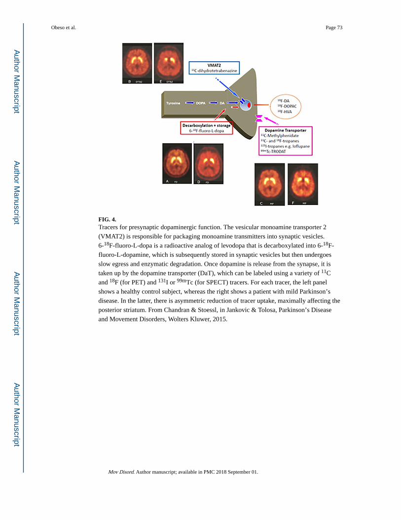
FIG. 4. Tracers for presynaptic dopaminergic function. The vesicular monoamine transporter 2
(VMAT2) is responsible for packaging monoamine transmitters into synaptic vesicles.
6-18F-fluoro-L-dopa is a radioactive analog of levodopa that is decarboxylated into 6-18F-
fluoro-L-dopamine, which is subsequently stored in synaptic vesicles but then undergoes
slow egress and enzymatic degradation. Once dopamine is release from the synapse, it is
taken up by the dopamine transporter (DaT), which can be labeled using a variety of 11C
and 18F (for PET) and 131I or 99mTc (for SPECT) tracers. For each tracer, the left panel
shows a healthy control subject, whereas the right shows a patient with mild Parkinson’s
disease. In the latter, there is asymmetric reduction of tracer uptake, maximally affecting the
posterior striatum. From Chandran & Stoessl, in Jankovic & Tolosa, Parkinson’s Disease
and Movement Disorders, Wolters Kluwer, 2015.
Obeso et al. Page 73
Mov Disord. Author manuscript; available in PMC 2018 September 01.
Author M
anuscriptA
uthor Manuscript
Author M
anuscriptA
uthor Manuscript
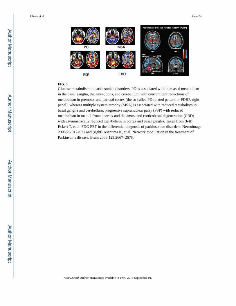
FIG. 5. Glucose metabolism in parkinsonian disorders. PD is associated with increased metabolism
in the basal ganglia, thalamus, pons, and cerebellum, with concomitant reductions of
metabolism in premotor and parietal cortex (the so-called PD related pattern or PDRP, right
panel), whereas multiple system atrophy (MSA) is associated with reduced metabolism in
basal ganglia and cerebellum, progressive supranuclear palsy (PSP) with reduced
metabolism in medial frontal cortex and thalamus, and corticobasal degeneration (CBD)
with asymmetrically reduced metabolism in cortex and basal ganglia. Taken from (left)
Eckert T, et al. FDG PET in the differential diagnosis of parkinsonian disorders. Neuroimage
2005;26:912–921 and (right) Asanuma K, et al. Network modulation in the treatment of
Parkinson’s disease. Brain 2006;129:2667–2678.
Obeso et al. Page 74
Mov Disord. Author manuscript; available in PMC 2018 September 01.
Author M
anuscriptA
uthor Manuscript
Author M
anuscriptA
uthor Manuscript
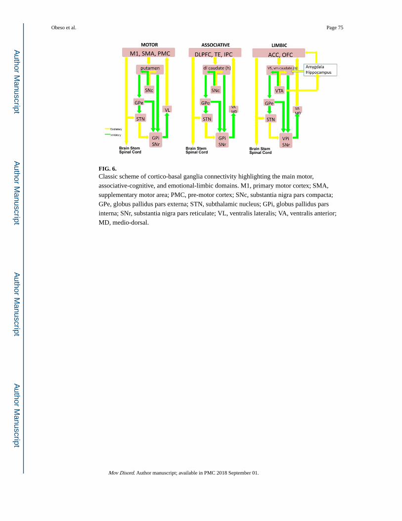
FIG. 6. Classic scheme of cortico-basal ganglia connectivity highlighting the main motor,
associative-cognitive, and emotional-limbic domains. M1, primary motor cortex; SMA,
supplementary motor area; PMC, pre-motor cortex; SNc, substantia nigra pars compacta;
GPe, globus pallidus pars externa; STN, subthalamic nucleus; GPi, globus pallidus pars
interna; SNr, substantia nigra pars reticulate; VL, ventralis lateralis; VA, ventralis anterior;
MD, medio-dorsal.
Obeso et al. Page 75
Mov Disord. Author manuscript; available in PMC 2018 September 01.
Author M
anuscriptA
uthor Manuscript
Author M
anuscriptA
uthor Manuscript
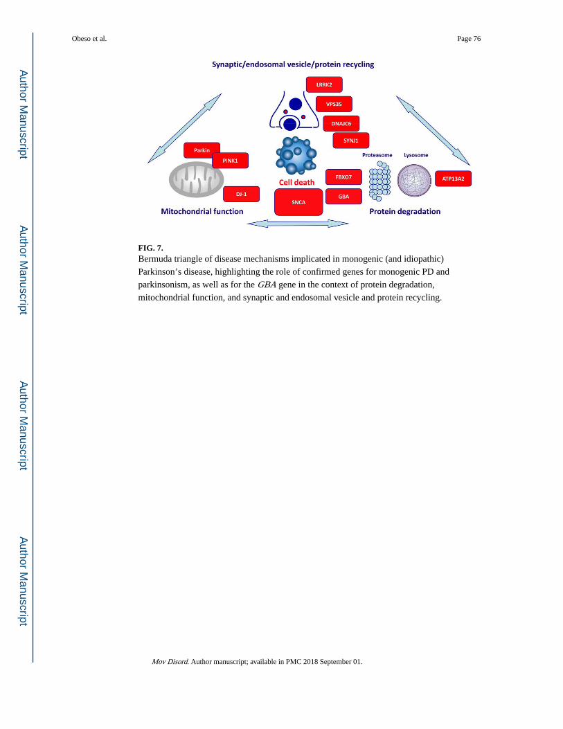
FIG. 7. Bermuda triangle of disease mechanisms implicated in monogenic (and idiopathic)
Parkinson’s disease, highlighting the role of confirmed genes for monogenic PD and
parkinsonism, as well as for the GBA gene in the context of protein degradation,
mitochondrial function, and synaptic and endosomal vesicle and protein recycling.
Obeso et al. Page 76
Mov Disord. Author manuscript; available in PMC 2018 September 01.
Author M
anuscriptA
uthor Manuscript
Author M
anuscriptA
uthor Manuscript

FIG. 8. (A) Schematic illustration of the similarity between the PrP and alpha-synuclein proteins in
terms of their potential to misfold to form betarich sheets, and polymerize to form
oligomers/rod, and amyloid plaques/Lewy Bodies (adapted from ref. 1). (B) Comparison of
Lewy pathology in grafted embryonic dopamine neurons (graft) and in the host substantia
nigra (host). Note the similarity in staining for alpha-synuclein, ubiquin, and thioflavin-S
(indicative of beta sheet formation). These observations raise the possibility that misfolded
alpha-synuclein has spread from affected neurons in the PD brain to unaffected implanted
dopamine neurons in a prion-like manner (adapted from ref. 266).
Obeso et al. Page 77
Mov Disord. Author manuscript; available in PMC 2018 September 01.
Author M
anuscriptA
uthor Manuscript
Author M
anuscriptA
uthor Manuscript

Author M
anuscriptA
uthor Manuscript
Author M
anuscriptA
uthor Manuscript
Obeso et al. Page 78
TAB
LE
1
Pest
icid
es, e
ffec
t mod
ific
atio
n, a
nd r
isk
of P
arki
nson
’s d
isea
se
Toxi
cant
Exp
osur
e ty
peE
ffec
t si
zeG
enet
ic m
odif
ier
Mod
ifie
d ef
fect
siz
eE
nvir
onm
enta
l mod
ifie
rM
odif
ied
effe
ct s
ize
Para
quat
Occ
upat
iona
l; re
side
ntia
l~
2 –
3 fo
ld ↑
risk
GST
T1
null
~ 11
fol
d ↑
risk
a.M
aneb
, Z
iram
b.H
ead
Inju
ry
c.L
ow D
ieta
ry
PUFA
s
d.N
ot u
sing
PP
E
a.~
3–4
fold
↑ri
sk
b.~
3 fo
ld ↑
risk
c.~
4 fo
ld
↑ris
k
d.~
4 fo
ld
↑ris
k
Rot
enon
eO
ccup
atio
nal
~ 2
– 3
fold
↑
risk
Unk
now
n—
a.H
igh
diet
ary
satu
rate
d fa
t
b.PP
E
•~
6 fo
ld
↑ris
k
•~
5 fo
ld
↑ris
k
Org
anoc
hlor
ines
: di
eldr
in, D
DT,
he
xach
loro
cycl
ohex
anes
Occ
upat
iona
l; ga
rden
ing;
res
iden
tial;
~ 2
fold
↑ r
isk
a.C
YP
2D6
PM
b.A
BC
B1
vari
ants
a.2
– 4
fold
↑
risk
b.2
– 5
fold
↑
risk
Unk
now
n—
Org
anop
hosp
hate
s:
mal
athi
on, p
arat
hion
, ch
lorp
yrif
os, d
iazi
non
Occ
upat
iona
l, re
side
ntia
l~
2 –
5 fo
ld ↑
risk
PON
1 va
rian
t~
2 –
3 fo
ld ↑ r
isk
Unk
now
n—
Perm
ethr
inO
ccup
atio
nal;
Impr
egna
ted
clot
hing
; ho
me
use
Not
sig
nifi
cant
——
No
PPE
~ 4
fold
↑ri
sk
Ben
omyl
Am
bien
t occ
cupa
tiona
l or
resi
dent
ial
~ 0.
5 fo
ld ↑ r
isk
SKP
1) v
aria
nt~
7 fo
ld ↑ r
isk
Unk
now
n—
DA
, dop
amin
e; P
UFA
s, p
olyu
nsat
urat
ed f
atty
aci
ds; G
ST T
1, G
luta
thio
ne S
Tra
nfer
ase
T1;
AB
C B
1, A
TP
bind
ing
cass
ette
B1;
CY
P, c
ytoc
hrom
e P
450;
PM
, poo
r m
etab
oliz
er; P
ON
1, P
arao
xona
se 1
; PPE
, pe
rson
al p
rote
ctiv
e eq
uipm
ent;
UPS
, ubi
quiti
n pr
otea
se s
yste
m; S
KP
1, S
pha
se k
inas
e as
soci
ated
pro
tein
1 v
aria
nt.
Mov Disord. Author manuscript; available in PMC 2018 September 01.

Author M
anuscriptA
uthor Manuscript
Author M
anuscriptA
uthor Manuscript
Obeso et al. Page 79
TABLE 2
List of monogenic PD and parkinsonism (adapted from ref. 210)
Designation and reference GeneReviews and OMIM Reference Clinical clues Inheritance Previous locus symbol
1. Classical PD
PARK-SNCA GeneReviews http://www.ncbi.nlm.nih.gov/books/NBK1223/OMIM 168601
Missense mutations cause classical parkinsonism. Duplication or triplication mutations in this gene cause early onset parkinsonism with prominent dementia.
AD PARK1
PARK-LRRK2 GeneReviews http://www.ncbi.nlm.nih.gov/books/NBK1208/OMIM 607060
Clinically typical PD AD PARK8
PARK-VPS35 GeneReviews http://www.ncbi.nlm.nih.gov/books/NBK1223/OMIM 614203
Clinically typical PD AD PARK17
2. Early-onset PD
PARK-Parkin GeneReviews http://www.ncbi.nlm.nih.gov/books/NBK1155/OMIM 600116
Often presents with dystonia, typically in a leg
AR PARK2
PARK-PINK1 GeneReviews http://www.ncbi.nlm.nih.gov/books/NBK1223/OMIM 605909
Often presents with psychiatric features
AR PARK6
PARK-DJ1 GeneReviews http://www.ncbi.nlm.nih.gov/books/NBK1223/OMIM 606324
AR PARK7
3. Parkinsonism
PARK-ATP13A2 GeneReviews http://www.ncbi.nlm.nih.gov/books/NBK1223/OMIM 606693
Kufor-Rakeb syndrome with parkinsonism and dystonia;Additional features:Supranuclear gaze palsy, spasticity/pyramidal signs, dementia, facial-faucial-finger mini-myoclonus, dysphagia, dysarthria, olfactory dysfunction
AR PARK9
PARK-FBXO7 GeneReviews http://www.ncbi.nlm.nih.gov/books/NBK1223/OMIM: 260300
Early onset parkinsonism with pyramidal signs
AR PARK15
PARK-DNAJC6 GeneReviews: n/a OMIM 615528 May present with mental retardation and seizures
AR PARK19
PARK-SYNJ1 GeneReviews: n/a OMIM 615530 May have seizures, cognitive decline, abnormal eye movements, and dystonia
AR PARK20
AD, autosomal dominant; AR, autosomal recessive; n/a, not available.
Mov Disord. Author manuscript; available in PMC 2018 September 01.

Author M
anuscriptA
uthor Manuscript
Author M
anuscriptA
uthor Manuscript
Obeso et al. Page 80
TAB
LE
3
Ove
rvie
w o
f th
e 26
gen
etic
ris
k va
rian
ts s
how
ing
cons
iste
nt a
ssoc
iatio
n w
ith P
arki
nson
’s d
isea
se in
gen
ome-
wid
e as
soci
atio
n st
udie
s
SNP
Loc
atio
n (h
g38)
Nea
rest
gen
eA
llele
sR
isk
alle
le f
req
OR
P v
alue
rs35
7490
111:
155,
162,
810
SLC
50A
1 (G
BA
)A
/G0.
017
1.82
41.
37 ×
10−
29
rs11
4138
760*
1:15
4,92
5,70
9PM
VK
(G
BA
)C
/G0.
012
1.57
43.
80 ×
10−
7
rs82
3118
1:20
5,75
4,44
4N
UC
KS1
T/C
0.55
91.
122
1.66
× 1
0−16
rs10
7975
761:
232,
528,
865
SIPA
1L2
T/C
0.14
01.
131
4.87
× 1
0−10
rs64
3053
82:
134,
782,
397
AC
MSD
C/T
0.57
01.
143
9.13
× 1
0−20
rs14
7405
52:
168,
253,
884
STK
39T
/C0.
128
1.21
41.
15 ×
10−
20
rs12
6374
713:
183,
044,
649
MC
CC
1G
/A0.
807
1.18
82.
14 ×
10−
21
rs34
3118
664:
958,
159
TM
EM
175
(GA
K)
G/A
0.19
11.
272
1.02
× 1
0−43
rs34
8842
17*
4:95
0,42
2T
ME
M17
5 (G
AK
)A
/C0.
913
1.24
71.
10 ×
10−
6
rs11
7246
354:
15,7
35,7
28B
ST1
A/C
0.55
31.
126
9.44
× 1
0−18
rs68
1219
34:
76,2
77,8
33FA
M47
EC
/T0.
636
1.10
32.
95 ×
10−
11
rs35
6182
4:89
,704
,960
SNC
AC
/T0.
367
1.31
64.
16 ×
10−
73
rs76
8115
4*4:
89,
842,
802
SNC
AC
/A0.
498
1.18
97.
09 ×
10−
19
rs92
7532
66:
32,6
98,8
83H
LA
-DQ
B1
C/T
0.90
61.
211
1.19
× 1
0−12
rs13
2011
01*
6:32
,375
,827
C6o
rf10
T/C
0.05
31.
192
3.84
× 1
0−6
rs19
9347
7:23
,254
,127
GPN
MB
A/G
0.59
01.
110
1.18
× 1
0−12
rs11
7896
735
7:11
9,77
7,06
5IN
PP5F
A/G
0.01
41.
624
4.34
× 1
0−13
rs32
9648
11:1
33,8
95,4
72M
IR46
97H
GT
/C0.
354
1.10
59.
83 ×
10−
12
rs76
9047
9812
: 40,
220,
882
LR
RK
2T
/C0.
143
1.15
55.
24 ×
10−
14
rs11
0601
8012
:122
,819
,039
CC
DC
62A
/G0.
558
1.10
56.
02 ×
10−
12
rs11
1580
2614
:54,
882,
151
GC
H1
C/T
0.66
51.
106
5.85
× 1
0−11
rs24
1473
915
:61,
701,
935
VPS
13C
A/G
0.73
41.
113
1.23
× 1
0−11
rs14
235
16:3
1,11
0,47
2B
CK
DK
A/G
0.38
11.
103
2.43
× 1
0−12
rs17
6495
5317
:45,
917,
282
MA
PTG
/A0.
774
1.30
02.
37 ×
10−
48
rs12
4564
9218
:43,
093,
415
RIT
2G
/A0.
307
1.10
67.
74 ×
10−
12
rs81
1800
820
:3,1
87,7
70D
DR
GK
1A
/G0.
657
1.11
13.
04 ×
10−
11
Mov Disord. Author manuscript; available in PMC 2018 September 01.

Author M
anuscriptA
uthor Manuscript
Author M
anuscriptA
uthor Manuscript
Obeso et al. Page 81
TABLE 4
Novel formulations and deliveries of levodopa
• IPX066 – Extended release carbidopa/levodopa containing immediate-release and extended-release levodopa
• XP21279 – actively transported levodopa prodrug that is actively absorbed by high-capacity nutrient transporters
• Accordion pill (AP09004) – extended release levodopa/carbidopa formulation with gastroretentive properties multilayer film, unfolds in the stomach
• DM1992 – gastroretentive, extended-release levodopa in bilayer tablet containing immediate-release and extended-release levodopa
• Carbidopa/Levodopa microtablet – dispersible carbidopa/levodopa 5/1.25 mg administered by means of electronic dispenser
• ODM-101 – levodopa/entacapone plus 65 or 105 mg of carbidopa
• Deuterated levodopa – deuterium–carbon bond is stronger than hydrogen–carbon bond, thus prolonging half-life of levodopa
• Levodopa methyl ester – transdermal delivery of levodopa
• Levodopa-carbidopa intestinal gel – carboxymethylcellulose aqueous gel delivered continuously to the proximal jejunum via a percutaneous gastrojejunostomy tube connected to a portable infusion pump
• CVT-301 – inhalable powder of levodopa without carbidopa
• ND0612 – liquid carbidopa/levodopa formulation delivered subcutaneously via patch-pump device
Mov Disord. Author manuscript; available in PMC 2018 September 01.

Author M
anuscriptA
uthor Manuscript
Author M
anuscriptA
uthor Manuscript
Obeso et al. Page 82
TABLE 5
Markers of prodromal PD
Marker Level of evidence Approximate relative risk Lead time Testing cost/burden
Olfaction High 5 ?? Low/Moderate
REM Sleep Behavior Disorder High 50 13 years Low (screens) to High (PSG)
Somnolence Moderate 1.8 ?? Low
Restless legs (late onset) Low 1.5 Short Low
Constipation High 2.5 > 15 years Low
Orthostatic hypotension Moderate ? 2–10? 2–5 years? Low
Urinary dysfunction Low-Moderate 2.1 ?? >5 years Low
Erectile dysfunction Low-Moderate 1.2 mild3.8 severe
5–10 years Low
Depression/anxiety High 1.8 Uncertain ?Biphasic Low, but follow-up higher
Color vision Low 2.5 >3 years? Moderate
Subtle parkinsonism Moderate 10 4–5 years Moderate - High (Expert)
Quantitative motor testing Moderate 3–4 5 years Moderate
SN ultrasound Moderate 15 Uncertain ? risk marker?
Moderate-High
Dopaminergic PET/SPECT Low (but high plausibility)
20 5 years High
PD-related pattern on SPECT/PET
Low ? ? High
Hippocampal hyperperfusion Low ? ? High
GI synuclein pathology Low 2? ? High
For this table, only markers with prospective evidence of predictive value are included. For level of evidence, low implies a single study, moderate implies >1 high-quality study, high implies >4 high-quality studies. Lead time refers to the approximate time that the marker deviates from normal values (the time at which testing is reliably abnormal cannot be estimated for most markers). For testing cost, low indicates can be screened by questionnaire (does not require visit), moderate implies in-person assessment required but low cost (eg, research assistant), high implies extensive or expensive evaluation (>$300).
Mov Disord. Author manuscript; available in PMC 2018 September 01.


![Parkinson's Disease[1] (1)](https://img.dokumen.tips/doc/110x75/55cf9295550346f57b97b1cc/parkinsons-disease1-1.jpg)