Embed Size (px)
Citation preview

PARTIAL, PROGRESSIVE AND COMPLETE OCCLUSIONOF THE AORTA AND OTHER LARGE ARTERIES
IN-THE DOG BY MEANS OF THE METAL BAND.'
BY W. S. HALSTED,
Professor of Surgery, Johns Hopkins University.
PLATES XI-XIII.
At the meeting of the Johns Hopkins Hospital Medical Society,March 20, I905, a brief preliminary report was made of the results ofexperiments in occlusion upon the abdominal aorta and other arteriesof sixty-three dogs, conducted during the previous winter and autumnby Dr. W. F. M. Sowers and me.2 During the following yearthese investigations were continued with the assistance of Dr. E. H.Richardson, thirty-nine dogs being operated upon. With the aidof a modified Brauer apparatus devised by Dr. Follis and Dr. Fisherwe were able successfully to constrict the thoracic aorta both inanimals and man. In June, 90o6, at the meeting of the AmericanMedical Association in Boston, attention was again called to. ourwork, the results to date being given in merest outlines Duringthe following two years, the exigencies of the surgical clinic seemedto demand experimental investigation in another direction ctnd henceonly a little time was found for prosecuting the arterial work underconsideration, although several problems which have arisen in con-nection with it we regard with unabated interest; and with the solu-tion of these we are again concerning ourselves. In the meantime,however, a number of experiments have been made, and severalanimals which were for many months under observation have fur-nished additional facts worthy of record. The method of applyingthe band has been improved and modified and new instruments forrolling it devised. The full report of this work will probably be
'Received for publication, January I4, I909.2Johns Hopkins Hospital Bulletin, 1905, xvi, 346.Jour. of the Anmer. Med. Assoc., 90o6, xlvii, 2147.
373
Dow
nloaded from http://rupress.org/jem
/article-pdf/11/2/373/1081562/373.pdf by guest on 22 March 2022

Occlusion of the Aorta in the Dog.
published during the year in the Johns Hopkins Hospital Reports.The incentive to the work was the desire, experienced by so many
surgeons of the past and present, to be able to occlude safely theabdominal aorta in the hope of curing thereby aneurisms of thisvessel and of the common iliac arteries. I shall write later of theattempts of Dubois, Assalini, Bujalsky, Pirogoff, Luigi Porta,Cooper and Keen to compress gradually the abdominal aorta bymeans of specially devised instruments which, passed through anincision in the abdominal wall, carried a snare of silk, catgut ormetal which might at any moment be tightened or loosened at will.A fault common to all of the methods hitherto devised is seeminglyan insurmountable one-the difficulty of preventing sepsis in thetrack of an instrument maintaining direct communication for daysor even weeks with the air. A better method might, we thought,be one permitting, in each entre-act, complete closure of the wound.The material compressing the aorta should not be bulky nor endan-ger by its form or substance the adjacent parts; and it should admitof easy readjustment at subsequent operations, should they be indi-cated. Metal bands of silver and then of aluminium were employedwith the hope that the amount of constriction might be regulatedto a nicety at prospective subsequent operations as well as at the pri-mary one. With the aid of an ingenious clock-maker, an instru-ment was devised to curl a metal strip, in perfect cylinder-form,about the vessel.
The tightening of the band is completed with the fingers; but inthe early experiments, when the metal employed was too thick andthe bands too broad, the aid of tweezers was required. We observedthe first group of dogs with some apprehension, fearing that theedges of the band would cut the much constricted and powerfullypulsating aorta, and were considering ways to obviate, if necessary,this danger. On the twelfth day after operation a dog died ofhemorrhage, the result of ulceration at the upper edge of the band.The experiments were thereupon discontinued for a while, to awaitresults in the other dogs carrying aortic bands. Further cases ofhemorrhage not occurring, we resumed, in a few weeks, the experi-ments. About three months later we investigated, at second opera-tions and at autopsy, the resultant conditions and found that the
374
Dow
nloaded from http://rupress.org/jem
/article-pdf/11/2/373/1081562/373.pdf by guest on 22 March 2022

W. S. Halsted.
aortic wall where the band had embraced it was, in each instanceof complete occlusion, atrophied, being reduced in some cases to afilm-like thinness. Notwithstanding these somewhat discouragingobservations the experiments were continued as actively as time per-mitted in the hope that, with an improved technique, derived fromgreater experience, particularly with reference to precision in thedetermination of the degree of closure brought about as the rolling(tightening) of the band proceeded, the walls of the arteries mightretain their vitality even in the case of complete arterial occlusion(vide Cases 27 and 28). By reducing the width and length of theband and the thickness of the metal, we were able with the fingers,and with ease, to occlude the artery to the extent desired. Fromthe first experiment we have endeavored, in each instance, to rollthe band as perfectly (as cylindrically) as possible, flattening of itbeing assiduously guarded against. To control with accuracy theamount of blood-flow under the band, the rolling must go onsmoothly and under the perfect command of the operator. Thethrills of various strength, the point at which the pulse disappears,are carefully noted in the course of the act of constricting the ves-sel; and, finally, when complete occlusion is desired, the pulse beingno longer distinguishable, the filling or not of the artery below theband, between it and a fine clamp placed central to the first distalbranch is our only clue as to the patency of the artery.
With the use of silk or even silver wire such delicate manipula-tion and determination were found impossible, irrespective of thegreat danger of injury to the vessel-wall from attempts to draw justa shade tighter a half-knotted thread which has constricted thevessel almost to the occlusion point.
At the time of my first report (March, I905)4 we stated that:( ) If applied tightly enough to interrupt completely the circulation,the band had usually caused atrophy and frequently complete absorp-tion of the aortic wall. In such cases hemorrhage was invariablyprevented by the formation of connective tissue enclosing the band.(2) Thrombosis had not been observed in a single instance, eitherin the cases of complete occlusion in which the arterial wall underthe band was found so greatly thinned, or in those in which it had
'Loc. cit.
375
Dow
nloaded from http://rupress.org/jem
/article-pdf/11/2/373/1081562/373.pdf by guest on 22 March 2022

Occlusion of the Aorta in the Dog.
been absorbed. (3) In a few cases (vide No. 28) aortic wallsdrawn together so snugly that at autopsy water could not be forcedthrough with a syringe were, on division of the band, found to benormal and could be easily smoothed out and the full lumen reestab-lished. But in these earlier cases of complete occlusion the aorticwall had almost invariably atrophied, having been so tightly con-stricted as to be deprived of its blood supply, and hence the " idealobliteration" (reported the following year) by adhesion of thefolded intima and the conversion of the constricted portion of theartery into a solid fibrous cylinder (vide Plate III, Fig. 2a), couldnot have taken place. (4) The less snugly, the loosely and the veryloosely applied bands might remain on the aorta, femorals and ca-rotids for months without apparent injury to the walls of these ves-sels, either externally or internally. For example, the band after onehundred days would be seen shimmering brightly under a quitenormal-looking peritoneum, having caused, as a rule, little if anyreaction, and could be as easily removed form the wall of the arteryas when originally applied. (5) We were encouraged to believethat there might be a place in surgery for the partially occludingband, and reported its application to the common carotid artery inthe human subject and the manifestation, thereupon, of slight brainsymptoms which persisted for several months. In this case theband was rolled more tightly than intended. It might easily havebeen removed and reapplied but, as I stated at the time, our notionsbeing somewhat vague as to the precise amount of constriction to bedesired and being unable to determine accurately the blood pressuredistal to the band we decided not to disturb it and to note results.On the appearance of the head symptoms I did not relieve the con-striction, believing that they would probably disappear; further-more, I was not perfectly sure that, rather than good, harm mightnot result from the release of the carotid.
Subsequently (in June, I9O6), in the report made in Bostonbefore the Section on Surgery and Anatomy of the AmericanMedical Association, the following newly observed facts wereemphasized:
I. The blood pressure in the aorta below the band is lowered inproportion to the amount of the occlusion. The rise in pressure
376
Dow
nloaded from http://rupress.org/jem
/article-pdf/11/2/373/1081562/373.pdf by guest on 22 March 2022

W. S. Halsted.
below the band is, at first, rapid, but varies considerably in thedifferent dogs. For example, in one dog, a rise below the band often millimeters (Hg. manometer) was noted in ten minutes, whereas,in another dog, two hours were required for a rise of fifteen milli-meters. For the return of the normal pulse wave and of the normalblood pressure as many as seven months have been insufficient (videhistory of Dog No. 96).6
2. Partially occluding bands produced as a rule no macroscopicalteration in the aortic wall under the band even after seven andeight months (vide Plate III, Fig. I). Under completely occlud-ing bands the arterial wall had (to the date of the second communi-cation referred to) usually atrophied and in the course of weeksor months been absorbed. " When the lumen had been, perhaps,not quite" occluded, complete obliteration might result spontaneouslywi.th the conversion of the arterial wall embraced by the band intoa solid cylinder of living tissue. This may," I stated, "be consid-ered the ideal closure of an artery." Although this form of arterialclosure had occurred only thrice in the long series of experiments, itmight, I thought, be accomplished frequently, and ultimately theband might be applied with such nicety that, unaided, further, bythe surgeon, a partial would be likely to proceed to total occlusion.
3. "The Effects on the Spinal Cord and Its Coverings.-Thestudy of the spinal cords was entrusted to Mr. P. KI Gilman (laterDr. Gilman and member of my staff), vWho discovered, in a numberof instances,7 about three months after operation, a deposit of extra-dural fat about the cord below the site of the aortic band. In three
B Since this statement was made we have had several opportunities to verifyit, and have noted in a dog with partially occluding band on the thoracic aorta,after seven months, a difference of 30 millimeters or more in the pressure im-mediately above and immediately below the band. Of particular interest arethe careful observations of Dr. Percy M. Dawson on the femoral and carotidpressures in Dog No. 96 of this series, reported below.
'We have not as yet learned to determine with the greatest precision thedegree of arterial occlusion effected, nor am I convinced that in all of the casesin which the "ideal occlusion" has resulted (we have now five such observa-tions) there may not have been, at the outset, complete obliteration of the lumen(vide history of Dog No. 27).
'These proved to be cases in which total or almost total occlusion had beenmade.
377
Dow
nloaded from http://rupress.org/jem
/article-pdf/11/2/373/1081562/373.pdf by guest on 22 March 2022

378 Occlusion of the Aorta in the Dog.
cases the production of fat was so great that it filled, seeminglyunder considerable tension, the vertebral canal."
If these observations should prove to be correct, Mr. Gilman hasmade a discovery of wide significance.
4. That in the human subject I had partially occluded the innomi-nate artery once and the common carotid four times.8 In the caseof a large popliteal aneurism I had employed the metal band toocclude completely the femoral artery. In the case of a womanasphyxiated to unconsciousness by an aneurism of the aortic arch,I had exposed, carefully and freely and without puncturing eitherpleural cavity, the heart and arch of the aorta, hoping to be able toencircle with a band the aortic arch between the regions of theinnominate and left carotid arteries, but the aneurism was so exten-sive and the patient's condition so desperate as to defeat the earnestand prolonged endeavor to execute the procedure.
THE ALUMINIUM BAND AND THE MANNER OF ITS APPLICATION TO
THE ARTERY.
We have tested only two of the metals, silver and aluminium,with reference to their adaptability to the procedure under consid-eration. After a few experiments with the former it was discarded.The greater weight and value of the silver (there is great waste inits use) and the inconstant results obtained as to nicety in rolling,particularly after repeated boilings, are the principal factors whichled to its disuse. The aluminium, usually purchased in sheets ofabout 25 degrees of thickness (American scale) should be cut,before being rolled down to the thinness desired, into strips of con-venient length, and of width not greater than three quarters of aninch. If much wider, the strips warp inconveniently and have tobe cut to waste in the selection of flat and regular parts for bandmaterial. It is well to stamp each strip with the numbers indicatingthe thickness of the metal and to have on hand a liberal supply ofthe various thicknesses from No. 25 to No. 46. The finest numbers
SThe aluminium band has now been successfully applied in man to the com-mon carotid artery twelve times, and once each to the thoracic aorta, the ab-dominal aorta, the common iliac, the femoral, and the innominate arteries.These cases will be reported later in the Amer. Jour. of the Med. Sciences.
Dow
nloaded from http://rupress.org/jem
/article-pdf/11/2/373/1081562/373.pdf by guest on 22 March 2022

W. S. alsted.
we have used on the very small femoral and renal arteries of thedog. In the average dog for the abdominal aorta Nos. 34 to 35are suitable, and for the thoracic aorta Nos. 33 and 34 we haveused most frequently. In the human subject for the abdominalaorta below an aneurism near its bifurcation No. 33 sufficed, butordinarily a heavier size would be required; for the common iliacNo. 32 answered the purpose admirably; for the thoracic aortaNos. 22 to 25 perhaps; for the common carotid we have almostinvariably selected No. 33. The length of the band should be aboutthat of the circumference of the full artery. The width varies fromabout 2 mm. for the renal arteries in the dog to about I cm. forthe thoracic aorta and innominate artery in man. Fig. b, Plate XI,depicts a band suitable in width for the human carotid and for theaverage dog's abdominal aorta. The band in this illustration weshould now regard as too long for its breadth by approximately onethird. It is best to sterilize the aluminium only once. It maybecome too brittle for perfect rolling by repeated boilings. Whenrolled down on the artery enough almost to obliterate the pulse, aband of seemingly proper dimensions has rarely described morethan two complete circles. The filing or "manicuring" of theband is of very great importance. It should be curved like a fingernail at the forward end and at the other cut precisely at right anglesto its long axis. With a file the edges should be made perfectlysmooth, but not sharp, and the rounded end symmetrical. A care-fully filed band coils more easily both in the. instrument and underthe fingers, and, what is more important, is not likely to cut theartery. The aorta is the only vessel that I have seen cut itself onthe band, and then, with the exception, perhaps, of two very youngdogs or puppies, only when the band was badly filed or clumsilyrolled, as in the early experiments, especially those in which forcepswere employed to supplement the work of the fingers in tighteningthe too broad and too heavy bands at that time employed. Silkligatures, even when occluding the aorta only partially, have in myexperiments repeatedly cut entirely through the aorta, and withoutcausing the death of the animal. They may leave in their wakevarious forms of diaphragm which more or less obstruct the lumenof this artery. I shall refer to a case of this kind depicted in PlateXII, Figs. 5 and 6.
379
Dow
nloaded from http://rupress.org/jem
/article-pdf/11/2/373/1081562/373.pdf by guest on 22 March 2022

Occlusion of the Aorta in the Dog.
The Band Curler.-The original instrument (vide Plate XI) had,we soon discovered, three major faults. It was (I) too broad atthe arterial end, (2) the band lacked anterior support as it wasbeing pressed onward by the driving blade, a (magnified a'), and(3) the latter did not always engage the former, owing to the factthat it was too springy and was insufficiently linked to its fellow onwhich it glides. To remedy the tendency for the band to buckleforward, one was compelled to support it with the finger during theprocess of curling. This was occasionally a difficult and usually anawkward performance.
Plate XI, Fig. 2 shows an improved and satisfactory band roller orcurler. In the full length drawing the instrument is not loaded; inthe abbreviated sketch the band projects from the end, half curled.The principal defects of the old instrument have been remedied inthe new. Buckling of the band is prevented by the boxing; thedriving-plate can not spring away from its fellow, and the width ofthe instrument has been sufficiently reduced to permit it to be passedfreely between the closely given off branches of the abdominal andthoracic aortas. When the thinner bands are used it is sometimesnecessary to give the faintest tip backwards to each of the two right-angled corners of the band to insure its engagement in the down-ward thrust of the piston or driving blade. After it has .encircledthe artery the band may, if its corners have been bent, be freshlysquared with the scissors before being curled tighter by the fingers.This is not, however, necessary. The curler being armed with thecarefully filed band of correct proportions the plunger is made to en-gage the band and to force its convex end into sight before the instru-ment is passed under the artery. In Plate XI, Fig. I, the act of curl-ing the band about the artery is shown, after the old manner, in itsfirst stage. As the curling proceeds the instrument is gradually with-drawn. In arranging for the tightening of the band, its convexend should lie on the wall of the artery and be overlapped by thesquare end. With a very little practice one learns to avoid bendingor flattening the band in the process of tightening it. The bandshould be long enough to encircle the artery in the expanded stateof the latter and the metal should be sufficiently thick and wide tosustain the curl given it. If perfectly rolled the inside and outside
380
Dow
nloaded from http://rupress.org/jem
/article-pdf/11/2/373/1081562/373.pdf by guest on 22 March 2022

W. S. Halsted.
circles of the metal touch each other at all points of the surfacesof contact and, in consequence, the cohesion force is greatest. Theartery should be raised from its bed by two tapes held far enoughapart to leave uncovered sufficient free space on the artery for theoccupancy of the band. Traction on the upper tape should be madeto interrupt the blood current and thus to reduce the size of thevessel. A band curler should be selected (we have, at present, foursizes of this instrument) which might make the metal describe acircle smaller than the distended or full artery but a little largerthan the empty one; then, with the return of the blood current, theartery expands and may fill the band quite snugly.
After complete occlusion of the abdominal aorta the femoral pulsedoes not, usually, return for weeks or even months. And afterincomplete occlusion of the thoracic aorta (Dog 96) the femoralpulse may be hardly discernible after seven months. The anas-tomotic circulation takes place through the vasa vasorum as discov-ered and so beautifully depicted by Luigi Porta,9 and by way ofthe internal mammary and epigastric arteries as especially empha-sized by Kast.10 We have repeatedly observed the great increasein the vascularity of the abdominal wall, particularly on splittingthe recti muscles but also in making mid-line incisions at operationssubsequent to the one at which the band was applied.
DOG No. 2.-Large, savage, collie-like dog. Operation I. Morphia and ether."November 4, 9o4. Silver band, partially occluding the vessel, applied to theabdominal aorta below (?) the inferior mesenteric artery. Radicles of thoracicduct not injured. Pulse in femorals easily countable at end of operation, thoughmuch reduced in volume.
November 6, I904. Dog in good condition, rather dull, walks about withoutapparent weakness in hind legs. Tests such as running up stairs were not made.
November Io, 904. Apparently perfectly well. Is quite savage and threat-ens to bite when cage is entered.
December 8, I9o4. Vigorous femoral pulse but seemingly weaker thanmight be expected in so large a dog. Health has been perfect. Operation II.Morphia and ether. Complete occlusion of aorta about I cm. above the silverband by heavy black silk ligature. The silver band glistened through a verythin and apparently normal peritoneum. It had excited little or no irritation.
'Luigi Porta, Delle alterazioni patologiche delle arterie per la legatura e latorsione, Milan, 845.
" Kast, Deut. Zeit. fiir Chirurgie, 879, xii, 405.'All the animals were anaesthetized in the same way preparatory to operation.
381
Dow
nloaded from http://rupress.org/jem
/article-pdf/11/2/373/1081562/373.pdf by guest on 22 March 2022

382 OccEusion of the Aorta in the Dog.
On coming out of ether the dog showed signs of unusual excitement. Respira- tions, 136 per minute. One hour after operation dog was able to climb a flight of stairs. The hind legs were, however, in spastic condition, flexed on the ab- domen, at the knees and hips, and very much weaker than the fore legs.
December 10, 1904. Dog is dull. Hind legs are dragged in walking but are not completely paralyzed. Movements still spastic.
December 14, 1904. I s lively and seems quite well. Bladder and rectum function normally. Scratches himself with left hind leg without apparent weakness.
January Q, 1905. Femoral pulses not yet definitely palpable but the arteries have become quite full.
February I, 1905. Femoral pulse faint and not countable. February XI, 1905. Fair pulse, countable. March 6, 1905. Found dead. Was not observed yesterday (Sunday). Au-
topsy.-A large flat cork causing intestinal obstruction is the cause of death. The silk ligature above the band has cut almost through the aorta, its track being apparently healed; part of the loop projects into a little cavity, with newly formed walls of connective tissue, adjacent to and in front of the wall of the aorta. A filmy substance, like decolorized blood clot, is adherent to the track taken by the ligature in its course through the artery. The aorta under the band is a little thinned. Its endothelial lining seems normal.
In the light of subsequent experience it seems probable that the thinning of the aorta may have been caused by the ligature applied so close above the band-by its interference with the circulation in the arterial wall.
DOG NO. 6.-November 19, 1904. Black bitch, length 31 inches. Operation I . Closure of abdominal aorta with aluminium band I cm. in width.
The pulse pressure in the left femoral artery as recorded by an assistant:
................................... Before operation.. 150 mm. ........................... After abdomen was opened I30 mm.
........................ Band incompletely tightened.. 80 mm. Tightening of band completed.. ...................... 20 mm. After abdominal manipulations were ended.. .......... 25 mm. After closure of abdomen.. .......................... 30 mm.
The operators could not feel the aortic pulse below the band after it was com- pletely curled, hence I have doubt as to the accuracy of the three observations on pulse pressure in the femoral artery made after the final tightening of the band.
November 21, 1904. Femoral pulse doubtful. Dog ran easily on level and mounted the first steps of a flight of stairs without definite signs of weakness. Her hind legs gave out before the top steps were reached. Temperature fore- feet, 70°, hind feet, 66", Fahrenheit.
November 22, 1904. Hind legs still weak. Sits down after slight exercise. Pulse in femorals doubtful. Surface temperature of hind-flanks, 9.5'; fore- flanks, 97' ; pads of feet, hind 97" ; front, 71".
November 25, 1904. Temperature axilla, 98.5" ; groin, 95.5". Hind legs less weak. Dog in good spirits. Operation I I , 11 :30 A. M. Ligature of black silk
Dow
nloaded from http://rupress.org/jem
/article-pdf/11/2/373/1081562/373.pdf by guest on 22 March 2022

W. S, Halsted.
obliterating aorta applied below the band. 3 P. M., bladder not distended. Bitchhas showed no excitement since operation. Temperature, axilla, 96° ; groin, 930 F.
December 13, I904. Slightly stiff in hind legs. Dog is dull and indisposedto walk much. We attributed the condition of the dog in part to one-half grainof morphia administered before operation.
December 14, 1904. Movements in hind legs greatly improved. Dog walkswith ease but is a little depressed and sits down frequently.
December 9, I9o4. Perfectly well and active.December 28, 904. Well and active.January 7, 1905. Well.January 2, 905. Well.January I9, I9o5. Well and strong. Faint femoral pulse observed for the
first time since the first operation performed two months ago.January 23, I905. Femoral pulse definite.February , I905. Femoral pulse countable but is still very small.March 6, I9o5. Dog well but quite thin. Killed with ether. Femoral
and thoracic conditions normal. On palpating aorta a small nodule is felt atsite of ligature. The band is found directly in line of the aorta above the liga-ture and filling completely a new-formed connective tissue cavity. The arterialwall under the band has been completely absqrbed and the aortic wall above theband is continuous with the new connective tissue which forms a strong capsuleabout the band. The capsule of fibrous tissue fits the band so closely that noblood has escaped from the patulous aorta above into the space between theband and the connective tissue which enclosed it. The arterial lumen just belowthe band has been obliterated by the silk ligature which has cut entirely throughthe arterial wall and is enclosed in a hard nodule of connective tissue.
DoG 2o.-Small puppy, male. December Io, 904. ii A. M. OperationAluminium band applied above the inferior mesenteric artery. In exposing theartery the sigmoid flexure was pulled to the right. The band had been cut toolong. It rolled beautifully to a certain point and then, before the aortic pulsecould be obliterated the metal cylinder began to flatten. The pulse then becameentirely obliterated but some sort of pulse-shock could be felt for a distance ofI cm. or more below the band. 1.30 P. M., dog runs about, but his hind legs arevery weak, though not completely paralyzed.
December I9, I904. Hind legs stronger.December 20, I904. Dog does not seem well. Is quite thin and disinclined
to walk. Can, however, stand and uses his hind legs in walking fairly well.December 23, 904. Not so well.December 26, I904. Quite ill.December 27. Dead.Autopsy.-The abdominal wound had opened down to the peritoneum but
was well sealed by momentum and the subperitoneal flap of fat. There is no peri-tonitis. The portion of the ileum lying in the pelvis, and the sigmoid flexureexhibit punctate hemorrhages in the fat along the vessels. The sigmoid flexureis particularly hemorrhagic and probably the cause of the bloody fluid in thepelvis. There is some blood escaping from the anus. The jejunum, at a pointnear the aorta where it may well have been pressed upon by the retractors andgauze, is also indurated and dark. The bladder is empty. The band has excitedvery little reaction. The peritoneum and fat over it are still ununited.
383
Dow
nloaded from http://rupress.org/jem
/article-pdf/11/2/373/1081562/373.pdf by guest on 22 March 2022

Occlusion of the Aorta in the Dog.
DOG No. 27.-Small, black puppy. December 26, 904. Operation. A verybroad band (7 mm. in width) was placed on the aorta about I cm. below theinferior mesenteric artery and rolled by tweezers until the demonstration of apulse below the band was questionable. We attempted in this case, prior to theapplication of the band, to produce occlusion by silver wire wrapped several timesabout the artery and over a small copper rod (the rod to be afterwards with-drawn), thinking that the size of the lumen might in such cases be definitelyregulated by the use of rods of various sizes. After several trials we were con-vinced that the liability of injury to the artery was too great, at least muchgreater than with the employment of the band. The wrapping of the wire wasalso troublesome. Hence the project was abandoned.
December 28, I904. Walks fairly well but has definite weakness in hind legs.Has 'fluid stools which contain a few drops of blood.
January 5, I905. Weak and emaciated. A very small, not countable, femoralpulse has developed. Dog uses hind legs a little better.
January 23, 905. Is very feeble and thin. Isolated in cage.January 27, I905. It is possible to-day for the first time to count the femoral
pulse; it is still very small.February I3, I905. Condition practically unchanged.February 20, 905. Seems a little better. Femoral pulse still very small, but
countable.February 23, I905. Improvement continues. Femoral artery feels fuller.April Io, 905. Dog is very thin and has the mange. Etherized. A very
faint femoral pulse was demonstrable on dissection of the artery, but through theskin it was not definitely palpable. Dog killed.
Autopsy.-Peritoneal cavity normal.
The aortic band shimmers quite clearly through the peritoneum,but about cm. below the band is a very delicate stellar cicatrix.The aorta was split to the band from both ends and the band itselfdivided. The vessel is entirely obliterated and the length of the ob-literated portion corresponds exactly to the width of the band. Theportion of the aorta enclosed by the band has become converted intoa solid fibrous cord (vide Plate XII, Fig. 4a). Outside the band isa peculiar, soft, yellowish-white material, about one drop in quan-tity, resembling aleuronat. The hard, white, living cord, into whichthe occluded portion of the aorta has been converted, is 2 mm. indiameter. The band, an unusually broad and thick one, had beenin place three and one-half months and might, without doing harm.have remained indefinitely. The form of the fibrous cord, soexactly cylindrical, under the band, is evidence that perfection incurling may be accomplished even with tweezers. Without especialeffort to bring it about we have in five instances obtained this ideal
384
Dow
nloaded from http://rupress.org/jem
/article-pdf/11/2/373/1081562/373.pdf by guest on 22 March 2022

W. S. Halsted. 386
form of obliteration-four times .in the aorta and once in the renal artery-and always in cases in which the completeness of the closure was in doubt at the time of operation. This case should be contrasted with Case No. 28, that of a small puppy whose aorta, though of intention completely closed by the band, was not found converted into a fibrous cord under the metal. T o our surprise, it presented no internal adhesions whatever on being laid open, al- though so tightly constricted that not a drop of water could be forced through this portion of the vessel while the band was in place. The foldings of the aortic wall under the band could be smoothed out so completely that no trace of them remained, nor was'there any abnormality of the wall to indicate that a band had been applied. W e must bear in mind that in Dog 28 only twenty- two days had elapsed from operation to autopsy, whereas in the cases cited of fibrous cylinder formation, the dogs had carried their bands for several months.
Doc No. a&-Small, black puppy, male. December 26, 1904 Operation. Aluminium band rolled (with tweezers) until pulse seemed .to be obliterated.
December 28, 1904. Dog seems fairly well, but marked weakness and stiffness in hind legs.
January 7, Igoj. Dog cross and disinclined to respond to attentions. Hind legs possibly a little less stiff.
January 12, 1905. Slight improvement noted. January 17, 1905. Dog looks badly. I s greatly emaciated. Indisposed to
move and hence the power in hind legs was not tested. January 18, 1905. Great emaciation. Autopsy.-Peritoneal cavity normal. Band shimmers through a perfcctly
normal-looking peritoneum. No trace of peritoneal or other scar over band. The healing about the band is the most absolutely perfect that we Ilave ns yet seen.
With a syringe connected to the aorta above the band we are un- able to force water through the site of obstruction. On removing the band we find, to our surprise, that not only could the point of a scissors readily be passed through the constricted part but that the aortic wall under the band could be so perfectly smoothed out that not a trace of the foldings -or wrinltlings of the lining remained. Nor was there apparent the slightest thinning or alteration in the wall of the vessel at the site of the band. We have not as yet had the time to complete experiments undertaken with a view to ac-
Dow
nloaded from http://rupress.org/jem
/article-pdf/11/2/373/1081562/373.pdf by guest on 22 March 2022

Occlusion of the Aorta in the Dog.
counting for the difference in the condition of the aorta at the siteof constriction in the cases (Nos. 27 and 28) referred to.
DoG. 30.-Medium-sized collie bitch. December 27, I9o4. Operation I. Alu-minium band tightened until the pulse was greatly reduced but not apparentlyobliterated. There was sufficient pulse just prior to the final tightening to givea faint thrill.
December 28, 9o4. Dog convalescing normally. Uses hind legs quite well.No definite sign of weakness in them.
January 7, I905. No weakness in hind legs.January 9, I9o5. Dog very well. Femoral pulse not yet distinguishable.January 23, 905. Operation II. ii A. M. There were a few adhesions of
momentum in the neighborhood of the band, which prevented the metal fromshimmering in the usual manner through the peritoneum. Pulse below the bandis feeble but perfectly definite. When the pulse just above the band is obliteratedwith the finger the pulse below the band disappears. We concluded consequentlythat the band had not completely closed the lumen of the aorta. Ligation withheavy black silk 3 or 4 mm. above band. Much to our astonishment the pulse,easily appreciable by finger, reappeared below the band in less than 30 secondsafter the aorta was ligated. It seems almost inconceivable that a ligature appliedso close above the band should have had such a marked influence on the pulsebelow it. We have made this observation in a number of cases in which wewere quite sure that no vessel of a size to be appreciated was given off from theaorta between the ligature and the band.
The almost immediate reappearance of the pulse after the appli-cation of the ligature indicates that the anastomotic circulation wasalready quite well established, for a pulse large enough to be appre-ciated by the finger has not been observed by us before the termi-nation of the operation, after complete closure of the aorta in oneact. We have, however, noted that within a minute after ligationof the abdominal aorta a faint pulse below the ligature can be seen(not felt), provided the aorta is cut open.
January 23, 1905. 3 P. M., Dog runs about. Weakness of hind legs notapparent.
January 24, I905. A very faint femoral pulse can be felt very high upon thefemoral arteries. No weakness of hind legs.
January 27, 905. Pulse more easily felt but still feeble.February I, 905. Femoral pulse can be counted. The artery is not full.February 20, I905. Artery much fuller. Pulse stronger. Dog quite thin.March Io, I905. Femoral pulse still small. Dog is thin but looks well.
Ether administered. Abdomen opened. Small indurated area about the ligatureand the band. A definite pulse below the band is promptly shut off by pressureimmediately above the band and at higher points, as noted in history of Dog No.6. A ligature was applied about 3 cm. above the band and tests for a pulse below
386
Dow
nloaded from http://rupress.org/jem
/article-pdf/11/2/373/1081562/373.pdf by guest on 22 March 2022

W. S. Halsted.
the band were made for three minutes but it had not returned in this period oftime. The dog was then killed. Above the ligature the aorta was entirely oc-cluded in a conical manner. The ligature lies in a small cavity lined with granu-lations. Below the band the aorta is closed off by a small transparent film. Theband lies in a connective tissue cavity and contains merely a film of opaque secre-tion. The aortic wall under the band has consequently been entirely absorbed.Between the two cavities a piece of aorta I3 mm. in length is felt with a partlyorganized clot which is translucent except for a red dot about I mm. broad, in itscenter.
It is evident that at the first operation I was probably mistakenin believing that the artery was not obliterated by the constrictingband. It would, a priori, seem reasonable to presume that a bandthough not altogether occluding the artery, might, nevertheless, sogreatly interfere with the circulation of its wall as to lead to necro-sis, but the findings in Dog. No. 28 would seem to make such a viewuntenable. It seems, however, not unlikely that the necrosis of thewall of the aorta may not have supervened until at the second opera-tion the ligation with silk a few millimeters above the band wasmade. If this were the case it may be that our impression that thepulse had not been obliterated by the band at the first operation wascorrect.
Thoracic Aorta Partially Occluded by Aluminium Band.-DogNo. 96.-Medium-sized Newfoundland bitch. May 22, 906.
Operation. Assisted by Drs. Richardson and Gilman. Lungs in-flated during the operation through tracheotomy tube. Incisionthrough seventh interspace. Ribs separated as usual with self-retaining retractor. Exposure of aorta very satisfactory. Alumi-nium band, thickness No. 32, width 6 mm., applied just above high-est point of diaphragm. Band was tightened until a continuousthrill below the band was produced.
May 24, i9o6. Slight stiffness in hind legs.May 25, 9o6. Improving; takes milk. Femoral pulse doubtful.May 27, Igo6. Cheerful; runs about freely. Percussion reveals no thoracic
signs. Chest not auscultated.June I, I9o6. Dog is lively and well.June 3, I9o6. I went to Boston.June iI, 9o6. On my return from Boston to-day dog was reported well. I
did not visit her.June 4, 9o6. On visiting dog surprised to find that she is emaciated, low
spirited and very weak. Lies in corner of kennel unless aroused. The keeper
387
Dow
nloaded from http://rupress.org/jem
/article-pdf/11/2/373/1081562/373.pdf by guest on 22 March 2022

Occlusion of the Aorta in the Dog.
states that, suddenly, two nights ago the dog became paralyzed. He assured methat she was perfectly well the preceding day. Has made no observations as toher stools, urine or feeding. I placed her at once in a separate room for observa-tion. Find that she can raise herself on hind legs and totter about with hindlegs stiff and held apart. Power in hind legs quickly becomes completely ex-hausted.
June I5, I9o6. Better. Takes milk and eats a little.June 6, I9o6. Improving. Has some appetite and a stronger gait. The
femoral artery is fairly full but a pulse is not palpable.June 8, I9o6. Stronger and in fairly good spirits. Is as lively as hind legs
permit but still has difficulty in getting in a standing position; but when balancehas been obtained runs about quite well, but for a few moments only, the hindlegs weakening rapidly, then a few steps alternating with dragging. Femoralarteries full and quite tense but no pulse is as yet discoverable although care-fully palpated for by several observers.
June I9, i9o6. At times there seemed to me to be a faint pulse in the femoralartery. Dog much livelier, eats better and gait is improved. She still experi-ences considerable difficulty in raising herself upon her hind legs, and after run-ning five or six seconds flops down behind. She is up again immediately andagain collapses as to hind legs. At no time have the stools been bloody ortarry. There has been no evidence either of retention of urine or difficulty inmicturition. The dog is to be kept all summer under careful observation.
December 21, 90o6. Since the last note the dog has been wellbut is still weak in the hind legs. No observations of especial inter-est were made during the summer. Careful examination by Dr.Sowers, Watts and myself determine that a femoral pulse is presentbut not easily countable, although it is now seven months since theoperation. Only the lightest pressure of the fingers can detect thisfemoral pulse. Ether is administered and the dog brought intothe surgical amphitheatre for demonstration to the class. Theover-pressure box of Drs. Follis and Fisher was successfully em-ployed throughout the observations which follow. On openingthe chest the left lung and pleura were normal. The band is seenshimmering clearly through the thin and normal-looking pleuracovering it. A very pronounced thrill is felt below the band on thethoracic aorta, just as at the termination of the first operation, henceour belief that the artery had suddenly closed about three weeksafter the first operation and during my absence in Boston is provedto be incorrect. Dr. Percy M. Dawson then made the followingobservations upon the blood pressure in the carotid and femoralarteries.
388
Dow
nloaded from http://rupress.org/jem
/article-pdf/11/2/373/1081562/373.pdf by guest on 22 March 2022

W. S. Halsted.
"Upon the circulation of this animal observations were made inthe physiologic laboratory and were reported as follows:
Maximum Pressure Mean Pressure. Minimum Pressure. Pulse Pressure.
Femoral 116 96 88 28Carotid I6o II3 83 77
These figures should be compared with values obtained in normalanimals,' 2 namely,
Femoral i88 120 95 93Carotid 162 122 103 49
" The pressure curves were also obtained and showed a total dis-appearance of the dicrotic elevation from the femoral pulse whereasin normal animals the dicrotic is more marked in the femoral thanin the carotid artery so that hyperdicrotism is more readily obtainedin the former than in the latter.la
" All these changes are to be attributed to the constriction due tothe band and are in complete accord with the unpublished experi-ments performed in this laboratory in which the aorta was partiallyoccluded for the purpose of studying the relation of the carotid andfemoral pulses.
" Considerable variations occur in the velocity of the pulse wavein different dogs. Nevertheless it is safe to say that a velocity ofonly 556 cm. per second as was found in this animal, owes its smallvalue to the aortic constriction."
Dr. Richardson and I, by the Huertle method with hypodermicneedle, observed, much to our surprise, that whereas the manom-eter registered 26 mm. above the band, it registered only 94below it. It seems remarkable that after so long a period (approxi-mately seven months) there should be such a difference in the bloodpressure above and below the band. Just before death the aortawas completely divided below the band to determine the degree ofpatency of this vessel at the constricted point. The heart pumpedthrough the band a small stream as large, perhaps, as that furnishedby the human radial artery.
Amter. Jour. of Physiol., 19o6, xv, 244.55Amer. Med., 1906, i, 152.
389
Dow
nloaded from http://rupress.org/jem
/article-pdf/11/2/373/1081562/373.pdf by guest on 22 March 2022

Occlusion of the Aorta in the Dog.
Autopsy.-There is a very great difference in the size and thick-ness of the thoracic aorta above and below the band. The wall ofthe artery above the band appears to be twice as thick and muchstiffer than the arterial wall below it. There is also great dilationof the artery above the band. There are no visible calcareousplaques. On cutting through the artery and band in the usual way,the probe point of a scissors being passed into the lumen of thevessel, there is found to be no adhesion between the folded surfacesalthough, until the artery is spread quite flat, the foldings andcreasings of the intima are evident. Were it not for the presenceof the band which reveals its form from within by pressure onthe wall of the artery from without it might be difficult if not im-possible to determine with the naked eye differences so far as thefree surface of the intima is concerned, below and beneath the band.The lining of the aorta under the metal seems perfectly normal, asindeed does the entire thickness of the arterial wall. The spinalcord was excised by Dr. Gilman in my presence. As to the extra-dural fat formation in this case a report will be made later.
EXPLANATION OF PLATES.
PLATE XI.
FIG. I. Drawn in 19o5. The original band roller in the act of curling ametal strip about an artery; a, the tip of the driving blade enlarged; b, the metalstrip; c, the band slightly tightened with the fingers as when a degree of incom-plete occlusion is desired. The proportions depicted are those observed at thetime the drawing was made. We should now regard the length of the metalstrip as about one-third too great for its width as well as for the size of theartery represented.
Fxa. 2. The improved band roller-the size usually employed in the ex-perimental work. The instrument shown in full length is unloaded. In theabbreviated cut the band is about to be expelled from the roller. This band isbroad enough for the abdominal aorta in man, and the diameter of the circleis too short for a vessel requiring such a broad band.
PLATE XII.
FIG. 3. Aorta of dog after partial occlusion by band for one month. Theband (b), in outline, is seen through the vessel's wall. Fig. 4. Aorta of Dog27 converted into a solid cylinder (a), the band (b) having embraced the arteryfor three and one-half months.
FIGS. 5 and 6. An aorta about which a partially occluding ligature of silk hadbeen placed. In Fig. 5, the wall of the artery is divided anteriorly only enough
390
Dow
nloaded from http://rupress.org/jem
/article-pdf/11/2/373/1081562/373.pdf by guest on 22 March 2022

THE JOURNAL OF EXPERIMENTAL MEDICINE VOL. XI. PLATE XI.
Dow
nloaded from http://rupress.org/jem
/article-pdf/11/2/373/1081562/373.pdf by guest on 22 March 2022

THE JOURNAL OF EXPERIMENTAL MEDICINE VOL. XI.
FIG. 3 Fic. 4.
1-c. 6.
PLATE Xll
Dow
nloaded from http://rupress.org/jem
/article-pdf/11/2/373/1081562/373.pdf by guest on 22 March 2022

THE JOURNAL OF EXPERIMENTAL MEDICINE VOL. XI. PLATE XI
Dow
nloaded from http://rupress.org/jem
/article-pdf/11/2/373/1081562/373.pdf by guest on 22 March 2022
W. S. Halsted. 391
to expose the diaphragm formed in the track of the ligature and perforated bytwo holes, a and a'. The silk ligature, at b, is more plainly seen in Fig. 6, inwhich the diaphragm has been divided as far as and into the anterior perforation.
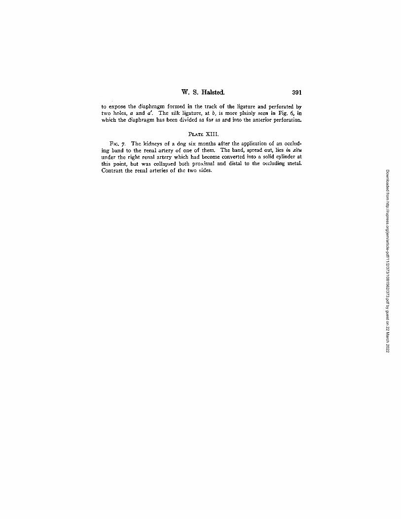
PLATE XIII.
FIG. 7. The kidneys of a dog six months after the application of an occlud-ing band to the renal artery of one of them. The band, spread out, lies in situunder the right renal artery which had become converted into a solid cylinder atthis point, but was collapsed both proximal and distal to the occluding metal.Contrast the renal arteries of the two sides. D
ownloaded from
http://rupress.org/jem/article-pdf/11/2/373/1081562/373.pdf by guest on 22 M
arch 2022


![Partial Face Recognition: An Alignment Free Approach · Alignment via landmarks 250 Cross-view [30,11,13,33] Limited FOV Skin texture [35] Frontal, partial face alignment 114 Occlusion,](https://img.dokumen.tips/doc/110x75/6001543a7033d50dfd266bbb/partial-face-recognition-an-alignment-free-approach-alignment-via-landmarks-250.jpg)