Embed Size (px)
Citation preview

Partial Anomalous Pulmonary Venous Connection in Adults: Evaluation with MDCT
e-Poster: 349
Congress: 2WCTI 2009
Type: Educational poster
Topic: Pulmonary circulation
Authors: , Maldjian P, Saric M; Newark/USBhatti W
MeSH:
Pulmonary Veins [A07.231.908.713]
Pulmonary Circulation [G09.330.582.163.770]
Keywords: Pulmonary circulation, PAPVC, ASD, MDCT
Any information contained in this pdf file is automatically generated from digital material submittedto e-Poster by third parties in the form of scientific presentations. References to any names, marks,products, or services of third parties or hypertext links to third-party sites or information are providedsolely as a convenience to you and do not in any way constitute or imply 2WCTI’s endorsement,sponsorship or recommendation of the third party, information, product, or service. 2WCTI is notresponsible for the content of these pages and does not make any representations regarding thecontent or accuracy of material in this file.As per copyright regulations, any unauthorised use of the material or parts thereof as well ascommercial reproduction or multiple distribution by any traditional or electronically basedreproduction/publication method is strictly prohibited.You agree to defend, indemnify, and hold 2WCTI harmless from and against any and all claims,damages, costs, and expenses, including attorneys’ fees, arising from or related to your use of thesepages.Please note: Links to movies, ppt slideshows and any other multimedia files are not available in the pdfversion of presentations.www.2wcti.org

1. 2. 3. 4.
1.
1. Educational Presentation
Educational Objective
The educational objectives of this exhibit are to present the CT appearance of partial anomalouspulmonary venous connection (PAPVC) in adults and discuss related issues assessable withmultidetector CT (MDCT).
Disclosure
The authors of this presentation do not have any affiliation with the manufacturer of any commercialproduct or services.
Background
Partial anomalous pulmonary venous connection is a congenital abnormality in which one ormore of the pulmonary veins connect to a systemic vein or directly to the right atriumpotentially producing a left to right shunt.The most common forms encountered in adults are PAPVC of the left upper lobe pulmonaryvein to the left brachiocephalic vein and PAPVC of the right upper lobe pulmonary vein to thesuperior vena cava (SVC).In this exhibit we show the appearances of various forms of PAPVC and discuss issues regardingthe anomaly evaluable by MDCT.
Presentation Overview
Discuss difficulty in discerning pulmonary venous connections to the SVC and strategies toimprove visualization of these connections.Association of PAPVC with sinus venosus atrial septal defect.Evaluation of the status of the remainder of the pulmonary venous circulation, as thisdetermines the extent of left to right shunting.Evaluation of the flow direction in the anomalous vessel. Under certain circumstances, flow inthe anomalous pulmonary vein can be away from the systemic venous connection producing aright to left shunt. We demonstrate how flow direction can be determined on MDCT.Complications secondary to PAPVC after surgery on the contralateral lung.Post-operative appearance and complications after surgical repair of PAPVC.We illustrate the use of multiplanar image reformatting and volume rendering to best depict theimaging features of PAPVC.
PAPVC: DEFINITION AND INCIDENCE
Partial anomalous pulmonary venous connection (PAPVC) is present when one or more, but not all, ofthe pulmonary veins connect to a systemic vein, the right atrium, or the coronary sinus.
Left Lung Veins connect to:
left innominate veincoronary sinushemiazygous veinanomalous vein that drains into the innominate vein.
Right Lung Veins connect to:

1. 2. 3. 4.
Superior/inferior vena cavaazygous veinright atriumhepatic vein
Less common variations:
Absence of the coronary sinus with pulmonary veins connecting to multiple systemic sites or tothe left atriumConnection to the portal vein
Incidence: 0.4-0.7% with higher rates in females.
PAPVC OF THE LEFT UPPER LOBE PULMONARY VEIN
On CT, this is seen as a vertically oriented anomalous vein to the left of the aortic arch.This is often mistaken for persistence of the left SVC.PAPVC of the left superior pulmonary vein drains to the left brachiocephalic vein.

Partial Anomalous Pulmonary Venous Connection (PAPVC) of the Left Upper LobePulmonary Vein
35-year-old man with PAPVC of the left superior pulmonary vein. CT demonstrates anomalousvessel (arrow) adjacent to the aortic arch.

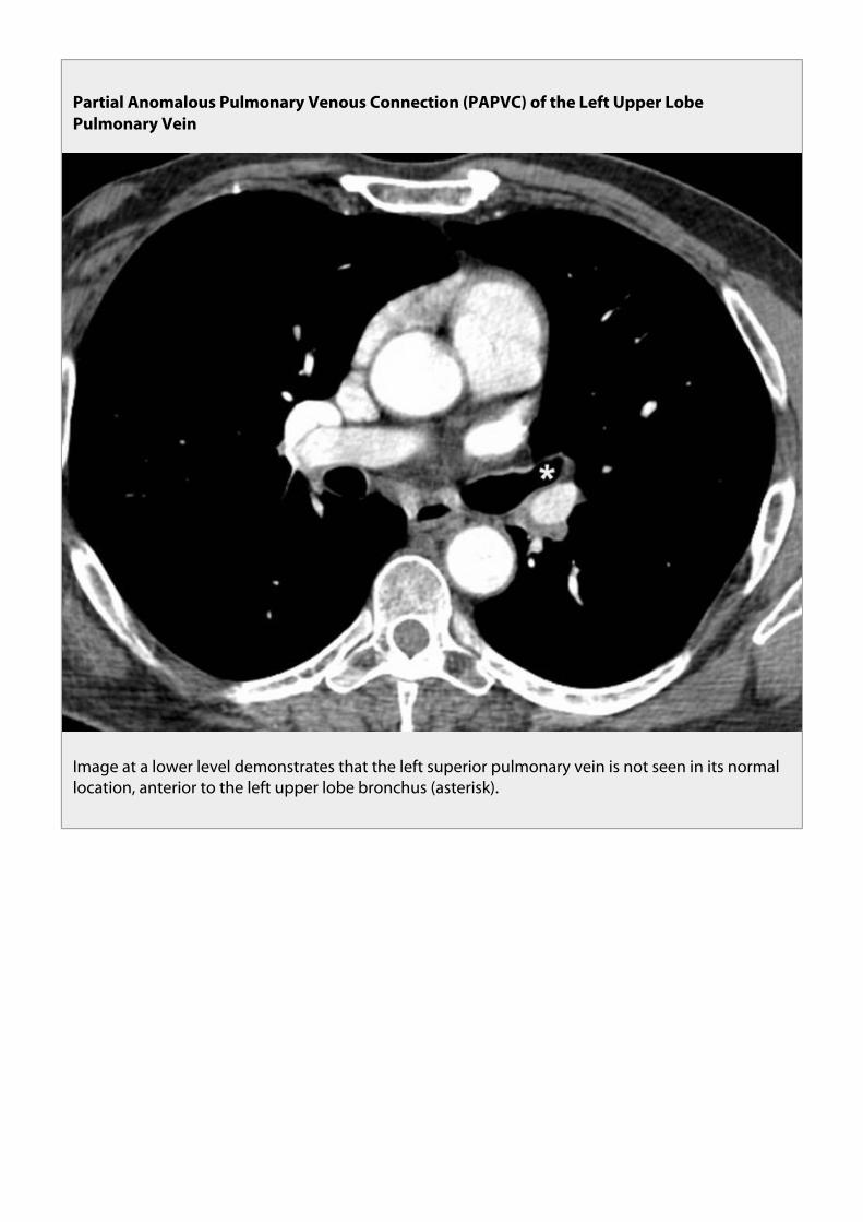
Partial Anomalous Pulmonary Venous Connection (PAPVC) of the Left Upper LobePulmonary Vein
Image at a lower level demonstrates that the left superior pulmonary vein is not seen in its normallocation, anterior to the left upper lobe bronchus (asterisk).

Partial Anomalous Pulmonary Venous Connection (PAPVC) of the Left Upper LobePulmonary Vein
Volume rendered view from anterior perspective shows anomalous left superior pulmonary vein(arrow) joining with left brachiocephalic vein (B). Arrowheads = tributaries of left upper lobepulmonary veins.
Clinical Implications:
Catheter placementsCHF from left heart failure spares the left upper lobeRight heart failure results in Left upper lobe pulmonary edemaObstruction of left brachiocephalic vein can result in right to left shunt (unoxygenated bloodfrom left subclavian vein drains to anomalous left superior pulmonary vein to collateral channelsto pulmonary veins to left atrium).[Levy JM, J Vasc Interv Radiol 2002; 13:423-425.]Right lung disease or right pneumonectomy results in significant left to right shunt.[Black MD.Ann Thor Surg 1992;53:689-91]

PAPVC of the Left Upper Lobe Pulmonary Vein: CXR Findings
PAPVC (arrow) of the Left Upper Lobe Pulmonary Vein on a chest radiology.

PAPVC of the Left Upper Lobe Pulmonary Vein
Volume rendering of PAPVC of the Left Upper Lobe Pulmonary Vein (arrowhead).

PAPVC Connection in Adults
PAPVC OF THE LEFT UPPER LOBE VS. PERSISTANT LEFT SUPERIOR VENA CAVA
Persistent Left SVC:
Most common venous anomaly of the thorax.Incidence 0.5 -2.0 % in normal individuals and up to 10% with congenital heart disease.Results from failure of embryologic regression of a portion of the left common and anteriorcardinal veins.Drains the left jugular and left subclavian vein and courses inferiorly anterior to the left hilum todrain into a dilated coronary sinus.Right superior vena cava may or may not be present, and may be small if present.In some cases, persistent left superior vena cava can connect to the left atrium producing a rightto left shunt.

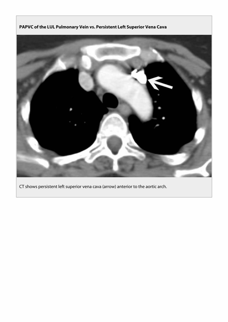
PAPVC of the LUL Pulmonary Vein vs. Persistent Left Superior Vena Cava
Chest radiograph shows a vascular catheter coursing along the left mediastinum in a persistent leftsuperior vena cava.

PAPVC of the LUL Pulmonary Vein vs. Persistent Left Superior Vena Cava
CT shows persistent left superior vena cava (arrow) anterior to the aortic arch.

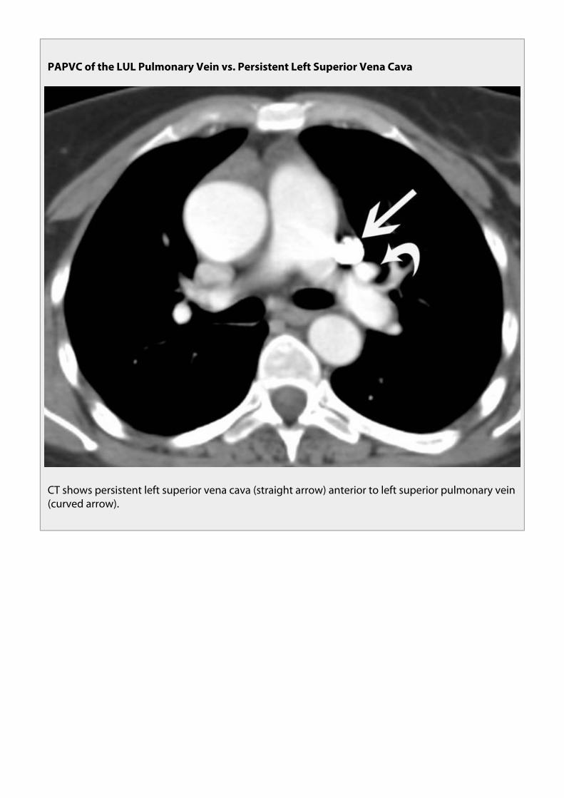
PAPVC of the LUL Pulmonary Vein vs. Persistent Left Superior Vena Cava
CT shows persistent left superior vena cava (straight arrow) anterior to left superior pulmonary vein(curved arrow).

PAPVC of the LUL Pulmonary Vein vs. Persistent Left Superior Vena Cava
Reformatted coronal MIP image shows persistent left superior vena cava (white arrow) draining tocoronary sinus (black arrow).
PAPVC - Left Upper Lobe PulmonaryVein
Persistent Left SVC
Left brachiocephalic vein isnormal or enlargedNo vessels anterior to the leftmain bronchusLeft upper lobe pulmonary Veinsenter aberrant vesselNormal Coronary Sinus
Left brachiocephalic vein is small or absent2 vessels anterior to Left main bronchus (Left superiorpulmonary vein and left SVC)Left superior pulmonary vein has normal courseCoronary sinus is enlarged
PAPVC - SHUNTING
With isolated PAPVC, CXR is usually normal since shunt is less than 2:1. With larger shunts findings aresimilar to ASD:
pulmonary overcirculationenlarged RA and RVenlarged main and hilar pulmonary arteries with small aortaenlarged systemic vein at site of connection (SVC, azygos vein)need to see the course of the abnormal PV in the lung to make the diagnosis on CXR
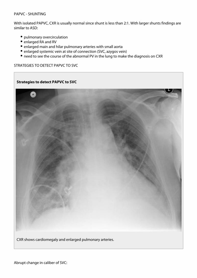
STRATEGIES TO DETECT PAPVC TO SVC
Strategies to detect PAPVC to SVC
CXR shows cardiomegaly and enlarged pulmonary arteries.
Abrupt change in caliber of SVC:
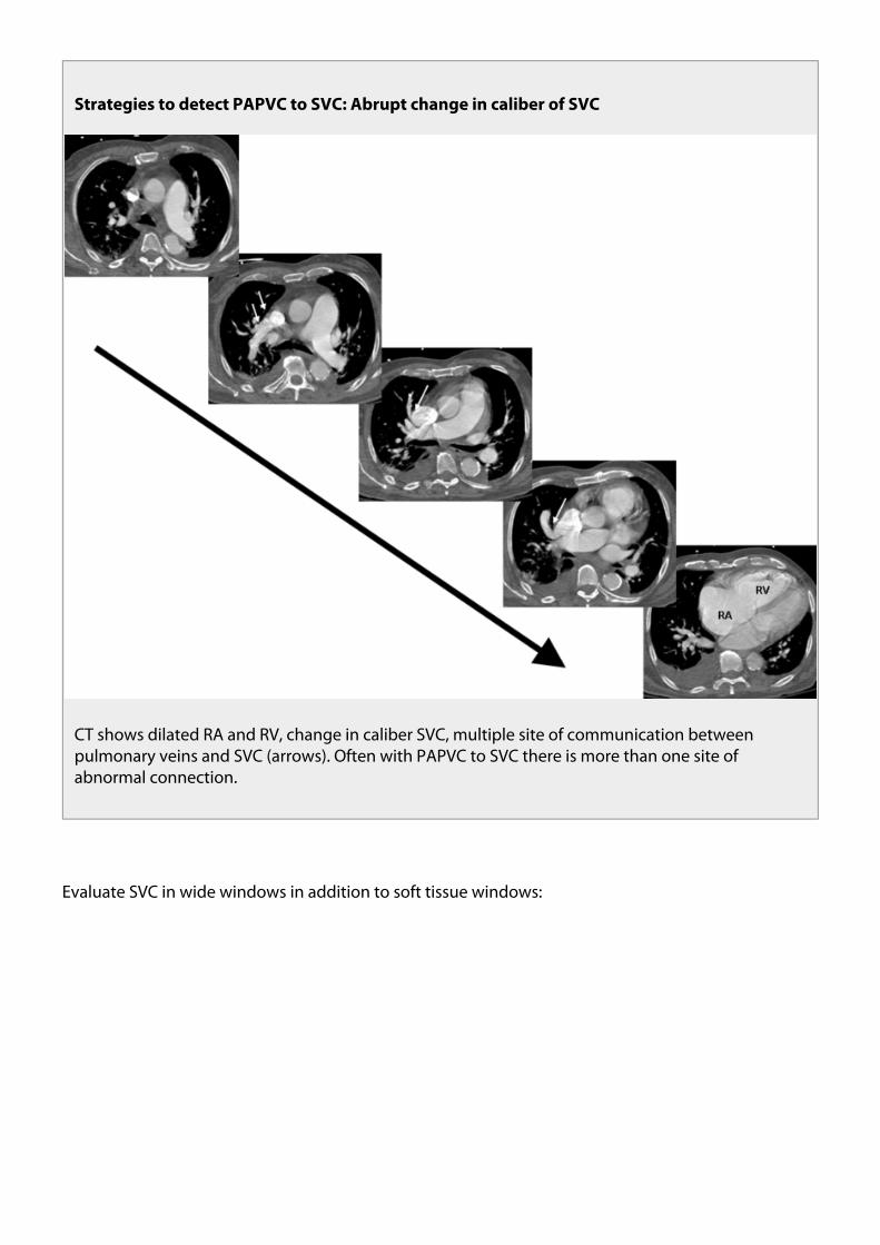
Strategies to detect PAPVC to SVC: Abrupt change in caliber of SVC
CT shows dilated RA and RV, change in caliber SVC, multiple site of communication betweenpulmonary veins and SVC (arrows). Often with PAPVC to SVC there is more than one site ofabnormal connection.
Evaluate SVC in wide windows in addition to soft tissue windows:

Strategies to detect PAPVC to SVC
Note that connections of anamalous vein to SVC (arrows) are more discernable with widerwindows.
PAPVC AND ATRIAL SEPTAL DEFECTS
Partial anomalous pulmonary venous connection (PAPVC) is associated with atrial septal defects(ASD).15% of secundum ASD’s and 85 - 100% of sinus venosus ASD’s are associated with PAPVC.
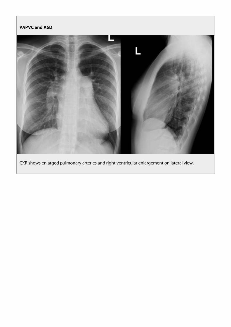
PAPVC and ASD
CXR shows enlarged pulmonary arteries and right ventricular enlargement on lateral view.

PAPVC and ASD
PAPVC and ASD

PAPVC and Atrial Septal Defects
Small sinus venosus ASD with dilated RA and RV
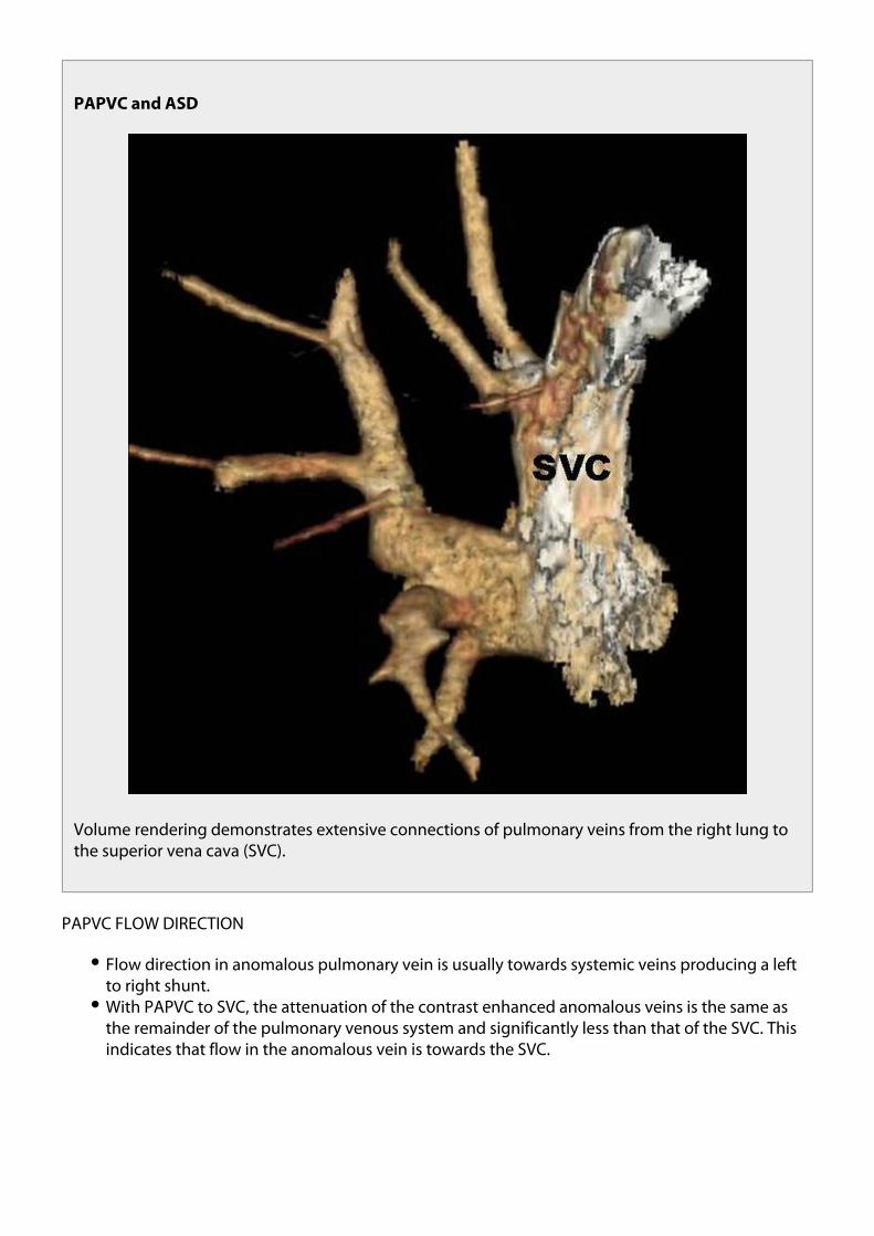
PAPVC and ASD
Volume rendering demonstrates extensive connections of pulmonary veins from the right lung tothe superior vena cava (SVC).
PAPVC FLOW DIRECTION
Flow direction in anomalous pulmonary vein is usually towards systemic veins producing a leftto right shunt.With PAPVC to SVC, the attenuation of the contrast enhanced anomalous veins is the same asthe remainder of the pulmonary venous system and significantly less than that of the SVC. Thisindicates that flow in the anomalous vein is towards the SVC.

PAPVC Flow Direction
Note that attenuation of the anomalous pulmonary veins is less than that of the SVC (arrows).
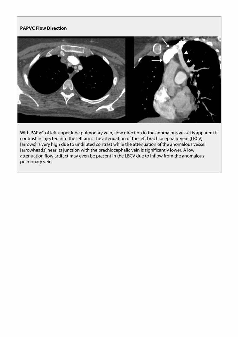
With PAPVC of left upper lobe pulmonary vein, flow direction in the anomalous vessel isapparent if contrast in injected into the left arm. The attenuation of the left brachiocephalic vein(LBCV) is very high due to undiluted contrast while the attenuation of the anomalous vessel nearits junction with the brachiocephalic vein is significantly lower. A low attenuation flow artifactmay even be present in the LBCV due to inflow from the anomalous pulmonary vein.

PAPVC Flow Direction
With PAPVC of left upper lobe pulmonary vein, flow direction in the anomalous vessel is apparent ifcontrast in injected into the left arm. The attenuation of the left brachiocephalic vein (LBCV)[arrows] is very high due to undiluted contrast while the attenuation of the anomalous vessel[arrowheads] near its junction with the brachiocephalic vein is significantly lower. A lowattenuation flow artifact may even be present in the LBCV due to inflow from the anomalouspulmonary vein.
In some cases flow can be retrograde in the anomalous pulmonary vein away from the LBCV.

PAPVC Flow Direction
CT in patient with heart failure from cardiomyopathy shows PAPVC of left upper lobe pulmonaryveins (LUL PV) [arrows] to the left brachiocephalic vein (LBCV) [arrowhead]. Images were obtainedearly after injection of IV contrast as study was performed to exclude pulmonary embolism. There isopacification of anomalous LUL PV before contrast has reached the remainder of the pulmonaryveins and left sided cardiac chambers. (Prolonged circulation time from heart failure likely alsocontributes to lack of contrast in normal pulmonary veins.) Contrast must have entered anomalouspulmonary vein via retrograde flow from LBCV. We speculate that elevated systemic venouspressure from patient’s cardiomyopathy also contributes to retrograde flow in anomalouspulmonary vein.

PAPVC Flow Direction
Oblique axial phase contrast MRI study on same patient performed after stabilization of heartfailure shows direction of flow in anomalous vein [arrow] has normalized to cephalad, towardsLBCV, in same direction as flow in ascending aorta [arrowhead] (phase image on left withmagnitude image on right).

PAPVC Flow Direction
CT scan in patient with history of tricuspid valve replacement for tricuspid valve endocarditisshows PAPVC [arrowhead] that forms conduit from LBCV to left inferior pulmonary vein [arrow].Flow direction in anomalous vessel is away from LBCV causing a right to left shunt. We speculatethat elevated systemic venous pressure from right heart disease is significantly contributory to thisphenomenon. Note opacification of left ventricle before opacification of the right cardiacchambers.

PAPVC Flow Direction
Axial image at level of main pulmonary artery shows early opacification of aorta (straight arrow)before opacification of pulmonary artery (PA) due to flow of contrast from LBCV to left atrium. Notethe dense contrast in the anamalous pulmonary vein (curved arrow).
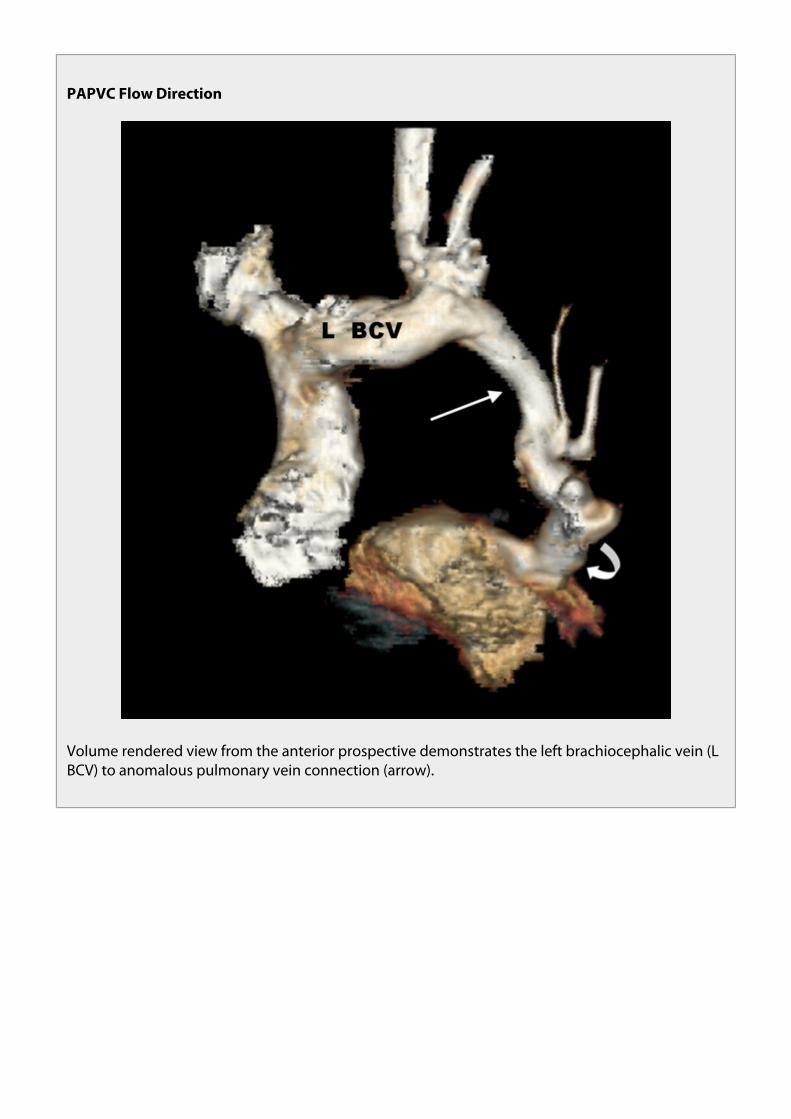
PAPVC Flow Direction
Volume rendered view from the anterior prospective demonstrates the left brachiocephalic vein (LBCV) to anomalous pulmonary vein connection (arrow).

PAPVC Flow Direction
Phase contrast image from MRI study confirms direction of flow in anomalous vessel [arrows] iscaudad away from LBCV, same direction as flow in descending thoracic aorta [arrowheads] ( phasecontrast image on top with magnitude image on bottom).
PAPVC SURGICAL CORRECTION
Cardiac gated CT on 25 year-old woman with sinus venosus atrial septal defect and multipleanomalous pulmonary veins in the right lung draining to the superior vena cava.

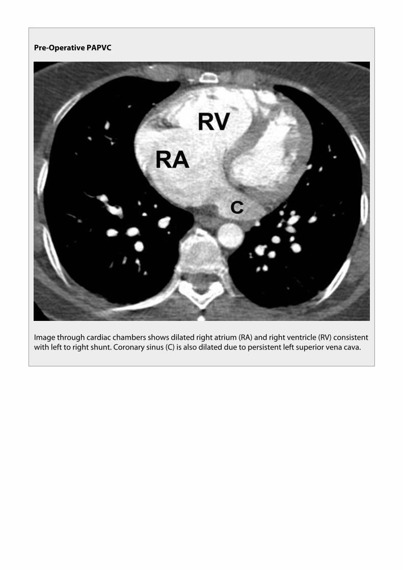
Pre-Operative PAPVC
CT shows sinus venosus atrial septal defect (long arrow) allowing communication of the left atrium(LA) and superior vena cava (S). Anomalous pulmonary veins (short arrows) are also seen drainingto the superior vena cava.

Pre-Operative PAPVC
At a slightly higher level additional anomalous pulmonary veins (short arrows) are seen draining tothe superior vena cava (long arrow).
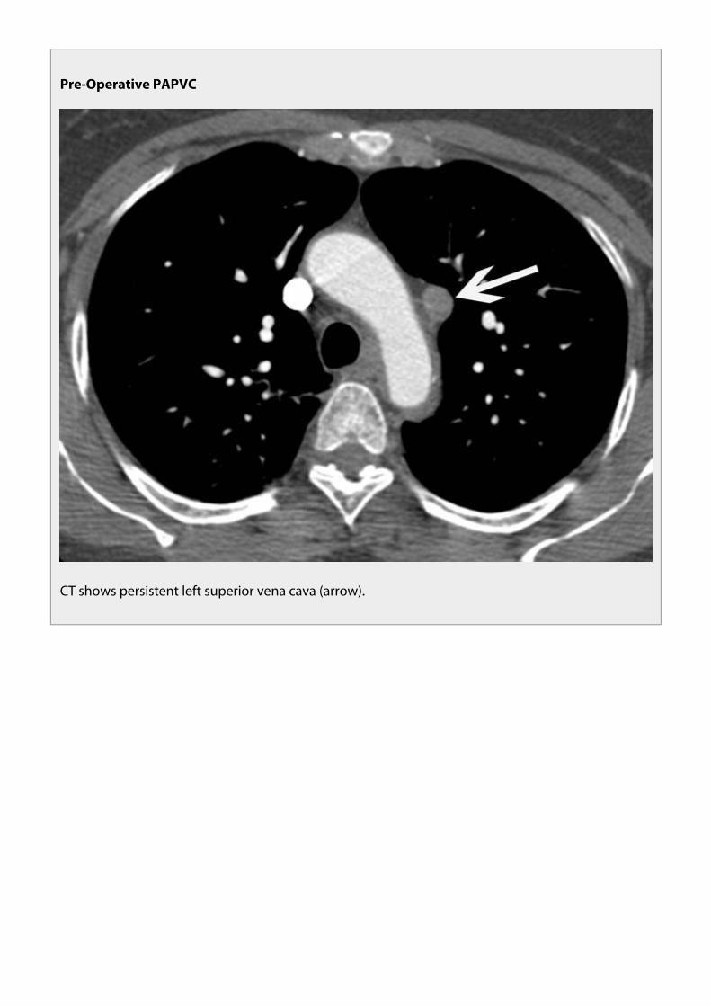
Pre-Operative PAPVC
CT shows persistent left superior vena cava (arrow).

Pre-Operative PAPVC
Image through cardiac chambers shows dilated right atrium (RA) and right ventricle (RV) consistentwith left to right shunt. Coronary sinus (C) is also dilated due to persistent left superior vena cava.
At surgery the right superior vena cava was ligated above the junction with the most superioranomalous right pulmonary vein and the communication between the inferior superior vena cava andthe right atrium was closed. Thus blood return from the anomalous pulmonary veins now drains to theinferior portion of the right superior vena cava and to the right atrium via the sinus venosus atrialseptal defect.

Post-Operative PAPVC
Post-operative CT scan shows anomalous pulmonary veins (short arrows) draining to inferiorportion of superior vena cava (long arrow) to left atrium (LA) via sinus venosus atrial septal defect(asterisk).
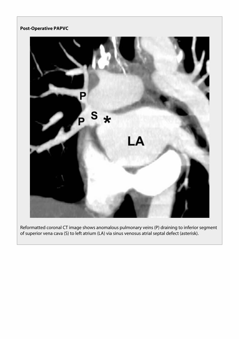
Post-Operative PAPVC
Reformatted coronal CT image shows anomalous pulmonary veins (P) draining to inferior segmentof superior vena cava (S) to left atrium (LA) via sinus venosus atrial septal defect (asterisk).

Post-Operative PAPVC
Reformatted coronal MIP image shows blood flow pattern above ligated portion of right superiorvena cava. Superior portion of right superior vena cava (short arrow) communicates with leftsuperior vena cava (long arrow) via collateral mediastinal veins.
55-year-old woman with PAPVC of left upper lobe pulmonary vein who had right pneumonectomy forlung cancer:

Post-Operative PAPVC
Coronal reformatted MIP image shows anomalous left upper lobe pulmonary vein (arrow) drainingto left brachiocephalic vein (B).

Post-Operative PAPVC
Volume rendered view of vasculature from anterior perspective shows anomalous left upper lobepulmonary vein (arrow) draining to left brachiocephalic vein (B). S = superior vena cava.

Post-Operative PAPVC
CT Image shows that main pulmonary artery (P) is larger in diameter than aorta (A) consistent withincreased pulmonary blood flow.
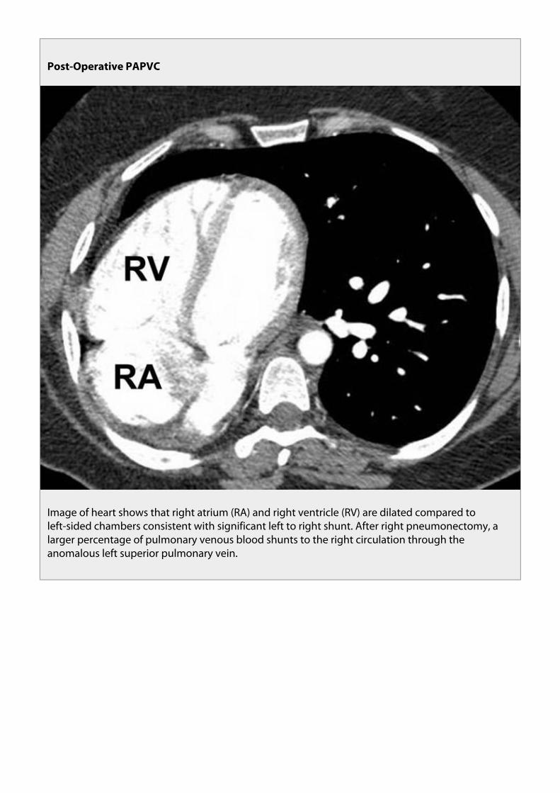
Post-Operative PAPVC
Image of heart shows that right atrium (RA) and right ventricle (RV) are dilated compared toleft-sided chambers consistent with significant left to right shunt. After right pneumonectomy, alarger percentage of pulmonary venous blood shunts to the right circulation through theanomalous left superior pulmonary vein.

Post-Operative PAPVC
Patient with history of PAPVC and repair with anastamosis of anomalous vein to left atrium. Patientdeveloped stenosis at junction [asterisk] of pulmonary vein and left atrium resulting in a largepulmonary varix [arrow].

Post-Operative PAPVC
Volume rendered from anterior perspective shows large pulmonary varix extending throughoutright lung [arrows].
2. Conclusions
After reviewing this exhibit, the participant should be familiar with the MDCT evaluation of partialanomalous pulmonary venous connection in adults.
3. References
Broy C, Bennett S. Partial anomalous pulmonary venous return. Mil Med. 2008 Jun;173(6):523-4
Moral S, Ortuño P, Aboal J. Multislice CT in Congenital Heart Disease: Partial Anomalous PulmonaryVenous Connection. Pediatr Cardiol. 2008 Jun 28
Maillard JO, Cottin V, Etienne-Mastroïanni B, Frolet JM, Revel D, Cordier JF. Pulmonary varix mimicking

pulmonary arteriovenous malformation in a patient with Turner syndrome. Respiration.2007;74(1):110-3.
Sungur M, Ceyhan M, Baysal K. Partial anomalous pulmonary venous connection of left pulmonaryveins to innominate vein evaluated by multislice CT. Heart. 2007 Oct;93(10):1292.
Haramati LB, Moche IE, Rivera VT, Patel PV, Heyneman L, McAdams HP, Issenberg HJ, White CS.Computed tomography of partial anomalous pulmonary venous connection in adults. J Comput AssistTomogr. 2003 Sep-Oct;27(5):743-9.
Schatz SL, Ryvicker MJ, Deutsch AM, Cohen HR. Partial anomalous pulmonary venous drainage of theright lower lobe shown by CT scans. Radiology. 1986 Apr;159(1):21-2.
Greene R, Miller SW. Cross-sectional imaging of silent pulmonary venous anomalies. Radiology. 1986Apr;159(1):279-81.
4. Personal Information
Waseem A. Bhatti, M.S., M.D.¹ Pierre D. Maldjian M.D. ¹ Muhamed Saric M.D.²Authors:
1: Department of Radiology. New Jersey Medical School. University of Medicine and Dentistry of NewJersey. Newark, New Jersey. USA.
2: Department of Cardiology. New Jersey Medical School. University of Medicine and Dentistry of NewJersey. Newark, New Jersey. USA.

5. Mediafiles
PAPVC Connection in Adults

PAPVC Flow Direction
CT in patient with heart failure from cardiomyopathy shows PAPVC of left upper lobe pulmonaryveins (LUL PV) [arrows] to the left brachiocephalic vein (LBCV) [arrowhead]. Images were obtainedearly after injection of IV contrast as study was performed to exclude pulmonary embolism. There isopacification of anomalous LUL PV before contrast has reached the remainder of the pulmonaryveins and left sided cardiac chambers. (Prolonged circulation time from heart failure likely alsocontributes to lack of contrast in normal pulmonary veins.) Contrast must have entered anomalouspulmonary vein via retrograde flow from LBCV. We speculate that elevated systemic venouspressure from patient’s cardiomyopathy also contributes to retrograde flow in anomalouspulmonary vein.
PAPVC Flow Direction
With PAPVC of left upper lobe pulmonary vein, flow direction in the anomalous vessel is apparent ifcontrast in injected into the left arm. The attenuation of the left brachiocephalic vein (LBCV)[arrows] is very high due to undiluted contrast while the attenuation of the anomalous vessel[arrowheads] near its junction with the brachiocephalic vein is significantly lower. A lowattenuation flow artifact may even be present in the LBCV due to inflow from the anomalouspulmonary vein.

PAPVC Flow Direction
CT scan in patient with history of tricuspid valve replacement for tricuspid valve endocarditisshows PAPVC [arrowhead] that forms conduit from LBCV to left inferior pulmonary vein [arrow].Flow direction in anomalous vessel is away from LBCV causing a right to left shunt. We speculatethat elevated systemic venous pressure from right heart disease is significantly contributory to thisphenomenon. Note opacification of left ventricle before opacification of the right cardiacchambers.

PAPVC Flow Direction
Axial image at level of main pulmonary artery shows early opacification of aorta (straight arrow)before opacification of pulmonary artery (PA) due to flow of contrast from LBCV to left atrium. Notethe dense contrast in the anamalous pulmonary vein (curved arrow).

PAPVC Flow Direction
Volume rendered view from the anterior prospective demonstrates the left brachiocephalic vein (LBCV) to anomalous pulmonary vein connection (arrow).

PAPVC Flow Direction
Oblique axial phase contrast MRI study on same patient performed after stabilization of heartfailure shows direction of flow in anomalous vein [arrow] has normalized to cephalad, towardsLBCV, in same direction as flow in ascending aorta [arrowhead] (phase image on left withmagnitude image on right).
PAPVC Flow Direction
Note that attenuation of the anomalous pulmonary veins is less than that of the SVC (arrows).

PAPVC Flow Direction
Phase contrast image from MRI study confirms direction of flow in anomalous vessel [arrows] iscaudad away from LBCV, same direction as flow in descending thoracic aorta [arrowheads] ( phasecontrast image on top with magnitude image on bottom).

PAPVC and ASD
Volume rendering demonstrates extensive connections of pulmonary veins from the right lung tothe superior vena cava (SVC).

PAPVC and ASD
PAPVC and ASD

PAPVC and ASD
CXR shows enlarged pulmonary arteries and right ventricular enlargement on lateral view.

PAPVC and Atrial Septal Defects
Small sinus venosus ASD with dilated RA and RV

PAPVC of the LUL Pulmonary Vein vs. Persistent Left Superior Vena Cava
Chest radiograph shows a vascular catheter coursing along the left mediastinum in a persistent leftsuperior vena cava.

PAPVC of the LUL Pulmonary Vein vs. Persistent Left Superior Vena Cava
Reformatted coronal MIP image shows persistent left superior vena cava (white arrow) draining tocoronary sinus (black arrow).
PAPVC of the LUL Pulmonary Vein vs. Persistent Left Superior Vena Cava
CT shows persistent left superior vena cava (arrow) anterior to the aortic arch.
PAPVC of the LUL Pulmonary Vein vs. Persistent Left Superior Vena Cava
CT shows persistent left superior vena cava (straight arrow) anterior to left superior pulmonary vein(curved arrow).

PAPVC of the Left Upper Lobe Pulmonary Vein
Volume rendering of PAPVC of the Left Upper Lobe Pulmonary Vein (arrowhead).

PAPVC of the Left Upper Lobe Pulmonary Vein: CXR Findings
PAPVC (arrow) of the Left Upper Lobe Pulmonary Vein on a chest radiology.
Partial Anomalous Pulmonary Venous Connection (PAPVC) of the Left Upper LobePulmonary Vein
Image at a lower level demonstrates that the left superior pulmonary vein is not seen in its normallocation, anterior to the left upper lobe bronchus (asterisk).

Partial Anomalous Pulmonary Venous Connection (PAPVC) of the Left Upper LobePulmonary Vein
35-year-old man with PAPVC of the left superior pulmonary vein. CT demonstrates anomalousvessel (arrow) adjacent to the aortic arch.

Partial Anomalous Pulmonary Venous Connection (PAPVC) of the Left Upper LobePulmonary Vein
Volume rendered view from anterior perspective shows anomalous left superior pulmonary vein(arrow) joining with left brachiocephalic vein (B). Arrowheads = tributaries of left upper lobepulmonary veins.

Post-Operative PAPVC
Reformatted coronal CT image shows anomalous pulmonary veins (P) draining to inferior segmentof superior vena cava (S) to left atrium (LA) via sinus venosus atrial septal defect (asterisk).

Post-Operative PAPVC
Post-operative CT scan shows anomalous pulmonary veins (short arrows) draining to inferiorportion of superior vena cava (long arrow) to left atrium (LA) via sinus venosus atrial septal defect(asterisk).

Post-Operative PAPVC
Reformatted coronal MIP image shows blood flow pattern above ligated portion of right superiorvena cava. Superior portion of right superior vena cava (short arrow) communicates with leftsuperior vena cava (long arrow) via collateral mediastinal veins.

Post-Operative PAPVC
Volume rendered from anterior perspective shows large pulmonary varix extending throughoutright lung [arrows].

Post-Operative PAPVC
Image of heart shows that right atrium (RA) and right ventricle (RV) are dilated compared toleft-sided chambers consistent with significant left to right shunt. After right pneumonectomy, alarger percentage of pulmonary venous blood shunts to the right circulation through theanomalous left superior pulmonary vein.

Post-Operative PAPVC
Coronal reformatted MIP image shows anomalous left upper lobe pulmonary vein (arrow) drainingto left brachiocephalic vein (B).

Post-Operative PAPVC
Patient with history of PAPVC and repair with anastamosis of anomalous vein to left atrium. Patientdeveloped stenosis at junction [asterisk] of pulmonary vein and left atrium resulting in a largepulmonary varix [arrow].

Post-Operative PAPVC
CT Image shows that main pulmonary artery (P) is larger in diameter than aorta (A) consistent withincreased pulmonary blood flow.

Post-Operative PAPVC
Volume rendered view of vasculature from anterior perspective shows anomalous left upper lobepulmonary vein (arrow) draining to left brachiocephalic vein (B). S = superior vena cava.
Pre-Operative PAPVC
Image through cardiac chambers shows dilated right atrium (RA) and right ventricle (RV) consistentwith left to right shunt. Coronary sinus (C) is also dilated due to persistent left superior vena cava.

Pre-Operative PAPVC
At a slightly higher level additional anomalous pulmonary veins (short arrows) are seen draining tothe superior vena cava (long arrow).

Pre-Operative PAPVC
CT shows persistent left superior vena cava (arrow).

Pre-Operative PAPVC
CT shows sinus venosus atrial septal defect (long arrow) allowing communication of the left atrium(LA) and superior vena cava (S). Anomalous pulmonary veins (short arrows) are also seen drainingto the superior vena cava.

Strategies to detect PAPVC to SVC
CXR shows cardiomegaly and enlarged pulmonary arteries.

Strategies to detect PAPVC to SVC
Note that connections of anamalous vein to SVC (arrows) are more discernable with widerwindows.

Strategies to detect PAPVC to SVC: Abrupt change in caliber of SVC
CT shows dilated RA and RV, change in caliber SVC, multiple site of communication betweenpulmonary veins and SVC (arrows). Often with PAPVC to SVC there is more than one site ofabnormal connection.