Embed Size (px)
Citation preview
CONGENITAL CARDIOLOGY TODAY
CALL FOR CASES AND OTHER ORIGINAL ARTICLES
Do you have interesting research results, observations, human interest stories, reports of meetings, etc. to share?
Submit your manuscript to: [email protected]
C O N G E N I T A L C A R D I O L O G Y T O D A YTime ly News and I n fo rma t i on f o r BC /BE Congen i t a l /S t ruc tu ra l Ca rd io l og i s t s and Su rgeons
June 2013; Volume 11; Issue 6International Edition Total Anomalous Pulmonary Venous
Connection in the NeonateIN THIS ISSUE
By Claudeen K. Whitfield, MD; P. Syamasundar Rao, MD
Introduction
In the previous issues of this publication, we fo-cussed on several general topics on Congenital Heart Disease (CHD) in the neonate.1-5 More recently, we began addressing individual car-diac lesions, namely: Transposition of the Great Arteries6 and Tetralogy of Fallot,7 Hypoplastic Left Heart Syndrome8 and Tricuspid Atresia.9 In this issue of Congenital Cardiology Today, we will discuss Total Anomalous Pulmonary Venous Connection (TAPVC). Other conditions with similar embryological and clinical findings, as well as therapeutic implications, such as atresia of the common pulmonary vein, cor triatriatum and stenosis/atresia of the individual pulmonary; veins will not be reviewed in this paper.
Total Anomalous Pulmonary Venous Connection
In TAPVC, all the pulmonary veins drain into systemic veins; most commonly they drain into a common pulmonary vein which is then con-nected to the left innominate vein, superior vena cava, coronary sinus, portal vein or other rare sites. Occasionally, individual veins drain direct-ly into the right atrium. TAPVC is the fifth most common cause of cyanotic congenital heart dis-ease (CHD), and the twelfth most common CHD in critically ill infants. TAPVC occurs in 0.6 to 1.2 per 10,000 live births. Sixty-eight percent are di-agnosed as neonates.10 TAPVC is an isolated le-sion in approximately two-thirds of patients and
occurs in association with other CHD, as well as heterotaxia syndromes in the remaining one-third of patients. The male to female ratio in the Baltimore-Washington infant study was 18:23.10 In other reports, a strong male preponderance of 3:1 was observed in patients with infra-di-aphargmatic connection. If left unrepaired, 50% of the babies may survive up to 3 months, and 20% up to 1 year of age, depending on the type and degree of obstruction.11
Embryology
During normal development, the lung buds are outgrowths of the primitive foregut, and early in fetal life venous drainage from the lungs is via the splanchnic plexus to the cardinal and um-bilicovi-telline venous systems. The common pulmonary vein arises from the posterior left atrium as a small pouch that enlarges and connects with the pulmonary venous component of the splanchnic plexus. When pulmonary venous drainage via the common pulmonary vein is established, the con-nections to the cardinal and umbilicovitelline ve-nous systems involute and eventually disappear. The common pulmonary vein is incorporated into the posterior left atrial wall by differential growth, and the pulmonary veins then connect directly to the left atrium. If the common pulmonary vein fails to develop or fails to connect to the splanchnic plexus, the primitive venous connections persist, and result in total anomalous pulmonary venous connection. The type of TAPVC is determined by which of the connections to the cardinal or umbili-covitelline venous system persists. Drainage of the pulmonary veins may be into the right atrium, the right common cardinal system (superior vena
Total Anomalous Pulmonary Venous Connection in the NeonateBy Claudeen K. Whitfield, MD; P. Syamasundar Rao, MD~Page 1
The Congenital Heart Intervention Mission Support Project: Aiming To Help Kids With CHD Around The WorldBy Karim Diab, MD; Damien Kenny, MD~Page 8
DEPARTMENTS
Medical News, Products and Information~Page 9
CONGENITAL CARDIOLOGY TODAY
Editorial and Subscription Offices16 Cove Rd, Ste. 200Westerly, RI 02891 USAwww.CongenitalCardiologyToday.com
© 2013 by Congenital Cardiology Today ISSN: 1544-7787 (print); 1544-0499 (on-line). Published monthly. All rights reserved.
UPCOMING MEDICAL MEETINGSSee website for additional meetings
iCi 2013 - Imaging in Structural, Valvular & Congenital InterventionsJun. 26, 2013; Frankfurt, Germany
www.ici-congress.org
CSI 2013 - Congenital, Structural & Valvular Interventions
Jun. 27–29, 2013; Frankfurt, Germanywww.csi-congress.org
International Academy of Cardiology, 18th World Congress on Heart Disease,
Annual Scientific Sessions 2013Jul. 26-29, 2012; Vancouver, Canada
www.cardiologyonline.com/
ESC (European Society of Cardiology) CongressAug. 31- Sep. 4, 2013; Amsterdam, Netherlands
www.escardio.org
TRENDS Asia Pacific - Renal Denervation, Device Based Treatment of Hypertension,
Neurohumoral StimulationSep. 21, 2013; Hong Kong, Hong Kong
www.csi-congress.org/trend-workshop.php

The Melody® TPV o� ers children and adults a revolutionary option for managing valve conduit failure without open heart surgery.
Just one more way Medtronic is committed to providing innovative therapies for the lifetime management of patients with congenital heart disease.
Innovating for life.
Melody® Transcatheter Pulmonary ValveEnsemble® Transcatheter Valve Delivery SystemIndications: The Melody TPV is indicated for use in a dysfunctional Right Ventricular out� ow Tract (RVOT) conduit (≥16mm in diameter when originally implanted) that is either regurgitant (≥ moderate) or stenotic (mean RVOT gradient ≥ 35 mm Hg)Contraindications: None known.Warnings/Precautions/Side E� ects:• DO NOT implant in the aortic or mitral position. • DO NOT use if patient’s anatomy precludes
introduction of the valve, if the venous anatomy cannot accommodate a 22-Fr size introducer, or if there is signi� cant obstruction of the central veins.
• DO NOT use if there are clinical or biological signs of infection including active endocarditis.
• Assessment of the coronary artery anatomy for the risk of coronary artery compression should be performed in all patients prior to deployment of the TPV.
• To minimize the risk of conduit rupture, do not use a balloon with a diameter greater than 110% of the nominal diameter (original implant size) of the conduit for pre-dilation of the intended site of deployment, or for deployment of the TPV.
• The potential for stent fracture should be considered in all patients who undergo TPV placement. Radiographic assessment of the stent with chest radiography or � uoroscopy should be included in the routine postoperative evaluation of patients who receive a TPV.
• If a stent fracture is detected, continued monitoring of the stent should be performed in conjunction with clinically appropriate hemodynamic assessment. In patients with stent fracture and signi� cant associated RVOT obstruction or regurgitation, reintervention should be considered in accordance with usual clinical practice.
Potential procedural complications that may result from implantation of the Melody device include: rupture of the RVOT conduit, compression of a coronary artery, perforation of a major blood vessel, embolization or migration of the device, perforation of a heart chamber, arrhythmias, allergic reaction to contrast media, cerebrovascular events (TIA, CVA), infection/sepsis, fever, hematoma, radiation-induced erythema, and pain at the catheterization site.Potential device-related adverse events that may occur following device implantation include: stent fracture resulting in recurrent obstruction, endocarditis, embolization or migration of the device, valvular dysfunction (stenosis or regurgitation), paravalvular leak, valvular thrombosis, pulmonary thromboembolism, and hemolysis.For additional information, please refer to the Instructions for Use provided with the product or call Medtronic at 1-800-328-2518 and/or consult Medtronic’s website at www.medtronic.com.
Humanitarian Device. Authorized by Federal law (USA) for use in patients with a regurgitant or stenotic Right Ventricular Out� ow Tract (RVOT) conduit (≥16mm in diameter when originally implanted). The e� ectiveness of this system for this use has not been demonstrated.
Melody and Ensemble are trademarks of Medtronic, Inc.UC201303735 EN © Medtronic, Inc. 2013; All rights reserved.
201303735_EN.indd 1 1/2/13 4:51 PM

3CONGENITAL CARDIOLOGY TODAY t www.CongenitalCardiologyToday.com t June 2013
cava or azygos vein), the left common cardinal system (left in-nominate vein or coronary sinus), or the umbilicovitelline system (portal vein, ductus venosus, etc.).12
Classification
Darling et al13 proposed a TAPVC classification system based on the site of pulmonary venous drainage: Type I in which there is a supra-car-diac connection and all four pulmonary veins are connected to the com-mon pulmonary vein, which is then drained by a connecting anomalous vein into the right superior vena cava, left superior vena cava, or their tributaries; Type II where there is a cardiac connection and the common pulmonary vein is connected directly to the right heart (coronary sinus or the right atrium); Type III is characterized by an infra-cardiac con-nection and the common pulmonary vein is drained by an anomalous channel which travels caudally anterior to the esophagus through the diaphragm to connect to the portal venous system; and Type IV where there are mixed connections, and the right and left pulmonary veins drain to different sites (for example, left pulmonary veins into the left vertical vein and then into the left innominate vein and right pulmonary veins directly into the right atrium or coronary sinus).13 Supra-cardiac connection is the most common (45%), followed by cardiac (25%), in-fra-cardiac (25%) and mixed (5%).14-18
A simpler classification was proposed by Smith and associates:19 su-pra-diaphragmatic without pulmonary venous obstruction and infra-diaphragmatic with obstruction. Although the supra-diaphragmatic forms are generally non-obstructive, obstruction can also occur in these as well, as reviewed elsewhere.20 However, the infra-diaphrag-matic forms are almost always obstructive. Connection to the left in-nominate vein is the most common type of TAPVC.21-23 Infra-diaphrag-matic type is most common form in neonates.21-23
Pathophysiology
In all types of TAPVC, entire pulmonary venous blood eventually re-turns to the right atrium. Intra-cardiac mixing of systemic and pulmo-nary venous returns occurs. Therefore, right-to-left shunt across the atrial septum (patent foramen ovale or atrial septal defect) must be present for survival. A restrictive atrial communication is not uncom-mon. If atrial communication is restrictive, the amount of blood reach-ing the left atrium is limited, and systemic blood flow (cardiac output) is reduced. If there is wide-open atrial communication, the flow dis-tribution to systemic and pulmonary circuits is dependent on relative compliances of the atria and ventricles; these compliances are even-tually linked to pulmonary and systemic vascular resistances.
In babies with a non-obstructive type of TAPVC, as the pulmonary vascular resistance decreases (with age), there is progressive in-crease in pulmonary blood flow. This causes pulmonary over-circu-lation and eventually congestive heart failure. Right atrial and right ventricular enlargement and dilatation of main and branch pulmonary arteries are usually seen. If untreated, increased pulmonary blood flow will result in pulmonary arteriolar medial hypertrophy and intimal proliferation, and the patients will develop pulmonary hypertension and eventually pulmonary vascular obstructive disease.
In babies with obstructive types of TAPVC, because of high pulmo-nary venous pressure, reflex pulmonary arteriolar constriction occurs resulting in high pulmonary artery pressure and decreased pulmonary blood flow. When the osmotic pressure of the blood exceeds the hy-drostatic pressure in the capillaries, pulmonary edema develops. This is partly compensated for by increased pulmonary lymphatic drain-age, development of alternative pulmonary venous bypass channels and altered capillary permeability.
Obstruction to pulmonary venous return is uniformly present in the infra-diaphragmatic type. It may be extrinsic at the level of the dia-phragm as the connecting vein passes through it, constriction of the ductus venosus, high resistance to the passage of the blood through
the hepatic sinusoids, intrinsic stenosis of the connecting vein or a combination thereof.20 The long connecting vein itself may offer im-pedance to the pulmonary venous return.20 In the supra-diaphrag-matic types, the obstruction can also occur and it may be at multiple sites and with varying degrees of severity. It may be intrinsic, within the connecting vein itself or extrinsic by compression (of the vertical vein) between the left bronchus and left pulmonary artery. The intrinsic stenosis of the anomalous connecting vein may be at its junction with
The Melody® TPV o� ers children and adults a revolutionary option for managing valve conduit failure without open heart surgery.
Just one more way Medtronic is committed to providing innovative therapies for the lifetime management of patients with congenital heart disease.
Innovating for life.
Melody® Transcatheter Pulmonary ValveEnsemble® Transcatheter Valve Delivery SystemIndications: The Melody TPV is indicated for use in a dysfunctional Right Ventricular out� ow Tract (RVOT) conduit (≥16mm in diameter when originally implanted) that is either regurgitant (≥ moderate) or stenotic (mean RVOT gradient ≥ 35 mm Hg)Contraindications: None known.Warnings/Precautions/Side E� ects:• DO NOT implant in the aortic or mitral position. • DO NOT use if patient’s anatomy precludes
introduction of the valve, if the venous anatomy cannot accommodate a 22-Fr size introducer, or if there is signi� cant obstruction of the central veins.
• DO NOT use if there are clinical or biological signs of infection including active endocarditis.
• Assessment of the coronary artery anatomy for the risk of coronary artery compression should be performed in all patients prior to deployment of the TPV.
• To minimize the risk of conduit rupture, do not use a balloon with a diameter greater than 110% of the nominal diameter (original implant size) of the conduit for pre-dilation of the intended site of deployment, or for deployment of the TPV.
• The potential for stent fracture should be considered in all patients who undergo TPV placement. Radiographic assessment of the stent with chest radiography or � uoroscopy should be included in the routine postoperative evaluation of patients who receive a TPV.
• If a stent fracture is detected, continued monitoring of the stent should be performed in conjunction with clinically appropriate hemodynamic assessment. In patients with stent fracture and signi� cant associated RVOT obstruction or regurgitation, reintervention should be considered in accordance with usual clinical practice.
Potential procedural complications that may result from implantation of the Melody device include: rupture of the RVOT conduit, compression of a coronary artery, perforation of a major blood vessel, embolization or migration of the device, perforation of a heart chamber, arrhythmias, allergic reaction to contrast media, cerebrovascular events (TIA, CVA), infection/sepsis, fever, hematoma, radiation-induced erythema, and pain at the catheterization site.Potential device-related adverse events that may occur following device implantation include: stent fracture resulting in recurrent obstruction, endocarditis, embolization or migration of the device, valvular dysfunction (stenosis or regurgitation), paravalvular leak, valvular thrombosis, pulmonary thromboembolism, and hemolysis.For additional information, please refer to the Instructions for Use provided with the product or call Medtronic at 1-800-328-2518 and/or consult Medtronic’s website at www.medtronic.com.
Humanitarian Device. Authorized by Federal law (USA) for use in patients with a regurgitant or stenotic Right Ventricular Out� ow Tract (RVOT) conduit (≥16mm in diameter when originally implanted). The e� ectiveness of this system for this use has not been demonstrated.
Melody and Ensemble are trademarks of Medtronic, Inc.UC201303735 EN © Medtronic, Inc. 2013; All rights reserved.
201303735_EN.indd 1 1/2/13 4:51 PM
Children with Heart Devices and Their Parents Struggle with Quality of Life
Children with implanted heart-rhythm devices and their parents suffer from a lower quality of life compared with their healthy counterparts and may benefit from psychotherapy according to new research in Circulation: Arrhythmia & Electrophysiology, an American Heart Association journal.
Researchers at the Cincinnati Children's Hospital Medical Center studied 173 children with either a pacemaker (40 patients) or implanted defibrillator (133 patients) to assess their quality of life compared to other children with Congenital Heart Disease and to healthy children. The children, ages 8 to 18 years old, and their parents completed quality of life questionnaires.
Compared with healthy children and their parents, children with heart devices and their parents reported significantly lower quality of life scores. Likewise, their scores were also lower than those of children with mild congenital heart disease. However, their quality of life scores were similar to those for children with more severe heart disease but no device.
For children, self-perception, self-worth, and athletic capability affected quality of life. For parents, their child's behavior was the biggest factor related to quality of life. Also, children with an implantable defibrillator tended to have lower quality of life scores than those with pacemakers.
"These findings should encourage us to consider the negative impact of devices, particularly defibrillators, on pediatric patients; and to develop strategies to mitigate these effects," said Richard J. Czosek, MD, study author and Assistant Professor of Pediatrics at the Cincinnati Children's Hospital Medical Center, Heart Institute in Ohio. "Whether these effects on quality of life can be reduced through the use of psychotherapy needs to be assessed."
Co-authors are: William J. Bonney, MD; Amy Cassedy PhD; Douglas Y. Mah, MD; Ronn E. Tanel, MD; Jason R. Imundo, MD; Anoop K. Singh, MD; Mitchell I. Cohen, MD, Christina Y. Miyake, MD; Kara Fawley, BS; and Bradley S. Marino, MD.
Author disclosures and sources of funding are on the manuscript.
Cartoons Reduce Anxiety in Children Undergoing Anesthesia
Newswise – Letting children watch a favorite cartoon is an effective and safe way to reduce anxiety before anesthesia and surgery, concludes a study in the November issue of Anesthesia & Analgesia, official journal of the International Anesthesia Research Society (IARS).
"Cartoon distraction" is an "inexpensive, easy to administer, and comprehensive" technique for reducing anxiety in young children before induction of anesthesia, according to the new research, led by Dr Joengwoo Lee of Chonbuk National University Hospital, South Korea.
The study evaluated the use of cartoons to reduce anxiety in 130 children, aged three to seven, undergoing routine surgical procedures—most commonly tonsillectomy. In a holding area, one group of patients were allowed to choose an animated movie to watch before induction of anesthesia. The children watched the movie on a tablet or
CONGENITAL CARDIOLOGY TODAY t www.CongenitalCardiologyToday.com t February 2013 17
Need to Recruit a Pediatric Cardiologist?
Advertise in Congenital Cardiology Today, the only monthly newsletter dedicated to pediatric and congenital cardiologists.
Reach the most Board Certified or Board Eligible pediatric cardiologists worldwide.
Recruitment advertising includes full color in either the North American print edition, or the electronic PDF International edition.
Available in 1/3 and 1/2 page vertical Recruitment ad sizes. We can create the advertisement for you at no extra charge!
Contact: Tony Carlson, Founder
Tel: +1.301.279.2005 or [email protected]

4 CONGENITAL CARDIOLOGY TODAY t www.CongenitalCardiologyToday.com t June 2013
the common pulmonary vein, at its entry into the left innominate vein, superior vena cava, azygos vein or right atrium, somewhere within the vein itself or a combination thereof. Also, the left innominate vein, superior vena cava or azygos vein may themselves be narrowed. For further details, the reader is referred elsewhere.20 Obstruction is least likely to occur when the pulmonary veins drain into the coronary si-nus. The potential for obstruction at the atrial septal defect level has already been mentioned above.
Clinical Features
Clinical features are largely determined by the degree of pulmonary venous obstruction. If obstruction is present, the majority (~75%) of patients will present within the first few days of life and the reminder at a later time. The presentation is shortly after the first 12 hours of life; this is in contradistinction to Respiratory Distress Syndrome which usually presents at birth. These babies are acutely ill and manifest tachypnea, dyspnea, hypoxemia and metabolic acidosis. These signs and symptoms appear to be related to severe pulmo-nary venous congestion. Physical examination is significant for rales and rhonchi in both lung fields. Cardiovascular findings include wide-ly split second heart sound with an accentuated pulmonary compo-nent and no murmurs. Sometimes a non-specific ejection systolic murmur along the left sternal border may be heard. Hepatomegaly is usually present. Obstructive TAPVC is present in almost all infra-diaphragmatic types and in only 50% of supra-diaphragmatic types.
In the absence of pulmonary venous obstruction, the presentation is within the first month of life in more than half of the patients and the reminder during the first year of life. They usually present with signs of congestive heart failure. Tachypnea, tachycardia, feeding difficul-ties and failure to thrive are usual presenting symptoms. Findings on physical examination are similar to those in patients with a secundum atrial septal defect in that there is a prominent right ventricular im-pulse (hyperdynamic), widely split and fixed second heart sound, an ejection systolic murmur at the left upper sternal border and a mid-di-astolic flow rumble at the left lower sternal border. In addition, pulmo-nary component (P2) of the second heart sound is accentuated, and an ejection systolic click plus third and/or fourth heart sounds (mul-tiple cardiac sounds) may be present. Cyanosis is minimal and may not be clinically detectable because of markedly increased pulmonary blood flow. Signs of cardiac failure are usual. Another clinical feature is a venous hum heard at the left or right upper sternal borders or in the infraclavicular regions in the supra-cardiac types of TAPVC; the venous hum is not altered by changes in the position of the patient.
Non-Invasive Evaluation
Chest X-ray
In the obstructive type, the size of the heart is small and normal or mildly enlarged. There is evidence of marked pulmonary edema with stippled densities and reticular pattern in the lung parenchyma, par-tially obscuring the cardiac borders (Figure 1). The reticular pattern may some- tococcal infection. In the non-obstructive type where there is unrestricted pulmonary blood flow, there is cardiomegaly and in-creased pulmonary vascular markings, but usually no pulmonary ede-ma is seen. In the supracardiac type draining into the left innominate vein, a snowman-type of cardiac silhouette may be seen; however, this may take several weeks/months to develop and may not be obvi-ous in the neonatal period.
Electrocardiogram
Electrocardiogram reveals right ventricular hypertrophy in the ob-structive type; however, it may be difficult to distinguish it from normal neonatal right ventricular preponderance. In the non-obstructive type, right axis deviation, right atrial enlargement as seen by tall P waves in lead II and right precordial leads and right ventricular hypertrophy, manifested tall R waves in right precordial leads, sometime with rSR pattern are usually seen.
Echocardiogram
Echocardiographic studies are useful in confirming the diagnosis, and in defining various issues germane to the management of these sick babies. Inability to easily visualize the entry of pulmonary veins into the left atrium by two-dimensional (2D) and color flow mapping should arouse the suspicion of the diagnosis of TAPVC. Enlargement of the right atrium, right ventricle and pulmonary artery is seen in all types of TAPVC (Figure 2). The left atrium and left ventricle usually appear relatively small compared to the very large right ventricle.25,26 The en-larged right ventricle encroaches onto the left ventricle, compressing it posteriorly (Figure 2 A & C) and to the left (Figure 2D). The left atrium is smaller than normal (because of lack of contribution of the common pulmonary vein), but is easily seen (Figure 2 A, B & D).
The right ventricular and pulmonary artery pressures are elevated, as demonstrated by high tricuspid valve regurgitant velocity (Figure 3A). The right ventricular and pulmonary artery systolic pressure may be estimated by modified Bernoulli equation:
Right ventricular and pulmonary artery systolic pressure = 4V2 + 5 mmHg
Where V is peak velocity of the regurgitant tricuspid jet, and 5 is the estimated right atrial pressure.
Right to left shunt across the patent foramen ovale is also seen in all types of TAPVC (Figure 3B).
The common pulmonary vein is seen behind the left atrium (Figure 2 A & B) and every effort should be made to demonstrate the entry of all pulmonary veins into this chamber by using a combination of 2D imaging and color flow mapping (Figures 4 & 5) in multiple views. Parasternal, subcostal and suprasternal notch views are most helpful in this regard. The size and orientation (horizontal or vertical) of the common pulmonary vein should also be determined. Careful color flow imaging should be used to demonstrate the anomalous connect-ing vein and the site of its drainage.
Figure 1.Chest x-ray of a neonate with total anomalous pulmonary venous connection of an infra-diaphragmatic type demonstrating severe pulmonary venous congestion. Mild cardiac enlargement is also seen.

5CONGENITAL CARDIOLOGY TODAY t www.CongenitalCardiologyToday.com t June 2013
Examples of TAPVC with connection to infra-diaphragmatic (Figure 4), left innominate (Figure 5) and coronary sinus (Figure 6) sites are shown. Stenosis of the connecting vein can occur and may be dem-onstrated by imaging actual narrowing, by dilated proximal portion of the connecting vein, by turbulent, continuous and increased Doppler flow velocity (Figure 7) or a combination thereof.
If the ductus arteriosus is patent, right to left shunt across it is usu-ally seen, particularly in patients with obstructed TAPVC; this might partially bypass pulmonary circuit with high pulmonary vascular resis-tance in the obstructed TAPVC and support the cardiac output.
Magnetic Resonance Imaging
Because precise anatomic details are often outlined by echocardio-graphic studies in the neonate, there is little need for MRI in this age group irrespective of pulmonary venous obstruction. When echo-Doppler studies can’t, for certain, demonstrate all pulmonary veins,26 particularly when connection to multiple sites (mixed type of TAPVC) is suspected or in patients with poor echo windows, MRI can provide vital anatomic information.
Cardiac Catheterization
Cardiac catheterization is not usually necessary to confirm the diag-nosis in the neonate. However, beyond infancy, it may be indicated in order to measure the pulmonary vascular resistance and study its responses to vasodilators.
Management
The initial management of neonates with TAPVC is similar to that of any cyanotic/distressed infant with suspected serious heart disease and is discussed in depth elsewhere4 and will not be detailed here. Maintenance of neutral thermal environment, normal acid-base sta-tus, normoglycemia, and normocalcemia should be undertaken by appropriate monitoring and correction as needed.4 No more than 0.4 F1O2 is necessary unless Pulmonary Parenchymal Disease is pres-ent. Metabolic acidosis, defined as pH < 7.25 should be corrected with sodium bicarbonate (usually 1-2 mEq/kg diluted half and half with 5% or 10% dextrose solution) immediately. In the presence of respiratory acidosis, appropriate suctioning, intubation and assisted ventilation should be undertaken.
Figure 2. Selected video frames of a 2-dimensional echocardiogram in parasternal long (A) and short (B & C) axis and apical four cham-ber (D) views demonstrating a large right atrium (RA), right ventricle (RV) and main pulmonary artery (MPA) in an infant with total anoma-lous pulmonary venous connection. Note that the enlarged RV en-croaches onto the left ventricle compressing it posteriorly (A & C) and to the left (D). Common pulmonary vein (CPV) is located behind (A & B) and superior to the left atrium (LA) (D).
Figure 3. A. High tricuspid regurgitant Doppler flow velocity is shown and is suggestive of elevated pulmonary artery pressure in a neonate with an infra-diaphragmatic type of total anomalous pulmonary ve-nous connection. B. Color Doppler flow mapping demonstrating right to left shunting across the atrial septum. RA, right atrium; LA, left atrium.
Figure 4. Selected video frames from suprasternal notch views in a baby with infra-diaphragmatic type of total anomalous pulmonary venous connection showing pulmonary veins (PV) draining into a common pulmonary vein (CPV) which is located adjacent to the left atrium (LA); 2-dimensional (A) and color flow mapping (B) images are shown. Anomalous trunk (AT) traversing towards the diaphragm is shown in C.
o b s t r u c t i o n . A c t a P a e d i a t r 9 6 , 1 4 5 5 - 1 4 5 9 , d o i : 1 0 . 1 1 1 1 / j .1651-2227.2007.00439.x (2007).
22. Sebelius, K. Advancing Screening for C C H D , < h t t p : / / w w w. h r s a . g o v /advisorycommittees/mchbadvisory/h e r i t a b l e d i s o r d e r s /recommendations/correspondence/cyanot icheartsecre09212011.pdf> (2011).
23. State of New Jersey 214th Legislature. A s s e m b l y, N o . 3 7 4 4 , < h t t p : / /www.n j leg .s ta te .n j .us /2010/B i l l s /A4000/3744_I1.PDF> (2011).
24. M a r y l a n d G e n e r a l A s s e m b l y . HB714, <ht tp : / /167.102.242.144/s e a r c h ? q = h o u s e + b i l l+714+2011&site=all&btnG=Search &filter=0&client=mgaleg_default&output=xml_no_dtd&proxysty lesheet =mgaleg_default&getfields=author. t i t l e . k e y w o r d s & n u m = 1 0 0 & s o r t = d a t e % 3 A D % 3 A L%3Ad1&entqr=3&oe=UTF-8&ie=UTF-8&ud=1> (2012).
25. Indiana State Senate. Senate Bill 552, <http://www.in.gov/legislative/bills/2011/SB/SB0552.1.html> (2011).
26. New York State Assembly. A7941-2011, <http://m.nysenate.gov/legislation/bill/A7941-2011> (2011-2012).
27. The General Assembly of Pennsylvania. S e n a t e B i l l 1 2 0 2 , < h t t p : / /www. leg is . s ta te .pa .us /CFDOCS/L e g i s / P N / P u b l i c / b t C h e c k . c f m ?txtType=HTM&sessYr=2011&sessInd=0&billBody=S&billTyp=B&billNbr=1202&pn=1486> (2011).
28. New Hampshire Assembly. SB 348, <http: / /www.gencourt .state.nh.us/l e g i s l a t i o n / 2 0 1 2 / S B 0 3 4 8 . h t m l > (2012).
29. Missouri House of Representatives. HB 1058, <http://www.house.mo.gov/b i l l s u m m a r y . a s p x ?bil l=HB1058&year=2012&code=R> (2012).
30. Georgia State Assembly. House Bill 745, <http://www.legis.ga.gov/Legislation/20112012/118525.pdf> (2012).
31. Florida House of Representatives. H B 8 2 9 , < h t t p : / /www.myfloridahouse.gov/Sections/D o c u m e n t s / l o a d d o c . a s p x ?FileName=_h0829__.docx&DocumentType=Bill&BillNumber=0829&Session=2012> (2012).
32. Virg in ia Genera l Assembly. HB 399, <h t tp : / / l i s .v i rg in ia .gov /cg i -b i n / l e g p 6 0 4 . e x e ?
ses=121&typ=bi l&va l=HB399+&Submit2=Go> (2012) .
33. West Virginia Legislature. House Bill 4327, <http://www.legis.state.wv.us/B i l l _ S t a t u s / b i l l s _ h i s t o r y . c f m ?input=4327&year=2012&sessiontype=rs> (2012).
34. General Assembly of the State of Tennessee. HOUSE BILL 373 SENATE BILL 65, <http://www.capitol.tn.gov/Bills/107/Bill/HB0373.pdf> (2012).
35. Connecticut General Assembly. SB 56, < h t t p : / / w w w . c g a . c t . g o v / a s p /cgab i l l s t a t us / cgab i l l s t a t us .asp?selBillType=Bill&bill_num=56&which_year=2012&SUBMIT1.x=9&SUBMIT1.y=11> (2012).
36. Minnesota House of Representatives. HF No. 3008, <https://www.revisor.mn. g o v / b i n / b l d b i l l . p h p ?bill=H3008.0.html&session=ls87> (2012).
37. California State Assembly. AB1731, <http://www.leginfo.ca.gov/pub/11-12/b i l l / a s m / a b _ 1 7 0 1 - 1 7 5 0 /ab_1731_bill_20120424_amended_asm_v97.html> (2012).
38. N e w b o r n . . . C o a l i t i o n . cchdscreen ingmap.com, <ht tp : / /w w w. c c h d s c r e e n i n g m a p . c o m / > (2013).
39. Beissel, D. J., Goetz, E. M. & Hokanson, J. S. Pulse oximetry screening in Wisconsin. Congenital hear t d isease 7, 460-465, do i :10.1111/j.1747-0803.2012.00651.x (2012).
CCT
Mitchell Goldstein, MDAssociate Professor, PediatricsDivision of NeonatologyLoma Linda University Children's HospitalLoma Linda, CA USACell: 818-730-9309Office: 909.558.7448Fax: 909.558.0298
Archiving Working GroupInternational Society for Nomenclature of Paediatric and Congenital Heart Disease
ipccc-awg.net
Pediatric Interventional Cardiologist
The Boston Children's Heart Foundation of Boston Children's Hospital and Harvard Medical School is recruiting a pediatric interventional cardiologist to join a large, academic, and innovative practice. Candidates should be at the instructor or assistant professor level, should be board certified in pediatric cardiology, and should have completed advanced training in congenital heart catheterization. This position will focus on clinical activity and will offer the opportunity to lead clinical research projects and train fellows. We are particularly seeking individuals with a track record of an active role in helping develop new devices/procedures.
Please send letters of application and CV to:
Audrey C. Marshall, MD, Chief,
Invasive Cardiology, Boston Children’s Hospital
300 Longwood AvenueBoston, MA, 02115
10 CONGENITAL CARDIOLOGY TODAY www.CongenitalCardiologyToday.com March 2013
6 CONGENITAL CARDIOLOGY TODAY t www.CongenitalCardiologyToday.com t June 2013
Almost all patients have a large pulmonary venous confluence behind the left atrium. This structure is horizontal in babies with supra-cardiac and cardiac connection and vertical in those with infra-diaphragmatic connection. The surgical repair usually involves making an anastomo-sis between this pulmonary venous confluence and the posterior wall of the left atrium under cardiopulmonary bypass and/or hypothermia. In TAPVC connected to coronary sinus, surgical excision of the com-mon wall between the coronary sinus and left atrium is performed along with closure of orifice of the coronary sinus and the PFO.
In the obstructive type, initial stabilization by intubation and ventila-tion with high airway pressure should be initiated. Prostaglandin E1
(PGE1) infusion to open the ductus may decompress the pulmonary vascular bed and augment systemic blood flow. In addition, it may open the ductus venosus, thus decreasing pulmonary venous ob-struction. This effect is not as certain as with ductus arteriosus and is not reliable. Intravenous infusion of PGE1 may be started at a dose of 0.05 to 0.1 µg per kilogram of body weight per minute and the rate of infusion is reduced to 0.02 µg per kilogram once the ductus is open. This lower dose has been most helpful in reducing the incidence and severity of some of the drug’s bothersome side effects, namely, ap-nea and hyperpyrexia. It is important to emphasize, however, that most patients with infra-diaphragmatic type have severe obstruction and the main treatment mode is surgical correction. After initial stabi-lization, emergent surgical correction by anastomosis of the common pulmonary vein to the left atrium is mandatory. High mortality seen in early years with surgery has decreased over the years.27
In non-obstructive type, elective surgery is recommended after con-trol of cardiac failure is achieved and the patient is stabilized. Conges-tive heart failure is managed with ionotropic support and diuretics. The entire systemic flow must pass though the patent foramen ovale (PFO) and therefore, if the PFO is restrictive, systemic perfusion is significantly reduced. These patients with supra-diaphragmatic type of TAPVC with a restrictive PFO will benefit from a balloon atrial sep-tostomy.28,29 Surgical correction involves anastomosis of the common pulmonary vein with the left atrium. Ligation of the connecting vein is routinely performed. Depending on surgical preference, the PFO is usually, but not always, closed.
In the presence of mixed type of TAPVC, a single large posterior pulmonary venous confluence is absent. Therefore, if the patient is stable and without significant pulmonary hypertension or pulmonary venous obstruction then one management option is to follow these patients medically until individual anomalous veins are large enough to be an-astomosed to the left atrium.26
Clinical and echocardiographic follow-up is recommended to detect development of pulmonary venous obstruction.
Summary and Conclusions
In TAPVC, all pulmonary veins drain into systemic veins, most com-monly they drain into a common pulmonary vein which is then con-nected to the left innominate vein, superior vena cava, coronary sinus, portal vein or other rare sites. TAPVC is the fifth most common cya-notic CHD and occurs in 0.6 to 1.2 per 10,000 live births. Irrespective of the type, all pulmonary venous blood eventually gets back into right atrium, mixes with systemic venous return, and gets redistributed to the systemic (via patent foramen ovale) and pulmonary (via tricuspid valve) circulations. The TAPVC is classified based on the anatomic location to which the connecting veins drain, namely, supra-diaphrag-matic (supra-cardiac and cardiac) or infra-diaphragmatic and physi-ologic based on obstruction to the pulmonary venous return, namely, obstructive or non-obstructive. The supra-diaphragmatic forms are generally non-obstructive, and the infra-diaphragmatic forms are al-most always obstructive. Connection to the left innominate vein is the most common type of TAPVC. Infra-diaphragmatic type is most com-mon form in the neonate.
The obstructive types present within the first few hours to days of life with signs of severe pulmonary venous congestion and manifest severe tachypnea, tachycardia and cyanosis. Examination reveals rales in the lung fields and a loud pulmonary component of the sec-ond heart sound. The non-obstructive TAPVC patients, on the other hand, usually present with symptoms of congestive heart failure later in the first month of life. On examination, they have very mild or no visible cyanosis and may have clinical signs of heart failure. Other findings on examination are similar to those seen in patients with secundum atrial septal defect. Clinical and chest x-ray findings are suggestive of the diagnosis and can be confirmed by echocar-diographic studies.
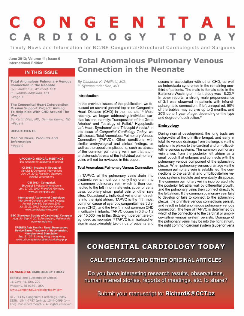
Figure 5. Selected video frames from suprasternal notch views in a baby with supra-diaphragmatic type of total anomalous pulmonary venous connection showing dilated vertical vein (VV), left innominate vein (L Inn) and superior vena cava (SVC); 2- dimensional (A) and color flow mapping (B) images are shown. 2-dimensional images of pulmonary veins (PV) draining into a common pulmonary vein (CPV) which is located adjacent to the left atrium (LA) are also shown (C).
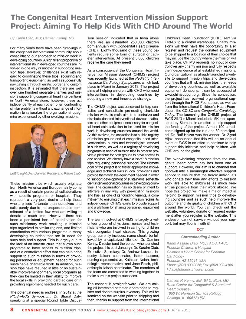
Figure 7. Continuous wave Doppler recordings from the obstructed anomalous trunks demonstrating high velocities in two different pa-tients with total anomalous pulmonary venous connection, infra-dia-phragmatic (A) and supra-diaphragmatic (B).
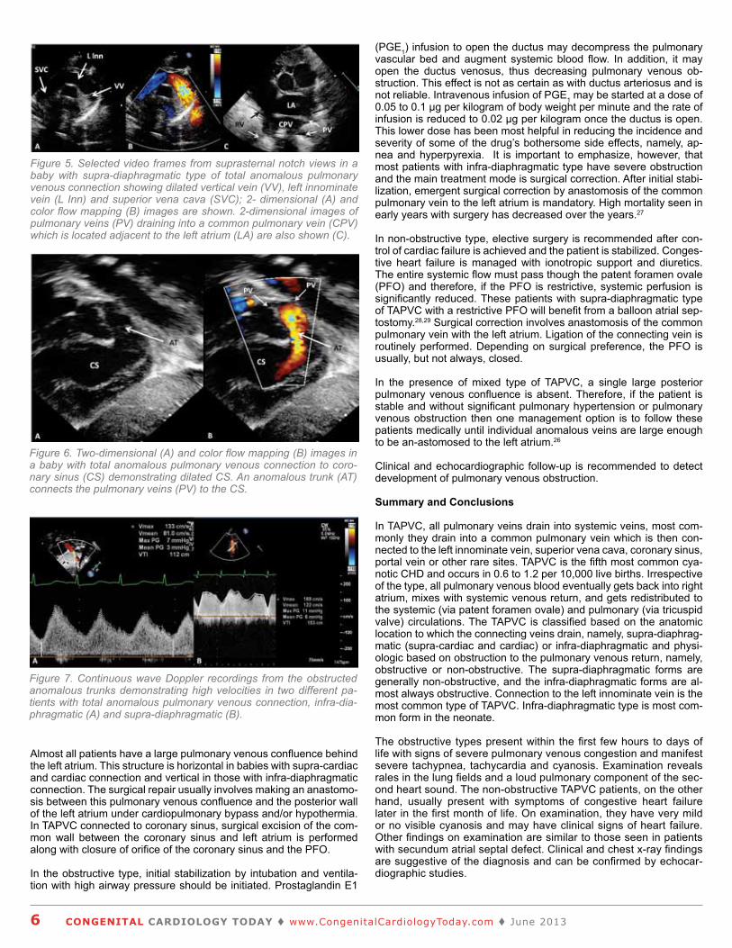
Figure 6. Two-dimensional (A) and color flow mapping (B) images in a baby with total anomalous pulmonary venous connection to coro-nary sinus (CS) demonstrating dilated CS. An anomalous trunk (AT) connects the pulmonary veins (PV) to the CS.

7CONGENITAL CARDIOLOGY TODAY t www.CongenitalCardiologyToday.com t June 2013
In the obstructive type, initial stabilization by intubation and ventilation with high airway pressure should be undertaken. This is fol-lowed by emergent surgical correction by anastomosis of the common pulmonary vein with the left atrium. In the non-obstructive type, control of congestive heart failure and stabilization of the patient, followed by elec-tive or semi-elective surgery is recommend-ed. Follow-up to detect development of pul-monary venous obstruction is recommended.
References
1. Rao PS. Perinatal circulatory physiol-ogy: It’s influence on clinical manifesta-tions of neonatal heart disease – Part I. Neonatology To-day 2008; 3(2):6-12.
2. Rao PS. Perinatal circulatory physiol-ogy: It’s influence on clinical manifesta-tions of neonatal heart disease – Part II. Neonatology Today 2008; 3(3):1-10.
3. Rao PS. An approach to the diagnosis of cyanotic neonate for the primary care provider. Neonatology Today 2007; 2 (6):1-7.
4. Rao PS. Principles of management of the neonate with congenital heart dis-ease Neonatology Today 2007; 2(8):1-10.
5. Rao PS. Neonatal cardiac emergencies: Management strategies, Neonatology Today 2008; 3(12):1-5.
6. Rao PS. Transposition of the great arter-ies in the neonate. Neona-tology Today 2010; 5(8):1-5.
7. Alapati S, Rao PS. Tetralogy of Fallot in the neonate. Neonatology Today 2011; 6(5):1-10.
8. Alapati S, Rao PS. Hypoplastic left heart syndrome in the neonate, Neonatology Today 2011; 6(12):1-9.
9. Rao PS, Alapati S. Tricuspid Atresia in the Neonate, Neonatology Today 2012; 7(5): 1-12.
10. Correa-Villasenor A, Ferencz C, Bough-man JA, Neill CA. Total anomalous pulmonary venous return: Familial and environmental factors. The Baltimore-Washington infant study group. Teratol-ogy. 1991; 44:415-28.
11. Kirklin JW. Total anomalous pulmonary venous connection. In: Cardiac Surgery. Third ed. New York: Churchill Living-stone; 2001. p. 753-759.
12. Bristow JD. Overview of cardiac de-velopment. In: Rudolph CD, Rudolph AM, Hostetter MK, Lister G, Siegel NJ, editors. Rudolph’s Pediatrics. 21st ed. 2003:1745-1749.
13. Darling RC, Rothney WB, Craig JM. To-tal pulmonary venous drain-age into the right side of the heart; report of 17 autop-sied cases not associated with other ma-jor cardiovascular anomalies. Lab Invest 1957; 6:44-64.
14. Choudhary SK, Bhan A, Sharma R, et al. Total anomalous pulmonary venous con-nection: Surgical experience in Indians. Indian Heart J. 2001; 53:754-760.
15. Hyde JA, Stumper O, Barth MJ, et al. Total anomalous pulmonary venous con-nection: Outcome of surgical correction and management of recurrent venous obstruction. Eur J Cardiothorac Surg 1999; 15:735-740; discussion 740-741.
16. Lupinetti FM, Kulik TJ, Beekman RH, 3rd, et al. Correction of total anomalous pulmonary venous connection in in-fancy. J Thorac Cardiovasc Surg 1993; 106:880-885.
17. Michielon G, Di Donato RM, Pasquini L, et al. Total anomalous pulmonary ve-nous connection: Long-term appraisal with evolving technical solutions. Eur J Car-diothorac Surg 2002; 22:184-191.
18. Sinzobahamvya N, Arenz C, Brecher AM, et al. Early and long-term results for cor-rection of total anomalous pulmo-nary venous drainage (TAPVD) in neo-nates and infants. Eur J Cardiothorac Surg 1996; 10:433-438.
19. Smith B, Frye TR, Newton WA, Jr. Total anomalous pulmonary venous return: Diagnostic criteria and a new classifica-tion. Am J Dis Child 1961; 101:41-51.
20. Rao PS, Silbert DR. Superior vena caval obstruction in total anomalous pulmo-nary venous return. Brit Heart J 1974; 36:228-232.
21. Burroughs JT, Edwards JE. Total anom-alous pulmonary venous connection. Am Heart J 1960; 59:913-31.
22. Lucas RV Jr, Lock JE, Tandon R, Ed-wards JE. Gross and histologic anat-omy of total anomalous pulmonary ve-nous connections. Am J Cardiol 1988; 62:292-300.
23. Delisle G, Ando M, Calder AL, et al. Total anomalous pulmonary venous connec-tion: Report of 93 autopsied cases with emphasis on diagnostic and surgical considerations. Am Heart J 1976; 91:99-122.
24. Oliveira Lima C, Valdes-Cruz LM, Allen HD, et al. Prognostic value of left ven-tricular size measured by echocardiog-raphy in infants with total anomalous pul-monary venous drainage. Am J Car-diol 1983; 51:1155-1159.
25. Rosenquist GC, Kelly JL, Chandra R, et al. Small left atrium and change in con-tour of the ventricular septum in total anomalous pulmonary venous connec-tion: A morphometric analysis of 22 in-fant hearts. Am J Cardiol 1985; 55:777-782.
26. Keane JF, Lock JE, Fyler DC, Nadas AS. Nadas’ Pediatric Cardiology. Second ed. Philadelphia: Saunders; 2006:773-781.
27. Bando K, Turrentine MW, Ensing GJ, et al. Surgical treatment of total anomalous pulmonary venous connection. Circula-tion 1996; 94, No. 9 (Suppl II):12-16.
28. Rashkind WJ, Miller WW. Creation of an atrial septal defect without thoracotomy. J Am Med Assoc 1966; 196:991-992.
29. Rao PS. Role of interventional cardiol-ogy in neonates: Part I. Non-surgical atrial septostomy. Neonatology Today 2007; 2(9):9-14.
CCT
Claudeen K. Whitfield, MDPediatric Cardiology FellowThe Division of Pediatric CardiologyDepartment of PediatricsUniversity of Texas-Houston Medical School/Children’s Memorial Hermann HospitalHouston, TX USA
Corresponding Author
P. Syamasundar Rao, MDProfessor of Pediatrics and Medicine Director, Pediatric Cardiology Fellowship Programs Emeritus Chief of Pediatric Cardiology University of Texas-Houston Medical School Children’s Memorial Hermann Hospital Houston, Texas
UT-Houston Medical School6410 Fannin St.UTPB Suite # 425 Houston, TX 77030 USAPhone: 713-500-5738Fax: [email protected]
Program topics will include management strategies of acute heart failure syndromes, methods of hemodynamic and physiologic monitoring, renal protective techniques, and updates on mechanical circulatory support in children.
When: October 10-12, 2013Where: Houston TexasWebsite: http://www.texaschildrenshospital.org/phfs2013/
8 CONGENITAL CARDIOLOGY TODAY t www.CongenitalCardiologyToday.com t June 2013
By Karim Diab, MD; Damien Kenny, MD
For many years there have been rumblings in the congenital interventional community about consolidating our approach to mission work in developing countries. A significant proportion of interventionalists in developed countries are in-volved in one way or another in supporting mis-sion trips; however, challenges exist with re-gard to coordinating these trips, acquiring and transporting equipment, as well as successfully navigating it through erratic border and custom inspection. It is estimated that there are well over one hundred separate charities and mis-sions supporting congenital heart interventions in North America alone, however, these act independently of each other, often confronting similar problems without any exchange of infor-mation to rationalize the organizational quag-mire experienced by other evolving missions.
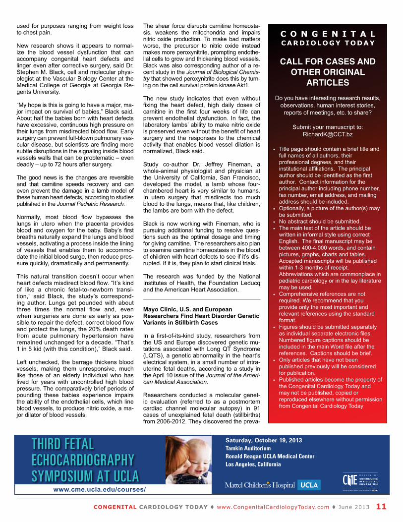
Left to right Drs. Damien Kenny and Karim Diab.
These mission trips which usually originate from North America and Europe mainly come as a result of certain personal collaborations with specific programs or hospitals. They represent a very pure desire to help those who are less fortunate than ourselves and exist purely due to the unquestionable com-mitment of a limited few who are willing to donate so much time. However, there has been a persistent lack of coordination for such missionary work resulting in mission trips organized to similar regions, and limited coordination with various programs in many developing countries that are in need for such help and support. This is largely due to the lack of an infrastructure that allows such programs to have access to mission trips, and of a centralized effort that can help bring support to such missions in terms of provid-ing personnel or equipment needed for such sustainable charitable work. In addition, mis-sion trips have resulted in little or no sustain-able improvement of many local programs as they can be limited in their ability to improve the local skills in providing cardiac care or in providing equipment needed for such care.
The potential need is endless. In 2012 at the PICS~AICS Symposium, Dr. Bharat Dalvi speaking at a special Round Table Discus-
sion session indicated that in India alone there are an estimated 250,000 children born annually with Congenital Heart Disease (CHD). Eighty thousand of these young pa-tients require some form of surgical or cath-eter intervention. At present 5,000 children receive the care they need!
As a consequence, the Congenital Heart In-tervention Mission Support (CHIMS) project was recently launched at the Pediatric Inter-ventional Cardiology Symposium, which took place in Miami in January 2013. The project aims at helping children with CHD who need interventions in developing countries by adopting a new and innovative strategy.
The CHIMS project was conceived to help cen-tralize and consolidate pre-existing charitable mission work. Its main aim is to centralize and distribute donated interventional devices, cathe-ters and other equipment necessary for congeni-tal heart catheterization to be used for mission work in developing countries around the world. As this evolves, the aspiration is to build a registry of mission groups and of congenital heart inter-ventionalists, nurses and technologists involved in such work, as well as a registry of developing programs in need of mission trips in order to pro-vide a platform for both groups to have access to one another. We already have a list of 18 mission trips requesting personnel support! The ultimate goal of the project is to foster appropriate knowl-edge and technical skills in local physicians and provide them with the equipment needed in order to support development of sustainable self-suffi-cient interventional programs in developing coun-tries. The organization has no desire or intent to interfere in any way with pre-existing missions and has, as one of its founding tenents, a com-mitment to ensuring that each mission retains its independence. CHIMS exists to provide support and a platform for sharing personnel, equipment and knowledge.
The team involved at CHIMS is largely a vol-unteer group of physicians, nurses and tech-nicians who are involved in caring for children with congenital heart disease. This growing group currently includes: name should be fol-lowed by a capitalized title ex. Dr. Damien Kenny, Director (and the person who launched the project this past January); Dr. Karaim Diab, Executive Secretary; Dr. Andrew Lashus, in-dustry liaison coordinator, Karen Lacono, nursing representative, Kathleen Nolan, tech-nologist representative, and Jane Reich, phil-anthropic liaison coordinator. The members of the team are committed to working together to make sure this project succeeds.
The concept is straightforward. We are ask-ing all interested catheter laboratories to reg-ister and donate surplus inventory. This will be itemized on the website prior to shipping and then, thanks to support from the International
Children’s Heart Foundation (ICHF), sent via Fed-Ex to a central warehouse. Charity mis-sions will then have the opportunity to also register and request the donated equipment to be shipped to a location of their choice that may include the country where the mission will take place. CHIMS requests no input or con-trol over any charity mission and fully respects the independence of all established missions. Our organization has already launched a web-site to support mission trips and developing countries that will list: mission trips, the needs of developing countries, as well as available equipment donations. It can be accessed at www.chimsupport.org. Since its conception, the project has received organizational sup-port through the PICS Foundation, as well as from the International Children’s Heart Foun-dation, the CCISC and Congenital Cardiology Today. The launching the CHIMS project at PICS 2013 in Miami, included a 5K race spon-sored by Siemens in an effort to help support the funding of the project. Almost 100 partici-pants signed up for the run and 80 participat-ed. Dr. Ralf Holzer was the winner! Dr. Ziyad Hijazi announced that this will be an annual event at PICS in an effort to continue to help support this initiative and help children with CHD around the globe.
The overwhelming response from the con-genital heart community has been one of support. However, our aim is to channel that goodwill into a meaningful effective support service to ensure that the heroic individuals who donate their time and efforts to mission work are able to maximize as much ben-efit as possible from their work abroad. We hope this project will make a major impact in helping to support mission trips to develop-ing countries and as such help improve the outcome and life quality of children with CHD around the world. You can check out the website, volunteer, donate or request equip-ment after you register at the website. This endeavor cannot survive without your sup-port, but may flourish with it!
CCT
Corresponding Author
Karim Assaad Diab, MD, FACC, FASEPhoenix Children’s Hospital Children’s Heart Center for Pediatric CardiologyPhoenix, AZ 85016 USAPhone: (602) 933-3366; Fax: (602) [email protected]
Damien P. Kenny, MB, BAO, BCH, MDRush Center for Congenital & Structural Heart Disease1650 W. Harrison St., 708 KelloggChicago, IL 60612 USA
The Congenital Heart Intervention Mission Support Project: Aiming To Help Kids With CHD Around The World

9CONGENITAL CARDIOLOGY TODAY t www.CongenitalCardiologyToday.com t June 2013
Medical News, Products and Information
Drug-Resistant MRSA Bacteria -- Here to Stay in Both Hospital and Community
The drug-resistant bacteria known as MRSA, once confined to hospitals but now wide-spread in communities, will likely continue to exist in both settings as separate strains, ac-cording to a new study.
The prediction that both strains will coexist is reassuring because previous projections indicated that the more invasive and fast-growing community strains would overtake and eliminate hospital strains, possibly pos-ing a threat to public health.
Researchers at Princeton University used mathematical models to explore what will happen to community and hospital MRSA strains, which differ genetically. Originally MRSA, which is short for methicillin-resistant Staphylococcus aureus, was confined to hospitals. However, community-associated strains emerged in the past decade and can spread widely from person to person in schools, athletic facilities and homes.
Both community and hospital strains cause diseases ranging from skin and soft-tissue infections to pneumonia and septicemia. Hospital MRSA is resistant to numerous anti-biotics and is very difficult to treat, while com-munity MRSA is resistant to fewer antibiotics.
The new study found that these differences in antibiotic resistance, combined with more ag-gressive antibiotic usage patterns in hospitals versus the community setting, over time will permit hospital strains to survive despite the competition from community strains. Hospital-based antibiotic usage is likely to successfully treat patients infected with community strains, preventing the newcomer strains from spread-ing to new patients and gaining the foothold they need to out-compete the hospital strains.
The researchers made their predictions by using mathematical models of MRSA trans-mission that take into account data on drug-usage, resistance profiles, person-to-person contact, and patient age.
Published February 28th in the journal PLOS Pathogens, the study was conducted by postdoctoral researcher Roger Kouyos, now a scholar at the University of Zurich, and Eili Klein, a graduate student who is now an as-sistant professor in the Johns Hopkins School of Medicine. They conducted the work under the advisement of Bryan Grenfell, Princeton’s Kathryn Briger and Sarah Fenton, Professor of Ecology and Evolutionary Biology and Public Affairs at Princeton’s Woodrow Wilson School of International and Public Affairs.
RK was supported by the Swiss National Sci-ence Foundation (Grants PA00P3_131498
and PZ00P3_142411). EK was supported by Princeton University (Harold W. Dodds Fellowship), as well as the Models of Infec-tious Disease Agent Study (MIDAS), under Award Number U01GM070708 from the Na-tional Institutes of General Medical Sciences. BG was supported by the Bill and Melinda Gates Foundation; the Research and Policy for Infectious Disease Dynamics (RAPIDD) program of the Science and Technology Di-rectorate, Department of Homeland Security; and the Fogarty International Center, Nation-al Institutes of Health.
Sudden Death in Young Athletes: Important Causes Not Identified by the Screening Process
Even though young athletes are required to receive health screens to be cleared to play sports, those tests failed to detect impor-tant cardiovascular abnormalities in cleared players, and many were allowed to play despite suspicions of dangerous cardiovas-cular conditions, according to a large regis-try study of patients who died from sudden death. The study results were presented on March 10th by Kevin Harris, MD, Research Cardiologist at the Minneapolis Heart Insti-tute Foundation (MHIF) at the annual Ameri-can College of Cardiology Scientific Ses-sions in San Francisco.
Aortic stenosis, which occurs when the aor-tic heart valve does not fully open, is consid-ered a rare, but important, cause of death in young people. Aortic dissection and rupture which occur when the aortic wall tears and ruptures respectively are catastrophic condi-tions that are not usually associated with the death of younger individuals. However, the role of these very serious conditions is not understood as causes of athletic field deaths, and their identification is often missed during routine pre-participation screening.
“While the majority of these young athletes are being screened, there is unfortunately great variability in the screening process, and we have had very sparse data on the effec-tiveness of these screening efforts,” explains Harris, who is also Co-Director of the Echo-cardiography Laboatory at the Minneapolis Heart Institute® at Abbott Northwestern Hos-pital in Minneapolis.
The American Heart Association has recom-mended specific historical questions and phys-ical examination components which should comprise pre-participation cardiac screening.
For this study, MHIF researchers analyzed the US National Registry of Sudden Death in Young Athletes for occurrences of sudden death due to aortic disease (including dissection, rupture or coarctation) and aortic stenosis.
Of the 2,588 deaths in the registry, 44 events were related to aortic stenosis (19) or aortic disease (25). On average, this group of ath-letes was 17.6 years old, and 40 were males. The most prominent sports represented in this group were football and basketball, fol-lowed baseball and softball.
Eighteen of the 19 deaths related to aortic stenosis occurred just after exercise, report-ed Harris. Also, 16 deaths attributed to aortic disease occurred during exercise, 6 occurred during sedentary activity, and 2 during sleep.
Data on pre-participation screening were available for 34 of the 44 athletes. Of the 34 deaths, 15 young athletes had been as-sessed specifically by cardiologists, 3 of the athletes had a known aortic abnormality and 8 had previously been diagnosed with aortic stenosis or bicuspid aortic valves—the latter of which occurs when an aortic valve only has two leaflets instead of three.
Based on their findings, the researchers concluded that aortic stenosis and aortic diseases are uncommon, but important, causes of sudden death among young, com-petitive athletes, usually while playing bas-ketball or football.
Twenty-five percent of the athletes (11 of 44) complained of symptoms of chest, back or abdominal pain in days prior to collapse. Three of the 11 had been seen in the emer-gency room. Two of the 11 had seen a cardi-ologist the day prior to death.
“We were able to identify the majority of the athletes in this study had been cleared to participate in sports and one-third had been evaluated by a cardiologist,” Harris reports. “The widespread screening process failed to detect important cardiovascular abnormali-ties in 19 of the deaths. In the remaining 15 cases, suspicion of cardiovascular conditions was raised, but the athletes were allowed to continue to compete in competitive sports.”
Shalom Jacobovitz Named CEO of American College of Cardiology The American College of Cardiology Board of Trustees announced in April that Shalom “Shal” Jacobovitz has been selected as the college’s Chief Executive Officer. “Shal has a track record that demonstrates he is the right person to lead a strong organi-zation like the ACC and to take it to the next level at a time when health care is undergo-ing massive changes,” said ACC President John Gordon Harold, MD, MACC. “He is an innovative and proven leader as well as a successful mentor and team builder. Shal brings a unique perspective at a time when

10 CONGENITAL CARDIOLOGY TODAY t www.CongenitalCardiologyToday.com t June 2013
the College is ramping up to meet the evolv-ing needs of cardiovascular professionals do-mestically and around the globe.” Jacobovitz comes to the ACC from Actelion Pharmaceuticals US, a biopharmaceutical company specializing in cardio-pulmonary therapies, where he has served as president since 2004. At Actelion, Jacobovitz developed a strong patient- and customer-centered corpo-rate strategy, which he implemented globally. “This is an exciting time for the College,” said former ACC President William Zoghbi, MD, MACC, who led the CEO Search Commit-tee. “Shal comes to the ACC with more than 25 years in health care, including extensive international and domestic experience. He shares the College’s commitment to quality, innovation and strategic management, and we are confident he will advance the ACC’s mission to transform cardiovascular care and improve heart health.”
Prior to Actelion, Jacobovitz held positions at F. Hoffmann La Roche, where he served as general manager for Central America and the Caribbean, led the Pharmaceutical, OTC and Diagnostic divisions, and served as the global lifecycle leader for cardiovascular products in Basel, Switzerland. He also held positions with Abbott Canada, Nordic Labs and Marion Merrill Dow (now known as Aventis) in Can-ada. Jacobovitz earned his Bachelor of Sci-ence Degree in Biology at the University of Western Ontario in Canada.
“It is a privilege for me to join the ACC,” Jaco-bovitz said. “The College is a thriving medical professional society that is well positioned to be at the center of rapid changes in health care. I look forward to working with the ACC’s dedicated professional staff and member leaders to build on the College’s legacy of leadership in quality improvement, patient-centered care, clinical education and practice excellence.” Jacobovitz was chosen as CEO following a year-long nationwide search led by the executive search firm, Korn/Ferry Interna-tional. He will assume the role beginning in May. During the search period, Tom Arend, COO and General Counsel served as Inter-im Chief Staff Officer.
For more information, visit cardiosource.org/ACC.
Physicians Interactive Introduces Omnio - A Customized, Comprehensive iPad App for Medical Professionals
Physicians Interactive (PI), a leading pro-vider of online and mobile clinical resources and solutions for healthcare professionals, announced in March the launch of Omnio, a versatile, new app that invigorates the medi-cal app market by unleashing the power of the iPad in a personalized point-of care tool.
The free Omnio app is available for download in the App Store: https://itunes.apple.com/us/app/omnio/id545775601?mt=8.
From the point of download, Omnio connects clinicians to comprehensive drug and disease references and calculators that are essential in their specialty. Omnio is easy to expand, connecting users to a worldwide medical marketplace of trusted publishers, allowing them to further customize Omnio into the op-timal point-of-care companion.
Since its mid-December debut in the App Store, word of mouth and social referral among medical professionals have driven Omnio to top the charts of free medical apps in the App Store, bolstered by a stellar rating near 5 stars.
“With more than 31 million new entrants coming into the American healthcare system under healthcare reform, clinicians have a pressing need for tools to simplify their work-flow,” said Physician Interactive’s CEO and Vice Chairman Donato Tramuto. “Yet, only 5% of medical professionals are satisfied with current medical apps (July 2012 Worl-dOne Research Survey). Omnio changes everything, allowing clinicians one-tap ac-cess to their favorite mobile tools and serving all the diverse roles clinicians now play. We chose the name Omnio because this app will become the omnipresent clinical assistant for the emerging healthcare workflow.”
Omnio is designed to be the modern-day digi-tal black bag for clinicians to keep all their most important “must haves” just a tap away on their iPads. The app was developed with extensive user research to ensure it met the needs of medical professionals who have been frus-trated with the limitations of other medical apps.
The research found that hundreds of apps designed for clinicians on the market today were too overwhelming to manage separate-ly. They often focused on just one function, did not allow clinicians to customize the app for their specialties, and too often failed to
provide the relevant information and resourc-es clinicians needed at the point-of-care.
“Clinicians don’t have time to scroll through five screens of medical apps to find the one tool they need,” said Physician Interactive’s Chief Medical Officer and Senior VP of Prod-uct Management Gautam Gulati, MD, MBA, MPH. “We provide those core tools, for each specialty, then apply the latest design and technology to make it easy to add, swap, drag and drop to customize Omnio into their optimal point-of-care resource. Clinicians are coming to Omnio to get the latest news in their field, check drug dosing and interac-tions, review evidence-based guidelines, and perform calculations at the point-of-care. We make it quick and easy for them to access the tools they need.”
Omnio’s free content includes: drug look-up, dosing recommendations, medical calcula-tors, drug interaction, formulary information, disease reference materials, curated spe-cialty news feeds, and much more with ongo-ing releases of new updates. All the offerings are personalized by profession and specialty to identify the most relevant resources from publishers, peers in healthcare, and profes-sional associations. Clinicians can bookmark and tag these key resources—which can range from books to medical calculators to entire Web sites—so they are just one tap away on the iPad. With Omnio, clinicians can spend less time looking for information and more time practicing medicine.
“I can use Omnio on my iPad every day to an-swer questions specific to my branch of car-diology,” said Jordan Safirstein, MD, FACC, FSCAI, RPVI, who specializes in cardiac and peripheral vascular intervention. “It is person-alized information chosen by me that is always accessible on my iPad. This type of function-ality is part of the appeal of the iPad—so it’s great to finally have this in a medical app.”
For more information - visit www.omnio.com.
Carnitine Supplement May Improve Survival Rates of Children with Heart Defects
A common nutritional supplement may be part of the magic in improving the survival rates of babies born with heart defects, re-searchers report.
Carnitine, a compound that helps transport fat inside the cell powerhouse, where it can be used for energy production, is currently
MED
ICA
L C
ON
FER
ENC
ES 2
013
SEPTEMBER 21www.csi-trends.org
NOVEMBER 22– 23www.csi-laa.org
JUNE 26www.ici-congress.org
JUNE 27– 29www.csi-congress.org
TRENDS ASIA-PACIFIC 2013 Catheter-based Neurohumoral Remodeling
Hong Kong, China
LAA 2013 Left Atrial Appendage Closure
Frankfurt, Germany
iCi 2013 Imaging in Cardiovascular Interventions
Frankfurt, Germany
CSI 2013 Congenital, Structural & Valvular Interventions
Frankfurt, Germany
www.ici-congress.orgwww.csi-congress.orgwww.csi-trends.orgwww.csi-laa.org
Taiwan, The Republic of China
SEPTEMBER 21www.csi-trends.org
NOVEMBER 22– 23www.csi-laa.org
JUNE 26www.ici-congress.org
JUNE 27– 29www.csi-congress.org
TRENDS ASIA-PACIFIC 2013 Catheter-based Neurohumoral Remodeling
Hong Kong, China
LAA 2013 Left Atrial Appendage Closure
Frankfurt, Germany
iCi 2013 Imaging in Cardiovascular Interventions
Frankfurt, Germany
CSI 2013 Congenital, Structural & Valvular Interventions
Frankfurt, Germany
www.ici-congress.orgwww.csi-congress.orgwww.csi-trends.orgwww.csi-laa.org
11CONGENITAL CARDIOLOGY TODAY t www.CongenitalCardiologyToday.com t June 2013
used for purposes ranging from weight loss to chest pain.
New research shows it appears to normal-ize the blood vessel dysfunction that can accompany congenital heart defects and linger even after corrective surgery, said Dr. Stephen M. Black, cell and molecular physi-ologist at the Vascular Biology Center at the Medical College of Georgia at Georgia Re-gents University.
“My hope is this is going to have a major, ma-jor impact on survival of babies,” Black said. About half the babies born with heart defects have excessive, continuous high pressure on their lungs from misdirected blood flow. Early surgery can prevent full-blown pulmonary vas-cular disease, but scientists are finding more subtle disruptions in the signaling inside blood vessels walls that can be problematic – even deadly – up to 72 hours after surgery.
The good news is the changes are reversible and that carnitine speeds recovery and can even prevent the damage in a lamb model of these human heart defects, according to studies published in the Journal Pediatric Research.
Normally, most blood flow bypasses the lungs in utero when the placenta provides blood and oxygen for the baby. Baby’s first breaths naturally expand the lungs and blood vessels, activating a process inside the lining of vessels that enables them to accommo-date the initial blood surge, then reduce pres-sure quickly, dramatically and permanently.
This natural transition doesn’t occur when heart defects misdirect blood flow. “It’s kind of like a chronic fetal-to-newborn transi-tion,” said Black, the study’s correspond-ing author. Lungs get pounded with about three times the normal flow and, even when surgeries are done as early as pos-sible to repair the defect, correct blood flow and protect the lungs, the 20% death rates from acute pulmonary hypertension have remained unchanged for a decade. “That’s 1 in 5 kid (with this condition),” Black said.
Left unchecked, the barrage thickens blood vessels, making them unresponsive, much like those of an elderly individual who has lived for years with uncontrolled high blood pressure. The comparatively brief periods of pounding these babies experience impairs the ability of the endothelial cells, which line blood vessels, to produce nitric oxide, a ma-jor dilator of blood vessels.
The shear force disrupts carnitine homeosta-sis, weakens the mitochondria and impairs nitric oxide production. To make bad matters worse, the precursor to nitric oxide instead makes more peroxynitrite, prompting endothe-lial cells to grow and thickening blood vessels. Black was also corresponding author of a re-cent study in the Journal of Biological Chemis-try that showed peroxynitrite does this by turn-ing on the cell survival protein kinase Akt1.
The new study indicates that even without fixing the heart defect, high daily doses of carnitine in the first four weeks of life can prevent endothelial dysfunction. In fact, the laboratory lambs’ ability to make nitric oxide is preserved even without the benefit of heart surgery and the responses to the chemical activity that enables blood vessel dilation is normalized, Black said.
Study co-author Dr. Jeffrey Fineman, a whole-animal physiologist and physician at the University of California, San Francisco, developed the model, a lamb whose four-chambered heart is very similar to humans. In utero surgery that misdirects too much blood to the lungs, means that, like children, the lambs are born with the defect.
Black is now working with Fineman, who is pursuing additional funding to resolve ques-tions such as the optimal dosage and timing for giving carnitine. The researchers also plan to examine carnitine homeostasis in the blood of children with heart defects to see if it’s dis-rupted. If it is, they plan to start clinical trials.
The research was funded by the National Institutes of Health, the Foundation Leducq and the American Heart Association.
Mayo Clinic, U.S. and European Researchers Find Heart Disorder Genetic Variants in Stillbirth Cases
In a first-of-its-kind study, researchers from the US and Europe discovered genetic mu-tations associated with Long QT Syndrome (LQTS), a genetic abnormality in the heart’s electrical system, in a small number of intra-uterine fetal deaths, according to a study in the April 10 issue of the Journal of the Ameri-can Medical Association.
Researchers conducted a molecular genet-ic evaluation (referred to as a postmortem cardiac channel molecular autopsy) in 91 cases of unexplained fetal death (stillbirths) from 2006-2012. They discovered the preva-
www.cme.ucla.edu/courses/
Saturday, October 19, 2013 Tamkin Auditorium Ronald Reagan UCLA Medical CenterLos Angeles, California
THIRD FETALECHOCARDIOGRAPHYSYMPOSIUM AT UCLA
C O N G E N I T A L CARDIOLOGY TODAY
CALL FOR CASES AND OTHER ORIGINAL
ARTICLESDo you have interesting research results,
observations, human interest stories, reports of meetings, etc. to share?
Submit your manuscript to: [email protected]
• Title page should contain a brief title and full names of all authors, their professional degrees, and their institutional affiliations. The principal author should be identified as the first author. Contact information for the principal author including phone number, fax number, email address, and mailing address should be included.
• Optionally, a picture of the author(s) may be submitted.
• No abstract should be submitted.• The main text of the article should be
written in informal style using correct English. The final manuscript may be between 400-4,000 words, and contain pictures, graphs, charts and tables. Accepted manuscripts will be published within 1-3 months of receipt. Abbreviations which are commonplace in pediatric cardiology or in the lay literature may be used.
• Comprehensive references are not required. We recommend that you provide only the most important and relevant references using the standard format.
• Figures should be submitted separately as individual separate electronic files. Numbered figure captions should be included in the main Word file after the references. Captions should be brief.
• Only articles that have not been published previously will be considered for publication.
• Published articles become the property of the Congenital Cardiology Today and may not be published, copied or reproduced elsewhere without permission from Congenital Cardiology Today
Need to Recruit a Pediatric Cardiologist?
Advertise in Congenital Cardiology Today, the only monthly newsletter dedicated to pediatric and congenital cardiologists.
Reach the most Board Certified or Board Eligible pediatric cardiologists worldwide.
Recruitment advertising includes full color in either the North American print edition, or the electronic PDF International edition.
Available in 1/3 and 1/2 page vertical Recruitment ad sizes. We can create the advertisement for you at no extra charge!
Contact: Tony Carlson, Founder
Tel: +1.301.279.2005 or [email protected]
CONGENITAL CARDIOLOGY TODAY is pleased to announce its first Chinese language edition, printed and distributed in China and available worldwide in a PDF file. It will be published four times a year. You can read the August premier issue at:
www.congeni ta lcardio logy.com/China-Aug12.pdf
If you would like to subscribe the the electronic PDF version, send an email to: [email protected] sure to include your name, titles, organization and its address.
If you would like to advertise in this special issue, send an email to: [email protected]
2012No.1

12 CONGENITAL CARDIOLOGY TODAY t www.CongenitalCardiologyToday.com t June 2013
Sponsored for CME credit by Rush University Medical Center
SAVE THE DATEJUNE 7-1O, 2O14Marriott Chicago D O W N T O W N
WWW.P ICSYMPOS IUM.COM
CHICAGO
LIVE CASE DEMONSTRATIONS • ABSTRACT
SESSIONS • “MY NIGHTMARE CASE IN THE
CATH LAB” • HOT DEBATES • WORKSHOPS
• SMALLER BREAKOUT SESSIONS
lence of mutations in the three most common LQTS-susceptible genes, KCNQ1, KCNH2 and SCN5A. Two of the most common genes were discovered in three cases (KCNQ1 and KCNH2); and five of the cases exhibited SC-N5A rare non-synonymous genetic variants.
Intrauterine fetal death or still birth happens in approximately one out of every 160 pregnan-cies and accounts for 50% of all perinatal deaths. “We know that the post-mortem evaluation often has not been able to explain these deaths,” says Michael J. Ackerman, MD, PhD, at Mayo Clinic and co-study senior author along with Peter J. Schwartz, MD, PhD, of the University of Pavia, It-aly. “Those of us who study LQTS and treat LQTS patients have often wondered whether LQTS may be the cause of some of these deaths.”
In the study, more than 1,300 ostensibly healthy individuals served as controls. In ad-dition, publicly available exome (the entire portion of the genome consisting of protein-coding sequences) databases were as-sessed for the general population frequency of identified genetic variances.
“Our preliminary evidence suggests that LQTS may be the cause for approximately 5% of oth-erwise unexplained stillbirths and points to the need for further large-scale studies,” says Dr. Ackerman, Director of Mayo’s LQTS Clinic and Windland Smith Rice Cardiovascular Genom-ics Research Professor. “With LQTS, when we know of its presence, it is a very treatable con-dition but still more work needs to be done to prevent the family’s first tragedy from occurring.”
In LQTS, which affects one in 2,000 people, the rapid heartbeats can trigger a sudden fainting spell, seizure, or sudden death. Life-threatening cardiac arrhythmias can occur unexpectedly, mainly during childhood or adolescence. Treatment can involve medica-tion, medical devices, or surgery.
Other study authors are: Lia Crotti, M.D., PhD, University of Pavia, Pavia, Italy; David Tester, Wendy White, MD, Melissa Will, Jen-nifer Blair, Daniel Van Dyke, PhD, Myra Wick, MD, PhD, Brian Brost, MD, all of Mayo Clinic; Daniel Bartos, Ellyn Velasco, Brian Delisle, PhD, all of University of Kentucky, Lexing-ton; Robert Insolia, PhD, and Alice Ghidoni, both University of Pavia; Alessandra Besana, PhD, IRCCS Instituto Auxologico Italiano, Mi-lan, Italy; Jennifer Kunic and Alfred L. George Jr., MD, Vanderbilt University, Nashville, Tenn.; Irene Cetin, MD, University of Milan, Italy; and Fabio Facchinetti, MD, University of Modena and Reggio Emilia, Italy.
CONGENITAL CARDIOLOGY TODAY
© 2013 by Congenital Cardiology Today (ISSN 1554-7787-print; ISSN 1554-0499-online). Published monthly. All rights reserved.
Corporate Offices:8100 Leaward Way,PO Box 444Manzanita, OR 97130 USATel: +1.301.279.2005; Fax: +1.240.465.0692
Editorial and Subscription Offices: 16 Cove Rd, Ste. 200, Westerly, RI 02891 USA
www.CongenitalCardiologyToday.com
Publishing Management:• Tony Carlson, Founder, President & Sr.
Editor - [email protected]• Richard Koulbanis, Group Publisher &
Editor-in-Chief - [email protected]• John W. Moore, MD, MPH, Medical Editor
- [email protected]• Virginia Dematatis, Assistant Editor• Caryl Cornell, Assistant Editor • Loraine Watts, Assistant Editor • Chris Carlson, Web Manager• William Flanagan, Strategic Analyst• Rob Hudgins, Graphic Designer -
Editorial Board: Teiji Akagi, MD; Zohair Al Halees, MD; Mazeni Alwi, MD; Felix Berger, MD; Fadi Bitar, MD; Jacek Bialkowski, MD; Philipp Bonhoeffer, MD; Mario Carminati, MD; Anthony C. Chang, MD, MBA; John P. Cheatham, MD; Bharat Dalvi, MD, MBBS, DM; Horacio Faella, MD; Yun-Ching Fu, MD; Felipe Heusser, MD; Ziyad M. Hijazi, MD, MPH; Ralf Holzer, MD; Marshall Jacobs, MD; R. Krishna Kumar, MD, DM, MBBS; John Lamberti, MD; Gerald Ross Marx, MD; Tarek S. Momenah, MBBS, DCH; Toshio Nakanishi, MD, PhD; Carlos A. C. Pedra, MD; Daniel Penny, MD, PhD; James C. Perry, MD; P. Syamasundar Rao, MD; Shakeel A. Qureshi, MD; Andrew Redington, MD; Carlos E. Ruiz, MD, PhD; Girish S. Shirali, MD; Horst Sievert, MD; Hideshi Tomita, MD; Gil Wernovsky, MD; Zhuoming Xu, MD, PhD; William C. L. Yip, MD; Carlos Zabal, MD
Statements or opinions expressed in Congenital Cardiology Today reflect the views of the authors and sponsors, and are not necessarily the views of Congenital Cardiology Today.
11615 Hesby StreetN. Hollywood, CA 91601
Tel: 818.754.0312 Fax: 818.754.0842
w w w . c a m p d e l c o r z a n . o r g
!
!& & & & & & & & &' ' ' ' ' ' ' ' ' ' ' '''''''''''''''''
'' '' ' ' ' ' ' ' ' ' ''' ' ' ' '
!!!!!!!!!!!!!!!!!
!!!!!!!!!!!!!!!!!
!!!!!!!!!!!!!!!!!!!!!!!!!
!!!!!!!!!!!!!!!!!!!!!!!!!!!!!!!!!!!!!
!!!!!!!!!!!!!!!!!!!!!!!!!!!!!!!!!!!!!!!!
!!!!"#$!%&'(#!)*+,'-.$!/-0+,&(1-+!!!"#"$%&'$()*+$!,--.+$/0$121*3$$$$$$$$$$$$$$$$$$$$$$$$$$$$$$$$$$$$$$$$$$$$$$$$$$$$
4,56$378"221"9923$$$$$$$:;<&=>?-@AB.;C-&D,"&-E$$$$$$$FFF">?-@AB.;C-&D,"&-E$$$$$$$$$$$$$$$$$$)*.2(-.34!G?-C:&D.&H?@A.+$I,J@-&H,;:?+$KJBL5,$M,?N;,BB+$O',-L:B,$P;@&5,-?;L,+$$$$$$$$$$$$$$
Q-&F@A$R,@?-C?@:&;!

• Transseptal Access Workshop from Cook Medical • Workshop: Past Present and Future of Pediatric Interventions
Cardiology - St. Jude & AGA Mmedical • Symposium on Prevention of Stroke Clinical Trials at the Heart of
the Matter - WL Gore Medical • Imaging in Congenital & Structural Cardiovascular Interventional
Therapies • Morphology of The Atrial Septum • Morphology of The Ventricular Septum • Pre-Selection of Patients of Pulmonic Valve Implantation and
Post-Procedural Follow-up • Echo Paravalvular Leakage (PVL) • ICE vs TEE ASD Closure in Children - PRO & CON ICE • 3D Rotational Angiography - Why Every Cath Lab Should Have This
Modality • PICS Doorway to the Past - Gateway to the Future • Follow-up From PICS Live Cases 2010 Presentation • Intended Intervention - Transcatheter TV Implantation - Live Case• Intended Intervention - LAA Closure Using Amplatzer Cardiac Plug
Under GA & Real Time 3D • Provided Intervention - LPA Stenting / Implantation of a Sapien
Valve • Intended Intervention - PV Implantation • Intended Intervention - COA Stent Using Atrium Advanta V12
Covered Stent - Live Case• Intended Intervention - ASD Closure - Live Case• Intended Intervention -Transcatheter VSD Device Closure - Live
Case Intended Intervention - COA Stenting Using Premounted Advanta V12 Covered Sten - Live Case
• Stunning Revelation - The Medical System is Changing - What Can You Do To Show Patients That Your Practice Does It Right? Patient Perspective
• Percutaneous Paravalvular Leak Closure Outcomes • Intensive Management of Critically Ill Infants Undergoing
Catheterization • Off Label Device Usage - Careful!
• Evolving Hybrid Programs Communications is the Key • Anti-Coagulation in Pediatric Interventions - Evidence Please! Are
we Flying Blind? • Shaping Your Structural Heart Interventional Career: How Do I Get
the Necessary Training and Start a Program? • Closure of Post-Infarct VSDs Limitations & Solutions • Percutaneous Interventions During Pregnancy • Percutaneous PVL Closure Techniques & Outcomes • Percutaneous Therapy for MR • Update on Percutaneous Aortic Valve Replacement: Time to Open
the Flood Gates? • 2011 PICS/AICS Achievement Achievement Award Presented To
Horst Sievert, MD. • The Cases I Learn The Most From • If I was Starting Now I Would... Contemplating the Question.• 10 Tips for the Trainee • The Perfect CV • Why I Left The Cath Lab • Intended Intervention Occlude Right High Flow Superior Segment
PAVM • Intended Intervention Edwards Sapien XT Transcatheter Heart
Valve - 3D Guidance - Live Case • Closure of the Silent PDA - Indicated or Not • Transcatheter Tricuspod Valve Replacement • Recommendations For Device Closure of MVSDs • Intended Intervention Diagnostic Angio/First Intervention Occlude
Left Lower Lobe - Live Case• Intended Intervention PFO Closure using Gore Helex Septal
Occluder Under GA and TEE Guidance - Live Case• Pulmonary Valvuloplasty - Pulmonary Oedema Presentation • Intended Intervention LAA Closure Using the Amplatzer Cardiac
Plug - Live Case • Transcatheter Alternatives in Sick Neonates With Coartation of the
Aorta; Balloon Angioplasty vs Surgery for Native Coartation of the Aorta in Newborns
• and many more!. Plus, live cases from other major meetings
www.CongenitalCardiologyToday.com www.CHDVideo.com
Watch over 300 Live Case Videos, Presentations and Workshops Online from Leading Congenital and Structural Medical Meetings
from Around the World - www.CHDVideo.com
Presented by CONGENITAL CARDIOLOGY TODAY