Embed Size (px)
Citation preview

SciELO Books / SciELO Livros / SciELO Libros FUGASSA, M.H. Paleoparasitological Diagnosis. In: FERREIRA, L.F., REINHARD, K.J., and ARAÚJO, A., ed. Foundations of Paleoparasitology [online]. Rio de Janeiro: Editora FIOCRUZ, 2014, pp. 223-254. ISBN: 978-85-7541-598-6. Available from: doi: 10.7476/9788575415986.0017. Also available in ePUB from: http://books.scielo.org/id/zngnn/epub/ferreira-9788575415986.epub.
All the contents of this work, except where otherwise noted, is licensed under a Creative Commons Attribution 4.0 International license.
Todo o conteúdo deste trabalho, exceto quando houver ressalva, é publicado sob a licença Creative Commons Atribição 4.0.
Todo el contenido de esta obra, excepto donde se indique lo contrario, está bajo licencia de la licencia Creative Commons Reconocimento 4.0.
Part II - Parasite Remains Preserved in Various Materials and Techniques in Microscopy and Molecular Diagnosis
15. Paleoparasitological Diagnosis
Martín H. Fugassa

Paleoparasitological Diagnosis
223
Paleoparasitological Diagnosis
Martín H. Fugassa
15
Paleoparasitology emerged in the early 20th century using light microscopy studies. Although numerous tools have been added since then, light microscopy is still the most widely used method for paleoparasitological examination
and study of microfossils.
Paleoparasitology has made significant progress in nearly a hundred years, partly through the adoption and adaptation of methodologies developed in other fields such as clinical parasitology and biology molecular. However, the inherent difficulties of archaeological materials require on-going improvement of methods and the introduction of new ones.
In the coming years, the extent to which paleoparasitology contributes to paleoecological and anthropological studies will depend largely on increasing the number of samples for examination. It is thus necessary to multiply the range of evidence beyond conventional sources like coprolites and mummified tissues. This will also require expanding the availability of powerful methods and techniques for recovering parasitological information. The following is a description of various methodologies and applied techniques in paleoparasitology, some still in the experimental stage.
SAMPLING
Enteroparasites (helminths and protozoa, or parasites of the gastrointestinal tract) commonly release eggs or other forms for dispersal in the feces. Helminth parasites of the respiratory tract can also be dispersed by passage of eggs into the host’s digestive system when they are transported with mucous, as in the case of Eucoleus aerophillus and Metastrongylus sp. (Miyazaki, 1991).
Coprolites and various archaeological sediments provide the clearest source of evidence in the paleoparasitological study of enteroparasites, while mummified tissues and bones are generally associated with paleopathological and tissue parasite studies.
Foundations of Paleoparasitology
224
Regardless of their nature, paleoparasitological samples should be extracted and handled under aseptic conditions, because molecular and optical studies of microfossils involve a certain risk of contamination from current and ancient sources. Thus, the sample design should consider not only the necessary statistical aspects, but also the aseptic conditions for procedures.
The following is a list of general recommendations for handling samples in fieldwork:
•Usesterile,hermetic,andresistantrecipients.
•Usenewsterileandpreferablydisposableinstruments(spoons,spatulas,tweezers,scalpels,etc.).
•Dependingonthesamplingconditions,inthefieldorlaboratory,andonthetypeofsample,itisnecessarytouseacapandmasktoprotectthefaceandhair.Duringexcavations,oneshouldusegloveswithouttalcumpowder(to avoid contamination with starch, which can influence the diagnosis of the use of corn by the prehistoric group), at least during the removal of samples for paleoparasitological examination and especially when they involve food remains.
•Samplesshouldbeextractedasquicklyaspossibletoavoidleavingthesamplingareauncoveredforprolongedperiods. One should avoid extracting samples under adverse weather conditions such as heavy wind or rain.
•Bothinthefieldandinmuseumsorotherinstitutionswherearchaeologicalcollectionsarekept,samplecollectionequipment and the resulting samples must be kept in clean, hermetic recipients, specific for the purpose.
•Itisbestifatleastonesubsampleineachcaseiskeptrefrigeratedat0ºCformolecularstudies(AlenaIñiguez,personal communication).
For statistical purposes, collecting samples from coprolites and sediments require a prior design. This design should be conducted carefully in order to reflect as closely as possible the properties of the sampled population from which one intends to obtain inferences. Extraction of samples from sediments or coprolites should consider the spatial distribution and density of the parasite remains. It is thus possible to establish some basic hypotheses: 1) coprolites contain more parasites than free sediments, with the exception of latrine deposits; 2) distribution of parasites in the coprolites may not be random; and 3) distribution of parasites within a particular sector of the stratigraphic unit is random, or steps can be taken for random distribution to occur. These assumptions mean establishing different sampling plans for free sediments and for coprolites and other concretions.
Extracting samples from coprolites
Before extracting samples for the studyofmicrofossils, one shouldmacroscopically examineanddescribe thecoprolites. This procedure is useful for both identifying them and for the detection of hairs, feathers, bone fragments, or any other remain from the individual’s diet and which could be associated with parasites.
Macroscopic observation also allows obtaining taphonomic data on the sample’s state of preservation, e.g., the presence of fungi or perforations resulting from scatophagous insects.
In order to interpret the distribution of parasites in coprolites, one must investigate the phenomena that allowed or inhibited their preservation over time. Morphometric studies of wildlife coprolites allowed obtaining criteria for the zoological identification of coprolites (Chame, 1988), while various experimental studies have helped assess the effects of desiccation and rehydration on the dimensions of the parasite remains (Confalonieri, 1988b; Araújo, 1988).
Incurrentsamples,Martín&Beaver(1968)showedthatparticlesenteringthedigestivetractmixuniformlywiththe feces at the upper colon or slightly before. Likewise, Ye et al. (1997) found a uniform distribution of Trichuris
Paleoparasitological Diagnosis
225
trichiura and Ascaris lumbricoides eggs inside and on the surface of current human feces. One can thus expect that the eggs of parasites living in the upper portion of the intestine, as well as parasites found in animals ingested as food, will be distributed uniformly in the fecal mass. However, parasite species located distally in the digestive tract, like Enterobius vermicularis and the tapeworms that release proglottids containing eggs, may be located mostly on the coprolite’s surface.
Therefore, in coprolites and concretions found in pelvic sediment – called enteroliths – it is useful to obtain asurface sampleandanother from the interior (Fugassa,2006).Sinceparasite remainsmaybeaggregated, it isconvenient to sample small amounts from various sites, both on the surface and inside the coprolite.
Usually,paleoparasitologicalexamination isonlydoneonpartsof thecoprolite,whichoftenmeans findingasmall number of eggs (Confalonieri et al., 1988b). The preservation of parasite remains can vary greatly between archaeological sites and between taxonomic groups of parasites. As a result, coprolite examination can yield a limited total number of parasites or of ones that preserve taxonomically valuable structures for identification.
Coprolite size can vary greatly, thus requiring different sampling strategies. As mentioned, parasites can be found both insideandon the surfaceof sample, or they can concentrate inoneof theseparts.Both insideandon thesurface, parasite remains can be distributed in an aggregated way as the result of differential taphonomic processes. One strategy might be to examine the entire coprolite, but this would impede future studies such as the application of molecular biology techniques and investigation of other microfossils in the same sample.
Therefore, the suggested strategy is to take a small sample from the surface and another from the interior, about 0.5-1.5 g each, consisting of numerous subsamples representing the entire coprolite. Exceptionally, in very large coprolites such as those of megafauna, one could use a design including the removal of more and larger samples (Fugassa,2006).
Thus, as we will describe below for free sediments, coprolites also require controls for adequate interpretation of the parasitological information recovered from them. A subsample from the coprolite surface can function as a control for the internal sample, supposedly less disturbed. Ideally, the sediment from the coprolite’s setting (in a radius of about 4 cm) would be highly useful as a control. The sediment removed from under the coprolite would serve to assess whether there was vertical migration of parasite remains, carried by water or other liquids (Dommelier-Espejo,2001).
Sampling of sediments
To date, the most widely used objects of study in paleoparasitology have been coprolites and enteroliths (Reinhard et al., 1988). However, paleoparasitological analysis of sediments can help elucidate the use of deposits such as latrines,garbageheaps,silosetc.(Bouchet,1995).Pike(1968)wasthefirsttoproposetheuseofparasitologicaltestsin archaeology as indicators of the use of space.
Taylor was the first to conduct paleoparasitological studies in latrines and wells, in the mid-20th century (Fernandes etal., 2005).Numerous studiesweredoneonother free sediments.Specifically, theantecedents for the studyofarchaeologicalsedimentsrelyonrecordsinlatrinesandothersoilsfromhumanoccupation(Bouchet,1995;Bouchetetal.,1999,2002;Fernandesetal.,2005;Fugassa&Barberena,2006;Moore,1981;Pike,1968;TaekHanetal.,2003),middens(Bathurst,2005),variousjarsandotherrecipients(Harteretal.,2003),andskeletons(Aspock,Auer&Picher,1996;Bouchetetal.,2001;Dittmar&Teejen,2003;Fugassa,Araújo&Guichón,2006).

Foundations of Paleoparasitology
226
Free sediments are generally assumed to involve greater dispersal and exposure to deterioration of parasites, with low probability of finding parasite specimens (Confalonieri et al., 1988b). For example, the sandy sediments of the Patagonian steppe display a greater degree of complication, such as the percolation and hydraulic and thermal stress to parasiteremains(Fugassa,2006).Thesetaphonomiceventsaffecttheexpectedpaleoparasitologicalyieldandshouldbe included in the sampling design for sediments, both from skeletons and those representing the archaeological soil.
Parasite vestiges inside the sediment sample (e.g., from the sacrum) can display random, uniform, or aggregated distribution.Definingthetypeofdistributionofsuchremainsallowsestablishingasamplingplan.Jones(1990)foundconcretions of fecal matter greater than 1 mm in diameter during excavations from 1983 to 1987 in York, England, might suggest contamination or aggregation in the sample. However, this depends on the size of the selected sample unit and the size of the concretions. For example, when extracting a 10 g sample, the sampling unit is many times greater than these concretions and will definitely behave as a random distribution. To ensure that the sample is random, one should first homogenize it.
Beforesampling,thesedimentshouldbeobservedwiththenakedeye,sinceitcancontaininterestingremainssuch as rodent coprolites (Fugassa, Araújo & Guichón, 2006; Fugassa, 2006) or coprolite fragments from theindividual himself.
Asemphasizedby Jones (1992),anycomparativestudy requiresusing thesame testing techniquesonall thesamples. This allows comparisons to assess the presence of parasites from distinct locations within the archaeological site using the chi-square (χ2)test(Daniel,2008).Inthiscase,thetestofindependenceisintendedtotestthehypothesisof dependence of the presence of parasites in relation to the samples’ location.
Samplingofsedimentsindepositsassociatedwiththearchaeologicalsite’soccupation(suchasdwellingorfood-processingareas)requiresmorecontrolsduetothediverseoriginsoftheparasiteremains.Suchremainscancomefrom different stratigraphic units or sectors from the same level, due to highly complex processes in the formation of the archaeological record.
SEDIMENTS ASSOCIATED WITH SKELETONS
Startingwith thenaturalorartificialdepositionof thecorpse, taphonomicprocessescondition the fateof thebony remains and soft tissues. After the individual’s death, the digestive system is generally deposited on the pelvic bones(Reinhardetal.,1992).Reinhardetal.(1992)andlaterBerg(2002)provedthatremainsofintestinalcontentsare usually deposited on the sacrum in skeletons, including those placed in the vertical position at the time of death (Shafer,Marek&Reinhard,1989).
Studiesonsedimentsfromthepelviccavityinhumanskeletonsshowedthepresenceofparasiteeggstogetherwith food remains. Examination of the pelvic cavity also revealed coprolites insidemummified bodies (Sianto etal., 2005). However, unlike the situation in mummified bodies, coprolites in skeletonized remains undergo greater dispersal due to various factors such as fragmentation and compression (Reinhard et al., 1992), although enteroliths canoccasionallybefoundinskeletonizedbodies(Shafer,Marek&Reinhard,1989).Itisthusrecommendedtoextractthe sediment without disaggregating the concretions that may be present.
Remains of parasites can be identified in various excavations and in sediments associated with pelvic bones (Bouchet et al., 2001;Dittmar&Teejen,2003; Jones1982b;Reinhard et al., 1992).Usingpalynological studies,macrobotanicalremains,andfauna,Berg(2002)alsodemonstratedtheimportanceofsedimentsfromthesacrum,particularly from the sacral foramina, for studying digestive remains in skeletons.

Paleoparasitological Diagnosis
227
Although sediments from skeletal remains usually display a lower density of parasite remains when compared to other deposits, they offer an opportunity to contribute results to the elaboration of epidemiological studies. In some regions there are more skeletons than coprolites available, and they can be used to establish sex and age by osteological examination, as well as information on location in time and space, allowing an ideal association between parasites and individuals. Thus, the analysis of a larger number of individuals allows interpreting this information from a population perspective.
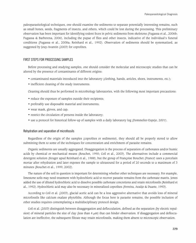
The samples examined by Reinhard et al. (1992) were obtained from a column of sediment located between the sacrum and the pubic symphysis (Figure 1), in supine position. The sample close to the sacrum showed the largest amount of intestinal content.
Figure 1 – Obtaining sediment from the pelvic girdle of a skeleton
(1) sediment at the level of the last lumbar vertebrae
(2) column of sediment over the sacrum, separated into two fractions: i) sacral concavity and ii) over the sacrum.
Modified from Reinhard et al. (1992).
It is possible to add a column of sediment at the level of the 4th and 5th lumbar vertebrae and the hepatic region (Figure 2) to this sampling scheme, although these locations have the disadvantage of lacking a bony structure to surround the deposited remains. As for the individual’s position, the sacrum can also retain material in bodies close to the anatomical position (Reinhard et al., 1992).
Foundations of Paleoparasitology
228
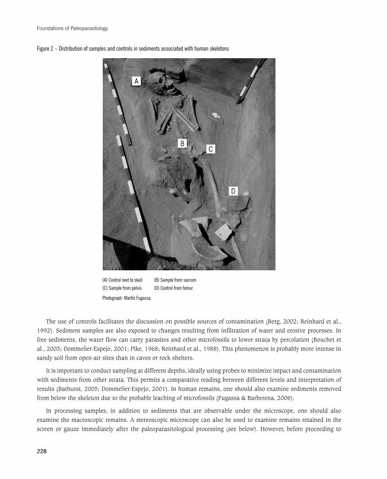
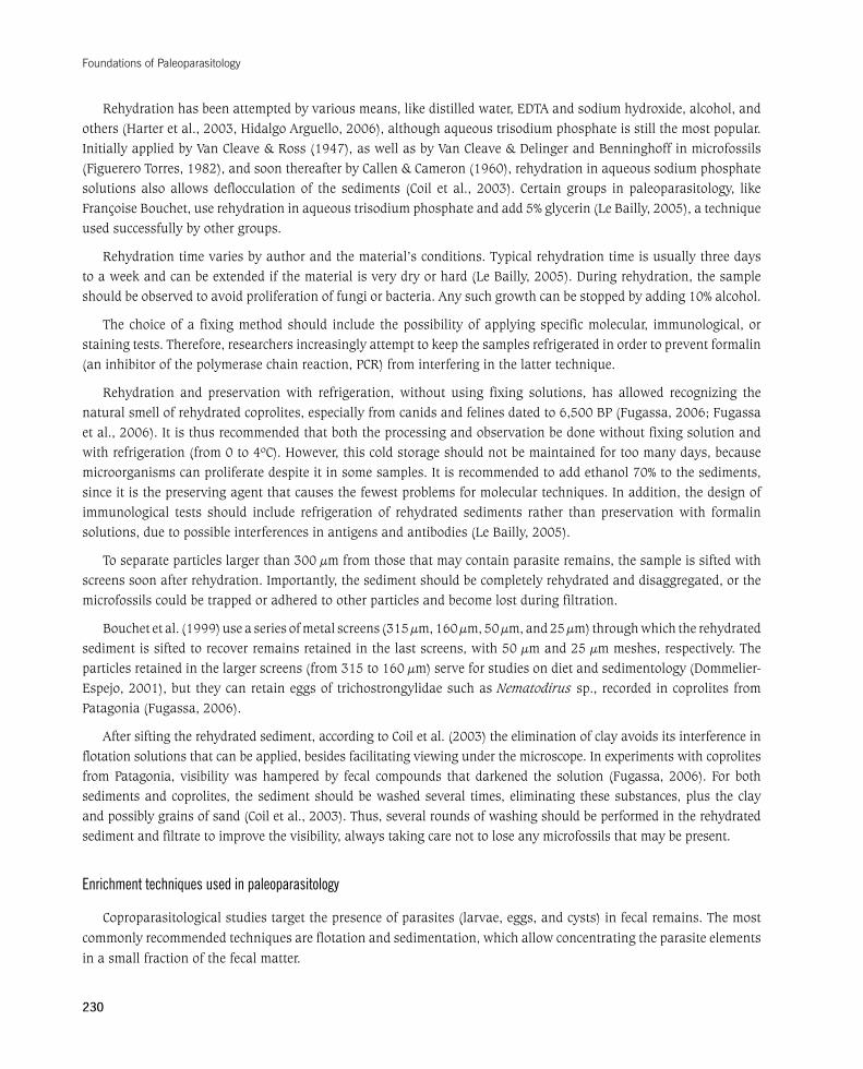
Figure 2 – Distribution of samples and controls in sediments associated with human skeletons
(A) Control next to skull (B) Sample from sacrum
(C) Sample from pelvis (D) Control from femur
Photograph: Martín Fugassa.
Theuseofcontrolsfacilitatesthediscussiononpossiblesourcesofcontamination(Berg,2002;Reinhardetal.,1992).Sedimentsamplesarealsoexposedtochangesresultingfrominfiltrationofwateranderosiveprocesses.Infreesediments,thewaterflowcancarryparasitesandothermicrofossilstolowerstratabypercolation(Bouchetetal.,2003;Dommelier-Espejo,2001;Pike,1968;Reinhardetal.,1988).Thisphenomenonisprobablymoreintenseinsandy soil from open-air sites than in caves or rock shelters.
It is important to conduct sampling at different depths, ideally using probes to minimize impact and contamination with sediments from other strata. This permits a comparative reading between different levels and interpretation of results(Bathurst,2005;Dommelier-Espejo,2001).Inhumanremains,oneshouldalsoexaminesedimentsremovedfrombelowtheskeletonduetotheprobableleachingofmicrofossils(Fugassa&Barberena,2006).
In processing samples, in addition to sediments that are observable under the microscope, one should also examine the macroscopic remains. A stereoscopic microscope can also be used to examine remains retained in the screen or gauze immediately after the paleoparasitological processing (see below). However, before proceeding to
Paleoparasitological Diagnosis
229
paleoparasitological techniques, one should examine the sediments to separate potentially interesting remains, such as small bones, seeds, fragments of insects, and others, which could be lost during the processing. This preliminary observationhasbeenimportantforidentifyingrodentfecesinpelvicsedimentsfromskeletons(Fugassaetal.,2006b;Fugassa&Barberena,2006), including thepupaeof fliesandother insects, indicativeof the individual’s funeralconditions (Fugassa et al., 2008a; Reinhard et al., 1992). Observation of sediments should be systematized, as suggestedbyJouy-Avantin(2003)forcoprolites.
FIRST STEPS FOR PROCESSING SAMPLES
Beforeprocessingandstudyingsamples,oneshouldconsiderthemolecularandmicroscopicstudiesthatcanbealtered by the presence of contaminants of different origins:
•contaminatedmaterialsintroducedintothelaboratory(clothing,hands,articles,shoes,instruments,etc.);
•inefficientcleaningofthestudyinstruments.
Cleaning should thus be performed in microbiology laboratories, with the following most important precautions:
•reducetheexposureofsamplesoutsidetheirrecipients;
•preferablyusedisposablematerialandinstruments;
•wearmask,gloves,andcap;
•restrictthecirculationofpersonsinsidethelaboratory;
•useaprotocolforhistoricalfollow-upofsampleswithadailylaboratorylog(Dommelier-Espejo,2001).
Rehydration and separation of microfossils
Regardless of the origin of the samples (coprolites or sediments), they should all be properly stored to allow submitting them to some of the techniques for concentration and enrichment of parasite remains.
Organicsedimentsareusuallyaggregated.Disaggregationistheprocessofseparationofcarbonatesand/orhumicacidsby chemicalormechanicalmeans (Bouchet,1995;Coil etal.,2003).Thealternatives includea commercialdetergentsolution(KrugerapudReinhardetal.,1988),butthegroupofFrançoiseBouchet(France)usesaporcelainmortar after rehydration and later exposes the sample to ultrasound for a period of 20 seconds to a maximum of 3 minutes(Bouchetetal.,1999,2002).
The nature of the soil in question is important for determining whether other techniques are necessary. For example, limestonesoilsmayneedtreatmentwithhydrochloricacidtorecoverparasiteremainsfromthecarbonatematrix.Jonesadded the use of diluted hydrochloric acid to dissolve possible carbonate concretions and retain microfossils (Reinhard et al.,1992).Hydrochloricacidmayalsobenecessaryinmineralizedcoprolites(Ferreira,Araújo&Duarte,1993).
According to Coil et al. (2003), glacial acetic acid can be a less aggressive alternative that avoids loss of mineral microfossils like calcium oxalate phytoliths. Although the focus here is parasite remains, the possible inclusion of other studies requires contemplating a multidisciplinary protocol design.
Coil et al. (2003) distinguish between disaggregation and deflocculation, defined as the separation (by electric repul-sion) of mineral particles the size of clay (less than 4 µm) that can hinder observation. If disaggregation and defloccu-lation are ineffective, the subsequent filtrate may retain microfossils, making them absent to microscopic observation.
Foundations of Paleoparasitology
230
Rehydrationhasbeenattemptedbyvariousmeans,likedistilledwater,EDTAandsodiumhydroxide,alcohol,andothers(Harteretal.,2003,HidalgoArguello,2006),althoughaqueoustrisodiumphosphateisstillthemostpopular.InitiallyappliedbyVanCleave&Ross(1947),aswellasbyVanCleave&DelingerandBenninghoffinmicrofossils(FiguereroTorres,1982),andsoonthereafterbyCallen&Cameron(1960),rehydrationinaqueoussodiumphosphatesolutions also allows deflocculation of the sediments (Coil et al., 2003). Certain groups in paleoparasitology, like FrançoiseBouchet,userehydrationinaqueoustrisodiumphosphateandadd5%glycerin(LeBailly,2005),atechniqueused successfully by other groups.
Rehydration time varies by author and the material’s conditions. Typical rehydration time is usually three days toaweekandcanbeextendedifthematerialisverydryorhard(LeBailly,2005).Duringrehydration,thesampleshouldbeobservedtoavoidproliferationoffungiorbacteria.Anysuchgrowthcanbestoppedbyadding10%alcohol.
The choice of a fixing method should include the possibility of applying specific molecular, immunological, or staining tests. Therefore, researchers increasingly attempt to keep the samples refrigerated in order to prevent formalin (an inhibitor of the polymerase chain reaction, PCR) from interfering in the latter technique.
Rehydration and preservation with refrigeration, without using fixing solutions, has allowed recognizing the naturalsmellofrehydratedcoprolites,especiallyfromcanidsandfelinesdatedto6,500BP(Fugassa,2006;Fugassaetal.,2006).Itisthusrecommendedthatboththeprocessingandobservationbedonewithoutfixingsolutionandwithrefrigeration(from0to4ºC).However,thiscoldstorageshouldnotbemaintainedfortoomanydays,becausemicroorganismscanproliferatedespiteitinsomesamples.Itisrecommendedtoaddethanol70%tothesediments,since it is the preserving agent that causes the fewest problems for molecular techniques. In addition, the design of immunological tests should include refrigeration of rehydrated sediments rather than preservation with formalin solutions,duetopossibleinterferencesinantigensandantibodies(LeBailly,2005).
To separate particles larger than 300 µm from those that may contain parasite remains, the sample is sifted with screens soon after rehydration. Importantly, the sediment should be completely rehydrated and disaggregated, or the microfossils could be trapped or adhered to other particles and become lost during filtration.
Bouchetetal.(1999)useaseriesofmetalscreens(315µm,160µm,50µm,and25µm)throughwhichtherehydratedsediment is sifted to recover remains retained in the last screens, with 50 µm and 25 µm meshes, respectively. The particlesretainedinthelargerscreens(from315to160µm)serveforstudiesondietandsedimentology(Dommelier-Espejo, 2001), but they can retain eggs of trichostrongylidae such as Nematodirus sp., recorded in coprolites from Patagonia(Fugassa,2006).
After sifting the rehydrated sediment, according to Coil et al. (2003) the elimination of clay avoids its interference in flotation solutions that can be applied, besides facilitating viewing under the microscope. In experiments with coprolites fromPatagonia, visibilitywashampered by fecal compounds that darkened the solution (Fugassa, 2006). For bothsediments and coprolites, the sediment should be washed several times, eliminating these substances, plus the clay and possibly grains of sand (Coil et al., 2003). Thus, several rounds of washing should be performed in the rehydrated sediment and filtrate to improve the visibility, always taking care not to lose any microfossils that may be present.
Enrichment techniques used in paleoparasitology
Coproparasitological studies target the presence of parasites (larvae, eggs, and cysts) in fecal remains. The most commonly recommended techniques are flotation and sedimentation, which allow concentrating the parasite elements in a small fraction of the fecal matter.
Paleoparasitological Diagnosis
231
For clinical analyses, Thienpont, Rochette & Vanparijs (1979) recommend using concentration techniques when direct techniques yield negative results (direct techniques are defined as those that analyze a minimal portion of the sample without concentrating the target elements before observation). The advantages of direct techniques are speed, ease, and minimal disturbance of delicate parasite remains, such as trophozoites of Giardia sp. or Entamoeba sp. (although the latter are relatively uncommon in archaeological samples).
Direct techniques are recommended for tests in recent reptile and bird droppings, but normally not forpaleoparasitologicaltests,althoughtheyareusedinsomecasessuchasHidalgoArguello(2006).
Among the enrichment techniques, sedimentation consists of concentrating parasite remains by deposition in an aqueous solution, less dense than the eggs or other parasite forms. The most important sedimentation techniques in clinical parasitology are spontaneous sedimentation (Lutz, 1919) and the Telemann formalin-ether technique (Thienpont,Rochette&Vanparijs,1979).Spontaneoussedimentationinvolvesaprolongedreadingtimeduetotheabundance of debris, but it is more sensitive, quick, and economical than other available techniques.
The formalin-ether techniques (Ritchie technique) and its modifications (formalin-acetate [Telemann technique] and formalin-tween) consist of disaggregation of an amount of fecal matter in a solution of an organic substance (ether, tween) in a polar medium (formalin or acetate). These substances should be shaken vigorously with the fecal matter, separating the suspension into two phases (Kaminsky, 2003; Thienpont, Rochette & Vanparijs, 1979).
The upper, less dense phase is the organic phase, while the lower phase is the polar compound. Parasite eggs andcystssettleonthebottomofthetube,andvariousdebrisconcentrateinbothphases.Horne&Tuck(1996)usedthe formalin-ether technique to examine sediments from historical latrines in North America, recovering eggs of Ascaris sp., Trichuris sp., Dicrocoelium dendriticum, and Taenia sp. However, Reinhard, Ambler & McGuffie (1985) had tested this sedimentation technique, observing that simple or spontaneous sedimentation was more efficient. There is limited paleoparasitological experience with this technique, widely used in clinical parasitology, essentially due to efficient separation of parasite remains from debris and fat.
Enrichment techniques using flotation are based on the relative density of the parasite remains. For the remains to float, the flotation solution must be denser than they are. Various saturated solutions are used, in which one introduces the fecal matter previously sifted in a screen or gauze and rehydrated, in the case of paleoparasitological studies.
It is common to place the sample in the test tube and add a little flotation solution, then mix and fill the tube to the brim with solution, avoiding bubbles that could hamper visualization. The slide cover is placed on the surface such that it makes contact with the solution. After some time, varying from 15 minutes to two hours, the parasite remains float to the surface of the solution. Mild centrifugation can be used to shorten the time for the parasite remains to float (Kaminsky, 2003; Thienpont, Rochette & Vanparijs, 1979). The slide cover is then removed, and if there are any parasite remains, they are adhered to the lower surface of the slide cover, which is then placed on the slide for observation under the microscope.
Different groups of parasites have different densities and thus behave differently in flotation solutions,withtrematode eggs requiring denser solutions (Thienpont, Rochette & Vanparijs, 1979). In addition, the techniques’ effectivenessvariesaccordingtotheconditionsofthesedimentbeingexamined.Bouchetetal.(1999)usedsolutionswith specific gravity ranging from 1.1 to 2.0, since taphonomic factors typically make the density of ancient eggs vary greatlyfrommodernones.Sucrosesolutionsatvariousconcentrations,sodiumchloride,sodiumnitrateinsucrosesolutionandothershavebeenusedinarchaeologicalsamplesinFrance(Dommelier-Espejo,2001).Importantly,theeggs tend to warp, fade, and lose the operculum (in the case of operculated eggs) when very dense solutions are used,

Foundations of Paleoparasitology
232
or for a very long time (Thienpont, Rochette & Vanparijs, 1979). In ancient samples, the use of zinc sulfate causes deformations, breakage, or peeling in eggs of Hymenolepis sp. (Reinhard et al., 1988).
Although the flotation technique in sodium chloride solution (Willis technique) is more common, Marder et al. (2000)foundnodifferencesinrelationtotheSheathertechnique(sucrosesolution)andFaust(sodiumsulfatesolution)for the detection of modern Toxoplasmacystsandnematodeeggs.However,usingtheWillistechnique,Binda,Moriena& Alvarez (2003) obtained more negative results and fewer Giardia sp. cysts than using the zinc phosphate technique. Other solutions have been used in paleoparasitology, such as zinc chloride and zinc phosphate (Reinhard et al. 1988), with variations in their osmotic potential and density.
Various authors, like Navone et al. (2005), recommend the simultaneous use of sedimentation and flotation techniques for stool tests in current human samples. In archaeological contexts, the use of two techniques means greater consumption of sediment, which is not recommendable and can only be suggested in specific situations, forexamplewithnegativeresultsandanabundantamountofsediment.Asanalternative,Bouchetetal. (2001)suggest taking samples from the flotation solution surface and simultaneously from the sediment in the bottom of thetube.Thus,inarchaeologicalsediments,thesuggestedmethodisthesequentialapplicationofthemodifiedStollquantitativetechnique(Fugassaetal.,2006)andtheflotationtechniqueonthesamerehydratedsediment(seelaterin this chapter).
As mentioned, spontaneous sedimentation has the most advantages. However, the final decision depends on various factors like the samples’ characteristics, the context from which they come, and the availability of equipment for examination. The selected technique should cause minimum destruction or alteration in the microfossils, especially in parasites.
As for the number of preparations performed for each sample, usually 10 to 20 slides are made (Gonçalves, Araújo & Ferreira, 2003; Holliday, Guillen & Richardson, 2003; Taek-Han et al., 2003). More preparations do not appear to increase the number of species recorded (Harter, 2002) and consume more laboratory time. However, since ancient samples suffer various taphonomic processes, parasites are often found in small numbers, and expanding the number of slides increases the likelihood of positive parasite finds.
The number of slide preparations also depends on the study’s objectives and availability of samples. For example, when hundreds of coprolites are available, one can plan to examine only a few slides per coprolite. However, when considering the material’s limitations and study objectives, one should not overlook the inherent conditions of sampling. If only a few slides are examined they will probably not be very representative. The study design should address these limitations and prioritize the examination of fewer specimens and more slide preparations.
More preparations are needed in free sediments than in coprolites, because (as discussed previously) the dispersal and deterioration of parasite elements are greater. While some 20 slides are prepared for coprolites, in sediments this figure reaches 50 slides per sample location. In sediments from latrines, Fernandes et al. (2005) examined 20 slides per sample, while Taek Han et al. (2003) made 10 slides. Latrine sediments require fewer slides because they usually have a higher concentration of parasites compared to skeletons or soil samples from archaeological sites.
Quantification in paleoparasitology
Paleoparasitological studies should use a quantitative methodology to obtain comparable results. Quantification in paleoparasitology helps assess the comparative density between locations in the same site and thus discuss possible sources of contamination or the origin of the parasites.
Paleoparasitological Diagnosis
233
BothReinhardetal.(1988)andAraújoetal.(1998)claimthatquantificationofparasiteremainscancontributeto a paleoepidemiological reconstruction, since it allows comparing egg density between skeletons from burials in the same site, even though parasite density is not highly related to an individual’s parasite burden in life. Likewise, Moore (1981) described findings in a medieval latrine and observed a certain stable quotient between the number of Ascaris sp. and Trichuris sp. eggs. This could suggest comparisons of rates in the number of parasite eggs found, or the use of such an index as a parameter in studying ancient human remains in a given region.
Importantly, quantification does not serve to discuss the intensity of infections, because taphonomic processes extensively modify the initial conditions. Even in current samples, fecal parasite burden is not a good indicator of intensity of infection, since the elimination of eggs, larvae, and cysts in feces depends on numerous parasite, host, and environmental factors, e.g., the number of parasites in the individual, the host immune status, time of year, etc. (Thienpont, Rochette & Vanparijs, 1979). However, parasite burden may be a useful indicator in some circumstances.
Quantitative techniques were first used by Taylor and soon after by Pike (Reinhard et al., 1988), who estimated the number of eggs per gram of sediment by directly counting the parasite eggs in a gram of sample.
Inthesearchforappropriatemethods,Warnock&Reinhard(1992),Dittmar&Teejen(2003),andSiantoetal.(2005) used a quantitative palynological technique that consists of adding a known amount of a tracer – Lycopodium spores – to a known amount of sediment. The number of eggs per gram of sediment can by estimated by the formula
HPG = h x ELM / ELC x PM
where h is the number of eggs counted on the slide, ELM is the number of Lycopodium spores in the sample, ELC is the number of spores counted on the slide, and PM is the weight of the sample.
In addition to serving as a quantitative tool, the state of preservation of the Lycopodium spores measures the possible aggressiveness of the paleoparasitological processing conditions on the microfossils, especially parasites. A possible disadvantage to this modified sedimentation technique is that it assumes homogeneous distribution of Lycopodium spores in the column of rehydrated sediment. As with eggs and cysts, spores deposit differentially, and their abundance can vary according to the depth at which the sediment is obtained.
QuantificationofsedimentscanbeperformedwiththeuseormodificationofclinicaltechniquessuchasStoll(Thienpont,Rochette&Vanparijs,1979).Jones(1984)firstusedthisproceduretoanalyzearchaeologicalsamplesinEngland.Heused150µlaliquotsofamixtureof3gsedimentin42mlofwater,assumingasedimentdensityof1g/ml. The number of eggs per gram (HPG) is estimated by the formula
HPG = no. of eggs counted x 100
As stated previously, themodified Stoll technique (Jones, 1982a) assumes that the sediment density is equalto 1. However, this assumption has not proven true for sediments of various origins analyzed in Patagonia. This demonstrates the need to measure the sediment density before calculating the number of eggs per gram of sediment and to make the necessary corrections in the HPG formula.
Recently,Fugassa,Araújo&Guichón(2006)successfullymodifiedtheStolltechniquewiththefollowingchanges:1) using 5 g of sediment in 10 ml of solution, which increases the density of sediment in the solution and thus the likelihood of recovering parasites, 2) reducing the size of the aliquot examined per slide, making the technique more operational, and 3) using aqueous trisodium phosphate as the liquid medium instead of water.

Foundations of Paleoparasitology
234
The modified Stoll quantitative technique (Fugassa, Araújo & Guichón, 2006) showed acceptable sensitivity
comparedtothetechniqueappliedbyJones.Bouchet,Harter&LeBailly(2003)suggestthatthelatterisespecially
useful in samples with parasite densities greater than 400 eggs per gram.
The quantitative technique also allows quantification of other remains, such as soil mites and pollen grains
(Fugassa,Sardella&Denegri,2007).Themodifiedtechniqueledtoanoperationaladvantage:meanobservationtime
fora24x36mmslidecoverwasonly13minutes,duetotheuniformdensityofthematerial.Equallyimportantis
the ease in slide preparation due to the use of an automatic micropipette and absence of grains of sand in preparations
of sandy soil samples.
Taek Han et al. (2003) used another modification of Stoll. Samples with 10 g of sediment were previously
rehydratedin50mloftrisodiumphosphate(TSP),whichwasthenreplacedwith20mltoeliminatetheturbidity.The
authors used 20 µl aliquots for the preparations. Washing the sediments can be a useful modification in soils with
large amounts of organic matter, although unnecessary in sandy soils.
ThemodifiedStolltechniquehasproventobeausefulquantitativetool.However,itshouldbeaccompaniedbya
highlysensitivequalitativetechniquesuchastheSheatherflotationtechnique.Ifdebrisisrecoveredandsomeparasite
remains are deformed, the risk of false negatives decreases. Thus, the combination of a quantitative technique with a
qualitative one is highly useful both to expand the results and to prove the former’s sensitivity.
In the pelvic sediment of a skeleton from the Alero Mazquiaráns site in Chubut, Argentina, with large amounts
oforganicmatter(Fugassa,2006),themodifiedStolltechniquefailedtoyieldpositiveresults,whiletheSheather
technique proved efficient for recovering helminth eggs. In the latter case, the large amount of organic matter had
retained the scarce parasite remains, and the flotation technique thus produced more efficient results.
Ideally,variousflotationtechniquesshouldbetested,assuggestedbyBouchetetal.(2001),althoughthetime
spent and the greater consumption of the sample are significant disadvantages.
Observation and diagnosis
Once the archaeological sample has been properly processed, one should use a small aliquot for microscopic
examination. Next, drops of sediment are mounted on slides covered with slide covers, available in various sizes
accordingtotheplanneduse.Smallslidecovers(18or20mm)dehydratelessaftersealedandareresistantandform
athinlayerofsolution,especiallybyaddingasmalldropofglycerin(Fugassa,2006).Largerslidecovers,24to36
mm, allow the drop of sediment to dehydrate more easily, although they form a layer of preparation that is thinner
and easier to examine. Larger slide covers are recommended for quantitative techniques using larger volumes, such
as50to150ml(Fugassa,Araújo&Guichón,2006;Jones,1984).
Slides should be examined exhaustively undermagnification 10 in the ocular lens and 10 in the objective
lens. Correct measurement of the parasite elements is essential for diagnosis and should be performed under a
magnificationof400.Deformedorbrokenparasiteremainsshouldnotbemeasured.Inspecieswithoperculated
eggs as in genera Trichuris and Calodium, the eggs should be measured without considering the polar opercula,
since they can vary widely; even more importantly, they are often absent from archaeological material (although
many authors consider the measurements of these species with the plugs in opercula, so the measurements should
be taken with and without them). Additional measurements such as the width of the operculum or egg wall may
be useful in some cases.

Paleoparasitological Diagnosis
235
During parasite identification, the use of photographs is indispensable for guaranteeing the results and forconsultation with other specialists. Kliks (1990) criticized numerous paleoparasitological findings, sparking an interestingdebate (Ferreira&Araújo,1996;Hawdon& Johnston,1996).Hisobservationemphasized the lackofimages to back the diagnoses. Findings should thus be properly recorded, especially when they contain species with numerically low representation in the samples.
Someauthorssealtheslideswithtransparentcommercialnailpolish(Bathurst,2005;Dittmar&Teejen,2003).ThepaleoparasitologylaboratoryattheOswaldoCruzFoundationinRiodeJaneirousesahotmixtureofequalpartsof beeswax and resin (Araújo, personal communication), which is very useful. However, nail polish is quick and can be used in unexpected situations when the dehydration needs to be stopped. Care should be taken to avoid contamination of the brush.
In samples containing interesting remains, a gelatin-glycerin solution can be used (Ruzin, 1999), which is good forpreservation.Themixtureshouldbeheatedtoapproximately50ºCinwaterbath,andoncefluidityisobtained,one drop of the preparation and one drop of gelatin-glycerin solution are mixed on a slide. The slide cover is then placed on top, and the preparation can be preserved for a long time. Preliminary results indicate that ancient eggs of Ascaris lumbricoides and Trichuris trichiura, as well as current Hymenolepis nana eggs and Giardia sp. cysts are well-preserved when immersed in gelatin-glycerin.
Eggs and cysts of given taxa have typical characteristics that facilitate their identification. For example, it is relatively easy to diagnose genera Calodium sp. and Trichuris sp. eggs by the opercula on the two extremities, their shape, and the ornamentation on the wall. However, species determination is less simple and should be measured in probabilistic terms (with the eggs’ length, width, and morphology as parameters) and comparative ones. The addition of other circumstantial evidence (e.g., diet items, host, parasite biogeography) can lead to a more robust probabilistic diagnosis.
As suggested by Noronha et al. (1994), identification should also consider the coprolite’s morphological characteristics and distribution of possible hosts in the region, besides the other above-mentioned circumstantial evidence. Concerning possible hosts, the distribution of both current and past wildlife should be observed.
Eggs from some different parasite species can be confusing. Certain ascarids such as Lamanema and Nematodirus, or Ancylostoma and Trichostrongylus, have similar eggs that require careful measurements of numerous specimens to enable estimating their mean morphometry and if necessary apply an appropriate statistical technique. Other genera can also be confusing if there is no microscopic method to help distinguish between them. For example, Necator and Ancylostoma cannot be distinguished by the appearance of their eggs, nor can species of genus Taenia from other tapeworms such as Echinococcus.
Distinctionswithin a genus are evenmore difficult. For example, the eggmorphometry ofAscaris suum and A. lumbricoides or Trichuris suis and T. trichiura does not serve to diagnose the eggs’ species. Importantly, the paleoparasitological study of coprolites from wild animals can prove even more complex, since there are fewer parasitological studies that serve as the basis or reference for the paleoparasitological findings. One should also consider the possibility of changes in the parasites’ life cycle and morphological alterations in the eggs, larvae, and cysts,whichcanhinderthecorrectdiagnosis(Dommelier-Espejo,2001).
Although less common, other difficulties in paleoparasitological diagnosis relate to some parasite remains than can resemble artifacts and other structures like pollen grains, air bubbles, fungal spores, and plant fragments (Thienpont, Rochette & Vanparijs, 1979).
Foundations of Paleoparasitology
236
Although the dehydration process may not alter the eggs’ morphometry (Araújo, 1988; Confalonieri, 1988b; Confalonieri et al., 1988a), taxonomic diagnosis assumes that morphometry is a stable characteristic in each species. Still,somestudieshaveproventhateggsizecanvaryinsomehelminthspecies,duetothehostamongotherreasons.For example, Fasciola hepatica eggs differ significantly in size, depending on the host’s body mass (Valero et al., 2002). In pseudophyllid cestodes, egg size varies according to the host species and intensity of infection (Andersen & Halvorsen, 1978). Although this variability cannot be generalized to all helminth taxa, one should recall the morphological plasticity that some species can display.
Eggs containing first-stage larvae can often be found. This is common in such parasites as hookworms, ascarids, and pinworms and is a useful criterion for differentiating them from artifacts and other helminth eggs that are only found embryonated. Third-stage larvae have been found in coprolites and sediments (Ferreira, Araújo & Confalonieri, 1980;Reinhardetal.,1988),someasoldas1.5millionyears(Ferreira,Araújo&Duarte,1993),thusdemonstratingtheir potential for preservation, although the diagnosis is difficult.
On this point, Reinhard, Hevly & Anderson (1987) emphasize the need to differentiate between parasite larvae and free-living nematodes. Kliks (1990) contends that some paleoparasitological diagnoses of parasite larvae may actually correspond to free-living nematodes. However, several characteristics can be used determine whether they arefree-livinglarvaeorparasites(Fiel,Steffan&Ferreyra,1998),oreventodistinguishthemfromplantparasites(Chaves, Echeverria & Torres, 1995).
Duringmicroscopicobservation,itiscommontofindvariousmitesthatcanprovideimportantinformationonectoparasitism, such as the finding of Demodexsp.inaregurgitationpelletfromabirdofprey(Fugassa,Sardella&Denegri,2007),oronintermediatehosts,suchasoribatidmites,intermediatehostsofAnoplocephalidaetapeworms(Fugassa et al., 2006). The presence of mites in coprolites of diverse zoological origins has provided relevantpaleoecological information (Guerra et al., 2003).
The preceding paragraphs highlight some difficulties that can arise during diagnosis and that relate basically to confusion between parasite remains and artifacts, pollen grains, or fungi and errors in species diagnosis. Taxonomic determination of eggs, larvae, and cysts is often difficult in both ancient settings and in modern clinical diagnostic laboratories.Forexample,inarecentqualitycontrolexerciseinSpain,humanstoolsamplescontainingeggsofahelminthspeciesandcystsfromtwospeciesofprotozoaweresentto200laboratories.Only6.9%identifiedallthreeparasites,andin27.2%ofthesamplesdifferentparasiteswereidentifiedfromthoseknowntothereferencelaboratory(SEIMC,1998).
Observation and identification of parasite remains require great attention and caution at the moment of taxonomic determination. It is important and highly useful to consult several colleagues and discuss the identity of ambiguous findings. A detailed description and illustration should precede a rigorous and solidly based discussion on the possible diagnosis. The erroneous communication of a finding for a given region and time can alter the correct understanding of the biogeographical history of certain parasites and their hosts.
Staining and electron microscopy
In paleoparasitology, microscopic observation allows diagnosing helminth eggs and only occasionally protozoan cysts(Faulkner,1991;Ferreiraetal.,1992;Fugassaetal.,2008c;Leguía,Casas&Jane,1995).Thelatterisduetothe fact that most cyst-producing species are not only differentially preserved, but also difficult to recognize due to their small size or morphological similarity to other microfossils. Pollen grains, fungal spores, crystals, mite eggs,
Paleoparasitological Diagnosis
237
free-living nematodes, and phytoliths are present in the sediments, so microscopic analysis involves some degree of uncertainty in identifying the observed structures.
The use of stains that react with given components of microfossils facilitates diagnosis. However, experience with staininginpaleoparasitologyisverylimitedandrelatedtoafewstudiesthatusedLugol’ssolution(Moore,1969;Taylor,1955),merthiolate-iodine-formalinsolution(MIF)(Harter,2002),orironhematoxylin(Horne&Tuck,1996).
Although trichrome staining has not been recorded in paleoparasitology, it is one of the most widely used stains inmodernmaterial.Somestudies recommend it fordetectingvarious intestinalparasites in fecalmatter (Kellogg& Elder, 1999), thus suggesting its potential use in ancient samples. Protozoa of the phylum Apicomplexa, like Cryptosporidium sp., Cyclospora sp. and Isospora belli may require special staining techniques, for example with modified acid-fast solutions (Kaminsky, 2003).
The lack of standardized protocols for differential staining in paleoparasitology further increases the complexity ofitsuseinsampletesting.Despitelimitedattemptsinpaleoparasitology,differentialstainingdeservesinclusioninroutine paleoparasitological examination.
BasedonthedesignbyHorne&Tuck(1996),whousedironhematoxylininthreeof15preparations,thisstainingcanberecommendedin50%oftheslidesforeachsample,examiningunder400xmagnification.Examinationofprotozoaincurrentsamplesuses20slidecoverfieldswithmagnificationof1,000(DelCocoetal.,2006).
In archaeological samples, it is recommended to examine a large area due to the expected low density of cysts. The proposal for each stained slide is to examine 20 more fields under magnification of 1000, i.e., a total of 40 fields.
In parasite genera with morphometrically similar eggs, microstructural characteristics can help distinguish between them (Bouchet et al., 1999), as exemplified by scanning electronmicroscopy (SEM) in ancientDiphyllobothrium tapeworms(LeBaillyetal.,2005)andincurrentToxocarasp.eggs(Ubelaker&Allison,1975).Theauthorsemphasizethattaphonomicfactorscanalterthesurfacestructurecharacteristics.SEMcanalsobeusedsatisfactorilytoobservethird-stage hookworm larvae in coprolites (Araújo et al., 1988b).
Immunologyhascontributedothertechniquestopaleoparasitologicalresearch.Directimmunofluorescenceusesantibodies marked with fluorescent substances and allows the specific detection of surface antigens in parasite forms (Atías, 1998). In paleoparasitology, it has been used to diagnose Giardia duodenalis(Faulkner,Sharon&Johnson,1989), G. duodenalis and Cryptosporidium parvum(Allison,Bergman&Gerszten,1999;LeBaillyetal.,2008;Ortega&Bonavia,2003),andlaterCryptosporidium/GiardiaincoprolitesfromFrance(LeBailly,2005).
INTERPRETATION OF PALEOPARASITOLOGICAL RESULTS
When one reaches a degree of diagnostic certainty or probability with parasite remains, it is not the end of the job, but only the beginning. The identified remains represent forms of dispersal of a life cycle in a parasite species.
Eggs, larvae, or cysts found in samples can be the result of patent infections or (in the case of eggs and cysts) the ingestion of prey infected with the parasites in question. Thus, the presence of parasites in sediments from archaeological sites involves more possible origins, so their analysis requires both ecological and cultural knowledge. Contamination from unknown sources can introduce a bias into the results. For example, finding rodent coprolites in sediment from the pelvis of human skeletons can alert the researcher to contamination of material with their parasites (Fugassa & Barberena, 2006; Fugassa, 2006). To reiterate, painstaking planning of sampling and macroscopicobservation of the sediment can help predict such situations.
Foundations of Paleoparasitology
238
Even under ideal conditions with highly sensitive and specific methods, false-negative results may occur. For example, in addition to the taphonomic phenomena specific to ancient samples, a mild infection, oviposition behavior as in Enterobius vermicularis, the reduced number of eggs in trichostrongylids of herbivores, intermittent oviposition, elimination of eggs in gravid proglottids, parasites in pre-patent periods, and other situations can lead to underestimation of ancient parasitic infections.
Adequate interpretation of results definitely requires in-depth knowledge of parasite diversity and biogeography, the natural history of the species under study, and the regional archaeology. The latter requires effective communication among paleoparasitologists and archaeologists studying the region.
STATISTICAL ANALYSIS APPLIED TO PARASITE REMAINS
As discussed previously, paleoparasitological diagnosis poses various challenges, from determination of the coprolite’s zoological origin to that of the respective parasite species. We have described various methodological tools to overcome such problems. We will now present some available statistical techniques for analyzing the observed parasite remains.
Egg morphometry is commonly employed for diagnosing intestinal parasites (Thienpont, Rochette & Vanparijs, 1979).Statisticalanalysisofeggandcystmeasurementscanbehighlyusefulfordiagnosisincontextswherebothhuman and other animal coprolites are present (Confalonieri et al., 1988b).
To compare eggs measurements between two situations, for example, eggs found in a body and in the surrounding sediment – the chi-square test can be used, with the median as the reference (Araújo, 1988), mounting a contingency table with the eggs’ length and width.
When only a single egg is available (a frequent situation in paleoparasitology), Confalonieri et al. (1988b) used the Studentt-testseparatelyforlengthandwidthtodeterminewhethertheeggbelongedtoagivenspecies.Ifafrequencytable is available for a large population of eggs with length and width measurements, this statistical test can be applied to the species to which the egg supposedly belongs. The authors show that this method is equally useful for the identification of single coprolites.
Mounting these tables requires comparative samples of reliably identified parasite eggs and coprolites.
Inanother studyon the taxonomical determinationof a set of parasite remains, Joyner (apudConfalonieri etal., 1988b) used the linear regression coefficient of the measurements obtained from a set of Eimeria sp. oocysts to determine whether they belonged to a given species for which the oocyst morphometry was known. This method can complement other statistical tests.
Case study: discriminant analysis applied to capillarid eggs
In some cases, the eggs’ qualitative characteristics are as important as their morphometric ones. In capillarids, the egg’s ornamentation is highly useful for diagnosis (Moravec, 2001). Capillarid species have cycles that include a host from a lower trophic level and a carnivore as disseminator. They are thus commonly identified as parasites in transit in predator feces. Coprological examination in carnivores or omnivores can often show eggs from different capillarid species, some produced by parasites in transit, or false parasitism, and others as true parasitism, causing infection in the host.
Paleoparasitological records of eggs belonging to capillarid species are rare. In the Americas, Confalonieri described eggs from this genus in animal coprolitesinBrazil(Confalonieri,1988a;Araújo,Confalonieri&Ferreira,1998).Bouchet
Paleoparasitological Diagnosis
239
(1997)laterfoundeggsfromthisgenusin21coprolitesfromFrance.Stilllater,Dittmar&Teejen(2003),Fernandesetal.(2005),andRochaetal.(2006)alsorecordedCapillaria sp. in archaeological sediments.
With the exception of an endemic of Pseudocapillaria philippinensis in the Philippines, reports of capillariasis in humansarenowrare(Atías,1998;Benenson,1992),althoughriskofinfectionwasprobablygreaterinthepastdueto different ecological conditions.
Gonçalves, Araújo & Ferreira (2003) contend that the occurrence of capillariasis in humans is due mainly to the ingestion of raw meat, especially liver, where the eggs of Calodium hepaticum are located (Miyazaki 1991). However, numerous species of capillarids located in the gut are recorded in other mammals, for example Capillaria puttori and Pearsonema plica(Soulsby,1987).VariousstooltestswithCapillaria sp. eggs have been recorded in Amerindian groups (Coimbra & Mello, 1981; Coimbra et al., 1985).
From the trophic point of view, the situation would be similar in canids, although more species of parasites have been reported in these hosts. Capillaria puttori and Eucoleus aerophilus have been recorded in canids (Thienpont, Rochette & Vanparijs, 1979), and Calodiumm hepaticumwasfoundinnecropsiesofwilddogsinBrazil(Ruasetal.,2003).
As mentioned, the presence of capillarid eggs in archaeological remains can be interpreted as either true parasitism or the product of parasites in transit. The current impossibility of elucidating the origin of such eggs precludes an interpretationoftheirculturaland/orecologicalmeaning.Abetterdiagnosiswouldallowdiscussingthepresenceorabsence of true parasitism, thus clarifying the meaning of such findings in the host population.
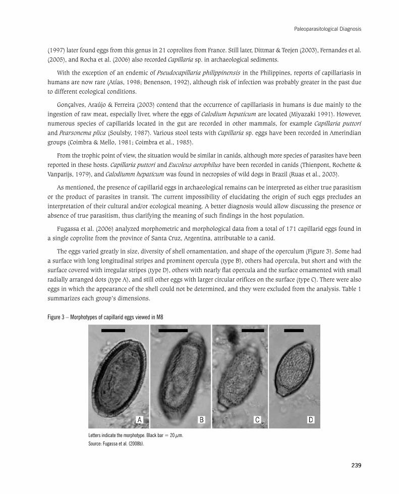
Fugassaetal.(2006)analyzedmorphometricandmorphologicaldatafromatotalof171capillarideggsfoundinasinglecoprolitefromtheprovinceofSantaCruz,Argentina,attributabletoacanid.
Theeggsvariedgreatlyinsize,diversityofshellornamentation,andshapeoftheoperculum(Figure3).Somehadasurfacewithlonglongitudinalstripesandprominentopercula(typeB),othershadopercula,butshortandwiththesurfacecoveredwithirregularstripes(typeD),otherswithnearlyflatoperculaandthesurfaceornamentedwithsmallradially arranged dots (type A), and still other eggs with larger circular orifices on the surface (type C). There were also eggs in which the appearance of the shell could not be determined, and they were excluded from the analysis. Table 1 summarizes each group’s dimensions.
Figure 3 – Morphotypes of capillarid eggs viewed in M8
Letters indicate the morphotype. Black bar = 20 µm.
Source: Fugassa et al. (2008b).
Foundations of Paleoparasitology
240
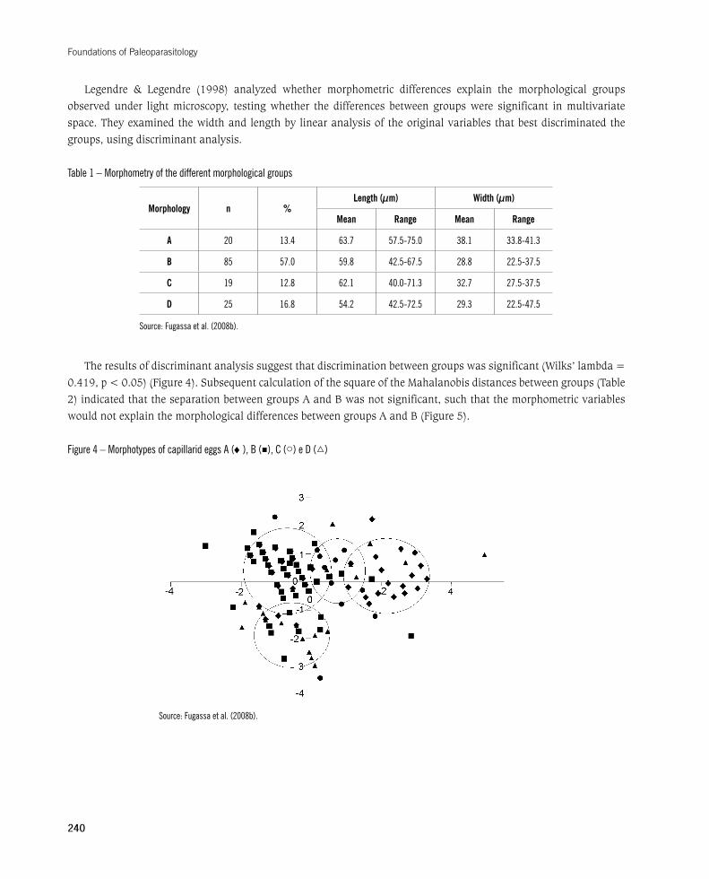
Legendre & Legendre (1998) analyzed whether morphometric differences explain the morphological groups observed under light microscopy, testing whether the differences between groups were significant in multivariate space. They examined the width and length by linear analysis of the original variables that best discriminated the groups, using discriminant analysis.
Table 1 – Morphometry of the different morphological groups
Morphology n %Length (µm) Width (µm)
Mean Range Mean Range
A 20 13.4 63.7 57.5-75.0 38.1 33.8-41.3
B 85 57.0 59.8 42.5-67.5 28.8 22.5-37.5
C 19 12.8 62.1 40.0-71.3 32.7 27.5-37.5
D 25 16.8 54.2 42.5-72.5 29.3 22.5-47.5
Source: Fugassa et al. (2008b).
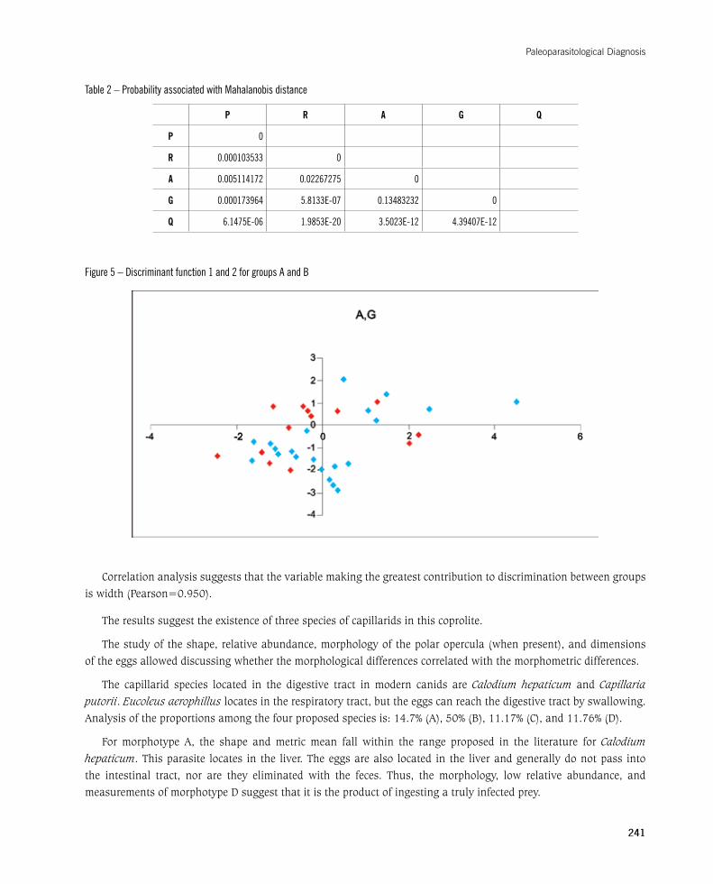
The results of discriminant analysis suggest that discrimination between groups was significant (Wilks’ lambda = 0.419,p<0.05)(Figure4).SubsequentcalculationofthesquareoftheMahalanobisdistancesbetweengroups(Table2)indicatedthattheseparationbetweengroupsAandBwasnotsignificant,suchthatthemorphometricvariableswouldnotexplainthemorphologicaldifferencesbetweengroupsAandB(Figure5).
Figure 4 – Morphotypes of capillarid eggs A (♦), B (■), C (○) e D (▵)
Source: Fugassa et al. (2008b).
Paleoparasitological Diagnosis
241
Table 2 – Probability associated with Mahalanobis distance
P R A G Q
P 0
R 0.000103533 0
A 0.005114172 0.02267275 0
G 0.000173964 5.8133E-07 0.13483232 0
Q 6.1475E-06 1.9853E-20 3.5023E-12 4.39407E-12
Figure 5 – Discriminant function 1 and 2 for groups A and B
Correlation analysis suggests that the variable making the greatest contribution to discrimination between groups is width (Pearson=0.950).
The results suggest the existence of three species of capillarids in this coprolite.
The study of the shape, relative abundance, morphology of the polar opercula (when present), and dimensions of the eggs allowed discussing whether the morphological differences correlated with the morphometric differences.
The capillarid species located in the digestive tract in modern canids are Calodium hepaticum and Capillaria
putorii. Eucoleus aerophillus locates in the respiratory tract, but the eggs can reach the digestive tract by swallowing. Analysisoftheproportionsamongthefourproposedspeciesis:14.7%(A),50%(B),11.17%(C),and11.76%(D).
For morphotype A, the shape and metric mean fall within the range proposed in the literature for Calodium
hepaticum. This parasite locates in the liver. The eggs are also located in the liver and generally do not pass into the intestinal tract, nor are they eliminated with the feces. Thus, the morphology, low relative abundance, and measurementsofmorphotypeDsuggestthatitistheproductofingestingatrulyinfectedprey.

Foundations of Paleoparasitology
242
MorphotypeBcouldcorrespondtoCapillaria putorii based on its measurements, ornamentation, and rela-
tive abundance.
Morphotype C is within the categories of measurements of Eucoleus aerophilus. The low proportion of eggs
can be explained by their location in the airways and their release into the environment through the digestive and
respiratory tracts.
MorphotypeDcannotbeassignedtoaknownspeciesandprobablyrepresentsimmatureeggsfromsomeofthe
species present, most likely morphotype A.
Discriminantanalysiscanbeuseful in thediagnosisofeggsbelonging to theCapillaria genus that cannot be
classified simply by their morphology, due to taphonomic processes or other causes. Although capillarids are one of
the most complex groups of parasites (Moravec, 2001), experience has shown that the combination of morphological
and morphometric analysis of capillarid eggs allows improving paleoparasitological diagnosis.
NEW SOURCES OF EVIDENCE
As shown, coprolites have been the main source of evidence in paleoparasitology, although sediments of various
origins have also been used sporadically. Paleoparasitological studies have also used mummified tissues (Araújo
et al., 1988a; Araújo, Reinhard& Ferreira, 2000; Aufderheide et al., 2004; Bastos et al., 1996; Cockburn et al.,
1975), intestinal mucosa (Alisson, Pezia & Hasegawa, 1974), and even taxidermied animals from museum collections
(Cantarino, 1998; Persing et al., 1990).
Together with the increase in the number of samples and available techniques, new archaeological materials
should be explored for their usefulness as sources of paleoparasitological evidence. The next section presents new
sources of evidence for paleoparasitological research based on two case studies.
Case study: sampling in sacral foramina from skeletons deposited in bone collections
Skeletons deposited in institutions have usually been cleaned, and thus paleoparasitological studies (at least
forenteroparasites)cannotbeperformed.ByexaminingvarioussacrafromcollectionsinPatagonia,thehypothesis
has been tested that parasite eggs can be found in the small amounts of sediment remaining on the thighbones and
sacrumaftertheskeletons’excavationandcleaning(Fugassa,2006;Fugassa,Sardella&Denegri,2007).
It is important to study sediments from bones in the pelvic region because paleoparasitological studies can be
expanded to more individuals with sufficient bioanthropological and contextual information to increase the sample
sizeneededinpaleoepidemiologicalstudies.Fugassa,Sardella&Denegri(2007)alsocallattentiontotheneedfor
preservation strategies that include the recovery of this type of information.
The sediments were recovered in the laboratory, mainly from sacral foramina of skeletons from southern Patagonia
(Figure6,Table3).Theboneshadbeentotallycleaned,andasmallsamplewastakenfromthesacralorificesand
bony processes. The sediment was weighed and rehydrated in double volume aqueous trisodium phosphate for seven
days.Next,themodifiedStolltechniquewasapplied,asdescribedpreviously(Fugassa,Araújo&Guichón,2006).

Paleoparasitological Diagnosis
243
Figure 6 – Localization of the samples used in the examination of sacral foramina. Skeleton recovered from the Misión La Candelaria cemetery (Río Grande, Tierra del Fuego, Argentina)
Bar = 50 mm.
Photograph: Martín Fugassa.
Table 3 – Close-up of samples used in parasitological analyses of dry bones
No. Site Dating Weight (g)
1 Nombre de Jesús-IV (NJ-IV) Historical 8.99
6 Nombre de Jesús-I (NJ-I) 515 +/- 45 years 5.02
7 Caleta Falsa 850 years1 0.405
8 Las Mandíbulas Historical 1.78
10 Nombre de Jesús-II (NJ-II) Historical 5.84
12 Nombre de Jesús-III (NJ-III) Historical 7.15
Dating performed in clamshell substrate (Chapman & Hester, 1973).
Source: Modified from Fugassa et al. (2008a).
InNJ-1,bodiessimilartocapillarideggswererecovered.ScarceeggsconsistentwithAscaris lumbricoides were also found.NoparasiteswereobservedineitherNJ-2orNJ-3.Largeamountsoffungiwereobservedinthelattertwocases.At the Las Mandíbulas site, capillarid eggs were found with ornamentation similar to that of Calodium hepaticum. In thiscase,quantitativeanalysisshowed120-360(208±95.5;n=5)HPG(eggspergramofsediment).Charcoalandcharred fragments were also found. The Caleta Falsa site yielded numerous charcoal particles and capillarid eggs with adensityof0-111.11(22.22±49.69;n=5)HPG.
Foundations of Paleoparasitology
244
SomeAscaris lumbricoides eggs, although mostly with eroded mamelons, keep their characteristic shell and an opercularregion,asdescribedbyUbelaker&Allison(1975).Thisfindingalsoprovidedevidenceontheindividual’sidentity, initially described as belonging to the indigenous population; however, the presence of this nematode strongly suggestsEuropeanoriginduetothehigherexpectedprevalenceinEuropeanpopulations.AncientDNAtestslaterconfirmed this hypothesis (Guichón, personal communication). In addition, the Laboratory for Molecular Genetics of MicroorganismsattheOswaldoCruzFoundationinRiodeJaneirolaterreconfirmedthepresenceofAscaris sp. using ancientDNAstudiesfortheparasite(Iñiguez,personalcommunication).
The results corroborated the hypothesis that parasite eggs can be preserved in clean pelvic bones. The observations also suggest that it is possible to simultaneously recover some evidence on eating behavior, such as hair fragments, starch,pollen,charcoal,andboneandplanttissues.AshighlightedbyJones(1982b),cleaningarchaeologicalremainsdestroys valuable information, as also observed during this study. It is thus important to reconcile preservation of archaeological material with the recovery of relevant paleobiological information.
Case study: analysis of regurgitated pellets
Concretions found in the Cerro Casa de Piedra archaeological site 5, located in the Perito Moreno National Park in SantaCruzprovince,Argentina,displayedasmoothtexturecoatedwithhairs.Closerexaminationshowedthattheywere regurgitated pellets, typical of birds of prey that first swallow their prey and then regurgitate the undigested remainsthroughtheirbeaks,includinghairs,feathers,scales,and/orbones(Marti,1987).Theseparticularspecimensbelongedtoadepositdatedto6,540±110BP(Aschero,1996).
The sample was described, measured, and weighed just as coprolites (Jouy-Avantin, 2003). Two samples ofapproximately 0.5 g each were extracted from it, corresponding to the surface and interior of the concretion. The sampleswerethenrehydratedinaqueoustrisodiumphosphateaccordingtoCallen&Cameron(1960).Thesedimentwas later concentrated by spontaneous sedimentation (Lutz, 1919). The macroscopic remains were separated and dried at room temperature. Ten slides each were made from the surface and inner samples.
Very little sediment was obtained by sedimentation, but the sample from inside the pellet showed capillarid eggs. A Demodex sp. mite was also identified, tmeasuring 112.5 x 32.5 µm. Fragments of rodent hairs predominated on all the slides from both places in the concretion.
Paleoparasitological examination was performed with little hope of finding parasites, since the sample had been identified immediately as a regurgitation pellet. However, examination yielded both enteroparasites and ectoparasites, common in rodents.
The limited amount of sediment and the large amount of hairs and bones are consistent with the diagnosis of a regurgitation pellet (Figuerero Torres, 1981). The size indicates that the zoological source was a large bird of prey. The state of fragmentation of the preys’ bones also serves as an indicator of the bird species (Figuerero Torres, 1981). Rock shelters are usually occupied by different owls such as Tyto alba and Bubo virginianus (Narosky & Yzurieta, 1987). The site is currently occupied by barn owls (T. alba) (Civalero, personal communication).
Capillarid eggs found in the sample are consistent with Calodium hepaticum (Thienpont, Rochette & Vanparijs, 1979), common parasites of rodents. Numerous species parasitize birds, although dispersal of the eggs generally involves their eliminationwiththefecesandsubsequentingestionbyearthworms,asinvertebratehosts(Soulsby,1987).
The presence of a mite from genus Demodex sp. was recorded for the first time in an ancient sample. Its species identification is usually based on its size and the host inwhich it is found (Soulsby, 1987). Perez Tort & Sigal
Paleoparasitological Diagnosis
245
Escalada(2006)reportedfindingeggsofthesemitesinthefecalmatterofinfecteddogs,sotheirpresenceshouldbeinvestigated in future paleoparasitological studies of mammalian coprolites, since they can be ingested accidentally by these animals when they lick themselves.
Although regurgitated pellets do not reflect the bird’s own parasite fauna, they do provide a parasitological record of their prey. Indirectly, it is possible to learn data on parasitism in mammals that inhabited the region around the archaeological site. The capillarid eggs regurgitated by the bird were also probably in the infective stage, thus representing a potential source of infection for humans inhabiting the caves in the past.
TAPHONOMY AND PRESERVATION
Ancient organic material is preserved when microbial activity is at least partially inhibited (Heizer & Napton, 1969).Interruptionoftheprocessoffecaldecompositionmusthappenquicklyinorderforthecoprolitestoformandfortheirmicrofossilcontenttobepreserved(Bouchetetal.,2003).Mountainousareasanddryandcoldecosystemsprovidethebestconditionsforpreservationoffecalmatter(Bang&Dahlstrom,apudChame,2003;Reinhard,1992).However, some events prevent preservation of the material (despite favorable climatic conditions), such as certain changes in the consistency of feces that can be explained by seasonal variations in the availability of various food sources(Jouy-Avantinetal.,1999).
Various types of archaeological sediments, including coprolites, represent microenvironments with a differential impactonthepreservationofparasiteremains(Bouchetetal.,2003).Asoccurswiththepreservationofacoprolite,anaerobic wet environments such as latrines or cold and dry environments show the best preservation of parasite remains (Gonçalves, Araújo & Ferreira, 2003).
On the other hand, open-air sites lead to exposure to more intense changes in temperature and humidity, besides the risk of percolation of parasite elements (Reinhard et al., 1988). Percolation from a recent stratum to another deeperandolderonecanalsooccurinlatrines,duetothewaterflowinsidethem(Bouchet,Harter&LeBailly,2003).Differenceshavealsobeenrecordedinthepreservationofeggsfromdifferentparasitesinthesamearchaeologicalsite. For example, where fungi were present in latrines, Trichuris trichiura eggs showed alterations and cracks that were absent in Ascaris lumbricoides eggs (Reinhard et al., 1988). This same article reports the recovery of rare Taenia sp. eggs in this type of environment.
Coprolites may yield fewer parasites than sediment under certain conditions, possibly due to the percolation of parasiteremains(Bouchet,1995).However,insomenematodespeciesthelarvaeleavetheeggsandmigratethroughthe soil; thus, as commented previously, samples should be taken from the sediment around the coprolite.
Among the most frequently recovered parasite groups, ascarids, capillarids, and trichurids are preserved best, while the eggs of Strongyloides sp., Enterobius vermicularis, and Trichostrongylussp.aremorefragile(Bouchetetal.,2003). In Patagonia, eggs of capillarid species have been found with the greatest frequency. They are commonly found infelinesandcamelidsandhavealsobeenrecordedinpalynologicalpreparationsfromPatagoniancoprolites(Burry,personal communication). Their wide distribution and abundance probably relate more to taphonomic aspects than to a higher prevalence compared to other helminths. These observations may reflect the diversity of factors influencing parasitetaphonomy.Severalcombinationsoffactorsprobablyaffectthepreservationofeggs,cysts,andlarvae.
Although obvious, it is important to emphasize that paleoparasitology has access to the remains that have survived over time. Likewise, coprolite findings can represent a sample, the product of taphonomic phenomena related to the
Foundations of Paleoparasitology
246
climate and favorable to the preservation of parasites in the environment. As an extreme example, the only species represented in the material may be those with eggs and cysts that resist over time, subject to the coldest and driest episodes in the region’s history.
After coprolites are extracted by archaeologists, they are exposed to new sources of deterioration such as disintegration by mechanical pressure during transportation, lack of asepsis in handling, and variable humidity and temperature during storage. Preservation of coprolites should follow similar procedures to those of other dehydrated materials. For example, one should avoid handling dry samples in humid environments, and it is advisable to have a storage facility with controlled temperature and humidity.
Storingflaskswiththesamplesinsidehermeticallysealedboxesallowscontrollingthehumidityandplacingsilicagel inside the boxes. The samples should also not be exposed to lower humidity than in their original environment. Thus, samples should be stored under constant conditions, since changes can cause alterations in organic remains (Doro&Corvalan,2006).
Paleoparasitological studies result in new materials such as microscope slides, macroscopic remains for studying the diet, and remains from rehydrated sediment, preferably stored in Eppendorf tubes. In all cases, immediately after the paleoparasitological analysis, the sediments from each location that have been exposed to given processes can be preserved, washed, and dried for subsequent storage.
Preservation should include not only maintenance of the sample’s initial characteristics, but also protection of the information obtained from the sample and its availability for future studies.
Preservation requires planning an efficient strategy for concentrating and managing the information produced on eachsample.Jouy-Avantin(2003)proposessuchastrategy,recommendingastandardizedseriesofobservationsoneachcoprolite. The author proposes a standard descriptive form for future use in order to build a global database that can help determine the zoological origin of new samples. Assembling and maintaining an image bank is also essential.
In summary, the preservation of archaeological materials and their parasitological contents can be designed according to the following recommendations:
1.Useaminimumfractionofthesampleineachstudy.
2. Coordinate the paleoscatological studies (phytoliths, pollen, charcoal, plant tissue, hairs, bones, parasites etc.) so as to allow simultaneous use in the same coprolite fraction to be processed.
3. Keep a subsample in cold storage for molecular studies.
4. Maintain the original sample under stable physical conditions, similar to the original ones.
5. Coprolites and organic remains should be kept in proper areas for avoiding fungal and bacterial proliferation (Dommelier-Espejo,2001).
6.Store the processed remains from each sample (macroscopic remains, rehydrated sediments, images, hairs,slides, and other preparations).
7. Create an information system connected to each sample (inventories of rehydrated sediment, preparations, unprocessed sediment, macroscopic remains, comparative samples, images, and lab notes).
8.Sharethegeneralrecommendationsforobtainingandpreservingsampleswitharchaeologistsandanthropologists.
The above list summarizes the highlights in classical paleoparasitological methodology. Studies should alsoconsider various specific situations that are beyond the scope of this chapter.
Paleoparasitological Diagnosis
247
As general criteria, before beginning a sampling or examination, it is important to recall that obtaining samples is a unique moment, generally incapable of being repeated. The use of specific methods on a sample will probably prevent the application of other subsequent techniques with that same sample.
METHODOLOGICAL PERSPECTIVES IN PALEOPARASITOLOGY
In a second paleoparasitological review, Araújo et al. (1998) highlighted that progress in the field depends on the creation of new techniques and their application to new materials. The field first emerged with findings in mummified human tissues (Ruffer, 1910), but most of the later work was done in coprolites.
Various studies have been conducted in mummified tissues. Ferreira, Araújo & Confalonieri (1983) were the first to employ a rectosigmoidoscope to obtain tissues and sediments from the interior of a mummy in order to avoid damaging the body. It was possible to view tissue helminths such as Taenia solium, Trichinella spiralis, and Echinococcus sp. bydetailedhistologicalexaminationinmummies,andBruschietal.(2006)recentlydescribedcasesofcysticercosis.
Hair and pottery have also been the focus of intense paleoparasitological study in recent years (Harter, 2002; Harter et al., 2003). In addition, the search for ectoparasites was extended to textiles, and Pediculus humanus humanus was recorded in archaeological tissues from Israel (Mumcuoglu et al., 2003). Historical documents can provide further information that proves the results of parasitological tests (Araújo et al., 1988a).
As discussed above, paleoparasitological examination of atypical archaeological materials (regurgitated pellets and sacral foramina) has yielded positive results. Various sediments such as sambaquis (shell middens) and food-processing areas, although still rarely used, have shown important results that reinforce their usefulness for archaeological studies.
Someprotozoanparasiteshavebeendescribedonrareoccasionsinpaleoparasitologicalstudiesusinglightmicroscopy.Suchrare findingsfeatureEimeriasp. indeercoprolitesdated9,000BP(Ferreiraetal.,1992),E. macusaniensis in camelid coprolites (Leguía, 1999; Fugassa et al., 2008c), and Giardia sp. in coprolites from Tennessee (Faulkner, 1991). The scarcity of such evidence is being overcome with immunological techniques for the detection of protozoa in coprolites using specific antibodies or molecular biology techniques such as polymerase chain reaction (PCR).
The inclusion of molecular techniques is a fundamental stage in the set of studies involved in paleoparasitological knowledge at the population level.
Paleogeneticshascontributednewandpowerfulanalyticaltechniques.ThefindingofDNAfromAscaris sp. in archaeological deposits from 14th-century latrines (Loreille et al., 2001) gave great impetus to the development of moleculartechniquesinpaleoparasitology.Later, Iñiguezetal.(2002)demonstratedtheusefulnessofPCRforthedetection of Enterobius vermicularis. Molecular techniques are also useful for the paleoparasitological diagnosis of microparasites such as the etiological agents of Chagas disease and leishmaniasis (Ferreira et al., 2000; Aufderheide et al., 2004; Cantarino, 1998). The presence of Trypanosoma cruzi was recently recorded in Pediculus humanus found in humanhairsandcoprolitesinChile(Aufderheideetal.,2005),andDNAfromBorrelia burgdorferi was detected in fleas found on rodents belonging to museum collections (Persing et al., 1990), which means a new stage in paleogenetics, consistent with the search for new sources of molecular evidence.
GenomicsequencingstudiesinparasitesandthedetectionofparasiteDNAalsolaunchedthetracingofphylogeneticstudies that include changes in virulence in relation to pathoecological studies (Gonçalves et al., 2002). The molecular studies launched in paleoparasitology are obviously expanding the scope of studies with traditional microscopy.
Foundations of Paleoparasitology
248
Independently, other disciplines are producing ecological and cultural information through the microscopic examinationofsediments.Studiesofmicrofossils(pollen,diatomaceae,charcoal,fungi,parasites,phytoliths,starch,spherulites, plant tissues, hairs etc.) have limited interpretative power but are enhanced by the combination of various sources of evidence (Coil et al., 2003).
Palynology is perhaps the field that has developed the most, providing evidence on the consumption of plant species, their seasonality, and other issues such as their possible pharmacological use in the past (Chaves & Reinhard, 2003; Reinhard, Ambler & McGuffie, 1985). As an example of the latter, Reinhard, Hevly & Anderson (1987) discuss the use of anthelminthics by ancient populations given the low occurrence of parasites and the presence of Chenopodium sp. seeds in coprolites that have been analyzed.
Sediments and coprolites also displaymultiplemacrofossils that are objects of study (charcoal,wood, bones,feathers, hairs, seeds, fly pupae, scatophagous insects, etc.). Entomological, zoological, botanical, and anthracological studies have produced important knowledge, thus reinforcing interdisciplinary collaboration as a valuable characteristic of modern paleoparasitology.
Although paleoparasitology promises to produce valuable information in the coming years, the extent of such information is limited by the quality of both the available techniques and the material for study. This limitation relates closely to the fluidity of relations between archaeologists, paleontologists, and paleoparasitologists. The dissemination of paleoparasitology in the archaeological and paleontological arena is indispensable for excavations to include the recovery and preservation of organic sediments.
A comparison of the first published review of paleoparasitological techniques (Reinhard et al., 1988) and the review conducted ten years later (Araújo et al., 1998) highlights the important strides made in the field. Gonçalves, Araújo & Ferreira (2003) conducted a new review on human paleoparasitology and incorporated new findings from 894 coprolites, offering an overview of the parasites found on the different continents, with the European colonization of the Americas as the timeline.
In short, when planning a new review of another “paleoparasitological decade”, the most striking differences may be the incorporation of new research groups, molecular studies in routine paleoparasitological work, and lasting interdisciplinary collaboration in the field’s approach. For an overview of new research groups, see Memórias do Instituto Oswaldo Cruz, 98, suppl. I, 2003 and 101, suppl. II, 2006, focusing on paleoparasitological andpaleopathological studies.
REFERENCES
ALLISON,M.J.;BERGMAN,T.&GERSZTEN,E.Furtherstudiesonfecalparasitesinantiquity.American Society of Clinical Pathology, 112:605-609,1999.
ALLISON,M. J.; PEZIA,A.&HASEGAWA, I.A case ofhookworm infection in apre-ColumbianAmerican.American Journal of Physical Anthropology,41:103-106,1974.
ANDERSEN, K. & HALVORSEN, O. Egg size and form as taxonomic criteria in Diphyllobothrium (Cestoda, Pseudophyllidea). Parasitology,76:229-240,1978.
ARAÚJO, A. Dessecação experimental de fezes contendo ovos de ancilostomídeos. In: FERREIRA, L. F.; ARAÚJO, A. &CONFALONIERI,U.(Eds.).Paleoparasitologia no Brasil.RiodeJaneiro:PEC/ENSP,1988.
ARAÚJO,A.;FERREIRA,L.F.&CONFALONIERI,U.AcontributiontothestudyofhelminthfindingsinarchaeologicalmaterialinBrazil.Revista Brasileira de Biologia, 41: 873-881, 1981.
Paleoparasitological Diagnosis
249
ARAÚJO,A.;CONFALONIERI,U.&FERREIRA,L.F.EncontrodeovosdeTrichostrongylidaeeTrichuris trichiura em corpo mumificado do períoodo colonial brasileiro. In:FERREIRA,L.F.;ARAÚJO,A.&CONFALONIERI,U.(Eds.).Paleoparasitologia no Brasil. Rio deJaneiro:PEC/ENSP,1988.
ARAÚJO,A.;REINHARD,K.J.&FERREIRA,L.F.TheroleofmummystudiesinPaleoparasitology.Chungará, 32: 111-115, 2000.
ARAÚJO,A.etal.Examepaleoparasitológico.In:FERREIRA,L.F.;ARAÚJO,A.&CONFALONIERI,U.(Eds.).Paleoparasitologia no Brasil.RiodeJaneiro:PEC/Ensp,1988a.
ARAÚJO,A.etal.Microscopiadevarreduradelarvasdeancilostomídeosencontradasemcoprólitoshumanosdatadosde3490±120a430±70anos.In:FERREIRA,L.F.;ARAÚJO,A.&CONFALONIERI,U.(Eds.).Paleoparasitologia no Brasil.RiodeJaneiro:PEC/Ensp, 1988b.
ARAÚJO,A.etal.Paleoparasitology:perspectiveswithnewtechniques.Revista do Instituto de Medicina Tropical de São Paulo, 40: 371-376,1998.
ASCHERO,C.A.EláreaRíoBelgrano-LagoPosadas(SantaCruz):problemasyestadodeproblemas.In:GÓMEZOTERO,J.(Ed.).Arqueología: solo Patagonia.BuenosAires:Conicet,1996.
ASPOCK,H.;AUER,H.&PICHER,O.Trichuris trichiura eggs in the Neolithic glacier mummy from the Alps. Parasitology Today, 12: 255-256,1996.
ATÍAS,A.Parasitología Médica.SantiagodeChile:Mediterraneo,1998.
AUFDERHEIDE,A.C.etal.A9,000yearrecordofChagas’disease.Proceedings of the National Academy of Sciences, 101: 2.034-2.039, 2004.
AUFDERHEIDE, A. C. et al. Aspects of ingestion transmission of Chagas disease identified in mummies and their coprolites.Chungará, 37: 85-90, 2005.
BASTOS, O. M. et al. Experimental paleoparasitology: identification of Trypanosoma cruzi DNA in desiccated mouse tissue.Paleopathology Newsletter,94:5-8,1996.
BATHURST,R.R.Archaeologicalevidenceofintestinalparasitesfromcoastalshellmiddens.Journal of Archaeological Science, 32: 115-123, 2005.
BENENSON,A.S.Control of Communicable Diseases in Man. Washington: Pan-American Health Organization, 1995.
BERG,G.E.Lastmeals:recoveringabdominalcontentsfromskeletonizedremains.Journal of Archaeological Science, 29: 1.349-1.365,2002.
BINDA,J.A.;MORIENA,R.A.&ALVAREZ,J.D.Comparacióndelaeficienciadedostécnicasdediagnósticodegiardiosiscanina.Revista de Veterinaria, 14: 88-89, 2003.
BOUCHET, F. Recovery of helminth eggs from archaeological excavations of the Grand Louvre (Paris, France). The Journal of Parasitology, 81: 785-787, 1995.
BOUCHET,F.IntestinalcapillariasisinNeolithicinhabitantsofChalain(Jura,France).The Lancet,349:256,1997.
BOUCHET,F.;HARTER,S.&LEBAILLY,M.ThestateoftheartofpaleoparasitologyresearchintheOldWorld.Memórias do Instituto Oswaldo Cruz, 98, supl. 1: 95-102, 2003.
BOUCHET,F.etal.FirstpaleoparasitologicalanalysisofamiddenintheAleutianIslands(Alaska):resultsandlimits.The Journal of Parasitology,85:369-372,1999.
BOUCHET,F.etal.IdentificationofparasitosisinachildburialfromAdakIsland(CentralAleutianIslands,Alaska).Life Sciences, 324: 123-127, 2001.
BOUCHET, F. et al. First recovery of Schistosoma mansoni eggs from a latrine in Europe (15-16th centuries). The Journal of Parasitology, 88: 404-405, 2002.
Foundations of Paleoparasitology
250
BOUCHET, F. et al. Parasite remains in archaeological sites. Memórias do Instituto Oswaldo Cruz, 98, supl.1: 47-52, 2003.
BRUSCHI,F.etal.Shortreport:cysticercosis inanEgyptianmummyoftheLatePtolemaicperiod.American Journal of Tropical Medicine and Hygiene,74:598-599,2006.
CALLEN, E. O. & CAMERON, T. W. M. A prehistoric diet revealed in coprolites. New Scientist,8:35-40,1960.
CANTARINO, L. M. C. Leishmaniose Tegumentar Americana: uso de técnicas da biologia molecular no diagnóstico de infecção de roedores de coleção do Museu Nacional,1998.Master’s thesis,Riode Janeiro:EscolaNacionaldeSaúdePública,FundaçãoOswaldo Cruz.
CHAME, M. Estudo Comparativo das Fezes e CoprólitosnãoHumanosdaRegiãoArqueológicadeSãoRaimundoNonato,SudestedoPiauí,1988.Master’sthesis,RiodeJaneiro:MuseuNacional,UniversidadeFederaldoRiodeJaneiro.
CHAME, M. Terrestrial mammal feces: a morphometric summary and description. Memórias do Instituto Oswaldo Cruz, 98, supl. 1: 71-94, 2003.
CHAPMAN,A.&HESTER,T.R.NewdataonthearchaeologyoftheHaush,TierradelFuego.Journal de la Societé des Américanistes, 62:185-208,1973.
CHAVES,E.J.;ECHEVERRIA,M.M.&TORRES,M.S.Clave para la Determinación de Nematodes del Suelo de la República Argentina. MardelPlata:EdicionesUniversidadNacionaldeMardelPlata,1995.
CHAVES,S.A.M.&REINHARD,K.Paleopharmacologyandpollen:theory,method,andapplication.Memórias do Instituto Oswaldo Cruz, 98, supl. 1: 207-211, 2003.
COCKBURN,A.etal.AutopsyofanEgyptianmummy.Science,187:1.155-1.160,1975.
COIL, J. et al. Laboratorygoalsand considerations formultiplemicrofossil extraction inarchaeology. Journal of Archaeological Science, 30: 991-1.008, 2003.
COIMBRAJR,C.E.A.&MELLO,D.A.EnteroparasitaseCapillariasp.entreogrupoSuruí,ParqueIndígenaAripuanã,Rondônia.Memórias do Instituto Oswaldo Cruz,76:299-302,1981.
COIMBRAJR,C.E.A.etal.EstudosepidemiológicosentregruposindígenasdeRondônia.II.BactériasenteropatogênicasegastrenteritesentreosSuruíeKaritiána.Revista da Fundação SESP, 30: 111-119, 1985.
CONFALONIERI,U.Odiagnósticodatrichuríasehumanaeanimal.In:FERREIRA,L.F.;ARAÚJO,A.&CONFALONIERI,U.(Eds.).Paleoparasitologia no Brasil.RiodeJaneiro:PEC/Ensp,1988a.
CONFALONIERI,U.Theexperimentalapproachtopaleoparasitology:desiccationofTrichuris trichiura eggs. In: FERREIRA, L. F.; ARAÚJO,A.&CONFALONIERI,U.(Eds.).Paleoparasitologia no Brasil.RiodeJaneiro:PEC/Ensp,1988b.
CONFALONIERI,U.etal.Theuseofstatisticaltestsfortheidentificationofhemintheggsincoprolites.In:FERREIRA,L.F.;ARAÚJO,A.&CONFALONIERI,U.(Eds.).Paleoparasitologia no Brasil.RiodeJaneiro:PEC/Ensp,1988a.
CONFALONIERI,U.etal.Trendsandperspectivesinpaleoparasitologicalresearch.In:FERREIRA,L.F.;ARAÚJO,A.&CONFALONIERI,U.(Eds.).Paleoparasitologia no Brasil.RiodeJaneiro:PEC/Ensp,1988b.
DANIEL,W.W.Biostatistics: A Foundation for Analysis in the Health Sciences,JohnWileyandSons,Inc.,9thEdition,USA,2008.
DELCOCO,V.F.etal.CryptosporidiumsppenternerosdeunárearuraldelaprovinciadeBuenosAires.Acta Bioquímica Clínica Latinoamericana,3,supl.:208,2006.
DITTMAR,K.&TEEJEN,W.R.ThepresenceofFasciola hepatica (liver fluke) in human and cattle from a 4500 years old archaeological siteintheSaale-UnstrutValley,Germany.Memórias do Instituto Oswaldo Cruz, 98, supl. 1: 141-144, 2003.
DOMMELIER-ESPEJO, S.Contribution à l’Étude Paléoparasitologique des Sites Néolithiques en Enviromement Lacustre dans les Domaines Jurassien et Peri-Alpin,2001.ThèseDocteur,Reims:UniversitédeReims.
DORO,R.A.&CORVALAN,C.P.Aproximaciónalestudiodelosrestosmomificados,2006.Availableat:<www.momias.com.ar/articulos/ramomias.htm>(lastupdateEquipoNayA1996-2008).
Paleoparasitological Diagnosis
251
FAULKNER,C.T.Prehistoric diet and parasitic infection in Tennessee: evidence from the analysis of desiccated human paleofeces. American Antiquity,56:687-700,1991.
FAULKNER,C.T.;SHARON,P.&JOHNSON,S.S.PrehistoricparasitisminTennessee:evidencefromtheanalysisofdesiccatedfaecalmaterialcollectedfromBigBoneCave,VanBuren,Tennessee.The Journal of Parasitology,75:461-463,1989.
FERNANDES,A.etal.Intestinalparasiteanalysisinorganicsedimentscollectedfroma16thcenturyBelgianarchaeologicalsite.Cadernos de Saúde Pública, 21: 329-332, 2005.
FERREIRA,L.F.&ARAÚJO,A.OnhookwormsintheAmericasandtrans-Pacificcontact.Parasitology Today,12:454,1996.
FERREIRA,L.F.;ARAÚJO,A.&CONFALONIERI,U.ThefindingofeggsandlarvaeofparasitichelminthsinarchaeologicalmaterialfromUnaí,MinasGerais,Brazil.Transactions of the Royal Society of Tropical Medicine and Hygiene, 75: 798-800, 1980.
FERREIRA,L.F.;ARAÚJO,A.&CONFALONIERI,U.ThefindingofhelmintheggsinaBrazilianmummy.Transactions of the Royal Society of Tropical Medicine and Hygiene,77:65-67,1983.
FERREIRA,L.F.;ARAÚJO,A.&DUARTE,A.N.NematodelarvaeinfossilizedanimalcoprolitesfromLowerandMiddlePleistocenesites, Central Italy. The Journal of Parasitology, 79: 440-442, 1993.
FERREIRA, L. F. et al. Eimeriaoocystsindeercoprolitesdatedfrom9,000yearsBP.Memórias do Instituto Oswaldo Cruz, 87: 105-106,1992.
FERREIRA, L. F. et al. Paleoparasitology of Chagas disease revealed by infected tissues from Chilean mummies. Acta Tropica, 75: 79-84, 2000.
FIEL,C.A.;STEFFAN,P.E.&FERREYRA,D.A.Manual para el Diagnóstico de Nematodes en Bovinos.Argentina:Bayer,1998.
FIGUEREROTORRES,M.J.AnálisisdelcoprolitodelsitioAlerodelDique,provinciadelNeuquén.Universidad de Buenos Aires, Facultad de Filosofía y Letras. Trabajos de Prehistoria, Publicación,1:165168,1981.
FIGUEREROTORRES,M.J.Análisisdecoprolitos:elcasodeLasBuitreras.In:CONGRESONACIONALDEARQUEOLOGÍA,VII,1982,ColoniadelSacramento,Uruguay.
FUGASSA,M.H.Examenpaleoparasitológicodesedimentosdeunsitioarqueológico,RíoMayo,Chubut,Argentina.Parasitología Latinoamericana,61:172-175,2006.
FUGASSA,M. H. & BARBERENA, R. Cuevas y zoonosis antiguas: paleoparasitología del sitio Orejas de Burro 1 (Santa Cruz,Argentina). Magallania,34:57-62,2006.
FUGASSA,M.H.;ARAÚJO,A.&GUICHÓN,R.A.Quantitativepaleoparasitologyappliedtoarchaeologicalsediments.Memórias do Instituto Oswaldo Cruz,101,supl.2:29-33,2006.
FUGASSA,M.H.;SARDELLA,N.H.&DENEGRI,G.M.PaleoparasitologicalanalysisofaraptorpelletfromSouthernPatagonia.The Journal of Parasitology, 93: 421-422, 2007.
FUGASSA,M.H.etal.PaleoparasitologicalrecordsinCanidcoprolitefromPatagonia,Argentina.The Journal of Parasitology, 92: 1.110-1.111,2006.
FUGASSA,M.H.etal.PaleoparasitologicalanalysisappliedtoskeletalsedimentsofMeridionalPatagoniancollections.Journal of Archaeological Science, 35: 1.408-1.411, 2008a.
FUGASSA, M. H. et al. Capillaria spp. findings in Patagonian archaeological sites: statistical analysis of morphometric data. Memórias do Instituto Oswaldo Cruz, 103: 104-105, 2008b.
FUGASSA,M.H.etal.EimeridoocystsfromarchaeologicalsamplesinPatagonia,Argentina.The Journal of Parasitology, 94: 1.418-1.420, 2008c.
GONÇALVES,M.L.C.;ARAÚJO,A.&FERREIRA,L.F.Humanintestinalparasitesinthepast:newfindingsandareview.Memórias do Instituto Oswaldo Cruz, 98, supl.1: 103-118, 2003.

Foundations of Paleoparasitology
252
GONÇALVES,M.L.C.etal.DetectionofGiardia duodenalis antigen in coprolites using a commercially available enzyme-linked immunosorbent assay. Transactions of the Royal Society of Tropical Medicine and Hygiene,96:640-643,2002.
GUERRA,R.M.S.N.C.etal.EcologicalanalysisofacarirecoveredfromcoprolitesfromarchaeologicalsitesofNortheastBrazil.Memórias do Instituto Oswaldo Cruz, 98, supl. 1: 181-190, 2003.
HARTER,S.Implications de la Paléoparasitologie dans l’Étude des Populations Anciennes de la Vallée du Nil et du Proche-Orient: études de cas,2002.Thèse,Champagne-Ardenne:UniversitédeReims.
HARTER,S. etal. Firstpaleoparasitological studyofanembalming rejects jar found inSaqqara,Egypt.Memórias do Instituto Oswaldo Cruz, 98, supl. 1: 119-121, 2003.
HAWDON,J.M.&JOHNSTON,S.A.Reply.Parasitology Today,12:454,1996.
HEIZER,R.F.&NAPTON,L.K.Biologicalandculturalevidencefromprehistorichumancoprolites.Science,165:563-568,1969.
HIDALGOARGUELLO,M.R.Newpaleoparasitologicaltechniques.Journal of Archaeological Science,33:372-377,2006.
HOLLIDAY,D.M.;GUILLEN,S.&RICHARDSON,D.J.DiphyllobothriasisoftheChiribaiaculture(700–1476AD)ofSouthernPeru.Comparative Parasitology,70:167-171,2003.
HORNE,P.D.&TUCK,J.A.Archaeoparasitologyata17th Century site in Newfoundland. The Journal of Parasitology,82:512-515,1996.
IÑIGUEZ,A.M.etal.Enterobius vermicularis:specificdetectionbyamplificationofaninternalregionof5SribosomalRNAintergenicspacer and trans-splicing leader RNA analysis. E. vermicularis:specificdetectionbyPCRandSL1RNAanalysis.Experimental Parasitology, 102: 218-222, 2002.
JONES,A.K.G.RecentfindsofintestinalparasiteovaatYork,England.In:EUROPEANMEMBERSMEETING,4,1982a,Middelburg,Antwerpen. (Papers on Paleopathology)
JONES, A. K. G. Human parasite remains: prospect for a quantitative approach. In: HALL, A. R. & KENWARD, H. K. (Eds.).Environmental Archaeology in the Urban Context.London:CouncilforBritishArchaeology,1982b.(ResearchReport,43)
JONES,A.K.G.Intestinalparasiteovafromarchaeologicaldeposits.PaleopathologyAssociation.In:EUROPEANMEETING,5,1984,Siena.
JONES,A.K.G.Coprolitesandfaecalconcretions.In:BELL,M.(Ed.).Brean Down Excavations 1983-1987. London: English Heritage, 1990. (Archaeological Report, 15)
JONES,A.K.G.Coprolitesandfaecalmaterialinarchaeologicaldeposits:amethodologicalapproach.Quaderni del Dipartimento di Archeologia e Storia delle Arti,sez.archeologica-UniversitàdiSiena,1992.
JOUY-AVANTIN,F.Astandardizedmethodforthedescriptionandstudyofcoprolites.Journal of Archaeological Science, 30: 367-372,2003.
JOUY-AVANTIN,F.etal.HelmintheggsinanimalcoprolitesfromaMiddlePleistocenesiteinEurope.The Journal of Parasitology, 85:376-379,1999.
KAMINSKY,R.D.Manual de Parasitología.Tegucigalpa:UniversidadNacionalAutónomadeHonduras,2003.
KELLOGG,J.A.&ELDER,C.J.Justificationforuseofasingletrichromestainasthesolemeansforroutinedetectionofintestinalparasites in concentrated stool specimens. Journal of Clinical Microbiology, 37: 835-837, 1999.
KLIKS,M.M.Helminthsasheirloomsandsouvenirs:areviewofNewWorldpaleoparasitology.Parasitology Today,6(4):93-100,1990.
LE BAILLY,M.Evolution de la Relation Hôte/Parasite dans les Systèmes Lacustres Nord Alpins au Néolitique (3900–2900BC), et Nouvelles Données dans la Détection des Paléoantigènes de Protozoa, 2005. ThésisDoctoral,Reims:Université deReinsChampagne-Ardenne.
LEBAILLY,M.etal.Diphyllobothrium: Neolithic parasite? The Journal of Parasitology, 91: 957-959, 2005.
Paleoparasitological Diagnosis
253
LEBAILLY,M. et al. New finding of Giardia intestinalis (Eukaryote, Metamonad) in Old World archaeological site using immunofluo-rescence and enzyme-linked immunosorbent assays. Memórias do Instituto Oswaldo Cruz, 103: 298-300, 2008.
LEGENDRE,P.&LEGENDRE,L.Numerical Ecology. 2ndEd.Amsterdam:ElsevierScience,1998.
LEGUÍA,P.G.Enfermedades Parasitarias de Camélidos Sudamericanos. Lima: Editorial del Mar, 1999.
LEGUÍA,P.G.;CASAS,A.E.&JANE,W.Parasitismoencamélidosprehispánicos.Parasitología al Día, 19: 435, 1995.
LOREILLE,O.etal.AncientDNAfromAscaris: extraction amplification and sequences from eggs collected in coprolites. International Journal for Parasitology,31:1.101-1.106,2001.
LUTZ,A.Schistosoma mansonieaschistosomatosesegundoobservaçõesfeitasnoBrasil.Memórias do Instituto Oswaldo Cruz, 11: 121-155, 1919.
MARDER,G.etal.Determinación Parasitaria en Materia Fecal de Perros y Gatos de la Ciudad de Corrientes.Chaco:UniversidadNacional del Nordeste, 2000. (Comunicación Científica y Tecnológica)
MARTI,C.D.Raptorfoodhabitsstudies.In:PENDLETON,B.A.etal.(Eds.).Raptor Management Techniques Manual. Washington: NationalWildlifeFederation,1987.(ScientificTechniqueSeries,10)
MARTÍN,L.&BEAVER,P.EvaluationofKatothick-smeartechniqueforquantitativediagnosisofhelminthinfections.American Journal of Tropical Medicine and Hygiene,17:382-391,1968.
MIYAZAKI,I.An Illustrated Book of Helminthic Zoonoses.Tokyo:InternationalMedicalFoundationofJapan,1991.
MOORE,J.G.Thorny-headedworminfectioninNorthAmericanprehistoricman.Science,163:1.324-1.325,1969.
MOORE,P.D.Lifeseenfromamedievallatrine.Nature,294:614,1981.
MORAVEC, F. Trichillenoid Nematodes: Parasitic in Cold-Blooded Vertebrates. Prague: Academia, 2001.
MUMCUOGLU,K.Y.etal.BodylouseremainsfoundintextilesexcavatedatMasada,Israel.Journal of Medical Entomology, 40: 585-587, 2003.
NAROSKY,Y.&IZURIETA,D.Guía para la Identificación de las Aves de Argentina y Uruguay.BuenosAires:AsociaciónOrnitológicadel Plata, 1987.
NAVONE,G.T.etal.Estudiocomparativoderecuperacióndeformasparasitariasportresdiferentesmétodosdeenriquecimientocoproparasitológico. Parasitología Latinoamericana,60:178-181,2005.
NORONHA,D.etal.Echinopardalis sp. (Acanthocephala, Oligacanthorhynchidae) eggs in felid coprolites dated from 9000 years beforepresent,foundintheBraziliannortheast.Memórias do Instituto Oswaldo Cruz, 89: 119-120, 1994.
ORTEGA,Y.R.&BONAVIA,D.Cryptosporidium, Giardia, and Cyclospora in ancient Peruvians. The Journal of Parasitology, 89: 635-636,2003.
PEREZTORT,D.&SIGALESCALADA,G.Demodicosis en Caninos y Felinos.BuenosAires:Inter-Médica,2006.
PERSING,D.H.etal.DetectionofBorrelia burgdoferiDNAinmuseumspecimensofIxodes dammini ticks. Science, 249: 1.420-1.423, 1990.
PIKE, A. W. Recovery of helminth eggs from archaeological excavations, and their possible usefulness in providing evidence for the purpose of an occupation. Nature,219:303-304,1968.
REINHARD,K.J.Parasitologyasaninterpretativetoolinarchaeology.American Antiquity, 57: 231-245, 1992.
REINHARD,K.J.;AMBLER,J.R.&MCGUFFIE,M.DietandparasitismatDustDevilCave.American Antiquity, 50: 819-824, 1985.
REINHARD,K.;HEVLY,R.H.;ANDERSON,G.A.HelminthremainsfromprehistoricIndiancoprolitesontheColoradoPlateau.The Journal of Parasitology,73:630-639,1987.
Foundations of Paleoparasitology
254
REINHARD,K.J.etal.Recoveryofparasiteremainsfromcoprolitesandlatrines:aspectsofpaleoparasitologicaltechnique.Homo, 37: 217-239, 1988.
REINHARD,K.etal.Discoveryofcoloncontentsinaskeletonizedburial:soilsamplingfordietaryremains.Journal of Archaeological Science,19:697-705,1992.
ROCHA,G.C.etal.PaleoparasitologicalremainsrevealedbysevenhistoriccontextsfromPlaced’Armes,Namur,Belgium.Memórias do Instituto Oswaldo Cruz,101,supl.2:43-52,2006.
RUAS,J.L.etal.InfecçãoporCapillaria hepatica em carnívoros silvestres (Lycalopex gymnocercus e Cerdocyon thous)naregiãosuldoRioGrandedoSul.Arquivos do Instituto do Biologia, 70: 127-130, 2003.
RUFFER,M.A.NoteonthepresenceofBilharzia haematobiainEgyptianmummiesoftheTwentiethDynasty(1250–1000BC).British Medical Journal,1:16,1910.
RUZIN,S.E.Plant Microtechniques and Microscopy.NewYork:OxfordUniversityPress,1999.
SANTORO,C.;VINTON,S.H.D.&REINHARD,K.J.IncaexpansionandparasitismintheLlutaValley:preliminarydata.Memórias do Instituto Oswaldo Cruz,98,supl.1:161-163,2003.
SOCIEDADEESPAÑOLADEENFERMEDADSINFECCIOSASYMICROBIOLOGÍACLÍNICA(SEIMC).ControldecalidaddeparasitologíaP-1/98,1998.Availableat:<www.seimc.org>.Accessed6Sept.2010.
SHAFER,H.J.;MAREK,M.&REINHARD,K.J.AMimbresburialwithassociatedcolonremainsfromtheNANRanchRuin,NewMexico. Journal of Field Archaeology,16:17-30,1989.
SIANTO,L.etal.ThefindingofEchinostoma(Trematoda:Digenea)andhookwormeggsincoprolitescollectedfromaBrazilianmummifiedbodydated600–1,200yearsbeforepresent.Journal of Parasitology, 91: 972-975, 2005.
SOULSBY,E.J.Helminths, Arthropods, and Protozoa of Domesticated Animals.Lond:BallèreTindall,DivisionofCassellLtd.,1982.
TAEKHAN,E.etal.DetectionofparasiteeggsfromarchaeologicalexcavationsintheRepublicofKorea.Memórias do Instituto Oswaldo Cruz,98,supl.1:123-126,2003.
TAYLOR, E. L. Parasitic helminthes in medieval remains. Veterinary Records,67:218-228,1955.
THIENPONT,D.;ROCHETTE,F.&VANPARIJS,O.F.J.Diagnosing Helminthiasis through Coprological Examination.Beerse:JanssenResearch Foundation, 1979.
UBELAKER,J.E.&ALLISON,V.F.ScanningelectronmicroscopyoftheeggsofAscaris lumbricoides, A. suum, Toxocara canis, and T. mystax. The Journal of Parasitology,61:802-807,1975.
VALERO, M. A. et al. Patterns in size and shedding of Fasciola hepatica eggs by naturally and experimentally infected murid rodents. The Journal of Parasitology, 88: 308-313, 2002.
VANCLEAVE,H.J.&ROSS,J.A.Amethodforreclaimingdriedzoologicalspecimens.Science, 105(2.725): 318, 1947.
WARNOCK,P.J.&REINHARD,K.J.Methodsforextractingpollenandparasiteeggsfromlatrinesoils.Journal of Archaeological Science,19:261-264,1992.
YE, X. P. et al. The non-randomness of the distribution of Trichuris trichiura and Ascaris lumbricoides eggs in faeces and the effect of stirring faecal specimens. Tropical Medicine and International Health,2:261-264,1997.