Embed Size (px)
Citation preview

S465
IntroductionThe recommendations in this 2015 American Heart Association (AHA) Guidelines Update for Cardiopulmonary Resuscitation and Emergency Cardiovascular Care are based on an extensive evidence review process that was begun by the International Liaison Committee on Resuscitation (ILCOR) after the publication of the 2010 International Consensus on Cardiopulmonary Resuscitation and Emergency Cardiovascular Care Science With Treatment Recommendations1,2 and was completed in February 2015.3,4
In this in-depth evidence review process, ILCOR exam-ined topics and then generated a prioritized list of questions for systematic review. Questions were first formulated in PICO (population, intervention, comparator, outcome) for-mat,5 and then search strategies and inclusion and exclusion criteria were defined and a search for relevant articles was performed. The evidence was evaluated by the ILCOR task forces by using the standardized methodological approach proposed by the Grading of Recommendations Assessment, Development and Evaluation (GRADE) Working Group.6
The quality of the evidence was categorized based on the study methodologies and the 5 core GRADE domains of risk of bias, inconsistency, indirectness, imprecision, and other con-siderations (including publication bias). Then, where possible, consensus-based treatment recommendations were created.
To create this 2015 Guidelines Update, the AHA formed 15 writing groups, with careful attention to manage conflicts of interest, to assess the ILCOR treatment recommendations and to write AHA treatment recommendations by using the AHA Class of Recommendation (COR) and Level of Evidence (LOE) system. The recommendations made in the Guidelines are informed by the ILCOR recommendations and GRADE classification, in the context of the delivery of medical care in North America. The AHA writing group made new recom-mendations only on topics specifically reviewed by ILCOR in 2015. This chapter delineates instances where the AHA writ-ing group developed recommendations that are significantly stronger or weaker than the ILCOR statements. In the online version of this publication, live links are provided so the reader can connect directly to the systematic reviews on the
Scientific Evidence Evaluation and Review System (SEERS) website. These links are indicated by a combination of letters and numbers (eg, ALS 790). We encourage readers to use the links and review the evidence and appendixes, including the GRADE tables.
This update uses the newest AHA COR and LOE classi-fication system, which contains modifications of the Class III recommendation and introduces LOE B-R (randomized stud-ies) and B-NR (nonrandomized studies) as well as LOE C-LD (limited data) and LOE C-EO (consensus of expert opinion). All recommendations made in this 2015 Guidelines Update, as well as in the 2010 Guidelines for post‒cardiac arrest care, are listed in the Appendix. For further information, see “Part 2: Evidence Evaluation and Management of Conflicts of Interest” in this 2015 Guidelines Update.
Overview of Post–Cardiac Arrest CareThe 2010 Guidelines emphasized that cardiac arrest can result from many different diseases. Regardless of cause, the hypoxemia, ischemia, and reperfusion that occur during car-diac arrest and resuscitation may cause damage to multiple organ systems.7 The severity of damage can vary widely among patients and among organ systems within individual patients. Therefore, effective post–cardiac arrest care consists of identification and treatment of the precipitating cause of cardiac arrest combined with the assessment and mitigation of ischemia-reperfusion injury to multiple organ systems. Care must be tailored to the particular disease and dysfunction that affect each patient. Therefore, individual patients may require few, many, or all of the specific interventions discussed in the remainder of this Part.
Cardiovascular CareAcute Cardiovascular InterventionsACS 340, ACS 885
The 2010 Guidelines recommended obtaining a 12-lead elec-trocardiogram (ECG) as soon as possible after return of spon-taneous circulation (ROSC) to identify if acute ST elevation is present, and to perform urgent coronary angiography with prompt recanalization of any infarct-related artery in select
(Circulation. 2015;132[suppl 1]:S465–S482. DOI: 10.1161/CIR.0000000000000262.)© 2015 American Heart Association, Inc.
Circulation is available at http://circ.ahajournals.org DOI: 10.1161/CIR.0000000000000262
The American Heart Association requests that this document be cited as follows: Callaway CW, Donnino MW, Fink EL, Geocadin RG, Golan E, Kern KB, Leary M, Meurer WJ, Peberdy MA, Thompson TM, Zimmerman JL. Part 8: post–cardiac arrest care: 2015 American Heart Association Guidelines Update for Cardiopulmonary Resuscitation and Emergency Cardiovascular Care. Circulation. 2015;132(suppl 2):S465–S482.
Part 8: Post–Cardiac Arrest Care2015 American Heart Association Guidelines Update for Cardiopulmonary
Resuscitation and Emergency Cardiovascular Care
Clifton W. Callaway, Chair; Michael W. Donnino; Ericka L. Fink; Romergryko G. Geocadin; Eyal Golan; Karl B. Kern; Marion Leary; William J. Meurer; Mary Ann Peberdy;
Trevonne M. Thompson; Janice L. Zimmerman
by guest on June 20, 2018http://circ.ahajournals.org/
Dow
nloaded from
by guest on June 20, 2018http://circ.ahajournals.org/
Dow
nloaded from
by guest on June 20, 2018http://circ.ahajournals.org/
Dow
nloaded from

S466 Circulation November 3, 2015
post–cardiac arrest patients in whom ST-segment elevation was identified. Acute coronary syndromes are a common etiology for out-of-hospital cardiac arrest (OHCA) in adults with no obvious extracardiac cause of arrest8–10 and also can precipitate some in-hospital cardiac arrest. In series in which consecutive post–cardiac arrest patients with suspected car-diovascular cause were taken to coronary angiography, a coronary artery lesion amenable to emergency treatment was found in 96% of patients with ST elevation and in 58% of patients without ST elevation.10
The 2015 ILCOR systematic review examined immediate coronary angiography for patients after cardiac arrest.
2015 Evidence SummaryNumerous observational studies evaluate the relationship between coronary angiography, survival, and functional out-come in post–cardiac arrest patients, but there are no prospec-tive randomized trials evaluating an interventional strategy in postarrest patients. The timing of immediate coronary angi-ography was defined in various ways in different studies, but all studies considered immediate angiography as a procedure performed on the same day as the cardiac arrest, as opposed to later in the hospital stay. Fifteen observational studies reported improved survival to hospital discharge associated with emergency coronary angiography in patients with ST elevation after cardiac arrest.11–25 Nine observational studies showed improved neurologically favorable outcome associ-ated with emergency coronary angiography in patients with ST elevation after cardiac arrest.11–13,16,18–21,23
Fewer data are available to evaluate coronary angiogra-phy in patients without ST elevation on the initial ECG. Two observational studies reported improved survival to hospital discharge and improved neurologically favorable outcome associated with emergency coronary angiography in patients without ST elevation on initial ECG.11,16
2015 Recommendations—UpdatedCoronary angiography should be performed emergently (rather than later in the hospital stay or not at all) for OHCA patients with suspected cardiac etiology of arrest and ST ele-vation on ECG (Class I, LOE B-NR).
Emergency coronary angiography is reasonable for select (eg, electrically or hemodynamically unstable) adult patients who are comatose after OHCA of suspected cardiac origin but without ST elevation on ECG (Class IIa, LOE B-NR).
Coronary angiography is reasonable in post–cardiac arrest patients for whom coronary angiography is indicated regard-less of whether the patient is comatose or awake (Class IIa, LOE C-LD).
Early invasive approaches are preferred for patients with ST-segment elevation myocardial infarction (STEMI), mak-ing these recommendations for post–cardiac arrest patients consistent with global recommendations for all patients with STEMI.26 Early invasive approaches also are suggested for treatment of select post–cardiac arrest patients with acute coronary syndromes without ST elevation. Considerations for selecting patients are complex and may consider factors such as hemodynamic or electrical instability as well as comorbidi-ties, evidence of ongoing ischemia, and other patient charac-teristics.27 Knowledge of coronary anatomy and opportunity
for placement of temporary support devices are other potential benefits derived from early catheterization. Therefore, these recommendations for post–cardiac arrest care are consis-tent with recommendations for all patients with non-STEMI acute coronary syndromes. Both the European Society of Cardiology and the combined entity of the American College of Cardiology Foundation and the AHA have published STEMI guidelines recommending immediate coronary angi-ography, and percutaneous coronary intervention when indi-cated, for resuscitated OHCA patients whose ECGs show STEMI.26,28 None of these guidelines recommended different treatment of patients based on the initial cardiac arrest rhythm (ventricular fibrillation [VF] or non-VF).
Previous consensus statements have discussed how public reporting of postprocedure death creates an incentive to avoid emergency coronary angiography in comatose patients who are at higher risk of death as a consequence of poor neurologic recovery.29 However, the probability of neurologic recovery cannot be determined reliably at the time that emergency car-diovascular interventions are performed (see Prognostication of Outcome section in this Part). Therefore, the best care for the patient requires separation of decisions about cardiovas-cular intervention from assessment of neurologic prognosis.
Hemodynamic GoalsALS 570
Post–cardiac arrest patients are often hemodynamically unsta-ble, which can occur for multiple reasons that include the underlying etiology of the arrest as well as the ischemia-reper-fusion injury from the arrest. Management of these patients can be challenging, and optimal hemodynamic goals remain undefined. In 2015, ILCOR evaluated the optimal hemody-namic targets in post–cardiac arrest patients, primarily consid-ering blood pressure goals.
2015 Evidence SummaryThere are several observational studies evaluating the rela-tionship between blood pressure and outcome in post–cardiac arrest patients, but there are no interventional studies targeting blood pressure in isolation and no trials evaluating one spe-cific strategy for improving blood pressure over another (ie, fluids, vasopressors). Observational studies found that post–cardiac arrest systolic blood pressure less than 90 mm Hg30,31 or less than 100 mm Hg32 was associated with higher mortality and diminished functional recovery. One observational study found that mean arterial pressure (MAP) greater than 100 mm Hg during 2 hours after ROSC was associated with better neurologic recovery at hospital discharge.33 Another observa-tional study found that survivors, compared with nonsurvi-vors, had higher MAP at 1 hour (96 versus 84 mm Hg) and at 6 hours (96 versus 90 mm Hg).34
While no studies evaluated blood pressure in isolation, several before-and-after studies implemented bundles of care that included blood pressure goals. In these studies, the indi-vidual effect of blood pressure was impossible to separate from the effects of the remainder of the bundle. One bundle with a MAP target of greater than 80 mm Hg improved mor-tality and neurologic outcome at hospital discharge.35 One bundle with a goal of MAP over 75 mm Hg found no change in functional recovery at hospital discharge.36 One bundle with
by guest on June 20, 2018http://circ.ahajournals.org/
Dow
nloaded from

Callaway et al Part 8: Post–Cardiac Arrest Care S467
MAP greater than 65 mm Hg increased survival to hospital discharge, with a favorable neurologic outcome at 1 year.37 Another bundle with a goal MAP greater than 65 mm Hg within 6 hours found no change in in-hospital mortality or functional recovery at hospital discharge.38
2015 Recommendation—NewAvoiding and immediately correcting hypotension (systolic blood pressure less than 90 mm Hg, MAP less than 65 mm Hg) during postresuscitation care may be reasonable (Class IIb, LOE C-LD).
A specific MAP or systolic blood pressure that should be targeted as part of the bundle of postresuscitation interventions could not be identified, although published protocols targeted MAP goals of greater than 65 mm Hg to greater than 80 mm Hg. Moreover, identifying an optimal MAP goal for the overall patient population may be complicated by individual patient vari-ability, because baseline blood pressures vary among patients. The true optimal blood pressure would be that which allows for optimal organ and brain perfusion, and different patients and dif-ferent organs may have different optimal pressures.
Targets for other hemodynamic or perfusion measures (such as cardiac output, mixed/central venous oxygen saturation, and urine output) remain undefined in post–cardiac arrest patients. The systematic reviews did not identify specific targets for other variables, and individual goals likely vary based on patient-spe-cific comorbidities and underlying physiology. In the absence of evidence for specific targets, the writing group made no recommendations to target any hemodynamic goals other than those that would be used for other critically ill patients.
Targeted Temperature ManagementThe 2010 Guidelines strongly advised induced hypothermia (32ºC to 34ºC) for the subgroup of patients with out-of-hospi-tal VF/pulseless ventricular tachycardia (pVT) cardiac arrest and post-ROSC coma (the absence of purposeful movements), and encouraged that induced hypothermia be considered for most other comatose patients after cardiac arrest. Precise duration and optimal temperature targets were unknown, and the Guidelines recommended 12 to 24 hours at 32ºC to 34ºC based on the regimens studied in prior trials. The 2015 ILCOR systematic review identified multiple new randomized controlled trials testing different target temperatures and dif-ferent timing for initiation of temperature control after cardiac arrest.39 Reflecting that a variety of temperature targets are now used, the term targeted temperature management (TTM) has been adopted to refer to induced hypothermia as well as to active control of temperature at any target.
Induced Hypothermia ALS 790, ALS 791
2015 Evidence SummaryFor patients with VF/pVT OHCA, combined outcome data from 1 randomized and 1 quasi-randomized clinical trial reported increased survival and increased functional recovery with induced hypothermia to 32ºC to 34ºC.40,41
For patients with OHCA and nonshockable rhythms, observational data were conflicting and no randomized data were available. Three observational studies found no differ-ence in neurologic outcome at hospital discharge in patients
treated with induced hypothermia.42–44 One study reported an increase in poor neurologic outcome at hospital discharge; however, the analysis of this study was confounded perhaps most notably by lack of information on whether analyzed patients were eligible for induced hypothermia (ie, unknown if patients were following commands).45 One study reported reduced mortality at 6 months with induced hypothermia.43
For patients with in-hospital cardiac arrest, no randomized data were available. One observational study found no associa-tion between induced hypothermia and survival or functionally favorable status at hospital discharge. However, the analysis of this study was also confounded by multiple factors, including the lack of information on which patients were comatose and, therefore, potential candidates for induced hypothermia.46
One well-conducted randomized controlled trial found that neurologic outcomes and survival at 6 months after OHCA were not superior when temperature was controlled at 36ºC versus 33ºC.47 Both arms of this trial involved a form of TTM as opposed to no TTM.
There are no direct comparisons of different durations of TTM in post–cardiac arrest patients. The largest trials and studies of TTM maintained temperatures for 24 hours40 or 28 hours47 followed by a gradual (approximately 0.25ºC/hour) return to normothermia.
2015 Recommendations—UpdatedWe recommend that comatose (ie, lack of meaningful response to verbal commands) adult patients with ROSC after cardiac arrest have TTM (Class I, LOE B-R for VF/pVT OHCA; Class I, LOE C-EO for non-VF/pVT (ie, “nonshockable”) and in-hospital cardiac arrest).
We recommend selecting and maintaining a constant temperature between 32ºC and 36ºC during TTM (Class I, LOE B-R).
In making these strong recommendations, the writing group was influenced by the recent clinical trial data enroll-ing patients with all rhythms, the rarity of adverse effects in trials, the high neurologic morbidity and mortality without any specific interventions, and the preponderance of data suggest-ing that temperature is an important variable for neurologic recovery. Of note, there are essentially no patients for whom temperature control somewhere in the range between 32oC and 36oC is contraindicated. Specific features of the patient may favor selection of one temperature over another for TTM. Higher temperatures might be preferred in patients for whom lower temperatures convey some risk (eg, bleeding),48,49 and lower temperatures might be preferred when patients have clinical features that are worsened at higher temperatures (eg, seizures, cerebral edema).50–52 Therefore, all patients in whom intensive care is continued are eligible. The initial temperature of the patient may influence selection of the temperature for TTM. For example, those who present at the lower end of the TTM range might be maintained at that lower temperature (as opposed to warming them to a higher target). Alternatively, passive warming to a maximum temperature of 36oC might be acceptable as well. Of note is that the recent randomized trial did not use active warming for the 36ºC group.47 Therefore, while it is stated that choosing a temperature within the 32ºC to 36ºC range is acceptable, actively or rapidly warming patients is not suggested. Conversely, patients who present on
by guest on June 20, 2018http://circ.ahajournals.org/
Dow
nloaded from

S468 Circulation November 3, 2015
the higher end of the TTM range might be kept at 36ºC with-out much additional effort. Providers should note that allow-ing patients to warm to temperatures above 36ºC would be more akin to the control group of the earlier trials and not consistent with the current TTM recommendations.
The recommendations for TTM for nonshockable rhythms and for patients following in-hospital arrest are stronger than those made in 2015 by ILCOR3,4 and are stronger than the recommendations in “Part 9: Post–Cardiac Arrest Care” in the 2010 Guidelines. The writing group felt that the option for TTM at 36ºC diminished theoretical concerns about side effects of TTM for these populations. In addition, the writing group was influenced by the high rate of neurologic morbidity in historical cohorts that did not use TTM.
It is reasonable that TTM be maintained for at least 24 hours after achieving target temperature (Class IIa, LOE C-EO).
Even if the selected target temperature is not achieved dur-ing this time frame, clinicians should still try to control tem-perature for at least 24 hours after cardiac arrest. Temperature sensitivity of the brain after cardiac arrest may continue for as long as brain dysfunction (ie, coma) is present, mak-ing the upper limit of duration for temperature management unknown. The duration of at least 24 hours was used in 2 of the largest trials, although there are no comparative data for this duration. For these reasons, 24 hours was selected as the minimum recommended time for TTM.
Hypothermia in the Prehospital SettingALS 802
The initiation of hypothermia has been popularized in the pre-hospital setting, though the original studies showing efficacy from induced hypothermia did not systematically study the prehospital setting. A logical assumption for the widespread implementation of this practice stemmed from the concept that earlier provision of an effective intervention would be more beneficial. However, induction of prehospital hypother-mia was not extensively evaluated by large-scale randomized trials in 2010. Since that time, a number of additional trials have been published, including at least 1 large-scale investiga-tion. In 2015, ILCOR examined the question of whether early provision of TTM was beneficial, with a focus on the prehos-pital period.
2015 Evidence SummaryFive randomized controlled trials53–57 compared the post-ROSC use of cold intravenous fluids to induce hypothermia to no fluids. One trial compared cold intravenous fluid dur-ing resuscitation to no cooling,58 and another trial compared intra-arrest intranasal cooling to no cooling.59 When cooling maneuvers were initiated in the prehospital setting, neither survival nor neurologic recovery differed for any of these trials alone or when combined in a meta-analysis. One trial found an increase in pulmonary edema and rearrest among patients treated with a goal of prehospital infusion of 2 L of cold fluids.57
2015 Recommendation—NewWe recommend against the routine prehospital cooling of patients after ROSC with rapid infusion of cold intravenous fluids (Class III: No Benefit, LOE A).
During the past few years, infusion of cold intravenous fluids has become a popular prehospital intervention that may influence the system of care. Initiation of a temperature management strategy en route to the hospital may increase the probability that temperature management continues dur-ing the hospitalization. Adverse effects of the rapid infusion of cold intravenous fluids in the prehospital setting must be weighed against this potential positive effect of earlier inter-vention. Current evidence indicates that there is no direct patient benefit from these interventions and that the intrave-nous fluid administration in the prehospital setting may have some potential harm, albeit with no increase in overall mor-tality. Whether different methods or devices for temperature control outside of the hospital are beneficial is unknown.
Avoidance of HyperthermiaALS 879
After the completion of TTM for a set duration (such as 24 hours), the optimal approach to subsequent temperature man-agement remains unknown. In 2015, the ILCOR systematic review evaluated both the approach to hyperthermia on pre-sentation (before initiation of TTM) and after rewarming. The treatment recommendation to maintain a targeted temperature between 32ºC and 36ºC for postarrest patients will prevent early hyperthermia. Therefore, treatment recommendations for the avoidance of hyperthermia focus on the post-rewarm-ing period.
2015 Evidence SummaryObservational studies consistently report that fever in the post–cardiac arrest patient who is not treated with TTM is associated with poor outcome.60–64
After rewarming to normothermia from TTM, many stud-ies have noted that fever occurs in a significant proportion of patients.64–71 Occurrence of hyperthermia during the first few days after cardiac arrest was associated with worse outcome in 2 studies70,71 but not in others.64–69
2015 Recommendation—NewIt may be reasonable to actively prevent fever in comatose patients after TTM (Class IIb, LOE C-LD).
Fever will not occur during the first 24 to 48 hours after cardiac arrest when patients are treated with TTM. Though the evidence that supports avoiding hyperthermia is weak in postarrest patients, the intervention is relatively benign. In addition, fever is associated with worsened neurologic injury in comatose patients receiving intensive care for other con-ditions.72,73 Therefore, the recommendation of the avoidance of fever is based on expert opinion that a relatively benign procedure is reasonable to perform in the face of a potential for worsening ischemic brain injury. The simplest method to accomplish prolonged hyperthermia prevention may be to leave the devices or strategies used for TTM in place.
Other Neurologic CareThe 2010 Guidelines emphasized advanced neurocritical care for patients who have brain injury after cardiac arrest, includ-ing electroencephalography (EEG) for detection of seizures, and prompt treatment of seizures. The 2015 ILCOR system-atic review considered detection and treatment of seizures.
by guest on June 20, 2018http://circ.ahajournals.org/
Dow
nloaded from

Callaway et al Part 8: Post–Cardiac Arrest Care S469
Seizure ManagementALS 868, ALS 431
2015 Evidence SummaryThe prevalence of seizures, nonconvulsive status epilepticus, and other epileptiform activity among patients who are coma-tose after cardiac arrest is estimated to be 12% to 22%.74–76 Nonconvulsive status epilepticus may be a reason that patients are not awakening from coma. Three case series looked at 47 post–cardiac arrest patients who were treated for seizures or status epilepticus and found that only 1 patient survived with good neurologic function.77–79
Available evidence does not support prophylactic adminis-tration of anticonvulsant drugs. Two randomized clinical trials comparing anticonvulsants (thiopental80 in one study and diaz-epam74 in the other study) to placebo found no difference in any outcome when these drugs were administered shortly after ROSC. In addition, 1 nonrandomized clinical trial with historic controls did not find outcome differences when a combination of thiopental and phenobarbital81 was provided after ROSC.
Prolonged epileptiform discharges are associated with secondary brain injury in other situations, making detection and treatment of nonconvulsive status epilepticus a priority.82 However, there are no direct comparative studies in post–car-diac arrest patients of treating seizures versus not treating sei-zures. The 2015 ILCOR systematic review did not identify any evidence that 1 specific drug or combination of drugs was supe-rior for treatment of epileptiform activity after cardiac arrest.
2015 Recommendations—UpdatedAn EEG for the diagnosis of seizure should be promptly performed and interpreted, and then should be monitored frequently or continuously in comatose patients after ROSC (Class I, LOE C-LD).
The same anticonvulsant regimens for the treatment of sta-tus epilepticus caused by other etiologies may be considered after cardiac arrest (Class IIb, LOE C-LD).
Respiratory CareThe 2010 Guidelines emphasized the identification of pul-monary dysfunction after cardiac arrest. The 2015 ILCOR systematic review evaluated whether a particular strategy of ventilator management should be employed for postarrest patients, with a specific focus on a target range for Paco
2.
VentilationALS 571
2015 Evidence SummarySystematic reviews examined whether ventilation to achieve and maintain a particular Paco
2 was associated with improved
outcome. Two observational studies83,84 found hypocapnia to be associated with a worse neurologic outcome, and 1 obser-vational study found hypocapnia was associated with failure to be discharged home.85 Observational studies did not find any consistent association between hypercapnia and outcome.83–86
2015 Recommendation—UpdatedMaintaining the Paco
2 within a normal physiological range,
taking into account any temperature correction, may be rea-sonable (Class IIb, LOE B-NR).
Normocarbia (end-tidal CO2 30–40 mm Hg or Paco
2 35–
45 mm Hg) may be a reasonable goal unless patient factors
prompt more individualized treatment. Other Paco2 targets
may be tolerated for specific patients. For example, a higher Paco
2 may be permissible in patients with acute lung injury
or high airway pressures. Likewise, mild hypocapnia might be useful as a temporizing measure when treating cerebral edema, but hyperventilation might cause cerebral vasocon-striction. The need to avoid potential hyperventilation-induced cerebral vasoconstriction needs to be weighed against the cor-rection of metabolic acidosis by hyperventilation. Providers should note that when patient temperature is below normal, laboratory values reported for Paco
2 might be higher than the
actual values in the patient.
OxygenationALS 448
Previous guidelines suggested that the optimal titration of sup-plementary oxygen targets avoidance of prolonged hyperoxia. Episodes of hypoxia that can add to organ injury should also be prevented.
2015 Evidence SummaryThe systematic review identified recent observational studies suggesting that excessively high arterial oxygen concentrations (hyperoxia) may harm various organs or worsen outcomes.87–89 Other studies did not confirm this finding.83,86,90–92 One small randomized trial comparing 30% inspired oxygen for 60 min-utes after ROSC versus 100% inspired oxygen for 60 minutes after ROSC found no difference in either survival to hospital discharge or survival with favorable neurologic outcome.93 Most studies defined hypoxia as Pao
2 less than 60 mm Hg, and
hyperoxia as a Pao2 greater than 300 mm Hg. However, the
optimum upper and lower limits of Pao2 are not known.
The 2010 Guidelines defined an arterial oxygen satura-tion (Sao
2) of less than 94% as hypoxemia, and there were
no new data to suggest modifying this threshold. Minimizing risk of hyperoxia must be weighed against the need to avoid hypoxia, which has a well established detrimental effect.88,91,94 Preventing hypoxic episodes is considered more important than avoiding any potential risk of hyperoxia.
2015 Recommendations—New and UpdatedTo avoid hypoxia in adults with ROSC after cardiac arrest, it is reasonable to use the highest available oxygen concentration until the arterial oxyhemoglobin saturation or the partial pres-sure of arterial oxygen can be measured (Class IIa, LOE C-EO).
When resources are available to titrate the Fio2 and to mon-
itor oxyhemoglobin saturation, it is reasonable to decrease the Fio
2 when oxyhemoglobin saturation is 100%, provided
the oxyhemoglobin saturation can be maintained at 94% or greater (Class IIa, LOE C-LD).
Shortly after ROSC, patients may have peripheral vaso-constriction that makes measurement of oxyhemoglobin saturation by pulse oximetry difficult or unreliable. In those situations, arterial blood sampling may be required before titration of Fio
2. Attempts to limit the concentration of inspired
oxygen rely on having proper equipment available. For exam-ple, oxygen blenders may not be available immediately after return of pulses, and these recommendations remind providers using bag-mask devices and oxygen cylinders to simply pro-vide the highest available oxygen concentration until titration is possible.
by guest on June 20, 2018http://circ.ahajournals.org/
Dow
nloaded from
S470 Circulation November 3, 2015
Other Critical Care InterventionsGlucose ControlALS 580
The 2010 Guidelines acknowledged that the optimum blood glucose concentration and interventional strategy to manage blood glucose in the post–cardiac arrest period are unknown. Glycemic control in critically ill patients is controversial, and efforts to tightly control glucose at low levels have been asso-ciated with increased frequency of hypoglycemic episodes that may be detrimental.
2015 Evidence SummaryThe 2015 ILCOR systematic review found no new evidence that a specific target range for blood glucose management improved relevant clinical outcomes after cardiac arrest. One randomized trial in post–cardiac arrest patients comparing strict (72 to 108 mg/dL) versus moderate (108 to 144 mg/dL) glucose control found no difference in 30-day mortality.95 One before-and-after study of a bundle of care that included a tar-get glucose range (90 to 144 mg/dL) reported better survival and functional recovery at hospital discharge, but the effects of glucose control could not be separated from the remainder of the bundle.37 No data suggest that the approach to glucose management chosen for other critically ill patients should be modified for cardiac arrest patients.96–98
2015 Recommendation—UpdatedThe benefit of any specific target range of glucose manage-ment is uncertain in adults with ROSC after cardiac arrest (Class IIb, LOE B-R).
Prognostication of OutcomeThe 2010 Guidelines discussed the use of clinical examination, electrophysiologic measurements, imaging studies, and evalu-ation of blood or cerebrospinal fluid markers of brain injury to estimate the prognosis for neurologic improvement in patients who are comatose after cardiac arrest. The 2015 ILCOR sys-tematic review examined numerous studies of the diagnostic accuracy of clinical findings, electrophysiologic modalities, imaging modalities, and blood markers for predicting neuro-logic outcome in comatose post–cardiac arrest patients who receive TTM, and examined recent studies of these modalities in comatose post–cardiac arrest patients who do not receive TTM. Updated guidelines for prognostication have also been proposed by other international organizations.99
Most studies examined the accuracy of diagnostic tests for predicting a poor outcome (as defined by a Cerebral Performance Category score of 3 to 5) and focused on patients receiving TTM with a goal of 32°C to 34°C. The writing group assumed that the accuracy of prognostic tests is similar in patients receiving TTM with a goal of 36°C when similar sedation and paralysis are used as in patients receiving TTM with a goal of 32°C to 34°C. Recognizing the need for high certainty when predicting that outcomes will be poor, the writing group focused recommendations on diagnostic tests for which the systematic review identified false-positive rates (FPRs) close to 0%, with narrow 95% confidence intervals (CIs; 0%–10%).
Experienced clinicians should select the proper tests and studies for individual patients. Some patients will recover
quickly and will require no special testing. For other patients, prediction of their recovery trajectory may be impossible despite collecting every available test and imaging study. The following recommendations are designed to provide guidance to clinicians about the performance of specific findings and tests, recognizing that not every patient will require every study.
Timing of Outcome PredictionALS 450, ALS 713
It is important to consider the optimal timing for prognos-tication in post–cardiac arrest patients. In 2015, the ILCOR task force evaluated the timing of prognostication for patients receiving TTM and for those not receiving TTM.
2015 Evidence SummarySedatives or neuromuscular blockers received during TTM may be metabolized more slowly in post–cardiac arrest patients, and injured brains may be more sensitive to the depressant effects of various medications. Residual sedation or paralysis can confound the accuracy of clinical examina-tions.100,101 The optimal time for prognostication is when the FPRs of the various prognostic tools approach zero. Multiple investigations suggest that it is necessary to wait to prognos-ticate for a minimum of 72 hours after ROSC to minimize the rate of false-positive results in patients who had not undergone TTM102 and to wait for some period of time after return of normothermia for those using TTM.103
2015 Recommendations—New and UpdatedThe earliest time for prognostication using clinical examina-tion in patients treated with TTM, where sedation or paralysis could be a confounder, may be 72 hours after return to normo-thermia (Class IIb, LOE C-EO).
We recommend the earliest time to prognosticate a poor neurologic outcome using clinical examination in patients not treated with TTM is 72 hours after cardiac arrest (Class I, LOE B-NR). This time until prognostication can be even longer than 72 hours after cardiac arrest if the residual effect of sedation or paralysis confounds the clinical examination (Class IIa, LOE C-LD).
Operationally, the timing for prognostication is typically 4.5 to 5 days after ROSC for patients treated with TTM. This approach minimizes the possibility of obtaining false-positive results (ie, inaccurately suggesting a poor outcome) because of drug-induced depression of neurologic function. In making this recommendation, it is recognized that in some instances, withdrawal of life support may occur appropriately before 72 hours because of underlying terminal disease, brain hernia-tion, or other clearly nonsurvivable situations.
Clinical Examination Findings That Predict OutcomeALS 450, ALS 713
Prediction of outcome based on clinical examination may be challenging. In 2015, the ILCOR Advanced Life Support Task Force evaluated a series of clinical exam findings to determine their value in outcome prediction.
2015 Evidence SummaryThe 2015 ILCOR systematic review examined pupillary light reflexes, corneal reflexes, and motor response for prediction
by guest on June 20, 2018http://circ.ahajournals.org/
Dow
nloaded from
Callaway et al Part 8: Post–Cardiac Arrest Care S471
of poor functional recovery in patients treated with TTM. Bilaterally absent pupillary light reflex at 72 to 108 hours after cardiac arrest predicted poor outcome, with an FPR of 1% (95% CI, 0%–3%).104–108 Bilaterally absent corneal reflexes at 72 to 120 hours after cardiac arrest predicted poor outcome, with a 2% FPR (95% CI, 0%–7%).106–109 Extensor posturing or no motor response to pain at 36 to 108 hours after cardiac arrest predicted poor outcome, with a 10% FPR (95% CI, 7%–15%).104,106,108,110–112 Only the absent pupillary light reflex at 72 to 108 hours achieved an FPR of 0% (95% CI, 0%–3%).
In patients not treated with TTM, absent pupillary light reflex 72 hours after cardiac arrest predicts poor outcome, with 0% FPR (95% CI, 0%–8%).113,114 Absent corneal reflex at 24 hours and 48 hours after cardiac arrest predicted poor outcome, with an FPR of 17% (95% CI, 9%–27%) and an FPR of 7% (95% CI, 2%–20%), respectively.114–116 Extensor posturing or no motor response to pain at 72 hours after car-diac arrest predicted a poor outcome, with 15% FPR (95% CI, 5%–31%).114,117 As in TTM-treated patients, only the absent pupillary light reflex at 72 to 108 hours achieved 0% FPR (95% CI, 0%–8%).
The 2015 ILCOR systematic review distinguished myoc-lonus from status myoclonus (continuous, repetitive myo-clonic jerks lasting more than 30 minutes) in patients treated with TTM. Any myoclonus within 72 hours after cardiac arrest predicted a poor outcome, with a 5% FPR (95% CI, 3%–8%).78,104,110,111,118,119 In 1 study,112 presence of myoclonus within 7 days after ROSC predicted poor outcome, with 11% FPR (95% CI, 3%–26%) and 54% FPR (95% CI, 41%–66%) sensitivity. In 3 studies,75,107,108 presence of status myoclonus (defined as a continuous prolonged and generalized myoclo-nus) within 72 to 120 hours after ROSC predicted poor out-come, with a 0% FPR (95% CI, 0%–4%). However, some series report good neurologic recovery in which an early-onset and prolonged myoclonus evolved into a chronic action myoclonus (Lance-Adams syndrome).118,121–123 Therefore, the presence of any myoclonus is not a reliable predictor of poor functional recovery, but status myoclonus during the first 72 hours after cardiac arrest achieved an FPR of 0% (95% CI, 0%–4%).
In patients not treated with TTM, status myoclonus on admission (FPR, 0%; 95% CI, 0%–5%)124 at 24 hours after cardiac arrest116 (FPR, 0%; 95% CI, 0%–7%) or within 72 hours of cardiac arrest114,125 (FPR, 0%; 95% CI, 0%–14%) is associated with poor outcome. The older studies were less precise in distinguishing myoclonus from status myoclonus, lowering confidence in their estimated predictive value.
2015 Recommendations—New and UpdatedIn comatose patients who are not treated with TTM, the absence of pupillary reflex to light at 72 hours or more after cardiac arrest is a reasonable exam finding with which to pre-dict poor neurologic outcome (FPR, 0%; 95% CI, 0%–8%; Class IIa, LOE B-NR).
In comatose patients who are treated with TTM, the absence of pupillary reflex to light at 72 hours or more after cardiac arrest is useful to predict poor neurologic outcome (FPR, 1%; 95% CI, 0%–3%; Class I, LOE B-NR).
We recommend that, given their unacceptable FPRs, the findings of either absent motor movements or extensor postur-ing should not be used alone for predicting a poor neurologic outcome (FPR, 10%; 95% CI, 7%–15% to FPR, 15%; 95% CI, 5%–31%; Class III: Harm, LOE B-NR). The motor exami-nation may be a reasonable means to identify the population who need further prognostic testing to predict poor outcome (Class IIb, LOE B-NR).
We recommend that the presence of myoclonus, which is distinct from status myoclonus, should not be used to predict poor neurologic outcomes because of the high FPR (FPR, 5%; 95% CI, 3%–8% to FPR, 11%; 95% CI, 3%–26%; Class III: Harm, LOE B-NR).
In combination with other diagnostic tests at 72 or more hours after cardiac arrest, the presence of status myoclonus during the first 72 to 120 hours after cardiac arrest is a reason-able finding to help predict poor neurologic outcomes (FPR, 0%; 95% CI, 0%–4%; Class IIa, LOE B-NR).
EEG Findings to Predict OutcomeALS 450, ALS 713
EEG is a widely used tool to assess brain cortical activity and diagnose seizures. EEG is the standard tool used to assess brain electrical activity (ie, EEG rhythms) and paroxysmal activity (ie, seizures and bursts). While EEG has been used widely in the diagnosis of seizures and prognostication after cardiac arrest, the lack of standardized EEG terminology con-tinues to be a major limitation in research and practice.126
2015 Evidence SummaryIn patients treated with TTM, the 2015 ILCOR systematic review identified EEG with burst suppression, epileptiform activity, and reactivity as potential predictors of poor out-come. Two studies reported that burst suppression on ini-tial EEG predicted poor outcome, with a 0% FPR (95% CI, 0%–5%),127,128 but 2 other studies reported that EEG during TTM predicted poor outcome, with a 6% FPR (95% CI, 1%–15%).111,129 Burst suppression after rewarming was associated with poor outcome128 (FPR, 0%; 95% CI, 0%–5%). Some studies reported good outcome despite the presence of epi-leptiform discharges during TTM.110,130 In several case series, no patients with electrographic seizures during or after TTM had good outcome,78,110,130–132 but other studies reported cases with good outcome when seizures occurred in the presence of a reactive EEG background.118,128 Absence of EEG reac-tivity during TTM predicted poor outcome, with an FPR of 2% (95% CI, 1%–7%),78,111,119 and absence of EEG reactivity after rewarming predicted poor outcome, with an FPR of 0% (95% CI, 0%–3%).78,110,111 Low-voltage EEG,128 low bispectral index,133 and EEG grades78 were not reliably associated with poor outcome.
In patients not treated with TTM, the 2015 ILCOR sys-tematic review identified EEG grades, burst suppression, and amplitude as potential predictors of poor outcome. EEG grades 4 to 5 at 72 hours or less after cardiac arrest predicted poor outcome, with a 0% FPR (95% CI, 0%–8%),134–136 and burst suppression at 72 hours after cardiac arrest predicted poor outcome, with a 0% FPR (95% CI, 0%–11%).114 EEG grades were not defined consistently between studies. Low-voltage EEG (≤20 to 21 μV) predicted poor outcome, with 0%
by guest on June 20, 2018http://circ.ahajournals.org/
Dow
nloaded from

S472 Circulation November 3, 2015
FPR (95% CI, 0%–5%) within 48 hours after cardiac arrest (1 study)116 and with 0% FPR (95% CI, 0%–11%) at 72 hours after cardiac arrest.114 However, low-voltage EEG is not reli-able, because a variety of technical factors can affect EEG amplitude.
2015 Recommendations—UpdatedIn comatose post–cardiac arrest patients who are treated with TTM, it may be reasonable to consider persistent absence of EEG reactivity to external stimuli at 72 hours after cardiac arrest, and persistent burst suppression on EEG after rewarm-ing, to predict a poor outcome (FPR, 0%; 95% CI, 0%–3%; Class IIb, LOE B-NR).
Intractable and persistent (more than 72 hours) status epi-lepticus in the absence of EEG reactivity to external stimuli may be reasonable to predict poor outcome (Class IIb, LOE B-NR).
In comatose post–cardiac arrest patients who are not treated with TTM, it may be reasonable to consider the pres-ence of burst suppression on EEG at 72 hours or more after cardiac arrest, in combination with other predictors, to pre-dict a poor neurologic outcome (FPR, 0%; 95% CI, 0%–11%; Class IIb, LOE B-NR).
Evoked Potentials to Predict OutcomeALS 450, ALS 713
The 2010 Guidelines advised that somatosensory evoked potentials (SSEPs) could be used as a prognostic tool in car-diac arrest survivors. The N20 waveform recorded from the primary cortical somatosensory area after median nerve stim-ulation was evaluated as a predictor of neurologic recovery in post–cardiac arrest patients.
2015 Evidence SummaryThe 2015 systematic review found that in patients who are comatose after resuscitation from cardiac arrest and who are treated with TTM, bilaterally absent N20 was highly pre-dictive of poor outcome. Absent N20 during TTM predicted poor outcome, with a 2% FPR (95% CI, 0%–4%).104,129,137,138 Absent N20 after rewarming predicted poor outcome, with a 1% FPR (95% CI, 0%–3%).104–106,108,110–112,119,139,140 One cau-tion about these data is that SSEP has been used by health-care providers and families as the parameter for withdrawal of life-sustaining therapies both in studies103 and in bedside care, a practice that may inflate the apparent predictive accu-racy of the test.
In patients not treated with TTM, bilateral absence of the N20 predicts poor outcome at 24, 48, or 72 hours after cardiac arrest (FPR, 0%; 95% CI, 0%–3% and 0%–12%).115,138,141–149 Only 1 case of a false-positive result from absent SSEP in a patient not treated with TTM was identified.116 Again, these studies may have allowed treating teams to act on the results of the SSEP, potentially inflating the accuracy of this test.
2015 Recommendations—UpdatedIn patients who are comatose after resuscitation from cardiac arrest regardless of treatment with TTM, it is reasonable to consider bilateral absence of the N20 SSEP wave 24 to 72 hours after cardiac arrest or after rewarming a predictor of poor outcome (FPR, 1%; 95% CI, 0%–3%; Class IIa, LOE B-NR).
SSEP recording requires appropriate skills and experi-ence, and utmost care should be taken to avoid electrical inter-ference from muscle artifacts or from the intensive care unit environment. However, sedative drugs or temperature manip-ulation affect SSEPs less than they affect the EEG or clinical examination.138,150
Imaging Tests to Predict OutcomeALS 450, ALS 713
Previous guidelines did not suggest specific imaging tests for prognosis in post–cardiac arrest coma. Brain imaging studies, including computed tomography (CT) or magnetic resonance imaging (MRI) can define structural brain injury or detect focal injury. On brain CT, some post–cardiac arrest patients exhibit brain edema, which can be quantified as the gray-white ratio (GWR), defined as the ratio between the x-ray attenuation measured in Hounsfield units of the gray matter and the white matter. Normal brain has GWR around 1.3, and this number decreases with edema.105 Brain edema on MRI is a sensitive marker of focal injury and is detected by restricted diffusion on diffusion-weighted imaging (DWI) sequences151 and can be quantified by using apparent diffusion coefficient (ADC). Normal ADC values range between 700 and 800 × 10−6 mm2/s and decrease with edema.152
2015 Evidence SummaryThe 2015 ILCOR systematic review identified 4 studies of CT scan performed within 2 hours after cardiac arrest in patients treated with TTM. A reduced GWR at the level of the basal ganglia on brain CT predicted poor outcome, with FPR rang-ing from 0% to 8%.105,153–155 Measurement techniques and thresholds for GWR varied among studies. Global cerebral edema on brain CT at a median of 1 day after cardiac arrest also predicted poor outcome107 (FPR, 0%; 95% CI, 0%–5%).
The 2015 ILCOR systematic review found 3 studies of CT scan on patients not treated with TTM. At 72 hours after car-diac arrest, the presence of diffuse brain swelling on CT pre-dicted a poor outcome, with a 0% FPR (95% CI, 0%–45%).156 In 2 studies, a GWR between the caudate nucleus and the posterior limb of internal capsule below 1.22 within 24 hours (FPR, 0%; 95% CI, 0%–28%) or below 1.18 within 48 hours (FPR, 17%; 95% CI, 0%–64%) after cardiac arrest predicted poor outcome.157,158
In patients treated with TTM, the 2015 systematic review identified two studies relating MRI findings to outcome. Presence of more than 10% of brain volume with ADC less than 650 × 10−6 mm2/s predicted poor outcome159 (FPR, 0%; 95% CI, 0%–78%). Low ADC at the level of putamen, thal-amus, or occipital cortex predicted poor outcome, with 0% FPR160 (95% CIs, from 0%–24%), although the ADC thresh-old in each region varied.
In patients not treated with TTM, 6 studies related MRI findings to poor outcome. Diffuse DWI abnormalities in cortex or brainstem at a median of 80 hours after cardiac arrest pre-dicted poor outcome, with a 0% FPR (95% CI, 0%–35%).151 Extensive (cortex, basal ganglia, and cerebellum) DWI changes predicted poor outcome, with a 0% FPR (95% CI, 0%–45%).161 Whole-brain ADC less than 665 × 10−6 mm2/s predicted poor outcome, with 0% FPR (95% CI, 0%–21%).162 More than 10% of brain volume with ADC less than 650 × 10−6 mm2/s predicted
by guest on June 20, 2018http://circ.ahajournals.org/
Dow
nloaded from

Callaway et al Part 8: Post–Cardiac Arrest Care S473
poor outcome, with 0% FPR (95% CI, 0%–28%).159 ADC below various thresholds at the level of putamen, thalamus, or occipital cortex at less than 120 hours after cardiac arrest pre-dicted poor outcome, with 0% FPR (95% CI, 0%–31%). The presence of extensive cortical global DWI or fluid-attenuated inversion recovery changes within 7 days from arrest-predicted poor outcome, with a 0% FPR (95% CI, 0%–78%).117,152
MRI testing may be difficult in unstable patients, which may lead to selection bias. Studies report that DWI changes are most apparent more than 48 hours after cardiac arrest,159 with most studies examining patients 3 to 7 days after cardiac arrest.
2015 Recommendations—NewIn patients who are comatose after resuscitation from cardiac arrest and not treated with TTM, it may be reasonable to use the presence of a marked reduction of the GWR on brain CT obtained within 2 hours after cardiac arrest to predict poor outcome (Class IIb, LOE B-NR).
It may be reasonable to consider extensive restriction of diffusion on brain MRI at 2 to 6 days after cardiac arrest in combination with other established predictors to predict a poor neurologic outcome (Class IIb, LOE B-NR).
Acquisition and interpretation of imaging studies have not been fully standardized and are subject to interob-server variability.163 In addition, the recommendations for brain imaging studies for prognostication are made with the assumption that images are performed in centers with expertise in this area.
Blood Markers to Predict OutcomeALS 450, ALS 713
Many blood markers have been examined for the prognos-tication of post–cardiac arrest patients. In 2015, the ILCOR Advanced Life Support Task Force evaluated whether blood markers can be used alone or in conjunction with other neuro-logic testing to prognosticate outcome in postarrest patients.
2015 Evidence SummaryThe 2015 ILCOR systematic review examined many stud-ies of blood markers to predict neurologic outcomes at vari-ous times after cardiac arrest, both in patients treated and not treated with TTM.104,106–108,111,114,119,132,133,145,147,155,160,164–176 Neuron-specific enolase (NSE) and S-100B are the 2 most commonly examined blood markers.
Studies of NSE and S-100B reported that initial S-100B levels were higher in patients with poor outcome compared to patients with good outcome, and that NSE levels would increase over 72 hours in patients with poor outcome relative to patients with good outcome. However, studies did not identify specific blood levels of these proteins that enable prediction of poor neurologic outcome with perfect specificity and narrow confidence intervals. Therefore, no threshold values that enable prediction of poor outcome with confidence were identified.
2015 Recommendations—UpdatedGiven the possibility of high FPRs, blood levels of NSE and S-100B should not be used alone to predict a poor neurologic outcome (Class III: Harm, LOE C-LD).
When performed with other prognostic tests at 72 hours or more after cardiac arrest, it may be reasonable to consider high serum values of NSE at 48 to 72 hours after cardiac arrest
to support the prognosis of a poor neurologic outcome (Class IIb, LOE B-NR), especially if repeated sampling reveals per-sistently high values (Class IIb, LOE C-LD).
Laboratory standards for NSE and S-100B measure-ment vary between centers, making comparison of absolute values difficult. The kinetics of these markers have not been studied, particularly during or after TTM in cardiac arrest patients. Finally, NSE and S-100B are not specific to neuronal damage and can be produced by extra–central nervous sys-tem sources (hemolysis, neuroendocrine tumors, myenteric plexus, muscle, and adipose tissue breakdown). If care is not taken when drawing NSE levels and if multiple time points are not assessed, false-positive results could occur secondary to hemolysis. All of these limitations led the writing group to conclude that NSE should be limited to a confirmatory test rather than a primary method for estimating prognosis.
Organ DonationALS 449
The 2010 Guidelines emphasized that adult patients who progress to brain death after resuscitation from cardiac arrest should be considered as potential organ donors.
2015 Evidence SummaryThe 2015 ILCOR systematic reviews considered the suc-cess rate of transplants when organs are taken from adult and pediatric donors who progressed to death or brain death after cardiac arrest. Post–cardiac arrest patients are an increasing proportion of the pool of organ donors.177 When patients who have previously had cardiopulmonary resuscitation proceed to become organ donors, each donor provides a mean of 3.9178 or 2.9177 organs. Multiple studies found no difference in imme-diate or long-term function of organs from donors who reach brain death after cardiac arrest when compared with donors who reach brain death from other causes. In addition, some patients have withdrawal of life support after cardiac arrest as a consequence of failure to improve neurologically or as part of advanced directives, which can lead to cardiovascular death in a predictable time frame that may allow donation of kidney or liver. Organs transplanted from these donors also have success rates comparable to similar donors with other conditions. These studies examined adult hearts,177,179–185 pediatric hearts,177,186–189 adult lungs,177,183,190 pediatric lungs,177 adult kidneys,177,191 pedi-atric kidneys,177,188 adult livers,177,179 pediatric livers,177,188 adult intestines,177,192 and pediatric intestines.177 Finally, tissue dona-tion (cornea, skin, and bone) is almost always possible if post–cardiac arrest patients die.
A few programs have developed procedures for recovery of kidney and liver when return of pulses cannot be achieved. Existing programs rely on continued mechanical circulatory support and very rapid mobilization of surgeons and trans-plant teams after a patient is unexpectedly pronounced dead. The resources to accomplish these donations require signifi-cant institutional preparation. These programs also require careful and thoughtful safeguards to prevent donation efforts from interfering with ongoing resuscitation efforts. A mean of 1.5193 or 3.2194 organs were procured from each donor in these programs. Function of adult kidneys195–197 or adult livers193,196,198 from these donors was similar immediately, 1 year, and 5 years after transplantation.
by guest on June 20, 2018http://circ.ahajournals.org/
Dow
nloaded from

S474 Circulation November 3, 2015
2015 Recommendations—Updated and NewWe recommend that all patients who are resuscitated from cardiac arrest but who subsequently progress to death or brain death be evaluated for organ donation (Class I, LOE B-NR).
Patients who do not have ROSC after resuscitation efforts and who would otherwise have termination of efforts may be considered candidates for kidney or liver donation in settings where programs exist (Class IIb, LOE B-NR). The ethics and practical aspects of these programs are quite complex and beyond the scope of this review.
Conclusions and Future DirectionsThe field of post–cardiac arrest care has increased in rigor and depth over the past decade. Investigations over this period illustrate the heterogeneity of patients hospitalized after car-diac arrest in terms of etiology, comorbid disease, and ill-ness severity. Future interventional trials should ideally be designed to take into account patient heterogeneity and focus interventions on the specific subgroups most likely to benefit. By tailoring interventions to patient physiology and disease, a greater chance exists that the right therapies will be matched to the patients who will benefit.
Part 8: Post–Cardiac Arrest Care: 2015 Guidelines Update Writing Group Disclosures
Writing Group Member Employment Research Grant
Other Research Support
Speakers’ Bureau/Honoraria Expert Witness
Ownership Interest
Consultant/ Advisory Board Other
Clifton W. Callaway
University of Pittsburgh; UPMC
Health System
None None None None None None None
Ericka L. Fink Children’s Hospital of Pittsburgh of
UPMC
NIH†; Laerdal Foundation†
None None None None None None
Romergryko G. Geocadin
Johns Hopkins University School of
Medicine
NIH† None None Medicolegal consulting*
None None None
Eyal Golan University Health Network
None None None None None None None
Karl B. Kern University of Arizona Zoll Medical†; PhysioControl†;
Arizona Biomedical Research
Association†
None BARD, Inc. None None None None
Marion Leary University of Pennsylvania
American Heart Association†;
Laerdal†
None None None Resuscor* PhysioControl*; Laerdal*
None
William J. Meurer University of Michigan
None None None None None None None
Mary Ann Peberdy Virginia Commonwealth
University
Zoll Medical† None None None None None None
Trevonne M. Thompson
University of Illinois at Chicago
None None None None None None None
Janice L. Zimmerman
The Methodist Hospital Physician
Organization
None None None None None Decisio Health, Inc*
None
Consultant
Michael W. Donnino
Beth Israel Deaconess Med
Center
American Heart Association†
None None None None American Heart Association†
None
This table represents the relationships of writing group members that may be perceived as actual or reasonably perceived conflicts of interest as reported on the Disclosure Questionnaire, which all members of the writing group are required to complete and submit. A relationship is considered to be “significant” if (a) the person receives $10 000 or more during any 12-month period, or 5% or more of the person’s gross income; or (b) the person owns 5% or more of the voting stock or share of the entity, or owns $10 000 or more of the fair market value of the entity. A relationship is considered to be “modest” if it is less than “significant” under the preceding definition.
*Modest.†Significant.
Disclosures
by guest on June 20, 2018http://circ.ahajournals.org/
Dow
nloaded from
Callaway et al Part 8: Post–Cardiac Arrest Care S475
2015 Guidelines Update: Part 8 Recommendations
Year Last Reviewed Topic Recommendation Comments
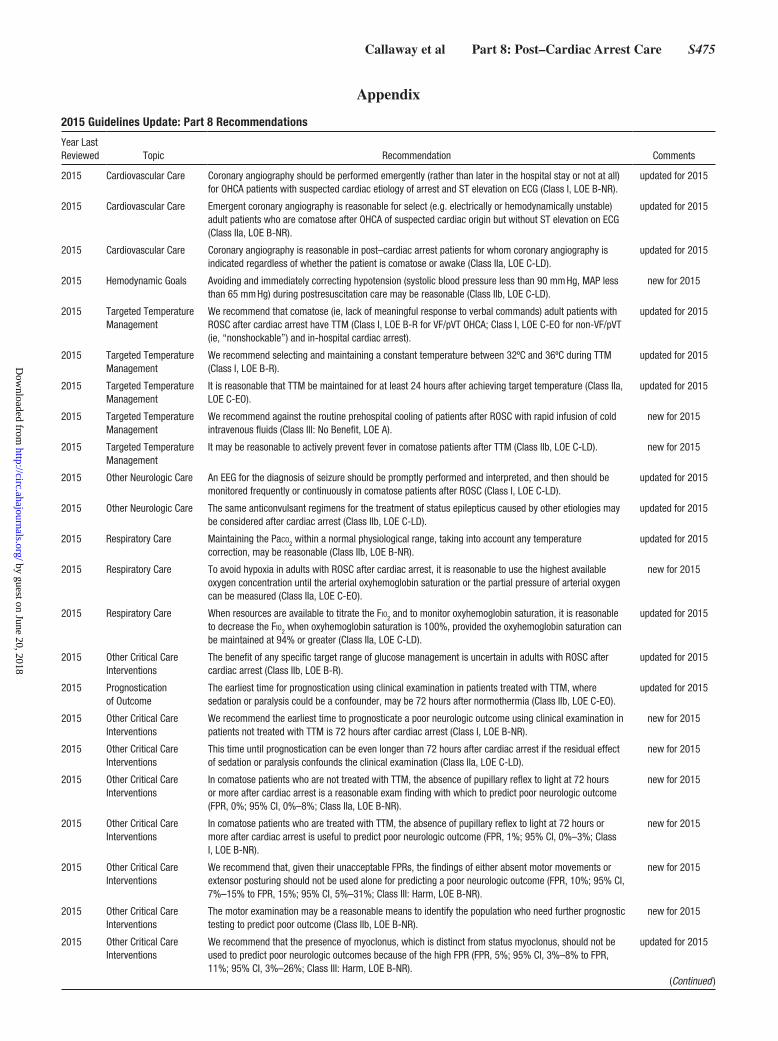
2015 Cardiovascular Care Coronary angiography should be performed emergently (rather than later in the hospital stay or not at all) for OHCA patients with suspected cardiac etiology of arrest and ST elevation on ECG (Class I, LOE B-NR).
updated for 2015
2015 Cardiovascular Care Emergent coronary angiography is reasonable for select (e.g. electrically or hemodynamically unstable) adult patients who are comatose after OHCA of suspected cardiac origin but without ST elevation on ECG (Class IIa, LOE B-NR).
updated for 2015
2015 Cardiovascular Care Coronary angiography is reasonable in post–cardiac arrest patients for whom coronary angiography is indicated regardless of whether the patient is comatose or awake (Class IIa, LOE C-LD).
updated for 2015
2015 Hemodynamic Goals Avoiding and immediately correcting hypotension (systolic blood pressure less than 90 mm Hg, MAP less than 65 mm Hg) during postresuscitation care may be reasonable (Class IIb, LOE C-LD).
new for 2015
2015 Targeted Temperature Management
We recommend that comatose (ie, lack of meaningful response to verbal commands) adult patients with ROSC after cardiac arrest have TTM (Class I, LOE B-R for VF/pVT OHCA; Class I, LOE C-EO for non-VF/pVT (ie, “nonshockable”) and in-hospital cardiac arrest).
updated for 2015
2015 Targeted Temperature Management
We recommend selecting and maintaining a constant temperature between 32ºC and 36ºC during TTM (Class I, LOE B-R).
updated for 2015
2015 Targeted Temperature Management
It is reasonable that TTM be maintained for at least 24 hours after achieving target temperature (Class IIa, LOE C-EO).
updated for 2015
2015 Targeted Temperature Management
We recommend against the routine prehospital cooling of patients after ROSC with rapid infusion of cold intravenous fluids (Class III: No Benefit, LOE A).
new for 2015
2015 Targeted Temperature Management
It may be reasonable to actively prevent fever in comatose patients after TTM (Class IIb, LOE C-LD). new for 2015
2015 Other Neurologic Care An EEG for the diagnosis of seizure should be promptly performed and interpreted, and then should be monitored frequently or continuously in comatose patients after ROSC (Class I, LOE C-LD).
updated for 2015
2015 Other Neurologic Care The same anticonvulsant regimens for the treatment of status epilepticus caused by other etiologies may be considered after cardiac arrest (Class IIb, LOE C-LD).
updated for 2015
2015 Respiratory Care Maintaining the Paco2 within a normal physiological range, taking into account any temperature correction, may be reasonable (Class IIb, LOE B-NR).
updated for 2015
2015 Respiratory Care To avoid hypoxia in adults with ROSC after cardiac arrest, it is reasonable to use the highest available oxygen concentration until the arterial oxyhemoglobin saturation or the partial pressure of arterial oxygen can be measured (Class IIa, LOE C-EO).
new for 2015
2015 Respiratory Care When resources are available to titrate the Fio2 and to monitor oxyhemoglobin saturation, it is reasonable to decrease the Fio2 when oxyhemoglobin saturation is 100%, provided the oxyhemoglobin saturation can be maintained at 94% or greater (Class IIa, LOE C-LD).
updated for 2015
2015 Other Critical Care Interventions
The benefit of any specific target range of glucose management is uncertain in adults with ROSC after cardiac arrest (Class IIb, LOE B-R).
updated for 2015
2015 Prognostication of Outcome
The earliest time for prognostication using clinical examination in patients treated with TTM, where sedation or paralysis could be a confounder, may be 72 hours after normothermia (Class IIb, LOE C-EO).
updated for 2015
2015 Other Critical Care Interventions
We recommend the earliest time to prognosticate a poor neurologic outcome using clinical examination in patients not treated with TTM is 72 hours after cardiac arrest (Class I, LOE B-NR).
new for 2015
2015 Other Critical Care Interventions
This time until prognostication can be even longer than 72 hours after cardiac arrest if the residual effect of sedation or paralysis confounds the clinical examination (Class IIa, LOE C-LD).
new for 2015
2015 Other Critical Care Interventions
In comatose patients who are not treated with TTM, the absence of pupillary reflex to light at 72 hours or more after cardiac arrest is a reasonable exam finding with which to predict poor neurologic outcome (FPR, 0%; 95% CI, 0%–8%; Class IIa, LOE B-NR).
new for 2015
2015 Other Critical Care Interventions
In comatose patients who are treated with TTM, the absence of pupillary reflex to light at 72 hours or more after cardiac arrest is useful to predict poor neurologic outcome (FPR, 1%; 95% CI, 0%–3%; Class I, LOE B-NR).
new for 2015
2015 Other Critical Care Interventions
We recommend that, given their unacceptable FPRs, the findings of either absent motor movements or extensor posturing should not be used alone for predicting a poor neurologic outcome (FPR, 10%; 95% CI, 7%–15% to FPR, 15%; 95% CI, 5%–31%; Class III: Harm, LOE B-NR).
new for 2015
2015 Other Critical Care Interventions
The motor examination may be a reasonable means to identify the population who need further prognostic testing to predict poor outcome (Class IIb, LOE B-NR).
new for 2015
2015 Other Critical Care Interventions
We recommend that the presence of myoclonus, which is distinct from status myoclonus, should not be used to predict poor neurologic outcomes because of the high FPR (FPR, 5%; 95% CI, 3%–8% to FPR, 11%; 95% CI, 3%–26%; Class III: Harm, LOE B-NR).
updated for 2015
Appendix
(Continued )
by guest on June 20, 2018http://circ.ahajournals.org/
Dow
nloaded from

S476 Circulation November 3, 2015
2015 Other Critical Care Interventions
In combination with other diagnostic tests at 72 or more hours after cardiac arrest, the presence of status myoclonus during the first 72 to 120 hours after cardiac arrest is a reasonable finding to help predict poor neurologic outcomes (FPR, 0%; 95% CI, 0%–4%; Class IIa, LOE B-NR).
new for 2015
2015 Other Critical Care Interventions
In comatose post–cardiac arrest patients who are treated with TTM, it may be reasonable to consider persistent absence of EEG reactivity to external stimuli at 72 hours after cardiac arrest, and persistent burst suppression on EEG after rewarming, to predict a poor outcome (FPR, 0%; 95% CI, 0%–3%; Class IIb, LOE B-NR).
updated for 2015
2015 Other Critical Care Interventions
Intractable and persistent (more than 72 hours) status epilepticus in the absence of EEG reactivity to external stimuli may be reasonable to predict poor outcome (Class IIb, LOE B-NR).
updated for 2015
2015 Other Critical Care Interventions
In comatose post–cardiac arrest patients who are not treated with TTM, it may be reasonable to consider the presence of burst suppression on EEG at 72 hours or more after cardiac arrest, in combination with other predictors, to predict a poor neurologic outcome (FPR, 0%; 95% CI, 0%–11%; Class IIb, LOE B-NR).
updated for 2015
2015 Other Critical Care Interventions
In patients who are comatose after resuscitation from cardiac arrest regardless of treatment with TTM, it is reasonable to consider bilateral absence of the N20 SSEP wave 24 to 72 hours after cardiac arrest or after rewarming a predictor of poor outcome (FPR, 1%; 95% CI, 0%–3%; Class IIa, LOE B-NR).
updated for 2015
2015 Other Critical Care Interventions
In patients who are comatose after resuscitation from cardiac arrest and not treated with TTM, it may be reasonable to use the presence of a marked reduction of the GWR on brain CT obtained within 2 hours after cardiac arrest to predict poor outcome (Class IIb, LOE B-NR).
new for 2015
2015 Other Critical Care Interventions
It may be reasonable to consider extensive restriction of diffusion on brain MRI at 2 to 6 days after cardiac arrest in combination with other established predictors to predict a poor neurologic outcome (Class IIb, LOE B-NR).
new for 2015
2015 Other Critical Care Interventions
Given the possibility of high FPRs, blood levels of NSE and S-100B should not be used alone to predict a poor neurologic outcome (Class III: Harm, LOE C-LD).
updated for 2015
2015 Other Critical Care Interventions
When performed with other prognostic tests at 72 hours or more after cardiac arrest, it may be reasonable to consider high serum values of NSE at 48 to 72 hours after cardiac arrest to support the prognosis of a poor neurologic outcome (Class IIb, LOE B-NR), especially if repeated sampling reveals persistently high values (Class IIb, LOE C-LD).
updated for 2015
2015 Other Critical Care Interventions
We recommend that all patients who are resuscitated from cardiac arrest but who subsequently progress to death or brain death be evaluated for organ donation (Class I, LOE B-NR).
updated for 2015
2015 Other Critical Care Interventions
Patients who do not have ROSC after resuscitation efforts and who would otherwise have termination of efforts may be considered candidates for kidney or liver donation in settings where programs exist (Class IIb, LOE B-NR).
new for 2015
The following recommendations were not reviewed in 2015. For more information, see the 2010 AHA Guidelines for CPR and ECC, “Part 9: Post–Cardiac Arrest Care.”
2010 Systems of Care for Improving Post–Cardiac Arrest Outcomes
A comprehensive, structured, multidisciplinary system of care should be implemented in a consistent manner for the treatment of post–cardiac arrest patients (Class I, LOE B).
not reviewed in 2015
2010 Treatment of Pulmonary Embolism After CPR
In post–cardiac arrest patients with arrest due to presumed or known pulmonary embolism, fibrinolytics may be considered (Class IIb, LOE C).
not reviewed in 2015
2010 Sedation After Cardiac Arrest
It is reasonable to consider the titrated use of sedation and analgesia in critically ill patients who require mechanical ventilation or shivering suppression during induced hypothermia after cardiac arrest (Class IIb, LOE C).
not reviewed in 2015
2010 Cardiovascular System
A 12-lead ECG should be obtained as soon as possible after ROSC to determine whether acute ST elevation is present (Class I, LOE B).
not reviewed in 2015
2010 Neuroprotective Drugs
The routine use of coenzyme Q10 in patients treated with hypothermia is uncertain (Class IIb, LOE B). not reviewed in 2015
2010 Evoked Potentials Bilateral absence of the N20 cortical response to median nerve stimulation after 24 hours predicts poor outcome in comatose cardiac arrest survivors not treated with therapeutic hypothermia (Class IIa, LOE A).
not reviewed in 2015
Year Last Reviewed Topic Recommendation Comments
2015 Guidelines Update: Part 8 Recommendations, Continued
by guest on June 20, 2018http://circ.ahajournals.org/
Dow
nloaded from
Callaway et al Part 8: Post–Cardiac Arrest Care S477
References 1. Morrison LJ, Deakin CD, Morley PT, Callaway CW, Kerber RE, Kronick
SL, Lavonas EJ, Link MS, Neumar RW, Otto CW, Parr M, Shuster M, Sunde K, Peberdy MA, Tang W, Hoek TL, Böttiger BW, Drajer S, Lim SH, Nolan JP; Advanced Life Support Chapter Collaborators. Part 8: advanced life support: 2010 International Consensus on Cardiopulmonary Resuscitation and Emergency Cardiovascular Care Science With Treatment Recommendations. Circulation. 2010;122(suppl 2):S345–S421. doi: 10.1161/CIRCULATIONAHA.110.971051.
2. Deakin CD, Morrison LJ, Morley PT, Callaway CW, Kerber RE, Kronick SL, Lavonas EJ, Link MS, Neumar RW, Otto CW, Parr M, Shuster M, Sunde K, Peberdy MA, Tang W, Hoek TL, Böttiger BW, Drajer S, Lim SH, Nolan JP; Advanced Life Support Chapter Collaborators. Part 8: advanced life support: 2010 International Consensus on Cardiopulmonary Resuscitation and Emergency Cardiovascular Care Science With Treatment Recommendations. Resuscitation. 2010;81 suppl 1:e93–e174. doi: 10.1016/j.resuscitation.2010.08.027.
3. Callaway CW, Soar J, Aibiki M, Böttiger BW, Brooks SC, Deakin CD, Donnino MW, Drajer S, Kloeck W, Morley PT, Morrison LJ, Neumar RW, Nicholson TC, Nolan JP, Okada K, O’Neil BJ, Paiva EF, Parr MJ, Wang TL, Witt J; on behalf of the Advanced Life Support Chapter Collaborators. Part 4: advanced life support: 2015 International Consensus on Cardiopulmonary Resuscitation and Emergency Cardiovascular Care Science With Treatment Recommendations. Circulation. 2015;132(suppl 1):S84–S145. doi: 10.1161/CIR.0000000000000273.
4. Soar J, Callaway CW, Aibiki M, Böttiger BW, Brooks SC, Deakin CD, Donnino MW, Drajer S, Kloeck W, Morley PT, Morrison LJ, Neumar RW, Nicholson TC, Nolan JP, Okada K, O’Neil BJ, Paiva EF, Parr MJ, Wang TL, Witt J; on behalf of the Advanced Life Support Chapter Collaborators. Part 4: advanced life support: 2015 International Consensus on Cardiopulmonary Resuscitation and Emergency Cardiovascular Care Science With Treatment Recommendations. Resuscitation. 2015. In press.
5. O’Connor D, Green S, Higgins JPT, eds. Chapter 5: defining the review questions and developing criteria for including studies. In: The Cochrane Collaboration. Higgins JPT, Green S, eds. Cochrane Handbook for Systematic Reviews of Interventions. Version 5.1.0. 2011. http://handbook.cochrane.org/. Accessed May 6, 2015.
6. Schünemann H, Brożek J, Guyatt G, Oxman A. GRADE Handbook. 2013. http://www.guidelinedevelopment.org/handbook/. Accessed May 6, 2015.
7. Neumar RW, Nolan JP, Adrie C, Aibiki M, Berg RA, Böttiger BW, Callaway C, Clark RS, Geocadin RG, Jauch EC, Kern KB, Laurent I, Longstreth WT Jr, Merchant RM, Morley P, Morrison LJ, Nadkarni V, Peberdy MA, Rivers EP, Rodriguez-Nunez A, Sellke FW, Spaulding C, Sunde K, Vanden Hoek T. Post-cardiac arrest syndrome: epidemiology, pathophysiology, treatment, and prognostication: a consensus statement from the International Liaison Committee on Resuscitation (American Heart Association, Australian and New Zealand Council on Resuscitation, European Resuscitation Council, Heart and Stroke Foundation of Canada, InterAmerican Heart Foundation, Resuscitation Council of Asia, and the Resuscitation Council of Southern Africa); the American Heart Association Emergency Cardiovascular Care Committee; the Council on Cardiovascular Surgery and Anesthesia; the Council on Cardiopulmonary, Perioperative, and Critical Care; the Council on Clinical Cardiology; and the Stroke Council. Circulation. 2008;118:2452–2483. doi: 10.1161/CIRCULATIONAHA.108.190652.
8. Davies MJ. Anatomic features in victims of sudden coronary death. Coronary artery pathology. Circulation. 1992;85(1 suppl):I19–I24.
9. Spaulding CM, Joly LM, Rosenberg A, Monchi M, Weber SN, Dhainaut JF, Carli P. Immediate coronary angiography in survivors of out-of-hos-pital cardiac arrest. N Engl J Med. 1997;336:1629–1633. doi: 10.1056/NEJM199706053362302.
10. Dumas F, Cariou A, Manzo-Silberman S, Grimaldi D, Vivien B, Rosencher J, Empana JP, Carli P, Mira JP, Jouven X, Spaulding C. Immediate per-cutaneous coronary intervention is associated with better survival after out-of-hospital cardiac arrest: insights from the PROCAT (Parisian Region Out of hospital Cardiac ArresT) registry. Circ Cardiovasc Interv. 2010;3:200–207. doi: 10.1161/CIRCINTERVENTIONS.109.913665.
11. Hollenbeck RD, McPherson JA, Mooney MR, Unger BT, Patel NC, McMullan PW Jr, Hsu CH, Seder DB, Kern KB. Early cardiac catheter-ization is associated with improved survival in comatose survivors of car-diac arrest without STEMI. Resuscitation. 2014;85:88–95. doi: 10.1016/j.resuscitation.2013.07.027.
12. Mooney MR, Unger BT, Boland LL, Burke MN, Kebed KY, Graham KJ, Henry TD, Katsiyiannis WT, Satterlee PA, Sendelbach S, Hodges JS, Parham WM. Therapeutic hypothermia after out-of-hospital
cardiac arrest: evaluation of a regional system to increase access to cooling. Circulation. 2011;124:206–214. doi: 10.1161/CIRCULATIONAHA.110.986257.
13. Gräsner JT, Meybohm P, Lefering R, Wnent J, Bahr J, Messelken M, Jantzen T, Franz R, Scholz J, Schleppers A, Böttiger BW, Bein B, Fischer M; German Resuscitation Registry Study Group. ROSC after cardiac arrest–the RACA score to predict outcome after out-of-hospital cardiac arrest. Eur Heart J. 2011;32:1649–1656. doi: 10.1093/eurheartj/ehr107.
14. Cronier P, Vignon P, Bouferrache K, Aegerter P, Charron C, Templier F, Castro S, El Mahmoud R, Lory C, Pichon N, Dubourg O, Vieillard-Baron A. Impact of routine percutaneous coronary intervention after out-of-hos-pital cardiac arrest due to ventricular fibrillation. Crit Care. 2011;15:R122. doi: 10.1186/cc10227.
15. Bulut S, Aengevaeren WR, Luijten HJ, Verheugt FW. Successful out-of-hospital cardiopulmonary resuscitation: what is the optimal in-hospital treatment strategy? Resuscitation. 2000;47:155–161.
16. Bro-Jeppesen J, Kjaergaard J, Wanscher M, Pedersen F, Holmvang L, Lippert FK, Møller JE, Køber L, Hassager C. Emergency coronary angi-ography in comatose cardiac arrest patients: do real-life experiences sup-port the guidelines? Eur Heart J Acute Cardiovasc Care. 2012;1:291–301. doi: 10.1177/2048872612465588.
17. Aurore A, Jabre P, Liot P, Margenet A, Lecarpentier E, Combes X. Predictive factors for positive coronary angiography in out-of-hospital cardiac arrest patients. Eur J Emerg Med. 2011;18:73–76. doi: 10.1097/MEJ.0b013e32833d469a.
18. Nanjayya VB, Nayyar V. Immediate coronary angiogram in comatose sur-vivors of out-of-hospital cardiac arrest–an Australian study. Resuscitation. 2012;83:699–704. doi: 10.1016/j.resuscitation.2011.12.004.
19. Reynolds JC, Callaway CW, El Khoudary SR, Moore CG, Alvarez RJ, Rittenberger JC. Coronary angiography predicts improved outcome fol-lowing cardiac arrest: propensity-adjusted analysis. J Intensive Care Med. 2009;24:179–186. doi: 10.1177/0885066609332725.
20. Strote JA, Maynard C, Olsufka M, Nichol G, Copass MK, Cobb LA, Kim F. Comparison of role of early (less than six hours) to later (more than six hours) or no cardiac catheterization after resuscitation from out-of-hospital cardiac arrest. Am J Cardiol. 2012;109:451–454. doi: 10.1016/j.amjcard.2011.09.036.
21. Tømte O, Andersen GØ, Jacobsen D, Drægni T, Auestad B, Sunde K. Strong and weak aspects of an established post-resuscitation treatment protocol-A five-year observational study. Resuscitation. 2011;82:1186–1193. doi: 10.1016/j.resuscitation.2011.05.003.
22. Waldo SW, Armstrong EJ, Kulkarni A, Hoffmayer K, Kinlay S, Hsue P, Ganz P, McCabe JM. Comparison of clinical characteristics and outcomes of cardiac arrest survivors having versus not having coronary angiography. Am J Cardiol. 2013;111:1253–1258. doi: 10.1016/j.amjcard.2013.01.267.
23. Nielsen N, Hovdenes J, Nilsson F, Rubertsson S, Stammet P, Sunde K, Valsson F, Wanscher M, Friberg H; Hypothermia Network. Outcome, timing and adverse events in therapeutic hypothermia after out-of-hos-pital cardiac arrest. Acta Anaesthesiol Scand. 2009;53:926–934. doi: 10.1111/j.1399-6576.2009.02021.x.
24. Werling M, Thorén AB, Axelsson C, Herlitz J. Treatment and outcome in post-resuscitation care after out-of-hospital cardiac arrest when a modern therapeutic approach was introduced. Resuscitation. 2007;73:40–45. doi: 10.1016/j.resuscitation.2006.08.018.
25. Zanuttini D, Armellini I, Nucifora G, Carchietti E, Trillò G, Spedicato L, Bernardi G, Proclemer A. Impact of emergency coronary angiogra-phy on in-hospital outcome of unconscious survivors after out-of-hos-pital cardiac arrest. Am J Cardiol. 2012;110:1723–1728. doi: 10.1016/j.amjcard.2012.08.006.
26. O’Gara PT, Kushner FG, Ascheim DD, Casey DE Jr, Chung MK, de Lemos JA, Ettinger SM, Fang JC, Fesmire FM, Franklin BA, Granger CB, Krumholz HM, Linderbaum JA, Morrow DA, Newby LK, Ornato JP, Ou N, Radford MJ, Tamis-Holland JE, Tommaso CL, Tracy CM, Woo YJ, Zhao DX. 2013 ACCF/AHA guideline for the management of ST-elevation myocardial infarction: executive summary: a report of the American College of Cardiology Foundation/American Heart Association Task Force on Practice Guidelines. Circulation. 2013;127:529–555. doi: 10.1161/CIR.0b013e3182742c84.
27. Jneid H, Anderson JL, Wright RS, Adams CD, Bridges CR, Casey DE Jr, Ettinger SM, Fesmire FM, Ganiats TG, Lincoff AM, Peterson ED, Philippides GJ, Theroux P, Wenger NK, Zidar JP. 2012 ACCF/AHA focused update of the guideline for the management of patients with unstable angina/non–ST-elevation myocardial infarction (updating the 2007 guideline and replacing the 2011 focused update): a report of the American College of Cardiology Foundation/American Heart Association
by guest on June 20, 2018http://circ.ahajournals.org/
Dow
nloaded from

S478 Circulation November 3, 2015
Task Force on Practice Guidelines. Circulation. 2012;126:875–910. DOI: 10.1161/CIR.0b013e318256f1e0.
28. Task Force on the Management of ST-Segment Elevation Acute Myocardial Infarction of the European Society of Cardiology, Steg PG, James SK, Atar D, Badano LP, Blomstrom-Lundqvist C, Borger MA, Di Mario C, Dickstein K, Ducrocq G, Fernandez-Aviles F, Gershlick AH, Giannuzzi P, Halvorsen S, Huber K, Juni P, Kastrati A, Knuuti J, Lenzen MJ, Mahaffey KW, Valgimigli M, van ‘t Hof A, Widimsky P, Zahger D. ESC Guidelines for the management of acute myocardial infarction in patients presenting with ST-segment elevation. Eur Heart J. 2012;33:2569–2619.
29. Peberdy MA, Donnino MW, Callaway CW, Dimaio JM, Geocadin RG, Ghaemmaghami CA, Jacobs AK, Kern KB, Levy JH, Link MS, Menon V, Ornato JP, Pinto DS, Sugarman J, Yannopoulos D, Ferguson TB Jr; on behalf of the American Heart Association Emergency Cardiovascular Care Committee; Council on Cardiopulmonary, Critical Care, Perioperative and Resuscitation. Impact of percutaneous coronary intervention performance reporting on cardiac resuscitation centers: a scientific statement from the American Heart Association. Circulation. 2013;128:762–773. doi: 10.1161/CIR.0b013e3182a15cd2.
30. Trzeciak S, Jones AE, Kilgannon JH, Milcarek B, Hunter K, Shapiro NI, Hollenberg SM, Dellinger P, Parrillo JE. Significance of arterial hypoten-sion after resuscitation from cardiac arrest. Crit Care Med. 2009;37:2895–903; quiz 2904.
31. Bray JE, Bernard S, Cantwell K, Stephenson M, Smith K; VACAR Steering Committee. The association between systolic blood pressure on arrival at hospital and outcome in adults surviving from out-of-hospital cardiac arrests of presumed cardiac aetiology. Resuscitation. 2014;85:509–515. doi: 10.1016/j.resuscitation.2013.12.005.
32. Kilgannon JH, Roberts BW, Reihl LR, Chansky ME, Jones AE, Dellinger RP, Parrillo JE, Trzeciak S. Early arterial hypotension is com-mon in the post-cardiac arrest syndrome and associated with increased in-hospital mortality. Resuscitation. 2008;79:410–416. doi: 10.1016/j.resuscitation.2008.07.019.
33. Müllner M, Sterz F, Binder M, Hellwagner K, Meron G, Herkner H, Laggner AN. Arterial blood pressure after human cardiac arrest and neu-rological recovery. Stroke. 1996;27:59–62.
34. Beylin ME, Perman SM, Abella BS, Leary M, Shofer FS, Grossestreuer AV, Gaieski DF. Higher mean arterial pressure with or without vasoac-tive agents is associated with increased survival and better neurological outcomes in comatose survivors of cardiac arrest. Intensive Care Med. 2013;39:1981–1988. doi: 10.1007/s00134-013-3075-9.
35. Gaieski DF, Band RA, Abella BS, Neumar RW, Fuchs BD, Kolansky DM, Merchant RM, Carr BG, Becker LB, Maguire C, Klair A, Hylton J, Goyal M. Early goal-directed hemodynamic optimization com-bined with therapeutic hypothermia in comatose survivors of out-of-hospital cardiac arrest. Resuscitation. 2009;80:418–424. doi: 10.1016/j.resuscitation.2008.12.015.
36. Laurent I, Monchi M, Chiche JD, Joly LM, Spaulding C, Bourgeois B, Cariou A, Rozenberg A, Carli P, Weber S, Dhainaut JF. Reversible myo-cardial dysfunction in survivors of out-of-hospital cardiac arrest. J Am Coll Cardiol. 2002;40:2110–2116.
37. Sunde K, Pytte M, Jacobsen D, Mangschau A, Jensen LP, Smedsrud C, Draegni T, Steen PA. Implementation of a standardised treatment protocol for post resuscitation care after out-of-hospital cardiac arrest. Resuscitation. 2007;73:29–39. doi: 10.1016/j.resuscitation.2006.08.016.
38. Walters EL, Morawski K, Dorotta I, Ramsingh D, Lumen K, Bland D, Clem K, Nguyen HB. Implementation of a post-cardiac arrest care bundle including therapeutic hypothermia and hemodynamic optimization in comatose patients with return of spontaneous circulation after out-of-hospital cardiac arrest: a feasibility study. Shock. 2011;35:360–366. doi: 10.1097/SHK.0b013e318204c106.
39. Donnino M, Andersen LW, Berg KM, et al. Temperature management after cardiac arrest: an advisory statement by the Advanced Life Support (ALS) Task Force of the International Liaison Committee on Resuscitation (ILCOR). Circulation. In press.
40. Mild therapeutic hypothermia to improve the neurologic outcome after cardiac arrest. N Engl J Med. 2002;346:549–556.
41. Bernard SA, Gray TW, Buist MD, Jones BM, Silvester W, Gutteridge G, Smith K. Treatment of comatose survivors of out-of-hospital cardiac arrest with induced hypothermia. N Engl J Med. 2002;346:557–563. doi: 10.1056/NEJMoa003289.
42. Dumas F, Grimaldi D, Zuber B, Fichet J, Charpentier J, Pène F, Vivien B, Varenne O, Carli P, Jouven X, Empana JP, Cariou A. Is hypothermia after cardiac arrest effective in both shockable and nonshockable patients?:
insights from a large registry. Circulation. 2011;123:877–886. doi: 10.1161/CIRCULATIONAHA.110.987347.
43. Testori C, Sterz F, Behringer W, Haugk M, Uray T, Zeiner A, Janata A, Arrich J, Holzer M, Losert H. Mild therapeutic hypothermia is associ-ated with favourable outcome in patients after cardiac arrest with non-shockable rhythms. Resuscitation. 2011;82:1162–1167. doi: 10.1016/j.resuscitation.2011.05.022.
44. Vaahersalo J, Hiltunen P, Tiainen M, Oksanen T, Kaukonen KM, Kurola J, Ruokonen E, Tenhunen J, Ala-Kokko T, Lund V, Reinikainen M, Kiviniemi O, Silfvast T, Kuisma M, Varpula T, Pettilä V; FINNRESUSCI Study Group. Therapeutic hypothermia after out-of-hospital cardiac arrest in Finnish intensive care units: the FINNRESUSCI study. Intensive Care Med. 2013;39:826–837. doi: 10.1007/s00134-013-2868-1.
45. Mader TJ, Nathanson BH, Soares WE 3rd, Coute RA, McNally BF. Comparative Effectiveness of Therapeutic Hypothermia After Out-of-Hospital Cardiac Arrest: Insight from a Large Data Registry. Ther Hypothermia Temp Manag. 2014;4:21–31. doi: 10.1089/ther.2013.0018.
46. Nichol G, Huszti E, Kim F, Fly D, Parnia S, Donnino M, Sorenson T, Callaway CW; American Heart Association Get With The Guideline-Resuscitation Investigators. Does induction of hypothermia improve out-comes after in-hospital cardiac arrest? Resuscitation. 2013;84:620–625. doi: 10.1016/j.resuscitation.2012.12.009.
47. Nielsen N, Wetterslev J, Cronberg T, Erlinge D, Gasche Y, Hassager C, Horn J, Hovdenes J, Kjaergaard J, Kuiper M, Pellis T, Stammet P, Wanscher M, Wise MP, Åneman A, Al-Subaie N, Boesgaard S, Bro-Jeppesen J, Brunetti I, Bugge JF, Hingston CD, Juffermans NP, Koopmans M, Køber L, Langørgen J, Lilja G, Møller JE, Rundgren M, Rylander C, Smid O, Werer C, Winkel P, Friberg H; TTM Trial Investigators. Targeted temperature management at 33°C versus 36°C after cardiac arrest. N Engl J Med. 2013;369:2197–2206. doi: 10.1056/NEJMoa1310519.
48. Watts DD, Trask A, Soeken K, Perdue P, Dols S, Kaufmann C. Hypothermic coagulopathy in trauma: effect of varying levels of hypo-thermia on enzyme speed, platelet function, and fibrinolytic activity. J Trauma. 1998;44:846–854.
49. Lavinio A, Scudellari A, Gupta AK. Hemorrhagic shock resulting in cardiac arrest: is therapeutic hypothermia contraindicated? Minerva Anestesiol. 2012;78:969–970.
50. Guilliams K, Rosen M, Buttram S, Zempel J, Pineda J, Miller B, Shoykhet M. Hypothermia for pediatric refractory status epilepticus. Epilepsia. 2013;54:1586–1594. doi: 10.1111/epi.12331.
51. Corry JJ, Dhar R, Murphy T, Diringer MN. Hypothermia for refrac-tory status epilepticus. Neurocrit Care. 2008;9:189–197. doi: 10.1007/s12028-008-9092-9.
52. Guluma KZ, Oh H, Yu SW, Meyer BC, Rapp K, Lyden PD. Effect of endovascular hypothermia on acute ischemic edema: morphometric analysis of the ICTuS trial. Neurocrit Care. 2008;8:42–47. doi: 10.1007/s12028-007-9009-z.
53. Kim F, Olsufka M, Longstreth WT Jr, Maynard C, Carlbom D, Deem S, Kudenchuk P, Copass MK, Cobb LA. Pilot randomized clinical trial of prehospital induction of mild hypothermia in out-of-hospital cardiac arrest patients with a rapid infusion of 4 degrees C normal saline. Circulation. 2007;115:3064–3070. doi: 10.1161/CIRCULATIONAHA.106.655480.
54. Kämäräinen A, Virkkunen I, Tenhunen J, Yli-Hankala A, Silfvast T. Prehospital therapeutic hypothermia for comatose survivors of car-diac arrest: a randomized controlled trial. Acta Anaesthesiol Scand. 2009;53:900–907. doi: 10.1111/j.1399-6576.2009.02015.x.
55. Bernard SA, Smith K, Cameron P, Masci K, Taylor DM, Cooper DJ, Kelly AM, Silvester W; Rapid Infusion of Cold Hartmanns (RICH) Investigators. Induction of therapeutic hypothermia by paramedics after resuscitation from out-of-hospital ventricular fibrillation cardiac arrest: a randomized controlled trial. Circulation. 2010;122:737–742. doi: 10.1161/CIRCULATIONAHA.109.906859.
56. Bernard SA, Smith K, Cameron P, Masci K, Taylor DM, Cooper DJ, Kelly AM, Silvester W; Rapid Infusion of Cold Hartmanns Investigators. Induction of prehospital therapeutic hypothermia after resuscitation from nonventricular fibrillation cardiac arrest. Crit Care Med. 2012;40:747–753. doi: 10.1097/CCM.0b013e3182377038.
57. Kim F, Nichol G, Maynard C, Hallstrom A, Kudenchuk PJ, Rea T, Copass MK, Carlbom D, Deem S, Longstreth WT Jr, Olsufka M, Cobb LA. Effect of prehospital induction of mild hypothermia on survival and neurological status among adults with cardiac arrest: a randomized clinical trial. JAMA. 2014;311:45–52. doi: 10.1001/jama.2013.282173.
58. Debaty G, Maignan M, Savary D, Koch FX, Ruckly S, Durand M, Picard J, Escallier C, Chouquer R, Santre C, Minet C, Guergour D, Hammer L, Bouvaist H, Belle L, Adrie C, Payen JF, Carpentier F, Gueugniaud PY,
by guest on June 20, 2018http://circ.ahajournals.org/
Dow
nloaded from

Callaway et al Part 8: Post–Cardiac Arrest Care S479
Danel V, Timsit JF. Impact of intra-arrest therapeutic hypothermia in out-comes of prehospital cardiac arrest: a randomized controlled trial. Intensive Care Med. 2014;40:1832–1842. doi: 10.1007/s00134-014-3519-x.
59. Castrén M, Nordberg P, Svensson L, Taccone F, Vincent JL, Desruelles D, Eichwede F, Mols P, Schwab T, Vergnion M, Storm C, Pesenti A, Pachl J, Guérisse F, Elste T, Roessler M, Fritz H, Durnez P, Busch HJ, Inderbitzen B, Barbut D. Intra-arrest transnasal evaporative cooling: a ran-domized, prehospital, multicenter study (PRINCE: Pre-ROSC IntraNasal Cooling Effectiveness). Circulation. 2010;122:729–736. doi: 10.1161/CIRCULATIONAHA.109.931691.
60. Zeiner A, Holzer M, Sterz F, Schörkhuber W, Eisenburger P, Havel C, Kliegel A, Laggner AN. Hyperthermia after cardiac arrest is associated with an unfa-vorable neurologic outcome. Arch Intern Med. 2001;161:2007–2012.
61. Langhelle A, Tyvold SS, Lexow K, Hapnes SA, Sunde K, Steen PA. In-hospital factors associated with improved outcome after out-of-hospital cardiac arrest. A comparison between four regions in Norway. Resuscitation. 2003;56:247–263.
62. Nolan JP, Laver SR, Welch CA, Harrison DA, Gupta V, Rowan K. Outcome following admission to UK intensive care units after cardiac arrest: a second-ary analysis of the ICNARC Case Mix Programme Database. Anaesthesia. 2007;62:1207–1216. doi: 10.1111/j.1365-2044.2007.05232.x.
63. Suffoletto B, Peberdy MA, van der Hoek T, Callaway C. Body temperature changes are associated with outcomes following in-hospital cardiac arrest and return of spontaneous circulation. Resuscitation. 2009;80:1365–1370. doi: 10.1016/j.resuscitation.2009.08.020.
64. Gebhardt K, Guyette FX, Doshi AA, Callaway CW, Rittenberger JC; Post Cardiac Arrest Service. Prevalence and effect of fever on outcome follow-ing resuscitation from cardiac arrest. Resuscitation. 2013;84:1062–1067. doi: 10.1016/j.resuscitation.2013.03.038.
65. Aldhoon B, Melenovsky V, Kettner J, Kautzner J. Clinical predictors of outcome in survivors of out-of-hospital cardiac arrest treated with hypo-thermia. Cor et Vasa. 2012;54:e68–e75.
66. Benz-Woerner J, Delodder F, Benz R, Cueni-Villoz N, Feihl F, Rossetti AO, Liaudet L, Oddo M. Body temperature regulation and outcome after cardiac arrest and therapeutic hypothermia. Resuscitation. 2012;83:338–342. doi: 10.1016/j.resuscitation.2011.10.026.
67. Bouwes A, Robillard LB, Binnekade JM, de Pont AC, Wieske L, Hartog AW, Schultz MJ, Horn J. The influence of rewarming after therapeutic hypothermia on outcome after cardiac arrest. Resuscitation. 2012;83:996–1000. doi: 10.1016/j.resuscitation.2012.04.006.
68. Leary M, Grossestreuer AV, Iannacone S, Gonzalez M, Shofer FS, Povey C, Wendell G, Archer SE, Gaieski DF, Abella BS. Pyrexia and neurologic outcomes after therapeutic hypothermia for cardiac arrest. Resuscitation. 2013;84:1056–1061. doi: 10.1016/j.resuscitation.2012.11.003.
69. Cocchi MN, Boone MD, Giberson B, Giberson T, Farrell E, Salciccioli JD, Talmor D, Williams D, Donnino MW. Fever after rewarming: inci-dence of pyrexia in postcardiac arrest patients who have undergone mild therapeutic hypothermia. J Intensive Care Med. 2014;29:365–369. doi: 10.1177/0885066613491932.
70. Bro-Jeppesen J, Hassager C, Wanscher M, Søholm H, Thomsen JH, Lippert FK, Møller JE, Køber L, Kjaergaard J. Post-hypothermia fever is associated with increased mortality after out-of-hospital cardiac arrest. Resuscitation. 2013;84:1734–1740. doi: 10.1016/j.resuscitation.2013.07.023.
71. Winters SA, Wolf KH, Kettinger SA, Seif EK, Jones JS, Bacon-Baguley T. Assessment of risk factors for post-rewarming “rebound hyperthermia” in cardiac arrest patients undergoing therapeutic hypothermia. Resuscitation. 2013;84:1245–1249. doi: 10.1016/j.resuscitation.2013.03.027.
72. Bohman LE, Levine JM. Fever and therapeutic normothermia in severe brain injury: an update. Curr Opin Crit Care. 2014;20:182–188. doi: 10.1097/MCC.0000000000000070.
73. Badjatia N. Hyperthermia and fever control in brain injury. Crit Care Med. 2009;37(7 suppl):S250–S257. doi: 10.1097/CCM.0b013e3181aa5e8d.
74. Longstreth WT Jr, Fahrenbruch CE, Olsufka M, Walsh TR, Copass MK, Cobb LA. Randomized clinical trial of magnesium, diazepam, or both after out-of-hospital cardiac arrest. Neurology. 2002;59:506–514.
75. Rittenberger JC, Popescu A, Brenner RP, Guyette FX, Callaway CW. Frequency and timing of nonconvulsive status epilepticus in comatose post-cardiac arrest subjects treated with hypothermia. Neurocrit Care. 2012;16:114–122. doi: 10.1007/s12028-011-9565-0.
76. Tomte O, Draegni T, Mangschau A, Jacobsen D, Auestad B, Sunde K. A comparison of intravascular and surface cooling techniques in comatose cardiac arrest survivors. Crit Care Med. 2011;39:443–449.
77. Hofmeijer J, Tjepkema-Cloostermans MC, Blans MJ, Beishuizen A, van Putten MJ. Unstandardized treatment of electroencephalographic status
epilepticus does not improve outcome of comatose patients after cardiac arrest. Front Neurol. 2014;5:39. doi: 10.3389/fneur.2014.00039.
78. Crepeau AZ, Rabinstein AA, Fugate JE, Mandrekar J, Wijdicks EF, White RD, Britton JW. Continuous EEG in therapeutic hypothermia after cardiac arrest: prognostic and clinical value. Neurology. 2013;80:339–344. doi: 10.1212/WNL.0b013e31827f089d.
79. Knight WA, Hart KW, Adeoye OM, Bonomo JB, Keegan SP, Ficker DM, Szaflarski JP, Privitera MD, Lindsell CJ. The incidence of seizures in patients undergoing therapeutic hypothermia after resuscitation from cardiac arrest. Epilepsy Res. 2013;106:396–402. doi: 10.1016/j.eplepsyres.2013.06.018.
80. Randomized clinical study of thiopental loading in comatose survivors of cardiac arrest. Brain Resuscitation Clinical Trial I Study Group. N Engl J Med. 1986;314:397–403.
81. Monsalve F, Rucabado L, Ruano M, Cuñat J, Lacueva V, Viñuales A. The neurologic effects of thiopental therapy after cardiac arrest. Intensive Care Med. 1987;13:244–248.
82. Friedman D, Claassen J, Hirsch LJ. Continuous electroencephalogram monitoring in the intensive care unit. Anesth Analg. 2009;109:506–523. doi: 10.1213/ane.0b013e3181a9d8b5.
83. Roberts BW, Kilgannon JH, Chansky ME, Mittal N, Wooden J, Trzeciak S. Association between postresuscitation partial pressure of arterial car-bon dioxide and neurological outcome in patients with post-cardiac arrest syndrome. Circulation. 2013;127:2107–2113. doi: 10.1161/CIRCULATIONAHA.112.000168.
84. Lee BK, Jeung KW, Lee HY, Lee SJ, Jung YH, Lee WK, Heo T, Min YI. Association between mean arterial blood gas tension and outcome in cardiac arrest patients treated with therapeutic hypothermia. Am J Emerg Med. 2014;32:55–60. doi: 10.1016/j.ajem.2013.09.044.
85. Schneider AG, Eastwood GM, Bellomo R, Bailey M, Lipcsey M, Pilcher D, Young P, Stow P, Santamaria J, Stachowski E, Suzuki S, Woinarski NC, Pilcher J. Arterial carbon dioxide tension and outcome in patients admitted to the intensive care unit after cardiac arrest. Resuscitation. 2013;84:927–934. doi: 10.1016/j.resuscitation.2013.02.014.
86. Vaahersalo J, Bendel S, Reinikainen M, Kurola J, Tiainen M, Raj R, Pettilä V, Varpula T, Skrifvars MB; FINNRESUSCI Study Group. Arterial blood gas tensions after resuscitation from out-of-hospital cardiac arrest: associ-ations with long-term neurologic outcome. Crit Care Med. 2014;42:1463–1470. doi: 10.1097/CCM.0000000000000228.
87. Janz DR, Hollenbeck RD, Pollock JS, McPherson JA, Rice TW. Hyperoxia is associated with increased mortality in patients treated with mild therapeutic hypothermia after sudden cardiac arrest. Crit Care Med. 2012;40:3135–3139. doi: 10.1097/CCM.0b013e3182656976.
88. Kilgannon JH, Jones AE, Shapiro NI, Angelos MG, Milcarek B, Hunter K, Parrillo JE, Trzeciak S; Emergency Medicine Shock Research Network (EMShockNet) Investigators. Association between arterial hyperoxia fol-lowing resuscitation from cardiac arrest and in-hospital mortality. JAMA. 2010;303:2165–2171. doi: 10.1001/jama.2010.707.
89. Elmer J, Scutella M, Pullalarevu R, Wang B, Vaghasia N, Trzeciak S, Rosario-Rivera BL, Guyette FX, Rittenberger JC, Dezfulian C; Pittsburgh Post-Cardiac Arrest Service (PCAS). The association between hyperoxia and patient outcomes after cardiac arrest: analysis of a high-resolution database. Intensive Care Med. 2015;41:49–57. doi: 10.1007/s00134-014-3555-6.
90. Rachmale S, Li G, Wilson G, Malinchoc M, Gajic O. Practice of excessive F(IO(2)) and effect on pulmonary outcomes in mechanically ventilated patients with acute lung injury. Respir Care. 2012;57:1887–1893. doi: 10.4187/respcare.01696.
91. Bellomo R, Bailey M, Eastwood GM, Nichol A, Pilcher D, Hart GK, Reade MC, Egi M, Cooper DJ; Study of Oxygen in Critical Care (SOCC) Group. Arterial hyperoxia and in-hospital mortality after resuscitation from cardiac arrest. Crit Care. 2011;15:R90. doi: 10.1186/cc10090.
92. Nelskylä A, Parr MJ, Skrifvars MB. Prevalence and factors correlating with hyperoxia exposure following cardiac arrest–an observational sin-gle centre study. Scand J Trauma Resusc Emerg Med. 2013;21:35. doi: 10.1186/1757-7241-21-35.
93. Kuisma M, Boyd J, Voipio V, Alaspää A, Roine RO, Rosenberg P. Comparison of 30 and the 100% inspired oxygen concentrations dur-ing early post-resuscitation period: a randomised controlled pilot study. Resuscitation. 2006;69:199–206. doi: 10.1016/j.resuscitation.2005.08.010.
94. Ihle JF, Bernard S, Bailey MJ, Pilcher DV, Smith K, Scheinkestel CD. Hyperoxia in the intensive care unit and outcome after out-of-hospital ventricular fibrillation cardiac arrest. Crit Care Resusc. 2013;15:186–190.
95. Oksanen T, Skrifvars MB, Varpula T, Kuitunen A, Pettilä V, Nurmi J, Castrén M. Strict versus moderate glucose control after resuscitation from ventricular fibrillation. Intensive Care Med. 2007;33:2093–2100. doi: 10.1007/s00134-007-0876-8.
by guest on June 20, 2018http://circ.ahajournals.org/
Dow
nloaded from

S480 Circulation November 3, 2015
96. Wiener RS, Wiener DC, Larson RJ. Benefits and risks of tight glucose control in critically ill adults: a meta-analysis. JAMA. 2008;300:933–944. doi: 10.1001/jama.300.8.933.
97. Marik PE, Preiser JC. Toward understanding tight glycemic control in the ICU: a systematic review and metaanalysis. Chest. 2010;137:544–551. doi: 10.1378/chest.09-1737.
98. Moghissi ES, Korytkowski MT, DiNardo M, Einhorn D, Hellman R, Hirsch IB, Inzucchi SE, Ismail-Beigi F, Kirkman MS, Umpierrez GE; American Association of Clinical Endocrinologists; American Diabetes Association. American Association of Clinical Endocrinologists and American Diabetes Association consensus statement on inpatient gly-cemic control. Diabetes Care. 2009;32:1119–1131. doi: 10.2337/dc09-9029.
99. Sandroni C, Cariou A, Cavallaro F, Cronberg T, Friberg H, Hoedemaekers C, Horn J, Nolan JP, Rossetti AO, Soar J. Prognostication in comatose survivors of cardiac arrest: an advisory statement from the European Resuscitation Council and the European Society of Intensive Care Medicine. Resuscitation. 2014;85:1779–1789. doi: 10.1016/j.resuscitation.2014.08.011.
100. Empey PE, de Mendizabal NV, Bell MJ, Bies RR, Anderson KB, Kochanek PM, Adelson PD, Poloyac SM; Pediatric TBI Consortium: Hypothermia Investigators. Therapeutic hypothermia decreases phenyt-oin elimination in children with traumatic brain injury. Crit Care Med. 2013;41:2379–2387. doi: 10.1097/CCM.0b013e318292316c.
101. Hostler D, Zhou J, Tortorici MA, Bies RR, Rittenberger JC, Empey PE, Kochanek PM, Callaway CW, Poloyac SM. Mild hypothermia alters midazolam pharmacokinetics in normal healthy volunteers. Drug Metab Dispos. 2010;38:781–788. doi: 10.1124/dmd.109.031377.
102. Sandroni C, Cavallaro F, Callaway CW, Sanna T, D’Arrigo S, Kuiper M, Della Marca G, Nolan JP. Predictors of poor neurological out-come in adult comatose survivors of cardiac arrest: a systematic re-view and meta-analysis. Part 1: patients not treated with therapeutic hypothermia. Resuscitation. 2013;84:1310–1323. doi: 10.1016/j.resuscitation.2013.05.013.
103. Sandroni C, Cavallaro F, Callaway CW, D’Arrigo S, Sanna T, Kuiper MA, Biancone M, Della Marca G, Farcomeni A, Nolan JP. Predictors of poor neurological outcome in adult comatose survivors of cardiac arrest: a systematic review and meta-analysis. Part 2: Patients treated with thera-peutic hypothermia. Resuscitation. 2013;84:1324–1338. doi: 10.1016/j.resuscitation.2013.06.020.
104. Bouwes A, Binnekade JM, Kuiper MA, Bosch FH, Zandstra DF, Toornvliet AC, Biemond HS, Kors BM, Koelman JH, Verbeek MM, Weinstein HC, Hijdra A, Horn J. Prognosis of coma after therapeutic hypothermia: a prospective cohort study. Ann Neurol. 2012;71:206–212. doi: 10.1002/ana.22632.
105. Choi SP, Youn CS, Park KN, Wee JH, Park JH, Oh SH, Kim SH, Kim JY. Therapeutic hypothermia in adult cardiac arrest because of drowning. Acta Anaesthesiol Scand. 2012;56:116–123. doi: 10.1111/j.1399-6576.2011.02562.x.
106. Cronberg T, Rundgren M, Westhall E, Englund E, Siemund R, Rosén I, Widner H, Friberg H. Neuron-specific enolase correlates with other prognostic markers after cardiac arrest. Neurology. 2011;77:623–630. doi: 10.1212/WNL.0b013e31822a276d.
107. Fugate JE, Wijdicks EF, Mandrekar J, Claassen DO, Manno EM, White RD, Bell MR, Rabinstein AA. Predictors of neurologic outcome in hypo-thermia after cardiac arrest. Ann Neurol. 2010;68:907–914. doi: 10.1002/ana.22133.
108. Samaniego EA, Mlynash M, Caulfield AF, Eyngorn I, Wijman CA. Sedation confounds outcome prediction in cardiac arrest survivors treat-ed with hypothermia. Neurocrit Care. 2011;15:113–119. doi: 10.1007/s12028-010-9412-8.
109. Bouwes A, van Poppelen D, Koelman JH, Kuiper MA, Zandstra DF, Weinstein HC, Tromp SC, Zandbergen EG, Tijssen MA, Horn J. Acute posthypoxic myoclonus after cardiopulmonary resuscitation. BMC Neurol. 2012;12:63. doi: 10.1186/1471-2377-12-63.
110. Rossetti AO, Oddo M, Logroscino G, Kaplan PW. Prognostication af-ter cardiac arrest and hypothermia: a prospective study. Ann Neurol. 2010;67:301–307. doi: 10.1002/ana.21984.
111. Rossetti AO, Carrera E, Oddo M. Early EEG correlates of neuronal injury after brain anoxia. Neurology. 2012;78:796–802. doi: 10.1212/WNL.0b013e318249f6bb.
112. Bisschops LL, van Alfen N, Bons S, van der Hoeven JG, Hoedemaekers CW. Predictors of poor neurologic outcome in patients after cardiac arrest treated with hypothermia: a retrospective study. Resuscitation. 2011;82:696–701. doi: 10.1016/j.resuscitation.2011.02.020.
113. Bertini G, Margheri M, Giglioli C, Cricelli F, De Simone L, Taddei T, Marchionni N, Zini G, Gensini GF. Prognostic significance of early clinical manifestations in postanoxic coma: a retrospective study of 58 patients resuscitated after prehospital cardiac arrest. Crit Care Med. 1989;17:627–633.
114. Zandbergen EG, Hijdra A, Koelman JH, Hart AA, Vos PE, Verbeek MM, de Haan RJ; PROPAC Study Group. Prediction of poor outcome within the first 3 days of postanoxic coma. Neurology. 2006;66:62–68. doi: 10.1212/01.wnl.0000191308.22233.88.
115. Edgren E, Hedstrand U, Nordin M, Rydin E, Ronquist G. Prediction of outcome after cardiac arrest. Crit Care Med. 1987;15:820–825.
116. Young GB, Doig G, Ragazzoni A. Anoxic-ischemic encephalopathy: clinical and electrophysiological associations with outcome. Neurocrit Care. 2005;2:159–164. doi: 10.1385/NCC:2:2:159.
117. Topcuoglu MA, Oguz KK, Buyukserbetci G, Bulut E. Prognostic value of magnetic resonance imaging in post-resuscitation encephalopathy. Intern Med. 2009;48:1635–1645.
118. Legriel S, Hilly-Ginoux J, Resche-Rigon M, Merceron S, Pinoteau J, Henry-Lagarrigue M, Bruneel F, Nguyen A, Guezennec P, Troché G, Richard O, Pico F, Bédos JP. Prognostic value of electrographic postan-oxic status epilepticus in comatose cardiac-arrest survivors in the thera-peutic hypothermia era. Resuscitation. 2013;84:343–350. doi: 10.1016/j.resuscitation.2012.11.001.
119. Oddo M, Rossetti AO. Early multimodal outcome prediction after cardiac arrest in patients treated with hypothermia. Crit Care Med. 2014;42:1340–1347. doi: 10.1097/CCM.0000000000000211.
120. Deleted in proof. 121. Accardo J, De Lisi D, Lazzerini P, Primavera A. Good functional out-
come after prolonged postanoxic comatose myoclonic status epilepticus in a patient who had undergone bone marrow transplantation. Case Rep Neurol Med. 2013;2013:872127. doi: 10.1155/2013/872127.
122. Greer DM. Unexpected good recovery in a comatose post-cardiac arrest patient with poor prognostic features. Resuscitation. 2013;84:e81–e82. doi: 10.1016/j.resuscitation.2013.02.011.
123. Lucas JM, Cocchi MN, Salciccioli J, Stanbridge JA, Geocadin RG, Herman ST, Donnino MW. Neurologic recovery after therapeutic hypo-thermia in patients with post-cardiac arrest myoclonus. Resuscitation. 2012;83:265–269. doi: 10.1016/j.resuscitation.2011.09.017.
124. Wijdicks EF, Parisi JE, Sharbrough FW. Prognostic value of myoclonus status in comatose survivors of cardiac arrest. Ann Neurol. 1994;35:239–243. doi: 10.1002/ana.410350219.
125. Krumholz A, Stern BJ, Weiss HD. Outcome from coma after cardiopul-monary resuscitation: relation to seizures and myoclonus. Neurology. 1988;38:401–405.
126. Hirsch LJ, LaRoche SM, Gaspard N, Gerard E, Svoronos A, Herman ST, Mani R, Arif H, Jette N, Minazad Y, Kerrigan JF, Vespa P, Hantus S, Claassen J, Young GB, So E, Kaplan PW, Nuwer MR, Fountain NB, Drislane FW. American Clinical Neurophysiology Society’s Standardized Critical Care EEG Terminology: 2012 version. J Clin Neurophysiol. 2013;30:1–27. doi: 10.1097/WNP.0b013e3182784729.
127. Kawai M, Thapalia U, Verma A. Outcome from therapeutic hypother-mia and EEG. J Clin Neurophysiol. 2011;28:483–488. doi: 10.1097/WNP.0b013e318231bfef.
128. Rundgren M, Westhall E, Cronberg T, Rosén I, Friberg H. Continuous amplitude-integrated electroencephalogram predicts outcome in hypo-thermia-treated cardiac arrest patients. Crit Care Med. 2010;38:1838–1844. doi: 10.1097/CCM.0b013e3181eaa1e7.
129. Cloostermans MC, van Meulen FB, Eertman CJ, Hom HW, van Putten MJ. Continuous electroencephalography monitoring for early prediction of neurological outcome in postanoxic patients after cardiac arrest: a pro-spective cohort study. Crit Care Med. 2012;40:2867–2875. doi: 10.1097/CCM.0b013e31825b94f0.
130. Mani R, Schmitt SE, Mazer M, Putt ME, Gaieski DF. The frequency and timing of epileptiform activity on continuous electroencephalogram in comatose post-cardiac arrest syndrome patients treated with thera-peutic hypothermia. Resuscitation. 2012;83:840–847. doi: 10.1016/j.resuscitation.2012.02.015.
131. Legriel S, Bruneel F, Sediri H, Hilly J, Abbosh N, Lagarrigue MH, Troche G, Guezennec P, Pico F, Bedos JP. Early EEG monitoring for detecting postanoxic status epilepticus during therapeutic hypother-mia: a pilot study. Neurocrit Care. 2009;11:338–344. doi: 10.1007/s12028-009-9246-4.
132. Wennervirta JE, Ermes MJ, Tiainen SM, Salmi TK, Hynninen MS, Särkelä MO, Hynynen MJ, Stenman UH, Viertiö-Oja HE, Saastamoinen KP, Pettilä VY, Vakkuri AP. Hypothermia-treated cardiac arrest
by guest on June 20, 2018http://circ.ahajournals.org/
Dow
nloaded from

Callaway et al Part 8: Post–Cardiac Arrest Care S481
patients with good neurological outcome differ early in quantitative variables of EEG suppression and epileptiform activity. Crit Care Med. 2009;37:2427–2435. doi: 10.1097/CCM.0b013e3181a0ff84.
133. Stammet P, Wagner DR, Gilson G, Devaux Y. Modeling serum level of s100β and bispectral index to predict outcome after cardiac arrest. J Am Coll Cardiol. 2013;62:851–858. doi: 10.1016/j.jacc.2013.04.039.
134. Bassetti C, Bomio F, Mathis J, Hess CW. Early prognosis in coma af-ter cardiac arrest: a prospective clinical, electrophysiological, and biochemical study of 60 patients. J Neurol Neurosurg Psychiatry. 1996;61:610–615.
135. Rothstein TL, Thomas EM, Sumi SM. Predicting outcome in hypoxic-ischemic coma. A prospective clinical and electrophysiologic study. Electroencephalogr Clin Neurophysiol. 1991;79:101–107.
136. Scollo-Lavizzari G, Bassetti C. Prognostic value of EEG in post-anoxic coma after cardiac arrest. Eur Neurol. 1987;26:161–170.
137. Bouwes A, Binnekade JM, Zandstra DF, Koelman JH, van Schaik IN, Hijdra A, Horn J. Somatosensory evoked potentials during mild hypo-thermia after cardiopulmonary resuscitation. Neurology. 2009;73:1457–1461. doi: 10.1212/WNL.0b013e3181bf98f4.
138. Tiainen M, Kovala TT, Takkunen OS, Roine RO. Somatosensory and brainstem auditory evoked potentials in cardiac arrest patients treated with hypothermia. Crit Care Med. 2005;33:1736–1740.
139. Leithner C, Ploner CJ, Hasper D, Storm C. Does hypothermia influ-ence the predictive value of bilateral absent N20 after cardiac arrest? Neurology. 2010;74:965–969. doi: 10.1212/WNL.0b013e3181d5a631.
140. Zanatta P, Messerotti Benvenuti S, Baldanzi F, Bosco E. Pain-related mid-dle-latency somatosensory evoked potentials in the prognosis of post an-oxic coma: a preliminary report. Minerva Anestesiol. 2012;78:749–756.
141. Brunko E, Zegers de Beyl D. Prognostic value of early cortical so-matosensory evoked potentials after resuscitation from cardiac arrest. Electroencephalogr Clin Neurophysiol. 1987;66:15–24.
142. Stelzl T, von Bose MJ, Hogl B, Fuchs HH, Flugel KA. A comparison of the prognostic value of neuron-specific enolase serum levels and somato-sensory evoked potentials in 13 reanimated patients. Eur J Emerg Med. 1995;2:24–27.
143. Rothstein TL. The role of evoked potentials in anoxic-ischemic coma and severe brain trauma. J Clin Neurophysiol. 2000;17:486–497.
144. Berek K, Lechleitner P, Luef G, Felber S, Saltuari L, Schinnerl A, Traweger C, Dienstl F, Aichner F. Early determination of neurologi-cal outcome after prehospital cardiopulmonary resuscitation. Stroke. 1995;26:543–549.
145. Zandbergen EG, Koelman JH, de Haan RJ, Hijdra A; PROPAC-Study Group. SSEPs and prognosis in postanoxic coma: only short or also long latency responses? Neurology. 2006;67:583–586. doi: 10.1212/01.wnl.0000230162.35249.7f.
146. Madl C, Kramer L, Domanovits H, Woolard RH, Gervais H, Gendo A, Eisenhuber E, Grimm G, Sterz F. Improved outcome prediction in uncon-scious cardiac arrest survivors with sensory evoked potentials compared with clinical assessment. Crit Care Med. 2000;28:721–726.
147. Zingler VC, Krumm B, Bertsch T, Fassbender K, Pohlmann-Eden B. Early prediction of neurological outcome after cardiopulmonary resus-citation: a multimodal approach combining neurobiochemical and elec-trophysiological investigations may provide high prognostic certainty in patients after cardiac arrest. Eur Neurol. 2003;49:79–84. doi: 68503.
148. Gendo A, Kramer L, Häfner M, Funk GC, Zauner C, Sterz F, Holzer M, Bauer E, Madl C. Time-dependency of sensory evoked poten-tials in comatose cardiac arrest survivors. Intensive Care Med. 2001;27:1305–1311.
149. Zhang Y, Su YY, Haupt WF, Zhao JW, Xiao SY, Li HL, Pang Y, Yang QL. Application of electrophysiologic techniques in poor outcome prediction among patients with severe focal and diffuse ischemic brain injury. J Clin Neurophysiol. 2011;28:497–503. doi: 10.1097/WNP.0b013e318231c852.
150. Kottenberg-Assenmacher E, Armbruster W, Bornfeld N, Peters J. Hypothermia does not alter somatosensory evoked potential amplitude and global cerebral oxygen extraction during marked sodium nitroprus-side-induced arterial hypotension. Anesthesiology. 2003;98:1112–1118.
151. Mlynash M, Campbell DM, Leproust EM, Fischbein NJ, Bammer R, Eyngorn I, Hsia AW, Moseley M, Wijman CA. Temporal and spatial profile of brain diffusion-weighted MRI after cardiac arrest. Stroke. 2010;41:1665–1672. doi: 10.1161/STROKEAHA.110.582452.
152. Wijdicks EF, Campeau NG, Miller GM. MR imaging in coma-tose survivors of cardiac resuscitation. AJNR Am J Neuroradiol. 2001;22:1561–1565.
153. Inamasu J, Miyatake S, Suzuki M, Nakatsukasa M, Tomioka H, Honda M, Kase K, Kobayashi K. Early CT signs in out-of-hospital cardiac arrest survivors: Temporal profile and prognostic significance. Resuscitation. 2010;81:534–538. doi: 10.1016/j.resuscitation.2010.01.012.
154. Kim SH, Choi SP, Park KN, Youn CS, Oh SH, Choi SM. Early brain com-puted tomography findings are associated with outcome in patients treated with therapeutic hypothermia after out-of-hospital cardiac arrest. Scand J Trauma Resusc Emerg Med. 2013;21:57. doi: 10.1186/1757-7241-21-57.
155. Lee BK, Jeung KW, Lee HY, Jung YH, Lee DH. Combining brain com-puted tomography and serum neuron specific enolase improves the prognostic performance compared to either alone in comatose cardiac arrest survivors treated with therapeutic hypothermia. Resuscitation. 2013;84:1387–1392. doi: 10.1016/j.resuscitation.2013.05.026.
156. Morimoto Y, Kemmotsu O, Kitami K, Matsubara I, Tedo I. Acute brain swelling after out-of-hospital cardiac arrest: pathogenesis and outcome. Crit Care Med. 1993;21:104–110.
157. Choi SP, Park HK, Park KN, Kim YM, Ahn KJ, Choi KH, Lee WJ, Jeong SK. The density ratio of grey to white matter on computed tomogra-phy as an early predictor of vegetative state or death after cardiac arrest. Emerg Med J. 2008;25:666–669. doi: 10.1136/emj.2007.053306.
158. Torbey MT, Geocadin R, Bhardwaj A. Brain arrest neurological out-come scale (BrANOS): predicting mortality and severe disability fol-lowing cardiac arrest. Resuscitation. 2004;63:55–63. doi: 10.1016/j.resuscitation.2004.03.021.
159. Wijman CA, Mlynash M, Caulfield AF, Hsia AW, Eyngorn I, Bammer R, Fischbein N, Albers GW, Moseley M. Prognostic value of brain dif-fusion-weighted imaging after cardiac arrest. Ann Neurol. 2009;65:394–402. doi: 10.1002/ana.21632.
160. Kim J, Choi BS, Kim K, Jung C, Lee JH, Jo YH, Rhee JE, Kim T, Kang KW. Prognostic performance of diffusion-weighted MRI combined with NSE in comatose cardiac arrest survivors treated with mild hypothermia. Neurocrit Care. 2012;17:412–420. doi: 10.1007/s12028-012-9773-2.
161. Els T, Kassubek J, Kubalek R, Klisch J. Diffusion-weighted MRI during early global cerebral hypoxia: a predictor for clinical outcome? Acta Neurol Scand. 2004;110:361–367. doi: 10.1111/j.1600-0404.2004.00342.x.
162. Wu O, Sorensen AG, Benner T, Singhal AB, Furie KL, Greer DM. Comatose patients with cardiac arrest: predicting clinical outcome with diffusion-weighted MR imaging. Radiology. 2009;252:173–181. doi: 10.1148/radiol.2521081232.
163. Greer D, Scripko P, Bartscher J, Sims J, Camargo E, Singhal A, Parides M, Furie K. Clinical MRI interpretation for outcome predic-tion in cardiac arrest. Neurocrit Care. 2012;17:240–244. doi: 10.1007/s12028-012-9716-y.
164. Hachimi-Idrissi S, Van der Auwera M, Schiettecatte J, Ebinger G, Michotte Y, Huyghens L. S-100 protein as early predictor of regain-ing consciousness after out of hospital cardiac arrest. Resuscitation. 2002;53:251–257.
165. Rosén H, Rosengren L, Herlitz J, Blomstrand C. Increased serum levels of the S-100 protein are associated with hypoxic brain damage after car-diac arrest. Stroke. 1998;29:473–477.
166. Steffen IG, Hasper D, Ploner CJ, Schefold JC, Dietz E, Martens F, Nee J, Krueger A, Jörres A, Storm C. Mild therapeutic hypothermia alters neuron specific enolase as an outcome predictor after resuscitation: 97 prospective hypothermia patients compared to 133 historical non-hypo-thermia patients. Crit Care. 2010;14:R69. doi: 10.1186/cc8975.
167. Reisinger J, Höllinger K, Lang W, Steiner C, Winter T, Zeindlhofer E, Mori M, Schiller A, Lindorfer A, Wiesinger K, Siostrzonek P. Prediction of neurological outcome after cardiopulmonary resuscitation by serial determination of serum neuron-specific enolase. Eur Heart J. 2007;28:52–58. doi: 10.1093/eurheartj/ehl316.
168. Tiainen M, Roine RO, Pettilä V, Takkunen O. Serum neuron-spe-cific enolase and S-100B protein in cardiac arrest patients treated with hypothermia. Stroke. 2003;34:2881–2886. doi: 10.1161/01.STR.0000103320.90706.35.
169. Rosén H, Sunnerhagen KS, Herlitz J, Blomstrand C, Rosengren L. Serum levels of the brain-derived proteins S-100 and NSE predict long-term outcome after cardiac arrest. Resuscitation. 2001;49:183–191.
170. Mussack T, Biberthaler P, Kanz KG, Wiedemann E, Gippner-Steppert C, Mutschler W, Jochum M. Serum S-100B and interleukin-8 as pre-dictive markers for comparative neurologic outcome analysis of patients after cardiac arrest and severe traumatic brain injury. Crit Care Med. 2002;30:2669–2674. doi: 10.1097/01.CCM.0000037963.51270.44.
171. Mörtberg E, Zetterberg H, Nordmark J, Blennow K, Rosengren L, Rubertsson S. S-100B is superior to NSE, BDNF and GFAP in predicting outcome of resuscitation from cardiac arrest with
by guest on June 20, 2018http://circ.ahajournals.org/
Dow
nloaded from

S482 Circulation November 3, 2015
hypothermia treatment. Resuscitation. 2011;82:26–31. doi: 10.1016/ j.resuscitation.2010.10.011.
172. Huntgeburth M, Adler C, Rosenkranz S, Zobel C, Haupt WF, Dohmen C, Reuter H. Changes in neuron-specific enolase are more suitable than its absolute serum levels for the prediction of neurologic outcome in hy-pothermia-treated patients with out-of-hospital cardiac arrest. Neurocrit Care. 2014;20:358–366. doi: 10.1007/s12028-013-9848-8.
173. Oksanen T, Tiainen M, Skrifvars MB, Varpula T, Kuitunen A, Castrén M, Pettilä V. Predictive power of serum NSE and OHCA score regarding 6-month neurologic outcome after out-of-hospital ventricular fibrilla-tion and therapeutic hypothermia. Resuscitation. 2009;80:165–170. doi: 10.1016/j.resuscitation.2008.08.017.
174. Rundgren M, Karlsson T, Nielsen N, Cronberg T, Johnsson P, Friberg H. Neuron specific enolase and S-100B as predictors of outcome after car-diac arrest and induced hypothermia. Resuscitation. 2009;80:784–789. doi: 10.1016/j.resuscitation.2009.03.025.
175. Storm C, Nee J, Jörres A, Leithner C, Hasper D, Ploner CJ. Serial mea-surement of neuron specific enolase improves prognostication in cardiac arrest patients treated with hypothermia: a prospective study. Scand J Trauma Resusc Emerg Med. 2012;20:6. doi: 10.1186/1757-7241-20-6.
176. Zellner T, Gärtner R, Schopohl J, Angstwurm M. NSE and S-100B are not sufficiently predictive of neurologic outcome after therapeutic hy-pothermia for cardiac arrest. Resuscitation. 2013;84:1382–1386. doi: 10.1016/j.resuscitation.2013.03.021.
177. Orioles A, Morrison WE, Rossano JW, Shore PM, Hasz RD, Martiner AC, Berg RA, Nadkarni VM. An under-recognized benefit of car-diopulmonary resuscitation: organ transplantation. Crit Care Med. 2013;41:2794–2799. doi: 10.1097/CCM.0b013e31829a7202.
178. Faucher A, Savary D, Jund J, Dorez D, Debaty G, Gaillard A, Atchabahian A, Tazarourte K. Out-of-hospital traumatic cardiac arrest: an underrecog-nized source of organ donors. Transpl Int. 2014;27:42–48. doi: 10.1111/tri.12196.
179. Adrie C, Haouache H, Saleh M, Memain N, Laurent I, Thuong M, Darques L, Guerrini P, Monchi M. An underrecognized source of organ donors: patients with brain death after successfully resuscitated cardiac arrest. Intensive Care Med. 2008;34:132–137. doi: 10.1007/s00134-007-0885-7.
180. Ali AA, Lim E, Thanikachalam M, Sudarshan C, White P, Parameshwar J, Dhital K, Large SR. Cardiac arrest in the organ donor does not nega-tively influence recipient survival after heart transplantation. Eur J Cardiothorac Surg. 2007;31:929–933. doi: 10.1016/j.ejcts.2007.01.074.
181. Hsu RB, Chu SH, Chien CY, Chou NK, Chen YS, Ko WJ, Wang SS. Heart transplantation with marginal recipients and donors. J Formos Med Assoc. 1999;98:663–667.
182. Quader MA, Wolfe LG, Kasirajan V. Heart transplantation outcomes from cardiac arrest-resuscitated donors. J Heart Lung Transplant. 2013;32:1090–1095. doi: 10.1016/j.healun.2013.08.002.
183. Pilarczyk K, Osswald BR, Pizanis N, Tsagakis K, Massoudy P, Heckmann J, Jakob HG, Kamler M. Use of donors who have suffered cardiopulmonary arrest and resuscitation in lung transplantation. Eur J Cardiothorac Surg. 2011;39:342–347. doi: 10.1016/j.ejcts.2010.06.038.
184. Sánchez-Lázaro IJ, Almenar-Bonet L, Martínez-Dolz L, Buendía-Fuentes F, Agüero J, Navarro-Manchón J, Raso-Raso R, Salvador-Sanz A. Can we accept donors who have suffered a resuscitated cardiac arrest? Transplant Proc. 2010;42:3091–3092. doi: 10.1016/j.transproceed.2010.05.054.
185. Southerland KW, Castleberry AW, Williams JB, Daneshmand MA, Ali AA, Milano CA. Impact of donor cardiac arrest on heart transplantation. Surgery. 2013;154:312–319. doi: 10.1016/j.surg.2013.04.028.
186. Conway J, Chin C, Kemna M, Burch M, Barnes A, Tresler M, Scheel JN, Naftel DC, Beddow K, Allain-Rooney T, Dipchand AI; Pediatric Heart Transplant Study Investigators. Donors’ characteristics and impact on outcomes in pediatric heart transplant recipients. Pediatr Transplant. 2013;17:774–781. doi: 10.1111/petr.12149.
187. de Begona JA, Gundry SR, Razzouk AJ, Boucek MM, Kawauchi M, Bailey LL. Transplantation of hearts after arrest and resuscitation. Early and long-term results. J Thorac Cardiovasc Surg. 1993;106:1196–1201; discussion 1200.
188. Finfer S, Bohn D, Colpitts D, Cox P, Fleming F, Barker G. Intensive care management of paediatric organ donors and its effect on post-transplant organ function. Intensive Care Med. 1996;22:1424–1432.
189. L’Ecuyer T, Sloan K, Tang L. Impact of donor cardiopulmonary re-suscitation on pediatric heart transplant outcome. Pediatr Transplant. 2011;15:742–745. doi: 10.1111/j.1399-3046.2011.01565.x.
190. Castleberry AW, Worni M, Osho AA, Snyder LD, Palmer SM, Pietrobon R, Davis RD, Hartwig MG. Use of lung allografts from brain-dead do-nors after cardiopulmonary arrest and resuscitation. Am J Respir Crit Care Med. 2013;188:466–473. doi: 10.1164/rccm.201303-0588OC.
191. Mercatello A, Roy P, Ng-Sing K, Choux C, Baude C, Garnier JL, Colpart JJ, Finaz J, Petit P, Moskovtchenko JF. Organ transplants from out-of-hospital cardiac arrest patients. Transplant Proc. 1988;20:749–750.
192. Matsumoto CS, Kaufman SS, Girlanda R, Little CM, Rekhtman Y, Raofi V, Laurin JM, Shetty K, Fennelly EM, Johnson LB, Fishbein TM. Utilization of donors who have suffered cardiopulmonary arrest and re-suscitation in intestinal transplantation. Transplantation. 2008;86:941–946. doi: 10.1097/TP.0b013e3181852f9a.
193. Fondevila C, Hessheimer AJ, Flores E, Ruiz A, Mestres N, Calatayud D, Paredes D, Rodríguez C, Fuster J, Navasa M, Rimola A, Taurá P, García-Valdecasas JC. Applicability and results of Maastricht type 2 donation after cardiac death liver transplantation. Am J Transplant. 2012;12:162–170. doi: 10.1111/j.1600-6143.2011.03834.x.
194. Mateos-Rodríguez AA, Navalpotro-Pascual JM, Del Rio Gallegos F, Andrés-Belmonte A. Out-hospital donors after cardiac death in Madrid, Spain: a 5-year review. Australas Emerg Nurs J. 2012;15:164–169. doi: 10.1016/j.aenj.2012.05.002.
195. Alonso A, Fernández-Rivera C, Villaverde P, Oliver J, Cillero S, Lorenzo D, Valdés F. Renal transplantation from non-heart-beating donors: a single-center 10-year experience. Transplant Proc. 2005;37:3658–3660. doi: 10.1016/j.transproceed.2005.09.104.
196. Casavilla A, Ramirez C, Shapiro R, Nghiem D, Miracle K, Bronsther O, Randhawa P, Broznick B, Fung JJ, Starzl T. Experience with liver and kidney allografts from non-heart-beating donors. Transplantation. 1995;59:197–203.
197. Nicholson ML, Metcalfe MS, White SA, Waller JR, Doughman TM, Horsburgh T, Feehally J, Carr SJ, Veitch PS. A comparison of the re-sults of renal transplantation from non-heart-beating, conventional cadaveric, and living donors. Kidney Int. 2000;58:2585–2591. doi: 10.1046/j.1523-1755.2000.00445.x.
198. Otero A, Gómez-Gutiérrez M, Suárez F, Arnal F, Fernández-García A, Aguirrezabalaga J, García-Buitrón J, Alvarez J, Máñez R. Liver trans-plantation from maastricht category 2 non-heart-beating donors: a source to increase the donor pool? Transplant Proc. 2004;36:747–750. doi: 10.1016/j.transproceed.2004.03.027.
KEY WORDS: cardiac arrest ◼ drug ◼ imaging ◼ moderate hypothermia
by guest on June 20, 2018http://circ.ahajournals.org/
Dow
nloaded from

Thompson and Janice L. ZimmermanGolan, Karl B. Kern, Marion Leary, William J. Meurer, Mary Ann Peberdy, Trevonne M.
Clifton W. Callaway, Michael W. Donnino, Ericka L. Fink, Romergryko G. Geocadin, Eyalfor Cardiopulmonary Resuscitation and Emergency Cardiovascular Care
Cardiac Arrest Care: 2015 American Heart Association Guidelines Update−Part 8: Post
Print ISSN: 0009-7322. Online ISSN: 1524-4539 Copyright © 2015 American Heart Association, Inc. All rights reserved.
is published by the American Heart Association, 7272 Greenville Avenue, Dallas, TX 75231Circulation doi: 10.1161/CIR.0000000000000262
2015;132:S465-S482Circulation.
http://circ.ahajournals.org/content/132/18_suppl_2/S465World Wide Web at:
The online version of this article, along with updated information and services, is located on the
/content/136/10/e197.full.pdfAn erratum has been published regarding this article. Please see the attached page for:
http://circ.ahajournals.org//subscriptions/
is online at: Circulation Information about subscribing to Subscriptions:
http://www.lww.com/reprints Information about reprints can be found online at: Reprints:
document. Permissions and Rights Question and Answer this process is available in the
click Request Permissions in the middle column of the Web page under Services. Further information aboutOffice. Once the online version of the published article for which permission is being requested is located,
can be obtained via RightsLink, a service of the Copyright Clearance Center, not the EditorialCirculationin Requests for permissions to reproduce figures, tables, or portions of articles originally publishedPermissions:
by guest on June 20, 2018http://circ.ahajournals.org/
Dow
nloaded from

Circulation. 2017;136:e197. DOI: 10.1161/CIR.0000000000000531 September 5, 2017 e197
CORRECTION
Correction to: Part 8: Post–Cardiac Arrest Care: 2015 American Heart Association Guidelines Update for Cardiopulmonary Resuscitation and Emergency Cardiovascular Care
© 2017 American Heart Association, Inc.
In the article by Callaway et al, “Part 8: Post–Cardiac Arrest Care: 2015 American Heart Association Guidelines Update for Cardiopulmonary Resuscitation and Emer-gency Cardiovascular Care,” which published ahead of print October 14, 2015, and appeared as a supplement to the October 20, 2015, issue of the journal (Cir-culation. 2015;132[suppl 2]: S465–S482. DOI: 10.1161/CIR.0000000000000262), a correction was needed.
On page S466, in the right column, under the heading “2015 Evidence Summa-ry,” in the first paragraph, second sentence, the phrase “or greater than 100 mm Hg” has been changed to “or less than 100 mm Hg.” The sentence now reads, “Observational studies found that post–cardiac arrest systolic blood pressure less than 90 mm Hg30,31 or less than 100 mm Hg32 was associated with higher mortality and diminished functional recovery.”
This correction has been made to the current online version of the article, which is available at http://circ.ahajournals.org/content/132/18_suppl_2/S465.
Circulation is available at http://circ.ahajournals.org.