Embed Size (px)
Citation preview
Seediscussions,stats,andauthorprofilesforthispublicationat:https://www.researchgate.net/publication/293646680
Parapharyngealmalignantperipheralnervesheathtumor:acasereportandreviewoftheliterature
Articleinmemo-MagazineofEuropeanMedicalOncology·February2016
DOI:10.1007/s12254-016-0253-9
CITATIONS
0
READS
18
7authors,including:
Someoftheauthorsofthispublicationarealsoworkingontheserelatedprojects:
HeadandNeckChondrosarcomaViewproject
InitialExperienceinDiagnosticUltrasonographyinpatientstreatedwithtotalThyroidectomyand
ComparisonwithresultsoffinalBiopsy.Viewproject
IlsonSepúlveda
HospitalClínicoRegionalDrGuillermoGrant…
38PUBLICATIONS17CITATIONS
SEEPROFILE
CesarGarcia
EsSalud
5PUBLICATIONS3CITATIONS
SEEPROFILE
EnriquePlatin
UniversityofNorthCarolinaatChapelHill
56PUBLICATIONS415CITATIONS
SEEPROFILE
AllcontentfollowingthispagewasuploadedbyIlsonSepúlvedaon11February2016.
Theuserhasrequestedenhancementofthedownloadedfile.Allin-textreferencesunderlinedinblueareaddedtotheoriginaldocument
andarelinkedtopublicationsonResearchGate,lettingyouaccessandreadthemimmediately.
1 23
memo - Magazine of EuropeanMedical OncologyAn International Journal for Oncologyand Haematology Professionals ISSN 1865-5041 memoDOI 10.1007/s12254-016-0253-9
Parapharyngeal malignant peripheralnerve sheath tumor: a case report andreview of the literature
Ilson Sepulveda, Alvaro Compan,Cesar Garcia, Enrique Platin, CarolinaDelgado, Francisco Mucientes & FelipeFredes
1 23
Your article is protected by copyright and
all rights are held exclusively by Springer-
Verlag Wien. This e-offprint is for personal
use only and shall not be self-archived
in electronic repositories. If you wish to
self-archive your article, please use the
accepted manuscript version for posting on
your own website. You may further deposit
the accepted manuscript version in any
repository, provided it is only made publicly
available 12 months after official publication
or later and provided acknowledgement is
given to the original source of publication
and a link is inserted to the published article
on Springer's website. The link must be
accompanied by the following text: "The final
publication is available at link.springer.com”.
case report
Parapharyngeal malignant peripheral nerve sheath tumor: a case report and review of the literature 11 3
Abstract We report on a patient who presented to the ear, nose, and throat (ENT) clinic with swelling on the right side of the neck. Imaging studies revealed a large expansive heterogeneous process encroaching on the parapharyngeal region. Postintravenous contrast administration revealed an ovoid mass with moderate enhancement without affecting the parotid gland. The patient underwent full tumor resection, and a biopsy confirmed the presence of a malignant peripheral nerve sheath tumor (MPNST). In addition, as part of the treat-ment the patient received radiation therapy and is cur-rently disease-free with no visible complications or signs of recurrence.
Keywords Parapharyngeal · Tumor · Magnetic reso-nance imaging (MRI) · Nerve · Sheath · Radiotherapy · Computed tomography (CT)
Introduction
Malignant peripheral nerve sheath tumors (MPNSTs) are uncommon soft tissue neoplasms that usually arise from peripheral nerves. They mainly affect males and females adults in a wide age distribution. Imaging studies are essential for preoperative staging and surgical planning. MPNSTs are staged and treated as malignant soft tissue sarcomas, and the treatment of choice is complete surgi-cal resection.
Case report
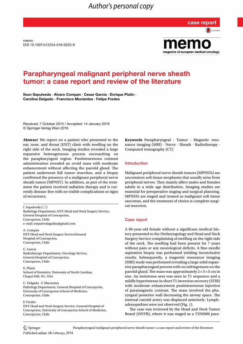
A 60-year-old female without a significant medical his-tory presented to the Otolaryngology and Head and Neck Surgery Service complaining of swelling on the right side of the neck. The swelling had been present for 7 years without pain or any neurological deficits. A fine-needle aspiration biopsy was performed yielding inconclusive results. Subsequently, a magnetic resonance imaging (MRI) study was performed revealing a large solid expan-sive parapharyngeal process with no infringement on the parotid gland. The mass was approximately 2 × 5 × 5 cm in size. An isointense area was seen in T1 sequence and a mildly hyperintense in short T1 inversion recovery (STIR) with moderate enhancement postintravenous injection of paramagnetic contrast. The mass involved the pha-ryngeal posterior wall decreasing the airway space. The internal carotid artery was displaced anteriorly. Lymph-adenopathies were not observed (Fig. 1).
The case was reviewed by the Head and Neck Tumor Board (HNTB), where it was staged as a T3N0M0 para-
I. Sepulveda ()Radiology Department; ENT-Head and Neck Surgery Service, General Hospital of Concepcion,Concepcion, Chilee-mail: [email protected]
A. CompanENT-Head and Neck Surgery ServiceGeneral Hospital of Concepcion,Concepcion, Chile
C. GarciaRadiotherapy Department, Oncology Service, General Hospital of Concepcion,Concepcion, Chile
E. PlatinSchool of Dentistry, University of North Carolina,Chapel Hill, NC, USA
C. Delgado · F. MucientesPathology Department, General Hospital of Concepción, University of Concepcion School of Medicine,Concepcion, Chile
F. FredesENT-Head and Neck Surgery Service, General Hospital of Concepcion, University of Concepcion School of Medicine,Concepcion, Chile
Received: 7 October 2015 / Accepted: 14 January 2016© Springer-Verlag Wien 2016
memoDOI 10.1007/s12254-016-0253-9
Parapharyngeal malignant peripheral nerve sheath tumor: a case report and review of the literature
Ilson Sepulveda · Alvaro Compan · Cesar Garcia · Enrique Platin · Carolina Delgado · Francisco Mucientes · Felipe Fredes
Author's personal copy
case report
2 Parapharyngeal malignant peripheral nerve sheath tumor: a case report and review of the literature 1 3
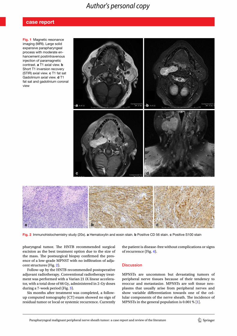
pharyngeal tumor. The HNTB recommended surgical excision as the best treatment option due to the size of the mass. The postsurgical biopsy confirmed the pres-ence of a low-grade MPNST with no infiltration of adja-cent structures (Fig. 2).
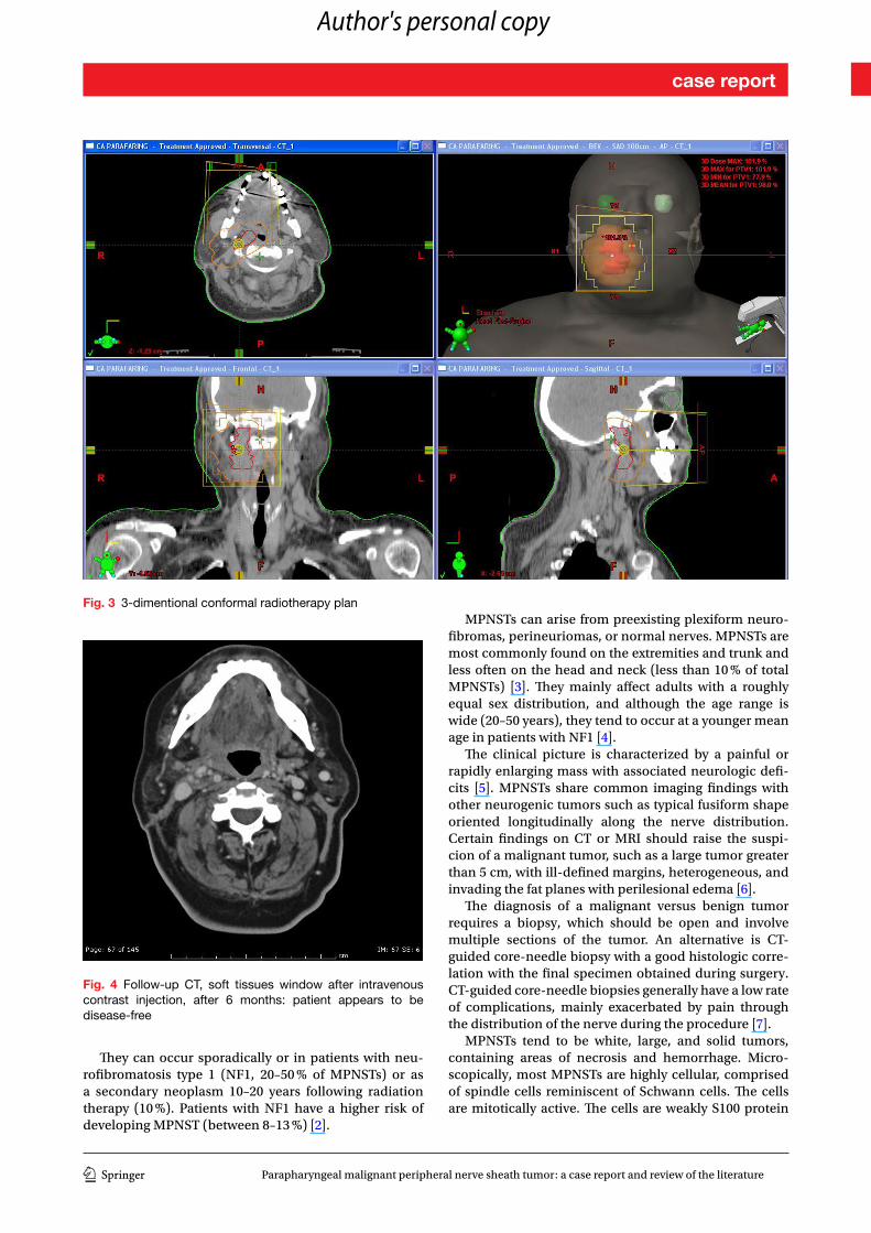
Follow-up by the HNTB recommended postoperative adjuvant radiotherapy. Conventional radiotherapy treat-ment was performed with a Varian 21 iX linear accelera-tor, with a total dose of 66 Gy, administered in 2-Gy doses during a 7-week period (Fig. 3).
Six months after treatment was completed, a follow-up computed tomography (CT) exam showed no sign of residual tumor or local or systemic recurrence. Currently
the patient is disease-free without complications or signs of recurrence (Fig. 4).
Discussion
MPNSTs are uncommon but devastating tumors of peripheral nerve tissues because of their tendency to reoccur and metastasize. MPNSTs are soft tissue neo-plasms that usually arise from peripheral nerves and show variable differentiation towards one of the cel-lular components of the nerve sheath. The incidence of MPNSTs in the general population is 0.001 % [1].
Fig. 2 Immunohistochemistry study (20x). a Hematoxylin and eosin stain. b Positive CD 56 stain. c Positive S100 stain
Fig. 1 Magnetic resonance imaging (MRI). Large solid expansive parapharyngeal process with moderate en-hancement postintravenous injection of paramagnetic contrast. a T1 axial view. b Short T1 inversion recovery (STIR) axial view. c T1 fat sat Gadolinium axial view. d T1 fat sat and gadolinium coronal view
Author's personal copy
case report
Parapharyngeal malignant peripheral nerve sheath tumor: a case report and review of the literature 31 3
MPNSTs can arise from preexisting plexiform neuro-fibromas, perineuriomas, or normal nerves. MPNSTs are most commonly found on the extremities and trunk and less often on the head and neck (less than 10 % of total MPNSTs) [3]. They mainly affect adults with a roughly equal sex distribution, and although the age range is wide (20–50 years), they tend to occur at a younger mean age in patients with NF1 [4].
The clinical picture is characterized by a painful or rapidly enlarging mass with associated neurologic defi-cits [5]. MPNSTs share common imaging findings with other neurogenic tumors such as typical fusiform shape oriented longitudinally along the nerve distribution. Certain findings on CT or MRI should raise the suspi-cion of a malignant tumor, such as a large tumor greater than 5 cm, with ill-defined margins, heterogeneous, and invading the fat planes with perilesional edema [6].
The diagnosis of a malignant versus benign tumor requires a biopsy, which should be open and involve multiple sections of the tumor. An alternative is CT-guided core-needle biopsy with a good histologic corre-lation with the final specimen obtained during surgery. CT-guided core-needle biopsies generally have a low rate of complications, mainly exacerbated by pain through the distribution of the nerve during the procedure [7].
MPNSTs tend to be white, large, and solid tumors, containing areas of necrosis and hemorrhage. Micro-scopically, most MPNSTs are highly cellular, comprised of spindle cells reminiscent of Schwann cells. The cells are mitotically active. The cells are weakly S100 protein
They can occur sporadically or in patients with neu-rofibromatosis type 1 (NF1, 20–50 % of MPNSTs) or as a secondary neoplasm 10–20 years following radiation therapy (10 %). Patients with NF1 have a higher risk of developing MPNST (between 8–13 %) [2].
Fig. 4 Follow-up CT, soft tissues window after intravenous contrast injection, after 6 months: patient appears to be disease-free
Fig. 3 3-dimentional conformal radiotherapy plan
Author's personal copy
case report
4 Parapharyngeal malignant peripheral nerve sheath tumor: a case report and review of the literature 1 3
Compliance with ethical standards
Conflict of interest I. Sepúlveda, A. Compan, C. García, E. Platin, C. Del-gado, F. Mucientes, and F. Fredes declare that there are no actual or potential conflicts of interest in relation to this this article.
References
1. Scheithauer BW, Louis DN, Hunter S, et al. Malignant peripheral nerve sheath tumor. In: Louis DN, Ohgaki H, Wiestler OD, Cavenee WK, editors. WHO classification of tumors of the central nervous system. Lyon: IARC Press; 2007. pp. 160–2.
2. Kim DH, Murovic JA, Tiel RL, Moes G, Kline DG. A series of 397 peripheral neural sheath tumors: 30-year experience at Louisiana State University Health Sciences Center. J Neuro-surg. 2005;102(2):246.
3. Touil H, Briki S, Karray F, Bahri I. Malignant peripheral nerve sheath tumor of the superficial cervical plexus with parotid extension. Eur Ann Otorhinolaryngol Head Neck Dis. 2015;132(2):93–5.
4. Thway K, Fisher C. Malignant peripheral nerve sheath tumor: pathology and genetics. Ann Diagn Pathol. 2014;18:109–16.
5. Baehring JM, Betensky RA, Batchelor TT. Malignant peripheral nerve sheath tumor: the clinical spectrum and outcome of treatment. Neurology. 2003;61(5):696.
6. Pilavaki M, Chourmouzi D, Kiziridou A, Skordalaki A, Zarampoukas T, Drevelengas A. Imaging of peripheral nerve sheath tumors with pathologic correlation. Eur J Radiol. 2004;52:229–39.
7. Pianta M, Chock E, Schlicht S, McCombe D. Accuracy and complications of CT-guided core needle biopsy of periph-eral nerve sheath tumors. Skeletal Radiol. 2015;44:1341–9.
8. Locketz G, Horowitz G, Abu-Ghanem S, Wasserzug O, Abergel A, Yehuda M, Fliss D. Histopathologic classifica-tion of parapharyngeal space tumors: a case series and review of the literature. Eur Arch Otorhinolaryngol. 2015. [Epub ahead of print].
9. Stark A, Mehdorn M. Leptomeningeal metastasis of an intradural malignant peripheral nerve sheath tumor. J Clin Neurosci. 2013;20:1181–3.
positive, consistent with dedifferentiation from Schwann cells [4, 8].
MPNSTs are staged and treated as malignant soft tis-sue sarcomas. Because of their rarity and the frequent need for multimodal treatment, the evaluation and management of MPNSTs ideally should be carried out in a facility with expertise in the treatment of sarcomas, including surgical, orthopedic, medical, and radiation oncology.
The treatment of choice is complete surgical resection [2, 5]. The role of adjuvant therapies such as radiotherapy and chemotherapy are not clear. Histologically, MPNSTs are radioresistant and chemoresistant. Nevertheless, post-surgery radiotherapy has been reported to provide some benefit in high-grade, large, or deep MPNSTs or in case of margin invasion [3, 5].
Even with aggressive surgical and radiation treat-ment, the prognosis is poor. Poor prognostic signs include tumors exceeding 5 cm in size, high tumor grade, association with NF1, older age, distant metas-tases at the time of diagnosis, and inability to achieve tumor-free margins. The rate of distant metastases is round 40 %. Metastases have been reported all over the body. Common sites are the lungs, liver, bone, and sub-cutaneous tissue. Less common sites are the endocrine organs, heart, and brain. The 5-year survival ranged from 34–64 % [9].
Conclusion
MPNSTs are rare tumors that mostly affect patients with NF1. They also occur sporadically or in patients who have undergone radiation therapy. The clinical presentation consists of a painfully rapidly enlarging mass accompa-nied by neurological deficits mainly in the extremities, trunk, head, and neck. A biopsy is required for a defini-tive diagnosis, and surgery is the treatment of choice. The role of adjuvant treatments is not well established, and the prognosis is poor with a 5-year survival rate ranging from 34–64 %.
Author's personal copy
View publication statsView publication stats