Embed Size (px)
Citation preview

--
Q Copyright 1989 by the American Chemical Society Volume 28, Number 7
Perspectives in Biochemistry
April 4, 1989
Parameters for the Molecular Recognition of Transfer RNAs? Paul Schimmel
Department of Biology, Massachusetts Institute of Technology, Cambridge, Massachusetts 021 39 Received January 9, 1989; Revised Manuscript Received February 3, 1989
T a n s f e r RNAs are highly differentiated nucleic acids com- prised of 74-93 nucleotides that are folded into a compact three-dimensional pattern which is believed to accommodate most of the known tRNA sequences (Sprinzl, 1987). The molecules are differentiated from each other according to their amino acid acceptance. This is determined in the two-step aminoacylation reaction whereby an amino acid is activated by its cognate aminoacyl-tRNA synthetase, which catalyzes formation of a tightly bound aminoacyl adenylate; this complex than reacts with the 2’- or 3’-hydroxyl of a tRNA to form the aminoacylated species. After this, aminoacyl-tRNAs react with components of the translation apparatus that recognize features common to all tRNAs and enable amino acids to be inserted into growing polypeptide chains through the precise base-pairing interaction of trinucleotide anticodons (within tRNAs) with codons in messenger RNAs. Thus, the trans- lation of mRNAs into polypeptides of defined sequences is a manifestation of the genetic code, but the code itself is based on the molecular recognition of transfer RNAs by amino- acyl-tRNA synthetases. This system of protein-nucleic acid recognition connects each amino acid with a trinucleotide sequence (anticodon) within the tRNA.
Because of degeneracy in the genetic code, whereby up to six trinucleotide sequences may code for a specific amino acid, there can be several tRNAs that are specific for a given amino acid. An example is serine, which in Escherichia coli has at least five distinct tRNA species that collectively recognize the six different codons for that amino acid (Sprinzl et al., 1987). These tRNAs differ not only in their anticodons but also in other parts of their respective sequences. These tRNA iso- acceptors are generally recognized by the one aminoacyl-tRNA synthetase which is specific for that amino acid, however (Schimmel & Soll, 1979). This implies that, for at least some of the enzymes, the anticodon is not the primary determinant for recognition.
Unlike tRNAs that have a common structural motif and
‘Support of research on aminoacyl-tRNA synthetases and recognition of transfer RNAs through National Institutes of Health Grants GM15539 and GM23562 is acknowledged.
typically vary in length by not more than 20%, the amino- acyl-tRNA synthetases are diverse proteins with four different types of quaternary structures (a, a2, CY&&, and cq) and subunit sizes that range from 334 to over 1000 amino acids (Schimmel, 1987). Although leucine, valine, and isoleucine tRNA synthetases have some sequence similarities (Heck & Hatfield, 1988), several of the enzymes have sequences which are not similar to that of any other synthetase. The enzymes arose early in evolution, probably appearing with the earliest life forms, and may never have been constrained to interact with elements of the protein synthesis apparatus other than tRNAs. In some cases, they have acquired additional biological functions. These considerations may explain in part the ap- parent uniqueness of the sequences and quaternary structures of many of the enzymes. However, similar structural motifs can in principle be assembled from diverse sequences, and there is evidence for a structural relatedness of portions of methi- onine and tyrosine tRNA synthetases, which have little se- quence similarity (Blow et al., 1983).
The basis for the recognition of t R N A by aminoacyl-tRNA synthetases has been difficult to solve [recent commentaries and summaries include de Duve (1988), Schulman and Abelson (1988), RajBhandary (1988), and Yarus (1988)l. There are significant differences from the system of recognition of DNA sequence elements by gene regulatory proteins. For protein-DNA complexes, the dissociation constant can be as small as lo-’* M, so that the complexes have long lifetimes (Pabo & Sauer, 1984). For synthetasetRNA complexes, the dissociation constants at pH 7.5 are on the order of 10” M, which is comparable to the dissociation constant or Michaelis constant for an enzyme-small molecule complex (Schimmel & Soll, 1979). This means that the enzyme-tRNA complexes have short lifetimes, which facilites rapid turnover. The relatively high enzyme-tRNA dissociation constant limits the degree to which discrimination can be achieved at the binding step. There is a second step in which specificity can be manifested, however. Because tRNA is a substrate for the enzyme, there can be discrimination during the transition state of aminoacylation, which is expressed through the catalytic parameter kat. Early work suggested that this played a role
0006-2960/89/0428-2747$01.50/0 0 1989 American Chemical Society

2748 Biochemistry, Vol. 28, No. 7, 1989 Perspectives in Biochemistry
in the overall recognition process (Schimmel & Soll, 1979). The tRNA molecule is folded, like many proteins, into a
globular structure where some elements which are dispersed in the sequence are brought into close proximity (Kim et al., 1974; Robertus et al., 1974). Dissection of tRNAs into smaller pieces, as has been done in successfully identifying recognition sites in linear DNA molecules, sacrifices many of the structural features that only are present in the intact tRNA. The rel- atively short lifetimes of the synthetase-tRNA complexes make it difficult to obtain, from a nuclease digestion, a nu- clease-resistant protein-nucleic acid complex-an approach that has also been successfully applied with the more stable DNA-protein complexes. Early work defined sites on tRNAs that are in close contact with bound synthetases [e.g., by using cross-linking among other approaches (Schimmel, 1977)] and investigated the basis for the mischarging of certain tRNAs when reacted with synthetases from another, heterologous organism [summarized in Schimmel and Soll (1979)l. The early application of an in vivo amber suppression assay also afforded an opportunity to explore determinants important for the recognition of tRNAs, although definitive conclusions were not reached at that time and the complexity of the recognition problem was highlighted (see below).
The recent availability of procedures for mutagenesis and expression of tRNAs in vivo, and for synthesis of tRNAs and variants in vitro, has stimulated a new generation of experi- ments. While much of the recent work has utilized in vivo amber suppression assays to study molecular recognition, the interpretation of some of these experiments is limited when there are no in vitro data. At the same time, in vitro exper- iments have their own limitations which can only be overcome by investigation of variant tRNAs in the context of all of the synthetases and tRNAs in vivo. Summarized below are recent experiments on the molecular recognition of transfer RNAs, with some consideration of binding and catalytic parameters that enable tRNAs to be distinguished from each other.
LOCATING DETERMINANTS FOR IDENTITY Amber Suppression Assay. Early experiments utilized the
E . coli supF amber suppressor, which is based on the sequence of a tyrosine tRNA with the anticodon altered from GUA to CUA so that the UAG amber codon is recognized [summa- rized by Ozeki et al. (1 980)]. This suppressor inserts tyrosine at amber codons, which demonstrates that the first position of the anticodon is not an essential determinant for the identity of a tyrosine tRNA. Mutants of supF that insert glutamine were isolated. The interpretation was not straightforward because some of the mutations that resulted in aminoacylation with glutamine did not create nucleotides that are in the analogous location of any E . coli tRNAGh species (Figure la). Subsequently, it was demonstrated that simple alteration of the anticodon of tRNATrp from CCA to CUA created an amber suppressor that inserts glutamine (Figure lb). These results demonstrated that amino acid acceptance could be manipulated by simple mutations. One interpretation was that the mutations disturbed the interaction with the cognate en- zyme and that glutamine tRNA synthetase had a relaxed specificity which was manifested on tRNA substrates that were less competitively aminoacylated by their cognate enzyme.
Conversion of a Leucine into a Serine Transfer RNA. Although difficulties of interpretation raised concerns about the early work on amber suppressors with altered amino acid acceptance capability, the approach has clear advantages. By restriction of amino acid substitutions of mutant suppressor tRNAs to amber codons, the possibility is removed for de- leterious substitutions in vivo in the translated regions of
a A r
A+G A e G C 4 A . U A 4 - G C + U
'A C G A G G A A G G
G CCCu cu Tyc G G ~ ~ ~ c G U c A 'c AA CG U
G C u"cc u ucc
&! 'AU$
CAyA
A C C G
b
1 A U G C G C 7 0 G C G C C G 6 0
G C U G , u c u c u c '4
G 5 8 G A G 1 0 A T Y C
D A A C U U G G A G C G u G A A C G U G
G D C G 2 0 G C
U A C A U A
3 0 G C 4 0
C C A
c U
FIGURE 1: (a) Mutations in E. coli tRNATFICUA that cause insertion of glutamine at amber codons. The nucleotide changes are concen- trated in the acceptor helix and in some cases do not recreate sequences found in any E. coli tRNAG'". The upper diagram shows the locations of single nucleotide substitutions that enable tRNATyr/CUA to be aminoacylated with glutamine, while the lower two diagrams show, separately, individual mutants that have two base changes. Data are summarized from Smith et al. (1970), Hooper et al. (1972), Shimura et al. (1972), Smith and Celis (1973), Celis et al. (1973), Ghysen and Celis (1974), Inokuchi et al. (1974), and Ozeki et al. (1980). (b) A single nucleotide substitution in E. coli tRNATV enables it to insert glutamine at amber codons. The substitution creates an amber CUA anticodon (Yaniv et al., 1974). See also Sol1 (1974), Celis et al. (1976), and Yarus et al. (1977).
mRNAs for cellular proteins. The approach is limited to those tRNAs whose amino acid acceptance is not altered by change of the anticodon to CUA. One example is serine tRNA, where there are six known isoacceptors (five encoded by the E. coli genome and one by phage T4) that can be aminoacylated by the E. coli serine tRNA synthetase and that have anticodons which include UGA, CGA, GGA, and GCU. These antico- dons collectively vary all three positions, and furthermore, a

Perspectives in Biochemistry
change to CUA does not change the identity of this tRNA. Normanly et al. (1986a) examined the 6 serine tRNAs and
concluded that, apart from the bases that are conserved in all tRNAs, only 12 were held in common. These 12 nucleotides, which are located in the acceptor stem and dihydrouridine stem and loop, were transferred into a leucine tRNA isoacceptor (with a CUA amber anticodon). The resulting tRNA sup- presses a serine-requiring amber allele at codon 68 of @-lac- tamase. The suppression efficiency of the strong parent leucine amber suppressor is 6076, and the transformed species with 12 substitutions has an efficiency of 0.5-1%. Direct infor- mation on the nature of the amino acid(s) inserted by the transformed tRNALcU/CUA was obtained by protein sequence analysis of a suppressed dihydrofolate reductase gene which has an amber mutation at codon 10. This showed that serine was inserted, with a possibility of, but little evidence for, minor amounts of valine and/or leucine. No glutamine was detected at the position corresponding to the amber codon. Thus, the transformation of a strong leucine-inserting suppressor into a weak serine-inserting suppressor was achieved. Further work has focused on defining the minimal subset of nucleotides from among the 12 defined in these experiments, which are sufficient to confer serine acceptance, and determining whether that subset will function in the context of other tRNA sequences (Schulman & Abelson, 1988).
A Single Base Pair Is a Major Determinant of the Identity of an Alanine tRNA. Alanine tRNA synthetase has been extensively studied by biochemical and genetic approaches, and the segment important for tRNA binding has been del- ineated (Schimmel, 1987, 1989). In order to define further the amino acids that are critical for tRNA recognition, a large population of tRNAAIa variants were created by site-directed mutagenesis and tested for recognition by alanine tRNA synthetase (Hou & Schimmel, 1988). Those that are defective in recognition were to be used to isolate second-site revertants in the enzyme which compensate for the defect in the tRNA. In the course of this work it was discovered that a single base pair is a major determinant of the identity of tRNAAIa.
A population of 28 mutant tRNAs were created that col- lectively varied over half of the nonconserved nucleotides. A tRNAA'a/CUA amber suppressor was used for these experi- ments. The sequence of this tRNA is based on that of tRNAAh/GGC [Mims et al., 1985; cf. Normanly et al. (1986b) and Masson and Miller (1986)l. This suppressor inserts alanine, even though all three anticodon nucleotides have been changed. The mutant tRNAs, which in some cases had as many as five substitutions within a single species, were first introduced on a multicopy plasmid and checked for suppression of a trpA(UAG2.34) amber allele. There is evidence that this allele is suppressed by insertion of glycine or alanine but not by other amino acids (Murgola & Hijazi, 1983). Any mutant that did not suppress the trpA(UAG234) allele was then checked to determine whether a stable tRNA was made. Those examples of a sup- phenotype for which the overpro- duced tRNA was not evident were not considered further. Of particular interest are those sup- species that are not defective in tRNA biosynthesis.
This screen and analysis yielded only one site in the entire molecule where mutations resulted in a tRNA which has a sup- phenotype on the trpA(UAG2.34) amber allele and which is clearly synthesized as a stable tRNA. This involved re- placements of the G3eU70 base pair in the amino acid acceptor helix with A3eU70 or G3C70 (Figure 2). In vitro amino- acylation measurements confirmed that the mutant tRNAs were not aminoacylated with catalytic amounts of purified
Biochemistry, Vol. 28, No. 7, 1989 2749
alanine tRNA synthetase (Hou & Schimmel, 1988). The G3mU70 base pair was introduced into tRNACyS/CUA
and tRNAPhe/CUA. Each of these tRNAs has a C3-G70 base pair and differs by 38 (tRNACp/CUA) and 31 (tRNAPhc/CUA) nucleotides from tRNAMaICUA. Introduction of the G3eU70 base pair into each of these tRNAs confers the ability to accept alanine in vivo. In the case of G34J70 tRNACJ's/CUA, only alanine was detected at the position of the suppressed amber codon in dihydrofolate reductase (Hou & Schimmel, 1988). This raises the possibility that the substitution at position 3.70 has perturbed a determinant for the cysteine tRNA synthetase in addition to conferring a determinant for alanine tRNA synthetase. Aminoacylation of G3eU70 tRNACyS/CUA with purified alanine tRNA synthetase was demonstrated. The G34J70 tRNAPhciCUA species is aminoacylated with both alanine and phenylalanine in vivo. This suggests that the determinants for the identity of phenylalanine tRNA are lo- cated, at least in part, elsewhere.
The results indicate that the G3sU70 base pair is a major determinant for the identity of an alanine tRNA. The result of introducing a G3sU70 base pair into tRNAPhc was con- firmed by McClain and Foss (1988a). These authors also did experiments with a variant of a glycine tRNA (which largely is aminoacylated with glutamine in vivo). Introduction of the G3LJ70 base pair into this tRNA does not confer acceptance of alanine. This may mean that, at least in the context of the variant sequence, the glutamine enzyme competes more ef- fectively than the alanine enzyme and, additionally or alter- natively, there are negative determinants in the variant tRNA which block interaction with the alanine enzyme (see Con- cluding Remarks).
A mutant lysine missense suppressor that inserts glycine and/or alanine at lysine codons has been reported (Prather et al., 1984). This suppressor also is believed to insert lysine. The mutation creates a G3sU70 base pair, and while the in- sertion of alanine was not established, Prather et al. (1984) suggested that this was likely. The aforementioned results and recent experiments with a lysine tRNA amber suppressor (McClain et al., 1988) support this possibility.
Among the published E. coli tRNA sequences the G3LJ70 base pair is unique to alanine. Further mutational analysis may uncover additional nucleotides that affect recognition by the alanine enzyme. If such nucleotides are uncovered, then it will be important to determine whether transfer of any of them into another tRNA framework will confer identity for alanine.
Role of the Anticodon in the Recognition of Methionine and Valine tRNAs. In vitro aminoacylation experiments have indicated that, for several tRNAs, single base changes in the anticodon affect the rate of aminoacylation. The availability of T7 RNA polymerase and synthetic DNA templates for transcription has made possible the preparation of synthetic tRNAs of any sequence whatsoever (Sampson & Uhlenbeck, 1988). This has afforded an opportunity to make substrates with varied anticodon sequences and to evaluate quantitatively the effect on aminoacylation.
The CAU anticodon of E . coli methionine tRNA is known to be important for in vitro aminoacylation with E. coli me- thionine tRNA synthetase (Schulman & Pelka, 1983, 1988). (This enzyme also aminoacylates initiator and elongator me- thionine tRNAs from other prokaryotes and from eukaryote organelles, all of which have the CAU anticodon.) At con- centrations of tRNA that are below the Michaelis constant K,, the initial rate of aminoacylation is given by (kw,/ K,)(E),(tRNA),, where (E), and (tRNA), are total enzyme

2750 Biochemistry. Vol. 28, No. 7, 1989
A C C A
1 G C
Perspectives in Biochemistry
k
C G A C U d D G A G G A
G G z r
G C G u 'O G C C G U A
U o A G C
G C
A U
U C G A G C c 5 0
G G A "G U A U A
C G A A U A
30 G C 40
C U A
E. coli t RNA
60
FIGURE 2: (a, left) Nucleotide sequence and cloverleaf structure of tRNAALajCUA. The sequence is based on tRNAALa/- (Mims et al., 1985) but where the GGC anticodon has been changed to CUA and a U38 - A substitution has been introduced to improve the efficiency of amber suppression (Raftery & Yarus, 1987). The G34J70 base pair is highlighted. This base pair is a major determinant of the identity of an alanine tRNA (Hou & Schimmel, 1988). (b, right) Depiction of the three-dimensional structure of tRNAhlCUA with the G3U70 base pair highlighted in white. The sequence of was built into the known coordinates of yeast tRNAWe (obtained from Brookhaven Data Bank) utilizing the PS300 FRODO program (Bush et al., 1987).
and tRNA concentrations, respectively, and kcat is the turnover number (unimolecular rate constant). The parameter kat/&, has units of a second-order rate constant. Table Ia shows that the relative kat/& is reduced 4-5 orders of magnitude by substitutions at the first position of the anticodon (Schulman & Pelka, 1988). The large effect of substitutions at the first position demonstrates its importance, but it alone is not suf- ficient for recognition by the methionine enzyme, however. None of the amber suppressors (CUA anticodon) and natural tRNAs with a first position C are known to insert methionine in vivo.
An even larger effect is found when the anticodon is reversed from CAU to UAC. The UAC anticodon corresponds to that for a valine tRNA. Replacement of that anticodon in tRNA?' with CAU confers methionine acceptance on tRNAy1ICAU (Table Ia). The efficiency of aminoacylation is quantitatively close to that of tRNAMet. In a reciprocal experiment, the CAU anticodon of elongator tRNA,M"' was replaced by UAC, and aminoacylation with valine tRNA synthetase was attempted. In this case the anticodon re- placement converts the hybrid tRNAMetIuAC into a substrate for valine tRNA synthetase, with a relative k,,/Km that is 10-fold less than that of tRNAVa1 (Table Ib). Thus the CUA - UAC substitution simultaneously eliminates aminoacylation with the methionine enzyme while conferring acceptance of valine. The tRNAMetIUAC and tRNAVdICAU species have been checked for aminoacylation in vitro with glutamic, glutamine, lysine, isoleucine, and phenylalanine tRNA synthetases. These
Table I: Relative Values of k,,/K, for Aminoacylation with Methionine and Valine of Anticodon Sequence Variants of tRNA,MC' and tRNAVa'
(a) Aminoacylation with Methionineu natural tRNAMet 1 .o synthetic tRNAMCt (CAU) 0.5 synthetic tRNAMC' (UAU) 0.000 1 synthetic tRNAMCt (GAU) 0.00001 synthetic tRNAMc' (UAC) 0.0000001 synthetic tRNAVa1 (CAU) 0.8
(b) Aminoacylation with Valineb natural tRNAVa' 1 .o synthetic tRNAV*' (UAC) 0.4 synthetic tRNAMCt (CAU) 0.000002 synthetic tRNAMC' (UAU) 0.00001 svnthetic tRNAMCt WAC) 0.04
a Data were obtained with purified E. coli methionine tRNA synthetase at pH 7.5, 37 OC, and are taken from Shulman and Pelka (1988). "Data were obtained with purified -E. coli valine tRNA synthetase at pH 7.5, 37 "C, and are taken from Schulman and Pelka (1988).
enzymes aminoacylate neither tRNAMetjUAC nor tRNAVd/CAU (Schulman & Pelka, 1988).
The data presented in Table I suggest that the anticodon has an important role in defining the identity of valine and methionine tRNAs and that the quantitative effects of simple nucleotide substitutions are large. In order to evaluate the role of the anticodon more fully, and to evaluate the significance

Perspectives in Biochemistry
of the anticodon nucleotide replacements in the presence of all of the enzymes and tRNAs, experiments are being at- tempted in vivo. This necessitates special techniques because the amber suppression system (which requires a CAU anti- codon) obviously cannot be used in these circumstances.
Recognition of E. coli Glutamine tRNA. The anticodon (CUG and UUG) has been implicated as a recognition site for glutamine tRNA synthetase. This apparently explains why certain amber suppressor tRNAs (CUA anticodon) mischarge at least partially with glutamine. The central U of the an- ticodon is believed to have an important role. Rogers and Soll (1 988) have manipulated a serine-inserting tRNASeT/CUA am- ber suppressor to examine one way to achieve discrimination of glutamine from serine tRNA synthetase. Because of the CUA anticodon, this suppressor potentially could be converted to a glutamine tRNA. A similar example had been previously demonstrated by isolation of mutations in the acceptor helix of a supF tRNATyr/CUA (Figure la).
The three base pairs at the end of the acceptor helix are implicated in recognition by the serine tRNA synthetase (Normanly et al., 1986a). The sequence at the beginning of the acceptor helix of tRNA? is Gl-C72:G2.C7 1:A3-U70. This was changed to Ul.A72:G2.C7 l:G3.C70, where the al- tered bases are indicated in bold type. These substitutions recreate the sequence of the first three base pairs of tRNA?'". While tRNASer/CUA inserts serine, the Ul.A72:G3-C70
inserts over 90% glutamine and about 5% serine in vivo (Rogers & Soll, 1988). One interpretation of this result is that substitution of four nucleotides has disrupted the in- teraction with the serine tRNA synthetase, thus making possible a more efficient competition by the glutamine enzyme. Additionally or alternatively, the substitutions have improved the interaction with glutamine tRNA synthetase. Analytical aminoacylation measurements would clarify this question, and further substitution and analysis will have to be done in order to define the determinants for the identity of a glutamine tRNA.
Determinants for Recognition of Yeast Phenylalanine tRNA. An in vitro analysis has been used to evaluate nu- cleotides important for recognition of Saccharomyces cere- uisiae tRNAPhe by the homologous phenylalanine tRNA synthetase. Initial experiments established that replacements of any of the three anticodon nucleotides decreased k,,/Km by a factor of 3-10-fold (Bruce & Uhlenbeck, 1982). Ad- ditional experiments showed that replacement of G20 (in the dihydrouridine loop) or of A73 (in the single-stranded ACCAoH 3' terminus) each resulted in a 12-fold reduction in catalytic efficiency [see Sampson and Uhlenbeck (1988, 1989)l. Because G20 is present in tRNAPhc but not in any of the other reported yeast tRNA sequences, it could act as an important discriminatory nucleotide.
E. coli tRNAPhe encodes four of the five aforementioned nucleotides, with a U replacing G20. Substitution of G for U20 improves k,,/K, for the yeast enzyme by 12-fold so that the yeast and E. coli substrates are almost equivalent (Sampson & Uhlenbeck, 1989) (Table 11). This corresponds to a relatively small amount (1.5 kcal mol-') of free energy.
Yeast tRNAh*, tRNAM", and tRNATy were reconstructed so that each contained a complete set of the five important nucleotides (Sampson & Uhlenbeck, 1989). Each is converted to a substrate for the homologous yeast phenylalanine tRNA synthetase (Table 11). The parameter k,,/K, for each of the substituted tRNAs is within 50% of that for the cognate yeast tRNAPhc. It is not known whether the nucleotide substitutions that were introduced in each case have an effect on the re-
Biochemistry, Vol. 28, No. 7, 1989 2751
Table 11: Relative Values of k,,/K,,, for Aminoacylation with Phenylalanine of Synthetic tRNAs with Different Subtitutions
starting sequence substitutions re1 k,,/K,,,'
yeast tRNAPhe u20 0.08 E . coli tRNAPhe none 0.04 E . coli tRNAPhe G20 0.5 yeast tRNAMet severalb 0.7 yeast tRNAAr* severalb 0.6
yeast tRNAPhe none 1 .o
yeast tRNAT"' severalb 1.5 OData were obtained with yeast phenylalanine tRNA synthetase at
pH 7.45 and are taken from Samson et al. (1989). bSubstitutions were introduced into the "starting sequence" so as to have G20, G34, A35, A36, and A73, which are the nucleotides believed important for the recognition of yeast tRNAphe.
spective cognate enzyme. This information would clarify whether the changes that improve each as a substrate for the phenylalanine enzyme are sufficient to convert the amino acid specificity to phenylalanine only or whether the resultant tRNAs are charged with phenylalanine and one or more ad- ditional amino acids. Analytical aminoacylation experiments with other synthetases should make possible a quantitative evaluation of the various competitive effects. Eventually, each mutant tRNA (which charges in vitro with phenylalanine) should be investigated in the context of all of the synthetases and tRNAs in vivo.
On the basis of an in vivo amber suppression assay, McClain and Foss (1988b) have indicated that more than 5 nucleotides (Le., 10) are important for the recognition of E. coli tRNAPhe by E. coli phenylalanine tRNA synthetase. Only two of these (positions 20 and 73) are at positions corresponding to the locations of the five sites studied by Sampson and Uhlenbeck (1989). The relative significance of each of these is unknown. It will be necessary to perform in vitro aminoacylation mea- surements similar to those done with the yeast enzyme in order to understand more fully the similarities and differences in the recognition by these two enzymes which are specific for the same amino acid.
Modvied Bases. Examples Where They Do and Do Not Have an Important Role. Transfer RNAs isolated from natural sources contain several bases at specific locations that are posttranscriptionally modified. Some of these are common to most tRNAs (such as 7-methylguanosine, dihydrouridine, and pseudouridine), and others are unique to a particular amino acid specific tRNA [such as the wybutosine at position 37 of certain eukaryote phenylalanine tRNAs (Sprinzl et al., 1987)]. In the aforementioned studies of E. coli tRNAMd and tRNAVa1 and of yeast tRNAPhe, the tRNA substrates were enzymatically synthesized in vitro. As a consequence, these substrates contain no modified bases. When these synthetic substrates were compared with their counterparts as isolated from natural sources (which thus contain the full complement of modified bases), only small differences in aminoacylation kinetics were observed (Sampson & Uhlenbeck, 1988; Schulman & Pelka, 1988). Thus, these are among the ex- amples [see also Samuelsson et al. (1 988) and Francklyn and Schimmel (1989)l where the modified bases do not play a major role in recognition by the cognate enzyme in vitro, although they could act to interfere with the interactions of noncognate enzymes.
In contrast, a modification of an isoacceptor of tRNA1IC is essential for recognition by isoleucine tRNA synthetase. In addition to promoting aminoacylation with isoleucine, the modification blocks misacylation by methionine tRNA synthetase. The tRNAp isoacceptor reads AUA codons. The gene for this isoacceptor encodes a CAT anticodon. Thus,

2752 Biochemistry, Vol. 28, No. 7, 1989 Perspectives in Biochemistry
A
C C A
1 G-C
G- C
G-U 7 0 G-C
P
C-G
A C C A
1 G-C
G- C
G-U 70 G-C
C-G
P
U 60 U-A
U A-U 60
U-A A-U
l o u G C G C C C A
I I I I I c G A C U C G A l o u y - y c A G C G A C U C G A
T T C U G C G G c 5 0
G 1 1 1 1 T T C G G A ~ ~ ~ ~ G
G I I I I F.,s G G
U U G G A ~ ~ ~ ~ G
20 C-GA GG 20 C-GA GG
E-R C- G U-A
30 G-C 4 0
C- G
A
U-A 30 G-C 4 0
C- G
u c IJ A
G C
~ A A ~ ~ ~ I C U A ~RNAAWUGC
( 6 . 2 x l o 5 ~ - l s e c - l ) ( 4 . 5 x l o 5 M-lsec-')
FIGURE 3: Nucleotide sequences and cloverleaf structures of tRNAMajUGC and tRNAMaICUA. The nine nucleotide sequence differences between the natural species (tRNAMajUGC) and the amber suppressor (tRNAmICUA) used in studies of Hou and Schimmel(l988) are shown by shading. Analytical aminoacylation measurements with purified alanine tRNA synthetase have shown that at pH 7.5, 37 OC, there is little difference in the apparent rate (kat/Km) of aminoacylation (given in parentheses) of these two tRNA species. Adapted from Park et al. (1989).
without posttranscriptional modification the anticodon is CAU, which is the same as the anticodon for tRNAMet. As shown by Schulman and co-workers, this triplet is recognized by the methionine tRNA synthetase (see Table Ia). Muramatsu et al. (1988a) have found that tRNAp/CAU is a substrate in vitro for methionine tRNA synthetase. By comparison, however, it is a poor substrate for isoleucine tRNA synthetase. In the mature tRNA, C34 is posttranscriptionally modified to lysidine L (Muramatsu et al., 1988b). This modification consists of a lysine substituted for 0 - 2 of the cytidine ring and attached through the e-amino group directly to C-2 of the pyrimidine base. With this modification, tRNApILAU is efficiently am- inoacylated with isoleucine and cannot be aminoacylated with methionine. Thus, the amino acid acceptance in vitro switches according to the state of modification.
The same E. coli isoleucyl-tRNA synthetase aminoacylates the major isoacceptor, which has a GAU anticodon. Thus, if the anticodon is the primary site of recognition, then the enzyme recognizes a structural feature common to GAU and LAU. EFFECT OF A SINGLE BASE PAIR ON THE BINDING AND CATALYTIC PARAMETERS FOR THE MOLECULAR RECOGNITION OF ALANINE TRANSFER RNA
Comparison of Kinetic Behavior of an Alanine-Inserting Amber Suppressor with a Naturally Occurring Isoacceptor. The alanine system has been used to explore the effects on tRNA binding and catalytic parameters of single nucleotide substitutions, which alter the G-U base pair, and thereby obtain greater insight into the molecular basis for discrimination of a simple structural feature. The tRNAAlaICUA amber sup- pressor described above was based on the sequence of the
tRNAAlalGGC isoacceptor. The second known naturally oc- curring isoacceptor is tRNAhIUGC. There are nine nucleotide differences between the synthetic tRNAA1a/CUA amber sup- pressor and tRNAAIaIUGC. Eight of these are located in the anticodon stem and loop, and the ninth is a position 49.65 base pair in the TUC stem (Figure 3). The kinetic parameters for these two tRNAs at pH 7.5, 37 OC, are kcat = 1.0 s-l and K , = 2.2 pM for tRNAAlaIUGC and k,,, = 1.8 s-l and K , = 2.9 p M for tRNAA1a/CUA (Park et al., 1989). The small differences between the respective parameters may be ex- perimentally insignificant, and the apparent second-order rate constants kcat/K, are close in value (Figure 3).
The binding of the two tRNA species has been measured at pH 5.5, where the nitrocellulose filter assay has a high efficiency for retention of synthetase-tRNA complexes. [Above pH 6.0 the efficiency drops so that the method cannot be used to measure binding (Yarus & Berg, 1970). In general, association of tRNAs with aminoacyl-tRNA synthetases in- creases at lower pH values (Schimmel & Soll, 1979).] At pH 5.5, 23 OC, the dissociation constants for the enzyme-tRNA complexes are within experimental error of each other (Park et al., 1989). Thus, by kinetic and equilibrium measurements, the nine nucleotide differences between tRNAA1a/CUA and tRNAAlaIUGC have little or no effect on binding and catalytic parameters. This is consistent with genetic studies which show that substitutions in the anticodon loop and stem, and of the position 49.65 base pair in the TUC stem, do not interfere with acceptance of alanine (Hou & Schimmel, 1988). It is of interest to note that the anticodon of yeast tRNAAIa is also not required for recognition by the yeast enzyme (Jin et al., 1987).

Perspectives in Biochemistry
7 5
5 0
2 5
Biochemistry, Vol. 28, No. 7, 1989 2753
-
.-
--
Ala "wild type" tRNA cw
Suppressor+
I / C6G7C66G67 Suppressor+
A3, C70, U3G70 and C6G7C66G67C70
Suppressor -
0 5 1 0 1 5 20 2 5 3 0 3 5
Time(minutes) FIGURE 4: Substrate levels of alanine tRNA synthetase do not sig- nificantly aminoacylate A30U70, G3C70, or U30G70 tRNAh at pH 7.5,37 OC. For these experiments, the enzyme and tRNA concen- trations were 20 and 4 pM, respectively. The suppressor' or sup- pressor- designation indicates which tRNAs are able to suppress the trpA(UAG234) amber allele in vivo. Note also that the C6G7C66G67 tRNAA1a/CUA has four substitutions in the acceptor helix and these do not prevent aminoacylation with alanine. Adapted from Park et al. (1989).
Effect of Sequence Variants of the G34J70 Base Pair on Kinetic Parameters. The initial characterization of sequence variants of the G34J70 base pair showed that they were de- fective for aminoacylation in vitro, but it was not determined whether the defect is in kcat, K,, or both (Hou & Schimmel, 1988). In further experiments, it was shown that addition of excess A34J70 tRNAA1a/CUA does not inhibit aminoacylation of tRNA*ICUA at pH 7.5,37 "C. The experiments were done under conditions where an inhibition constant of less than 95 pM would have been detected. Because the K , for tRNAMaiCUA is 2.2 pM, and because of evidence that the K , can be regarded as an enzyme-tRNA dissociation constant, the result implies that at pH 7.5 (37 "C) the binding constant for the A34J70 variant is at least 40-fold weaker than that for the natural tRNAAIaIUGC isoacceptor (Park et al., 1989).
This is not the only effect of the A34J70 substitution, however. An excess of enzyme over tRNA substrate has been used to attempt aminoacylation of A34J70, G3C70, U30G70, and C6C7G66G67C70 tRNAA1a/CUA. (The latter species has five substitutions in the acceptor stem, which collectively change three base pairs, including the one at position 3.70.) Because of the excess of enzyme, it does not have to turn over in order to achieve complete aminoacylation. With 20 pM enzyme and 4 pM tRNA substrate, the "wild-type" tRNAAIaICUA and C6C7G66G67 tRNAA1a/CUA species are immediately aminoacylated, as expected. Even after a 30-min incubation, however, the position 3.70 variants are not ami- noacylated (Figure 4). If the position 3-70 variants could be aminoacylated but the kinetic defect caused an extremely low product release, then "one shot" of aminoacylation would have been detected at these high enzyme concentrations. Thus, the
defect is at a step prior to stable aminoacyl-tRNA formation. The failure to observe aminoacylation of position 3-70
variants after prolonged incubations with excess enzyme at pH 7.5 suggests that, in addition to reduced binding, there could be a severly reduced kat. This was demonstrated in the following way. At pH 5.5, the binding of synthetases to tRNAs is generally stronger and can be independently mea- sured by the nitrocellulose filter assay. Thus, experiments were attempted at pH 5.5 to determine whether, under these con- ditions, the binding of the A34J70 variant might be enhanced so that a complex could be demonstrated and tested for cat- alytic competence. The rate of aminoacylation of the wild-type tRNAA1" is reduced at pH 5.5, but the Kd measured by the filter assay for the enzyme-tRNAA1" complex (0.28 pM) is in agreement with the K , measured in the aminoacylation assay under the same conditions (0.22 pM). Binding of A30U70 tRNAAla can be detected at pH 5.5 by the nitro- cellulose filter assay, and the Kd of 1.2 pM is only 4-fold less than that of the wild-type tRNAA1". However, no amino- acylation of the A34J70 species can be detected, even though binding was demonstrated. Moreover, the A34J70 variant is a competitive inhibitor of aminoacylation of wild-type tRNA* at pH 5.5. The inhibition constant KI = 1.5 pM is close to the independently measured Kd. Because the A30U70 species binds competitively, it is likely that it occupies the same site on the enzyme as wild-type tRNAA1" (Park et al., 1989).
These data show that, even when bound to alanine tRNA synthetase, A30U70 tRNA* cannot be aminoacylated in vitro. The discrimination against this species is a double barrier of both kat and K, parameters. Thus, there is a severe reduction in kat/Km for the aminoacylation with alanine of tRNAs that have A30U70 or G3C70 or other alternatives at position 3-70 (cf. Figure 4). Because the presence of the G30U70 pair in a number of tRNA sequence frameworks is sufficient to confer aminoacylation with alanine, the enzyme had to develop a rigorous way to distinguish tRNAAIa from those tRNAs that differ by only a single nucleotide at position 3.70. These tRNAs include those for glutamine, glycine, histidine, leucine, lysine, tryptophan, and valine, which have a G3C70 base pair, and specific isoacceptors of arginine and serine tRNAs, which have an A30U70 base pair (Sprinzl et al., 1987). The sharply reduced binding at pH 7.5 of those species that differ by only one nucleotide at the 3.70 position, as demonstrated for the A34J70 variant, also prevents them from being inhibitors of the enzyme under these conditions.
EVIDENCE FOR INTERACTION OF ALANINE TRANSFER RNA SYNTHETASE WITH THE AMINO ACID ACCEPTOR HELIX
RNA Footprinting. A three-dimensional structure of ala- nine tRNA synthetase or of the synthetase-tRNA complex is not available, although diffraction-grade crystals of a fragment of the enzyme have recently been reported (Frederick et al., 1988). In the absence of high-resolution structural information, RNA "footprint" methods can provide a rough model of sites on tRNAA1" that make contact with the bound enzyme. Nuclease digestion of the free and bound tRNA is done under conditions where approximately one cut per molecule is introduced. When end-labeled tRNA is used and the digested species are resolved by gel electrophoresis, the positions of the cleavages can be accurately determined. Because nucleases preferentially cleave at some sites and far less at others, not every position in the molecule can be in- vestigated by these methods.
Figure 5a shows the sequence and cloverleaf structure of tRNAAIalUGC and indicates by arrows the 32 phosphodiester

2754 Biochemistry, Vol. 28, No. 7, 1989 Perspectives in Biochemistry
jrC D
-GG
c6k " A I G G C
T Y
FIGURE 5: (a, left) Sequence and cloverleaf structure of tRNAA'a/UGC and the phosphodiester linkages that are protected from ribonuclease attack by bound alanine tRNA synthetase. The arrows designate the positions that have been examined by use of RNase A or RNase VI . The arrows with slashed bars are the sites protected by the bound synthetase. The heavy arrow denotes a site of enhanced cleavage in the presence of bound enzyme. Adapted from Park and Schimmel(l988). (b, right) Skeletal model of tRNAAIa as viewed from the 3' end with sites protected from nuclease cleavage by bound alanine tRNA synthetase. The backbone has been highlighted in white at the places that are protected by the bound enzyme. The protection of the 3' side of ?he acceptor helix [see (a)] is seen as a spiral that follows the course of the acceptor-TW helix and encompasses the G30U70 base pair.
linkages that have been probed by the nuclease digestion assay (Park & Schimmel, 1988). For this experiment, RNase A (specially cleaves on the 3' side of pyrimidines with a pref- erence for single-stranded regions) and cobra venom RNase VI (preferentially cleaves on the 5' side of a purine or pyri- midine base in a double-stranded region) were used. The arrows with cross bars denote those sites that are protected from cleavage by the bound enzyme. The anticodon loop is not protected, and this is consistent with genetic and in vitro kinetic results which show that the anticodon is not important for recognition. However, there is protection (and a site of synthetase-induced hypersensitivity) in the anticodon stem, even though genetic studies have suggested that sequence alterations in this region do not disrupt aminoacylation with alanine in vivo. This simply shows that regions of protection are not necessarily of major consequence for determining amino acid specificity. The greatest concentration of con- secutive protected sites is on the 3' side of the amino acid acceptor helix and extends into the T\kC helix. [These two helices are fused together as one helical branch of the L-shaped tRNA structure (Figure 2).] Both phosphodiester bonds that flank U70 are shielded by the bound enzymes. There is no protection of the 5' side of the acceptor helix, although the
internucleotide phosphodiester linkage between U8 and A9 is protected.
The pattern of protection of the acceptor helix implies that the enzyme spirals around the acceptor-TW helix. This spiral is evident by viewing the structure ((on end", looking from the 3' end down the axis of the helix (Figure 5b). The G30U70 base pair is visible in this projection. If the enzyme recognizes specifically this base pair, and if this pair is arranged in the wobble configuration, then it is formally possible to distinguish G34J70 from other base pairs. In the wobble configuration, the 4-keto oxygen of U70 is not hydrogen bonded and it projects into the major groove. Introduction of A70 introduces a standard Watson-Crick A-U base pair whereby the 4-keto of uracil is now shifted into a hydrogen bond. On the other hand, change of U70 to C70 introduces an exocyclic amino group at the 4-position which is hydrogen bonded in the standard Watson-Crick configuration with the 6-keto group of guanine. Similar considerations show that the exocyclic 2-amino group of guanine, which is not hydrogen bonded in a G34J70 wobble pair, can be a site for discrimination. These explanations of specificity are speculative, although they demonstrate how discrimination of subtle structural alterations is possible in principle.

Perspectives in Biochemistry Biochemistry, Vol. 28, No. 7, 1989 2755
Table 111: Apparent Kinetic Parameters for Aminoacylation of tRNAA1' and of Mini- and MicrohelicesO
kcatlKm substrate Km (pM) k,, (s-l) (M-ls-l)
tRNAA1' 2.0 0.89 4.4 x 10⁵ minihelixA" 11.4 1.5 1.3 x 10⁵ G34J70 minihelixTv 8.8 0.48 5.5 x 10⁴ microhelixA1' 35.9 0.28 7.8 x 10³
"Adapted from Francklyn and Schimmel (1989). Results were ob- tained at pH 7.5, 37 "C, at a saturating concentration of ATP and a subsaturating concentration (20 pM) of radioactive alanine. Because of the high K, for alanine, saturation requires prohibitively large amounts of radioactive substrate. Earlier work showed that the Km for tRNAA1* exhibited little sensitivity to the concentration of alanine (Jasin & Schimmel, 1985).
Recognition of the G 3 U 0 Base Pair andlor of a Structural Variation? Recognition of tRNANa may also be directed, at least in part, at a structural variation in the amino acid ac- ceptor helix that arises from the G34J70 base pair. McClain et al. (1988) report that other nucleotide combinations in- cluding U30G70 and A34J70, while weak or inactive sup- pressors of the alanine-requiring trpA( UAG234) amber allele, can nonetheless insert alanine at an amber codon of di- hydrofolate reductase. The interpretation of this result is unclear because of the poor or nonexistent suppression of the trpA( UAG234) amber by these tRNAA1" sequence variants and because of the inability to detect significant amino- acylation of the A30U70 or U70eG3 tRNAA1" species in vitro with substrate levels of enzyme (Figure 4). Possibly the en- zyme has a residual activity toward tRNA substrates that have structural irregularities in the acceptor helix and that, in vivo, a minor amount of the aminoacylated A3=U70 and U30G70 species is produced and is then sequestered by elongation factor Tu and carried to the ribosomes. AMINOACYLATION WITH ALANINE OF RNA MINIHELICES
Because of the aforementioned evidence that a major portion of the alanine tRNA synthetase-tRNAAla interaction is con- centrated in the acceptor-T\kC helix, RNA hairpin helices have been designed and synthesized to correspond to this part of the molecule (Francklyn & Schimmel, 1989). These minihelices have been tested as substrates for aminoacylation with alanine tRNA synthetase. In minihelixA1" the accep- tor-T\kC helix based on tRNAAlalGGC has been recreated (Figure 6). This has 12 base pairs with a loop of seven nucleotides and a single-stranded 3' end that terminates in the sequence ACCAoH. This construction effectively deletes the segment from A9 to C48 of tRNAA1"IGGC so that the highly conserved nucleotide U8 has been covalently joined to A49. A G30C70 minihelixA1" variant has also been constructed.
The wild-type minihelixA1" is efficiently and completely aminoacylated by alanine tRNA synthetase. When compared to tRNA&, the kcat parameter is similar and K, is about 6-fold higher for the minihelix (Table 111). The elevation in the K , corresponds to approximately 1 kcal mol-'. This is a small energy and could be due to one or two van der Waals contacts that are missing in the enzyme-minihelixAla complex.
A G3C70 variant of minihelixA1" was also synthesized and was found to be inactive for aminoacylation with alanine, even with elevated levels of alanine tRNA synthetase. This behavior is analogous to the effect of G3oU70 in the aminoacylation of tRNAA1". To determine whether the G39U70 base pair could confer alanine acceptance on an unrelated minihelix, a "tyrosine" minihelix with a G3eU70 variant was synthesized. The minihelixTYr is based on the acceptor-T\kC sequence of tRNAT)'' and differs at 7 of the 12 base pairs (in the helical region) from that of minihelixAa. The presence of the G34J70
20
30 G-C 40
ACmGU
'GG C A
tRNAwGGC
A 76 C C A
'G-C G C
&33@370 --+ c G-C
' G G U-A A-U
A U G C
u C l 3
Microhel ix*
A76 C C A
'G-C G-C
70-C
C-G U-A
aUA-U A-U 65
50G-C C-G G-C
UG-%
c G Au U
M i n i h e w
A 76 C C A
'G-C
G-C G-C
e u G C G C 65
'OA 4 A- U G-C G-C c 6o
55 u c G Au
G3:U70 Minihelixvr
FIGURE 6: RNA minihelices and a microhelix that can be amino- acylated by alanine tRNA synthetase. The alanine helices are based on the sequence of tRNAA1a/GGC. MinihelixA1" includes the accep- tor-TW helix and joins U8 to A49, while microhelixA" consists only of the acceptor helix (seven base pairs) and joins C 13 to U66. In each case, substitution of C70 for U70 eliminates efficient amino- acylation with alanine. The tyrosine minihelix (minihelixTy) is based on the acceptor-TW helix of tRNATyr; this minihelix becomes an efficient substrate for alanine tRNA synthetase when G30U70 is substituted for U30A70 (Francklyn & Schimmel, 1989).
base pair is required for the efficient aminoacylation of this hairpin. Relative to minihelix*, K, for G34J70 minihelixTJ" is comparable and kcat is reduced about 3-fold (Table 111). In terms of energy, the reduction in kcat is small and corresponds to less than 1 kcal mol-' (Francklyn & Schimmel, 1989).
Encouraged by the success of these experiments, an even smaller synthetic helix was constructed, which is based on just the amino acid acceptor stem. This consists of seven base pairs connected by a loop of six nucleotides (Figure 6). In this microhelixAa, the sequence of the loop starts at U8 and extends to C13 which is joined to U66. The microhelixAa is completely aminoacylated by alanine tRNA synthetase at a rate which is reduced relative to tRNA&. While the K, is 18-fold higher for microhelix*, the kat is reduced by only 3-fold (Table 111). On the basis of the relative K , parameters, the decrease in binding energy is about 1.7 kcal mol-'. The relatively small effect on kcat shows that, once bound to the enzyme, the rate of aminoacylation of the microhelix is comparable to that of intact tRNAA1".
Because the seven base pair microhelixA1" has a k,, for aminoacylation that is within a factor of 3 of that of tRNANa (Table 111), it is evident that this parameter is not greatly enhanced by sequences which lie outside of the acceptor helix. Moreover, most of the binding energy is derived from inter- actions with the acceptor-T\kC helix. However, because

2156
tRNAAla must be discriminated in vivo from the ensemble of noncognate tRNAs, it is possible that there are negative de- terminants outside of the acceptor-TQC helix which prevent tRNAAla from fitting into the tRNA binding sites of other synthetases. In the mapping of contacts between tRNANa and its cognate enzyme (Figure 5), sites are protected that are not present in the minihelices which are efficient substrates for the enzyme (Figure 6 and Table 111). These could be regions where tRNAs other than tRNAALa have nucleotides which block their binding to alanine tRNA synthetase and thereby lessen the likelihood that they will inhibit the enzyme.
It has been speculated that, at an early stage in the de- velopment of an organized system for protein synthesis, small oligonucleotides may have been aminoacylated and that se- quence information in those oligonucleotides could have con- ferred some specificity for amino acid attachment. The results described above show that, for at least one amino acid, an oligonucleotide can be enzymatically aminoacylated. The proximal location of the enzyme’s recognition site relative to the amino acid attachment site was critical to the success of these experiments. It is doubtful that mini- or microhelices analogous to those for alanine (Figure 6) could be amino- acylated in those cases where the major determinants for identity are located in the anticodon or another region (see below) which is distal to the amino acid acceptor end. In those instances it is conceivable that a determinant for identity was at one time proximal to the acceptor end of a small oligo- nucleotide and, as the tRNA structure became fully elabo- rated, was translocated to a distal position. Regardless of the origin of the structure of tRNA and of the basis for specific amino acid attachment, however, further investigation of ol- igonucleotide components of tRNAs will afford a deeper analytical understanding of recognition.
CONCLUDING REMARKS Positive and Negative Determinants for Recognition, the
Role of Competition, and the Difficulties of Interpretation. Conceptually, it is necessary to distinguish nucleotides that are recognized by an aminoacyl-tRNA synthetase from those that block or interfere with binding or catalysis. These can be considered as positive and negative determinants, respec- tively. It is possible for the context of a particular tRNA sequence to inhibit the recognition of an element which is important for identity; that is, a negative determinant may be dominant. For this reason it is necessary to test nucleotides believed important for recognition in more than one sequence framework. A clear example of context effects is shown in the different degree of aminoacylation with alanine in vivo when the G3eU70 base pair is transferred into tRNACyS/CUA versus into tRNAPhc/CUA (Hou & Schimmel, 1988). In ad- dition, the relative levels of aminoacyl-tRNA synthetases and tRNAs can play a role in determining the recognition of a particular tRNA and whether a given mutant species will be recognized [Yarus et al., 1986; Swanson et al., 1988; see also Yarus (1988)l.
There is a clear conceptual distinction between effects due to negative elements and those due to positive elements, and in some cases further experiments need to be done in order to make this distinction. Thus, nucleotide substitutions that allow tRNATyr/CUA (Figure la) and tRNASerlCUA (Rogers & Soll, 1988) to be aminoacylated with glutamine may be ef- fective by blocking the interaction of the mutant tRNAs with the cognate enzyme (tyrosine and serine tRNA synthetases, respectively) rather than by creating a site for the interaction with glutamine tRNA synthetase [that is, in addition to the middle base (U) of the anticodon which is present in the amber
Biochemistry, Vol, 28, No. 7 , 1989 Perspectives in Biochemistry
suppressor]. Similarly, mutations in the anticodon stem loop of an E . coli tRNAGh/CUA amber suppressor which allow some aminoacylation with tryptophan may weaken the interaction with glutamine tRNA synthetase rather than create specific sites for recognition by tryptophan tRNA synthetase, partic- ularly because some of these substitutions do not recreate nucleotides that are in the analogous locations of tRNATr* (Yamao et al., 1988a,b). If the nucleotides are acting as blocking elements for glutamine tRNA synthetase, then their transfer into another tRNA sequence framework is not likely to confer tryptophan acceptance.
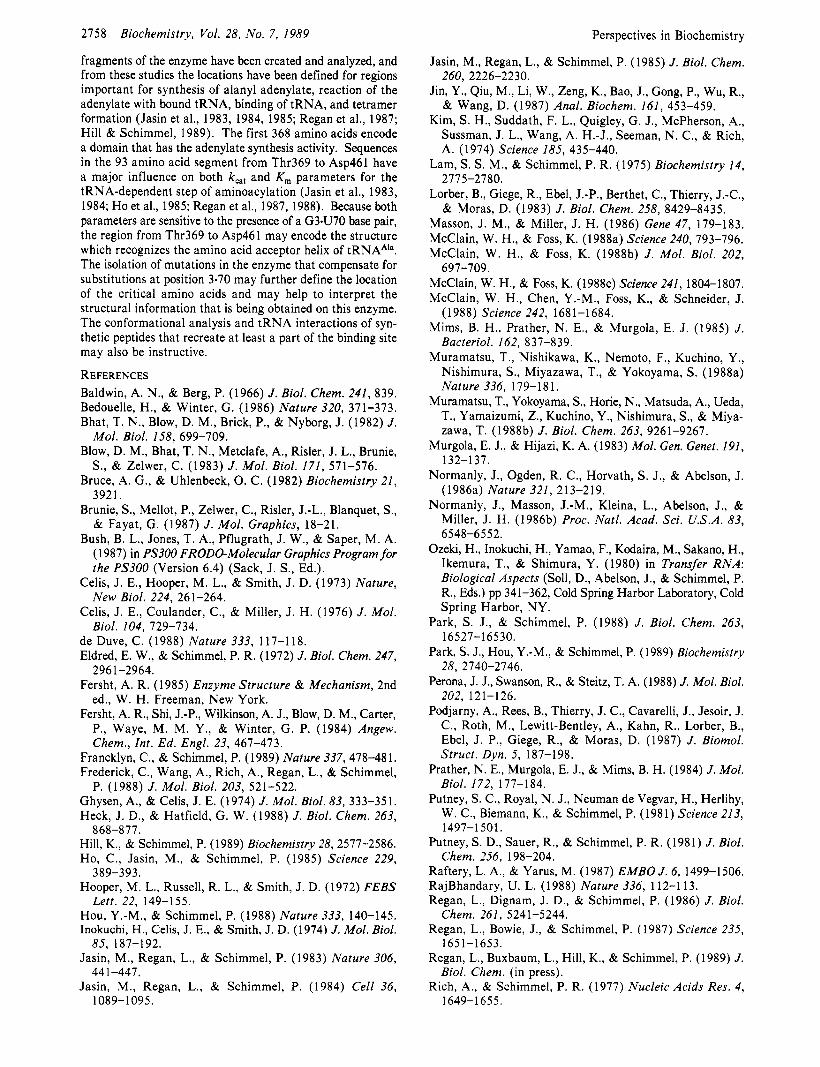
Limited Nucleotide Constellations Establish the Identities of Some Transfer RNAs. The standards of the field have been raised so that it is no longer sufficient to show only that a specific nucleotide substitution lowers the efficiency of ami- noacylation with a particular enzyme. This is because some substitutions that interfere with aminoacylation could create new, unfavorable enzyme-tRNA steric conflicts at sites which are close to but which do not bind to the enzyme through specific atomic interactions in the wild-type complex. The more recent conclusions are based upon the now accepted practice of making nucleotide substitutions into different tRNA sequence frameworks and establishing the effect on amino acid acceptance. This experiment attempts to identify sites that are dominant positive determinants in vivo and in vitro (thorough k,,, and/or K,,, parameters).
The locations of important nucleotides for some tRNAs are given in Table IV. The listing includes cases where a “transfer” experiment has been done and is provisional because further work may show that, for a particular tRNA, additional nucleotides are required. Moreover, context effects undoub- tedly have a major role (see above), and the full extent of these effects will not be known for some time. However, it is now clear that the determinants for identity are idiosyncratic. Also, for at least some tRNAs, a limited constellation of nucleotides is a major determinant of their identities. It is noteworthy that there is evidence that a simple structural feature (base-pair mismatch at the first position of the amino acid acceptor helix) is a major determinant for distinguishing an initiator from an elongator tRNA (Seong & RajBhandary, 1987).
Possible Role for Editing in the Determination of Transfer RNA Identity. An early observation by Baldwin and Berg (1 966) indicated that valine could be activated by isoleucine tRNA synthetase and that tRNA1le then induced hydrolysis of the bound valyl adenylate. There was no evidence that valine was actually transferred to tRNA””. Schreier and Schimmel (1 972) then showed that aminoacyl-tRNA synthetases have a hydrolytic site that removes an amino acid from transfer RNA, in the absence of AMP and pyro- phosphate. This was initially discovered as an activity which cleaves the ester bond which links the cognate amino acid to its tRNA, and evidence was presented that the activity is general to synthetases
Ile-tRNA1Ie + Ile-tRNA synthetase - Ile + tRNA1le + Ile-tRNA synthetase
Phe-tRNAPhe + Phe-tRNA synthetase - Phe + tRNAPhe + Phe-tRNA synthetase
and, similarly, for other aminoacyl-tRNA synthetases. Subsequently, Eldred and Schimmel (1972) showed that,
for isoleucine tRNA synthetase, this activity is much enhanced when the incorrect amino acid is attached to a tRNA:
Val-tRNA1le + Ile-tRNA synthetase - Val + tRNA1le + Ile-tRNA synthetase
Perspectives in Biochemistry Biochemistry, Vol. 28, No. 7, 1989 2757
Table IV: Nucleotides Implicated as Important for the Identities of Some Transfer RNAs tRNA
E . coli alanine
E . coli arginine
E . coli glutamine
E . coli isoleucine
E . coli methionine
yeast phenylalanine
E . coli serine
E . coli valine
important positions G3sU70
A20 and others
u 3 5
L34
anticodon
G20, G34, A35, A36, A13
Gl*C72, G2C71, A3mU70, C l l G 2 4
anticodon
evidence in vivo amber suppression with over 30 tRNAAia sequence variants and with G3mU70 tRNACyB/CUA
and G3sU70 tRNAPhc/CUA; in vitro aminoacylation data on several tRNAA1” variants on synthetic minihelix and microhelix substrates [Hou & Schimmel, 1988; Park et al., 1989; Francklyn & Schimmel, 1989; cf. also McClain and Foss (1988a)J
preliminary results in vivo based on introducing A20 and A59 into a tRNAphC amber suppressor; A59 may play a structural role rather than a role in direct recognition; other nucleotides such as those in anticodon may also be important (McClain & Foss, 1988c)
some tRNAs with the amber codon are misacylated with glutamine, and U35 appears responsible (see text); other nucleotides in the acceptor helix may be important as well
posttranscriptional modification of cytidine 34 to lysidine switches tRNA’IC2 from a methionine-accepting to an isoleucine-accepting tRNA in vitro (Muramatsu et al., 1988a)
in vitro aminoacylation data with anticodon sequence variants and transfer of the CAU anticodon into tRNAVa’ (Schulman & Pelka, 1988)
in vitro aminoacylation data with sequence variants of tRNAPhc and of four reconstructed tRNAs (Samson et al., 1989)
in vivo amber suppression with sequence variants of a tRNALCU that was converted to a serine-accepting tRNA [Normanly et al., 1986a; cf. Schulman and Abelson (1988)]
in vitro aminoacylation data with the UAC anticodon of tRNAVa’ transferred into tRNAMC‘ (Schulman & Pelka, 1988)
and Yarus (1972) demonstrated a rapid deacylation of Ile- tRNAPhc by phenylalanine tRNA synthetase: Ile-tRNAPhC + Phe-tRNA synthetase -
Ile + tRNAPhe + Phe-tRNA synthetase
Considerable investigation of editing reactions was subse- quently undertaken [see summaries by Soll and Schimmel (1974), Schimmel and Soll (1979), Yarus (1979), and Fersht (1 985)]. Editing can occur by hydrolysis of the aminoacyl adenylate or by charging followed by hydrolysis of the mis- charged aminoacyl-tRNA species. The editing activity of a specific enzyme is generally directed toward an amino acid whose steric bulk is not greater than that of the cognate amino acid. Such amino acids can potentially be activated by a specific enzyme (e.g., valine can be activated by isoleucine tRNA synthetase and threonine by the valine enzyme) because they “fit” into the amino acid binding site, albeit with a lower affinity.
The role of editing in vivo is affected by the presence of elongation factor Tu, which tightly binds and sesquesters aminoacyl-tRNA species (Schimmel & Soll, 1979). Thus, if a mischarged tRNA species is released from an enzyme, it can be carried into the ribosomal translation apparatus by Tu and insert a missense substitution in a growing polypeptide chain. The role of editing of charged mutant amber suppressors in vivo, and the influence of this potential reaction on the results obtained with the amber suppression assay, is unknown. When a G3.U70 base pair is transferred into tRNACyS/CUA, the re- sulting mutant amber suppressor can be aminoacylated (with alanine) in vitro with purified alanine tRNA synthetase (Hou & Schimmel, 1988). The overall yield of aminoacylation of Ala-G3*U70 tRNACys/cUA is dependent on the enzyme con- centration, however, and may reflect an editing reaction of the alanine tRNA synthetase that is due to enzyme-catalyzed deacylation of Ala-G34J70 tRNACPICUA and, additionally or alternatively, to some hydrolysis of the enzyme-bound alanyl adenylate when presented with G3U70 tRNA*/CUA. Specific editing reactions with “transformed” tRNAs remain to be demonstrated, but these considerations illustrate the role that editing may have in determining the identity of transfer RNAs.
Defining Sites on Aminoacyl-tRNA Synthetases That Determine Transfer RNA Identity. Structural information on aminoacyl-tRNA synthetases has been limited to Bacillus stearothermophilus tyrosine and E . coli methionine tRNA synthetases (Bhat et al., 1982; Risler et al., 1982; Blow et al., 1983; Brunie et al., 1987). A cocrystal with tRNA has not
been obtained in either case. For tyrosine tRNA synthetase, there is a nucleotide fold in the amino-terminal half which is the location of ATP and tyrosine binding sites which have been extensively analyzed by Fersht and co-workers by site-directed mutagenesis (Fersht et al., 1984; Fersht, 1985). The C-ter- minal half of the protein is required for binding of tRNATyr but is disordered in the crystal (Blow et al., 1982; Bedouelle & Winter, 1986). The tRNA binding site has not been definitively located in the structure of methionine tRNA synthetase, although the tRNA cross-linking experiments of Schulman and co-workers have provided evidence that at least a part of the interaction involves the C-terminal half of the protein (Valenzuela et al., 1984; Valenzuela & Schulman, 1986).
Cocrystals of yeast aspartyl-tRNA synthetase with tRNAASp (Lorber et al., 1983; Podjarny et al., 1987) and of E . coli glutamine tRNA synthetase with tRNAG’” (Perona et al., 1988) have been obtained. The latter crystals diffract to high resolution and will provide the first well-defined structure of a complex. The specific binding interactions in the synthe- tase-tRNA complexes are relatively weak, and the ones most critical will have to be differentiated from weak nonspecific protein-tRNA contacts. It should be possible from the model of a complex to define nucleotide replacements in the bound tRNA that would not be accommodated in the complex. Such nucleotides may constitute the “negative determinants” de- scribed above. The possibility of defining the structural basis for discrimination by the k,, parameter is more problematic, because this is influenced by interactions in a transition state and not in the form which is isolated in a cocrystal. The structural interpretation of mutants of glutamine tRNA synthetase that cause misacylation may be instructive (Ino- kuchi et al., 1984).
For alanine tRNA synthetase, much of the recognition is concentrated in the amino acid acceptor helix and is centered on the G3U70 base pair. It is not known whether the enzyme recognizes specific atoms in the base pair or a helix variation at this location. It is noteworthy that the U3sG70 tRNAAa/CUA is not aminoacylated in vitro by substrate levels of the enzyme (Figure 3). In experiments with nucleases that have respectively a strong preference for cleavage in single- or double-stranded regions, the G3sU70 base pair behaves as though it is part of a double-stranded section (Park & Schimmel, 1988).
Alanine tRNA synthetase is a tetramer of identical 875 amino acid polypeptides (Putney et al., 1981a,b). Eighteen
2758
fragments of the enzyme have been created and analyzed, and from these studies the locations have been defined for regions important for synthesis of alanyl adenylate, reaction of the adenylate with bound tRNA, binding of tRNA, and tetramer formation (Jasin et al., 1983, 1984, 1985; Regan et al., 1987; Hill & Schimmel, 1989). The first 368 amino acids encode a domain that has the adenylate synthesis activity. Sequences in the 93 amino acid segment from Thr369 to Asp461 have a major influence on both k,, and K , parameters for the tRNA-dependent step of aminoacylation (Jasin et al., 1983, 1984; Ho et al., 1985; Regan et al., 1987, 1988). Because both parameters are sensitive to the presence of a G34J70 base pair, the region from Thr369 to Asp461 may encode the structure which recognizes the amino acid acceptor helix of tRNAAIa. The isolation of mutations in the enzyme that compensate for substitutions at position 3.70 may further define the location of the critical amino acids and may help to interpret the structural information that is being obtained on this enzyme. The conformational analysis and tRNA interactions of syn- thetic peptides that recreate at least a part of the binding site may also be instructive.
REFERENCES Baldwin, A. N., & Berg, P. (1966) J . Biol. Chem. 241, 839. Bedouelle, H., & Winter, G. (1986) Nature 320, 371-373. Bhat, T. N., Blow, D. M., Brick, P., & Nyborg, J. (1982) J .
Mol. Biol. 158, 699-709. Blow, D. M., Bhat, T. N., Metclafe, A., Rider, J. L., Brunie,
S., & Zelwer, C. (1983) J . Mol. Biol. 171, 571-576. Bruce, A. G., & Uhlenbeck, 0. C. (1982) Biochemistry 21,
3921. Brunie, S., Mellot, P., Zelwer, C., Rider, J.-L., Blanquet, S.,
& Fayat, G. (1987) J . Mol. Graphics, 18-21. Bush, B. L., Jones, T. A., Pflugrath, J . W., & Saper, M. A.
(1 987) in PS300 FRODOMolecular Graphics Program for the PS300 (Version 6.4) (Sack, J. S., Ed.).
Celis, J. E., Hooper, M. L., & Smith, J. D. (1973) Nature, New Biol. 224, 261-264.
Celis, J . E., Coulander, C., & Miller, J. H. (1976) J . Mol. Biol. 104, 729-734.
de Duve, C. (1988) Nature 333, 117-1 18. Eldred, E. W., & Schimmel, P. R. (1972) J . Biol. Chem. 247,
Fersht, A. R. (1985) Enzyme Structure & Mechanism, 2nd ed., W. H. Freeman, New York.
Fersht, A. R., Shi, J.-P., Wilkinson, A. J., Blow, D. M., Carter, P., Waye, M. M. Y., & Winter, G. P. (1984) Angew. Chem., Int. Ed. Engl. 23, 467-473.
Francklyn, C., & Schimmel, P. (1989) Nature 337,478-481. Frederick, C., Wang, A., Rich, A., Regan, L., & Schimmel,
Ghysen, A., & Celis, J. E. (1974) J . Mol. Biol. 83, 333-351. Heck, J. D., & Hatfield, G. W. (1988) J . Biol. Chem. 263,
Hill, K., & Schimmel, P. (1989) Biochemistry 28,2577-2586. Ho, C., Jasin, M., & Schimmel, P. (1985) Science 229,
Hooper, M . L., Russell, R. L., & Smith, J. D. (1972) FEBS
Hou, Y.-M., & Schimmel, P. (1988) Nature 333, 140-145. Inokuchi, H., Celis, J. E., & Smith, J. D. (1974) J . Mol. Biol.
Jasin, M., Regan, L., & Schimmel, P. (1983) Nature 306,
Jasin, M., Regan, L., & Schimmel, P. (1984) Cell 36,
Biochemistry, Vol. 28, No. 7, 1989
296 1-2964.
P. (1988) J . Mol. Biol. 203, 521-522.
868-811.
389-393.
Lett. 22, 149-155.
85, 187-192.
44 1-447.
1089-1 095.
Perspectives in Biochemistry
Jasin, M., Regan, L., & Schimmel, P. (1985) J . Biol. Chem.
Jin, Y., Qiu, M., Li, W., Zeng, K., Bao, J., Gong, P., Wu, R., & Wang, D. (1987) Anal. Biochem. 161, 453-459.
Kim, S. H., Suddath, F. L., Quigley, G. J., McPherson, A., Sussman, J. L., Wang, A. H.-J., Seeman, N. C., & Rich, A. (1974) Science 185, 435-440.
Lam, S. S. M., & Schimmel, P. R. (1975) Biochemistry 14,
Lorber, B., Giege, R., Ebel, J.-P., Berthet, C., Thierry, J.-C., & Moras, D. (1983) J . Biol. Chem. 258, 8429-8435.
Masson, J. M., & Miller, J. H. (1986) Gene 47, 179-183. McClain, W. H., & Foss, K. (1988a) Science 240, 793-796. McClain, W. H., & Foss, K. (1988b) J . Mol. Biol. 202,
McClain, W. H., & Foss, K. (1988~) Science 241, 1804-1807. McClain, W. H., Chen, Y.-M., Foss, K., & Schneider, J.
(1988) Science 242, 1681-1684. Mims, B. H., Prather, N. E., & Murgola, E. J. (1985) J .
Bacteriol. 162, 837-839. Muramatsu, T., Nishikawa, K., Nemoto, F., Kuchino, Y.,
Nishimura, S., Miyazawa, T., & Yokoyama, S. (1988a) Nature 336, 179-1 8 1.
Muramatsu, T., Yokoyama, S., Horie, N., Matsuda, A., Ueda, T., Yamaizumi, Z., Kuchino, Y., Nishimura, S., & Miya- zawa, T. (1988b) J . Biol. Chem. 263, 9261-9267.
Murgola, E. J., & Hijazi, K. A. (1983) Mol. Gen. Genet. 191,
Normanly, J., Ogden, R. C., Horvath, S. J., & Abelson, J. (1986a) Nature 321, 213-219.
Normanly, J., Masson, J.-M., Kleina, L., Abelson, J., & Miller, J. H. (1986b) Proc. Natl. Acad. Sci. U.S.A. 83,
Ozeki, H., Inokuchi, H., Yamao, F., Kodaira, M., Sakano, H., Ikemura, T., & Shimura, Y. (1980) in Transfer RNA: Biological Aspects (Soll, D., Abelson, J., & Schimmel, P. R., Eds.) pp 341-362, Cold Spring Harbor Laboratory, Cold Spring Harbor, NY.
Park, S. J., & Schimmel, P. (1988) J . Biol. Chem. 263,
Park, S . J., Hou, Y.-M., & Schimmel, P. (1989) Biochemistry
Perona, J. J., Swanson, R., & Steitz, T. A. (1988) J. Mol. Biol.
Podjarny, A., Rees, B., Thierry, J. C., Cavarelli, J., Jesoir, J. C., Roth, M., Lewitt-Bentley, A., Kahn, R., Lorber, B., Ebel, J . P., Giege, R., & Moras, D. (1987) J . Biomol. Struct. Dyn. 5, 187-198.
Prather, N. E., Murgola, E. J., & Mims, B. H. (1984) J . Mol. Biol. 172, 177-184.
Putney, S. C., Royal, N. J., Neuman de Vegvar, H., Herlihy, W. C., Biemann, K., & Schimmel, P. (1981) Science 213,
Putney, S . D., Sauer, R., & Schimmel, P. R. (1981) J . Biol.
Raftery, L. A., & Yarus, M. (1987) EMBO J . 6, 1499-1506. RajBhandary, U. L. (1988) Nature 336, 112-113. Regan, L., Dignam, J. D., & Schimmel, P. (1986) J . Biol.
Regan, L., Bowie, J., & Schimmel, P. (1987) Science 235,
Regan, L., Buxbaum, L., Hill, K., & Schimmel, P. (1 989) J.
Rich, A., & Schimmel, P. R. (1977) Nucleic Acids Res. 4,
260, 2226-2230.
2775-2780.
697-709.
132-1 37.
6548-65 52.
16527-1 6530.
28, 2740-2746.
202, 121-126.
1497-1501.
Chem. 256, 198-204.
Chem. 261, 5241-5244.
1651-1653.
Biol. Chem. (in press).
1649-1 655.
Perspectives in Biochemistry
Risler, J. L., Zelwer, C., & Brunie, S. (1982) Nature 292, 383-386.
Robertus, J. D., Ladner, J. E., Finch, J. T., Rhodes, D., Brown, R. S . , Clark, B. F. C., & Klug, A. (1974) Nature 250,
Rogers, M. J., & Soll, D. (1988) Proc. Natl. Acad. Sci. U.S.A.
Sampson, J., & Uhlenbeck, 0. C. (1988) Proc. Natl. Acad.
Sampson, J. K., DeRenzo, A., Behlen, L., & Uhlenbeck, 0.
Samuelsson, T., Boren, T., Johansen, T.-I., & Lustig, F. (1988)
Schimmel, P. R. (1977) Acc. Chem. Res. 10, 411-418. Schimmel, P. (1987) Annu. Rev. Biochem. 56, 125-158. Schimmel, P. (1989) Adv. Enzymol. (in press). Schimmel, P. R., & Soll, D. (1979) Annu. Rev. Biochem. 48,
Schreier, A. A., & Schimmel, P. R. (1972) Biochemistry 1 1 ,
Schulman, L. H., & Abelson, J. (1988) Science 240,
Schulman, L. H., & Pelka, H. (1989) Science (in press). Seong, B. L., & RajBhandary, U. L. (1987) Proc. Natl. Acad.
Shimura, Y., Aono, H., Ozeki, H., Sarabhai, A., Lamfrom,
Smith, J. D., & Celis, J. E. (1973) Nature, New Biol. 243,
Smith, J. D., Barnett, L., Brenner, S. , & Russell, R. L. (1970)
546-55 1.
85, 6627-663 1.
Sci. U.S.A. 85, 1033-1037.
C. (1989) Science (in press).
J. Biol. Chem. 263, 13692-13699.
60 1-648.
1582-1589.
1591-1592.
Sci. U.S.A. 84, 8859-8863.
H., & Abelson, J . (1972) FEBS Lett. 22, 144-148.
66-7 1.
J . Mol. Biol. 54, 1-14.
Biochemistry, Vol. 28, No. 7, 1989 2759
Soll, D., & Schimmel, P. R. (1974) Enzymes 10, 489-538. Soll, L. (1974) J. Mol. Biol. 86, 233-243. Sprinzl, M., Hartmann, T., Meissner, F., Moll, H., & Vor-
derwulbecke, T. (1987) Nucleic Acids Res. 15, r53-rl88. Swanson, R., Hoben, P., Sumner-Smith, M., Uemura, H.,
Watson, L., & Soll, D. (1988) Science 242, 1548-1551. Valenzuela, D., & Schulman, L. H. (1986) Biochemistry 25,
Valenzuela, D., Leon, O., & Schulman, L. H. (1984) Biochem.
Waye, M. Y., Winter, G., Wilkinson, A. J., & Fersht, A. R.
Yamo, F., Inokuchi, H., & Ozeki, H. (1988a) Jpn. J. Genet.
Yamo, F., Inokuchi, H., Normanly, J., Abelson, J., & Ozeki,
Yaniv, M., Folk, W. R., Berg, P., & Soll, L. (1 974) J. Mol.
Yarus, M. (1972) Proc. Natl. Acad. Sci. U.S .A. 69,
Yarus, M. (1979) in Transfer RNA: Structure, Properties, & Recognition (Schimmel, P. R., Soll, D., & Abelson, J., Eds.) pp 501-515, Cold Spring Harbor Laboratory, Cold Spring Harbor, NY.
4555-4561.
Biophys. Res. Commun. 1 19, 677-684.
(1983) EMBO J. 2, 1827-1829.
63, 237-249.
H. (1988b) Jpn. J . Genet. 63, 251-258.
Biol. 86, 245-260.
1915-1919.
Yarus, M. (1986) J. Mol. Biol. 192, 235-255. Yarus, M. (1988) Cell 55, 739-741. Yarus, M., & Berg, P. (1970) Anal. Biochem. 35,450-465. Yarus, M., Knowlton, R., & Soll, L. (1977) in Nucleic
Acid-Protein Recognition (Vogel, H. J., Ed.) pp 391-408, Academic Press, New York.