Embed Size (px)
Citation preview
Medical and Pediatric Oncology 26:284292 (1996)
Key words: pancreatoblastoma, pancreatic tumors, pediatric oncology, chemotherapy, embryology
+
Pancreatic Blastomatous Tumor in a Child Responding to Therapy Used for Hepatoblastoma: Case Report and Review of the literature
Elaine R. Morgan, MD, John H. Perryman, MD, Marleta Reynolds, MD, and Frank J. Conzalez-Crussi, MD
A case is reported of a 7-year-old girl di- agnosed with initially inoperable metastatic embryonic pancreatic tumor, which showed a significant clinical response to chemother- apeutic agents commonly used to treat hepatoblastoma. This regimen was selected because certain histologic features of the tu- mor demonstrated characteristics seen in hepatic tissue. After two courses of chemo- therapy (cis-platinum and adriamycin), there
was a significant reduction of the primary mass, and it was completely resected, al- though the tumor subsequently recurred in the metastatic, unoperated site. The embry- ologic relationship between the tumor in this patient and hepatoblastoma, as well as the literature concerning treatment in pedi- atric pancreatoblastic tumors are reviewed. Complete eradication of tumor appears to be necessary for cure. 0 1996 Wiley-Liss, Inc.
INTRODUCTION
Pancreatoblastoma is a rare tumor of childhood of uncertain histogenesis [ 1,2]. Literature on this tumor is scant, mostly consisting of isolated case reports or small series, and primary modalities of therapy utilized are surgical resection and/or radiation therapy [2-111. A case is presented of an inoperable blastomatous abdominal tumor in a 7-year-old girl arising in the pancreas and differing in some histologic respects from pancreatoblas- toma. Treatment for hepatoblastoma (HBL) was used because of similar histologic features in this tumor with a good initial response.
CASE REPORT
The patient is a previously healthy 7-year-old white female who presented with a left upper quadrant abdomi- nal mass. There was no history of fever, unusual bruising or bleeding, or bone pain. There was a history of slightly decreased appetite and possible weight loss. Review of systems was otherwise unremarkable. Her past medical history was noncontributory. She was on no medications, ate a “balanced” diet, and was developmentally appropri- ate. Her family history was notable for aplastic anemia in a paternal great-grandfather at age 50, breast cancer in a maternal aunt, and a “blood-clotting” disorder in a mater- nal grandmother and great-grandfather. At the time of referral, the patient appeared comfortable; her tempera- ture was 36.8”C, heart rate 100/min, respiratory rate 201 0 1996 Wiley-Liss, Inc.
min, and blood pressure 100/70 mm Hg. Her head and neck exam was unremarkable. Her chest was clear with decreased breath sounds at the left base. Cardiovascular examination was unremarkable. Examination of her ab- domen revealed a 10 X 15 cm firm, nontender mass in the left upper quadrant. Her extremities were normal, neurological examination was intact, and she had normal Tanner stage I genitalia.
Laboratory examination demonstrated a hemoglobin of 11.3 g/dL (11.5-15.5), hematocrit of 34.1% (35.0- 45.0), platelet count of 335,OOO/cu mm (150450), and white blood cell count 7,200icu mm (4.5-13.5). The differential was 50% polymorphonuclear neutrophils, 13% band forms, 27% lymphocytes, 6% atypical lym- phocytes, and 4% eosinophils. Her BUN was 8 mg/dL [7-181, and creatinine was 0.6 mg/dL (0.3-0.8). Her serum calcium was 9.3 mg/dL (8.8-10.8), LDH 895 U/L
mg/dL, bilirubin (total/direct) 0.3/0.1 mg/dL (0.2-1 .O/ <0.3), and uric acid 6.5 mg/dL (2.0-6.0). Urinalysis demonstrated a specific gravity of 1.015, pH of 7.5, 2+ occult blood and trace protein. Microscopic examination
(163-304), SCOT 27 U/L (8-42), SGPT 7 U/L (3-36
From the Departments of Pediatrics (E.R.M., J .H.P . , M.R.) and Pa- thology (F.J.G.C.), Children’s Memorial Hospital, Chicago, Illinois.
Received October 27, 1994; accepted February 9, 1995.
Address reprint requests to Elaine R. Morgan, M.D., Division of Hematology/Oncology, Children’s Memorial Hospital, 2300 Chil- dren’s Plaza, Chicago, IL 60614.
Pancreatic Tumor Responding to Chemotherapy 285
of necrosis within the mass and complete resolution of liver metastasis. AFP levels fell from a pre-CDDP-ADR level of 8,532 ng/mL to 330 ng/mL. She underwent a second laparotomy and distal pancreatectomy , splenec- tomy, left nephrectomy, and left adrenalectomy. The pri- mary mass was completely excised, and the margins of resection were free of tumor. Approximately 10 days after surgery, her alpha-fetoprotein level was <5 .O ng/ mL; follow-up CT scans of the chest and abdomen 3 weeks later demonstrated no residual or metastatic neo- plasm. The patient received an additional three courses of this chemotherapy after which she manifested ototoxicity and nephrotoxicity. No cardiotoxicity was noted. Her therapy was then changed to a regimen of multiple courses of vincristine and 5-FU (vincristine 1.5 mg/M2 IV, 5-FU 500 mg/M2/day X 3 days) since initial partial response to 5FU could not be excluded.
Approximately five months after resection, a CT scan done to evaluate abdominal pain demonstrated a new mass in the operative bed. At this point, the patient had received one course of the vincristine/5-FU regimen, as well as one additional dose of vincristine. Findings at laparotomy included calcified greater omental masses and a 2 X 3 cm calcified mass in the greater curvature of the stomach. The liver, right kidney, and remainder of the stomach were normal. Pathologic examination re- vealed that these represented areas of fat necrosis and suture granulomas; no tumor was identified. Four addi- tional courses of vincristine and 5-FU were administered to complete an arbitrary 6 months of therapy.
The patient did well and remained disease-free for -2 months after discontinuing chemotherapy (-7 months after tumor resection). At that time, she suffered tumor recurrence in the liver at the site of initial metastatic disease. She had a partial response to carboplatin and etoposide (VP 16) and underwent wedge resection of tumor in the liver along with a lymph node in the porta hepatis, which was infiltrated by tumor. She subse- quently developed rapidly progressive tumor in the liver, which was nonresponsive to further chemotherapy and expired 18 months after diagnosis. Postmortem examina- tion was not performed.
showed 90-100 RBCs/HPF, 0-1 WBCs/HPF, 10-12 ep- ithelial cells/HPF, and few bacteria. An alpha-fetoprotein (AFP) level was 82,940 ng/mL (normal values 0-8.5). Bone marrow examination demonstrated mild erythroid hyperplasia but was otherwise normal. A bone scan dem- onstrated no evidence of bony involvement. A CT scan and MRI of the abdomen showed a large leftsided retro- peritoneal mass measuring 13 cm in its greatest diameter that appeared separate from the liver, spleen and kidney, although contiguity with the caudate lobe of the liver could not be totally excluded. The tail of the pancreas was not well visualized. Two lucent lesions were seen in the right lobe of the liver and were felt to represent metastases. A CT scan of the chest revealed no evidence of metastases.
The patient went to surgery for exploratory laparot- omy and possible resection. Intraoperatively , she was noted to have a firm, fungating mass involving the pan- creas, stomach, left kidney, spleen, adjacent retroperito- neum, and inferior vena cava. The mass was felt to be unresectable and was biopsied only. Postoperatively, she was managed with intravenous hydration, alkalinization, and allopurinol .
The initial pathologic diagnosis of the tumor was pan- creatoblastoma vs. adenocarcinoma of the pancreas (de- tailed discussion of the findings below). However, cer- tain features of the tumor were suggestive of hepatic origin, and hepatoblastoma was included in the differen- tial diagnosis. Treatment was initiated based on therapy used for adult pancreatic carcinoma and consisted of a course of external beam irradiation with concomitant flu- orouracil(5-FU) 500 mg/m2/d as a short IV infusion for 3 days at the beginning of radiation for sensitization. She received a total of 2,340 cGy divided into 13 fractions of a planned course of 4,000-5,OOO cGy but had no signifi- cant decrease in the size of the mass clinically or radio- logically despite a decrease in AFP from 67,305 ng/mL immediately prior to radiation to 8,532 ng/mL.
Because of the hepatoid features of the mass, the im- pression of an outside consultant (Dr. Juan Rosai, whose help we gratefully acknowledge), that the tumor was of liver cell origin, the poor clinical response to radiation therapy, and the dismal prognosis of pancreatic adenocar- cinoma, radiation therapy was discontinued and the pa- tient was started on a protocol successfully used for HBL in children. This therapy, consisting of, every 3rd week, courses of cis-platinum (CDDP) and Adriamycin (doxo- rubicin) (ADR) (90 mg/M2 of cis-platinum (CDDP) ad- ministered intravenously over 8 hours, followed by a 4-day infusion of Adriamycin at a dose of 20 mg/M2/day) was initiated within 1 week of termination of radiation therapy.
After receiving two courses of this chemotherapy, a CT of the abdomen demonstrated marked reduction of the primary mass (7 cm in greatest diameter) with evidence
MATERIALS A N D METHODS
Both the biopsy tissue and the surgical specimen ob- tained subsequently were processed in the same manner. Tissues were fixed in 10% buffer formalin or B-5 fixri- tive.
Paraffin-embedded tissue blocks were cut at 5 pni thickness and stained by conventional histologic tcch- nique with hematoxylin and eosin (WE), Gomori's rctic- ulum, and Masson's trichrome. For immunohistochcriiis- try, an avidin-biotin complex immunoperoxidase-linkctl detection kit was used following the manufacturers' iii-
286 Morgan et al.
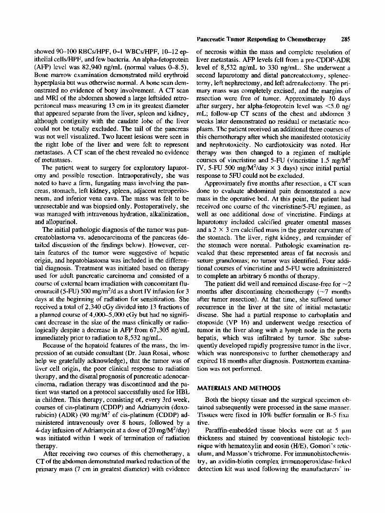
Fig. 1. macrophages (hematoxylin/eosin, X 225).
Solid area of the tumor showing cohesive epithelial cells with occasional interspersing of
structions (Vectastain ABC kit, Vector Laboratories, Burliname, CA). The primary antibodies were commer- cially available antibodies against alpha- 1 -fetoprotein, alpha- 1-antichymotrypsin, albumin, amylase, fibrinogen and neuron-specific enolase (Dako, Santa Barbara, CA); antimacrophage antibody HAM 56 (Enzo Biochem, New York, NY); chromogranin A (Boehringer Manheim, In- dianapolis IN); and pancreatic polypeptide. For electron microscopy, tissue was minced in 3% glutaraldehyde, postfixed in osmium tetroxide, dehydrated, and embed- ded in Spurr’s plastic according to standard technique. Ultrathin sections stained with uranyl acetate and lead citrate were examined with a Zeiss 10-A electron micro- scope.
PATHOLOGY
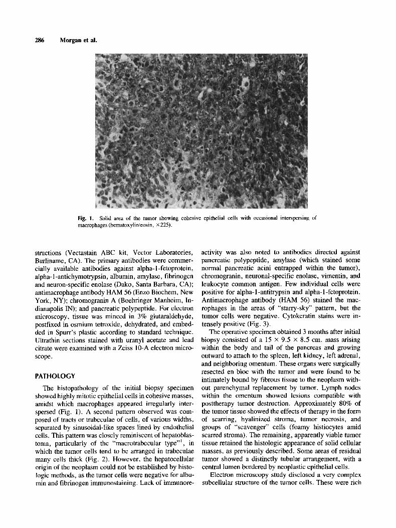
The histopathology of the initial biopsy specimen showed highly mitotic epithelial cells in cohesive masses, amidst which macrophages appeared irregularly inter- spersed (Fig. 1). A second pattern observed was com- posed of tracts or trabeculae of cells, of various widths, separated by sinusoidal-like spaces lined by endothelial cells. This pattern was closely reminiscent of hepatoblas- toma, particularly of the “macrotrabecular type”’, in which the tumor cells tend to be arranged in trabeculae many cells thick (Fig. 2). However, the hepatocellular origin of the neoplasm could not be established by histo- logic methods, as the tumor cells were negative for albu- min and fibrinogen immunostaining . Lack of immunore-
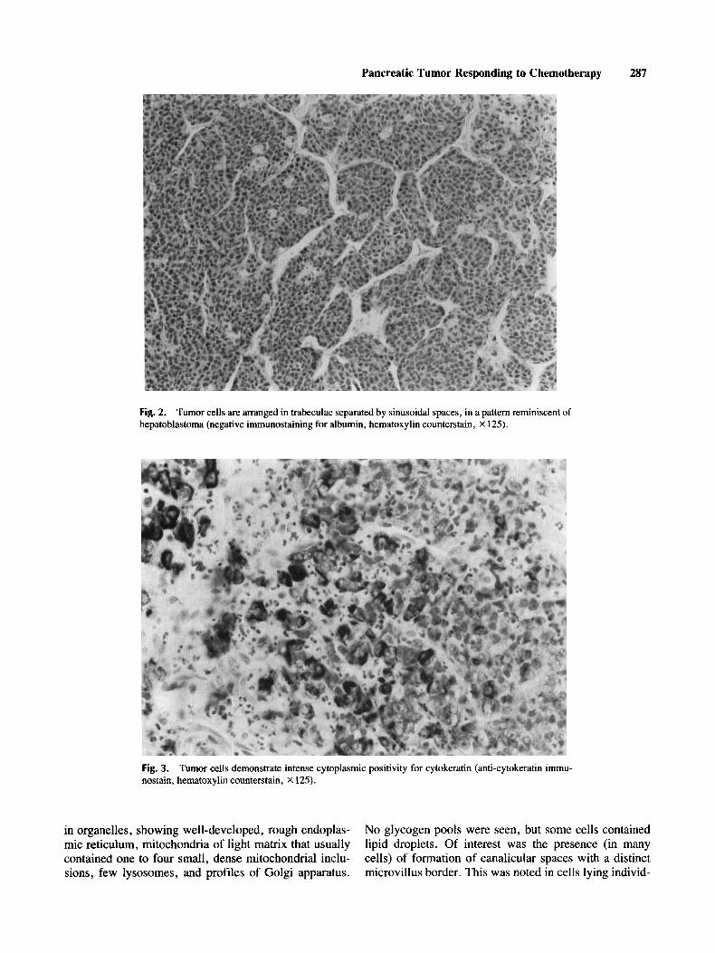
activity was also noted to antibodies directed against pancreatic polypeptide, amylase (which stained some normal pancreatic acini entrapped within the tumor), chromogranin, neuronal-specific enolase, vimentin, and leukocyte common antigen. Few individual cells were positive for alpha-1-antitrypsin and alpha- 1-fetoprotein. Antimacrophage antibody (HAM 56) stained the mac- rophages in the areas of “starry-sky’’ pattern, but the tumor cells were negative. Cytokeratin stains were in- tensely positive (Fig. 3).
The operative specimen obtained 3 months after initial biopsy consisted of a 15 X 9.5 X 8.5 em. mass arising within the body and tail of the pancreas and growing outward to attach to the spleen, left kidney, left adrenal, and neighboring omentum. These organs were surgically resected en bloc with the tumor and were found to be intimately bound by fibrous tissue to the neoplasm with- out parenchymal replacement by tumor. Lymph nodes within the omentum showed lesions compatible with posttherapy tumor destruction. Approximately 80% of the tumor tissue showed the effects of therapy in the form of scarring, hyalinized stroma, tumor necrosis, and groups of “scavenger” cells (foamy histiocytes amid scarred stroma). The remaining, apparently viable tumor tissue retained the histologic appearance of solid cellular masses, as previously described. Some areas of residual tumor showed a distinctly tubular arrangement, with a central lumen bordered by neoplastic epithelial cells.
Electron microscopy study disclosed a very complex subcellular structure of the tumor cells. These were rich
Pancreatic Tumor Responding to Chemotherapy 287
Fig. 2. Tumor cells are arranged in trabeculae separated by sinusoidal spaces, in a pattern reminiscent of hepatoblastoma (negative immunostaining for albumin, hematoxylin counterstain, X 125).
Fig. 3. Tumor cells demonstrate intense cytoplasmic positivity for cytokeratin (anti-cytokeratin immu- nostain, hematoxylin counterstain, X 125).
in organelles, showing well-developed, rough endoplas- mic reticulum, mitochondria of light matrix that usually contained one to four small, dense mitochondria1 inclu- sions, few lysosomes, and profiles of Golgi apparatus.
No glycogen pools were seen, but some cells contained lipid droplets. Of interest was the presence (in many cells) of formation of canalicular spaces with a distinct microvillus border. This was noted in cells lying individ-

288 Morgan et al.
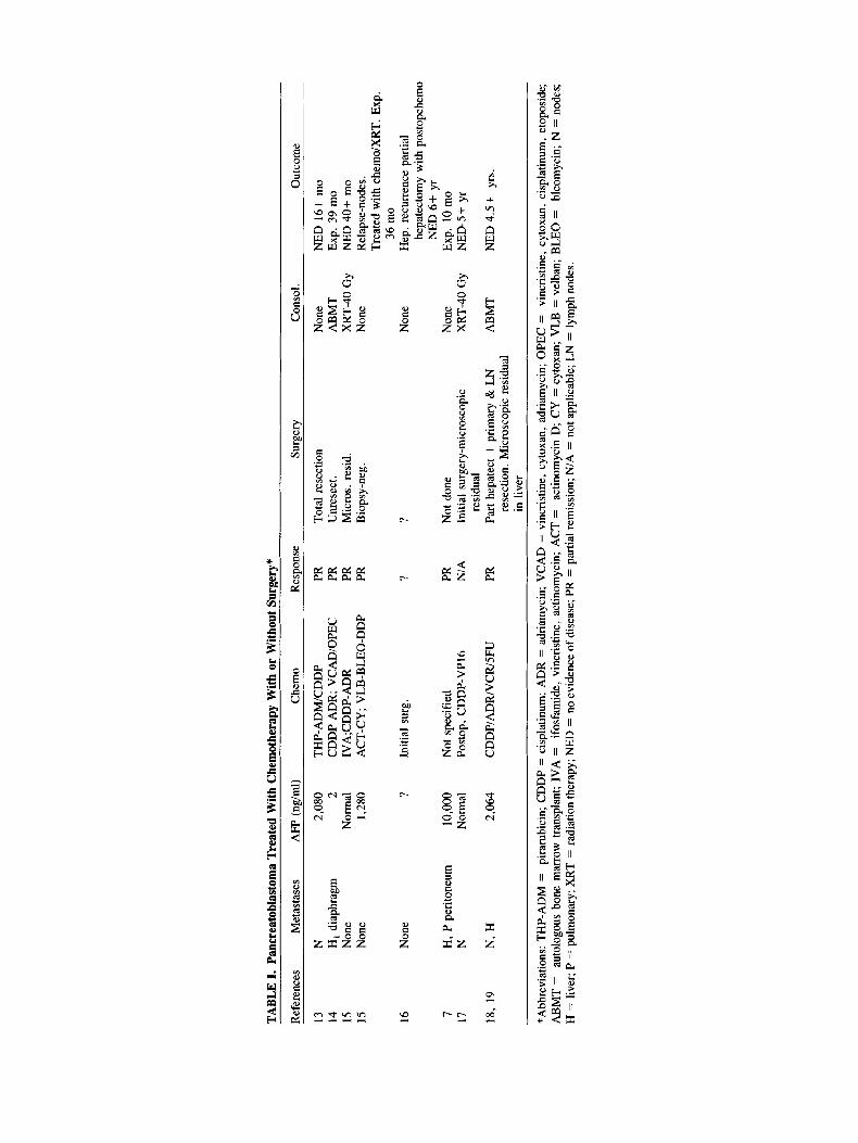
Fig. 4. between opposing cells (X20,OOO).
Detail of a canalicular structure, showing well-developed microvilli and tight junctions (mows)
ually, or at sites of cell-to-cell apposition. In the later case, it was not uncommon to observe the presence of tight junctions between cells bordering a canaliculus (Fig. 4).
DISCUSSION Malignant pancreatic tumors are very rare in children.
Pancreatoblastoma is histologically distinct from typical ductal or acinar pancreatic adenocarcinoma [ 11. Because of their varieties, it may be that the morphologic spec- trum of these neoplasms is incompletely known. With the reservations explained earlier, we approached this pa- tient’s management using therapy appropriate for HBL. Making accurate statements regarding prognosis in a tu- mor this rare is difficult, but there are data to suggest that surgical resection is more likely to be successful in pan- creatoblastoma than in adult pancreatic carcinoma. In adults, only 13% of these tumors are resectable at presen- tation, and median survival postresection is 14-17 months [ 121. Camprodon and Quintenilla [9] reviewed 28 cases of childhood pancreatic malignancy reported be- tween 1885 and 1981 and noted that resection was possi- ble in 12 (40%) of these cases. Follow-up information was obtainable for seven of those patients; all were alive a minimum of 5 years after surgery, with survival times ranging from 5 to 20 years. Nagaraj and Polk [3] report an overall survival rate of 80% in 20 children with re- sected pancreatoblastomalpancreatic carcinoma of the in- fantile type.
Other reviews paint a less optimistic picture. Tsuki- mot0 et al. [lo] reviewed 12 cases from the Japanese literature; surgery was impossible in three patients, six had a laparotomy only, and two had pancreaticoduo- denectomy performed. All died within 6 months of the onset of symptoms. Taxy reviewed 16 cases; in 14 where follow-up was available, only three had survived 9 months or longer [ 1 I].
There are several case reports in the literature indicat- ing responses to chemotherapy, most notably to regimens containing CPDD. These are summarized in Table I [7,13-18, M. Kletzel, pers. comm.]. Notably, the only patients surviving received chemotherapy followed by total surgical resection or surgical resection with micro- scopic residual followed by aggressive radiation ther- apy or high dose chemotherapy with autologous marrow rescue.
Comparing responses to other therapeutic modalities in adult and pediatric pancreatic malignancy is difficult because of the low incidence of this disease in childhood. Moreover, pancreatic tumors in adults and children are histologically different entities. In adults, radiation ther- apy plays a significant role in palliation and may improve survival [ 121. Griffin [4] reported successful control of locally recurrent pancreatoblastoma with radiation ther- apy. Chemotherapy in adult disease has failed to be of any benefit in disease control or improvement of survival [7]. Iseki et al. [7] reported control of metastatic pancre- atoblastoma with a regimen of vincristine, doxorubicin, and cyclosphosphamide. There has been, however, no
TABL
E I.
Panc
reat
obla
stom
a Tr
eate
d W
ith C
hem
othe
rapy
With
or
With
out S
urge
ry*
Ref
eren
ces
Met
asta
ses
AFP
(ng
/ml)
Che
mo
Res
pons
e Su
rger
y C
onso
l. O
utco
me
13
N
2,08
0 TH
P- A
DM
/CD
DP
PR
Tota
l res
ectio
n N
one
NED
16+
mo
14
H, di
aphr
agm
2
CD
DP
AD
R;
VC
AD
/OPE
C
PR
Unr
esec
t. A
BM
T Ex
p. 3
9 m
o 15
N
one
Nor
mal
1V
A;C
DD
P-A
DR
PR
M
icro
s. r
esid
. X
RT-
40 G
y N
ED 4
0+ m
o 15
N
one
1,28
0 A
CT-
CY
; V
LB-B
LEO
-DD
P PR
B
iops
y-ne
g.
Non
e R
elap
se-n
odes
. Tr
eate
d w
ith c
hern
o/X
RT.
Exp
. 36
mo
hepa
tect
omy
with
pos
topc
hem
o N
ED 6
+ y
r
16
Non
e ?
Initi
al s
urg.
?
? N
one
Hep
. rec
urre
nce
parti
al
7 H
, P p
erito
neum
10
,OOO
N
ot s
peci
fied
PR
Not
don
e N
one
Exp.
10
mo
17
N
Nor
mal
Po
stop
, C
DD
P-V
P16
N/A
In
itial
sur
gery
-mic
rosc
opic
X
RT-
40 G
y N
ED-5
+ yr
18,
19
N,
H
2,06
4 C
DD
P/A
DR
/VC
R/S
FU
PR
Part
hepa
tect
+ pr
imar
y &
LN
A
BM
T N
ED 4
.5+
yrs.
re
sidu
al
rese
ctio
n. M
icro
scop
ic r
esid
ual
in li
ver
*Abb
revi
atio
ns: T
HP-
AD
M =
pi
raru
bici
n; C
DD
P =
cis
plat
inum
; A
DR
= a
dria
myc
in;
VC
AD
= v
incr
istin
e, c
ytox
an, a
dria
myc
in; O
PEC
=
vinc
ristin
e, c
ytox
an,
cisp
latin
um,
etop
osid
e;
AB
MT
=
auto
logo
us b
one
mar
row
tra
nspl
ant;
IVA
=
ifosf
amid
e, v
incr
istin
e, a
ctin
omyc
in;
AC
T =
ac
tinom
ycin
D;
CY
= c
ytox
an;
VLB
= v
elba
n; B
LEO
=
bleo
myc
in;
N =
nod
es;
H =
live
r; P
= p
ulm
onar
y; X
RT
= ra
diat
ion
ther
apy;
NED
= n
o ev
iden
ce o
f dis
ease
; PR
= p
artia
l rem
issi
on; N
/A =
not
app
licab
le; L
N =
lym
ph n
odes
.
290 Morgan et al.
consistently successful chemotherapeutic regimen re- ported.
In short, the best chance for cure in pancreatoblastoma would appear to be complete surgical resection, with a possible role for radiation in disease control or palliation. Unresectable disease portends a poor prognosis.
In the case presented here, interpretation of the biopsy pathology was rendered difficult by uncertainty of the location of the primary tumor in the preoperative period. It was thought that the bulk of the neoplasm was in the pancreas, but the possibility of exophytic growth from a primary in the caudate lobe of the liver could not be ruled out with complete assurance before the second surgical intervention. The presence of a solid epithelial tumor, whose pathologic characteristics indicated a “blastoma- tous” nature, together with the clinical presumption of a pancreatic primary site and the age of the patient, justi- fied the tentative diagnosis of pancreatoblastoma. How- ever, there were features suggestive of hepatoblastoma, such as characteristic epithelial-sinusoidal trabecular his- tologic pattern, and the presence of canaliculi demon- strated by electron microscopy. This is not a feature rec- ognized in the generally available descriptions of the pathology of pancreablastoma [2,17,19,20], as further discussed below.
After biopsy, the patient was started on radiation ther- apy and 5-FU. Initial clinical response to this modality in our patient was minimal, although there was a marked reduction in the AFP. Since effective chemotherapy for hepatoblastoma does exist [2 13 and pathologic diagnosis was not clear, the patient was started on multiple courses of CDDP and ADR, which are two of the most active agents used in the treatment of HBL. There was a dra- matic response to these agents, with marked reduction of the mass on CT scan, a 10-fold reduction in alpha-feto- protein levels, and resolution of the lucent lesions seen in the liver. Following the postchemotherapeutic surgical intervention (3 months after biopsy), it became clear that the tumor had originated within the pancreas and had no relationship to the liver.
The epithelial, “liver-like” appearance was retained in some areas of the excised primary tumor, thus emphasiz- ing the “histogenetic kinship” of the blastomas primary to the liver and pancreas. Previous reports on the pathol- ogy of pancreatoblastoma have not pointed out the possi- bility of diagnostic confusion with hepatoblastoma. Our patient’s tumor did not conform with the recognized his- topathology of HBL, nor are there any documented ex- amples of true hepatoblastoma arising within the pan- creas [23]. Both of these eventualities, however, seem possible when one bears in mind the close embryologic relationship of these two organs. The embryonic hepatic diverticulum and the dorsal pancreas trace a common origin in the foregut. Two further diverticula originate
from the lower aspect of the hepatic diverticulum; one destined to become the gallbladder and cystic duct, the other the ventral pancreas. The adult pancreas thus origi- nates from two embryonic formations, the dorsal and ventral diverticula, which join each other to form the completed organ [23]. Moreover, there is experimental evidence that pancreatic ductal cells, at least in rodents, are able to transform into hepatocytes [24]. This phenom- enon, called “transdifferentiation,” has been studied ex- tensively in hamsters and rats, leaving no doubt that pancreatic ductular epithelium can give rise to cells with all the morphologic, functional, and biochemical at- tributes of hepatocytes, including the presence of “liver- specific” protein markers [24,25]. Conversely, the origi- nation of a pancreatoblastoma in the liver is theoretically possible, given the well-documented occurrence of pan- creatic tissue in the liver [26], either by heterotopia or metaplasia.
Intriguing similarities exist between pancreatoblas- toma and hepatoblastoma. Hepatoblastoma occurs in as- sociation with the Beckwith-Wiedemann syndrome (ex- omphalos-macroglossia-gigantism, or EMG), as do some other embryonal tumors. There are reports in the litera- ture of pancreatoblastoma occurring in patients with EMG [5,6]. Some authors have speculated that chromo- somal abnormalities found in certain patients with EMG, a loss of heterozygosity at chromosome 1 1 band p13, has been linked with development of rhabdomyosarcoma and hepatoblastoma [28]. Elevation in the serum AFP, an- other characteristic feature of hepatoblastoma, was present in our patient. Other cases in the literature have demonstrated this marker to be elevated in patients with pancreatoblastoma [7 $1, but it is not a universal finding occurring in -25% of patients [28], and does not appear to be correlated with response or outcome. It has been reported that the raised AFP in pancreatoblastoma is of the yolk sac type, differing from that of hepatoblastoma [29]. This distinction was not determined in our patients.
These considerations have practical relevance, since they are grounds to predict that therapy effective against hepatoblastoma [2 11 also may be effective against some “blastomas” of the pancreas in contrast to their lack of efficacy in adult pancreatic carcinoma, which probably represents a very different disease entity. Although tu- mors in adult medicine are generally treated primarily by site of origin, in pediatrics it is common to treat according to histology. Thus extrarenal Wilms’ tumor is treated similarly to its intrarenal counterpart, and extraosseous osteogenic sarcoma is treated as the skeletal tumor. Cis- platin as a single agent has no efficacy against pancreatic carcinoma in adults. Doxorubicin and various anthracy- cline analogues have demonstrated some objective effect in adult disease, but none have shown dramatic efficacy. Combination chemotherapy fails to demonstrate any sig-

Pancreatic Tumor Responding to Chemotherapy 291
nificant benefit in adults, although combinations of the two agents used in this patient have not been studied extensively [ 121. Nonetheless, these agents proved very effective as therapy for pancreatoblastoma in our patient. The role of radiation therapy (XRT) and 5 FU was not clear in this case. The initial lack of tumor shrinkage by palpation combined with our interest in trying therapy effective in HBL prompted an early change in therapy. However, XRT and 5FU have also been successfully used in the treatment of hepatoblastoma [21]. Although there appeared to be a dramatic response to CDDP and ADR, the initial therapy also might have contributed to this observed response, particularly in view of the 10-fold decrease in AFP prior to initiation of CDDP and ADR.
Our patient presented with a massive, inoperable, pan- creatic blastomatous malignancy for which nonsurgical therapy has been notoriously unrewarding in the past. The tumor was rendered operable by a therapeutic ap- proach of proven efficacy against hepatoblastoma, and the child was rendered free of disease.
Unfortunately, this child’s tumor recurred at the site of initial metastatic lesions in the absence of recurrence at the primary site. These lesions were not initially treated with either resection or irradiation therapy. Patients in the literature who have had a successful outcome have all undergone total resection of tumor and/or local irradia- tion therapy either primarily or after initial tumor shrink- age with chemotherapy. Perhaps related to this, the ex- tent of disease at diagnosis also may impact on survival, as most surviving patients have had only regional disease (local with or without regional nodal involvement).
CONCLUSIONS
It appears that total eradication of all tumor is required for cure. It is suggested by these cases that consolidation with megadose chemotherapy and involved field RT fol- lowed by autologous stem cell rescue may be successful when tumor cannot be totally eradicated by other modali- ties. Pancreatoblastoma is a chemotherapy responsive tu- mor and should be approached like other pediatric tumors with an aggressive combined modality approach.
ACKNOWLEDGMENTS
The authors gratefully acknowledge the assistance of Dina Jimenez and Charine Scott in preparation of this manuscript.
REFERENCES
1. Gonzalez-Crussi F, Upton MP, Maurer HS: Hepatoblastoma: At- tempts at characterization of histologic subtypes. Am J Surg ..
Pathol6599-612, 1982. L J .
2.
3.
4.
5.
6.
7.
8.
9.
10.
11.
12.
13.
14.
15.
16.
17.
18.
19.
20.
21.
22.
23.
24.
.7c
Horie H, Yano Y, Kotoo Y, Mowa A: Morphogenesis of pancre- atoblastoma, infantile carcinoma of the pancreas. Report of two cases. Cancer 39(1):247-254, 1977. Nagaraj H, Polk HC Jr: Pancreatic carcinoma in children. Surgery 95(4):505, 1984. Griffin BR, Wisbeck WM, Schaller RT, Benjamin DR: Radio- therapy for locally recurrent infantile pancreatic carcinoma (pan- creatoblastoma). Cancer 60(8): 17341736, 1987. Drut R, Jones MC: Congenital pancreatoblastoma in Beckwith- Wiedemann syndrome: An emerging association. Pediatr Pathol
Koh TH, et al: Pancreatoblastoma in a neonate with Wiedemann- Beckwith syndrome. Eur J Pediatr 145(5):435438, 1986. Iseki M, et al: Alpha-fetoprotein producing pancreatoblastoma, a case report. Cancer 57(9): 1833-1835, 1986. Morohoshi T, Sagawa F, Mitsuya T Pancreatoblastoma with marked elevation of serum alpha-fetoprotein: An autopsy case report with immunocytochemical study. Virchows Arch [A]
Camprodon R, Quintenilla E Successful long term results with resection of pancreatic carcinoma in children: Favorable prognosis for an uncommon neoplasm. Surgery 95(4):420426, 1984. Tsukimoto I, et al: Pancreatic carcinoma in children in Japan. Cancer 31(5):1203-1207, 1973. Taxy JB: Adenocarcinoma of the pancreas in childhood: Report of a case and a review of the English language literature. Cancer
Brennan MF, Kunsella T, Friedman M: Cancer of the pancreas. In DeVita VT, Hellmans-Rosenberg , SA (eds): “Cancer: Principles and Practice of Oncology,” 3rd ed. Philadelphia: Lippincott, 1989. Inomata Y, Nishizawa T, Takasa H, et al: Pancreatoblastoma resected by delayed primary operation after effective chemother- apy. J Pediatr Surg 27(12):157&1572, 1992. Eden OB, Shaw MP: Letter to the Editor: Chemotherapy for pancreaticoblastoma. Med Pediatr Oncol 20(4):357-359, 1992. Vannier JP, Flamant F, Hemet J , et al: Pancreatoblastoma: re- sponse to chemotherapy. Med Pediatr Oncol 19(3):187-191, 1991. Grosfeld JL, Vane DW, Rescorla FJ, et al: Pancreatic tumors in childhood: Analysis of 13 cases. J Pediatr Surg 25(10):1057- 1062, 1990. Silverman JR, Holbrook CT, Pones WJ, et al: Fine needle aspira- tion cytology of pancreatoblastoma with immunocytochemical and ultrastructural studies. Acta Cytologica 34(5):632-640, 1990. Stephenson CA, Kletzel M, Seibert JJ, Glasier CM: Pancreato- blastoma: MR appearance. J Comput Assist Tomogr 14(3):492- 194, 1990. Buchino JJ, Castello FM, Nagaraj HS: Pancreatoblastoma: A his- tochemical and ultrastructural analysis. Cancer 53:963-969, 1984. Ichyima K, Akaishi K, Toyanda N, et al: Carcinoma of the pan- creas with endocrine component in child: A case report. Am J Clin Pathol83:95-100, 1985. Greenberg M, Filler RM: P.A. Pizzo, D.G. Poplack (eds): “He- patic Tumors: Principles and Practices of Pediatric Oncology.” Philadelphia: Lippincott, 1989, pp. 569-580. Gonzalez-Crussi F, Upton MP, Mauer HS: Hepatoblastoma: At- tempt to characterization of histologic subtypes. Amer J Surg Pathol6:559-612, 1982. Harrison RG: The foregut. In: “A Textbook of Human Embryol- ogy” chap. 11. Oxford Blackwell, 1959, pp. 99-10, Rao MS, Scarpelli DG, Reddy JK: Transdifferentiated hepato- cytes in rat pancreas. Cum Topics Devel Biol20:63-78, 1986. Makino T, Usuda N, Rao S, Reddy JK, Scarpelli DG: Transdiffer-
8(3):331-339, 1988.
416(3):265-270, 1990.
37(3): 1508-15 18, 1976.
292 Morgan et al.
entiation of ductular cells into hepatocytes in regenerating hamster pancreas. Lab Invest 62(5):552-561, 1990.
26. Wolf HK, Burchette JL, Garcia JA, Michalopoulos G: Exocrine pancreatic tissue in human liver: A metaplastic process? Am J Surg Pathol 14(6):59&595, 1990.
27. Koufos A, et al: Loss of heterozygosity in three embryonal tumors suggest a common pathogenetic mechanism. Nature 3 16:330- 334, 1985.
28. Hone A, Haratake J, Jimi A, et al: Pancreatoblastoma in Japan, with differential diagnosis from papillary cystic tumor (ductuloac- inar adenoma) of the pancreas. Acta Pathologica Japonica 37(1): 47-63, 1987.
29. Tsuchida Y, Kaneko M, Fukui M, et al: Three different types of alpha-fetoprotein in the diagnosis of malignant solid tumors: Use of a sensitive lectin-affinity immunoelectrophoresis. J Pediatr Surg 24(4):35@355, 1989.