Embed Size (px)
Citation preview

PALMOXYLON SURANGEI, A NEW SPECIES OF PETRIFIEDPALMS FROM THE DECCAN INTERTRAPPEAN SERIES
R. ~. L\KHANPAL
Birbal Salmi Institute of Palaeobotany, Lucknow
ABSTRACT
A new species of Palmoxylon, P. surangei sp. nov.,is described from the Deccan Intertrappean series.The specimen was collected from Keria, a villageabout two miles south of Mohgaon Kalan, DistrictChhindwara, Madhya Pradesh. The species belongs to the subgroup Cordata of petrified palmstems.
INTRODUCTION
INFebruary 1951 Dr. K. R. Surange andI went to Madhya Pradesh (formerlyknown as Central Provinces) to make
collections of the Deccan Intertrappean flora.While working at Mohgaon Kalan, thefamous locality of this flora, we learnt thatapproximately two miles south there wasanother village called Keria where fossilplants could be collected in plenty. Oninvestigation we found that the actual locality lay about 500 yards to the north ofKeria and was very rich in fossil woods.Amongst the specimens collected here was alarge piece of a petrified palm comprising thebasal portion of a stem with roots attached.After detailed investigation it is found tobe a new species of Palmoxylon.
I have great pleasure in naming the speciesafter Dr. Surange who first spotted this fossilin the field and thus has the credit of discovering it.
DESCRIPTION
In the main the descriptive terminologyused here is the same as suggested by Professor B. Sahni (1943) in his paper onPalmoxylon sclerodermum.
External Characters
The specimen is a large rusty brown stumpwhich, before cutting, measured 37 em. inlength and 46 em. in diameter at the base,narrowing to about 33 em. at the top. Athick mantle of adventitious roots surroundsthe lower 15 em. of the stump. In the
15
upper part the mantle of roots is replacedby a cortical zone measuring about 4 em.in thickness. Next to the cortex inwardsare the three zones, dermal, subdermal andcentral.
Anatomy
Cortex: - The fibrovascular bundles arescattered in the outer part, becoming gradually crowded towards the dermal zone.They are of various shapes and sizes as shownin Text-fig. 1. An average bundle is ovalin cross-section and measures about 0·95 X0·45 mm. The base of the sclerenchyma iscordate. There are 2-4 vessels in the xylem.The f/v ratio varies from 5/1 to 10/1, theaverage being 6/1.
The fibrous bundles are usually circularto slightly oval in cross-section, measuringabout 80 fJ. in diameter and consisting ofabout 20 fibres. Stegmata are presentaround the fibres. There are some structureswhich in section look like fibrous bundlesbut are devoid of any cellular structurewithin. These may be sclereids or mucilagecanals.
The ground tissue is made up of parenchymatous cells which are generally tangentially elongated (TEXT-FIGS. 2, 3) exceptat the distal and proximal ends of fibrovascular bundles where they are radiallyelongated. Generally the cells are fairlyclosely packed but sometimes the intercellular spaces increase to form lacunae.
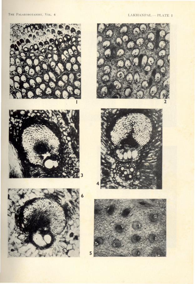
Dermal Zone - It is about 2 em. thick.The fibrovascular bundles are densely crowded towards the periphery, becoming less sotowards the centre (PL. 1, FIG. 1). Theaverage frequency is 90-95 per cm.2 but inthe outer region it is as much as 140 percm.2•In cross-section the bundles are elongateelliptical in the outer region, becomingbroadly ovate towards the interior. Butgenerally the normal shape of the bundlesin the crowded exterior region is deformeddue to pressure against each other. Anaverage bundle measures about 1 X 0·5 mm.

16 THE PALAEOBOTA T1ST
3
TEXT-FIGS. 1-3-1, outlines of some fibrovascular bundles from the cortical zone. X 31. 2, groundtissue cells of the cortex in cross-section. X 70. 3, the sam~ in longitudinal section. X 70.
All of them are normally orientated. Thef/v ratio is less in the exterior region, beingin some cases only about 7/8, and increasestowards the interior to about 7/1. Thefibrous part is usually pyriform. The mediansinus is rounded cordate. The vessels in thexylem number 1-4, normally 2-3, and areexcluded. On the distal side, along thesclerenchyma the bundles are surroundedby elongated parenchymatous cells arrangedradially (TEXT-FIG. 4). These cells arefound only around the bundles of the external
crowded region; in the inner region they arereplaced by the usual tabular parenchyma.
In the less crowded region fibrous bundlesmay occur sporadically. They are about110-120 [L in diameter. The stegmata arepresent in longitudinal rows on the fibrousbundles (TEXT-FIGS. 6, 7) as well as on thesclerenchyma of the fibrovascular bundles.
The parenchymatous ground tissue isscanty and compact. The cells are smallerthan those of the cortex. They are isodiametric to slightly rectangular in the outer

LAKHANPAL-PALMOXYLON SURANGEI, A NEW PETRIFIED PALM 17
TEXT-FIGS. 4-7 - 4, two fibrovascular bundles from the derm~l zonc showing the radially elongatedparenchymatous cells on the distal sidc. X 70. 5, end wall of a large vessel. X 115. 6, 7, cross-sectionand longitudinal section respectively of the fibrous bundles. Stegmata are visiblc on the surfacc inFig. 7. Roth X 115.
region, becoming longer and bigger towardsthe interior. At places some of the longercells arrange themselves in wavy horizontalrows as seen in transverse section.
Subdermal Zone-This zone is nearly4 em. in thickness. There are 45-50 fibro-
vascular bundles per cm.2• They are broadly oval to slightly ovate in cross-section( PL. 1, FIG. 3). Their normal size is 0·95 X0·65 mm. The flv ratio in average bundlesis 5/1 to 6/1. The sclerenchyma is broadlyround, pyriform to orbicular and the xylem

18 THE PALAEOBOTANIST
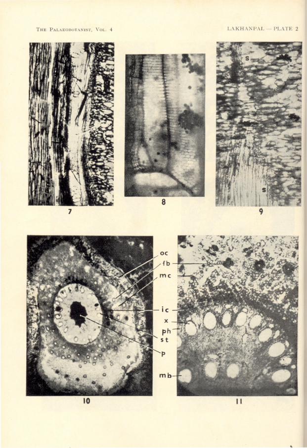
IS narrower, pointing towards the centre.Auricular lobes are round. Auricular sinuses are obtuse, being less prominent inbroader bundles. The tabular parenchymais present. The phloem which is wedged inbetween the xylem and the median sinus isvery poorly preserved, generally disorganized.The xylem usually consists of 2-3 vessels,excluded. The pitting of the metaxylem isscalariform in the narrow elements and reticulate in the wider ones. The proto xylemhas spiral thickening. The end walls ofthe vessels are slightly oblique and havescalariform or reticulate thickening withnearly half a dozen broad rounded meshes( PL. 2, FIG. 7; TEXT-l'IG. 5).
Occasionally leaf trace bundles (PL. 1,FIG. 4) also occur in the dermal as well asthe subdermal zone. In these bundles thexylem is greatly developed, almost doublethat of the fibrovascular bundles, and incross-section projects like a tongue. Thereis an arc of sclerenchymatous cells on theventral border.
Fibrous bundles occur sporadically, eachconsisting of about 20 fibres and measuringapproximately 80 p. in diameter. Stegmataare present as in the dermal zone. Idioblasts are not seen.
The ground tissue is quite compact andconsists of fairly big parenchymatous cellswhich in cross-section appear elongated withround edges. Wavy horizontal rows oflonger cells are seen in the outer region. Inwider areas between the fibrovascular bundlesthe cells tend to become broader, someattaining almost circular outline. An average cell is 150 p. long and 60 p. wide. Theround ones average about 80 p. across. Inlongitudinal section the cells are round,isodiametric to slightly elongated.
Central Zone - This is the widest part ofthe stem with a radius of about 6·5 cm.The fibrovascular bundles are irregularlyorientated (PL. 1, FIG. 5 ), broadly ovate incross-section, measuring about 0·95 X 0·8 mm.Aproximately 25 bundles occur per cm.2•The average flv ratio is 4/1. The sclerenchyma is broad and orbicular with broadrounded cordate base ( PL. 1, FIG. 6 ), sometimes appearing as reniform. The xylem isbetter developed than in the subdermal zone,with obtuse auricular sinuses. There areusually 3-4 vessels in the xylem.
The fibrous bundles and stegmata arepresent ( PL. 2, FIG. 9 ) as in the subdermalzone. The structure of the ground tissue
also is essentially the same as in the subdermal zone. However, because of morespace between the fibrovascular bundles, thecells become broader and more rounded asthey approach the centre.
Roots - The roots are closely packed,usually deformed by crowding together.They measure 5-7 mm. in diameter, the onescoming from the periphery being narrowerthan those arising from the central region.
Seen in cross-section ( PL. 2, FIGS. 10, 11 )the outermost layer, about 30 p. in thickness,is indistinguishable. Next to this is thecortex which can be distinguished into threeparts - outer, middle and inner. The outercortex, about 0·5 mm. thick, is formed ofcompact rows of thick-walled, dark-coloured,rounded cells, approximately 25 X 30 p. insize. The middle cortex is about 1 mm.thick. It is composed of round, thin-walledcells, about 35 p. in diameter, rather looselypacked and with air cavities between them.In this region also occur a number of fibrousbundles appearing almost circular in outline.They are slightly bigger than those of thestem region, usually measuring 110-120 p.
in diameter, while some may be as big as160 p. across. Stegmata are present aroundthese bundles. Some of the structureslooking like fibrous bundles in cross-sectionbut having no cellular structure within maybe sclereids. The inner cortex is a morecompact zone, about 0·2 mm. in thickness.Next to the cortical region inwards is a layerof rectangular cells constituting the endodermis. Next are two layers of thin-walled cellsof the pericycle. There are about 22 bundleseach of the xylem and phloem alternatingwith each other and arranged in a circleconcentric with the endodermis. Eachxylem bundle usually contains one bigvessel surrounded by a ring of parenchymatous cells. Occasionally one or twosmaller vessels may also be associated with,and situated on the outer side of, the bigvessel. The vessels are oval in cross-section,measuring about 140-190 p. in radial and 80115 p. in tangential direction. The phloembundles are not well preserved and occur aslight patches alternating with and a littleoutward of the xylem vessels. On the innerside the vascular bundles are embraced bythick-walled polygonal cells of the conjunctive parenchyma which extends by about0·2 mm. beyond the vascular row towardsthe centre where it meets the central coreof the pith composed of thin-walled cells.

LAKHAXPAL-PAL1IOXYLON SURANGEI, A NEW PETRIFIED PAUL 19
The inner boundary of the conjunctive tissueis irregularly sinuous. The cells in the pithare round, measuring about 20-25 [J. in diameter. In the pith region there may be 1-4medullary bundles each consisting of a vessel encircled by a ring of thick-walled cells.
DISCUSSION
So far there is no available system for thenatural classification of petrified palm stemswhich are all grouped under the form genusPalmoxylon. In 1935, based on an extensivestudy of modern palms, Kaul had announcedthat palm stems could be identified by thestructure of their ground tissue. But thiswork has not yet been published in full.At present the only course open is to classifythe genus artificially. For this purpose Professor Sahni's scheme (1943, pp. 218-219)based jointly on the classifications of vonMohl (1845)and Stenzel (1904) is very convenient and suitable. According to this schemeP. surangei falls under the subgroup Cordata.
Comparison with Indian Palmoxyla - Ofthe already described Palmoxyla from India,only P. sclerodermum ( SAHNI,1943; SHUKLA,1946) belongs to Cordata. It resembles ourspecies in (i) the general appearance, orientation and distribution of fibrovascularbundles, (ii) the sculpturing of the xylemelements (with some difference with regardto the end walls of the vessels), (iii) thepresence of fibrous bundles and stegmata,and (iv) the general form of the leaf-tracebundles, having a tongue-like vascular process with ventral sclerenchymatous arc.
In a number of other characters, however,the two species are distinctly separate. Theground tissue cells of the cortex are radiallyelongated and often occur in layers in P.sclerodermum while they are tangentiallyelongated in P. surangei. The fibrovascularbundles are bigger and more crowded, andtheir flv ratio is higher in P. sclerodermumthan in P. surangei. Auricular sinuses arealmost absent in P. sclerodermum while theyare usually present and obtuse in P. surangei.The end walls of the broad xylem vessels inP. sclerodermum are very oblique and havescalariform thickening. In P. surangei theyare less oblique and generally with reticulatethickening. In P. surangei the sclerenchymaof a few outer rows of the dermal bundles isexternally surrounded by radially elongatedparenchymatous cells. This character is notmet with in P. sclerodermum. The frequency
of fibrous bundles is higher in P. surangeithan in P. sclerodermum. The ground tissuecells in P. sclerodermum are smaller ( 0·08 X0·06 mm.) and roughly isodiametric while inP. surangei they are bigger (0·15 X 0·06mm.), oval, oblong and elongated. Themore rounded cells of the latter speciesaverage about 0·08 mm. in diameter. Thecells in P. sclerodermum are lobed while thistendency is not prevalent in P. surangei.
In the roots of P. surangei, fibrous bundlesare found in the middle cortex while no suchfibrous bundles occur in P. sclerodermum.The structure of the xylem bundles is different in the two species. While in P. scleroderm~tm there are 3-4 vessels placed end toend in radial direction, in P. surangei thereis usually only one big vessel. Another important difference is the presence of medullary bundles in the pith region of P. surangei.Such bundles have not been recorded in P.sclerodermum.
Comparison with Foreign Palmoxyla - Ofthe foreign known species only P. densum( UNGER) Schenk and P. speciosum (STENZEL) Schenk described by Stenzel (1904)can, to some extent, be compared with thepresent species.
P. densum is known only by the outerparts of the stem. This species resemblesP. surangei in having roughly comparableshape of the fibrovascular bundles, increasing distance between the bundles and decreasing flv ratio as we go inwards, the presence of fibrous bundles and stegmata, andthe general form of the leaf trace bundles.However, there are a number of differenceswhich distinguish it from P. surangei. Thefrequency of fibrovascular bundles in P.densum is higher than in P. surangei. InP. densum the auricular lobes are very prominent, the auricular sinuses being acute. InP. surangei the auricular sinuses are obtuseand not so prominent. The median sinusis reniform in P. dcnsum and cordate in P.surangei. No stegmata have been reportedfrom the fibrous part of the fibrovascularbundles of P. densum while they are presentin P. surangei. The ground tissue is quitedifferent in the two species. The cells aresmaller ( about 50-70 [J. in diameter), morerounded and often arranged in vertical rowsin P. densum. In P. surangei they are biggerand elongated. Their arrangement is different and not in vertical rows. Some of thecells are polygonal in P. densum while nopolygonal cells are seen in P. surangei.

20 THE PALAEOBOTANIST
REFERENCES
In the general shape of the bundles, presence of fibrous bundles with stegmata, andthe ground tissue consisting of elongatedcells, P. surangei also resembles P. speciosum.But, as enumerated below, there are a number of differences between the two species.In P. speciosum the fibrovascular bundlesare bigger (1-1·25 mm. across) and moresparsely distributed ( 15-21 per cm.2 in thesubdermal region). In cross-section although the fibrous part of some bundles ofP. speciosum does seem cordate as in P.surangei, in majority of them it is sagittate.The flv ratio is higher in P. speciosum, especially in the outer region. The auricularsinuses are acute in P. speciosum and obtusein P. suranRei. While a large number ofbundles in P. speciosum have only one bigvessel, those in P. surangei have 2-3. Fibrousbundles are more numerous and bigger ( 100166 fl in diameter) in P. speciosum. Theground tissue cells are longer in P. speciosum( 166-250 fl in length and 40-60 fl in width ).The arrangement of these cells is also different in the two. In P. speciosum there isusually one short polygonal cell at placeswhere the long cells from all the directionsmeet. Such an arrangement is not seen inP. surangei.
DIAGNOSIS
Genus - Palmoxylon
Sub-group - Cordata
Palmoxylon surangei sp. novoPlates 1, 2, Text-figs. 1-7
Stem thick ( in type specimen about 33 em.in diameter near the base). Cortex faid ythick, composed of thin-walled parenchymatous cells elongated horizontally and containing scattered fibrovascular and fibrousbundles. Dermal and subdermal zones fairlywell developed, central zone quite broad.Fibrovascular bundles in the dermal, subdermal and central zones almost of the samesize, becoming slightly bigger towards thecentre. Dermal bundles 90-95 per cm.2;flv ratio 9/2; external bundles with radially
RAUL, K. N. (1935). A classification of palmsbased upon the ground-tissue of the stem.Proc. 22nd Indian Sci. Congress: 285-286.
elongated parenchymatous cells on thedorsal side; inner bundles having tabularparenchyma; median sinus cordate; numberof vessels in the xylem usually 2-3, excluded.Subdermal bundles 45-50 per cm.2 measuringca. 0·95 X 0·65 mm.; flv ratio 5/1-6/1; tabularparenchyma present; auricular sinus obtuse;xylem usually consisting of 2-3 vessels, excluded; thickening of the metaxylem scalariform to reticulate; end walls of the vesselsslightly oblique with scalariform or reticulatethickenings with wide perforations betweenthem. Central bundles far apart, about25 per cm.2, broad, looking almost orbicularin cross-section, 0·95 X 0·8 mm. in size; flvratio 4/1; median sinus broad, almost reniform; xylem usually with 3-4 vessels.Fibrous bundles present in all the zones,measuring about 80 fl in diameter. Stegmata present on fibrous as well as thefibrovascular bundles. Leaf trace bundlesin the dermal and subdermal zones radiallystretched with well-developed xylem projecting as a tongue-like process. Groundtissue composed of thin-walled, elongated,oval-oblong cells with rounded corners,arranged horizontally; more compact in thedermal zone and rather loose in the subdermal and central zones; average subdermacell measuring 150X 60 fl in size; in widerareas between the fibrovascular bundles thecells tend to become broader, some attainingalmost circular outline in cross-section.
Roots running downwards, closely packed,5-7 mm. in diameter. Cortex made of 3zones; middle cortex with aerenchyma containing fibrous bundles. The stele consisting of about 22 xylem bundles alternatingwith phloem; the xylem of each bundleusually consisting of one large vessel andrarely one or two small ones. Pith composed of thin-walled cells, often with 1-4medullary bundles.
Locality - About 500 yards to the northof the village Keria in Chhindwara district,Madhya Pradesh.
Horizon - Deccan Intertrappean series.Collection - Holotype, specimen No. 17426
of the Birbal Sahni Institute of PalaeobotanyMuseum.
MORL, HUGO VON( 1845). Ueb?r den Bau des Palmenstammes. Vermischte Schriften b:JtanischenInhalts: 129-185. Tubingen.

LAKHA)<PAL-PALMOXYLON SURANGEI, A )<EW PETRIFIED PALM 21
MOHL,HUGOVON(1849). On the Structure of thePalm Stem. Ray Society Reports and Paperson Botany, 1849: 1-92. London.
SAHNI, B. (1943). A new species of petrifiedpalm stems, Palrnoxylon scle"oderrnurn sp. nov.,from the Deccan Intertrappean Series. Jour.Ind. Bot. Soc. 22 (2-4); 209-224.
SHUKLA, V. B. (1946). Palmoxylon sclerodermumSahni from the Eocene beds of Nawargaon, Wardha district, C.P. Ibid. 25(3): 105116.
STENZEL, K. G. (1904). Fossile Palmenholzer.Beitriige zur Palaeontologie und Geologie Osterreich-Ungarns und des Orients. 16: 107-287.
PLATE 1
EXPLANATION OF PLATES
PLATE 2
Palmoxylon surangei sp. novo
1. Part of cross-section through the dermal zone.Wavy horizontal rows of long parenchymatouscells are clearly visible. X7f.
2. Part of cross-section through the subdermalzone. X n.
3. A subdermal fibrovascular bundle seen incross-section. X 50.
4. A leaf-trace bundle from the subdermal zoneseen in cross-section. X 50.
5. Part of cross-section through the central zoneshowing irregular orientation of the bundles. 71.
6. A central fibrovascular bundle seen in crosssection. X 50.
Palmoxylon surangei sp. novo
7. Longitudinal section through a fibrovascularbundle with the vessel showing the end walls. X50.
8. Surface view of a vessel showing mixed scalariform and reticulate thickenings. X 270.
9. Longitudinal section through the central zoneshowing stegmata (s) on the fibrous cells.
10. Transverse section through a root. oc, outercortex; mc, middle cortex; ic, inner cortex; fb,fibrous bundle; st, stele; p, pith. X 23.
11. Magnified view of a part of the T.S. througha root. mc, middle cortex; ic, inner cortex;fb, fibrous bundle; ph, phloem bundle; x, xylembundle; mb, medullary bundle. X 50.
THE PAL.~EOBOTANJST, YOLo 4
6
5
LAKHAITPAL-PLATE 1
4
THE PALAEOBOTANIST, YOLo 4
78
L.\KHANPAL- PL.\TE 2
10 II