Embed Size (px)
Citation preview

Paediatric Research Group
Institute of Clinical Medicine
Antibiotic Therapy for Neonatal Sepsis Studies on epidemiology, gentamicin safety, and early adverse effects of antibiotics — Jon Widding Fjalstad A dissertation for the degree of Philosophiae Doctor – May 2018
1
List of Contents
Acknowledgements ................................................................................................................ 3 List of Papers .......................................................................................................................... 4 Abbreviations.......................................................................................................................... 5 Abstract ................................................................................................................................... 7 1 Introduction ..................................................................................................................... 9
1.1 Preface ..................................................................................................................... 9 1.2 Host Immunity in the Neonatal Period ...................................................................... 9 1.3 Neonatal Sepsis ...................................................................................................... 12 1.4 Early-Onset Sepsis ................................................................................................. 13
1.4.1 Epidemiology .................................................................................................. 13 1.4.2 Risk Factors and Prevention ........................................................................... 14
1.5 Late-Onset Sepsis .................................................................................................. 15 1.5.1 Epidemiology .................................................................................................. 15 1.5.2 Risk Factors and Prevention ........................................................................... 16
1.6 Necrotizing Enterocolitis ......................................................................................... 17 1.7 Diagnostic Challenges in Neonatal Sepsis ............................................................. 18
1.7.1 Biomarkers ...................................................................................................... 18 1.7.2 Detecting Pathogens in Sterile Sites ............................................................... 19 1.7.3 Deciding Who to Treat and How Long ............................................................ 20
1.8 Antibiotic Treatment in Neonates ............................................................................ 23 1.8.1 Beta-Lactams .................................................................................................. 24 1.8.2 Aminoglycosides ............................................................................................. 25 1.8.3 Glycopeptides ................................................................................................. 28 1.8.4 Empirical Antibiotic Regimens ........................................................................ 28
1.9 Adverse Effects of Antibiotic Treatment .................................................................. 29 1.9.1 Gut Microbiota and Gut Dysbiosis .................................................................. 30 1.9.2 Antibiotic Resistance ....................................................................................... 32
1.10 Evidence Based Medicine ...................................................................................... 34 2 Aims of the Study ......................................................................................................... 37 3 Materials and Methods ................................................................................................. 38
3.1 Study Design and Materials .................................................................................... 38 3.2 Gentamicin Dosing Regimen and Monitoring ......................................................... 39 3.3 Search Strategy in Systematic Reviews ................................................................. 40 3.4 Variables and Definitions ........................................................................................ 40 3.5 Audiology Assessment ........................................................................................... 43 3.6 Assessment of Methodological Quality ................................................................... 43 3.7 Statistical Analyses ................................................................................................. 43 3.8 Ethical Approval ...................................................................................................... 45
4 Main Results ................................................................................................................. 46 4.1 Paper 1 ................................................................................................................... 46 4.2 Paper 2 ................................................................................................................... 48 4.3 Paper 3 ................................................................................................................... 50 4.4 Paper 4 ................................................................................................................... 52
5 Discussion .................................................................................................................... 54 5.1 Epidemiology of Early Onset Sepsis ....................................................................... 54 5.2 Antibiotic Consumption and Potential Implications ................................................. 55

2
5.3 Choice of Antibiotic Regimen .................................................................................. 58 5.4 Gentamicin Pharmacokinetics and Toxicity ............................................................ 59 5.5 Prolonged Antibiotic Therapy .................................................................................. 61 5.6 Methodological and Ethical Considerations ............................................................ 63
5.6.1 Registry-Based Cohort Studies ....................................................................... 63 5.6.2 Retrospective Cohort Studies ......................................................................... 64 5.6.3 Systematic Review Methodology .................................................................... 65 5.6.4 Ethical Considerations .................................................................................... 67
6 Conclusions .................................................................................................................. 68 7 Future Perspectives ..................................................................................................... 69 8 References .................................................................................................................... 71 9 Appendix ....................................................................................................................... 84
9.1 Risk of Bias Evaluation Charts ............................................................................... 84 9.2 Flowcharts detailing Study Selection Process ........................................................ 87 9.3 Tables Summarizing Main Characteristics and Results from Studies Reporting Early Adverse Outcome Following Neonatal Antibiotic Therapy ........................................ 89 9.4 Risk of Bias Assessments in the Systematic Reviews of Early Adverse Effects .... 99
Paper 1-4 101

3
Acknowledgements
4
List of Papers
Paper 1 Fjalstad JW, Stensvold HJ, Bergseng H, Simonsen GS, Salvesen B, Ronnestad AE,
Klingenberg C. Early-onset Sepsis and Antibiotic Exposure in Term Infants: A Nationwide
Population-based Study in Norway. Pediatr Infect Dis J 2016; 35: 1-6.1
Paper 2 Fjalstad JW, Laukli W, van den Anker JN, Klingenberg C. High-dose gentamicin in newborn
infants: is it safe? Eur J Pediatr 2013; 173: 489-95.2
Paper 3 Esaiassen E, Fjalstad JW, Juvet LK, van den Anker JN, Klingenberg C. Antibiotic exposure in
neonates and early adverse outcomes: a systematic review and meta-analysis. J Antimicrob
Chemother 2017; 72: 1858-70.3
Paper 4 Fjalstad JW, Esaiassen E, Juvet LK, van den Anker JN, Klingenberg C. Antibiotic therapy in
neonates and impact on gut microbiota and antibiotic resistance development: a systematic
review. J Antimicrob Chemoter 2017 Nov 22 [Epub ahead of print].4
5
Abbreviations
AAP; American Academy of Pediatrics (United States)
AMP; antimicrobial peptides
AUC; area under the plasma drug concentration-time curve
BW; birth weight
CDC; Centers for Disease Control and Prevention
CI; confidence interval
CMV; cytomegalovirus
CoNS; coagulase-negative Staphylococci
CRP; C-reactive protein
EBM; evidence-based medicine
ELBW; extremely low birth weight (< 1000 g)
EOS; early-onset sepsis
ESBL; extended-spectrum beta-lactamase
GA; gestational age
GBS; group B Streptococci
GRADE; Grading of Recommendations Assessment, Development, and Evaluation
IAP; intrapartum antibiotic prophylaxis
IFI; invasive fungal infection
IQR; interquartile range
LB; live-born
LOS; late-onset sepsis
MDR; multi-drug resistant
MIC; minimum inhibitory concentration
MRSA; methicillin-resistant Staphylococcus aureus
NEC; necrotizing enterocolitis
NICE; National Institute for Health and Care Excellence (United Kingdom)
NICU; neonatal intensive care unit
NNN; Norwegian Neonatal Network
NNT; number needed to treat
NPV; negative predictive value
OAE; otoacoustic emissions
OR; odds ratio

6
PCT; procalcitonin
PMA; postmenstrual age
PNA; postnatal age
PPC; peak plasma concentration
PPV; positive predictive value
PRISMA; Preferred Reporting Items for Systematic Reviews and Meta-Analysis
PROM; prolonged rupture of membranes (> 18 hours)
QoE; quality of evidence
RCT; randomized controlled trial
SD; standard deviation
TLR; Toll like receptor
TPC; trough plasma concentration
VD; volume of distribution
VLBW; very low birth weight (< 1500 g)

7
Abstract
Background and Objectives: Sepsis is a prominent cause of neonatal mortality and morbidity
yet can be very hard to diagnose. The disease is rare, the symptoms are unspecific, the laboratory
tests are difficult to interpret, and blood cultures, which can potentially confirm an infection, may
take 36-48 hours before they demonstrate any growth. Therefore, antibiotics are the most
commonly used medications in neonatal medicine. While antibiotics can be life-saving, they can
also have potentially adverse effects. Several early adverse outcomes have been reported from
neonatal antibiotic treatment; among these necrotizing enterocolitis (NEC), invasive fungal
infection (IFI), death, changes in the gut microbiota, and development of antibiotic resistance. In
addition, gentamicin, a commonly used antibiotic in the neonatal period, has ototoxic and
nephrotoxic potential, in particular if trough plasma concentrations (TPCs) are elevated or the
infant receives prolonged therapy.
The overall aim of this thesis was to investigate different aspects of antibiotic therapy for
neonatal sepsis in order to obtain new knowledge that could improve and optimise care. The first
aim was to investigate the epidemiology of early onset sepsis (EOS) and exposure to systemic
antibiotics during the first week of life in an unselected national cohort of live-born term infants.
Secondly, we wished to evaluate a simplified high-dose extended-interval gentamicin dosing
regimen with focus on pharmacokinetic safety, potential ototoxicity, and the number of
prescription errors. Finally, we aimed to identify, critically appraise, and synthesize evidence from
studies reporting different categories of antibiotic exposure in neonates and their subsequent
impact on NEC, IFI, death, gut microbiota, and/or antibiotic resistance development.
Material and Methods: The epidemiology of EOS and systemic antibiotic exposure in the first
week of life was studied in a nationwide population-based study from the Norwegian Neonatal
Network. During the 3-year study period (2009-2011), 20 out of 21 Norwegian neonatal units
prospectively collected data. A high-dose extended-interval gentamicin regimen was studied in
the neonatal unit in Tromsø from 2004-2012. The main outcome measures were TPCs,
ototoxicity, and prescription errors. Early adverse effects of antibiotic therapy were studied in a
systematic review. We included observational studies and randomized controlled trials (RCTs)
that provided data on different categories of antibiotic therapy and either the risk of NEC, IFI,
death, antibiotic resistance development, or changes in the gut microbiota. Risks of bias were
assessed according to a modified version of the Cochrane Handbook. When appropriate, data
were meta-analysed using the random effect model or a semi-quantitative vote-counting method.

8
Results: There were 0.54 cases of culture-confirmed EOS per 1000 live-born term infants, and
the majority of these cases were caused by Gram-positive bacteria, most commonly group B
streptococci. Intravenous antibiotics were administered to 2.3% of all live-born term infants in
Norway, and 54% of these infants were not diagnosed with an infection. Empiric treatment
consisted of an aminoglycoside and either penicillin or ampicillin in 95% of cases. The EOS-
attributable mortality rate was 1%.
In the neonatal unit in Tromsø, gentamicin TPCs were above the threshold of 2 mg/L in 6% of
cases, mainly among term infants with renal impairment. Thirty-eight patients failed the neonatal
hearing screening, but only five patients had permanent hearing loss. One of these patients had a
gentamicin TPC > 2 mg/L. Gentamicin was prescribed correctly in 93% of cases.
The majority of the included studies in our systematic reviews had poor to moderate
methodological quality. Prolonged antibiotic exposure was significantly associated with NEC
and/or death in preterm infants. Third-generation cephalosporin treatment was associated with a
significantly higher risk of IFI than narrow-spectrum antibiotic treatment. Prolonged antibiotic
treatment was associated with reduced gut microbial diversity, while antibiotic treatment in
general was associated with reduced colonization rates of commensal anaerobic bacteria. All
categories of antibiotic exposure were associated with an increased risk of antibiotic resistance
development, particularly multi-drug resistant Gram-negative bacteria. Meta-analyses were limited
by few RCTs and significant heterogeneity between studies.
Main Conclusions: The incidence of culture-confirmed EOS in Norway was in line with
previous international reports, and the mortality was very low. A large proportion of infants were
treated with antibiotics without an infection. The extended-interval high-dose gentamicin
regimen studied in this thesis seems safe with low numbers of elevated TPCs, few prescription
errors, and no evidence for ototoxicity. Prolonged antibiotic exposure in uninfected preterm
infants is associated with an increased risk of NEC and/or death, while broad-spectrum
antibiotics are associated with an increased risk of IFI. Antibiotic treatment is associated with
antibiotic resistance development in neonates and appears to induce potentially disease-
promoting changes in the gut microbiota. Measures should be taken to spare neonates of
unnecessary antibiotic treatment.

9
1 Introduction
1.1 Preface
The overarching theme of this thesis are the challenges concerning treatment of neonatal sepsis
with antibiotics, and the potentially adverse effects that antibiotic treatment may have in newborn
infants. Neonatal sepsis is an important cause of morbidity and mortality world-wide, and
antibiotic treatment can be life-saving. Confirmed infections are, however, relatively rare
compared to the number of suspected infections, and it is difficult to determine which neonates
are truly infected at disease onset. Consequentially, many uninfected neonates are exposed to
antibiotics that they, in retrospect, did not need.
In Paper 1, we examined the epidemiology of neonatal sepsis and antibiotic treatment in the first
week of life of nearly all term-born neonates in Norway from 2009-2011. In Paper 2, we studied
drug concentrations and the rate of ototoxicity in newborn infants who were treated with
gentamicin, one of the most commonly used antibiotics in neonatal sepsis treatment. In Paper 3
and 4, we systematically reviewed the literature on early clinical and microbiological adverse
effects from antibiotic treatment in the first month of life. In the following introduction, I will
present the challenges in correctly diagnosing neonatal sepsis and important considerations
regarding antibiotic therapy of this potentially life-threatening condition.
1.2 Host Immunity in the Neonatal Period
The neonatal period, which are the first 28 days of life for term infants and up to 44 weeks
postmenstrual age (PMA) for preterm infants, is a particularly vulnerable period in life and
neonates are at risk of acquiring infections. The newborn infant is suddenly exposed to a plethora
of microorganisms during birth, after a relatively sterile existence in utero.5, 6 Following a normal,
vaginal birth, microorganisms from the maternal vaginal and gastrointestinal tracts, breast
feeding, parents’ skin, and (if hospitalized) the hospital environment begin to colonize the
neonate’s gastrointestinal tract, skin, and mucosal surfaces.7 This eventually develops into a
diverse and stable microbiota that largely exists in symbiosis with its host.8 However, many
bacteria are able to cause disease if they enter the blood stream, lungs, central nervous system,
urinary tract, or other sterile body parts. Our immune systems monitor and regulate the
interactions between microorganism and host and largely enable a peaceful coexistence.9

10
The human immune system can be divided into the innate and adaptive immune systems.9 The
innate immune system is non-specific and serves as a first line of defence with immediate
responses against microbial pathogens such as virus, bacteria, and fungi. The adaptive immune
system, on the other hand, takes more time to activate, but is more specific and potent. It grants
immunity against pathogens with a rapid response upon re-infection. While these two parts of the
immune system are discussed separately, it is important to emphasize that they are heavily
interlinked and depend on each other for their immune responses.
The innate immune system can largely be divided into two parts. The first part is the surface
barrier, which is formed by epithelial cells on skin and mucosal surfaces.10 The skin protects the
host from invading microbes by epithelial cells bound by tight junctions and the stratum
corneum layer. This layer is very thin in preterm infants. Additionally, the epidermidis has
important immunological functions, such as detecting microbes through pattern recognising
receptors and killing bacteria through antimicrobial peptides (AMPs). The mucosal surfaces are
protected by epithelial cells linked with tight junctions, but also contain a mucus layer that is
secreted by the epithelial cells.11 Mucus forms a relatively impenetrable gel, in addition to
containing bactericidal AMPs. The second part of the innate immune system consists of cells (e.g.
granulocytes, monocytes, macrophages, natural killer cells) and the complement system.9, 12
Neutrophilic granulocytes and macrophages are phagocytes that engulf and destroy
microorganisms. Additionally, macrophages and dendritic cells, which are both differentiated
from monocytes, are the foremost antigen presenting cells, which is crucial in the activation of an
adaptive immune response. The complement system is composed of several plasma and cell
surface proteins that are activated through three different pathways; the classical, the alternative,
and the lectin pathways.9 When activated, they promote inflammation, attack the plasma
membrane of pathogens, and enhance the abilities of phagocytic cells and antibodies through
opsonization.
The adaptive immune response is carried out by lymphocytes of two classes; B cells and T cells.9,
13 B cells secrete specific antibodies, glycoproteins of the immunoglobin (Ig) family that
neutralize pathogens, aid phagocytosis, and activate the complement system. T cells are divided
into several subtypes; prominently the cytotoxic T cells, or CD8+ T cells, and the T helper (TH)
cells, or CD4+ T cells. The cytotoxic T cells destroy virus-infected cells and tumour cells, while
the TH cells assist cytotoxic T cells, B cells, and macrophages. Some B and T cells are
differentiated into memory cells that enable a rapid response upon reinfection with a previously

11
encountered pathogen. Additionally, some T cells provide regulatory functions (Tregs) that
maintain immunological tolerance.
Toll like receptors (TLRs) are pattern recognising receptors that are important for both the
innate and adaptive immune systems to recognize pathogens and separate them from host cells.12
They are surface receptors expressed on the membranes of leukocytes, particularly dendritic cells
and macrophages, and they recognize molecules that are broadly shared by microbes, but not by
host molecules. For example, TLR2 recognizes lipoteichoic acid from Gram-positive bacteria and
TLR4 recognizes lipopolysaccharides from the outer membranes of Gram-negative bacteria.
Upon binding to a pathogen-associated molecular pattern, TLRs recruit adapter proteins that
ultimately lead to upregulation or suppression of genes that orchestrate inflammatory responses.
Despite an equal number of TLRs compared to adults, infants have widely different functional
responses to TLR stimulation, with lower secretion of pro-inflammatory cytokines, such as IL-6,
IFN-g, and TNF-a, and higher secretion of anti-inflammatory cytokines such as IL-4, IL-5, and
IL-10.13 This increased secretion of anti-inflammatory cytokines and lower secretion of pro-
inflammatory cytokines is partially caused by neonates having a skewed T-cell maturation towards
TH2 cells in favour of TH1 cells.13 Neonates also have diminished macrophage activation, lower
cytotoxic capacity of natural killer cells, and lower levels of complement proteins compared with
adults.12, 14, 15 The severity of these differences in functional response is inversely proportional to
gestational age (GA), leaving preterm infants even more exposed to infections than term
infants.16 Preterm infants also have diminished chemotaxis, which is the recruitment of other
immune cells, and diminished bactericidal effect from neutrophil granulocytes.14, 17
Transplacental transfer of antibiodies (IgG) peaks after 32 weeks’ gestation, leaving preterm
infants with low levels of circulating IgG.18 Additionally, the relatively lower rates of breast-
feeding in preterm infants compared to term infants may leave them more exposed to
infections.19 Breast milk and colostrum, which is a form of breast milk produced in the first few
days after birth, contain beneficial bacteria such as Bifidobacterium species and numerous immune
factors, including stem cells that help protect the newborn infant. Among these immune factors
are IgA, cytokines, AMPs and proteins, for example lactoferrin.20

12
1.3 Neonatal Sepsis
Neonatal sepsis is a clinical manifestation of systemic infection during the first 28 days of life.
There is no uniform definition for the disease, and it is varyingly defined by clinical signs,
laboratory markers, or isolation of a bacterial pathogen from the blood stream or another sterile
site.21 Many authors and publications only include culture-confirmed sepsis with positive blood
cultures and clinical signs of infection as a definite case of neonatal sepsis. However, others
include clinical cases not confirmed by a positive blood culture (culture-negative sepsis), which is
considered a separate entity causing a large proportion of neonatal sepsis cases.22-24 Neonatal
sepsis is the most common form of severe infection in the neonatal period, and its definition
often includes meningitis and pneumonia.25, 26
Neonatal sepsis is a major problem world-wide regardless of its definition, and approximately
413 000 neonates died from sepsis in 2015 according to UNICEF.27 This amounts to 15.3% of
the total neonatal deaths world-wide. These deaths are unevenly distributed as the majority of
sepsis-related neonatal deaths occur in developing countries.27, 28 In developed countries,
mortality rates from 8-18% have been reported, and mortality is highest among very low birth
weight (VLBW) infants (birth weight (BW) < 1500 g).26, 29-31
Neonatal sepsis is normally divided into two subtypes, early-onset sepsis (EOS) and late-onset
sepsis (LOS). These subtypes require different strategies for treatment and prevention due to
different modes of transmission, risk factors, and causative pathogens.32 EOS is most commonly
defined as sepsis with an onset of symptoms in the first 48/72 hours of life, and the neonate is
thought to be infected through contaminated amniotic fluid due to bacteria ascending from the
birth canal.32-34 LOS is often defined as sepsis with an onset between 3 and 28 days of life, and is
typically nosocomially acquired and closely linked to prematurity and low BW.29, 35, 36 Determining
a precise cut-off in timing of onset between the two subtypes of sepsis is not easy and some
authors, particularly those who study EOS caused by group-B Streptococci (GBS), define EOS as
having an onset in the first week of life.37, 38

13
1.4 Early-Onset Sepsis
1.4.1 Epidemiology
In developed countries, the incidence of EOS has steadily decreased during the last 30 years to an
incidence between 0.5 – 1.0 cases per 1000 live-born (LB) infants.26, 30, 39-41 The incidence of EOS
is inversely correlated to gestational age (GA) and BW, despite the majority of EOS patients
having a GA ≥ 30 and BW ≥ 1500 g.26, 31 EOS generally presents itself with respiratory distress,
lethargy, temperature instability, feeding difficulties, and irritability. These symptoms, however,
are not specific for EOS, as many uninfected neonates display similar symptoms.33
Gram-positive bacteria have been reported to cause between 60-80% of EOS-cases, with Gram-
negative bacteria causing the remaining cases.30, 31, 41 GBS is the most common cause of EOS in
industrialised countries, followed by Escherichia coli. GBS is reported to cause between 30-58% of
EOS cases, with an incidence rate between 0.2-0.5 cases per 1000 LB infants.26, 30, 37, 38, 41 E. coli is
reported to cause between 16-38% of EOS cases, with an incidence rate between 0.13-0.28 cases
per 1000 LB infants.26, 30, 31, 41 Other pathogens associated with EOS are Staphylococcus aureus,
coagulase-negative Staphylococci (CoNS), viridans-group Streptococci, group A Streptococci,
and species of Enterococcus, Listeria, Bacteriodes, and Klebsiella.25, 30
EOS mortality rates have fallen in developed countries, and a single-centre retrospective chart
review from a US hospital reported a decrease in sepsis related mortality from 87% in 1928 to
3% in 2003.40 Antibiotics are likely to be a major reason for the improved survival. Recent studies
present EOS-attributable mortality rates between 11-16% when both term and preterm infants
are included.26, 30 Preterm infant have the highest mortality rates, while mortality rates of 2-3%
have been reported for term infants.30, 42 EOS mortality rates vary between causative pathogens,
and Gram-negative bacteria reportedly cause higher mortality rates than Gram-positive bacteria.26,
43 Mortality rates up to 40% have been reported in patients with E. coli EOS.44 Prematurity
appears to have a confounding and/or interacting effect on the relationship between the
causative pathogen and mortality, as preterm infants are more likely to suffer Gram-negative
infections.30 EOS in VLBW infants is also associated with increased rates of prematurity
complications such as bronchopulmonary dysplasia, intraventricular haemorrhage, periventricular
leukomalacia, and retinopathy of prematurity.43, 45
14
1.4.2 Risk Factors and Prevention
The most commonly implicated risk factors for EOS are premature birth, prolonged rupture of
membranes (PROM; ≥ 18 hours), chorioamnionitis, maternal intrapartum pyrexia
(temperature > 38ºC), and maternal GBS carriage.26, 39 A nested case-control study with 350 cases
and 1063 controls found that the highest maternal antepartum temperature, the duration of
membrane rupture, prematurity, and maternal GBS carrier status were independently correlated
with EOS. This study also reported an association between intrapartum antibiotic prophylaxis
(IAP) and EOS in univariate analysis, but this effect disappeared when stratifying for treatment
indication.39 Additionally, it is possible that there is some interaction between the risk factors for
EOS, as chorioamnionitis can lead to PROM and premature birth.46
IAP is preferably commenced at least four hours prior to birth for GBS colonized mothers or
mothers with risk factors for having a GBS infected newborn baby. The aim is to prevent
transmission of GBS to the infant.47 IAP is a major cause of the declining EOS rates in
developed countries, but there are different opinions on how to identify women that should
receive IAP.48 The British Royal College of Obstetricians and Gynaecologists recommend a risk
based screening approach, where they recommend IAP for women with GBS carriage that is
incidentally or intentionally detected, GBS bacteriuria, infants with GBS infection after a previous
pregnancy, intrapartum pyrexia, known chorioamnionitis, or PROM after 37 weeks’ gestation.49
The American Centers for Disease Control and Prevention (CDC) guidelines, on the other hand,
recommend universal rectovaginal screening of all women at 35 to 37 weeks’ gestation, and IAP
for all GBS-colonized women.50 Both guidelines recommend benzylpenicillin as the first choice
IAP if the mother does not require treatment for suspected infection. The CDC also consider
ampicillin as an acceptable alternative to benzylpenicillin.
In Australia, the incidence of GBS EOS dropped from 1.43 per 1000 LB infants in 1993 to 0.25
per 1000 LB infants after implementing universal rectovaginal GBS-screening.48 After the
implementation of risk-based IAP guidelines in the US, GBS EOS incidence rates fell from 1.7
per 1000 LB infants in 1990 to 0.6 per 1000 LB infants in 1998.51 GBS EOS incidences have
fallen to between 0.22 - 0.41 cases per 1000 LB infants in the US after the CDC recommended
universal rectovaginal screening in 2002.26, 30 However, similarly low rates are reported in
countries with risk-based approaches to IAP, such as the Netherlands, New Zealand, Sweden,
Norway, and the UK. In these countries, GBS EOS rates between 0.19 - 0.49 cases per 1000 LB
infants have been reported.1, 38, 41, 52, 53 There is, however, a concern that opportunities to

15
administer IAP are missed when using the risk-based approach, and a strict adherence to
guidelines is important.53, 54
A surveillance study of ten US states found that the percentage of infants exposed to IAP
increased from 27% to 32% following the implementation of universal rectovaginal GBS-
screening.55 There are growing concerns that this widespread maternal antibiotic exposure may
cause increased rates of E. coli infections, as well as leading to increased ampicillin-resistance
among E. coli strains. US studies on VLBW infants have found unchanged total EOS incidence
rates, but increased rates of total LOS and E. coli EOS and LOS after formal IAP guidelines were
implimented.43, 56 A potential confounder, however, is that an increasing number of preterm
babies are able to survive due to improved health care.56 IAP has also been linked with increased
incidence rates of sepsis caused by ampicillin-resistant E. coli strains.43, 56 Determining the optimal
strategy for judicious IAP use is a huge challenge, and an effective GBS vaccine would aid greatly
in preventing GBS EOS, as well as reducing antibiotic exposure among neonates.
1.5 Late-Onset Sepsis
1.5.1 Epidemiology
Most LOS cases affect preterm infants, and the total LOS incidence increased after 1990 due to
improved survival for this population.40 More recently, however, incidence rates have fallen in
developed countries such as the US and the UK.41, 57 Among VLBW infants, 15-20% are reported
to have culture-confirmed LOS, with an even higher rate of ~35% in extremely low BW (ELBW)
infants (BW < 1000 g).35, 58, 59 There are few studies on LOS that include term born infants, but a
recent study from 30 UK NICUs reported 2.2 confirmed LOS cases per 1000 LB infants,
regardless of GA.41 The symptoms and signs are similar to EOS with respiratory distress,
pallor/grey skin, lethargy, feeding intolerance, hypoperfusion (capillary refill time > 2 seconds),
and temperature instability.22 The median age of disease onset has been reported between 11-17
days.60, 61
Gram-positive bacteria account for 70-83% of LOS cases, while Gram-negative bacteria and
fungi cause the remaining cases.41, 58, 61, 62 CoNS are the most common causative pathogens of
LOS and cause between 45-77% of LOS cases.41, 58, 61, 62 Other reported LOS pathogens are S.
aureus, E. coli, GBS, Candida albicans, and species of Enterococcus, Klebsiella, Enterobacter, Serratia,
Pseudomonas, and Acinetobacter. Invasive fungal infections (IFIs) are reported to account for
16
between 4-12% of LOS cases in VLBW infants, but rates of IFI are declining among neonates,
possibly due to the widespread introduction of routine anti-fungal prophylaxis.58, 59, 61, 63
LOS is reported to have mortality rates between 12-20% in VLBW infants, and mortality appears
to vary between different causative pathogens.29, 58, 62 Gram-negative infections have an
independently higher sepsis-attributable mortality than Gram-positive infections; Gram-negative
LOS is reported to have sepsis-attributable mortality rates up to 26% in infants with GA < 32
weeks, while Gram-positive LOS had a sepsis-attributable mortality rate of ~10%.62 LOS caused
by E. coli and species of Pseudomonas, Klebsiella, Serratia, and Candida are associated with the highest
sepsis-attributable mortality rates. CoNS, on the other hand, a group of staphylococci containing
species such as Staphylococcus epidermidis and Staphylococcus hominis, are associated with the lowest
sepsis-attributable mortality rates.59, 64 LOS, and particularly Gram-negative LOS, is also strongly
associated with increased rates of prematurity complications such as intraventricular
haemorrhage, bronchopulmonary dysplasia, patent ductus arteriosus, NEC, prolonged
hospitalization, and prolonged respiratory support.59, 62 IFIs, most commonly with Candida
species, are in addition associated with severe complications like endocarditis, meningitis, brain
parenchymal infection, and renal abcesses.65
1.5.2 Risk Factors and Prevention
The most important risk factors for LOS are prematurity, low BW, and forms of invasive
treatment.35, 62 Indwelling catheters, parenteral nutrition, surgery and mechanical ventilation
independently increase the risk of LOS. Prolonged durations of parenteral nutrition, indwelling
catheters, and ventilator support are also associated with LOS.35, 61 Indwelling catheters, such as
percutaneous catheters, central venous catheters, and umbilical catheters, provide a passageway
past the skin barrier for CoNS and other skin bacteria. These catheters also provide an ideal
surface for development of bacterial biofilms, which is one of the most important virulence
factors of CoNS as it increases their resilience to antibiotic treatment and host immune
responses.66, 67
Despite plausible explanations for a cause-effect relationship between invasive treatment and
LOS, it is important to note that these treatment variables may be partially confounded by factors
that increase the risk of LOS such as prematurity, low BW, and severe disease.35 Neither EOS
nor antibiotic treatment for EOS appear to increase the risk of LOS in general, but prior
antibiotic treatment, particularly with broad-spectrum antibiotics like cephalosporins and

17
carbapenems, increases the risk of fungemia through selection pressure.60, 68, 69 In addition, IAP
appears to increase the incidence rates of E. coli LOS in VLBW neonates.56
Minimizing the use of catheters and implementing proper hygiene are the primary strategies to
prevent LOS. Around 20-35% reductions in LOS rates have been reported after implementing
improved catheter care.36, 70 In a single centre study, something so simple and cheap as adding
gloves to a hand hygiene protocol was found to successfully lower the rate of LOS.71 Probiotics,
live microorganisms that provide health benefits to the host, were found to be protective against
LOS in a meta-analysis of randomized controlled trials (RCTs) and observational studies.72 Oral
lactoferrin was also found to be protective against LOS in a meta-analysis.73 A large UK multi-
centre RCT (ELFIN study) has recently completed recruitment of 2203 preterm infants below 32
weeks’ gestation in order to assess whether enteral lactoferrin supplements reduces the number
of late-onset invasive infections. The results are not yet published.74 Systemic antifungal
prophylaxis with fluconazole, and possibly oral nystatin, is effective in preventing IFI in VLBW
infants, and is particularly recommended for ELBW infants and VLBW infants who receive
broad-spectrum antibiotics.75, 76
1.6 Necrotizing Enterocolitis
Necrotizing enterocolitis (NEC) is a disease characterized by gut inflammation, which typically
affects extremely premature (GA < 28 weeks) and VLBW infants with clinical onset in the
second or third week of life.77 It affects approximately 5-7% of VLBW infants and is rare in term
born infants.78-80 The pathogenesis of NEC is multifactorial and not completely understood, but
there appears to be an interplay between an immature gut and immune system, unfavourable
changes in the gut microbiota, and type of feeding.81 Important risk factors include
prematurity/low BW, prior sepsis, assisted ventilation, and prolonged antibiotic treatment. In a
large cohort study of > 5600 VLBW neonates, each additional day of antibiotic treatment was
found to increase the risk of NEC.82 In contrast, probiotics and breast milk have been found to
have a protective effect against NEC.83, 84
Typical signs of NEC are a distended abdomen, periumbilical erythema, bloody stools, feeding
intolerance, and a generally unstable infant. The signs are non-specific, however, and the
diagnosis is usually based on radiographic findings such as intramural bowel gas.77 The severity of
NEC can range from mucosal ulceration to transmural necrosis, and NEC is classified according
to the modified Bell’s staging criteria from stages I to III.85 Stage I refers to suspected, but

18
unconfirmed NEC, while stage II is radiographically confirmed NEC requiring medical therapy.
This medical therapy includes broad spectrum antibiotics for Gram-positive, Gram-negative, and
anaerobic bacteria as well as supportive care.86 Stage III patients demonstrate clinical signs of
bowel necrosis, peritonitis, and septic shock or radiographic findings of gastrointestinal
perforation.85 These patients require surgery in addition to medical therapy. The mortality rate of
NEC has been reported between 15-42%, and is highest in infants with a low BW, concurrent
sepsis, and/or stage III NEC.78-80 Those who survive NEC have an increased risk of
neurocognitive impairment such as cerebral palsy, blindness, and deafness.86
1.7 Diagnostic Challenges in Neonatal Sepsis
Before discussing the diagnostic challenges of neonatal sepsis, it is important to define a few
commonly used epidemiological terms. When discussing neonatal sepsis and biomarkers,
sensitivity is the proportion of infected neonates with a positive test, while specificity is the
proportion of uninfected neonates with a negative test. The positive predictive value (PPV) is the
proportion of neonates with a positive test that are truly infected, while the negative predictive
value (NPV) is the proportion of neonates with a negative test that are truly uninfected. These
predictive values are heavily influenced by prevalence rates, while sensitivity and specificity are
not affected by prevalence.
As previously mentioned, symptoms that may cause a suspicion of neonatal sepsis are relatively
common and non-specific, while neonatal sepsis is rare.22 This causes symptoms to have a low
PPV for culture-confirmed neonatal sepsis. Additionally, some neonates initially appear
asymptomatic despite having an infection.87 The difficulty in correctly diagnosing neonatal sepsis
is further complicated by the lack of sensitive biomarkers in the early stage of the disease and the
limitations of blood-cultures in neonates.
1.7.1 Biomarkers
In NICUs, biomarkers such as C-reactive-protein (CRP) and complete blood-counts are very
frequently used, while procalcitonin (PCT) is also increasingly used.88 Other promising
biomarkers that are not properly tested clinically are acute-phase proteins such as serum
amyloid A and cytokines such as IL-6, IL-10, and TNF-a.88, 89 In a systematic review of
biomarkers for neonatal sepsis, CRP was shown to have relatively decent specificity (0.87-1.00),
but variable sensitivity at symptom onset (0.30-0.80).88 The sensitivity was improved after 24-48
19
hours, but the PPV (0.77-1.00) and NPV (0.73-0.98) remained variable. It is, however, possible
that this high specificity was somewhat overestimated as most of the studies in this review
included clinical sepsis, which was partially defined by elevated CRP, as part of their sepsis
definition.
PCT rises more rapidly following infection, and had a much higher sensitivity than CRP at
symptom onset (0.72-0.79). Therefore, PCT has a moderate NPV (0.88-0.99), and
implementation of PCT-guided decision-making has demonstrated a reduction in duration of
antibiotic therapy without affecting mortality.90 In contrast, a study of > 11 000 neonates found
no increase in empiric antibiotic prescription rates after reducing the use of CRP and complete
blood counts.91 Additionally a large, prospective before-after study found no difference in
outcome whether neonates were evaluated with laboratory tests and physical examination or with
physical examination alone.92
1.7.2 Detecting Pathogens in Sterile Sites
Neonatal sepsis is confirmed by a combination of clinical symptoms and demonstrable growth of
bacteria from a normally sterile site. This usually implies detection of pathogens in blood
cultures, but many authors include detection of pathogens in cerebrospinal fluid (CSF) in their
definition of neonatal sepsis.25, 30, 93, 94 Urine cultures are generally not used for neonatal sepsis
evaluation.94 Blood cultures need at least 24-36 hours inoculation before they can demonstrate
growth.95 When samples of ≥ 1 ml are taken, blood cultures are estimated to have a sensitivity
approaching 100% for common neonatal pathogens.96 Despite this, blood cultures have the
potential for both type I errors (false positive results) and type II errors (false negative results).
Type I errors can occur due to contamination with bacteria from the patient’s skin or health care
workers’ hands.97 CoNS are among the most common causes of sepsis in preterm neonates, but
they are also a part of the normal skin flora.25, 61 Because of this, it is difficult to correctly interpret
blood cultures with growth of CoNS or other skin bacteria. The Vermont Oxford Network, a
non-profit organization of world-wide NICU health care professionals, define CoNS sepsis as a
combination of clinical signs of sepsis, a blood culture or CSF sample with growth of CoNS, and
antibiotic treatment ≥ 5 days.98 An alternative definition is two positive blood cultures for CoNS
within five days or one positive blood culture with clinical evidence of infection (low white cell
count and hypothermia/hyperthermia or hypotension). This definition was tested by expert
neonatologists and achieved a sensitivity of 46% and a specificity of 96% in identifying CoNS

20
sepsis.97 Some studies on EOS, particularly those that study term infants, classify all CoNS cases
as contaminations for the sake of simplicity as CoNS is a rare cause of EOS.30
Type II errors can occur due to too small blood culture sample volume, unculturable bacteria, or
IAP exposure. Failure to obtain a blood volume ≥ 0.5 ml, which is considered necessary to
achieve a sufficient sensitivity, is reported to be frequent, especially in preterm infants.99 Due to a
fear of missed cases “clinical sepsis”, also called “culture-negative sepsis”, is a commonly used
diagnosis. Indeed, clinical sepsis is reported to cause the majority of EOS cases and a significant
minority of LOS cases.22, 23 However, the definition of this diagnosis is highly variable and poorly
defined. In 2006, neonatologists in the Norwegian Paediatric Association suggested the following
four criteria for the diagnosis of clinical sepsis: i) clinical signs of infection, ii) maximum CRP >
30 mg/L, iii) minimum duration of 5 days antibiotic treatment, and iv) exclusion of other
explanations for the clinical picture. Other studies simply define culture-negative sepsis as sepsis
in neonates with strong clinical suspicion and slightly elevated haematological markers.91
The potential consequences of false negative blood culture results and the delay before results are
available leads to a large potential for overtreatment. This caused high hopes for 16s rRNA
sequencing as a method with greater sensitivity and faster results than blood cultures. 16s rRNA
sequencing is a method where the 16s rRNA gene is amplified using polymerase chain reaction,
sequenced, and compared to annotated databases. With this method, the identity of bacterial
species, genus, families, or phylum can be inferred. A meta-analysis found that 16s rRNA
sequencing achieved a sensitivity of 0.85 (95% confidence interval (CI), 0.81-0.88) and a
specificity of 0.96 (95% CI 0.95-0.96) in neonates when compared with blood cultures.100 In
contrast to culture based methods, sequencing based techniques are able to detect unculturable
bacteria, dead bacteria, and bacteria that are present in small quantities. However, the clinical
relevance of bacteria that are not even able to grow on culture media is considered highly
uncertain, and sequencing based techniques are yet to be commonly used in NICUs.101
1.7.3 Deciding Who to Treat and How Long
Deciding which neonates should receive empiric antibiotics prior to culture results is a major
topic of discussion in neonatology.102, 103 Most guidelines and authors agree on treating clinically ill
infants, but the American Academy of Pediatrics (AAP) also recommend performing laboratory
tests on well-appearing neonates whose mothers were diagnosed with chorioamnionitis and
treating them for at least 48 hours.104 The UK National Institute for Health and Care Excellence
21
(NICE) recommend evaluating and empirically treating neonates who have more than one
clinical sign or risk factor indicating EOS. They also recommend treating neonates who have a
"red flag sign"; which are respiratory distress >4 hours after birth, seizures, shock, having a twin
with infection, or having a mother who was treated for suspected invasive bacterial infection
within the 24 hours before or after birth. If the neonate presents with one clinical sign or risk
factor, but no red flags, they leave it up to the clinician to decide whether antibiotics should be
administered.94
Neonatologists world-wide have large differences in opinion on when to initiate treatment for
suspected sepsis. In a survey of neonatologists from developed countries, 29% would start
treatment in a “low-risk scenario” where the neonate had two maternal risk factors and no
clinical signs of infection, while an additional 45% would initiate treatment if laboratory markers
were abnormal.105 In addition, 81% of US neonatologists consider an obstetric diagnosis of
chorioamnionitis to be a sufficient reason for empirical antibiotic treatment.106 Several studies
have found a minimal risk of culture-confirmed sepsis among asymptomatic neonates with risk
factors.107-109 Additionally, empirical treatment given for a low suspicion of sepsis is likely to
constitute a large amount of neonatal antibiotic exposure. In a 14-month surveillance of
antibiotic use in a US NICU, 63% of all antibiotic use was 48-hour treatment for suspected sepsis
that was later ruled-out.110 Recently, consensus has begun to shift towards withholding antibiotic
treatment for well-appearing neonates.102, 111
Another aspect in the effort to reduce neonatal antibiotic exposure is to reduce treatment length,
especially with negative cultures.101, 103 For culture-confirmed neonatal sepsis or strongly
suspected neonatal sepsis, the AAP guidelines recommend treatment for 10 days, while the
NICE guidelines recommend treatment for a minimum of 7 days.94, 104 With negative cultures and
a low likelihood of sepsis, both guidelines focus on early cessation of therapy. The NICE
guidelines recommend considering stopping antibiotics after 36 hours if blood cultures are
negative, the CRP remains low, and the neonate is clinically stable.94 The AAP guidelines
recommend discontinuing antibiotics after 48 hours if the probability of sepsis is low.104
Diagnosing neonatal sepsis more rapidly and precisely would greatly reduce the rate and length of
antibiotic treatment due to suspected infection. As the current laboratory tests have their
limitations regarding sensitivity, specificity, and time until results are available, alternative
strategies are needed to decide who to treat with antibiotics. For EOS, risk stratification schemes

22
have been developed based on maternal risk factors, or a combination of maternal risk factors
and clinical data in the first 12 hours of life.39, 112
A prediction model developed by Escobar and co-workers used objective maternal data (GA,
GBS status, time from rupture of membranes to birth, highest antepartum temperature, and type
of IAP) and neonatal data from the first 12 hours of life (Apgar scores, markers of respiratory
distress, need for respiratory support, heart rate, respiratory rate, and temperature) to stratify the
included neonates into three risk groups: (1) high-risk, should be treated immediately, (2)
medium-risk, should be further evaluated, or (3) low-risk, should be observed.112 When evaluated
in a large case-control study, 4% of their population were placed in the high-risk group with a
number needed to treat (NNT) of 118, 11% were placed in the medium-risk group with a NNT
of 823, and 85% placed in the low-risk group with a NNT of 9370. Theoretically, this approach
would reduce the rate of antibiotic treatment in the included NICUs from between 6-10% to 4%.
Taking this approach further, they developed an EOS calculator for neonates with GAs ≥ 35
weeks based on the same maternal risk factors, background incidence in the hospital/region, and
clinical signs of infection.113 The calculator estimates an incidence of EOS per 1000 LB infants.
The group behind it recommend obtaining blood cultures if the estimated incidence is ≥ 1 per
1000 LB infants and to institute empirical antibiotics if the estimated incidence is ≥ 3 per 1000
LB infants. The developers evaluated the EOS calculator in a 6-year before-after study of 204 485
neonates. In the first part of the study they followed the CDC guidelines. After applying the EOS
calculator, the rate of blood culture sampling declined from 14.5% to 4.9% of the included
neonates. Concurrently, the rate of antibiotic use decreased from 5.0% to 2.6% of the included
neonates. They also reduced the length of antibiotic treatment from 16.0 to 8.5 days per 100
neonates. Despite this, there were no changes in EOS mortality, signs of complications, or
readmissions.114 A small cohort study retrospectively evaluated the EOS calculator and supported
the notion that using it would have reduced the rate of empirical antibiotic therapy.115
There are currently no LOS calculators available, but several prediction models exist. In a
systematic review of LOS prediction models, the model that performed best required at least two
of the following factors; CRP ≥ 14 mg/L, neutrophil fraction > 50%, thrombocytopenia, fever >
38.2ºC, or exposure to parenteral nutrition ≥ 14 days to predict LOS.116 This model achieved a
sensitivity of 0.95 (95% CI, 0.86-0.99) and a specificity of 0.43 (95% CI, 0.30-0.56) when tested in
the NICU where it was developed. However, it did not perform as well in other NICUs.117
Another LOS model achieved a sensitivity of 97% and a specificity of 37% by requiring one of

23
the following four factors to be present; increased respiratory support, capillary refill time ≥ 2
seconds, pallor/grey skin, and/or a central venous catheter.22
1.8 Antibiotic Treatment in Neonates
Antibiotics are currently the most commonly used drugs in NICUs, and up to 72% of NICU
patients in general and 85% of VLBW infants specifically have been reported to receive
antibiotics.110, 118, 119 Antibiotics are antimicrobial drugs that kill or inhibit the growth of bacteria.
They can be classified into several categories based on their mode of action (Table 1). Because
treatment is started empirically, e.g. before infection is confirmed, the potential causative
pathogen is unknown. This necessitates an initial relatively broad-spectrum treatment that is
effective against the organisms that normally cause neonatal sepsis.
Table 1. Classification of Antibiotics Commonly Used in Neonates
Antibiotic Type Mode of Action Examples
BETA-LACTAMS Cell wall synthesis inhibition
Penicillins
Beta-lactamase labile Penicillin, ampicillin
Beta-lactamase stable* Dicloxacillin, cloxacillin,
flucloxacillin
Cephalosporins
1st generation Cephalotin
2nd generation Cefuroxime
3rd generation Cefotaxime, ceftazidime,
ceftriaxone
Carbapenems Meropenem, imipenem
AMINOGLYCOSIDES Protein synthesis inhibition Gentamicin, tobramycin,
netilmicin, amikacin
GLYCOPEPTIDES Cell wall synthesis inhibition Vancomycin, teicoplanin
Source: www.felleskatalogen.no *Does not include extended-spectrum beta-lactamases
The following segment is going to discuss pharmacokinetic and pharmacodynamic properties of
antibiotic classes that are commonly used in neonates. It is therefore important to define a few
terms.120 Minimum inhibitory concentration (MIC) is the lowest concentration of an antibiotic
drug that prevents visible growth of a bacteria. Time > MIC is the period where the plasma

24
concentration of the antibiotic drug is higher than the MIC. Peak plasma concentration (PPC) is
the maximum plasma concentration of a drug, and it is commonly measured shortly (0.5 - 1 hour)
after drug administration when the drug is in steady state. Trough plasma concentration (TPC) is
the lowest concentration of a drug during the treatment period, and it is commonly measured
shortly before the third dose. The area under the plasma drug concentration-time curve (AUC)
represents the total drug exposure over a specific time. It is displayed as an integral in a plot of
drug concentration versus time.
1.8.1 Beta-Lactams
Beta-lactams are a major class of antibiotics consisting of several sub-groups such as penicillins,
cephalosporins, and carbapenems. Alexander Fleming famously discovered penicillin in 1928, but
despite its age, penicillin G (benzylpenicillin), along with ampicillin and cefotaxime, remain
among the most commonly used antibiotics in NICUs.119, 121 Beta-lactams contain a beta-lactam
ring and achieve their bactericidal effect through inhibiting the formation of peptidoglycan cross-
links in the bacterial cell wall by binding to penicillin-binding proteins.122 This leads to a futile
cycle of peptidoglycan synthesis and degradation that depletes cellular resources and leads to cell
death.
Benzylpenicillin is a narrow-spectrum antibiotic that provides coverage against GBS, other
streptococci, most listeria strains and penicillin-susceptible staphylococci. The often used
empirical combination regimen benzylpenicillin plus an aminoglycoside provides coverage against
most EOS pathogens.123 Ampicillin and other aminopenicillins have relatively similar uses as
benzylpenicillin, with an added effect against Gram-negative bacteria due to their amino-group.124
Both benzylpenicillin and ampicillin are susceptible to the beta-lactamase enzyme commonly
found on the cell surface of staphylococci, common causative agents of both EOS and LOS.25, 125
Cloxacillin and flucloxacillin are stable against some types of beta-lactamases and are
consequently used against staphylococci.123 However, high rates methicillin-resistant S. aureus
(MRSA) and S. epidermidis threatens their effectiveness in many countries.126, 127
Cephalosporins are broad-spectrum antibiotics often used for treatment of neonatal infections.128
These antibiotics are grouped into several generations based on their antibacterial spectrums.
Cephalotin, a first-generation cephalosporin, is effective against staphylococci, other Gram-
positives, and some Gram-negatives, and is therefore a valid part of empiric LOS regimens.129
The third generation cephalosporins like cefotaxime have a broader antibacterial spectrum than

25
previous generations with coverage against both Gram-positive and Gram-negative organisms.123
Moreover, cefotaxime effectively penetrates the blood-brain barrier and is therefore a good
option for treatment of neonatal meningitis.130, 131 As a consequence, cefotaxime is one of the
most commonly used medications in NICUs.128 However, cephalosporins, and in particular third-
generation compounds, are associated with an increased selection of antibiotic resistant
bacteria.132
Amoxicillin and ceftriaxone are suspected of toxicity, despite toxicity being rare among beta-
lactams.133, 134 Ceftriaxone is a competitive inhibitor of bilirubin’s binding to albumin, which may
place neonates, particularly preterm neonates, at risk of bilirubin encephalopathy.134 Additionally,
co-administration of ceftriaxone and intravenous calcium has been associated with an increased
risk of thromboembolism and cardiopulmonary adverse events.134, 135 There are isolated reports of
amoxicillin causing renal toxicity in paediatric patients, but nephrotoxicity was extremely rare in a
US nation-wide study of children under 6 years old who received amoxicillin.133 To avoid toxicity,
PPCs < 140 mg/L have been proposed as a target for amoxicillin therapy, despite beta-lactam
PPCs rarely being measured and toxicity being too rare to demonstrate a dose-dependent
effect.133, 136
The bactericidal effect of beta-lactams is dependent on time > MIC, and it is commonly
recommended to keep concentrations above the MIC for at least 40-50% of the time for
penicillins and 50-60% of the time for cephalosporins.120, 137, 138 Beta-lactams are water-soluble and
have a large volume of distribution (VD) in neonates than older children and adults.138 They are
eliminated through the kidneys, and half time is increased in neonates, particularly in preterm
neonates.137, 138 To maintain a sufficient time > MIC while avoiding potentially toxic
concentrations, small doses are given with 8-12 hour intervals.120 The British National Formulary
for Children recommends beta-lactam dosing intervals of 12 hours for neonates < 7 days of age
and 8 hours for neonates ≥ 7 days of age.139
1.8.2 Aminoglycosides
Aminoglycosides are a class of antibiotics that consist of tobramycin, gentamicin, netilmicin, and
amikacin, among others.140 Aminoglycosides achieve bactericidal effect through irreversibly
binding to the 30S subunit of bacterial ribosomes, thereby inhibiting protein synthesis and
altering the integrity of the bacterial cell membrane.141 They are a mainstay of empiric neonatal
sepsis treatment due to their coverage for Gram-negative bacteria.94, 104 In contrast to beta-

26
lactams, all aminoglycosides have a very similar antimicrobial spectrum. Gentamicin is currently
the most commonly used aminoglycoside in neonates.119 Despite aminoglycosides effectiveness
and relatively low rates of resistance, there has often been some concern about their potential
nephrotoxicity and ototoxicity.142, 143
Aminoglycosides have a concentration-dependent effect, and achieving a high PPC in relation to
the MIC is vital for effective bacterial killing.144, 145 Aminoglycosides also have a post-antibiotic
effect, meaning that bacterial killing continues after the serum concentration has fallen below the
MIC.145 PPCs > 5-10 mg/L is a commonly proposed target for gentamicin, netilmicin, and
tobramycin to maintain the bactericidal and post-antibiotic effects,.146-149 In contrast,
aminoglycoside toxicity occurs through saturation of proximal tubule cells (nephrotoxicity) and
cochlear cells (ototoxicity).150, 151 Saturation occurs with prolonged durations of aminoglycoside
treatment and high TPCs. Consequently, many authors suggest maintaining TPCs < 2.0 mg/L to
prevent potential toxicity.147, 148
Aminoglycoside ototoxicity in humans initially affects hearing at the higher frequencies, before
progressing to the middle frequencies.152 The hearing loss is caused by hair cell apoptosis inside
the cochlea and is typically irreversible.153 Hearing loss in early childhood could potentially go
undetected until teachers and parents notice delayed language development. Therefore, most
developed countries screen neonates for hearing loss with an otoacoustic emissions (OAE) test
followed by an auditory brain stem response (ABR) if infants fail the OAE test. Combined, this
two-step diagnostic process has been reported to have an estimated sensitivity of 92% and
specificity of 98%.154 Due to the low prevalence of hearing loss in neonates, however, the PPV of
this screening is reported to lie between 2-40%.155
In general, 2-7% of all tested neonates fail their OAE screening, but sensorineural hearing loss
has a reported prevalence of only 0.5-3.6 cases per 1000 LB infants.156-158 In addition to
aminoglycosides, a family history of hearing loss, parental consanguinity, maternal intoxication
during pregnancy, medications such as loop diuretics and glycopeptides, cytomegalovirus (CMV)
infections, congenital anomalies, prematurity, and respiratory distress are considered risk factors
for sensorineural hearing loss in neonates.158, 159 Moreover, relatively rare mutations in the
mitochondrial 12S rRNA gene and some other mitochondrial genes have been associated with
aminoglycoside-induced ototoxicity.160 However, the evidence on aminoglycoside ototoxicity is
currently limited, and several studies actually report no associations between aminoglycosides and
hearing loss in infants.158-160 It is possible that there are interactions or additive effects between

27
risk factors, as aminoglycosides have been found to cause hearing loss in neonates when used
concurrently with other ototoxic drugs.161
In contrast to ototoxicity, aminoglycoside nephrotoxicity is largely reversible. Aminoglycosides
are excreted through the kidneys, and high concentrations over time may cause apoptosis of renal
cells in the proximal tubule.152 In neonates, aminoglycoside nephrotoxicity is poorly
documented.152 While high TPCs are correlated with high serum creatinine in some studies, the
correlation may be a case of reverse causality.146, 162 An unrelated acute renal injury may cause high
gentamicin TPCs through impaired clearance, as aminoglycosides are excreted renally.140
Previously, administering small doses multiple times daily was the norm for aminoglycoside
treatment in neonates.147 However, this was irrational for a few reasons. Firstly, aminoglycosides
are water-soluble drugs and neonates, particularly VLBW neonates, have proportionally larger VD
than children or adults.163 Therefore, proportional to body weight, larger doses are needed to
achieve therapeutic PPCs. Secondly, aminoglycosides are cleared through the kidneys, and
clearance is impaired in neonates shortly after birth, particularly with low BW and postnatal age
(PNA).163 Therefore, neonates need larger time intervals between doses. A Cochrane systematic
review reported that multiple doses per day regimens are inferior to one-dose daily regimens in
achieving therapeutic PSCs and TSCs in neonates.147
Over the last 20 years, larger doses given once daily have become widely established for
aminoglycoside treatment in neonates.149 However, aminoglycoside dosing regimens vary
greatly.139, 147, 148, 164 To achieve satisfactory PPCs and TPCs, a dosing regimen has to account for
varying GAs and PNAs. This often leads to complicated dosing-regimens with increased risk of
erroneous administration.165 Additionally, most current neonatal gentamicin dosing regimens
recommend 4-5 mg/kg at intervals between 24-48 hours, but dosing regimens for older children
beyond the neonatal period recommend larger doses despite these children having
proportionately lower VD.147, 149, 166 These factors emphasize the need for a simplified high-dose
extended-interval dosing regimen in neonates.
28
1.8.3 Glycopeptides
Glycopeptides are a class of antibiotics that achieve bactericidal effect on Gram-positive bacteria
by inhibiting cell wall synthesis.167 There are concerns regarding empiric vancomycin treatment
due to increasing rates of vancomycin-resistant enterococci and staphylococci.168, 169 In Norway,
vancomycin is seldom used empirically as S. aureus is largely susceptible to cloxacillin and
gentamicin.125 In some countries, however, high rates of methicillin resistant staphylococci have
caused vancomycin to become one of the most commonly used antibiotics in NICUs.43, 119 Beta-
lactams such as cephalotin, however, can be clinically effective against CoNS that are methicillin
resistant in vitro.129
There are many unexplained factors in vancomycin pharmacokinetics in neonates, but their
efficacy seems to be best predicted by the AUC/MIC-ratio.167 Vancomycin is potentially ototoxic
and nephrotoxic, especially with large doses, prolonged treatment, and concurrent use of other
ototoxic and nephrotoxic medications. These side-effects are, however, rarely seen in neonates.158,
170 Vancomycin is, similarly to other antibiotics, water-soluble and cleared through the kidneys.
Consequently, neonates have higher VD and longer clearance of vancomycin compared with older
children or adults.171 VD and clearance vary greatly among neonates, due to variable protein-
binding capacities for vancomycin and variable kidney functions.158 Consequently, therapeutic
drug monitoring is vital to account for this inter-individual variability. Trough concentrations
have been found to be predictive of the AUC/MIC ratio, and vancomycin troughs between 10-
15 mg/L appear adequate to achieve satisfactory AUC/MIC ratios in neonates.172
1.8.4 Empirical Antibiotic Regimens
In many countries, the most commonly used empiric antibiotic regimen for EOS is a
combination of an aminoglycoside and either benzylpenicillin or ampicillin.25, 30, 173 This is
supported by the NICE and AAP guidelines. In contrast, third-generation cephalosporins are not
recommended as part of empirical sepsis treatment because of their association with increased
development of antibiotic resistance.94, 104, 132 Moreover, in a large retrospective cohort study of
~130 000 neonates, cefotaxime treatment was independently associated with an increased risk of
death compared with gentamicin treatment.174
While the NICE guidelines recommend benzylpenicillin and gentamicin for suspected EOS, the
AAP guidelines recommend ampicillin and gentamicin.94, 104 Both regimens provide excellent

29
coverage against common EOS pathogens, with an exception of CoNS, which is more
commonly seen in LOS.123, 175 Ampicillin has traditionally had better Gram-negative coverage
than penicillin, but ampicillin-resistance rates among E. coli strains are high. According to the
Norwegian Surveillance System for Antibiotic Resistance in Microbes, 43.5% of E. coli blood
culture isolates in Norway were resistant to ampicillin in 2016.125
IAP with ampicillin is reported to be a significant risk factor for developing ampicillin resistant E.
coli.43, 56 For EOS treatment, however, there is little evidence whether penicillin or ampicillin
should be preferred as a part of an empirical regimen. A RCT with treatment failure as the
primary outcome compared benzylpenicillin and gentamicin with ampicillin and gentamicin. The
rate of treatment failure, defined as the need to change antibiotics within 72 hours or death
within seven days, was 14% regardless of empiric antibiotic regimen. In this RCT, with limited
number of participants, the authors did not find any significant differences in antibiotic resistance
development.176
In contrast to EOS treatment, there are few LOS guidelines and the choice of empiric antibiotics
is highly variable.177 However, the British National Formulary for children recommend
flucloxacillin and gentamicin for empiric LOS treatment.139 Except for CoNS, 95% of LOS
organisms were susceptible to this combination in a survey of 90% of the hospitals in England
and Wales.123 LOS is usually nosocomially acquired, which causes higher resistance rates among
LOS pathogens than EOS pathogens.175 Variations in empiric LOS regimens are understandable,
as LOS pathogens’ resistance rates are likely to vary between different countries. In a prospective
cohort of suspected LOS cases from five southern- or eastern-European countries, the empiric
regimen was meropenem-based in 27% of cases, vancomycin-based in 23% of cases, third-
generation cephalosporin-based in 18% of cases, and ampicillin based in 10% of cases.177 In an
American study from 1998 to 2000, 44% of all VLBW infants who survived for at least three
days received vancomycin.59
1.9 Adverse Effects of Antibiotic Treatment
While antibiotic treatment is potentially life-saving, overuse can lead to adverse effects. In the
short-term, prolonged antibiotic therapy in uninfected preterm infants has been implicated as a
risk factor for NEC, and broad-spectrum antibiotic therapy has been associated with an increased
risk of IFI.69, 178 Antibiotics may also have long term consequences, such as an increased spread
and development of antibiotic resistance. In the last few years, more and more emphasis has been
30
placed on the gut microbiota and how its composition may affect human health. Antibiotics early
in life are thought to disrupt the development of the gut microbiota.179
1.9.1 Gut Microbiota and Gut Dysbiosis
The human gut microbiota can be described as the sum of all life living in or on the human body.
More practically, it is a complex system of bacteria, virus, fungi, and other microorganisms that
colonise the human gut. Bacteria are the most studied part of the gut microbiota, and a common,
but poorly documented cliché is that the gut bacteria outnumber the cells of their host by ten to
one.8 In a stable resilient gut microbiota after 2-3 years of age, gut bacteria are estimated to be
composed of 1000 species from 40-50 genera.180 They perform vital functions for the host,
including colonisation resistance against potential pathogens and antibiotic resistant bacteria,
aiding in digestive functions, and developing and shaping the immune system.181, 182 In contrast,
gut dysbiosis, which can be defined as a microbial imbalance in the gut microbiota, is associated
with imbalanced and disease promoting immune responses.182
The foetal gut was considered sterile until a unique placental microbiome was discovered using
modern sequencing techniques.6, 183 Some authors, however, consider these findings to be caused
by contamination.5 Nevertheless, during birth the neonate is exposed to a plethora of bacteria
from its mother’s birth canal, including species of Bifidobacterium and Lactobacillus. Colonization
with maternal bacteria causes a rapid development of the infant’s gut microbiota with increasing
diversity as the infant encounters bacteria from breast feeding and its environment. The child’s
microbiota begins to resemble that of an adult one year after birth, and after 2.5 years it is
considered stable and adult-like.184
In healthy adults, the gut microbiome is highly diverse and is largely comprised of bacteria from
three phyla; Bacteriodetes, Firmicutes, and Proteobacteria.185, 186 The phyla are the major lineages
of the bacterial kingdom, and they are further subdivided into classes, orders, families, and
genera. Proteobacteria, a phylum of Gram-negative bacteria that includes E. coli, Klebsiella species,
and Enterobacter species, only makes up a small proportion of bacteria in the healthy gut.186 The
vast majority of gut bacteria are anaerobes, and Bacteriodes is by far the most prevalent genus.185
Figure 1 displays the hierarchical distribution of relevant gut bacteria.
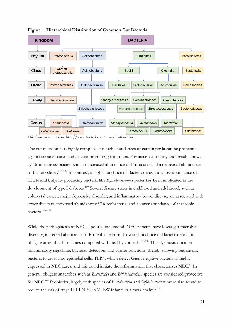
31
Figure 1. Hierarchical Distribution of Common Gut Bacteria
This figure was based on http://www.bacterio.net/-classification.html
The gut microbiota is highly complex, and high abundances of certain phyla can be protective
against some diseases and disease-promoting for others. For instance, obesity and irritable bowel
syndrome are associated with an increased abundance of Firmicutes and a decreased abundance
of Bacteriodetes.187, 188 In contrast, a high abundance of Bacteriodetes and a low abundance of
lactate and butyrate producing bacteria like Bifidobacterium species has been implicated in the
development of type I diabetes.189 Several disease states in childhood and adulthood, such as
colorectal cancer, major depressive disorder, and inflammatory bowel disease, are associated with
lower diversity, increased abundance of Proteobacteria, and a lower abundance of anaerobic
bacteria.190-192
While the pathogenesis of NEC is poorly understood, NEC patients have lower gut microbial
diversity, increased abundance of Proteobacteria, and lower abundance of Bacteriodetes and
obligate anaerobic Firmicutes compared with healthy controls.193-196 This dysbiosis can alter
inflammatory signalling, bacterial detection, and barrier functions, thereby allowing pathogenic
bacteria to cross into epithelial cells. TLR4, which detect Gram-negative bacteria, is highly
expressed in NEC cases, and this could initiate the inflammation that characterizes NEC.81 In
general, obligate anaerobes such as Bacteriodes and Bifidobacterium species are considered protective
for NEC.194 Probiotics, largely with species of Lactobacillus and Bifidobacterium, were also found to
reduce the risk of stage II-III NEC in VLBW infants in a meta-analysis.72

32
Several factors may cause dysbiosis in the developing gut microbiota during the neonatal and
infant period. An obvious example is the mode of delivery, as infants born via caesarean section
are not exposed to commensal bacteria from the maternal vaginal tract. Consequently, newborn
babies delivered by caesarean section have lower abundance of Bifidobacterium species.7 Instead,
these neonates may be more influenced by bacteria in their environment, such as bacteria from
their mother’s skin. Neonates delivered by caesarean section that are hospitalized after birth may
also be more heavily colonized by bacteria from the NICU environment, including genera from
the NICU itself and skin bacteria from caregivers’ hands.197
Breastfeeding has been associated with an increased diversity of the gut microbiota at one year of
age.198 The introduction of cow milk and a full adult diet causes shifts in the developing
microbiota, such as increasing the abundance of Bacteriodetes.184 Premature infants have a
different development of the gut microbiota than term infants with higher abundances of
Proteobacteria and Firmicutes, and lower abundances of Bacteriodetes.199 However, preterm
infants also have higher risks of being born via caesarean section, being formula fed and receiving
antibiotics, so significant confounding and interaction may occur.
Antibiotic treatment, particularly long-term treatment with broad-spectrum antibiotics, can cause
a selection pressure that causes antibiotic susceptible pathogens to die while other pathogens
survive.7 Antibiotic treatment causes an overgrowth of Proteobacteria at the expense of
commensal anaerobes.200 This may be partially due to losing the colonization resistance that
obligate anaerobes offer against pathogenic and antibiotic resistant bacteria.201 Indeed, antibiotics
early in life, including IAP and neonatal antibiotic treatment, have been associated with an
increased risk of obesity, allergies, inflammatory bowel disease, behavioural difficulties, IFI, and
NEC.69, 178, 202-205
1.9.2 Antibiotic Resistance
Most antibiotics are derived from antimicrobial substances that are naturally produced by
microorganisms. As these substances have existed for millennia, bacteria have naturally occurring
resistance mechanisms.206 However, selection pressure from the wide-spread use of antibiotics in
human medicine, veterinary medicine, and agriculture has made antibiotic resistance a developing
global health crisis. An estimated 214 500 neonates die yearly due to sepsis with antibiotic
resistant bacteria.207

33
Bacteria develop antibiotic resistance primarily through two different pathways; spontaneous
mutation and horizontal gene transfer.208 Spontaneous mutations can develop antibiotic
resistance through altering the drug targets, thereby coding for enzymes that change the structure
of the antibiotic or up-regulate efflux pumps.209 A classic example of enzymes changing the
structure of antibiotics are the beta-lactamases; enzymes that break down the central beta-lactam
ring of beta-lactam antibiotics. Horizontal gene transfer occurs through several different
mechanisms, but the transfer of plasmids is perhaps the most important.208 Antibiotics apply a
selection pressure that not only favours bacteria with antibiotic resistance genes, but also induces
transfer of resistance genes.210
According to a WHO surveillance report from 2014, extended-spectrum beta-lactamase (ESBL)-
producing E. coli and Klebsiella pneumoniae are among the most concerning antibiotic resistant
bacteria.211 In addition to penicillins, ESBL may hydrolyse third-generation cephalosporins and
even carbapenems. ESBL-rates are highest in South-East Asia, and 20-61% of E. coli isolates and
53-100% of K. pneumoniae isolates in this part of the world are resistant to third-generation
cephalosporins.211 In Europe, rates are more variable, and 4.9% of K. pneumoniae and 5.8% of E.
coli were ESBL-producing in Norwegian blood culture isolates in 2016.125 ESBL-producing
Enterobacteriaceae infection have a mortality rate of approximately 31-43% in neonates.212
Currently, 73% of Norwegian S. aureus isolates produce beta-lactamase and, therefore, cloxacillin
is commonly used to treat staphylococcal infections.125 However, the emergence of methicillin-
resistant staphylococci has made glycopeptide treatment necessary in many cases. In Japan, for
instance, MRSA causes 88% of S. aureus LOS.213 In contrast, only 11% of S. aureus LOS in the
UK is caused by MRSA, while 99% of Norwegian S. aureus blood-stream isolates are methicillin-
sensitive.25, 125 Other emerging threats are carbapenem-resistant Enterobacteriaceae and
vancomycin-resistant enterococci.214 Broad-spectrum antibiotics have been found to induce more
multi-drug resistant (MDR) Gram-negative bacteria in neonatal populations than narrow
spectrum antibiotics.132, 215 Moreover, both antibiotic treatment versus no treatment and
prolonged treatment versus shorter treatment have been found to increase the rate of MDR
Gram-negative bacteria in neonates.216
34
1.10 Evidence Based Medicine
Evidence-based medicine (EBM) is an approach to medical practice, and the Oxford Dictionary
of Epidemiology defines it as "the consistent use of knowledge derived from biological, clinical,
and epidemiological research in the management of patients".217 Clinical epidemiology is one of
the foundations of EBM, and it is the study of occurrences and distribution of health related
effects in a clinical setting. The highest achievement in epidemiology is to discover and
understand the cause-effect relationships behind diseases. Such understanding makes it possible
to treat or even prevent disease.
In epidemiology, a cause is something that alters the frequency of a disease or a health status. A
necessary step is finding associations between potential causes and the studied outcome, but
associations alone do not imply causality. Sometimes an association is erroneously interpreted as
causal when it is in fact the result of confounding (a third factor that is the true cause of the
association between exposure and outcome), interaction (two or more exposures working
together to affect the outcome), or bias (a systematic deviation of results from the truth).217, 218 To
establish causality, certain factors need to be present. A cause needs to precede the disease, show
a consistent effect, increase the incidence of the disease, have a dose-response effect (greater
effect in greater quantity), and its effects should be consistent across several studies. Bias exists in
many forms, and Table 2 explains the kinds of bias that are most relevant for this thesis.
Table 2. Types of Bias Relevant for This Thesis
Type of Bias Description
Confounding A variable that causes a spurious association by influencing both the
dependent and the independent variable
Selection bias Choice of study population leads to an uneven distribution of
confounding factors
Performance bias Systematic differences in care provided to members of different study
groups that is not the studied exposure
Detection bias Systematic differences between study groups in assessment,
ascertainment, diagnosis, or verification of outcomes
Reporting bias Selective revelation or concealment of information or results from a
study
Source: Porta M. A Dictionary of Epidemiology. New York: Oxford University Press, 2014.

35
Robust study designs are needed to minimize the risks and impact of bias and confounding.
Different study designs have different advantages, but central to EBM is a hierarchy of evidence
where meta-analysis, systematic reviews, and RCTs are at the top of the hierarchy.218 Systematic
reviews are critical appraisals of the scientific evidence that apply strategies to limit bias in
collection, synthesising, and critically appraising relevant studies.217 The core premise of this
method is to develop a research question and perform systematic searches according to
previously established criteria to uncover relevant studies. Studies are included or excluded based
on previously established criteria. A meta-analysis is a statistical analysis of results from several
studies, which can increase statistical power.217 Meta-analyses are commonly a part of the
systematic review process, but studies with low risks of bias and comparable populations,
exposures, and outcomes are required for such methods.219 RCTs are usually well suited for this,
due to their lower risks of bias. Typically, systematic reviews are based on RCTs, but
observational studies generally have longer follow-up time and larger population sizes and are
therefore well suited to study rare adverse effects.220
The Cochrane Collaboration is an esteemed international collaboration of researchers that work
to summarise evidence from health research. They have developed the Cochrane Handbook for
Systematic Reviews of Interventions; a handbook that aims to improve the methodological
quality of systematic reviews.219 The Cochrane Handbook includes tools for assessing
methodological quality in included studies and the quality of evidence. Additionally, it
recommends following the Preferred Reporting Items for Systematic Reviews and Meta-Analysis
(PRISMA) statement; a checklist for transparent reporting of systematic reviews.221 Several
journals demand that systematic reviews follow this statement, which increases the transparency
of systematic reviews.
Publishing the study protocol prior to performing the systematic review is part of the PRISMA
checklist.221 Several databases allow researchers to publish their protocols, and one of the largest
and most frequently used is PROSPERO.222 It is an international database of prospectively
registered systematic reviews in several academic fields, among them health care. Prospective
registration helps to counter publication bias as systematic reviews are searchable, regardless of
whether they were published or not. Additionally, it increases transparency and reduces reporting
bias as it allows the reader to compare the finished study with how the review was planned in the
protocol.

36
The Cochrane Handbook recommends using the Grading of Recommendations Assessment,
Development, and Evaluation (GRADE) system for evaluating the quality of evidence (QoE) in
a body of literature.219 This approach specifies four levels of quality from high to very low. These
levels of QoE define the degree to which estimates of effects or associations can be trusted.
Findings based on RCTs are initially assigned a high QoE, while findings based on observational
studies are initially assigned a low QoE. Several factors could upgrade or downgrade the quality
rating. For example, a dose-response effect or large effect estimates could increase the QoE,
while a high risk of bias or inconsistent results could decrease the QoE.223
An estimated 700-800 articles about antibiotic treatment in neonates are published in the
PubMed database every year, which makes it challenging for both clinicians and researchers to
stay up to date.224 Systematic reviews summarize, critically evaluate, and appraise the evidence,
and in doing so they can be important and very useful for caregivers. In many cases the evidence
is not of a sufficient quality to enable strong conclusions, but thorough and methodologically
robust systematic reviews will none the less give a summary of the current evidence in a field and
potentially pinpoint the need for further research. To our knowledge, no systematic reviews on
the adverse effects of neonatal antibiotic treatment have been published previously.

37
2 Aims of the Study
The overall aim of this thesis was to investigate different aspects of antibiotic therapy for
neonatal sepsis in order to obtain new knowledge that could improve and optimise care.
The specific objectives were:
• To investigate the epidemiology of EOS and exposure to systemic antibiotics during the first
week of life in an unselected national cohort of LB term infants.
• To evaluate a simplified high-dose extended-interval gentamicin dosing regimen with focus
on pharmacokinetic safety, potential ototoxicity, and the number of prescription errors.
• To identify, critically appraise, and synthesize evidence from studies reporting different
categories of antibiotic exposure in neonates and the subsequent risk of developing the
following three early adverse outcomes: NEC, IFI, and/or death.
• To identify, critically appraise, and synthesize evidence from studies reporting different
categories of antibiotic therapy in neonates and their impact on the gut microbiota and/or
antibiotic resistance development.

38
3 Materials and Methods
3.1 Study Design and Materials
Paper 1
Paper 1 is a registry-based cohort study of LB term infants admitted to neonatal units in Norway
during the three-year period from January 1, 2009 to December 31, 2011. Detailed clinical data
were prospectively collected by the Norwegian Neonatal Network (NNN), a web based public
registry maintained by the Norwegian Institute of Public Health. Twenty of the 21 neonatal units
in Norway contributed data during this period. In the NNN, clinical data are entered daily on all
infants admitted to each participating neonatal unit. In Norway, all infants receiving intravenous
antibiotic therapy are admitted to a neonatal unit situated in one of four regional health-care
trusts (South-East, West, Mid and North). Data on the total number of LB infants in Norway
were obtained from the Medical Birth Registry of Norway. Supplementary information on
systemic GBS infections, a notifiable disease in Norway, was obtained from the Norwegian
Surveillance System for Communicable Diseases. Mortality data were compared with data
obtained from the Norwegian Cause of Death Registry.
Paper 2
Paper 2 is a retrospective single-centre cohort study of neonates up to 50 weeks PMA that
received gentamicin and had ≥1 TPC measured between January 1, 2004 through May 31, 2012.
Patients were recruited from the NICU at the University Hospital of Northern Norway in
Tromsø. This NICU is the only tertiary neonatal unit in the two northernmost counties in
Norway, covering a population of 230 000 with around 3000 births per year.225 All infants <34
weeks GA and all infants receiving mechanical ventilation in the catchment area are treated in
this unit, so for these infants our data are population-based.
Paper 3 and 4
Paper 3 and 4 are systematic reviews of adverse effects following antibiotic treatment in the
neonatal period. Both reviews were reported according to the PRISMA statement following a
joint, prospectively registered protocol (study protocol registration: PROSPERO
CRD42015026743).226 Our primary research questions were:
• Paper 3: ‘Are different types of antibiotic exposure in neonates associated with increased risks
of the adverse outcomes NEC, IFI, and/or death in the neonatal period?’

39
• Paper 4: ‘Are different categories of antibiotic treatment in neonates associated with different
changes in gut microbiota composition and/or differences in antibiotic resistance
development?’
A study was eligible for review if it reported on groups of neonates, preterm or term, with
different categories of intravenous antibiotic exposure and examined their impact on either NEC,
IFI, or death in the neonatal period or up to discharge from the neonatal unit (Paper 3) or
changes in the gut microbiota or antibiotic resistance development (Paper 4). Both RCTs and
observational studies such as cohorts, case-control studies, and cross-sectional studies were
eligible for inclusion. We excluded case reports and case series, studies with a non-human or non-
neonatal population, studies that were not written in English, and studies that investigated
antenatal antibiotics, oral antibiotics, or low-dose intravenous vancomycin prophylaxis.
3.2 Gentamicin Dosing Regimen and Monitoring
In Paper 2, we administered gentamicin 6 mg/kg as a 30-min infusion, regardless of GA and
PNA. The dosing intervals ranged from 24-48 hours, depending on PNA and GA (Table 3).
TPCs were obtained right before the third dose. During the study period, two different
immunoassays with a lower limit of detection <0.3 mg/L were used to analyse gentamicin plasma
concentration (2004-09: GENT2, Roche, Mannheim, Germany, 2010–2012: CEDIA®
Gentamicin II Assay, Microgenics, Passau, Germany). An internal validation showed a good
correlation between both methods.
Table 3. Gentamicin Dosing Protocol
Postnatal age Gestational age (GA)/
Postmenstrual age (PMA)
Dosage Dosing Interval
Group A 0-7 days GA > 36 weeks 6 mg/kg 24 hours
Group B 0-7 days GA 29-36 weeks 6 mg/kg 36 hours
Group C 0-7 days GA < 29 weeks 6 mg/kg 48 hours
Group D >7 days PMA ≥ 29 weeks 6 mg/kg 24 hours
Group E >7 days PMA < 29 weeks 6 mg/kg 36 hours

40
3.3 Search Strategy in Systematic Reviews
In Paper 3 and 4, we developed our search strategy in consultation with an epidemiologist, a
librarian, a paediatric pharmacologist, and a neonatologist. We searched PubMed, Medline,
Embase, and the Cochrane database using MeSH-terms and free-text searches with no time
restrictions (last search December 22, 2016). The first search was conducted using MeSH terms.
The search strategy in PubMed, Medline and the Cochrane Database was to combine ‘Infant,
Newborn’ and ‘Anti-Bacterial Agents’ with either ‘Enterocolitis, Necrotizing’, ‘Fungemia’,
‘Candidiasis, Invasive’, ‘Meningitis, Fungal’, or ‘Mortality’ (Paper 3) or ‘Drug Resistance,
Bacterial’ or ‘Microbiota’ (Paper 4). The Embase database uses its own key words, and we
combined ‘Newborn’ and ‘Antibiotic Agent’ with either ‘Necrotising Enterocolitis’, ‘Fungemia’,
‘Invasive Candidiasis’, ‘Fungal Meningitis’, or ‘Mortality’ (Paper 3) or ‘Antibiotic Resistance’ or
‘Microbiome’ (Paper 4).
The second search was conducted using free text in PubMed, Medline and Embase by combining
the keywords ‘Infant, Low Birth Weight’, ‘Infant, Postmature’, ‘Infant, Premature’ or ‘Infant,
Newborn’ with ‘Anti-Bacterial Agents’ or ‘Antibiotics’ and one of the following outcome terms:
‘Necrotizing Enterocolitis’, ‘Fungaemia’, ‘Fungemias’, ‘Candidemia’, ‘Invasive Candidiasis’,
‘Fungal Meningitis’, or ‘Mortality’ (Paper 3) or ‘Antibiotic Resistance’, ‘Antibacterial Drug
Resistance’, ‘Microbiota’, ‘Microbiome’, ‘Microbiomes’, or ‘Gut Flora’ (Paper 4). We examined
reference lists of included studies and relevant reviews to identify additional eligible studies. We
then combined all citations and excluded duplicates or triplicates. We did not contact authors for
supplementary information and we did not perform searches in the “grey literature”, e.g.
materials and research produced by organizations outside of the traditional commercial or
academic publishing and distribution channels; thus not controlled by commercial publishers.
3.4 Variables and Definitions
Paper 1
Information on GA, birth weight by 500 g groups, Apgar scores, blood culture results and
information on treatment and clinical diagnoses were included in the analysis. We did not have
information on maternal fever or chorioamnionitis. Clinical diagnoses registered in NNN were
defined according to the International Classification of Diseases, 10th Revision. Bacterial sepsis
in the newborn (P36.0–P36.8) is defined as growth of bacteria in blood cultures together with
clinical signs and symptoms compatible with infection. Unspecified bacterial sepsis (P36.9) is

41
applied when there are clinical and biochemical signs of sepsis without growth of bacteria in
blood cultures or when blood cultures are not obtained.
For infants with EOS, infection onset was defined as the day antibiotic treatment began. We
defined EOS as infection onset in the first week of life. Infants diagnosed with sepsis (P36) who
did not receive intravenous antibiotics were considered misclassified. We ascertained all cases of
P36.0 - P36.8 by evaluating blood culture results and requested the neonatal units to register
blood culture results if they were missing. Cases of unspecified bacterial sepsis (P36.9) with
antibiotic treatment <5 days were not defined as EOS. Coagulase-negative staphylococci,
micrococci, Propionibacterium and Corynebacterium/diphtheroids in a single blood culture were
classified as contaminants, in line with suggestions by Stoll et al.30 Data on culture-confirmed
EOS in preterm infants were also collected to present incidence rates for all infants, irrespective
of GA.
Paper 2
Two of the authors (Jon W. Fjalstad and Claus Klingenberg) reviewed the medical records of all
eligible patients. We registered background data (sex, age, weight, diagnoses and complications
including acute renal failure) and gentamicin TPCs. Gentamicin TPCs <0.3 mg/L were assigned
a value of 0.2 mg/L. We took extra care to assess medical staff prescription and to evaluate
whether dosing (mg/kg) and dosing intervals were in line with the dosing protocol (Table 3). We
evaluated nursing staff administration and we defined a dose given >3 h earlier or later than
scheduled as an administration error.
Paper 3 and 4
Two reviewers (Jon W. Fjalstad and Eirin Esaiassen) independently screened search results and
assessed each potentially eligible study per our predetermined inclusion and exclusion criteria. We
only excluded studies that we agreed were irrelevant according to our predefined criteria. A third
researcher (Claus Klingenberg) had the deciding vote in cases of disagreement. We extracted the
following information from included studies: author, year, country, study design, study
population, including GA and BW, comparison of outcomes between groups with different
categories of antibiotic treatment, and, if available, risk estimates with 95% confidence intervals
(CI) for the specific outcome.
We compared three different categories of antibiotic therapy: (i) antibiotics yes versus no; (ii)
antibiotics long versus short duration; and (iii) broad-spectrum versus narrow-spectrum antibiotic

42
regimens. For category (ii), we suggested in advance that ‘prolonged’ antibiotic exposure was
either ≥3 days or the longest of two antibiotic regimens. For category (iii), we always defined
regimens including third-generation cephalosporins or carbapenems as a broad-spectrum regimen
when compared with regimens containing aminoglycosides for coverage against Gram-negative
bacteria. This definition was based on previous reports indicating that empirical treatment using a
third-generation cephalosporin for Gram-negative coverage induces significantly more antibiotic
resistance than regimens containing an aminoglycoside.132 If two similar regimens were
compared, the regimen with the broadest spectrum was labelled broad-spectrum.
We defined the neonatal period as up to 44 weeks PMA if the neonate was born prematurely.
NEC was defined as Bell’s stage 2–3.227 IFI was defined as fungaemia or detection of fungi in
otherwise sterile body sites. Death as an adverse outcome was defined as any cause of death,
including death attributed to infection during antibiotic therapy in the neonatal period or up to
discharge from the neonatal unit. Gut microbiota analyses were based on faecal samples using
both standard culture-based methods and culture-independent methods relying on DNA
amplification and sequencing. We decided to present data on the gut microbiota in three main
categories acknowledging some clear overlap; i) microbial load, ii) microbial diversity, and
iii) microbial composition. We defined microbial load as the total number of bacteria in a sample,
microbial diversity as the number of different bacterial genus or species in a sample, and
microbial composition as the taxonomic composition in a sample.
Antibiotic resistance development was based on detection of antibiotic susceptibility patterns in
bacteria isolated from blood, urine, CSF, faeces, tracheal aspirates, or the skin surface. We
defined MDR bacteria as bacteria resistant to either ≥ 2 unrelated classes of antibiotics or broad-
spectrum antibiotics. Included in this category were carbapenem resistant Acinetobacter baumannii,
ESBL-producing Gram-negative bacteria, and other third-generation cephalosporin resistant
Gram-negative bacteria. Antibiotic-resistant bacteria that did not meet any of these criteria were
defined as 'other antibiotic resistant bacteria'.
43
3.5 Audiology Assessment
In Paper 2, all infants were screened for ototoxicity with a transient-evoked OAE test (Madsen,
AccuScreen, GN Otometrics, Denmark) before discharge. Prior to 2007, a risk based screening
approach was used, including all neonates treated with gentamicin. Since January 2007, OAE has
been implemented as a universal screening test for all newborn infants. Patients who failed OAE
screening had an automatic ABR test as the first follow-up test. Further follow-up was then
individualised in the audiology unit. We carefully reviewed hearing data for all patients referred
for follow-up. An experienced audiologist reassessed all cases with possible persistent hearing
problems. To ensure that no patients with severe ototoxicity were missed, the audiologist also
identified all children who went on to have hearing aids or cochlear implants and were born
during the audit period. Furthermore, all patient at risk for neurological sequelae (GA <32 weeks,
VLBW, or severe perinatal asphyxia) were seen at regular intervals in the outpatient clinic up to 2
years of age, and sensory impairment was recorded.
3.6 Assessment of Methodological Quality
In Paper 3 and 4, the methodological quality of included studies was assessed by using the
Cochrane Handbook of Systematic Reviews of Interventions and recently published
recommendations on how to assess risk of bias and confounding in observational studies.219, 228
Five domains related to risk of bias were assessed for each study included: selection bias,
performance bias, detection bias, reporting bias, and confounding. Risks of bias were judged as
low, high or unclear for each domain (Appendix 9.1). The risk of reporting bias was considered
unclear in studies that did not have a previously published protocol. The risk of detection bias
was considered high in studies that examined the gut microbiota with culture-based methods,
unclear in studies that applied 16S rRNA sequencing techniques, and low in studies that applied
shotgun metagenome sequencing techniques. Two reviewers (CK and either EE or JWF)
assessed the risks of bias for each study. In Paper 4, we applied the GRADE approach to
evaluate the QoE for each relevant outcome category.223
3.7 Statistical Analyses
Paper 1 and 2
Data were analysed using IBM-SPSS (IBM, Armonk, NY) statistical software, versions 20
(Paper 2) and 22 (Paper 1). Continuous variables are expressed as mean (standard deviation (SD))
if variables were normally distributed or median (interquartile range (IQR)) if variables were not

44
normally distributed. Categorical variables are displayed as frequency (%). Paper 2 is purely
descriptive, and we did not test any variables for statistical significance.
In Paper 1, interval data were tested for normality using the Shapiro–Wilks test. Paired t-tests
were used to compare continuous data, and proportions were compared using c2 test. Correlation
was calculated using Spearman correlation. We used Kruskal–Wallis to test differences between
multiple groups. A post hoc analysis with Tamhanes T2 test, catering for unequal variances, was
used to test differences between individual groups. We calculated the number of antibiotics that
accounted for 90% of the total volume used. P values < 0.05 were considered statistically
significant.
Paper 3
We classified studies according to their outcome categories, including comparisons of different
categories of antibiotic therapy. In each outcome category, we combined adverse outcomes of
interest from studies we considered sufficiently homogeneous to provide a meaningful summary
and calculated combined effect estimates. Data entry and meta-analysis were performed using
RevMan version 5.3 (The Nordic Cochrane Centre, Copenhagen, Denmark). In the meta-
analyses, we pooled RCTs and non-randomized studies, the latter only if clinical baseline
characteristics of patient groups that experienced different antibiotic exposures (categories i–iii)
were similar and the studies reported dichotomous outcomes. Subgroup analysis was performed
for RCTs and observational studies.
We quantified inconsistency between the results of the studies by using the I2 test. Interpretation
of thresholds for statistical heterogeneity was as follows: I2 values between 0% and 40% might
not be important, whereas higher I2 values may represent moderate (30%–60%), substantial
(50%–90%) or considerable heterogeneity (75%–100%).219 We calculated odds ratios (ORs) with
95% CIs for the outcomes of interest. We present the effect estimates by using the random-effect
model due to assumption of clinical and methodological diversity among the studies,
subsequently often leading to statistical heterogeneity. Most non-randomized studies are reported
separately and were not pooled for meta-analysis because of marked clinical and methodological
diversity regarding interventions, antibiotics used, study design, and reported outcomes.
45
Paper 4
The large heterogeneity in study designs, comparisons, and outcomes made it impossible to
perform traditional meta-analysis of the included studies. Vote-counting methods can be used for
studies that do not contain enough information to compute an effect size estimate but do contain
information about the direction and the statistical significance of results, or that contain just the
direction of results.229 We therefore applied a vote-counting method to meta-analyse and
investigate whether the different categories of antibiotic therapy had any effect on the outcomes
of interest. Studies were classified based on whether they showed a reduction in the outcome
measure, no effect, or an increase in the outcome measure. When appropriate, outcomes were
presented in vote-count figures. The size of the squares in the vote-count figures were
proportional to the relative number of infants included in that study.
3.8 Ethical Approval
The regional ethical committee approved the study leading to Paper 1 (2013/358/REK nord).
The regional ethical committee also considered the retrospective study leading to Paper 2, but
characterized this study as a “quality assurance project” (2013/713/REK nord). The study was
consequently approved by the hospital institutional review board. Paper 3 and 4 did not require
ethical approval as they were systematic reviews with no patient interactions and did not contain
any confidential data.

46
4 Main Results
4.1 Paper 1
A total of 168 877 LB infants were born with GA ≥37 weeks in the catchment areas of the 20
units reporting data to the NNN during the three-year study period, and 10 175 of these (6.0%)
were hospitalized in their first week of life. There were 91 cases of culture-confirmed EOS (0.54
per 1000 term LB infants) and 1447 cases classified as culture-negative EOS (8.57 per 1000 term
LB infants). Among preterm infants (GA < 37 weeks), there were 50 cases of culture-confirmed
EOS among 11 649 infants (4.29 per 1000 preterm LB infants). This gave a total incidence rate of
0.78 culture-confirmed EOS cases per 1000 LB in all infants, irrespective of GA.
Gram-positive bacteria caused 83 of 91 (91%) culture-confirmed EOS cases among term infants.
Gram-negative bacteria caused 8 cases (9%). Figure 2 shows the distribution of EOS pathogens
in blood cultures. GBS was the most frequently isolated pathogen, with an incidence of 0.31
GBS-EOS cases per 1000 term LB infants. Seven preterm infants also had GBS-EOS; the total
incidence rate of GBS-EOS was 0.33 cases per 1000 LB infants in all infants, irrespective of GA.
There was one single EOS-attributable death (GBS-sepsis) among of 91 cases of culture-
confirmed EOS in term infants. Three patients with culture-negative EOS died; however, the
primary cause of death was a non-infectious condition for all three.
Intravenous antibiotic treatment was commenced during the first week of life in 3964 out of
10 175 (39.0%) infants included in the study, corresponding to an incidence of 2.3% of term LB
infants in Norway. Of these, 3725 (94.0%) commenced treatment within the first 72 hours of life.
Among 3964 neonates receiving antibiotic therapy, 2128 (53.7%) were never diagnosed with a
bacterial infection, but still received antibiotic therapy for a median (IQR) duration of 4 (3–5)
days. Table 4 shows the regional variations in antibiotic consumption and EOS incidence, as well
as differences in treatment depending on blood culture results and EOS diagnosis.
Empiric therapy consisted of an aminoglycoside and either benzylpenicillin or ampicillin in 3746
of 3964 cases (94.5%) (Table 4). Change of antibiotic regimen during the course of therapy was
more frequent in the patients receiving benzylpenicillin with an aminoglycoside (66/724; 9.1%)
compared with patients receiving ampicillin with an aminoglycoside (160/3022; 5.3%) (P <
0.001), but we observed no difference in mortality between these groups (aminoglycoside and
benzylpenicillin: 9/724=1.2% versus ampicillin and aminoglycoside: 29/3022=1.0%; p=0.41).

47
Figure 2. Distribution of Bacteria in Blood Culture-confirmed EOS
GBS; group B streptococci, S. aureus; Staphylococcus aureus, E. coli; Escherichia coli. Created using Microsoft Excel
(version 15.40).
Table 4. Regional Variations in Incidence of EOS and Antibiotic Consumption

48
4.2 Paper 2
We identified 546 treatment episodes from 457 neonates who had one or more gentamicin TPC
registered during the 8-year study period. 37 episodes (37/546; 6.7 %) were excluded from final
analyses on TPC and ototoxicity due to incorrect medical staff prescriptions. We included a total
of 509 treatment episodes (≥ three doses gentamicin) belonging to 440 patients. For the whole
study population, the mean (SD) GA was 36.4 (5.3) weeks and the mean (SD) BW was 2739
(1326) gram. There were 85 (19 %) patients with a very low birth weight (<1500 g) and 61
patients (14 %) with GA <29 weeks. Table 5 shows population and outcome data among the five
different treatment groups.
The mean (SD) gentamicin TPC for all treatment episodes during the first week of life was 1.1
(0.5) mg/L and after first week of life 0.8 (0.6) mg/L. Figure 3 shows pharmacokinetic data on all
509 treatment episodes, divided by the five treatment groups. We observed a potential toxic TPC
(≥2.0 mg/L) in 31/509 (6.1 %) treatment episodes. Of these, 22 were observed in group A and
predominantly in children with perinatal asphyxia (n =13) or acute renal injury for other reasons,
including congenital renal malformations (n =4).
Thirty-eight of 440 patients (8.6 %) failed the OAE screening before discharge and were referred
for follow-up in the audiology unit. Four patients who failed their OAE test were suspected to
have permanent sensorineural hearing loss, and one additional patient who passed the OAE test
later received a cochlear implant. Two of these five patients probably have small unilateral
hearing losses, two received hearing aids and one received a cochlear implant. Only one out of 31
patients with a TPC ≥ 2.0 mg/L suffered a permanent hearing loss, but this patient was also
diagnosed with a congenital CMV infection.
Thirty-one of 37 treatment episodes with medical staff prescription errors involved ordering a
12-h too long interval. Mean (SD) TPC among these was 0.6 (0.4) mg/L, and none had a TPC
≥2.0 mg/L. Six treatment episodes were prescribed with too short intervals (12 h). Mean (SD)
among these was 1.5 (0.9) mg/L, and in two episodes, TPC was ≥2.0 mg/L (33 %). We identified
81/509 (16 %) episodes with nursing staff errors regarding timing of administration. Gentamicin
was administered too late in 59 episodes (mean (SD) TPC, 0.9 (0.4) mg/L) and too early in 22
episodes (mean (SD) TPC, 1.0 (0.5) mg/L).

49
Table 5. Treatment Groups, Population Data, and Audiology Assessment
Figure 3. Gentamicin Trough Plasma Concentrations in Treatment Groups
Box plots show median values (solid bar), interquartile ranges (margins of box), and 5 and 95 percentile (whiskers).
Created using IBM SPSS (version 20.0)

50
4.3 Paper 3
47 studies met our inclusion criteria: 9 RCTs176, 230-237 and 38 observational non-randomized
studies (Appendix 9.2).1, 82, 118, 174, 238-271 There was a large diversity between the studies regarding
antibiotics used, as well as onset and duration of antibiotic exposure after birth (Appendix 9.3 a-
c). The majority of the included studies were judged to be of moderate to poor quality due to
many risks of bias (Appendix 9.4 a-c).
In the NEC category, there were highly divergent results in the six studies comparing antibiotic
therapy yes versus no,82, 230, 234, 260-262 and between the seven studies comparing broad- versus
narrow-spectrum antibiotic regimens.174, 176, 236, 237, 243, 264, 268 There was no significant difference
between antibiotics broad versus narrow regarding risk for NEC in the pooled analysis (Figure
4a). However, five studies comprising more than 5000 preterm infants showed significant
associations between duration of antibiotic exposure and NEC or the composite outcome of
NEC, LOS, or death.82, 238, 241, 261, 262 In contrast, five studies did not show a significant difference
in NEC rates.242, 260, 263, 268, 271 However, one of these five studies (2502 neonates total)
predominantly contained infants with GAs >34 weeks. 271 Moreover, three of these five studies
(448 neonates total) showed a trend towards higher NEC rates in patients with prolonged
antibiotic therapy, but all these studies were too small to detect significant differences.242, 260, 268
In the IFI category, twelve out of 15 studies reported an increased risk of IFI after broad-
spectrum antibiotic treatment, mainly third-generation cephalosporins or carbapenems,
compared with narrow spectrum treatment.232, 233, 240, 246, 248, 253-257, 266-270 Five studies reported an
increased risk of IFI following prolonged antibiotic therapy,240, 247, 249, 251, 252 while eight studies
found no significant difference.231, 246, 248, 250, 253, 265, 268, 270
In the mortality category, two studies, one of them extremely large (128 914 neonates), found an
increased risk of death after broad-spectrum antibiotic treatment.174, 243 However, seven studies
found no difference between antibiotics broad versus narrow and there was no significant
difference in the pooled analysis (Figure 4b).1, 176, 233, 235, 236, 264, 268 Four studies containing 12 832
preterm infants reported an increase in mortality following prolonged antibiotic therapy,82, 118, 241,
268 while seven studies containing 7506 neonates found no significant difference.231, 238-240, 242, 263, 271
However, one of the larger studies (2502 neonates) showing no difference included
predominantly term infants with a low risk of death.271

51
Figure 4. Forest Plots Stratified by Outcomes
(a) Pooled results of six studies comparing risk of NEC between neonates who received broad- versus narrow-spectrum antibiotic regimens
(b) Pooled results of eight studies comparing risk of death between neonates who received broader- versus narrower-spectrum antibiotic regimens
Subgroup analysis of RCTs and observational studies. The sizes of the squares are proportional to study weights.
Diamond markers indicate pooled effect sizes.

52
4.4 Paper 4
48 studies met our inclusion criteria: 3 RCTs132, 272, 273 and 45 observational studies (Appendix
9.2).179, 196, 215, 216, 263, 271, 274-313 The included studies were highly heterogeneous in both exposures and
outcomes (Appendix 9.3 d & e). Moreover, a large proportion of studies had a high risk of bias,
particularly selection bias, reporting bias, and confounding (Appendix 9.4 d & e).
Four studies examined the impact of antibiotic therapy on microbial loads with inconclusive
results.273, 279, 285, 286 Two out of four studies that compared antibiotic treatment yes versus no
found reduced microbial diversity following antibiotic treatment.196, 263, 285, 288 Three studies
examined the impact of antibiotic therapy duration (long versus short) on microbial diversity and
all three found decreased diversity following prolonged therapy.286, 288, 291 Nine studies focused on
Enterobacteriaceae; four reported an increase and five studies reported unchanged composition
after antibiotic treatment (yes versus no), mainly ampicillin plus an aminoglycoside (Figure 5a).179,
263, 278, 279, 281, 282, 285, 287, 290 Five studies focused on different commensal obligate anaerobes, showing
a clear trend towards reduced colonization rates following antibiotic treatment.280, 281, 283, 285, 287 Two
studies found lower colonization rates of Enterobacteriaceae after treatment with third-
generation cephalosporin compared with narrow-spectrum antibiotics.282, 290 We graded the QoE
as very low for outcomes in the gut microbiota category due to inclusion of observational studies
with serious risk of bias and/or inconsistent results.
In the antibiotic resistance category, 20 out of 31 studies focused on MDR Gram-negative
bacteria.132, 215, 216, 271, 272, 274-277, 282, 292-312 Nine studies reported data after antibiotic treatment yes
versus no, and seven of them reported increased rates of MDR Gram-negative bacteria following
treatment.216, 276, 296, 297, 299, 303, 307, 309, 310 Thirteen studies reported data after treatment with broad-
versus narrow-spectrum antibiotics, and the overwhelming majority reported higher rates of
MDR Gram-negative bacteria following treatment with broad-spectrum antibiotics (Figure 5b).132,
215, 274-277, 292, 296, 298, 304, 305, 307, 311 Five studies reported data after long versus shorter duration of
treatment, and four of them found significantly more MDR Gram-negative bacteria after
prolonged treatment.216, 271, 275, 297, 304 We graded the QoE as moderate for the outcomes relating to
antibiotic resistance development due to inclusion of observational studies that either had large
effect sizes or a dose–response effect.
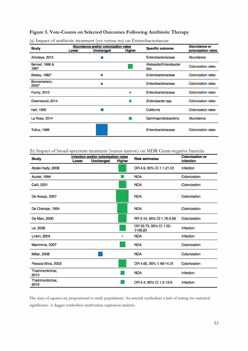
53
Figure 5. Vote-Counts on Selected Outcomes Following Antibiotic Therapy
(a) Impact of antibiotic treatment (yes versus no) on Enterobacteriaceae
(b) Impact of broad-spectrum treatment (versus narrow) on MDR Gram-negative bacteria
The sizes of squares are proportional to study populations. An asterisk symbolizes a lack of testing for statistical
significance. A dagger symbolizes multivariate regression analysis.

54
5 Discussion
The studies included in this thesis focused on the epidemiology of EOS and antibiotic use in the
first week of life in term born infants (Paper 1), the pharmacokinetics and potential toxicity of an
extended-interval gentamicin dosing regimen in neonates (Paper 2), and clinical (Paper 3) and
microbiological (Paper 4) adverse effects following neonatal antibiotic treatment.
We used different study designs in the different papers included in this thesis. Paper 1 is a
registry-based study with clinical and demographic data from a Norwegian cohort of term
infants. In Paper 2, we retrospectively collected clinical and pharmacokinetic data related to
gentamicin therapy in term and preterm infants from a single NICU. Paper 3 and 4 were
systematic reviews of RCTs and observational studies reporting adverse effects of antibiotic
therapy in the neonatal period, and the reviews followed a previously published protocol.
5.1 Epidemiology of Early Onset Sepsis
Using data that included all Norwegian neonates born during a three-year period (Paper 1), we
found an incidence rate of culture-confirmed EOS of 0.78 per 1000 LB infants. For term born
infants, the incidence was 0.54 culture-confirmed EOS per 1000 LB infants. This rate is in line
with data published from both England and the US. A UK multi-centre study reported an
incidence rate of 0.70 culture-confirmed EOS cases (0-48 hours) per 1000 LB infants, regardless
of GA.41 In the US, incidence rates between 0.78-0.98 culture-confirmed EOS cases (0-48/72
hours) per 1000 LB infants have recently been reported, with an incidence of 0.58 cases per 1000
LB infants for neonates with GAs ≥ 34 weeks.26, 30, 39 We applied a wider definition of EOS (0–6
days) than comparable studies, but the overwhelming majority of our EOS cases received
treatment within the first 3 days of life.
In Paper 1, GBS was the most commonly isolated pathogen, with an incidence rate of 0.31 GBS-
EOS per 1000 term LB infants. Including preterm infants, the Norwegian incidence rate was 0.33
GBS-EOS per 1000 LB infants. This is comparable with rates reported in US multi-centre studies
(0.41 per 1000 LB infants and 0.22 per 1000 LB infants), a UK study (0.30 per 1000 LB infants),
a Dutch nation-wide study (0.19 per 1000 LB infants), data from Sweden in 2009-2011(0.30 per
1000 LB infants) and data from a meta-analysis spanning several countries (0.43 per 1000 LB
infants).26, 30, 37, 38, 41 In accordance with guidelines from the Royal College of Obstetrics and
Gynaecology in United Kingdom, Norwegian health authorities recommend a risk-based

55
approach to identify women who may benefit from IAP for prevention of GBS EOS.49, 314 This is
in contrast to the Centers for Disease Control and Prevention’s guidelines who recommend
universal rectovaginal GBS-screening and IAP for all colonized women.50
Studies from the US and Australia indicate that universal swab-based screening programs have
lowered the rate of GBS EOS.30, 48, 315 However, some authors report an unchanged overall rate of
EOS with an increase in EOS caused by Gram-negative bacteria associated with higher
mortality.56, 316 In our study, the prevalence of E. coli and other Gram-negative EOS cases was
very low and the rate of GBS EOS was similar to or lower than that reported in countries using
universal swab-based screening programs. It is however, worth noting that our study consisted of
term neonates that have a lower risk of Gram-negative EOS.30 Additionally, the incidence rates of
GBS EOS may also be affected by clones with increased virulence or epidemic potential.38
We found a low EOS attributable mortality among Norwegian term infants. Only one neonate
(1%) died from culture-confirmed EOS after suffering from GBS sepsis. An additional three
infants with culture negative EOS died, but none of these deaths were attributable to infection
according to the Norwegian Cause of Death Registry. Other studies have found much higher
mortality rates from EOS (11-16%), but these studies also included preterm infants, which is
likely to be one major reason for the discrepancy.26, 30 Indeed, other studies on term-born infants
have reported mortality rates between 2-3% among EOS patients.30, 42
5.2 Antibiotic Consumption and Potential Implications
Overall, approximately 39% of hospitalized term infants in Norway received intravenous
antibiotics at some point during first week of life, with regional variations ranging from 36% to
41% (Paper I). We have no explanation for the regional differences in antibiotic use. However,
we have reasons to believe that regional differences may reflect differences in antibiotic policy,
including the use of CRP to guide treatment, as it is not likely that these differences reflect
disease severity in such a large, homogenous population-based study. In total, 2.3% of all term
infants in Norway received intravenous antibiotics in the first week of life.
There are few other population-based studies examining antibiotic consumption in the neonatal
population. In a selected population of newborns delivered at ≥34 weeks’ gestation at the Kaiser
Permanente Northern California network of hospitals, almost 6% of all infants received systemic
antibiotics in the neonatal period, and an even larger proportion receive antibiotics in other US
56
hospitals.112 There are no national Norwegian guidelines on when to start antibiotics in the
newborn infant at risk of or with clinical suspicion of EOS. In contrast, the guidelines from the
British National Institute for Health and Care Excellence (NICE) and the American Academy of
Pediatrics (AAP) specifically address these issues.94, 104 However, guidelines are often non-
dynamic, challenging to follow and may lead to overtreatment.317 Indeed, a study from the US
reported that when using the Centers for Disease Control and Prevention’s 2010 guidelines, 13%
of all infants were evaluated for EOS and 11% were treated empirically with antibiotics, although
only 0.04% of the cohort of infants had blood culture-confirmed infection.87
In retrospect, it is worth noting that 54% of the neonates who received antibiotics were not
diagnosed with an infection. Only 91 neonates had an infection with demonstrable growth in
blood-cultures, while ~1400 neonates were treated for an infection with negative blood-cultures.
Considering that blood cultures with samples above 1 ml have been reported to have a sensitivity
approaching 100% and that all included neonates in our study were term born, it is unlikely that a
large proportion of these culture-negative cases were severe infections with false-negative blood
cultures.96 Overall, ~3 term neonates were exposed to intravenous antibiotics for each case of
diagnosed, but unconfirmed infection, while ~44 neonates were exposed to antibiotics for every
case of confirmed EOS that was treated. Escobar et al. used a stratification scheme based on
maternal risk factors and objective neonatal clinical data to reduce the NNT to 118 per proven
EOS case, while a study from 18 North American and European hospitals reported a NNT of 63
per proven EOS case.90, 112 These findings therefore imply that Norwegian neonatologists are
relatively judicious in their antibiotic use.
It is important to consider the potential side-effects of antibiotic treatment in light of the high
rate of antibiotic exposure in neonates. Based on findings in Paper 4 we are moderately confident
that neonatal antibiotic therapy increases the risk of antibiotic resistance development, in
particular ESBL-producing Gram-negative bacteria and other MDR bacteria.216, 276, 296, 297, 299, 303, 307,
309, 310 Antibiotics overuse may lead to increased antibiotic resistance through several mechanisms.
Antibiotic resistance genes exist even in the absence of antimicrobial drugs, but antibiotics apply
a direct selection pressure that gives significant advantages to bacteria expressing resistance
genes.318-320 A recent study reported that only a fraction of the enriched antibiotic resistance genes
following antibiotic therapy are specific to the particular antibiotics given.321
Antibiotic treatment also contributes to changes in the human gut-associated resistome, which
comprises numerous functional antibiotic resistance genes in the gut microbiota.322 An antibiotic

57
induced increase in the gut resistome and decrease in colonization resistance could theoretically
increase horizontal transfer of antibiotic resistance genes from commensals to potential
pathogens.323 Although in vivo horizontal transfer between commensals and pathogens in the gut
microbiota remains to be proven, there is evidence of exchange of antibiotic resistance genes
between environmental bacteria and human pathogens.324
We are less confident about our findings related to antibiotic therapy and changes in the gut
microbiota (Paper 4). Neonatal antibiotic treatment was associated with an increased abundance
and/or colonization rates of Enterobacteriaceae in four out of nine included studies, whereas
none of the studies reported reduced abundance.179, 263, 278, 279, 281, 282, 285, 287, 290 Neonatal antibiotic
treatment was also associated with reduced colonization rates of protective commensal anaerobic
bacteria such as bifidobacteria, lactobacilli, or bacteriodes in four out of five included studies.280,
281, 283, 285, 287 It is possible that neonatal antibiotic therapy, regardless of treatment length, leads to
reduced microbial diversity, but the studies included in this category were small and two out of
four studies did not detect a significant difference.196, 263, 285, 288
All included studies in our systematic review (Paper 4) published prior to 2007 used culture-based
techniques to examine the gut microbiota composition. It has been estimated that <20% of
environmental bacteria can be grown in defined growth media.325 However, sequencing-based
techniques also have limitations. Studies relying on 16S rRNA analysis allow only a coarse sorting
of bacteria, mainly at phylum level. Deep shotgun metagenome sequencing allows for finer
distinction at the genus or species level, but it is of crucial importance to standardize sampling
and temperature control during the pipeline up to DNA extraction in order to obtain valid
results.326 Moreover, bioinformatic presentations are often challenging to understand and
interpret.
The frequent use of culture-based techniques added a significant detection bias for many of the
included studies (Paper 4), and the high risk of bias in the included studies was often the reason
for the very low QoE in the gut microbiota category. Nonetheless, our results are in line with
findings in adult populations showing decreased diversity, reduced colonization rates of obligate
anaerobes and increased colonization rates of Enterobacteriaceae following antibiotic
exposure.327-329 In contrast with the adult gut microbiota, the early-life gut microbiota is thought
to be less resilient and more susceptible to antibiotic treatment, causing larger shifts in the
microbial composition if antibiotics are administered in the neonatal period rather than later in
life.202

58
5.3 Choice of Antibiotic Regimen
We found that approximately 95% of term infants in Norway received an aminoglycoside
combined with either ampicillin or benzylpenicillin as their initial antibiotic therapy (Paper 1). We
found no difference in overall mortality between the regimens, but because of low mortality the
study was not powered for this comparison. It was slightly more common to change antibiotic
regimen during the course of therapy for neonates that were started on penicillin and gentamicin.
This was, however, a soft endpoint, and it may reflect differences in attitude and culture between
neonatal units, as the choice of empiric antibiotic regimens in neonates are based on local policy
in Norway.
To minimize harmful ecological effects of antibiotic therapy, some experts recommend using
empiric therapy with the narrow-spectrum combination of benzylpenicillin plus gentamicin for
suspected EOS.94 Others, including the AAP, recommend ampicillin and gentamicin.104 We
included two papers based on the same RCT comparing ampicillin with benzylpenicillin in our
systematic reviews (Paper 3 and 4).176, 272 In this RCT the researchers found no differences in
mortality, dysbiotic changes in gut microbiota, or development of MDR bacteria between the two
regimens.176, 272 However, this RCT was underpowered to detect clinical differences, and gut flora
analysis was performed with conventional culture-based methods.
In Norway, GBS isolates are uniformly susceptible to both benzylpenicillin and ampicillin.125
Neonatal listeria infection, a notifiable disease, is extremely rare in Norway, but listeria strains are
often susceptible to benzylpenicillin. However, a steady rise in gentamicin resistance among
E. coli blood culture strains in Norway (~6% in 2016) is of great concern.125 Furthermore, 96% of
gentamicin resistant E. coli isolates are also resistant to ampicillin. The prevalence of E. coli sepsis
was low in our term infant population, but it is more frequent in preterm infants.26 A further
increase in gentamicin resistance could potentially threaten the value of gentamicin as Gram-
negative back-bone coverage in the traditional empiric regimens.
The alternative to gentamicin-based regimens would be to use a more broad-spectrum antibiotic
such as a third-generation cephalosporin, piperacillin-tazobactam, or a carbapenem. Norwegian
E. coli blood culture isolates have similar resistance rates to cefotaxime (6%) as gentamicin, but in
2016 they were all susceptible to meropenem.125 There are, however, findings in our systematic
reviews (Paper 3 and 4) that indicate an increase in adverse effects following treatment with
broad-spectrum antibiotics. First, there is evidence from ten observational studies that previous

59
exposure to third-generation cephalosporins or carbapenems is associated with an increased risk
of developing IFI.240, 246, 253-256, 266-269 Preterm infants are more prone to early colonization of fungi
than term infants due to an immature immune system and impaired skin and mucosal integrity.330
Broad-spectrum antibiotics may foster IFIs by suppressing normal flora and allowing fungi to
occupy muco-epithelial niches that facilitate invasion and dissemination.331 Cephalosporin use has
been associated with intestinal colonization with Candida among neonates, and colonization is a
risk factor for IFIs.247, 250 Moreover, twelve out of 13 studies found a higher chance of infection
or colonization with MDR Gram-negative bacteria in neonates who were treated with broad-
spectrum antibiotics rather than narrow-spectrum antibiotics (Paper 4).132, 215, 274-277, 292, 296, 298, 304, 305,
307, 311 Taken together, the results from Paper 3 and 4 imply that there are substantial data
indicating that broad-spectrum antibiotics may pave the way for IFI and development of MDR
Gram-negative bacteria. In light of these findings, it is reassuring that cefotaxime appears to be
less commonly used for empirical EOS treatment than ten years ago.119, 128
5.4 Gentamicin Pharmacokinetics and Toxicity
Potential ototoxicity and nephrotoxicity has traditionally been a concern with aminoglycoside
based regimens.143 In neonates, this toxicity has never been proven, and aminoglycosides are not
associated with increased rates of hearing loss with high-dose extended interval dosing
regimens.332 In our evaluation of a simplified high-dose extended-interval gentamicin regimen
(Paper 2), we found that 6 % of all treatment episodes had a TPC ≥2 mg/l. This proportion is
similar or lower than in most comparable studies,148, 149, 162, 333, 334 but two studies reported even
lower rates of potential toxic TPCs.335, 336 In one of these studies, gentamicin was administered
every 24 hours with 4 mg/kg to infants with a GA ≥35 weeks and 3 mg/kg to infants with a GA
<35 weeks. All patients had TPCs < 2.0 mg/L, but 20 of the preterm infants with GA <35
weeks had PPCs < 6mg/L.335 A dosing protocol from Christchurch, New-Zealand has complex
dosing equations based on birth weight, leading to higher dose (mg/kg) and longer intervals (up
to 60 h) for infants with the lowest body weight. In their evaluation of more than 1,000 TPCs,
they reported high PPCs and low TPCs, but 87 % of all patients had only received one dose of
gentamicin.336
Impaired renal function and high plasma creatinine values are well-known risk factors for high
aminoglycoside TPCs.146, 162 Accordingly, we found that most term infants in the first week of life
with a TPC >2 mg/L had perinatal asphyxia and renal impairment (Paper 2). When renal failure
is likely, it may be advisable to either check TPCs already before the second dose of gentamicin,

60
to routinely increase dosing intervals to 36 hours, or to use a different empiric antibiotic until
renal function is clarified. In the NICU in Tromsø cefotaxime is routinely used for empiric
treatment of infants with severe perinatal asphyxia, in particular infants undergoing hypothermia
who are already at high risk for later hearing impairment.146
The gentamicin dosing regimen in this thesis (Paper 2) has a higher dosage (mg/kg) than what is
commonly recommended for neonates. Higher peak levels most likely optimise the efficacy of
gentamicin treatment. In contrast, there is little support in the literature for an association
between high peak levels and toxicity in neonates.161, 337, 338A lack of data on peak gentamicin
levels diminished our ability to fully assess the pharmacokinetic efficacy of our dosage regimen.
However, in the NICU in Tromsø we felt it was unnecessary to continue measuring peak levels
in this high-dose regimen after already having evaluated peak levels in a previous study.146
Repeated blood tests for therapeutic drug monitoring increases the patient’s pain and may cause
clinically important blood loss. Furthermore, 75 % of the cost of gentamicin therapy is due to
therapeutic drug monitoring.339 Based on previous results from a study in Tromsø using the same
dose (mg/kg) for netilmicin, and other studies using gentamicin 4–5 mg/kg, we would expect
that the majority of peak levels with the current dosing regimen (Paper 2) are >10 mg/L.146, 148, 149
Newborn infants treated with aminoglycosides are at risk of developing hearing impairment.
However, there are many other potential risk factors for hearing impairment including perinatal
asphyxia, CMV infections, intracranial complications, congenital malformations, prematurity and
treatment with loop diuretics.161, 340, 341 A combination of more than one risk factor is often found
in children who later develop hearing impairment. In one study, gentamicin did not seem to
induce any ototoxicity, and in fact, a protective effect against ototoxicity was proposed.158
OAE is considered an effective screening test for detecting aminoglycoside-induced cochlear
ototoxicity, but PPV is low due to low prevalence.155 In Paper 2, 38 (8.6%) infants failed the
OAE test. Only 4 out of 38 patients who failed the OAE tests were later diagnosed having
permanent hearing impairment, and all four had TPCs < 2 mg/L. The only child who had a TPC
≥2 mg/L and acquired a hearing impairment passed the OAE test, but gradually evolved hearing
impairment due to a congenital CMV infection. The low rate of hearing impairment among our
high-risk intensive care infants, and in particular among patients with potential toxic TPCs, is a
strong indication that gentamicin treatment is safe. Long-term follow-up studies with detailed
hearing evaluation are still needed to confirm this.

61
We did not perform serial creatinine measurement or analyse urinary biomarkers for detailed
assessment of potential gentamicin nephrotoxicity. Gentamicin nephrotoxicity, however, is
challenging to assess in the first week of life when plasma creatinine values are unstable and
influenced by renal maturity and changes in systemic circulation of sick neonates.342 Furthermore,
it seems that in neonates, aminoglycosides rarely induce clinically relevant renal injury in a normal
course of treatment when TPC is in a safe range.147 In contrast, when infants have high TPCs
gentamicin is often discontinued as these infants usually already have an impaired renal function
and one does not want to further exaggerate this with gentamicin.
Gentamicin is one of the drugs most commonly associated with prescription errors in the
paediatric setting, increasing the risk of high TPCs.343 Simpler dosing protocols are associated
with less prescription errors.165 In Paper 2 we found that 93 % of all treatment episodes were
correctly prescribed. Among the cases where we detected prescription errors, almost 2/3 were
made in preterm infants after the first week of life, leading to a too large dosing interval and less
potential toxicity. It is likely that medical staff only considered the low GA and failed to recognise
and assess the PNA. Improvements in education of medical staff may reduce such errors.
5.5 Prolonged Antibiotic Therapy
In our epidemiological study of Norwegian term infants (Paper 1) median treatment duration was
8 (7–10) days for culture-confirmed EOS and 6 (5–7) days for culture-negative EOS. In contrast,
a study from Switzerland reported a substantially longer duration of antibiotic treatment (mean
13 days) for infants with confirmed infection.344 The AAP guidelines recommend a minimum of
10 days treatment for culture-confirmed sepsis, while the NICE guidelines recommend a
minimum of 7 days for culture-confirmed sepsis and culture-negative neonates with a strong
clinical suspicion of sepsis.94, 104 We believe that the low mortality among term infants in Paper 1
indicates that most infants with culture-confirmed EOS can be treated safely with 7–10 days
systemic antibiotics, and that a shorter course may be appropriate for culture-negative EOS with
rapid clinical improvement.
Recent guidelines on neonatal sepsis emphasize the importance of stopping antibiotics after 36–
48 hours if there is no longer suspicion of sepsis.94, 104 In Paper 1, 26% of all admitted infants
received a median of 4 days antibiotics without being diagnosed with an infection. Furthermore,
it is likely that among the infants in our study diagnosed with a culture-negative EOS there were a
substantial number of infants not being truly infected, but still treated with a 5–7-day course of

62
antibiotic therapy. In many of these cases it is therefore likely that treatment could have been
safely stopped several days earlier.
Stopping antibiotics some days earlier would shorten the average length of stay in the neonatal
unit, leading to a significant reduction in hospital expenditures. Further advantages are reductions
in maternal–infant separation and the pain for the infants associated with frequent blood samples
and insertion of intravenous lines. However, in spite of guidelines emphasizing early cessation of
antibiotics if sepsis is ruled out, the effects of guidelines may be different. A recent report
showed that after implementing NICE guidelines, more investigations and increased length of
stay were observed in newborns with suspected EOS when following the new guidelines.317
Prolonged antibiotic treatment was associated with several adverse effects in our systematic
reviews (Paper 3 and 4). First, five observational studies including around 5000 infants showed
that prolonged duration of antibiotic exposure for uninfected preterm infants is associated with
an increased risk of developing NEC later in the neonatal period (Paper 3).82, 238, 241, 261, 262 NEC has
previously been associated with dysbiotic changes in the gut microbiota such as low diversity,
overgrowth of Proteobacteria and decreased abundance of obligate anaerobic bacteria from the
Bacteriodetes and Firmicute phyla.193, 196 In Paper 4, prolonged antibiotic therapy seemed to
reduce gut microbial diversity, but QoE according to GRADE evaluations was very low.286, 288, 291
We did not find any conclusive evidence that prolonged antibiotic treatment caused more
changes in the abundance of specific gut bacteria than shorter treatment durations, but very few
studies examined this.263, 284, 291, 313 However, shorter courses of antibiotic therapy are associated
with a more rapid recovery from suppression of the gut microbiota.263, 345
Several biological mechanisms have been proposed to explain the association between gut
dysbiosis and the massive gut inflammatory response seen in NEC. NEC cases have been
reported to have an overexpression and dysregulation of TLR4.81 An increased abundance of
Enterobacteriaceae could lead to overexpression and increased activation of TLR4, resulting in
the excessive inflammation that characterizes NEC. Antibiotic-induced killing of obligate
anaerobes can potentially also lead to an increased abundance of Enterobacteriaceae due to a loss
of colonization resistance.201 It is also well known that bifidobacteria may reduce expression of
inflammatory response genes and stimulate genes promoting the integrity of the mucosal
barrier.346 Moreover, certain lactobacilli appear to lower the inflammatory response from LPS
stimulation, and these factors might explain why probiotics are associated with lower risks of

63
NEC.72, 347 There were, however, few studies included in paper 4 that examined the impact of
prolonged antibiotic therapy specifically on Enterobacteriace or commensal anaerobes.
Prolonged antibiotic treatment was also associated with an increased risk of colonization or
infection with MDR Gram-negative bacteria, and this outcome had a moderate QoE (Paper 4).216,
271, 275, 297, 304 We also found an association between prolonged antibiotic therapy and the risk of
death in four studies including very preterm infants (Paper 3).82, 118, 241, 268 Two of these studies
were extremely large retrospective cohorts with a total population of 12 863 VLBW infants. They
specifically examined the impact of antibiotic treatment for uninfected neonates.82, 118 In contrast,
seven studies found no significant difference, but many of these studies were small or largely
contained term infants with a lower risk of death. It is possible that the associations between
prolonged treatment and mortality were statistical anomalies, as even small differences can
produce p-values <0.05 if the study population is large enough. On the other hand, it is possible
that the studies that did not find a significant difference were underpowered to detect an actual
difference. If it happens to be real, there are several possible explanations for an association
between prolonged antibiotic treatment and mortality in uninfected neonates, including higher
risk of NEC, LOS, IFI, infection with MDR bacteria, or immune-related diseases secondary to a
certain degree of immune suppression.182
We did not study the impact of prolonged gentamicin treatment on potential hearing loss in
Paper 2. This was due to both the very low incidence of permanent hearing loss in the study
population and also the low rate of prolonged gentamicin therapy (≥ 5 days). Other studies have
examined the relationship between prolonged gentamicin treatment and hearing loss, and a
recent cohort study detected a non-significant trend for increased rates of hearing loss following
gentamicin treatment ≥ 5 days compared with shorter durations of treatment.332
5.6 Methodological and Ethical Considerations
5.6.1 Registry-Based Cohort Studies
Norway has several nationwide medical registries that cover practically the entire population.348
The NNN is one of the newer nationwide medical registries in Norway, and has covered all
Norwegian neonatal units since 2011. Nationwide registries enable medical research on large
cohorts over long time periods, which is especially useful when studying rare diseases such as
neonatal sepsis. Indeed, the main strength of Paper 1 was the population-based design that

64
captured approximately 97% of all term LB infants admitted to a neonatal unit in Norway during
the 3-year study period. This large and unselected study population minimizes the risk of
selection bias.
The main limitation of registry-based studies is that data has already been collected when the
study is planned. This could potentially increase the risk of detection bias as the researcher
depends on the judgments of multiple clinicians for the accuracy of outcomes, as well as their
zealousness in reporting exposures. In Paper 1, we relied on a substantial number of clinicians
performing the daily web-based registration in the NNN and concluding with diagnoses at
discharge. However, the data in the NNN was registered prospectively and the data on antibiotic
therapy was registered on a daily basis in the NNN. This makes underestimation of treatment
length unlikely. We also took steps to verify the outcome data we collected from the NNN by
comparing it to data from other Norwegian public registries. In fact, the NNN managed to
capture all cases of GBS EOS in term infants according to data from the Norwegian Surveillance
System for Communicable Diseases. We also confirmed diagnoses of culture-confirmed EOS by
examining blood culture results.
The diagnosis of culture-negative sepsis (P36.9) is particularly controversial, and the definition
proposed by Norwegian neonatologists was not universally followed in NNN. Data on CRP
levels that could have supported or refuted a clinical sepsis diagnosis were not included in the
NNN during the study period. In addition, it was difficult to determine whether skin flora
isolates in blood cultures were causes of actual infection or contaminants in a registry based
study. We chose to define all skin isolates as blood culture contaminants, in line with a
comparable US study.30 It is possible, however, that some cases of CoNS bacteraemia
represented true infections, despite our entire population being term born. We also lacked
information on maternal risk factors for EOS, such as maternal fever, rupture of membranes,
and chorioamnionitis, which we could have added in a truly prospective study.
5.6.2 Retrospective Cohort Studies
Retrospective cohorts are possible to perform when medical records allow accurate assessment
of both exposures and outcomes without any additional data collection.218 Retrospective cohorts
are, similarly to registry-based cohort studies, cheap and data can be collected rapidly. Paper 2
was a retrospective cohort study. Paper 2 was, to our knowledge, the largest study ever to analyse
an extended-interval gentamicin dosing regimen in neonates that included infants with all GAs

65
and a large number of infants with PNAs of at least one week. These data were population based
for infants born in the two northernmost counties in Norway with GA < 34 weeks or
requirement of mechanical ventilation. Again, this minimized the risk for selection bias.
In Paper 2, the retrospective nature of the study made it difficult to fully assess all levels of
ototoxicity. Infants with severe hearing impairment were identified, but we may have missed less
severe ototoxicity in the neonates who were born towards the end of our study with less than 21
months of observation. While OAE is an effective screening tool for detecting hearing loss, it is
possible that high-frequency hearing loss, which was not clinically apparent may have been
missed.155 These issues increased the risk of detection bias. We are currently performing a
prospective long-term follow-up with a complete audiological assessment of the same cohort
now in the age between 5-15 years in order to get an even more reliable assessment of whether
this high-dose and extended interval regimen has an ototoxic potential (ClinicalTrials.gov
identifier: NCT03253614).349
5.6.3 Systematic Review Methodology
The primary strengths of our systematic review (Paper 3 and 4) were our rigorous and sensitive
search strategy. The fact that we published our study protocol in advance of the reviews
themselves increased transparency and shows that our research questions and methodology were
decided a priori. We also used two to three authors to decide whether to include or exclude
studies based on our protocol, and to evaluate the methodological quality of included studies
based on a modified version of the Cochrane Handbook.219, 228 This reduced the risk of mistakes
causing deviations from protocol.
The main challenges for both reviews were the low number of RCTs, and the heterogeneity in
study designs, sample sizes, outcomes, categories of antibiotic treatment and methodological
quality. These challenges meant that traditional meta-analysis was only possible for a small subset
of studies in Paper 3 and that we had to use the vote-counting method in Paper 4 to assess the
effect of neonatal antibiotic treatment on relevant outcomes. The vote-counting method has
limitations as it usually fails to account for the population size and methodological quality of
pooled studies. Nevertheless, vote-counting may be an effective method to assess the ranking of
outcomes.350 Moreover, we attempted to improve the method by presenting the differential
weight of each study with squares corresponding to sample size.
66
Observational studies are prone to biases and confounding, and many of the included studies
attempted to adjust for confounders, such as risk factors and illness severity, through
multivariable regression analysis. This reduced the risk of random findings in our reviews, but we
cannot rule out residual confounding and confounding by indication: sicker neonates receive
more antibiotics, but antibiotic exposure does not make them sicker.118 According to the
GRADE approach, evidence from observational studies is usually considered to be of low
quality.223 However, well-designed observational studies have been shown to provide similar
results to RCTs and they can therefore be useful for detecting rare adverse outcomes by allowing
larger sample sizes and longer lengths of follow-up than RCTs for lower costs.351 We included
observational studies due to our intention to collect as much evidence related to our research
questions as possible.
The evidence of a significant association between prolonged duration of antibiotic therapy and
increased risk of NEC and/or death is mainly supported by retrospective studies in preterm
infants, and we cannot conclude that there is a causal relationship. This also applies to the
association between broad-spectrum antibiotics and increased risk of IFI. However, antibiotic
exposure was identified before the outcomes and cohort studies potentially have a temporal
framework to assess causality. We decided a priori to include studies with both term and preterm
infants as we anticipated that some studies would include a mix of both, and we did not want to
exclude these. Term infants, however, rarely develop NEC and IFI, and have a low mortality in
general. The differences in study populations therefore need careful consideration when
interpreting the results of our systematic review. Based on studies in Paper 3, we believe that it is
possible to draw conclusions about the association between antibiotic exposure and early adverse
outcomes in preterm infants, whereas data on NEC, IFI, and death are more limited in term
infants and do not justify clear conclusions. We feel more able to draw conclusions in term
infants regarding changes in gut microbiota and antibiotic resistance development (Paper 4), as
these changes are not exclusive to preterm infants.
In Paper 4, we used the GRADE approach to assess the QoE. Overall, we graded the QoE as
very low for all outcomes presented in the gut microbiota category. In contrast, we considered
the QoE to be moderate in the antibiotic resistance category owing to large effect sizes and a
dose–response effect. Based on current evidence we are therefore moderately confident that all
types of antibiotic treatment lead to increased rates of antibiotic resistance. We felt that the
GRADE approach strengthened our interpretations in Paper 4, and the fact that we did not use
this method in Paper 3 is a limitation.

67
We also acknowledge that our definition of broad-spectrum and narrow-spectrum antibiotics is
somewhat arbitrary as most of the narrow-spectrum regimens covered both Gram-negative and
Gram-positive bacteria. However, Paper 4 confirms previous findings, clearly suggesting that
antibiotic regimens containing third-generation cephalosporins or carbapenems are more
frequently associated with antibiotic resistance development than regimens with aminoglycosides
for Gram-negative coverage.132, 215, 274-277, 292, 296, 298, 304, 305, 307, 311 Finally, we decided to exclude studies
from Paper 3 and 4 that only examined antenatal antibiotic treatment, despite the frequent use of
IAP for prevention of neonatal infections and its reported effects on the infant gut microbiota
and carriage of antibiotic resistance genes.352 The focus of these reviews was neonatal antibiotic
treatment given for suspected neonatal infection, and the isolated effects of antenatal antibiotics
given to infants who did not receive antibiotics after birth were beyond the scope of these
studies.
5.6.4 Ethical Considerations
None of the studies that formed this thesis were ethically controversial. Papers 3 and 4 were
systematic reviews of already published studies, and as such there were no ethical aspects to
consider. Paper 1 was based on the NNN, and all the information in this registry was
anonymized. We chose to contact neonatal units for blood culture results for patients with a
diagnosis of culture-confirmed EOS when blood culture results were missing in the NNN, but
we did not directly access confidential information. This study was approved by the regional
ethical committee.
Paper 2 was based on medical records, and there was no contact with study subjects. We did,
however, need to access to confidential information to collect data for the study. Access to
confidential patient information is regulated by the Health Personnel Law in Norway. However,
the ability to grant dispensation to access confidential information for medical research is
delegated to the Regional Ethical Committees. The Regional Ethical Committee considered in
their feedback to the study protocol that our study was a “quality assurance project”, and they
suggested that we only needed approval from the institutional review board. The institutional
review board granted us access to this data. Information that could be traced back to individual
patients was stored separately and safely and was not part of the published study.

68
6 Conclusions
• The incidence of culture-confirmed EOS in term born infants was low in Norway (0.54 per
1000 live-born term infants), and in line with comparable reports from other developed
countries. Gram-positive bacteria caused 90% of culture-confirmed EOS, and GBS was the
most common causative pathogen. The EOS-attributable mortality rate was very low (1%).
• Of all Norwegian term infants, 2.3 % were treated with antibiotics in the first week of life,
primarily with an aminoglycoside and either penicillin or ampicillin. Over half of these were
never diagnosed with an infection. Guidelines commonly recommend ending treatment if
blood cultures are negative after 36-48 hours, but the median treatment length was 4 days for
neonates that received antibiotics without infection and 6 days for infants with culture-
negative EOS.
• We found no evidence for ototoxicity from gentamicin treatment following a high-dose
extended interval regimen. Only 6% of trough plasma concentrations were above the
commonly recommended 2 mg/L threshold. Our simplified dosing regimen resulted in a low
number of prescription errors.
• Prolonged antibiotic therapy was associated with an increased risk of NEC and/or death in
preterm infants and broad-spectrum antibiotics were associated with an increased risk of
invasive fungal infections.
• All types of increased antibiotic exposure in the neonatal period, whether it was antibiotics
versus no antibiotics, prolonged treatment versus shorter treatment, or broader-spectrum
antibiotics versus narrower-spectrum antibiotics, increased the rates of colonization and/or
infection with MDR Gram-negative bacteria (moderate quality of evidence.
• Neonatal antibiotic therapy, in general, appeared to induce various potentially disease
promoting alterations in the gut microbiota, in particular a reduced microbial diversity and a
reduction in “protective” commensal obligate anaerobes (very low quality of evidence).

69
7 Future Perspectives
While antibiotic can be life-saving, our findings strongly emphasize the need to reduce
unnecessary antibiotic treatment in neonates. In addition, they illustrate that while Norwegian
neonatologists are relatively judicious in their use of antibiotics, there remains further potential
for reducing neonatal antibiotic exposure. Preventing infections, antibiotic stewardship, and
knowledge-based use of today’s antibiotics are central principles to avoid overuse and adverse
outcomes related to antibiotic exposure in the neonatal period, and to maintain safe and effective
treatment for those who need it.
In general, it is better to prevent rather than treat disease. Development of a GBS vaccine could
potentially reduce rates of EOS and the amount of antibiotics neonates are exposed to. Until
such a vaccine is developed however, the debate on whether to use a universal screening
approach or a risk-based approach for IAP would be greatly informed by studies that directly
compare their effectiveness. It is possible that a large amount of IAP exposure causes more harm
than benefit for neonates, and a systematic review of the potential adverse effects from IAP
treatment would be an important step in determining this.
Development of new diagnostic tools could lead to a faster and more precise diagnosis of
neonatal sepsis, which in turn would reduce antibiotic exposure for healthy neonates. As it
remains difficult to decide early on whether a neonate is truly infected or not with current
diagnostic tools, it is vital to find safe ways to reduce unnecessary antibiotic exposure for
neonates. Strategies that separate neonates into different risk categories for EOS appear to be
promising in reducing the proportion of antibiotic treated neonates in a safe manner. Moreover,
further studies could determine whether it is safe to withhold treatment for well-appearing
neonates with maternal risk factors for EOS. Measures should also be taken to discontinue
antibiotic treatment early (36-48 hours) if a clinically suspected infection is not confirmed.
It is important to restrict the empirical use of broad-spectrum antibiotic treatment.
Aminoglycoside-based regimens cause less resistance than cephalosporin- or carbapenem-based
regimens, but have often been thought to cause hearing loss and renal failure. While gentamicin
in the neonatal period appears to be safe regarding ototoxicity in retrospective studies,
prospective follow-up studies with audiometry testing could help to determine whether
aminoglycosides cause subclinical hearing loss. Development of new antibiotics and new ways to

70
combat antibiotic resistance could ensure effective treatment for neonatal infections in the future
as increasing resistance rates threaten the effectiveness of aminoglycoside-based regiments.

71
8 References
1. Fjalstad JW, Stensvold HJ, Bergseng H et al. Early-onset Sepsis and Antibiotic Exposure in Term Infants: A Nationwide Population-based Study in Norway. Pediatr Infect Dis J 2016; 35: 1-6. 2. Fjalstad JW, Laukli E, van den Anker JN et al. High-dose gentamicin in newborn infants: is it safe? Eur J Pediatr 2013; 173: 489-95. 3. Esaiassen E, Fjalstad JW, Juvet LK et al. Antibiotic exposure in neonates and early adverse outcomes: a systematic review and meta-analysis. J Antimicrob Chemother 2017; 1: 1858-70. 4. Fjalstad JW, Esaiassen E, Juvet LK et al. Antibiotic therapy in neonates and impact on gut microbiota and antibiotic resistance development: a systematic review. J Antimicrob Chemother 2017 [Epub Ahead of print]. 5. Lauder AP, Roche AM, Sherrill-Mix S et al. Comparison of placenta samples with contamination controls does not provide evidence for a distinct placenta microbiota. Microbiome 2016; 4: 29. 6. Collado MC, Rautava S, Aakko J et al. Human gut colonisation may be initiated in utero by distinct microbial communities in the placenta and amniotic fluid. Sci Rep 2016; 6: 23129. 7. Penders J, Thijs C, Vink C et al. Factors influencing the composition of the intestinal microbiota in early infancy. Pediatrics 2006; 118: 511-21. 8. Gritz EC, Bhandari V. The human neonatal gut microbiome: a brief review. Front Pediatr 2015; 3: 17. 9. Chaplin DD. Overview of the immune response. J Allergy Clin Immunol 2010; 125: S3-23. 10. Ono S, Kabashima K. Novel insights into the role of immune cells in skin and inducible skin-associated lymphoid tissue (iSALT). Allergo J Int 2015; 24: 170-9. 11. Sansonetti PJ. War and peace at mucosal surfaces. Nat Rev Immunol 2004; 4: 953-64. 12. Marodi L. Innate cellular immune responses in newborns. Clin Immunol 2006; 118: 137-44. 13. Basha S, Surendran N, Pichichero M. Immune responses in neonates. Expert Rev Clin Immunol 2014; 10: 1171-84. 14. Kumar SK, Bhat BV. Distinct mechanisms of the newborn innate immunity. Immunol Lett 2016; 173: 42-54. 15. Granslo HN, Klingenberg C, Fredheim EA et al. Staphylococcus epidermidis biofilms induce lower complement activation in neonates as compared with adults. Pediatr Res 2013; 73: 294-300. 16. Segura-Cervantes E, Mancilla-Ramirez J, Gonzalez-Canudas J et al. Inflammatory Response in Preterm and Very Preterm Newborns with Sepsis. Mediators Inflamm 2016; 2016: 6740827. 17. Strunk T, Currie A, Richmond P et al. Innate immunity in human newborn infants: prematurity means more than immaturity. J Matern Fetal Neonatal Med 2011; 24: 25-31. 18. Malek A, Sager R, Schneider H. Maternal-fetal transport of immunoglobulin G and its subclasses during the third trimester of human pregnancy. Am J Reprod Immunol 1994; 32: 8-14. 19. Lee HC, Jegatheesan P, Gould JB et al. Hospital-wide breastfeeding rates vs. breastmilk provision for very-low-birth-weight infants. Acta Paediatr 2013; 102: 268-72. 20. Maertens K, De Schutter S, Braeckman T et al. Breastfeeding after maternal immunisation during pregnancy: providing immunological protection to the newborn: a review. Vaccine 2014; 32: 1786-92. 21. Wynn JL, Wong HR, Shanley TP et al. Time for a neonatal-specific consensus definition for sepsis. Pediatr Crit Care Med 2014; 15: 523-8. 22. Bekhof J, Reitsma JB, Kok JH et al. Clinical signs to identify late-onset sepsis in preterm infants. Eur J Pediatr 2013; 172: 501-8. 23. Hofer N, Muller W, Resch B. Neonates presenting with temperature symptoms: role in the diagnosis of early onset sepsis. Pediatr Int 2012; 54: 486-90. 24. Drageset M, Fjalstad JW, Mortensen S et al. Management of early-onset neonatal sepsis differs in the north and south of Scandinavia. Acta Paediatr 2017; 106: 375-81. 25. Vergnano S, Menson E, Kennea N et al. Neonatal infections in England: the NeonIN surveillance network. Arch Dis Child Fetal Neonatal Ed 2011; 96: F9-F14. 26. Schrag SJ, Farley MM, Petit S et al. Epidemiology of Invasive Early-Onset Neonatal Sepsis, 2005 to 2014. Pediatrics 2016; 138. 27. UNICEF W, World Bank, UN-DESA Population Division,. Levels and trends in child mortality 2015. 2015; 1-36. 28. Seale AC, Blencowe H, Manu AA et al. Estimates of possible severe bacterial infection in neonates in sub-Saharan Africa, south Asia, and Latin America for 2012: a systematic review and meta-analysis. Lancet Infect Dis 2014; 14: 731-41. 29. Verstraete EH, Mahieu L, De Coen K et al. Impact of healthcare-associated sepsis on mortality in critically ill infants. Eur J Pediatr 2016; 175: 943-52.

72
30. Stoll BJ, Hansen NI, Sanchez PJ et al. Early onset neonatal sepsis: the burden of group B Streptococcal and E. coli disease continues. Pediatrics 2011; 127: 817-26. 31. Berardi A, Baroni L, Bacchi Reggiani ML et al. The burden of early-onset sepsis in Emilia-Romagna (Italy): a 4-year, population-based study. J Matern Fetal Neonatal Med 2016; 29: 3126-31. 32. Russell AB, Sharland M, Heath PT. Improving antibiotic prescribing in neonatal units: time to act. Arch Dis Child Fetal Neonatal Ed 2012; 97: F141-6. 33. van Herk W, Stocker M, van Rossum AM. Recognising early onset neonatal sepsis: an essential step in appropriate antimicrobial use. J Infect 2016; 72 Suppl: S77-82. 34. Hammoud MS, Al-Taiar A, Al-Abdi SY et al. Culture-proven early-onset neonatal sepsis in Arab states in the Gulf region: two-year prospective study. Int J Infect Dis 2017; 55: 11-5. 35. Troger B, Gopel W, Faust K et al. Risk for late-onset blood-culture proven sepsis in very-low-birth weight infants born small for gestational age: a large multicenter study from the German Neonatal Network. Pediatr Infect Dis J 2014; 33: 238-43. 36. Sinha AK, Murthy V, Nath P et al. Prevention of Late Onset Sepsis and Central-line Associated Blood Stream Infection in Preterm Infants. Pediatr Infect Dis J 2016; 35: 401-6. 37. Edmond KM, Kortsalioudaki C, Scott S et al. Group B streptococcal disease in infants aged younger than 3 months: systematic review and meta-analysis. Lancet 2012; 379: 547-56. 38. Bekker V, Bijlsma MW, van de Beek D et al. Incidence of invasive group B streptococcal disease and pathogen genotype distribution in newborn babies in the Netherlands over 25 years: a nationwide surveillance study. Lancet Infect Dis 2014; 14: 1083-9. 39. Puopolo KM, Draper D, Wi S et al. Estimating the probability of neonatal early-onset infection on the basis of maternal risk factors. Pediatrics 2011; 128: e1155-63. 40. Bizzarro MJ, Raskind C, Baltimore RS et al. Seventy-five years of neonatal sepsis at Yale: 1928-2003. Pediatrics 2005; 116: 595-602. 41. Cailes B, Kortsalioudaki C, Buttery J et al. Epidemiology of UK neonatal infections: the neonIN infection surveillance network. Arch Dis Child Fetal Neonatal Ed 2017. 42. Weston EJ, Pondo T, Lewis MM et al. The burden of invasive early-onset neonatal sepsis in the United States, 2005-2008. Pediatr Infect Dis J 2011; 30: 937-41. 43. Stoll BJ, Hansen N, Fanaroff AA et al. Changes in pathogens causing early-onset sepsis in very-low-birth-weight infants. N Engl J Med 2002; 347: 240-7. 44. Jones B, Peake K, Morris AJ et al. Escherichia coli: a growing problem in early onset neonatal sepsis. Aust N Z J Obstet Gynaecol 2004; 44: 558-61. 45. Klinger G, Levy I, Sirota L et al. Outcome of early-onset sepsis in a national cohort of very low birth weight infants. Pediatrics 2010; 125: e736-40. 46. Goldenberg RL, Hauth JC, Andrews WW. Intrauterine infection and preterm delivery. N Engl J Med 2000; 342: 1500-7. 47. Committee on Infectious Diseases, Committee on Fetus and Newborn, Baker CJ et al. Policy statement-Recommendations for the prevention of perinatal group B streptococcal (GBS) disease. Pediatrics 2011; 128: 611-6. 48. Daley AJ, Isaacs D. Ten-Year Study on the Effect of Intrapartum Antibiotic Prophylaxis on Early Onset Group B Streptococcal and Escherichia coli Neonatal Sepsis in Australasia. The Pediatric Infectious Disease Journal 2004; 23: 630-4. 49. Hughes RG BP, Steer PJ, Heath P, Stenson BM on behalf of the Royal College of Obstetricians and Gynaecologists. Prevention of Early-onset Neonatal Group B Streptococcal Disease: Green-top Guideline No. 36. BJOG 2017. 50. Verani JR, McGee L, Schrag SJ et al. Prevention of perinatal group B streptococcal disease--revised guidelines from CDC, 2010. MMWR Recomm Rep 2010; 59: 1-36. 51. Schrag SJ, Zywicki S, Farley MM et al. Group B streptococcal disease in the era of intrapartum antibiotic prophylaxis. N Engl J Med 2000; 342: 15-20. 52. Darlow BA, Voss L, Lennon DR et al. Early-onset neonatal group B streptococcus sepsis following national risk-based prevention guidelines. Aust N Z J Obstet Gynaecol 2016; 56: 69-74. 53. Hakansson S, Lilja M, Jacobsson B et al. Reduced incidence of neonatal early-onset group B streptococcal infection after promulgation of guidelines for risk-based intrapartum antibiotic prophylaxis in Sweden: analysis of a national population-based cohort. Acta Obstet Gynecol Scand 2017; 96: 1475-83. 54. Vergnano S, Embleton N, Collinson A et al. Missed opportunities for preventing group B streptococcus infection. Arch Dis Child Fetal Neonatal Ed 2010; 95: F72-3. 55. Van Dyke MK, Phares CR, Lynfield R et al. Evaluation of universal antenatal screening for group B streptococcus. N Engl J Med 2009; 360: 2626-36. 56. Bizzarro MJ, Dembry LM, Baltimore RS et al. Changing patterns in neonatal Escherichia coli sepsis and ampicillin resistance in the era of intrapartum antibiotic prophylaxis. Pediatrics 2008; 121: 689-96.

73
57. Stoll BJ, Hansen NI, Bell EF et al. Trends in Care Practices, Morbidity, and Mortality of Extremely Preterm Neonates, 1993-2012. JAMA 2015; 314: 1039-51. 58. Greenberg RG, Kandefer S, Do BT et al. Late-onset Sepsis in Extremely Premature Infants: 2000-2011. Pediatr Infect Dis J 2017; 36: 774-9. 59. Stoll BJ, Hansen N, Fanaroff AA et al. Late-onset sepsis in very low birth weight neonates: the experience of the NICHD Neonatal Research Network. Pediatrics 2002; 110: 285-91. 60. Wynn JL, Hansen NI, Das A et al. Early sepsis does not increase the risk of late sepsis in very low birth weight neonates. J Pediatr 2013; 162: 942-8 e1-3. 61. Wojkowska-Mach J, Gulczynska E, Nowiczewski M et al. Late-onset bloodstream infections of Very-Low-Birth-Weight infants: data from the Polish Neonatology Surveillance Network in 2009-2011. BMC Infect Dis 2014; 14: 339. 62. Shah J, Jefferies AL, Yoon EW et al. Risk Factors and Outcomes of Late-Onset Bacterial Sepsis in Preterm Neonates Born at < 32 Weeks' Gestation. Am J Perinatol 2015; 32: 675-82. 63. Pana ZD, Roilides E, Warris A et al. Epidemiology of Invasive Fungal Disease in Children. J Pediatric Infect Dis Soc 2017; 6: S3-S11. 64. Makhoul IR, Sujov P, Smolkin T et al. Pathogen-specific early mortality in very low birth weight infants with late-onset sepsis: a national survey. Clin Infect Dis 2005; 40: 218-24. 65. Benjamin DK, Jr., Poole C, Steinbach WJ et al. Neonatal candidemia and end-organ damage: a critical appraisal of the literature using meta-analytic techniques. Pediatrics 2003; 112: 634-40. 66. Dong Y, Speer CP. The role of Staphylococcus epidermidis in neonatal sepsis: guarding angel or pathogenic devil? Int J Med Microbiol 2014; 304: 513-20. 67. Klingenberg C, Aarag E, Ronnestad A et al. Coagulase-negative staphylococcal sepsis in neonates. Association between antibiotic resistance, biofilm formation and the host inflammatory response. Pediatr Infect Dis J 2005; 24: 817-22. 68. Kung YH, Hsieh YF, Weng YH et al. Risk factors of late-onset neonatal sepsis in Taiwan: A matched case-control study. J Microbiol Immunol Infect 2016; 49: 430-5. 69. Cotten CM, McDonald S, Stoll B et al. The association of third-generation cephalosporin use and invasive candidiasis in extremely low birth-weight infants. Pediatrics 2006; 118: 717-22. 70. Kaplan HC, Lannon C, Walsh MC et al. Ohio statewide quality-improvement collaborative to reduce late-onset sepsis in preterm infants. Pediatrics 2011; 127: 427-35. 71. Janota J, Sebkova S, Visnovska M et al. Hand hygiene with alcohol hand rub and gloves reduces the incidence of late onset sepsis in preterm neonates. Acta Paediatr 2014; 103: 1053-6. 72. Dermyshi E, Wang Y, Yan C et al. The "Golden Age" of Probiotics: A Systematic Review and Meta-Analysis of Randomized and Observational Studies in Preterm Infants. Neonatology 2017; 112: 9-23. 73. Pammi M, Abrams SA. Oral lactoferrin for the prevention of sepsis and necrotizing enterocolitis in preterm infants. Cochrane Database Syst Rev 2015: CD007137. 74. McGuire W, Griffiths J. Enteral LactoFerrin In Neonates. https://www.npeu.ox.ac.uk/elfin (January 2018, date last accessed). 75. Howell A, Isaacs D, Halliday R et al. Oral nystatin prophylaxis and neonatal fungal infections. Arch Dis Child Fetal Neonatal Ed 2009; 94: F429-33. 76. Manzoni P, Mostert M, Jacqz-Aigrain E et al. The use of fluconazole in neonatal intensive care units. Arch Dis Child 2009; 94: 983-7. 77. Neu J. Necrotizing enterocolitis: the mystery goes on. Neonatology 2014; 106: 289-95. 78. Qian T, Zhang R, Zhu L et al. Necrotizing enterocolitis in low birth weight infants in China: Mortality risk factors expressed by birth weight categories. Pediatr Neonatol 2017. 79. Holman RC, Stoll BJ, Curns AT et al. Necrotising enterocolitis hospitalisations among neonates in the United States. Paediatr Perinat Epidemiol 2006; 20: 498-506. 80. Fitzgibbons SC, Ching Y, Yu D et al. Mortality of necrotizing enterocolitis expressed by birth weight categories. J Pediatr Surg 2009; 44: 1072-5; discussion 5-6. 81. Tanner SM, Berryhill TF, Ellenburg JL et al. Pathogenesis of necrotizing enterocolitis: modeling the innate immune response. Am J Pathol 2015; 185: 4-16. 82. Cotten CM, Taylor S, Stoll B et al. Prolonged duration of initial empirical antibiotic treatment is associated with increased rates of necrotizing enterocolitis and death for extremely low birth weight infants. Pediatrics 2009; 123: 58-66. 83. AlFaleh K, Anabrees J. Probiotics for prevention of necrotizing enterocolitis in preterm infants. Evid Based Child Health 2014; 9: 584-671. 84. Samuels N, van de Graaf RA, de Jonge RCJ et al. Risk factors for necrotizing enterocolitis in neonates: a systematic review of prognostic studies. BMC Pediatr 2017; 17: 105. 85. Neu J, Walker WA. Necrotizing enterocolitis. N Engl J Med 2011; 364: 255-64. 86. Agnoni A, Amendola CL. Necrotizing enterocolitis: Current concepts in practice. JAAPA 2017.

74
87. Mukhopadhyay S, Eichenwald EC, Puopolo KM. Neonatal early-onset sepsis evaluations among well-appearing infants: projected impact of changes in CDC GBS guidelines. J Perinatol 2013; 33: 198-205. 88. Hedegaard SS, Wisborg K, Hvas AM. Diagnostic utility of biomarkers for neonatal sepsis--a systematic review. Infect Dis (Lond) 2015; 47: 117-24. 89. Khaertynov KS, Boichuk SV, Khaiboullina SF et al. Comparative Assessment of Cytokine Pattern in Early and Late Onset of Neonatal Sepsis. J Immunol Res 2017; 2017: 8601063. 90. Stocker M, van Herk W, El Helou S et al. Procalcitonin-guided decision making for duration of antibiotic therapy in neonates with suspected early-onset sepsis: a multicentre, randomised controlled trial (NeoPIns). Lancet 2017; 390: 871-81. 91. Duvoisin G, Fischer C, Maucort-Boulch D et al. Reduction in the use of diagnostic tests in infants with risk factors for early-onset neonatal sepsis does not delay antibiotic treatment. Swiss Med Wkly 2014; 144: w13981. 92. Cantoni L, Ronfani L, Da Riol R et al. Physical examination instead of laboratory tests for most infants born to mothers colonized with group B Streptococcus: support for the Centers for Disease Control and Prevention's 2010 recommendations. J Pediatr 2013; 163: 568-73. 93. Sgro M, Shah PS, Campbell D et al. Early-onset neonatal sepsis: rate and organism pattern between 2003 and 2008. J Perinatol 2011; 31: 794-8. 94. Caffrey Osvald E, Prentice P. NICE clinical guideline: antibiotics for the prevention and treatment of early-onset neonatal infection. Arch Dis Child Educ Pract Ed 2014; 99: 98-100. 95. Mukhopadhyay S, Puopolo KM. Clinical and Microbiologic Characteristics of Early-onset Sepsis Among Very Low Birth Weight Infants: Opportunities for Antibiotic Stewardship. Pediatr Infect Dis J 2017; 36: 477-81. 96. Schelonka RL, Chai MK, Yoder BA et al. Volume of blood required to detect common neonatal pathogens. J Pediatr 1996; 129: 275-8. 97. Beekmann SE, Diekema DJ, Doern GV. Determining the clinical significance of coagulase-negative staphylococci isolated from blood cultures. Infect Control Hosp Epidemiol 2005; 26: 559-66. 98. Vermont Oxford Network. Manual of Operations, Part 2: Data Definitions and Infant Data Forms. https://public.vtoxford.org/wp-content/uploads/2017/04/Manual_of_Operations_Part2_v22-1.pdf (January 2018, date last accessed). 99. Connell TG, Rele M, Cowley D et al. How reliable is a negative blood culture result? Volume of blood submitted for culture in routine practice in a children's hospital. Pediatrics 2007; 119: 891-6. 100. Su G, Fu Z, Hu L et al. 16S Ribosomal Ribonucleic Acid Gene Polymerase Chain Reaction in the Diagnosis of Bloodstream Infections: A Systematic Review and Meta-Analysis. PLoS One 2015; 10: e0127195. 101. Cantey JB, Baird SD. Ending the Culture of Culture-Negative Sepsis in the Neonatal ICU. Pediatrics 2017; 140. 102. Hooven TA, Polin RA. Time to Overhaul the "Rule Out Sepsis" Workup. Pediatrics 2017; 140. 103. Dona D, Mozzo E, Mardegan V et al. Antibiotics Prescriptions in the Neonatal Intensive Care Unit: How to Overcome Everyday Challenges. Am J Perinatol 2017; 34: 1169-77. 104. Polin RA, Committee on F, Newborn. Management of neonates with suspected or proven early-onset bacterial sepsis. Pediatrics 2012; 129: 1006-15. 105. van Herk W, el Helou S, Janota J et al. Variation in Current Management of Term and Late-preterm Neonates at Risk for Early-onset Sepsis: An International Survey and Review of Guidelines. Pediatr Infect Dis J 2016; 35: 494-500. 106. Mukhopadhyay S, Taylor JA, Von Kohorn I et al. Variation in Sepsis Evaluation Across a National Network of Nurseries. Pediatrics 2017; 139. 107. Ottolini MC, Lundgren K, Mirkinson LJ et al. Utility of complete blood count and blood culture screening to diagnose neonatal sepsis in the asymptomatic at risk newborn. Pediatr Infect Dis J 2003; 22: 430-4. 108. Flidel-Rimon O, Galstyan S, Juster-Reicher A et al. Limitations of the risk factor based approach in early neonatal sepsis evaluations. Acta Paediatr 2012; 101: e540-4. 109. Hashavya S, Benenson S, Ergaz-Shaltiel Z et al. The use of blood counts and blood cultures to screen neonates born to partially treated group B Streptococcus-carrier mothers for early-onset sepsis: is it justified? Pediatr Infect Dis J 2011; 30: 840-3. 110. Cantey JB, Wozniak PS, Sanchez PJ. Prospective surveillance of antibiotic use in the neonatal intensive care unit: results from the SCOUT study. Pediatr Infect Dis J 2015; 34: 267-72. 111. Benitz WE, Wynn JL, Polin RA. Reappraisal of guidelines for management of neonates with suspected early-onset sepsis. J Pediatr 2015; 166: 1070-4. 112. Escobar GJ, Puopolo KM, Wi S et al. Stratification of risk of early-onset sepsis in newborns >/= 34 weeks' gestation. Pediatrics 2014; 133: 30-6. 113. Probability of neonatal early-onset sepsis based on maternal risk factors and the infant's clinical presentation. https://neonatalsepsiscalculator.kaiserpermanente.org/ (January 2018, date last accessed).

75
114. Kuzniewicz MW, Puopolo KM, Fischer A et al. A Quantitative, Risk-Based Approach to the Management of Neonatal Early-Onset Sepsis. JAMA Pediatr 2017; 171: 365-71. 115. Warren S, Garcia M, Hankins C. Impact of neonatal early-onset sepsis calculator on antibiotic use within two tertiary healthcare centers. J Perinatol 2017; 37: 394-7. 116. Verstraete EH, Blot K, Mahieu L et al. Prediction models for neonatal health care-associated sepsis: a meta-analysis. Pediatrics 2015; 135: e1002-14. 117. Mahieu LM, De Dooy JJ, Cossey VR et al. Internal and external validation of the NOSEP prediction score for nosocomial sepsis in neonates. Crit Care Med 2002; 30: 1459-66. 118. Ting JY, Synnes A, Roberts A et al. Association Between Antibiotic Use and Neonatal Mortality and Morbidities in Very Low-Birth-Weight Infants Without Culture-Proven Sepsis or Necrotizing Enterocolitis. JAMA Pediatr 2016; 170: 1181-7. 119. Hsieh EM, Hornik CP, Clark RH et al. Medication use in the neonatal intensive care unit. Am J Perinatol 2014; 31: 811-21. 120. de Hoog M, Mouton JW, van den Anker JN. New dosing strategies for antibacterial agents in the neonate. Semin Fetal Neonatal Med 2005; 10: 185-94. 121. Metsvaht T, Nellis G, Varendi H et al. High variability in the dosing of commonly used antibiotics revealed by a Europe-wide point prevalence study: implications for research and dissemination. BMC Pediatr 2015; 15: 41. 122. Cho H, Uehara T, Bernhardt TG. Beta-lactam antibiotics induce a lethal malfunctioning of the bacterial cell wall synthesis machinery. Cell 2014; 159: 1300-11. 123. Muller-Pebody B, Johnson AP, Heath PT et al. Empirical treatment of neonatal sepsis: are the current guidelines adequate? Arch Dis Child Fetal Neonatal Ed 2011; 96: F4-8. 124. Danelon C, Nestorovich EM, Winterhalter M et al. Interaction of zwitterionic penicillins with the OmpF channel facilitates their translocation. Biophys J 2006; 90: 1617-27. 125. NORM / NORM-VET 2016. Usage of Antimicrobial Agents and Occurence of Antimicrobial Resistance in Norway. Tromsø/Oslo, 2017. 126. Sharma P, Kaur P, Aggarwal A. Staphylococcus aureus- the predominant pathogen in the neonatal ICU of a tertiary care hospital in amritsar, India. J Clin Diagn Res 2013; 7: 66-9. 127. Dolapo O, Dhanireddy R, Talati AJ. Trends of Staphylococcus aureus bloodstream infections in a neonatal intensive care unit from 2000-2009. BMC Pediatr 2014; 14: 121. 128. Clark RH, Bloom BT, Spitzer AR et al. Reported medication use in the neonatal intensive care unit: data from a large national data set. Pediatrics 2006; 117: 1979-87. 129. Krediet TG, Jones ME, Gerards LJ et al. Clinical outcome of cephalothin versus vancomycin therapy in the treatment of coagulase-negative staphylococcal septicemia in neonates: relation to methicillin resistance and mec A gene carriage of blood isolates. Pediatrics 1999; 103: E29. 130. Trang JM, Jacobs RF, Kearns GL et al. Cefotaxime and desacetylcefotaxime pharmacokinetics in infants and children with meningitis. Antimicrob Agents Chemother 1985; 28: 791-5. 131. National Institute for Health and Care Excellence. Meningitis and Meningococcal Septicaemia in under 16s: Recognition, Diagnosis and Management. https://www.nice.org.uk/guidance/cg102?unlid=501397575201621231125 (January 2018, date last accessed). 132. de Man P, Verhoeven BA, Verbrugh HA et al. An antibiotic policy to prevent emergence of resistant bacilli. Lancet 2000; 355: 973-8. 133. Mrvos R, Pummer TL, Krenzelok EP. Amoxicillin renal toxicity: how often does it occur? Pediatr Emerg Care 2013; 29: 641-3. 134. Donnelly PC, Sutich RM, Easton R et al. Ceftriaxone-Associated Biliary and Cardiopulmonary Adverse Events in Neonates: A Systematic Review of the Literature. Paediatr Drugs 2017; 19: 21-34. 135. Steadman E, Raisch DW, Bennett CL et al. Evaluation of a potential clinical interaction between ceftriaxone and calcium. Antimicrob Agents Chemother 2010; 54: 1534-40. 136. Pullen J, Stolk LM, Nieman FH et al. Population pharmacokinetics and dosing of amoxicillin in (pre)term neonates. Ther Drug Monit 2006; 28: 226-31. 137. Pacifici GM. Pharmacokinetics of cephalosporins in the neonate: a review. Clinics (Sao Paulo) 2011; 66: 1267-74. 138. Pacifici GM, Labatia J, Mulla H et al. Clinical pharmacokinetics of penicillins in the neonate: a review of the literature. Eur J Clin Pharmacol 2009; 65: 191-8. 139. Paediatric Formulary Committee. BNF for Children. https://www.medicinescomplete.com/mc/bnfc/current/ (January 2018, date last accessed). 140. Pacifici GM. Clinical pharmacokinetics of aminoglycosides in the neonate: a review. Eur J Clin Pharmacol 2009; 65: 419-27.

76
141. Roberts JK, Stockmann C, Constance JE et al. Pharmacokinetics and pharmacodynamics of antibacterials, antifungals, and antivirals used most frequently in neonates and infants. Clin Pharmacokinet 2014; 53: 581-610. 142. Blackburn RM, Verlander NQ, Heath PT et al. The changing antibiotic susceptibility of bloodstream infections in the first month of life: informing antibiotic policies for early- and late-onset neonatal sepsis. Epidemiol Infect 2014; 142: 803-11. 143. Begg EJ, Barclay ML. Aminoglycosides--50 years on. Br J Clin Pharmacol 1995; 39: 597-603. 144. Moore RD, Lietman PS, Smith CR. Clinical response to aminoglycoside therapy: importance of the ratio of peak concentration to minimal inhibitory concentration. J Infect Dis 1987; 155: 93-9. 145. Lacy MK, Nicolau DP, Nightingale CH et al. The pharmacodynamics of aminoglycosides. Clin Infect Dis 1998; 27: 23-7. 146. Klingenberg C, Smabrekke L, Lier T et al. Validation of a simplified netilmicin dosage regimen in infants. Scand J Infect Dis 2004; 36: 474-9. 147. Rao SC, Srinivasjois R, Moon K. One dose per day compared to multiple doses per day of gentamicin for treatment of suspected or proven sepsis in neonates. Cochrane Database Syst Rev 2016; 12: CD005091. 148. Hoff DS, Wilcox RA, Tollefson LM et al. Pharmacokinetic outcomes of a simplified, weight-based, extended-interval gentamicin dosing protocol in critically ill neonates. Pharmacotherapy 2009; 29: 1297-305. 149. Nestaas E, Bangstad HJ, Sandvik L et al. Aminoglycoside extended interval dosing in neonates is safe and effective: a meta-analysis. Arch Dis Child Fetal Neonatal Ed 2005; 90: F294-300. 150. Lopez-Novoa JM, Quiros Y, Vicente L et al. New insights into the mechanism of aminoglycoside nephrotoxicity: an integrative point of view. Kidney Int 2011; 79: 33-45. 151. Tabuchi K, Nishimura B, Nakamagoe M et al. Ototoxicity: mechanisms of cochlear impairment and its prevention. Curr Med Chem 2011; 18: 4866-71. 152. Kent A, Turner MA, Sharland M et al. Aminoglycoside toxicity in neonates: something to worry about? Expert Rev Anti Infect Ther 2014; 12: 319-31. 153. Zimmerman E, Lahav A. Ototoxicity in preterm infants: effects of genetics, aminoglycosides, and loud environmental noise. J Perinatol 2013; 33: 3-8. 154. Kennedy C, McCann D, Campbell MJ et al. Universal newborn screening for permanent childhood hearing impairment: an 8-year follow-up of a controlled trial. Lancet 2005; 366: 660-2. 155. Akinpelu OV, Peleva E, Funnell WR et al. Otoacoustic emissions in newborn hearing screening: a systematic review of the effects of different protocols on test outcomes. Int J Pediatr Otorhinolaryngol 2014; 78: 711-7. 156. Arslan S, Isik AU, Imamoglu M et al. Universal newborn hearing screening; automated transient evoked otoacoustic emissions. B-ENT 2013; 9: 122-31. 157. Korres S, Nikolopoulos TP, Peraki EE et al. Outcomes and efficacy of newborn hearing screening: strengths and weaknesses (success or failure?). Laryngoscope 2008; 118: 1253-6. 158. Vella-Brincat JW, Begg EJ, Robertshawe BJ et al. Are gentamicin and/or vancomycin associated with ototoxicity in the neonate? A retrospective audit. Neonatology 2011; 100: 186-93. 159. Vos B, Senterre C, Lagasse R et al. Newborn hearing screening programme in Belgium: a consensus recommendation on risk factors. BMC Pediatr 2015; 15: 160. 160. Johnson RF, Cohen AP, Guo Y et al. Genetic mutations and aminoglycoside-induced ototoxicity in neonates. Otolaryngol Head Neck Surg 2010; 142: 704-7. 161. Robertson CM, Tyebkhan JM, Peliowski A et al. Ototoxic drugs and sensorineural hearing loss following severe neonatal respiratory failure. Acta Paediatr 2006; 95: 214-23. 162. Liu X, Borooah M, Stone J et al. Serum gentamicin concentrations in encephalopathic infants are not affected by therapeutic hypothermia. Pediatrics 2009; 124: 310-5. 163. Treluyer JM, Merle Y, Tonnelier S et al. Nonparametric population pharmacokinetic analysis of amikacin in neonates, infants, and children. Antimicrob Agents Chemother 2002; 46: 1381-7. 164. Ettlinger JJ, Bedford KA, Lovering AM et al. Pharmacokinetics of once-a-day netilmicin (6 mg/kg) in neonates. J Antimicrob Chemother 1996; 38: 499-505. 165. Wong E, Taylor Z, Thompson J et al. A simplified gentamicin dosing chart is quicker and more accurate for nurse verification than the BNFc. Arch Dis Child 2009; 94: 542-5. 166. Zakova M, Pong S, Trope A et al. Dose derivation of once-daily dosing guidelines for gentamicin in critically ill pediatric patients. Ther Drug Monit 2014; 36: 288-94. 167. Jacqz-Aigrain E, Zhao W, Sharland M et al. Use of antibacterial agents in the neonate: 50 years of experience with vancomycin administration. Semin Fetal Neonatal Med 2013; 18: 28-34. 168. Ranotkar S, Kumar P, Zutshi S et al. Vancomycin-resistant enterococci: Troublemaker of the 21st century. J Glob Antimicrob Resist 2014; 2: 205-12.

77
169. Lawrence SL, Roth V, Slinger R et al. Cloxacillin versus vancomycin for presumed late-onset sepsis in the Neonatal Intensive Care Unit and the impact upon outcome of coagulase negative staphylococcal bacteremia: a retrospective cohort study. BMC Pediatr 2005; 5: 49. 170. Mergenhagen KA, Borton AR. Vancomycin nephrotoxicity: a review. J Pharm Pract 2014; 27: 545-53. 171. Rodvold KA, Everett JA, Pryka RD et al. Pharmacokinetics and administration regimens of vancomycin in neonates, infants and children. Clin Pharmacokinet 1997; 33: 32-51. 172. Frymoyer A, Hersh AL, El-Komy MH et al. Association between vancomycin trough concentration and area under the concentration-time curve in neonates. Antimicrob Agents Chemother 2014; 58: 6454-61. 173. Carr JP, Burgner DP, Hardikar RS et al. Empiric antibiotic regimens for neonatal sepsis in Australian and New Zealand neonatal intensive care units. J Paediatr Child Health 2017; 53: 680-4. 174. Clark RH, Bloom BT, Spitzer AR et al. Empiric use of ampicillin and cefotaxime, compared with ampicillin and gentamicin, for neonates at risk for sepsis is associated with an increased risk of neonatal death. Pediatrics 2006; 117: 67-74. 175. Cailes B, Kortsalioudaki C, Buttery J et al. Antimicrobial resistance in UK neonatal units: neonIN infection surveillance network. Arch Dis Child Fetal Neonatal Ed 2017. 176. Metsvaht T, Ilmoja ML, Parm U et al. Comparison of ampicillin plus gentamicin vs. penicillin plus gentamicin in empiric treatment of neonates at risk of early onset sepsis. Acta Paediatr 2010; 99: 665-72. 177. Lutsar I, Chazallon C, Carducci FI et al. Current management of late onset neonatal bacterial sepsis in five European countries. Eur J Pediatr 2014; 173: 997-1004. 178. Cotten CM, Taylor S, Stoll B et al. Prolonged duration of initial empirical antibiotic treatment is associated with increased rates of necrotizing enterocolitis and death for extremely low birth weight infants. Pediatrics 2009; 123: 58-66. 179. La Rosa PS, Warner BB, Zhou Y et al. Patterned progression of bacterial populations in the premature infant gut. Proc Natl Acad Sci USA 2014; 111: 12522-7. 180. Qin J, Li R, Raes J et al. A human gut microbial gene catalogue established by metagenomic sequencing. Nature 2010; 464: 59-65. 181. Hollister EB, Gao C, Versalovic J. Compositional and functional features of the gastrointestinal microbiome and their effects on human health. Gastroenterology 2014; 146: 1449-58. 182. Belkaid Y, Hand TW. Role of the microbiota in immunity and inflammation. Cell 2014; 157: 121-41. 183. Aagaard K, Ma J, Antony KM et al. The placenta harbors a unique microbiome. Sci Transl Med 2014; 6: 237ra65. 184. Koenig JE, Spor A, Scalfone N et al. Succession of microbial consortia in the developing infant gut microbiome. Proc Natl Acad Sci U S A 2011; 108 Suppl 1: 4578-85. 185. Human Microbiome Project Consortium. Structure, function and diversity of the healthy human microbiome. Nature 2012; 486: 207-14. 186. Goodrich JK, Waters JL, Poole AC et al. Human genetics shape the gut microbiome. Cell 2014; 159: 789-99. 187. Labus JS, Hollister EB, Jacobs J et al. Differences in gut microbial composition correlate with regional brain volumes in irritable bowel syndrome. Microbiome 2017; 5: 49. 188. Turnbaugh PJ, Hamady M, Yatsunenko T et al. A core gut microbiome in obese and lean twins. Nature 2009; 457: 480-4. 189. Tai N, Wong FS, Wen L. The role of gut microbiota in the development of type 1, type 2 diabetes mellitus and obesity. Rev Endocr Metab Disord 2015; 16: 55-65. 190. Jiang H, Ling Z, Zhang Y et al. Altered fecal microbiota composition in patients with major depressive disorder. Brain Behav Immun 2015; 48: 186-94. 191. Cho M, Carter J, Harari S et al. The interrelationships of the gut microbiome and inflammation in colorectal carcinogenesis. Clin Lab Med 2014; 34: 699-710. 192. Gevers D, Kugathasan S, Denson LA et al. The treatment-naive microbiome in new-onset Crohn's disease. Cell Host Microbe 2014; 15: 382-92. 193. Warner BB, Deych E, Zhou Y et al. Gut bacteria dysbiosis and necrotising enterocolitis in very low birthweight infants: a prospective case-control study. Lancet 2016; 387: 1928-36. 194. Torrazza RM, Ukhanova M, Wang X et al. Intestinal microbial ecology and environmental factors affecting necrotizing enterocolitis. PLoS One 2013; 8: e83304. 195. Grishin A, Bowling J, Bell B et al. Roles of nitric oxide and intestinal microbiota in the pathogenesis of necrotizing enterocolitis. J Pediatr Surg 2016; 51: 13-7. 196. Zhou Y, Shan G, Sodergren E et al. Longitudinal analysis of the premature infant intestinal microbiome prior to necrotizing enterocolitis: a case-control study. PLoS ONE [Electronic Resource] 2015; 10: e0118632. 197. Hartz LE, Bradshaw W, Brandon DH. Potential NICU Environmental Influences on the Neonate's Microbiome: A Systematic Review. Adv Neonatal Care 2015; 15: 324-35.
78
198. Azad MB, Konya T, Persaud RR et al. Impact of maternal intrapartum antibiotics, method of birth and breastfeeding on gut microbiota during the first year of life: a prospective cohort study. BJOG 2016; 123: 983-93. 199. Arboleya S, Sanchez B, Solis G et al. Impact of Prematurity and Perinatal Antibiotics on the Developing Intestinal Microbiota: A Functional Inference Study. Int J Mol Sci 2016; 17. 200. Gibson MK, Crofts TS, Dantas G. Antibiotics and the developing infant gut microbiota and resistome. Curr Opin Microbiol 2015; 27: 51-6. 201. Pamer EG. Resurrecting the intestinal microbiota to combat antibiotic-resistant pathogens. Science 2016; 352: 535-8. 202. Russell SL, Gold MJ, Hartmann M et al. Early life antibiotic-driven changes in microbiota enhance susceptibility to allergic asthma. EMBO Rep 2012; 13: 440-7. 203. Hviid A, Svanstrom H, Frisch M. Antibiotic use and inflammatory bowel diseases in childhood. Gut 2011; 60: 49-54. 204. Slykerman RF, Thompson J, Waldie KE et al. Antibiotics in the first year of life and subsequent neurocognitive outcomes. Acta Paediatr 2017; 106: 87-94. 205. Saari A, Virta LJ, Sankilampi U et al. Antibiotic exposure in infancy and risk of being overweight in the first 24 months of life. Pediatrics 2015; 135: 617-26. 206. Aminov RI. The role of antibiotics and antibiotic resistance in nature. Environ Microbiol 2009; 11: 2970-88. 207. Laxminarayan R, Matsoso P, Pant S et al. Access to effective antimicrobials: a worldwide challenge. Lancet 2016; 387: 168-75. 208. Holmes AH, Moore LS, Sundsfjord A et al. Understanding the mechanisms and drivers of antimicrobial resistance. Lancet 2016; 387: 176-87. 209. Patel SJ, Saiman L. Antibiotic resistance in neonatal intensive care unit pathogens: mechanisms, clinical impact, and prevention including antibiotic stewardship. Clin Perinatol 2010; 37: 547-63. 210. Beaber JW, Hochhut B, Waldor MK. SOS response promotes horizontal dissemination of antibiotic resistance genes. Nature 2004; 427: 72-4. 211. World Health Organization. Antimicrobial resistance - Global Report on Surveillance. Bulletin of the World Health Organization, 2014; 1-232. 212. Stapleton PJ, Murphy M, McCallion N et al. Outbreaks of extended spectrum beta-lactamase-producing Enterobacteriaceae in neonatal intensive care units: a systematic review. Arch Dis Child Fetal Neonatal Ed 2016; 101: F72-8. 213. Babazono A, Kitajima H, Nishimaki S et al. Risk factors for nosocomial infection in the neonatal intensive care unit by the Japanese Nosocomial Infection Surveillance (JANIS). Acta Med Okayama 2008; 62: 261-8. 214. Folgori L, Bielicki J, Heath PT et al. Antimicrobial-resistant Gram-negative infections in neonates: burden of disease and challenges in treatment. Curr Opin Infect Dis 2017; 30: 281-8. 215. Abdel-Hady H, Hawas S, El-Daker M et al. Extended-spectrum beta-lactamase producing Klebsiella pneumoniae in neonatal intensive care unit. J Perinatol 2008; 28: 685-90. 216. Giuffre M, Geraci DM, Bonura C et al. The Increasing Challenge of Multidrug-Resistant Gram-Negative Bacilli: Results of a 5-Year Active Surveillance Program in a Neonatal Intensive Care Unit. Medicine 2016; 95: e3016. 217. Porta M. A Dictionary of Epidemiology. New York: Oxford University Press, 2014. 218. Bhopal RS. Concepts of Epidemiology: Integrating the ideas, theories, principles, and methods of epidemiology. New York: Oxford University Press, 2016. 219. Higgins JPT, Green S. Cochrane Handbook for Systematic Reviews of Interventions Version 5.1.0 [updated March 2011]. http://www.handbook.cochrane.org/(January 2018, date last accessed). 220. Loke YK, Price D, Herxheimer A et al. Systematic reviews of adverse effects: framework for a structured approach. BMC Med Res Methodol 2007; 7: 32. 221. Liberati A, Altman DG, Tetzlaff J et al. The PRISMA statement for reporting systematic reviews and meta-analyses of studies that evaluate healthcare interventions: explanation and elaboration. BMJ 2009; 339: b2700. 222. National Institute for Health Research. PROSPERO: International prospective register of systematic reviews. https://www.crd.york.ac.uk/prospero/ (January 2018, date last accessed). 223. GRADE Working Group. Handbook for grading the quality of evidence and the strength of recommendations using the GRADE approach. http://gdt.guidelinedevelopment.org/app/handbook/handbook.html - h.g2dqzi9je57e (January 2018, date last accessed). 224. Alexandru DC. Medline trend: automated yearly statistics of PubMed results for any query. http://dan.corlan.net/medline-trend.html (January 2018, date last accessed).
79
225. Klingenberg C, Kaaresen PI, Dahl LB. International Perspectives: Neonatology Above the Arctic Circle. NeoReviews 2009; 10: e323-9. 226. Klingenberg C, Fjalstad J, Esaiassen E et al. A systematic review of early adverse effects associated with antibiotic exposure in the neonatal period. http://www.crd.york.ac.uk/PROSPERO/display_record.asp?ID=CRD42015026743 (January 2018, date last accessed). 227. Bell MJ, Ternberg JL, Feigin RD et al. Neonatal necrotizing enterocolitis. Therapeutic decisions based upon clinical staging. Ann Surg 1978; 187: 1-7. 228. Viswanathan M, Berkman ND, Dryden DM et al. Assessing Risk of Bias and Confounding in Observational Studies of Interventions or Exposures: Further Development of the RTI Item Bank. Rockville (MD): Agency for Healthcare Research and Quality (US), 2013. 229. Bushman B, Wang MC. Vote-counting procedures in meta-analysis. The handbook of research synthesis and meta-analysis, 2009. 230. Harms K, Herting E, Kron M et al. Randomized, controlled trial of amoxicillin prophylaxis for prevention of catheter-related infections in newborn infants with central venous silicone elastomer catheters. J Pediatr 1995; 127: 615-9. 231. Auriti C, Rava L, Di Ciommo V et al. Short antibiotic prophylaxis for bacterial infections in a neonatal intensive care unit: a randomized controlled trial. J Hosp Infect 2005; 59: 292-8. 232. Snelling S, Hart CA, Cooke RW. Ceftazidime or gentamicin plus benzylpenicillin in neonates less than forty-eight hours old. J Antimicrob Chemother 1983; 12 Suppl A: 353-6. 233. Tewari VV, Jain N. Monotherapy with amikacin or piperacillin-tazobactum empirically in neonates at risk for early-onset sepsis: A randomized controlled trial. Journal of Tropical Pediatrics 2014; 60: 297-302. 234. Tagare A, Kadam S, Vaidya U et al. Routine antibiotic use in preterm neonates: a randomised controlled trial. J Hosp Infect 2010; 74: 332-6. 235. De Louvois J, Dagan R, Tessin I. A comparison of ceftazidime and aminoglycoside based regimens as empirical treatment in 1316 cases of suspected sepsis in the newborn. European Journal of Pediatrics 1992; 151: 876-84. 236. Millar MR, MacKay P, Levene M et al. Enterobacteriaceae and neonatal necrotising enterocolitis. Arch Dis Child 1992; 67: 53-6. 237. Hall MA, Ducker DA, Lowes JA et al. A randomised prospective comparison of cefotaxime versus netilmicin/penicillin for treatment of suspected neonatal sepsis. Drugs 1988; 35 Suppl 2: 169-77. 238. Kuppala VS, Meinzen-Derr J, Morrow AL et al. Prolonged initial empirical antibiotic treatment is associated with adverse outcomes in premature infants. J Pediatr 2011; 159: 720-5. 239. Cordero L, Ayers LW. Duration of empiric antibiotics for suspected early-onset sepsis in extremely low birth weight infants. Infect Control Hosp Epidemiol 2003; 24: 662-6. 240. Cotten CM, McDonald S, Stoll B et al. The association of third-generation cephalosporin use and invasive candidiasis in extremely low birth-weight infants. Pediatrics 2006; 118: 717-22. 241. Abdel Ghany EA, Ali AA. Empirical antibiotic treatment and the risk of necrotizing enterocolitis and death in very low birth weight neonates. Ann Saudi Med 2012; 32: 521-6. 242. Shah P, Nathan E, Doherty D et al. Prolonged exposure to antibiotics and its associations in extremely preterm neonates--the Western Australian experience. J Matern Fetal Neonatal Med 2013; 26: 1710-4. 243. Chong E, Reynolds J, Shaw J et al. Results of a two-center, before and after study of piperacillin-tazobactam versus ampicillin and gentamicin as empiric therapy for suspected sepsis at birth in neonates </= 1500 g. J Perinatol 2013; 33: 529-32. 244. Feja KN, Wu F, Roberts K et al. Risk factors for candidemia in critically ill infants: a matched case-control study. J Pediatr 2005; 147: 156-61. 245. Singh K, Chakrabarti A, Narang A et al. Yeast colonisation & fungaemia in preterm neonates in a tertiary care centre. Indian J Med Res 1999; 110: 169-73. 246. Ariff S, Saleem AF, Soofi SB et al. Clinical spectrum and outcomes of neonatal candidiasis in a tertiary care hospital in Karachi, Pakistan. Journal of Infection in Developing Countries 2011; 5: 216-23. 247. Faix RG, Kovarik SM, Shaw TR et al. Mucocutaneous and invasive candidiasis among very low birth weight (less than 1,500 grams) infants in intensive care nurseries: a prospective study. Pediatrics 1989; 83: 101-7. 248. Manzoni P, Farina D, Leonessa M et al. Risk factors for progression to invasive fungal infection in preterm neonates with fungal colonization. Pediatrics 2006; 118: 2359-64. 249. Pera A, Byun A, Gribar S et al. Dexamethasone therapy and Candida sepsis in neonates less than 1250 grams. J Perinatol 2002; 22: 204-8. 250. Saiman L, Ludington E, Pfaller M et al. Risk factors for candidemia in Neonatal Intensive Care Unit patients. The National Epidemiology of Mycosis Survey study group. Pediatr Infect Dis J 2000; 19: 319-24.

80
251. Warris A, Semmekrot BA, Voss A. Candidal and bacterial bloodstream infections in premature neonates: a case-control study. Med Mycol 2001; 39: 75-9. 252. Weese-Mayer DE, Fondriest DW, Brouillette RT et al. Risk factors associated with candidemia in the neonatal intensive care unit: a case-control study. Pediatr Infect Dis J 1987; 6: 190-6. 253. Yu Y, Du L, Yuan T et al. Risk factors and clinical analysis for invasive fungal infection in neonatal intensive care unit patients. Am J Perinatol 2013; 30: 589-94. 254. Benjamin Jr DK, DeLong ER, Steinbach WJ et al. Empirical therapy for neonatal candidemia in very low birth weight infants. Pediatrics 2003; 112: 543-7. 255. Benjamin DK, Jr., Stoll BJ, Fanaroff AA et al. Neonatal candidiasis among extremely low birth weight infants: risk factors, mortality rates, and neurodevelopmental outcomes at 18 to 22 months. Pediatrics 2006; 117: 84-92. 256. Lee JH, Hornik CP, Benjamin Jr DK et al. Risk factors for invasive candidiasis in infants >1500g birth weight. Pediatric Infectious Disease Journal 2013; 32: 222-6. 257. Linder N, Levit O, Klinger G et al. Risk factors associated with candidaemia in the neonatal intensive care unit: a case-control study. J Hosp Infect 2004; 57: 321-4. 258. Krediet TG, Van Lelyveld N, Vijlbrief DC et al. Microbiological factors associated with neonatal necrotizing enterocolitis: Protective effect of early antibiotic treatment. Acta Paediatrica, International Journal of Paediatrics 2003; 92: 1180-2. 259. Mufti P, Bhutta ZA. Necrotizing enterocolitis in infants weighing less than 2000 G. J Pak Med Assoc 1992; 42: 37-9. 260. Stoll BJ, Kanto WP, Jr., Glass RI et al. Epidemiology of necrotizing enterocolitis: a case control study. J Pediatr 1980; 96: 447-51. 261. Wang Y, Hoenig JD, Malin KJ et al. 16S rRNA gene-based analysis of fecal microbiota from preterm infants with and without necrotizing enterocolitis. ISME J 2009; 3: 944-54. 262. Alexander VN, Northrup V, Bizzarro MJ. Antibiotic exposure in the newborn intensive care unit and the risk of necrotizing enterocolitis. J Pediatr 2011; 159: 392-7. 263. Greenwood C, Morrow AL, Lagomarcino AJ et al. Early empiric antibiotic use in preterm infants is associated with lower bacterial diversity and higher relative abundance of Enterobacter. J Pediatr 2014; 165: 23-9. 264. Allen TR, da Silva OP. Choice of antibiotics in late neonatal sepsis in the extremely low birth weight infant. Can J Infect Dis 2003; 14: 28-31. 265. Lin CHW, L.W.; Wang, S.T.; Lin, Y.J.; Lin, C.C.; Yeh, T.F. Duration of endotracheal intubation and candidemia in very low-birth-weight infants. Clinical Neonatology 1998; 5: 1-5. 266. Benjamin DK, Jr., Ross K, McKinney RE, Jr. et al. When to suspect fungal infection in neonates: A clinical comparison of Candida albicans and Candida parapsilosis fungemia with coagulase-negative staphylococcal bacteremia. Pediatrics 2000; 106: 712-8. 267. Benjamin DK, Jr., Stoll BJ, Gantz MG et al. Neonatal candidiasis: epidemiology, risk factors, and clinical judgment. Pediatrics 2010; 126: e865-73. 268. Chang YJ, Choi IR, Shin WS et al. The control of invasive Candida infection in very low birth weight infants by reduction in the use of 3rd generation cephalosporin. Korean J Pediatr 2013; 56: 68-74. 269. Aliaga S, Clark RH, Laughon M et al. Changes in the incidence of candidiasis in neonatal intensive care units. Pediatrics 2014; 133: 236-42. 270. Fu J, Wang X, Wei B et al. Risk factors and clinical analysis of candidemia in very-low-birth-weight neonates. American Journal of Infection Control 2016; 44: 1321-5. 271. Cantey JB, Wozniak PS, Pruszynski JE et al. Reducing unnecessary antibiotic use in the neonatal intensive care unit (SCOUT): a prospective interrupted time-series study. Lancet Infect Dis 2016; 16: 1178-84. 272. Parm U, Metsvaht T, Sepp E et al. Impact of empiric antibiotic regimen on bowel colonization in neonates with suspected early onset sepsis. Eur J Clin Microbiol Infect Dis 2010; 29: 807-16. 273. Westerbeek EA, Slump RA, Lafeber HN et al. The effect of enteral supplementation of specific neutral and acidic oligosaccharides on the faecal microbiota and intestinal microenvironment in preterm infants. Eur J Clin Microbiol Infect Dis 2013; 32: 269-76. 274. de Araujo OR, da Silva DC, Diegues AR et al. Cefepime restriction improves gram-negative overall resistance patterns in neonatal intensive care unit. Braz J Infect Dis 2007; 11: 277-80. 275. Mammina C, Di Carlo P, Cipolla D et al. Surveillance of multidrug-resistant gram-negative bacilli in a neonatal intensive care unit: prominent role of cross transmission. Am J Infect Control 2007; 35: 222-30. 276. Millar M, Philpott A, Wilks M et al. Colonization and persistence of antibiotic-resistant Enterobacteriaceae strains in infants nursed in two neonatal intensive care units in East London, United Kingdom. J Clin Microbiol 2008; 46: 560-7.

81
277. Thatrimontrichai A, Apisarnthanarak A, Chanvitan P et al. Risk factors and outcomes of carbapenem-resistant Acinetobacter baumannii bacteremia in neonatal intensive care unit: a case-case-control study. Pediatr Infect Dis J 2013; 32: 140-5. 278. Arboleya S, Sanchez B, Milani C et al. Intestinal microbiota development in preterm neonates and effect of perinatal antibiotics. J Pediatr 2015; 166: 538-44. 279. Bennet R, Eriksson M, Nord CE et al. Fecal bacterial microflora of newborn infants during intensive care management and treatment with five antibiotic regimens. Pediatric Infectious Disease 1986; 5: 533-9. 280. Bennet R, Nord CE. Development of the faecal anaerobic microflora after Caesarean section and treatment with antibiotics in newborn infants. Infection 1987; 15: 332-6. 281. Blakey JL, Lubitz L, Barnes GL. Development of gut colonisation in pre-term neonates. Journal of Medical Microbiology 1982; 15: 519-29. 282. Bonnemaison E, Lanotte P, Cantagrel S et al. Comparison of fecal flora following administration of two antibiotic protocols for suspected maternofetal infection. Biology of the Neonate 2003; 84: 304-10. 283. Butel MJ, Suau A, Campeotto F et al. Conditions of bifidobacterial colonization in preterm infants: a prospective analysis. J Pediatr Gastroenterol Nutr 2007; 44: 577-82. 284. Ferraris L, Butel MJ, Campeotto F et al. Clostridia in premature neonates' gut: incidence, antibiotic susceptibility, and perinatal determinants influencing colonization. PLoS One 2012; 7: e30594. 285. Fouhy F, Guinane CM, Hussey S et al. High-throughput sequencing reveals the incomplete, short-term recovery of infant gut microbiota following parenteral antibiotic treatment with ampicillin and gentamicin. Antimicrob Agents Chemother 2012; 56: 5811-20. 286. Gewolb IH, Schwalbe RS, Taciak VL et al. Stool microflora in extremely low birthweight infants. Arch Dis Child Fetal Neonatal Ed 1999; 80: F167-73. 287. Hall MA, Cole CB, Smith SL et al. Factors influencing the presence of faecal lactobacilli in early infancy. Arch Dis Child 1990; 65: 185-8. 288. Jacquot A, Neveu D, Aujoulat F et al. Dynamics and clinical evolution of bacterial gut microflora in extremely premature patients. J Pediatr 2011; 158: 390-6. 289. Jenke AC, Postberg J, Mariel B et al. S100A12 and hBD2 correlate with the composition of the fecal microflora in ELBW infants and expansion of E. coli is associated with NEC. Biomed Res Int 2013; 2013: 150372. 290. Tullus K, Berglund B, Fryklund B et al. Influence of antibiotic therapy of faecal carriage of P-fimbriated Escherichia coli and other Gram-negative bacteria in neonates. J Antimicrob Chemother 1988; 22: 563-8. 291. Ward DV, Scholz M, Zolfo M et al. Metagenomic Sequencing with Strain-Level Resolution Implicates Uropathogenic E. coli in Necrotizing Enterocolitis and Mortality in Preterm Infants. Cell Rep 2016; 14: 2912-24. 292. Acolet D, Ahmet Z, Houang E et al. Enterobacter cloacae in a neonatal intensive care unit: account of an outbreak and its relationship to use of third generation cephalosporins. J Hosp Infect 1994; 28: 273-86. 293. Bergin SP, Thaden JT, Ericson JE et al. Neonatal Escherichia coli Bloodstream Infections: Clinical Outcomes and Impact of Initial Antibiotic Therapy. Pediatr Infect Dis J 2015; 34: 933-6. 294. Burman LG, Haeggman S, Kuistila M et al. Epidemiology of plasmid-mediated beta-lactamases in enterobacteria Swedish neonatal wards and relation to antimicrobial therapy. Antimicrob Agents Chemother 1992; 36: 989-92. 295. Burman LG, Berglund B, Huovinen P et al. Effect of ampicillin versus cefuroxime on the emergence of beta-lactam resistance in faecal Enterobacter cloacae isolates from neonates. J Antimicrob Chemother 1993; 31: 111-6. 296. Calil R, Marba ST, von Nowakonski A et al. Reduction in colonization and nosocomial infection by multiresistant bacteria in a neonatal unit after institution of educational measures and restriction in the use of cephalosporins. Am J Infect Control 2001; 29: 133-8. 297. Crivaro V, Bagattini M, Salza MF et al. Risk factors for extended-spectrum beta-lactamase-producing Serratia marcescens and Klebsiella pneumoniae acquisition in a neonatal intensive care unit. J Hosp Infect 2007; 67: 135-41. 298. De Champs C. Clinical and bacteriological survey after change in aminoglycoside treatment to control an epidemic of Enterobacter cloacae. J Hosp Infect 1994; 28: 219-29. 299. Duman M, Abacioglu H, Karaman M et al. Beta-lactam antibiotic resistance in aerobic commensal fecal flora of newborns. Pediatr Int 2005; 47: 267-73. 300. Gaynes RP, Simpson D, Reeves SA et al. A nursery outbreak of multiple-aminoglycoside-resistant Escherichia coli. Infect Control 1984; 5: 519-24. 301. Isaacs D, Catterson J, Hope PL et al. Factors influencing colonisation with gentamicin resistant gram negative organisms in the neonatal unit. Arch Dis Child 1988; 63: 533-5. 302. Kalenic S, Francetic I, Polak J et al. Impact of ampicillin and cefuroxime on bacterial colonization and infection in patients on a neonatal intensive care unit. J Hosp Infect 1993; 23: 35-41.
82
303. Kumar A, Randhawa VS, Nirupam N et al. Risk factors for carbapenem-resistant Acinetobacter baumanii blood stream infections in a neonatal intensive care unit, Delhi, India. J Infect Dev Ctries 2014; 8: 1049-54. 304. Le J, Nguyen T, Okamoto M et al. Impact of empiric antibiotic use on development of infections caused by extended-spectrum beta-lactamase bacteria in a neonatal intensive care unit. Pediatr Infect Dis J 2008; 27: 314-8. 305. Linkin DR, Fishman NO, Patel JB et al. Risk factors for extended-spectrum beta-lactamase-producing Enterobacteriaceae in a neonatal intensive care unit. Infect Control Hosp Epidemiol 2004; 25: 781-3. 306. Noy JH, Ayliffe GA, Linton KB. Antibiotic-resistant gram-negative bacilli in the faeces of neonates. J Med Microbiol 1974; 7: 509-20. 307. Pessoa-Silva CL, Meurer Moreira B, Camara Almeida V et al. Extended-spectrum beta-lactamase-producing Klebsiella pneumoniae in a neonatal intensive care unit: risk factors for infection and colonization. J Hosp Infect 2003; 53: 198-206. 308. Raz R, Sharir R, Shmilowitz L et al. The elimination of gentamicin-resistant gram-negative bacteria in a newborn intensive care unit. Infection 1987; 15: 32-4. 309. Rettedal S, Hoyland Lohr I, Natas O et al. Risk factors for acquisition of CTX-M-15 extended-spectrum beta-lactamase-producing Klebsiella pneumoniae during an outbreak in a neonatal intensive care unit in Norway. Scand J Infect Dis 2013; 45: 54-8. 310. Sehgal R, Gaind R, Chellani H et al. Extended-spectrum beta lactamase-producing gram-negative bacteria: clinical profile and outcome in a neonatal intensive care unit. Ann Trop Paediatr 2007; 27: 45-54. 311. Thatrimontrichai A, Techato C, Dissaneevate S et al. Risk factors and outcomes of carbapenem-resistant Acinetobacter baumannii ventilator-associated pneumonia in the neonate: A case-case-control study. J Infect Chemother 2016; 22: 444-9. 312. Toltzis P, Dul MJ, Hoyen C et al. Molecular epidemiology of antibiotic-resistant gram-negative bacilli in a neonatal intensive care unit during a nonoutbreak period. Pediatrics 2001; 108: 1143-8. 313. Goldmann DA, Leclair J, Macone A. Bacterial colonization of neonates admitted to an intensive care environment. J Pediatr 1978; 93: 288-93. 314. Hordnes K, Stray-Pedersen B, Øian P et al. Group B Streptococci in Pregancy and Labour. Guidelines from the Norwegian Society for Gynecology and Obstetrics. http://legeforeningen.no/Fagmed/Norsk-gynekologisk-forening/Veiledere/Veileder-i-fodselshjelp-2014/Gruppe-B-streptokokker-hos-gravide-og-fodende/ (January 2018, date last accessed). 315. Schrag SJ, Verani JR. Intrapartum antibiotic prophylaxis for the prevention of perinatal group B streptococcal disease: experience in the United States and implications for a potential group B streptococcal vaccine. Vaccine 2013; 31 Suppl 4: D20-6. 316. Bromiker R, Ernest N, Meir MB et al. Correlation of bacterial type and antibiotic sensitivity with maternal antibiotic exposure in early-onset neonatal sepsis. Neonatology 2013; 103: 48-53. 317. Mukherjee A, Davidson L, Anguvaa L et al. NICE neonatal early onset sepsis guidance: greater consistency, but more investigations, and greater length of stay. Arch Dis Child Fetal Neonatal Ed 2015; 100: F248-9. 318. Pallecchi L, Bartoloni A, Paradisi F et al. Antibiotic resistance in the absence of antimicrobial use: mechanisms and implications. Expert Rev Anti Infect Ther 2008; 6: 725-32. 319. Zhang L, Kinkelaar D, Huang Y et al. Acquired antibiotic resistance: are we born with it? Appl Environ Microbiol 2011; 77: 7134-41. 320. Davies J, Davies D. Origins and evolution of antibiotic resistance. Microbiol Mol Biol Rev 2010; 74: 417-33. 321. Gibson MK, Wang B, Ahmadi S et al. Developmental dynamics of the preterm infant gut microbiota and antibiotic resistome. Nat Microbiol 2016; 1: 16024. 322. Sommer MO, Church GM, Dantas G. The human microbiome harbors a diverse reservoir of antibiotic resistance genes. Virulence 2010; 1: 299-303. 323. Martinez JL. General principles of antibiotic resistance in bacteria. Drug Discov Today Technol 2014; 11: 33-9. 324. Forsberg KJ, Reyes A, Wang B et al. The shared antibiotic resistome of soil bacteria and human pathogens. Science 2012; 337: 1107-11. 325. Ward DM, Weller R, Bateson MM. 16S rRNA sequences reveal numerous uncultured microorganisms in a natural community. Nature 1990; 345: 63-5. 326. Hanage WP. Microbiology: Microbiome science needs a healthy dose of scepticism. Nature 2014; 512: 247-8. 327. Yap TW, Gan HM, Lee YP et al. Helicobacter pylori Eradication Causes Perturbation of the Human Gut Microbiome in Young Adults. PLoS One 2016; 11: e0151893.

83
328. Zaura E, Brandt BW, Teixeira de Mattos MJ et al. Same Exposure but Two Radically Different Responses to Antibiotics: Resilience of the Salivary Microbiome versus Long-Term Microbial Shifts in Feces. MBio 2015; 6: e01693-15. 329. Jakobsson HE, Jernberg C, Andersson AF et al. Short-term antibiotic treatment has differing long-term impacts on the human throat and gut microbiome. PLoS One 2010; 5: e9836. 330. Waggoner-Fountain LA, Walker MW, Hollis RJ et al. Vertical and horizontal transmission of unique Candida species to premature newborns. Clin Infect Dis 1996; 22: 803-8. 331. Leibovitz E, Livshiz-Riven I, Borer A et al. A prospective study of the patterns and dynamics of colonization with Candida spp. in very low birth weight neonates. Scand J Infect Dis 2013; 45: 842-8. 332. El-Barbary MN, Ismail RI, Ibrahim AA. Gentamicin extended interval regimen and ototoxicity in neonates. Int J Pediatr Otorhinolaryngol 2015; 79: 1294-8. 333. Konig K, Lim A, Miller A et al. Gentamicin trough levels using a simplified extended-interval dosing regimen in preterm and term newborns. Eur J Pediatr 2015; 174: 669-73. 334. Glover ML, Shaffer CL, Rubino CM et al. A multicenter evaluation of gentamicin therapy in the neonatal intensive care unit. Pharmacotherapy 2001; 21: 7-10. 335. Hansen A, Forbes P, Arnold A et al. Once-daily gentamicin dosing for the preterm and term newborn: proposal for a simple regimen that achieves target levels. J Perinatol 2003; 23: 635-9. 336. Stickland MD, Kirkpatrick CM, Begg EJ et al. An extended interval dosing method for gentamicin in neonates. J Antimicrob Chemother 2001; 48: 887-93. 337. de Hoog M, van Zanten BA, Hop WC et al. Newborn hearing screening: tobramycin and vancomycin are not risk factors for hearing loss. J Pediatr 2003; 142: 41-6. 338. McCormack JP, Jewesson PJ. A critical reevaluation of the "therapeutic range" of aminoglycosides. Clin Infect Dis 1992; 14: 320-39. 339. Thureen PJ, Reiter PD, Gresores A et al. Once- versus twice-daily gentamicin dosing in neonates >/=34 Weeks' gestation: cost-effectiveness analyses. Pediatrics 1999; 103: 594-8. 340. Dollard SC, Grosse SD, Ross DS. New estimates of the prevalence of neurological and sensory sequelae and mortality associated with congenital cytomegalovirus infection. Rev Med Virol 2007; 17: 355-63. 341. Lieu JE, Ratnaraj F, Ead B. Evaluating a prediction model for infant hearing loss. Laryngoscope 2013; 123: 2873-9. 342. Allegaert K, Kuppens M, Mekahli D et al. Creatinine reference values in ELBW infants: impact of quantification by Jaffe or enzymatic method. J Matern Fetal Neonatal Med 2012; 25: 1678-81. 343. O'Meara MO, Lyons E. An audit of prescribing errors in neonates and paediatrics. 8th Neonatal and Paediatric Pharmacists Group (NPPG) Annual Conference. Liverpool Marriott Hotel: Arch Dis Child, 2013. 344. Zingg W, Pfister R, Posfay-Barbe KM et al. Secular trends in antibiotic use among neonates: 2001-2008. Pediatr Infect Dis J 2011; 30: 365-70. 345. Dardas M, Gill SR, Grier A et al. The impact of postnatal antibiotics on the preterm intestinal microbiome. Pediatr Res 2014; 76: 150-8. 346. Kavanaugh DW, O'Callaghan J, Butto LF et al. Exposure of Bifidobacterium longum subsp. infantis to Milk Oligosaccharides Increases Adhesion to Epithelial Cells and Induces a Substantial Transcriptional Response. PLoS One 2013; 8: e67224. 347. Ganguli K, Collado MC, Rautava J et al. Lactobacillus rhamnosus GG and its SpaC pilus adhesin modulate inflammatory responsiveness and TLR-related gene expression in the fetal human gut. Pediatr Res 2015; 77: 528-35. 348. Maret-Ouda J, Tao W, Wahlin K et al. Nordic registry-based cohort studies: Possibilities and pitfalls when combining Nordic registry data. Scand J Public Health 2017; 45: 14-9. 349. Klingenberg C, Hemmingsen D. Auditive and Renal Long Term Outcomes - Risk After Aminoglycoside Therapy in Neonates (AURORA-study). https://clinicaltrials.gov/ct2/show/NCT03253614?term=gentamicin&cond=Hearing+Loss&cntry=NO&rank=1 (January 2018, date last accessed). 350. Rikke BA, Wynes MW, Rozeboom LM et al. Independent validation test of the vote-counting strategy used to rank biomarkers from published studies. Biomark Med 2015; 9: 751-61. 351. Cameron C, Fireman B, Hutton B et al. Network meta-analysis incorporating randomized controlled trials and non-randomized comparative cohort studies for assessing the safety and effectiveness of medical treatments: challenges and opportunities. Syst Rev 2015; 4: 147. 352. Nogacka A, Salazar N, Suarez M et al. Impact of intrapartum antimicrobial prophylaxis upon the intestinal microbiota and the prevalence of antibiotic resistance genes in vaginally delivered full-term neonates. Microbiome 2017; 5: 93.
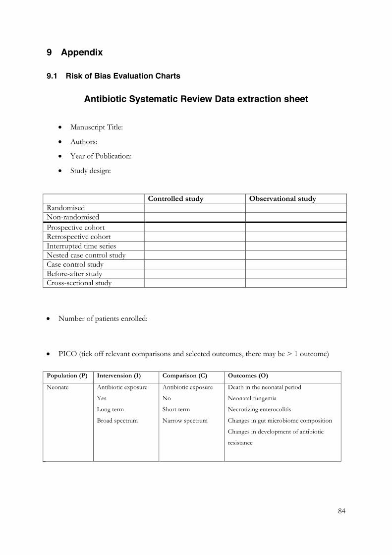
84
9 Appendix
9.1 Risk of Bias Evaluation Charts
Antibiotic Systematic Review Data extraction sheet
• Manuscript Title:
• Authors:
• Year of Publication:
• Study design:
Controlled study Observational study Randomised Non-randomised Prospective cohort Retrospective cohort Interrupted time series Nested case control study Case control study Before-after study Cross-sectional study
• Number of patients enrolled:
• PICO (tick off relevant comparisons and selected outcomes, there may be > 1 outcome)
Population (P) Intervension (I) Comparison (C) Outcomes (O)
Neonate Antibiotic exposure
Yes
Long term
Broad spectrum
Antibiotic exposure
No
Short term
Narrow spectrum
Death in the neonatal period
Neonatal fungemia
Necrotizing enterocolitis
Changes in gut microbiome composition
Changes in development of antibiotic
resistance

85
Risk of bias assessment
For each study, a risk of bias assessment was performed by one investigator using a tool based on the Cochrane handbook (Cochrane), which we adapted and clarified to also assess observational studies (Viswanathan M, 2013). We categorised for each study the risks of bias as high, low or unclear Selection bias: High Low Unclear Performance bias: High Low Unclear Detection bias: High Low Unclear Reporting bias: High Low Unclear Confounding : High Low Unclear Selection bias: Controlled studies: Low risk if random sequence generation and allocation concealment Uncontrolled studies: Low or high risk if patients had been enrolled or not enrolled as consecutively observed based on a pre-existent study protocol and if numbers and reasons for possible exclusions were reported or not reported specifically. High risk when the association between exposure and outcome is different for those who participate compared with those who do not participate in a study (i.e., all those who are theoretically eligible). This includes inappropriate selection of controls in a case-control study, differential loss to follow-up for groups being compared (attrition bias), incidence-prevalence bias, nonresponse bias, and in- or exclusion of specific groups for study. Performance bias Controlled studies: High risk if not blinding of the study personnel as to which intervention a neonate had received. Uncontrolled studies: High risk if systematic differences in the care provided to participants and protocol deviation. Examples include contamination of the control group with the exposure or intervention, unbalanced provision of additional interventions or co-interventions, difference in co-interventions, and inadequate blinding of providers and participants. Detection bias Controlled studies: High risk if not blinding of personnel evaluating outcomes Uncontrolled studies: High risk if systematic differences in outcomes assessment among groups being compared, including misclassification of the exposure or intervention, covariates, or outcomes because of variable definitions and timings, diagnostic thresholds, recall from memory, inadequate assessor blinding, and faulty measurement techniques. Erroneous statistical analysis might also affect the validity of effect estimates.

86
Reporting bias Controlled studies: High risk if not reporting of the study’s prespecified or expected outcomes of interest to the review. Including attrition bias; high risk if not completeness of reporting data, reason and balance across groups of missing data. Uncontrolled studies: High risk if systematic differences between reported and unreported findings (e.g., differential reporting of outcomes or harms, incomplete reporting of study findings, potential for bias in reporting through source of funding). Confounding Low risk if any attempt to (if necessary) to balance the design or allocation between the groups or match groups (e.g., through stratification, matching, propensity scores or other statistical adjustment such as instrumental variables) are done (When selection bias produces imbalances in prognostic factors associated with the outcome of interest then ‘confounding’ is said to occur. Statistical methods are sometimes used to counter bias introduced from confounding by producing ‘adjusted’ estimates of intervention effects, and part of the assessment of study quality may involve making judgements about the appropriateness of the analysis as well as the design and execution of the study) Important confounding factors that should be similar between groups • Age • Feeding • Disease severity • Same/different environment (hospital, country) • Antifungal prophylaxis used
These charts were modified from the Cochrane Handbook by Claus Klingenberg
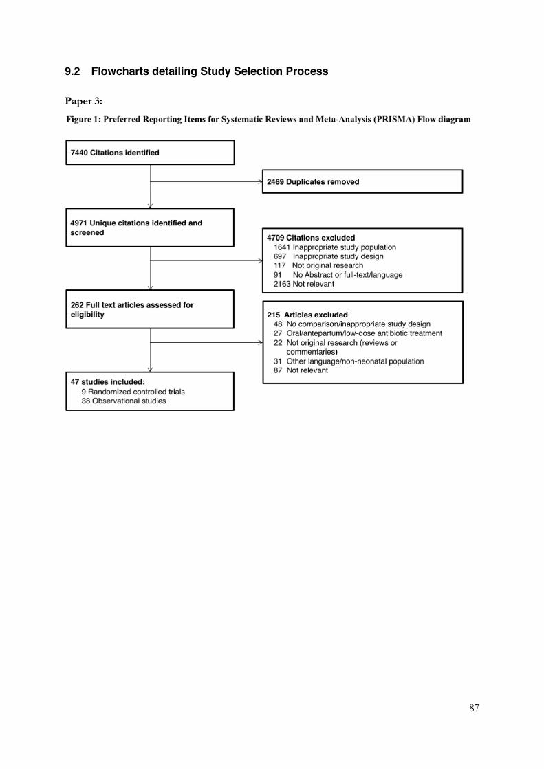
87
9.2 Flowcharts detailing Study Selection Process
Paper 3:
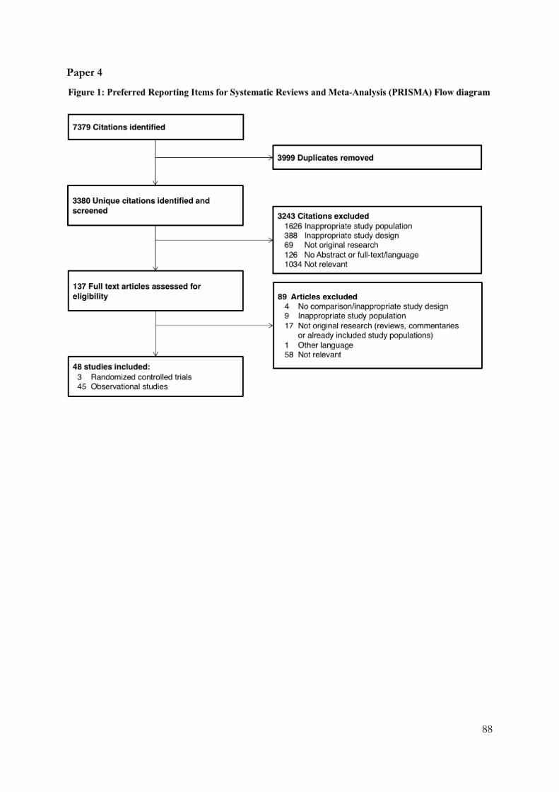
88
Paper 4
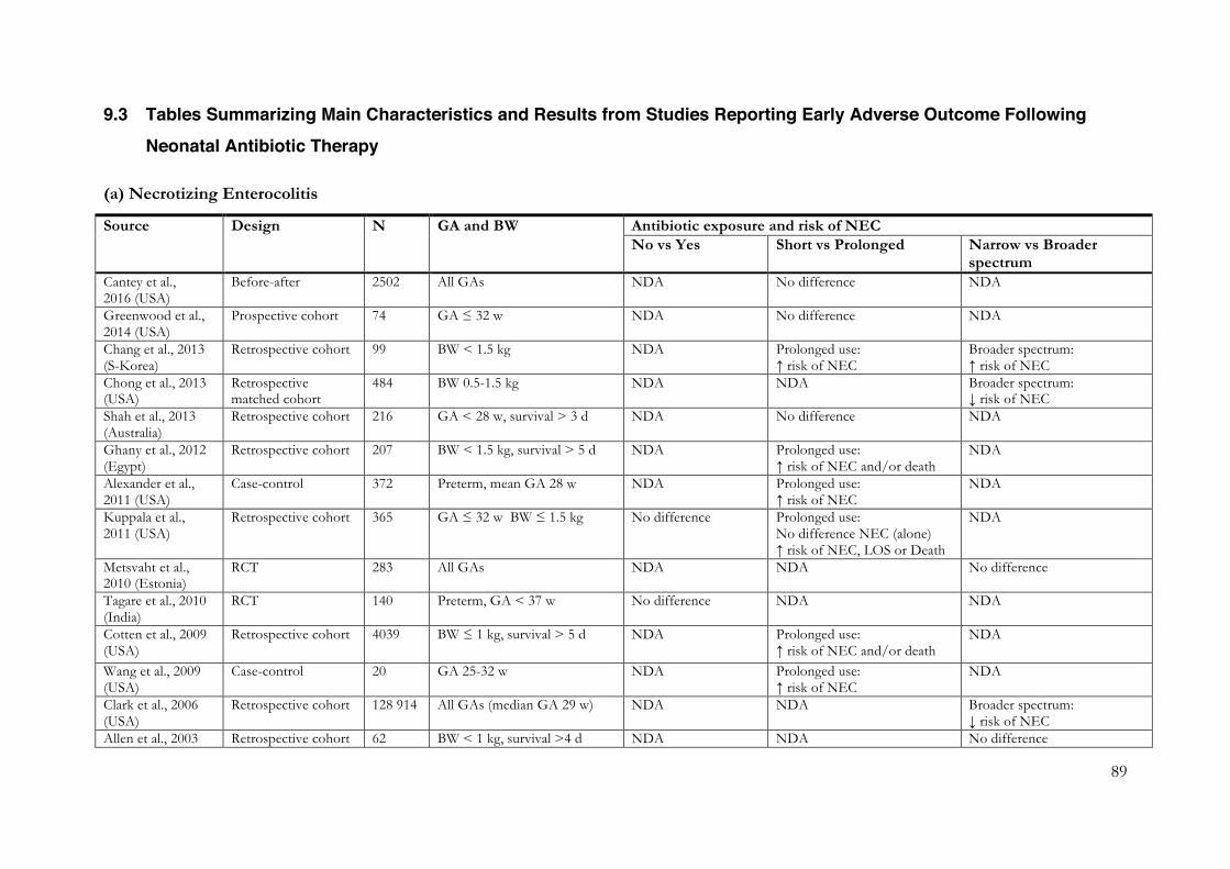
89
9.3 Tables Summarizing Main Characteristics and Results from Studies Reporting Early Adverse Outcome Following Neonatal Antibiotic Therapy
(a) Necrotizing Enterocolitis
Source Design N GA and BW Antibiotic exposure and risk of NEC No vs Yes Short vs Prolonged Narrow vs Broader
spectrum Cantey et al., 2016 (USA)
Before-after 2502 All GAs NDA No difference NDA
Greenwood et al., 2014 (USA)
Prospective cohort 74 GA ≤ 32 w NDA No difference NDA
Chang et al., 2013 (S-Korea)
Retrospective cohort 99 BW < 1.5 kg
NDA Prolonged use: ↑ risk of NEC
Broader spectrum: ↑ risk of NEC
Chong et al., 2013 (USA)
Retrospective matched cohort
484 BW 0.5-1.5 kg NDA NDA Broader spectrum: ↓ risk of NEC
Shah et al., 2013 (Australia)
Retrospective cohort 216 GA < 28 w, survival > 3 d NDA No difference NDA
Ghany et al., 2012 (Egypt)
Retrospective cohort 207 BW < 1.5 kg, survival > 5 d NDA Prolonged use: ↑ risk of NEC and/or death
NDA
Alexander et al., 2011 (USA)
Case-control 372 Preterm, mean GA 28 w NDA Prolonged use: ↑ risk of NEC
NDA
Kuppala et al., 2011 (USA)
Retrospective cohort 365 GA ≤ 32 w BW ≤ 1.5 kg No difference Prolonged use: No difference NEC (alone) ↑ risk of NEC, LOS or Death
NDA
Metsvaht et al., 2010 (Estonia)
RCT 283 All GAs NDA NDA No difference
Tagare et al., 2010 (India)
RCT 140 Preterm, GA < 37 w No difference NDA NDA
Cotten et al., 2009 (USA)
Retrospective cohort 4039 BW ≤ 1 kg, survival > 5 d NDA Prolonged use: ↑ risk of NEC and/or death
NDA
Wang et al., 2009 (USA)
Case-control 20 GA 25-32 w NDA Prolonged use: ↑ risk of NEC
NDA
Clark et al., 2006 (USA)
Retrospective cohort 128 914 All GAs (median GA 29 w) NDA
NDA Broader spectrum: ↓ risk of NEC
Allen et al., 2003 Retrospective cohort 62 BW < 1 kg, survival >4 d NDA NDA No difference

90
(Canada) Krediet et al., 2003(Netherlands)
Case-control 208 All GAs, median GA 29 w
Early use: ↓ risk NEC NDA NDA
Harms et al., 1995 (Germany)
RCT 148 Preterm, mean GA 29 w No difference NDA NDA
Millar et al., 1992 (England)
RCT 81 GA < 33 w NDA NDA No difference
Mufti et al., 1992 (Pakistan)
Case-control 39 BW ≤ 2 kg No difference NDA NDA
Hall et al., 1988 (England)
RCT 222 All GAs NDA NDA No difference
Stoll et al., 1980 (USA)
Case-control 133 All GAs No difference NDA No difference
GA; Gestational age, BW; birth weight, d; days, w; weeks, kg; kilogram, LOS; late-onset sepsis, NEC; necrotizing enterocolitis, NDA; no data available

91
(b) Invasive Fungal Infection
Source Design N GA and BW Antibiotic exposure and risk of IFI No vs Yes Short vs Prolonged Narrow vs Broader
spectrum Fu et al., 2016 (China)
Case-control 96 BW < 1.5 kg NDA Prolonged use: ↑ risk of IFI
Broader spectrum: ↑ risk of IFI
Tewari et al., 2014 (India)
RCT
187 GA ≥ 28 w, BW ≥ 1 kg NDA NDA No difference
Aliaga et al., 2013 (USA)
Retrospective cohort 709 325 All GAs NDA NDA Broader spectrum: ↑ risk of IFI
Chang et al., 2013 (S-Korea)
Retrospective cohort 99 Preterm, BW < 1.5 kg
NDA No difference Broader spectrum: ↑ risk of IFI
Lee et al., 2013 (USA)
Retrospective cohort 530 162 BW > 1.5 kg NDA NDA Broader spectrum: ↑ risk of IFI
Yu et al., 2013 (China)
Case-control
135 All GAs NDA No difference Broader spectrum: ↑ risk of IFI
Ariff et al., 2011 (Pakistan)
Case-control
81 All GAs NDA No difference Broader spectrum: ↑ risk of IFI
Benjamin et al., 2010 (USA)
Retrospective cohort 1515 BW ≤ 1 kg, survival > 3 d NDA NDA Broader spectrum: ↑ risk of IFI
Benjamin et al., 2006 (USA)
Retrospective cohort 4579 BW ≤ 1 kg, survival > 3 d NDA NDA Broader spectrum: ↑ risk of IFI
Cotten et al., 2006 (USA)
Retrospective cohort 3702 BW ≤ 1 kg, survival > 3 d NDA Prolonged use: ↑ risk of IFI
Broader spectrum: ↑ risk of IFI
Manzoni et al., 2006 (Italy)
Nested case-control 201 Preterm, BW < 1.5 kg
NDA No difference No difference
Feja et al., 2005 (USA)
Case-control
180 Preterm, mean GA 30 w
No difference NDA NDA
Linder et al., 2004 (Israel)
Case-control
112 Preterm, mean GA 28-29 w
NDA NDA No difference
Auriti et al., 2003 (Italy)
RCT 130 GA < 32 w NDA No difference NDA
Benjamin et al., 2003 (USA)
Retrospective cohort 6172 BW < 1.25 kg, survival ≥ 3 d
NDA NDA Broader spectrum: ↑ risk of IFI
Pera et al., 2002 (USA)
Case-control 334 Preterm, BW < 1.25 kg
NDA Prolonged use: ↑ risk of IFI
NDA
Warris et al., Case-control 24 GA ≤ 33 w NDA Prolonged use: NDA
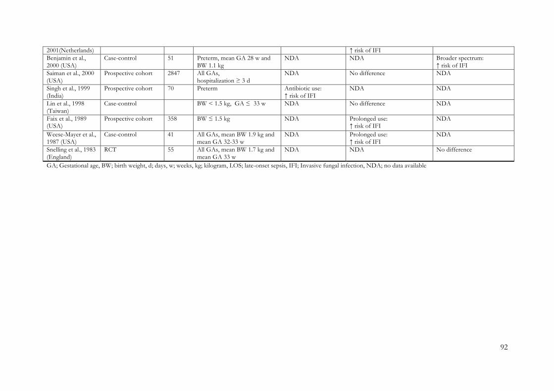
92
2001(Netherlands) ↑ risk of IFI Benjamin et al., 2000 (USA)
Case-control 51 Preterm, mean GA 28 w and BW 1.1 kg
NDA NDA Broader spectrum: ↑ risk of IFI
Saiman et al., 2000 (USA)
Prospective cohort 2847 All GAs, hospitalization ≥ 3 d
NDA No difference NDA
Singh et al., 1999 (India)
Prospective cohort 70 Preterm Antibiotic use: ↑ risk of IFI
NDA NDA
Lin et al., 1998 (Taiwan)
Case-control BW < 1.5 kg, GA ≤ 33 w
NDA No difference NDA
Faix et al., 1989 (USA)
Prospective cohort 358 BW ≤ 1.5 kg NDA Prolonged use: ↑ risk of IFI
NDA
Weese-Mayer et al., 1987 (USA)
Case-control 41 All GAs, mean BW 1.9 kg and mean GA 32-33 w
NDA Prolonged use: ↑ risk of IFI
NDA
Snelling et al., 1983 (England)
RCT 55 All GAs, mean BW 1.7 kg and mean GA 33 w
NDA NDA No difference
GA; Gestational age, BW; birth weight, d; days, w; weeks, kg; kilogram, LOS; late-onset sepsis, IFI; Invasive fungal infection, NDA; no data available
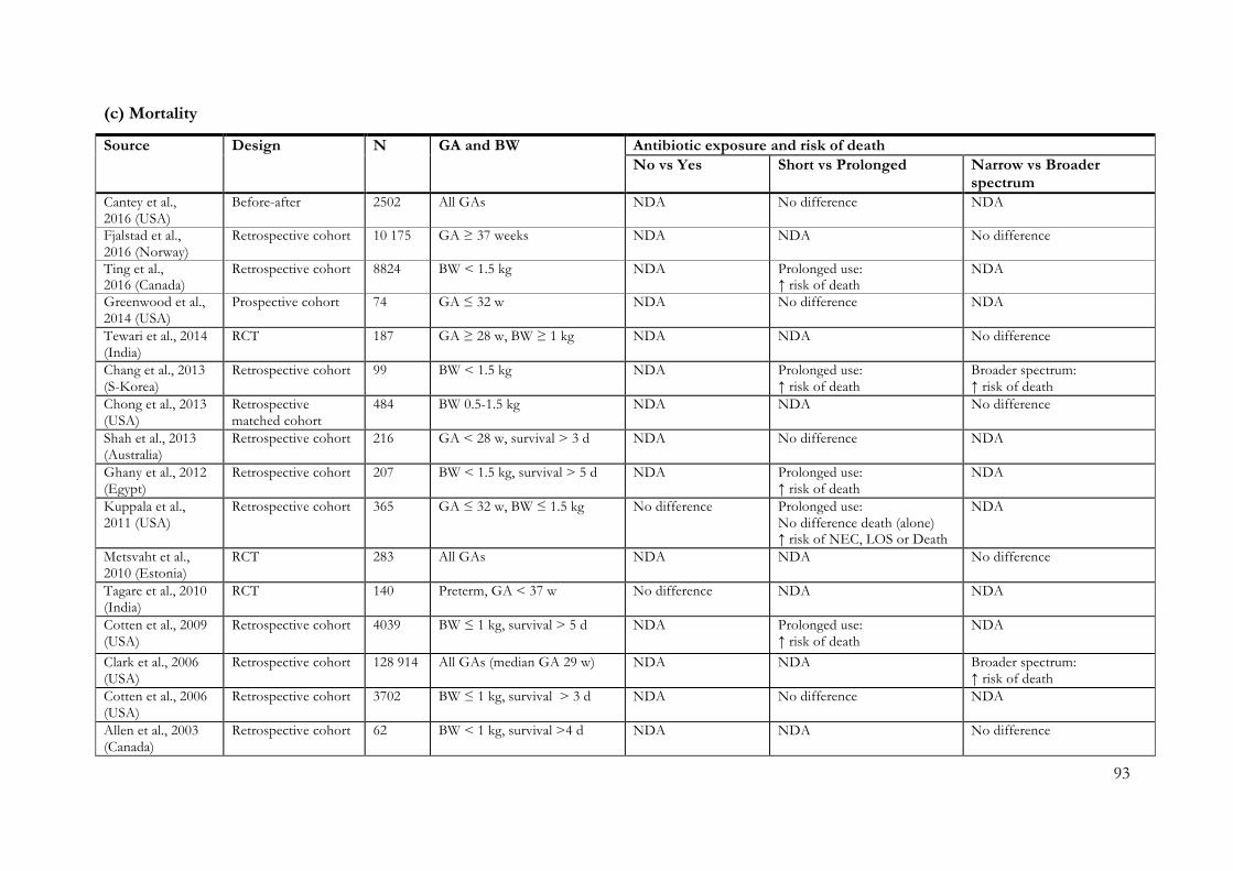
93
(c) Mortality
Source Design N GA and BW Antibiotic exposure and risk of death No vs Yes Short vs Prolonged Narrow vs Broader
spectrum Cantey et al., 2016 (USA)
Before-after 2502 All GAs NDA No difference NDA
Fjalstad et al., 2016 (Norway)
Retrospective cohort 10 175 GA ≥ 37 weeks NDA NDA No difference
Ting et al., 2016 (Canada)
Retrospective cohort 8824 BW < 1.5 kg NDA Prolonged use: ↑ risk of death
NDA
Greenwood et al., 2014 (USA)
Prospective cohort 74 GA ≤ 32 w NDA No difference NDA
Tewari et al., 2014 (India)
RCT
187 GA ≥ 28 w, BW ≥ 1 kg NDA NDA No difference
Chang et al., 2013 (S-Korea)
Retrospective cohort 99 BW < 1.5 kg
NDA Prolonged use: ↑ risk of death
Broader spectrum: ↑ risk of death
Chong et al., 2013 (USA)
Retrospective matched cohort
484 BW 0.5-1.5 kg NDA NDA No difference
Shah et al., 2013 (Australia)
Retrospective cohort 216 GA < 28 w, survival > 3 d NDA No difference NDA
Ghany et al., 2012 (Egypt)
Retrospective cohort 207 BW < 1.5 kg, survival > 5 d NDA Prolonged use: ↑ risk of death
NDA
Kuppala et al., 2011 (USA)
Retrospective cohort 365 GA ≤ 32 w, BW ≤ 1.5 kg No difference Prolonged use: No difference death (alone) ↑ risk of NEC, LOS or Death
NDA
Metsvaht et al., 2010 (Estonia)
RCT 283 All GAs NDA NDA No difference
Tagare et al., 2010 (India)
RCT 140 Preterm, GA < 37 w No difference NDA NDA
Cotten et al., 2009 (USA)
Retrospective cohort 4039 BW ≤ 1 kg, survival > 5 d NDA Prolonged use: ↑ risk of death
NDA
Clark et al., 2006 (USA)
Retrospective cohort 128 914 All GAs (median GA 29 w) NDA
NDA Broader spectrum: ↑ risk of death
Cotten et al., 2006 (USA)
Retrospective cohort 3702 BW ≤ 1 kg, survival > 3 d NDA No difference NDA
Allen et al., 2003 (Canada)
Retrospective cohort 62 BW < 1 kg, survival >4 d
NDA
NDA No difference

94
Auriti et al., 2003 (Italy)
RCT 130 GA < 32 w NDA No difference NDA
Cordero et al., 2003 (USA)
Retrospective matched cohort
517 BW < 1 kg NDA No difference NDA
Harms et al., 1995 (Germany)
RCT 148 Preterm, mean GA 29 w No difference NDA NDA
De Louvois et al., 1992 (Europe)
RCT 1316 All GAs
NDA NDA No difference
Millar et al., 1992 (England)
RCT 81 GA < 33 w NDA NDA No difference
GA; Gestational age, BW; birth weight, d; days, w; weeks, kg; kilogram, LOS; late-onset sepsis, NDA; no data available

95
(d) Gut Microbiota
Study Design N GA and BW Empiric regimen Categories of antibiotic exposure and changes in gut microbiota Arboleya et al., 2015 (Spain)
Prospective cohort
40 All GAs EOS: AMP + GEN, LOS: VAN + AMK
Yes vs. no: Composition: ¯ Staphylococcus spp. & Comamonadaceae
Bennet et al., 1986 & 1987 (Sweden)
Prospective cohort
164 All GAs NDA Yes vs. no: Load: ; Composition: Klebsiella/Enterobacter spp., ¯ Anaerobes, ¯Bifidobacterium spp., ¯Lactobacillus spp., ¯Bacteriodes spp. Broad vs. narrow: Composition: Enterococcus spp., S. faecalis
Blakey et al., 1982 (Australia)
Prospective cohort
28 GA ≤ 36 weeks EOS: PEN + GEN Yes vs. no: Composition: No difference*
Bonnemaison et al., 2003 (France)*
Prospective cohort
30 All GAs EOS: AMX + NET ± CTX Yes vs. no: Composition: No difference Broad vs. narrow: Composition: No difference*
Butel et al., 2007 (France)
Prospective case-control
52 GA 30 - 35 weeks NDA Yes vs. no: Composition: No significant difference
Ferraris et al., 2012 (France)
Retrospective cohort
76 GA < 36 weeks NDA Yes vs. no: Composition: C. butyricum Long vs. short: Composition: ¯ Clostridium spp.
Fouhy et al., 2012 (Ireland)
Prospective cohort
18 GA ≥ 37 weeks AMP + GEN Yes vs. no: Composition: Enterobacteriaceae, Gammaproteobacteriae, Peptostreptococcaceae, Enterococcus spp., Clostridium spp., ¯ Lactobacillus spp., ¯ Bifidobacterium spp., ¯ Bacteriodetes
Gewolb et al., 1999 (USA)
Prospective cohort
29 BW < 1000 g EOS: AMP + GEN, LOS: VAN + CTX
Long vs. short: Load: ¯; Diversity: ¯
Goldmann et al., 1978 (USA)
Prospective cohort
63 All GAs NDA Long vs. short: Composition: Klebsiella spp., Enterobacter spp., and/or Citrobacter spp.
Greenwood et al., 2014 (USA)
Prospective cohort
74 GA ≤ 32 weeks EOS: AMP + GEN Yes vs. no: Diversity: ¯; Composition: Enterobacter spp. Long vs. short: Composition: Enterobacter spp., ¯ Staphylococcus spp.
Hall et al., 1990 (UK)
Prospective cohort
42 GA ≤ 33 weeks NDA Broad vs. narrow: Composition: ¯ Lactobacillus spp.
Jacquot et al., 2011 (France)
Prospective cohort
29 GA ≤ 30 weeks EOS: AMK + (1) PEN or (2) AMP or (3) CTX, LOS: VAN + AMK
Yes vs. no: Diversity: No significant effect Long vs. short: Diversity: ¯
Jenke et al., 2013 (Germany)
Prospective cohort
68 GA < 27 weeks NDA Yes vs. no: Composition: C. difficile
La Rosa et al., 2014 (USA)
Prospective cohort
58 BW ≤ 1500 g NDA Yes vs. no: Composition: Gammaproteobacteria (GA ≥ 26 weeks), ¯ Clostridium spp. (GA ≤ 28 weeks)
Parm et al., 2010 (Estonia)
RCT 276 All GAs EOS: (1) PEN + GEN or (2) AMP + GEN
Broad vs. narrow: Composition: S. haemolyticus, S. hominis, K. pneumonia, ¯ Enterococcus spp. S. aureus
Tullus et al., 1988 Retrospective 953 All GAs AMP + GEN Yes vs. no: Composition: ¯ E. coli Broad vs. narrow: Composition: No

96
(Sweden) cohort significant difference Ward et al., 2016 (USA)
Case-control 166 All GAs EOS: AMP + GEN Long vs. short: Diversity: ¯
Westerbeek et al., 2013 (Netherlands)
RCT 113 GA < 32 weeks ± BW < 1500 g
NDA Yes vs. no: Load: ¯
Zhou et al., 2015 (USA)
Case-control 38 GA < 32 weeks NDA Yes vs. no: Diversity: ¯
Outcomes: Load; the total number of bacteria in a sample, Diversity; the number of bacterial genus or species in a sample, and Composition; the taxonomical composition in a
sample. Categories: Yes vs. no compares neonates exposed to antibiotics with non-exposed neonates, Long vs. short compares long and short treatment durations, Broad vs.
narrow compares broad spectrum antibiotic treatment to narrow spectrum treatment. *; did not test for statistical significance, RCT; randomized controlled trial, GA; gestational
age, PNA; post-natal age, BW; birth weight, g; gram, EOS; early onset sepsis, AMP; ampicillin, GEN; gentamicin, LOS; late onset sepsis, VAN; vancomycin, AMK; amikacin, NDA;
no data available, PEN; penicillin, AMX; amoxicillin, NET; netilmicin, CTX; cefotaxime

97
(e) Antibacterial Resistance
Study Design N Empiric regimen Categories of antibiotic exposure and changes in antibacterial resistance
Abdel-Hady et al., 2008 (Egypt)
Prospective cohort
380 NDA Broad vs. narrow: ESBL producing K. pneumoniae infection
Acolet et al., 1994 (UK)
Case-control 60 EOS: AMX + CTX, LOS: CTX Broad vs. narrow: CREC colonization
Bergin et al., 2015 (USA)
Case-control 258 NDA Broad vs. narrow: No significant difference
Bonnemaison et al, 2003 (France)
Prospective cohorts
30 EOS: AMX + NET ± CTX Yes vs. no: Did not assess significance Broad vs. narrow: Did not assess significance
Burman et al., 1992 (Sweden)
Retrospective cohort
953 EOS: (1) AMP + GEN or (2) CTX Yes vs. no: TEM-1 in E. coli Broad vs. narrow: No significant difference
Burman et al., 1993 (Sweden)
Retrospective cohort
46 EOS: (1) AMP + GEN or (2) CTX Yes vs. no: E. cloacae: MIC to ampicillin, cephalotin, cephalexin
Calil et al., 2001 (Brazil)
Prospective cohort
342 EOS: AMX + (1) GEN or (2) CRO, LOS: OXA + (1) GEN or (2) CRO
Yes vs. no: MDR E. cloacae colonization Broad vs. narrow: MDR E. cloacae colonization
Cantey et al., 2016 (USA)
Before-after study
2502 EOS: AMX + GEN, LOS: OXA + GEN Long vs. short: No significant difference
Crivaro et al., 2007 (Italy)
Case-control 167 AMP + GEN Yes vs. no: ESBL-producing S. marcescens and K. pneumoniae Long vs. short: ESBL-producing S. marcescens and K. pneumoniae
De Araujo et al., 2007 (Brazil)
Before-after study
995 PEN & GEN Broad vs. narrow: MDR GNB
De Champs et al., 1994 (France)
Before-after study
636 (1) AMP + GEN or (2) AMP + AMK Broad vs. narrow: Gentamicin-resistant, cephalosporin-resistant, and MDR E. cloacae, Amikacin-resistant P. aerunginosa; ¯ Gentamicin & amikacin-resistant GNB, MRSE
De Man et al., 2000 (Netherlands)
RCT 436 EOS: (1) PEN + TOB or (2) AMX + CTX, LOS: FLU + (1) TOB or (2) CTX
Broad vs. narrow: Colonization with cefotaxime-resistant Enterobacter spp. & GNB
Duman et al., 2005 (Turkey)
Prospective cohort
118 NDA Yes vs. no: ESBL-producing Enterobacteriaceae colonization
Gaynes et al., 1984 (USA)
Case-control 32 (1) PEN or (2) AMP + (1) GEN or (2) KAN
Yes vs. no: Aminoglycoside-resistant E. coli
Giuffrè et al., 2016 (Italy)
Prospective cohort
1152 SAM + GEN Yes vs. no: MDR GNB colonization Long vs. short: MDR & ESBL-producing GNB colonization
Isaacs et al., 1988 (UK)
Before-after study
NDA EOS: PEN + (1) NET or (2) GEN, LOS: FLU + (1) NET or (2) GEN
Long vs. short: No significant difference

98
Kalenic et al., 1993 (Croatia)
Before-after study
440 (1) AMP + GEN or (2) CXM + GEN Broad vs. narrow: ¯ Ampicillin-resistant GNB, cefuroxime-resistant GNB & cefuroxime-resistant K. pneumoniae
Kumar et al., 2014 (India)
Case-control 65 NDA Yes vs. no: CRAB blood stream infections
Le et al., 2008 (USA) Before-after study
250 EOS: AMP + GEN, LOS: VAN + (1) CTX or (2) TOB
Long vs. short: ESBL-producing Enterobacteriaceae infection Broad vs. narrow: ESBL-producing Enterobacteriaceae infection
Linkin et al., 2004 (USA)
Case-control 10 NDA Yes vs. no: ESBL-producing Enterobacteriaceae
Mammina et al., 2007 (Italy)
Prospective cohort
210 EOS: SAM + GEN Long vs. short: MDR GNB colonization Broad vs. narrow: MDR GNB colonization
Millar et al., 2008 (UK)
Prospective cohort
124 EOS: PEN + GEN, LOS: (1) TZP + VAN or (2) FLU + GEN
Yes vs. no: No significant difference Broad vs. narrow: MDR Enterobacteriaceae colonization
Noy et al., 1974 (UK)
Prospective cohort
584 NDA Yes vs. no: Antibiotic-resistant E. coli & Klebsiella spp. colonization
Parm et al., 2010 (Estonia)
RCT 276 EOS: (1) PEN + GEN or (2) AMP + GEN Broad vs. narrow: ¯ Ampicillin-resistant Acinetobacter spp. colonization
Pessoa-Silva et al., 2003 (Brazil)
Prospective cohort
379 EOS: AMP + GEN, LOS: Varying antibiotics
Yes vs. no: ESBL-producing K. pneumoniae colonization
Raz et al., 1987 (Israel)
Before-after study
118 (1) AMP + GEN or (2) AMP + AMK Broad vs. narrow: Gentamicin-resistant GNB and E. cloacae
Rettedal et al., 2013 (Norway)
Case-control 99 NDA Yes vs. no: ESBL-producing K. pneumoniae colonization
Sehgal et al., 2007 (India)
Case-control 63 EOS: AMP + GEN, LOS: 3rd gen. cephalosporin + AMK
Yes vs. no: ESBL-producing GNB blood stream infection
Thatrimontrichai et al., 2013 (Thailand)
Case-control 96 EOS: AMP + GEN, LOS: 3rd gen. cephalosporin + AMK
Broad vs. narrow: CRAB blood stream infection
Thatrimontrichai et al., 2016 (Thailand)
Case-control 101 EOS: AMP + GEN, LOS: varying antibiotics
Broad vs. narrow: odds of CRAB ventilator associated pneumonia
Toltzis et al., 2001 (USA)
Prospective cohort
1180 NDA Long vs. short: antibiotic resistant GNB colonization
Categories: Yes vs. no; compares neonates exposed to antibiotics with non-exposed neonates, Long vs. short; compares long and short treatment durations, and Broad vs.
narrow; compares broad spectrum antibiotic treatment to narrow spectrum treatment. RCT; randomized controlled trial, NDA; no data available, EOS; early onset sepsis, AMX;
amoxicillin, CTX; cefotaxime, LOS; late onset sepsis, NET; netilmicin, AMP; ampicillin, GEN; gentamicin, CRO; ceftriaxone, OXA; oxacillin, TOB; tobramycin, FLU;
flucloxacillin, KAN; kanamycin, SAM; ampicillin/sulbactam, CXM; cefuroxime, TZP; piperacillin/tazobactam, CREC; cephalosporin-resistant Enterobacter cloacae, GNB; Gram-
negative bacteria, CRAB; carbapenem-resistant Acinetobacter baumannii

99
9.4 Risk of Bias Assessments in the Systematic Reviews of Early Adverse Effects
Risk of bias graph: review of authors’ judgements about each risk of bias item for each included study and the five
outcomes. (a) Studies reporting on risk of necrotizing enterocolitis (n=20). (b) Studies reporting on risk of invasive
fungal infection (n=24). (c) Studies reporting on risk of death (n=21). (d) Studies reporting on changes in gut
microbiota (n=20). (e) Studies reporting on changes in antibiotic resistance development (n=31).
(a) (b)
Aliaga 2013 - + + + -Ariff 2011 - - + + -Auriti 2005 - + + ? -Benjamin 2000 + - + ? -Benjamin 2003 - + + ? -Benjamin 2006 - + + ? -Benjamin 2010 - + + ? -Chang 2013 + - + - -Cotten 2006 - + + ? -Faix 1989 - - - - +Feja 2005 - + + ? -Lee 2013 - + + + -Lin 1998 - - + + +Linder 2004 - - + + -Manzoni 2006 - - + + -Pera 2002 - - + + -Saiman 2000 - + + + -Singh 1999 + + - ? +Snelling 1983 - + + + +Tewari 2014 - + + ? -Warris 2001 - - + + +Weese-Mayer 1987 - - + + -Yu 2013 - - + + -
Selecti
on
Perform
ance
Detecti
ng
Report
ing
Confou
nding
Alexander 2011 + - ? + -Allen 2003 - - + ? -Cantey 2017 - + - + -Chang 2013 + - + - -Chong 2013 + + + ? +Clark 2006 + + + - -Cotten 2009 - + + - -Ghany 2012 - - + ? -Greenwood 2014 + - - ? +Hall 1988 - + + - +Krediet 2003 - - + + +Harms 1995 - + + ? -Kuppala 2011 - + + - -Metsvath 2010 - + + ? -Millar 1992 - + + ? -Mufti 1992 + - + + +Shah 2013 - - + ? -Stoll 1980 - - + + -Tagare 2010 - + - ? -Wang 2009 + - + + +
Selecti
on
Perform
ance
Detecti
ng
Report
ing
Confou
nding

100
(c) (d)
(e)
- - - ? -+ - + ? +- + + ? -+ - + ? ++ + + ? ++ + + ? ++ - - ? -- - - ? ++ - + ? -+ - + ? +- - + ? +- + + ? -+ - - ? -- - + ? -- - - ? -+ - + ? ++ + + ? +- - + ? -- - + ? -+ - + ? +- - - ? -- + - ? ++ + - ? +- + - ? -
Abdel-Hady 2008Acolet 1994Bergin 2015Bonnemaison 2003Burman 1992
Millar 2008Noy 1974Parm 2010
Burman 1993
Kumar 2014Le 2008
Calil2001Cantey2016Crivaro 2007DeAraujo2007DeChamps1994DeMan2000Duman2005Gaynes1984Giuffrè2016Isaacs1988Kalenic1993
Linkin 2004Mammina 2007
- - - ? -Pessoa-Silva 2003+ - + ? ++ - + ? -+ - + ? -- - + ? -- - + ? -+ - - ? +
Rettedal 2013Sehgal 2007Thatrimontrichai 2013Thatrimontrichai 2016Toltzis 2001
Raz 1987
Selecti
on
Perform
ance
Detecti
ng
Report
ing
Confou
nding
Allen 2003 - - + ? -Auriti 2005 - + + ? -Cantey 2017 - + - + -Chang 2013 + - + - -Chong 2013 + + + ? +Clark 2006 + + + - -Cordero 2003 - + + + +Cotten 2006 - + + - -Cotten 2009 - + + - -De Louvois 1992 - + + ? -Fjalstad 2016 - + - + +Ghany 2012 - - + ? -Greenwood 2014 + - - ? +Harms 1995 - + + ? -Kuppala 2011 - + + - -Metsvath 2010 - + + ? -Millar 1991 - + + ? -Shah 2013 - - + ? -Tagare 2010 - + - ? -Tewari 2014 - + + ? -Ting 2016 - + + + -
Selecti
on
Perform
ance
Detecti
ng
Report
ing
Confou
nding
+ - ? ? ++ + + ? ++ + + ? ++ - + ? ++ + + ? ++ + + ? -+ - ? ? +- ? + ? -- - + ? ++ - ? ? +- - + ? +- - ? ? ++ + ? ? +- - - ? -+ + + ? -+ + + ? +- + - ? -- - ? ? -- - ? ? -
Greenwood 2014
Arboleya 2014Bennet 1986 & 1987Blakey 1982Bonnemaison 2003Butel 2007Ferraris 2012Fouhy 2012Gewolb 1999Goldman 1978
Ward 2016Westerbeek 2013Zhou 2015
Hall 1990Jacquot 2011Jenke 2013La Rosa 2014Parm 2010Tullus 1988
Selecti
on
Perform
ance
Detecti
ng
Report
ing
Confou
nding