Embed Size (px)
Citation preview

PACEMAKER LEAD ENDOCARDITIS
(CDR-IE)
EuroValve 2016
Brussels
I. Vilacosta.
Hospital Clínico San Carlos
Madrid. Spain.

Clinical case• An 80-year-old obese woman with diabetes, HTN, and
permanent AF was admitted to the H. for PM generator replacement. She had SOB and fatiguetwo weeks before admission.
• Past history: LA myxoma surgery in 1988; 2nd heart surgery (myxoma recurrence) in 2000. During postop a permanent VVI PM was implanted due to persistent nodal rhythm.
• Therapy: acenocumarol, digoxin, furosemide, and metformin.
• After the procedure, she developed progressive heart failure and was admitted to the CCU.
• Eight days after the procedure she became febrile (39ºC) and had orthopnea. A chest X-ray was done.

How should this case be managed ?

Clinical case
• BC were obtained and she was treated with iv.
furosemide + antibiotics (vancomycin &
gentamicin).
• BC were positive for MRSA.
• There were no signs of infection at the pocket
site.
• TTE & TEE were performed to rule-out PM
infection.

Clinical case
• The patient continued with fever and got worse with hypotension and hypoperfusion. She also complained of mild lumbar pain.
• Rifampin was added to the antibiotic regimen.
• FDG PET/CT was performed and PM explantation was planned.

FDG PET/CT & CDR-IE

Clinical case

Clinical case

Clinical case

Clinical case
• Percutaneous removal of the whole PM system was
performed by simple traction. No vegetations were
seen along the PM lead.
• Lead tip culture was positive for MRSA.
• After explantation, the patient was treated with iv
antibiotics during 6 weeks and she was discharged
with OST.
• A TEE before being discharged was normal.
• A new PM system was not necessary.

CDR-IE-Introduction• CIED infection seems to be raising out of
proportion to the increase in device implantation rates.
• Incidence: varies widely among studies: 1,9‰ device-
years (1). Incidence of CDR-IE: 20%-25% of all
device related infections.
• ICD are associated with a greater risk of infection
than are PM. Infection risk in epicardial systems is
similar to that of transvenous systems (2).
• Main pathogenic mechanism of CDR-IE:
contamination by local bacteriological flora at the
time of device implantation.
Baddour LM, et al. N Engl J Med 2012; Nof E, Epstein LM. Eur Heart J 2013; (1) Uslan
DZ, et al.Arch Intern Med 2007; (2) Silvetti MS, et al. Europace 2006.

Risk factors for CIED infections
• Patient-related:
– Diabetes mellitus.
– Heart failure.
– Chronic renal failure.
– Corticosteroids.
– Central lines (dialysis).
– Anticoagulants.
– Fever within 24 h of
implantation.
• Procedural factors:
– Longer procedure time.
– Op. inexperience.
– Temporary pacing leads.
– Dual or triple-chamber devices.
– Lack of antibiotic prophylaxis.
– Pocket hematomaSohail MR, et al. CID 2007; Bloom H, et al. Pacing Clin Electrophysiol 2006;
Kug D, et al. Circulation 1997; Greenspon AJ, et al. JACC 2012; Nery PB, et
al. J Cardiovasc Electrophysiol 2010; Johansen JB, et al. Eur Heart J 2011.

CDR-IE; Diagnosis.
2015 ESC Guidelines.

ECHO-DIAGNOSIS• Technique of choice for detection & sizing of
vegetations: TEE.
• Evaluation of the whole infectious lead course
(SVC-RA junction): TEE.
• Assess (TTE & TEE): degree of TR, right
chambers dilatation, quantitation of PAP, other
valves (left-sided, prosthesis).
• TTE: poor sensitivity and a low NPV.
• TEE: higher sensitivity & specificity than TTE.Vilacosta I, et al. Circulation 1994; Klug D, et al. Circulation 1997; Cacoub P, et
al. Am J Cardiol 1998; Victor F, et al. Heart 1999. Sohail MR, et al. JACC 2007;
Grammes JA, et al. JACC 2010.

CDR-IE-DIAGNOSIS.
Duke criteria
• Lower sensitivity of Duke criteria.
• Modifications proposed:
– Local signs of infection.
– Pulmonary embolism.
• PET/CT to assess lead & pocket infection, presence of pulmonary embolism, and other extrapulmonary septic foci: should be considered (IIa).
Klug D, et al. Circulation 1997; Massoure P-L, et al. Pacing Clin Electrophysiol 2007;
Sohail MR, et al. Mayo Clin Proc 2008.

Conclusions• CIED infection rate is increasing.
• Main risk factors for CDR-IE include complications
at the generator pocket site and device
manipulation.
• Staphylococci account for most CDR-IE.
• TEE remains the technique of choice in cases of
CDR-IE.
• PET/CT has a complementary role and should be
considered (IIa) in the diagnostic work-up.
• Complete removal of the device is strongly
recommended, even when signs of infection are
limited to the generator pocket site.


Principles of treatment
2015 ESC Guidelines.

Mode of device removal.
2015 ESC Guidelines.

Percutaneous explantation• Percutaneous success rate: 93-97%.
• Fibrosis: in areas of direct contact between the lead
and the vasculature and endocardium.
• Complications: < 2%.
– Myocardial avulsion, tricuspid valve lesions, venous
lacerations, lead fracture / disruption, tamponade, pulmonary
embolism, arrythmias, pneumothorax, death.
• Team approach (CT surgeon, etc). OR vs EP lab.
• Several extraction tools are available (simple traction,
locking stylets, powered sheaths…).
Wilkoff BL, et al. (PLEXES trial). JACC 1999; Byrd CL, et al. Pacing Clin Electrophysiol
1999; Jones SO, et al. Heart Rhythm 2008; Grammes JA, et al. JACC 2010;
Rodriguez Y, et al. Intern J Cardiol 2013.


What to do after device removal
in CDR-IE ?
• After extraction of an infected device in
patients with CDR-IE:
– At least 2 weeks of parenteral therapy.
– Blood cultures.
– Chest X-ray (PA & lateral).
– TEE to rule out RV-RA-SVC residual masses
(ghost).
Baddour LM, et al. N Engl J Med 2012; Le Dolley Y, et al. J Am Coll Cardiol Img 2010;
Grammes JA, et al. JACC 2010.
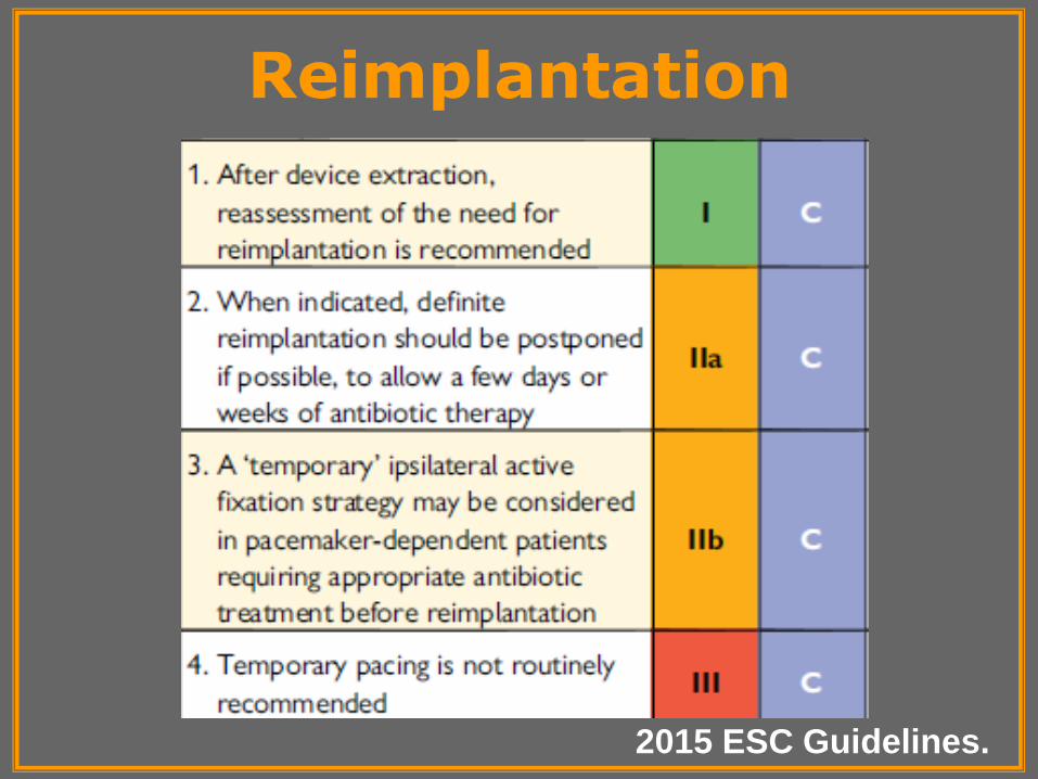
Reimplantation
2015 ESC Guidelines.

Generator pocket site

Re-implantation of CIED after
device explantation for infection• Re-assessment before re-implanting.
• Re-implantation has to be performed at another site
(contralateral site, epicardial).
• Avoid immediate reimplantation.
• Timing: on a case-by-case basis depending on indication
for extraction, urgency, BC results, pocket infection
control, and clinical status.
• Temporary pacing: risk factor for subsequent CD infection.
• PM-dependent: “immediate epicardial vs temporary
transvenous pacing” not well defined.
Klug, et al. Circulation 2007; ESC Guidelines on IE-2009; ESC Guidelines on CP/CRT-2013;
Baddour LM, et al. N Engl J Med 2012.

Diagnosis of CDR-IE• Misleading presentation, with frequent
respiratory or rheumathological
symptoms, as well as local signs of
infection.
• CDR-IE must be suspected in any
patient with a CD and unexplained fever.
• Echocardiography (TTE & TEE) and
BC are the cornerstone of diagnosis.Habib G, et al. Eur Heart J 2009; Baddour LM, et al. N Engl J Med 2012.

CLINICAL PRESENTATION
• Varies with the virulence of the infecting organism.
• Symptoms from local pocket erosion to full-blown
sepsis.
• Main symptoms: fever, chills, malaise, rheumathologic and
pulmonary symptoms, and findings at the generator
pocket site: erythema, pain, swelling, warmth, drainage, and
skin and soft-tissue ulceration.
• Pocket erosion should be treated as pocket infection.
• The interval between CIED placement or revision and
the onset of infection varies widely, from days to years.
Sohail MR, et al. Mayo Clin Proc 2008; Le KY, et al. Am J Cardiol 2012; Tarakji
KG, et al. Heart Rhythm 2010; Baddour LM, et al. N Engl J Med 2012.

Clinical suspicion of CDR-IE
Fever of unknown origin
Chills
+
Respiratory symptoms
+
0 5 10
Respiratory tract infection
Fever without focus
Pneumonia
COPD exacerbation
Urinary tract infection
Erroneous diagnosis at discharge

Blood cultures & CDR-IE• Blood cultures are recommended in all
suspected cases of CIED infection.
• BC may be negative despite CIED infection: local
infection or previous antibiotics.
• Positive BC ≠ CDR-IE.
• Likelihood of CIED infection when BC +:
– ≥ 35% in staphylococcal species.
– < 20% in nonstaphylococcal or GNB.
• A single + BC for CoNS: contamination.
• Multiple + BC for CoNS: CDR-IE.Baddour LM, et al. N Engl J Med 2012.
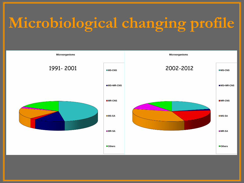
Microbiological changing profile
Microorganisms
MS-CNS
MS+MR-CNS
MR-CNS
MS-SA
MR-SA
Others
Microorganisms
MS-CNS
MS+MR-CNS
MR-CNS
MS-SA
MR-SA
Others
1991- 2001 2002-2012

CIED infection-classification (1)
• 1-. Superficial skin infection: limited to the skin or suture.
• 2-. Pocket infection: local inflammatory or infectious signs
or exposure of hardware through the skin.
• 3-. CDR-IE (Duke criteria):
– Definite.
– Possible.
• * In the presence of a lead vegetation, local device infection
was considered a major criterion (2).
• * Positive lead culture is a major criterion only in the absence
of a pocket infection or when the leads are not removed
through the generator pocket.
(1) Baddour LM, et al. Circulation 2010; (2) Sohail MR, et al. Mayo Clin Proc 2008.
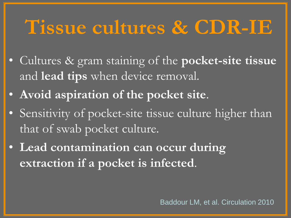
Tissue cultures & CDR-IE
• Cultures & gram staining of the pocket-site tissue
and lead tips when device removal.
• Avoid aspiration of the pocket site.
• Sensitivity of pocket-site tissue culture higher than
that of swab pocket culture.
• Lead contamination can occur during
extraction if a pocket is infected.
Baddour LM, et al. Circulation 2010

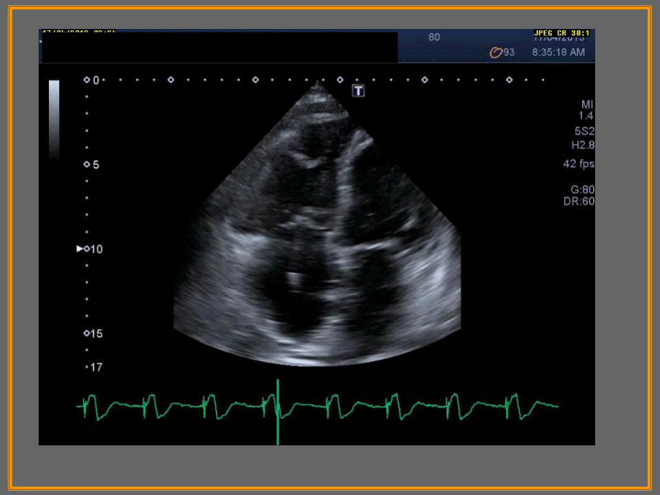
Echocardiography & CDR-IE
Klug D, et al. Circulation 1997; Cacoub P, et al. Am J
Cardiol 1998; Victor F, et al. Heart 1999.

ECHO-DIAGNOSIS
Vilacosta I, et al. Circulation 1994; Klug D, et al. Circulation 1997; Cacoub P, et al.
Am J Cardiol 1998; Victor F, et al. Heart 1999.
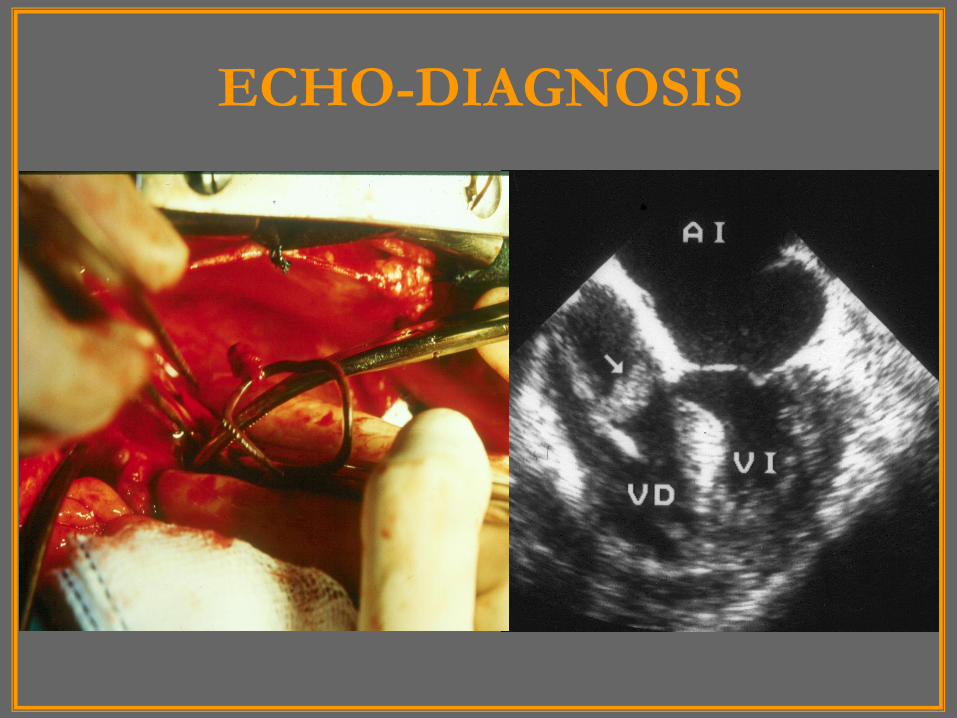
ECHO-DIAGNOSIS

ECHO-DIAGNOSIS• Why TTE has difficulties in CDR-IE?
– Reverberation lead echoes.
– Atypical location of vegetations.
– Inadequate transthoracic window.
• TTE & TEE may be falsely negative.
• When clinical suspicion of CDR-IE is high and TEE is negative: repeat TEE.
• Bland clots on leads have been found on echo exam in 1,4-10 % in CIED recipients without infection. No possible distinction by echocardiography.
• Accretions on the RA segment of pacing leads (28% by TEE): no effect on mortality.
Lo R, et al. J Invasive Cardiol 2006; Habib G, et al. Eur J Echocardiogr 2010; Downey BC, et al.
Pacing Clin Electrophysiol 2011; Dundar C, et al. JASE 2011; Rahbar AS, et al.PACE 2013.

Percutaneous explantation
• The preferred method of removal.
• It can be a high-risk procedure if performed in unprepared centres.
• The sooner the better for extracting an infected device.
• It is essential to remove all hardware to avoid recurrence of infection. In one study (1), 71% of patients with retained material showed recurrence of infection.
(1) Pichlmaier M, et al. J Thorac Cardiovasc Surg 2011; Nof E, et al. Eur Heart J 2013.
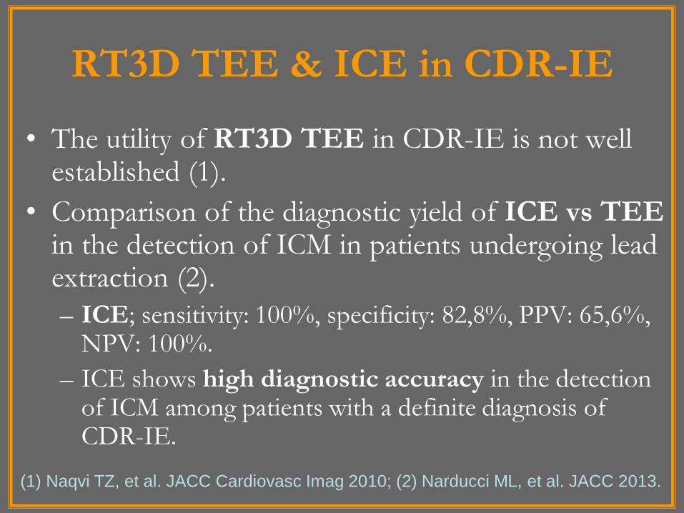
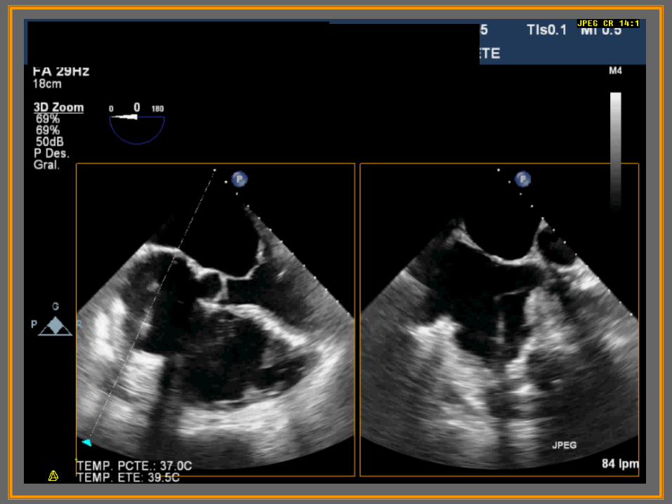
RT3D TEE & ICE in CDR-IE
• The utility of RT3D TEE in CDR-IE is not well established (1).
• Comparison of the diagnostic yield of ICE vs TEEin the detection of ICM in patients undergoing lead extraction (2).
– ICE; sensitivity: 100%, specificity: 82,8%, PPV: 65,6%, NPV: 100%.
– ICE shows high diagnostic accuracy in the detection of ICM among patients with a definite diagnosis of CDR-IE.
(1) Naqvi TZ, et al. JACC Cardiovasc Imag 2010; (2) Narducci ML, et al. JACC 2013.
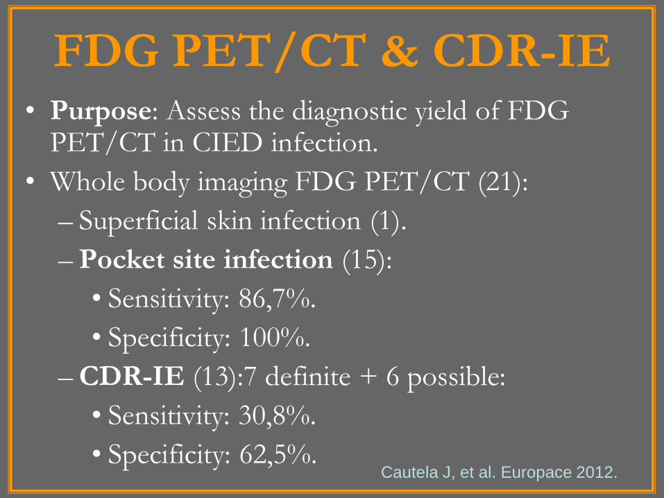
FDG PET/CT & CDR-IE• Purpose: Assess the diagnostic yield of FDG
PET/CT in CIED infection.
• Whole body imaging FDG PET/CT (21):
– Superficial skin infection (1).
– Pocket site infection (15):
• Sensitivity: 86,7%.
• Specificity: 100%.
– CDR-IE (13):7 definite + 6 possible:
• Sensitivity: 30,8%.
• Specificity: 62,5%.Cautela J, et al. Europace 2012.

CDR-IE: ESC treatment guidelines
Habib G, et al. Eur Heart J 2009

Empirical antibiotic therapy in CDR-IE
Clinical
presentation
Main
pathogens
Antibiotic
therapy
Acute clinical
course
S. Aureus
CoNS
Dapto/Vanco
+
Gentamicin
Subacute clinical
course CoNS (MS)
Cloxacillin
+
Gentamicin
Healthcare-
associated; severe
sepsis.
S. Aureus (MR); CoNS
(MR); enterococci;
GNB.
Dapto/Vanco +
Carbapenem +
rifampin

Antibiotic therapy in CDR-IE
• Antimicrobial choice should be based on in vitro susceptibility of the infectious agent.
• Duration of antimicrobial therapy:
– Non-complicated infection: 2 weeks.
– Complicated infection: 4-6 weeks.
• Persistent bacteremia.
• Thrombophlebitis.
• Septic metastasis / lung complications.
• Immunosuppression.
• Resistant microorganisms.
Baddour LM, et al. Circulation 2010

CDR-IE: AHA guidelines
Circulation 2010
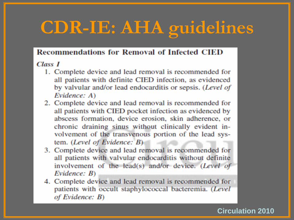
CDR-IE: AHA guidelines
Circulation 2010

Potential surgical indications
• Vegetation size ≥ 25 mm.
• Extension to mural endocardium, SVC,
severe tricuspid valve regurgitation.
• Previous incomplete extraction.
• Large vegetations and PFO /ASD.
• Associated left-sided IE.
• Other surgical reasons (Ao. stenosis).
• Mortality rates: 12,5%-40%.
Baddour L, et al. Circulation 2010; del Rio A, et al. Chest 2003; Verma A,
Wilkoff BL. Heart Rhythm 2004.

Clinical case

Tissue cultures & CDR-IE
• Cultures & gram staining of the pocket-site tissue
and lead tips when device removal.
• Avoid aspiration of the pocket site.
• Sensitivity of pocket-site tissue culture higher than
that of swab pocket culture.
• Lead contamination can occur during
extraction if a pocket is infected.
Baddour LM, et al. Circulation 2010