Embed Size (px)
Citation preview
22
Oxidative Therapy Against Cancer
Manuel de Miguel and Mario D. Cordero Departamento de Citología e Histología Normal y Patológica,
Facultad de Medicina, Universidad de Sevilla, Sevilla Spain
1. Introduction
Although a moderate increase of reactive oxygen species (ROS) may induce cell proliferation, excessive amounts of ROS can cause oxidative damage to lipids, proteins, and DNA, provoking oncogenic transformation, increased metabolic activity, and mitochondrial dysfunction (Dreher and Junod, 1996; Behrend et al., 2003; Pelicano et al, 2003). Many reports suggest that cancer cells are under a continuous oxidative stress (Pervaiz and Clement, 2004; Schumacker et al., 2006; Kryston et al., 2011). Studies with human tumor cell lines clearly show that these cells produce ROS at a much higher rate than healthy cells (Oberley and Buettner, 1979; Lu et al., 2007). ROS have been established as important molecules involved in the multistage process of carcinogenesis (Klaunig and Kamendulis, 2004). Mitochondria are the major consumers of molecular oxygen in cells, representing an important source of ROS. It is well accepted that cancer cells present mitochondrial alterations which result in respiration injury. This mitochondrial dysfunction may induce a low coupling efficiency of the mitochondrial electron chain, increasing electron leakage and leading to enhanced ROS formation. The resulting oxidative stress may cause further damage to both mitochondrial DNA (mtDNA) and the respiratory chain, amplifying the ROS generation (Zorov et al., 2006).
The higher oxidative stress observed in cancer cells can also result from a decrease or inactivation of antioxidants (Huang et al., 2003; Conklin, 2004). The majority of tumor cells usually present very few antioxidative enzymes, such as catalase, superoxide dismutase, and glutathione peroxidase, which are known to play a protective role against ROS in normal cells (Sato et al., 1992; Hasegawa et al., 2002; Pelicano et al., 2004). The lack of proper antioxidant defences makes tumor cells very vulnerable to oxidative stress. Nevertheless, some studies have revealed increased expression of antioxidants, probably as a consequence of selective pressure towards stress adaptation. The sources of ROS in cancer cells and the consequences of oxidative stress in the carcinogenesis process are still under debate.
What seems accepted is that cancer cells have increased ROS steady state level and are likely to be more vulnerable to damage by further ROS insults induced by exogenous agents. Thus, manipulating ROS levels by redox modulation could be a way to selectively kill cancer cells without causing significant toxicity to normal cells. A promising anticancer strategy named “oxidation therapy” has been developed by inducing cytotoxic oxidative stress for cancer treatment. This could be achieved by two different methods: inducing the
www.intechopen.com
Oxidative Stress and Diseases
498
generation of cytotoxic levels of ROS, and inhibiting the antioxidant system of tumor cells (Fang et al., 2007; Trachootham et al., 2009).
It is well known that ROS, such as hydrogen peroxide (H2O2) and superoxide anion, induce apoptosis to a wide range of tumor cells through the activation of the caspase cascade. It has been described that mitochondrial damage induced by the use of certain drugs provokes an increment of oxidative stress and cell death (Chandra et al., 2000; Conklin, 2004). Major ROS-modulating agents are based on the capacity to induce high ROS generation or to reduce the antioxidant defence machinery of cancer cells.
An interesting drug for oxidative cancer therapy is amitriptyline, a tricyclic antidepressant commonly prescribed for depression and therapeutic treatments of several neuropathic and inflammatory illnesses. Chlorimipramine, another tricyclic antidepressant, has been already proposed as a novel anticancer agent targeted to mitochondria as it induces caspase-3-dependent apoptosis (Daley et al., 2005). Several reports showed that the toxicity of this drug is due to an increase in oxidative stress by the generation of high amounts of ROS (Daley et al., 2005; Moreno-Fernández et al., 2008; Cordero et al., 2009). Therefore, amitriptyline has being proposed to be used for anticancer oxidant therapy against tumors that present significant oxidative stress and/or low antioxidant defences (Cordero et al., 2010).
2. Mitochondria and Reactive Oxygen Species (ROS)
Mitochondria are dynamic organelles that play a central role in many cellular functions including the generation of chemical energy (adenosine triphosphate, ATP), heat, and intracellular calcium homeostasis. They are also responsible for the formation of ROS and for triggering the programmed cell death or apoptosis (Turrens, 2003). The primary metabolic function of mitochondria is oxidative phosphorylation, an energy-generating process that couples oxidation of respiratory substrates to the synthesis of ATP (Pieczenik & Neustadt, 2007). The mitochondrial respiratory chain (MRC) is composed of five multisubunit enzyme complexes. Both the mtDNA and the nuclear DNA (nDNA) encode for polypeptide components of these complexes. Electron transport between MRC complexes I–IV is coupled to the extrusion of protons across the inner mitochondrial membrane by proton pump components of the respiratory chain. This movement of protons creates an electrochemical gradient (m) across the inner mitochondrial membrane. Protons return to the mitochondrial matrix by flowing through ATP synthase (complex V), which utilizes the energy thus produced to synthesize ATP from adenosine diphosphate (ADP) and inorganic phosphate. Both the mtDNA and the nDNA encode for polypeptide components of these complexes. As a consequence, mutations in either genome can cause MRC dysfunction that impairs transport of electrons and/or protons and decreases ATP synthesis. Primary or secondary genetic diseases affecting MRC or secondary mitochondrial dysfunctions usually affect brain and skeletal muscle because of their energy requirements. Besides MRC enzyme complexes, two electron carriers, coenzyme Q10 (CoQ) and cytochrome c, are essential for mitochondrial synthesis of ATP. CoQ transports electrons from complexes I and II to complex III and is essential for the stability of complex III. CoQ is a lipid-soluble component of virtually all cell membranes. CoQ also functions as an antioxidant that protects cells both by direct ROS scavenging and by regenerating other antioxidants such as vitamins C and E (Turunen et al., 2004). Given the critical role of CoQ in mitochondria function, it has been suggested that CoQ levels could be a useful biological
www.intechopen.com
Oxidative Therapy Against Cancer
499
marker of mitochondrial function (Haas et al., 2008). CoQ deficiency induces decreased mitochondrial respiratory enzymes activity, reduced expression of mitochondrial proteins involved in oxidative phosphorylation, decreased mitochondrial membrane potential, increased production of ROS, mitochondrial permeabilization, mitophagy of dysfunctional mitochondria, reduced growth rates and cell death (Rodriguez-Hernandez et al., 2009, Cotan et al, 2011).
In addition to energy, mitochondrial oxidative phosphorylation also generates ROS. When the MRC becomes highly reduced, the excess electrons from complex I or complex III may increase substantially, passing directly to O2 to generate high amounts of superoxide anion (O2
−). Superoxide is transformed to hydrogen peroxide (H2O2) by the detoxification enzymes manganese superoxide dismutase (MnSOD) or copper/zinc superoxide dismutase (Cu/Zn SOD), and then to water by catalase, glutathione peroxidase (GPx) or peroxiredoxin III (PRX III). However, when these enzymes cannot convert ROS such as the superoxide radical to water fast enough, oxidative damage occurs and accumulates in the mitochondria. If H2O2 encounters a reduced transition metal or is mixed with O2�−, the H2O2 can be further reduced to hydroxyl radical (HO�), the most potent oxidizing agent among ROS. Additionally, nitric oxide (�NO) is produced within the mitochondria by mitochondrial nitric oxide synthase (mtNOS) and also freely diffuses into the mitochondria from the cytosol. �NO reacts with O2
− to produce peroxynitrite (ONOO−). Together, these two radicals as well as others can do great damage to mitochondria and other cellular components (Turrens, 2003).
Under normal physiological conditions, ROS production is highly regulated. However, if the MRC is inhibited, or key mitochondrial components, such as CoQ, are deficient, then, electrons accumulate on the MRC carriers, greatly increasing the rate of a single electron being transferred to O2 to generate O2
−. An excessive mitochondrial ROS production can exceed the cellular antioxidant defense and the cumulative damage can ultimately destroy the cell by necrosis or apoptosis.
Mitochondria play an important role in cell bioenergetics and life signaling. Mitochondria are necessary for cell survival but they can also trigger cell death, thus exerting decisive control over the biochemistry of several cascades that lead to cell death, specifically the intrinsic pathway of apoptosis. The particular biochemical properties of these organelles are closely related to their segmented structures, which provide an optimal environment for multiple pathways of biosynthesis and bioenergetics. Consequently, it is possible that these organelles are involved in the process of carcinogenesis through alterations in cell metabolism and cell death pathways (Pilkington et al., 2008). Cancer cells show alterations in mtDNA, in oxidative phosphorylation and in energetic metabolism, all triggered by a pro-oxidative change (Indo et al., 2007). The “respiratory damage” in cancer cells predicts low coupling efficiency in electron transfer at the inner membrane of mitochondria and, consequently, greater loss of electrons, leading to the formation of more O2
− (Pelicano et al., 2003). The superoxide anion will thus generate more free radicals. Generated ROS can be released into cytosol and trigger “ROS-induced ROS-release” (RIRR) in neighbouring mitochondria. This mitochondrion-to-mitochondrion ROS-signaling constitutes a positive feedback mechanism for enhanced ROS production potentially leading to significant mitochondrial injury (Zorov et al., 2006). Recent studies by a number of groups have demonstrated that ROS can directly modify signaling proteins through different
www.intechopen.com
Oxidative Stress and Diseases
500
modifications, for example by nitrosylation, carbonylation, disulphide bond formation and glutathionylation. Moreover, redox modification of proteins permits further regulation of cell signaling pathways (England and Cotter, 2005).
3. Oxidative stress and cancer
3.1 ROS and carcinogenesis
In general, oxidative/nitrosative stress could be defined as an imbalance between the presence of high levels of ROS and reactive nitrogen species (RNS), and the antioxidative defense mechanisms. These toxic molecules are formed via oxidation-reduction reactions and are highly reactive since they have an odd number of electrons. ROS generated under physiological conditions are essential for life, as they are involved in bactericidal activity of phagocytes, and in signal transduction pathways, regulating cell growth and reduction–oxidation (redox) status (Davies, 1995). ROS includes free radicals, such as hydroxyl and superoxide radicals, and non-radicals, including hydrogen peroxide and singlet oxygen. Oxidative stress and generation of free radicals, as primary or secondary event, have been related in a great number of diseases, including cancer (Floyd, 1990).
At the beginning of the carcinogenic process, tumor cells accumulate mutations that allow them to proliferate in an uncontrolled way. Moreover, these alterations contribute to increase the susceptibility to accumulate additional genetic modifications, facilitating tumor progression and cancer development. Cancer could be defined as a cell-cycle desease, where its misregulation is considered an essential step (Sandhu et al., 2000). Carcinogenesis is a complex process of different sequencies that allow a cell to evolve from a healthy state into a pre-cancerous state and eventually reach a cancerous state. In this sense, there are several theories about the process of carcinogenesis. For instance, an increase of DNA synthesis and mitosis triggered by non-genotoxic agents could induce mutations in new cells. These mutations could spread through new cell divisions, evolving from an initial pre-neoplastic state into a neoplastic state. Another theory explains the existence of an imbalance between proliferation and cell death, where proliferation is favoured. If DNA damage is too high, there are important mechanisms, such as apoptosis, by which the altered cells are selectively eliminated. Protein p53 plays a fundamental role in this process, as it initiates mechanisms that eliminate, for example, those oxidized DNA bases that could cause mutations. If cell damage is too high, p53 initiates the mechanisms of apoptosis, although uncontrolled processes of apoptosis can be harmful for the organism, since healthy cells could also be eliminated. Therefore, there are systems for regulating apoptosis that consist of both pro-apoptotic and anti-apoptotic factors. Alterations that affect the function of gene p53 have been found in more than half of all types of cancer. This fact supports the idea that carcinogenesis would be caused by an imbalance between proliferation and cell death, in favour of proliferation.
Studies of epidemiology and animal experimentation have shown that carcinogenesis could occur in several stages characterized by different mechanisms. Thus, the model of carcinogenesis based on the hypothesis of three stages: initiation, promotion and progression, should be highlighted. Genotoxic agents are mainly chemical substances that damage DNA directly, inducing the generation of a mutation and/or a set of structural changes. On the other hand, there is a second category of non-genotoxic carcinogenic agents,
www.intechopen.com

Oxidative Therapy Against Cancer
501
whose role in the carcinogenic process is not related directly to DNA damage. These compounds modulate mechanisms of cell growth and death. Thus, the development process of a cancer would consist of the accumulation of multiple events, and the ROS could act at different levels and in all stages (Klauning and Kamendulis, 2004). Thereby, ROS can induce both genomic instability, caused by DNA damage, and alterations in cell signaling processes related to survival, proliferation, resistance to apoptosis, angiogenesis and metastasis, thus contributing to cancer initiation, promotion and progression.
Initiation involves a DNA mutation that is not lethal, but it produces a cell alteration followed by at least one round of DNA synthesis that allows fixing the damage done. At this point, the cell can stop its cycle temporally to undo DNA damage and then resume cell division. DNA damage may be done by ROS, like hydroxyl radicals formed by the Fenton´s reaction. Several studies have revealed an interesting correlation between tumor size and the amount of 8-OHdG (8-hydroxy-2'-desoxyguanosine; also known as 8-oxo-deoxyguanosine, 8-oxo-dG), a nucleotide modified by the activity of free radicals (Kennedy et al., 1998; Yano et al., 2009). The promotion stage is characterized by the expansion of initiated cells, stimulating cell proliferation and/or apoptosis inhibition. As a result of this process, an identificable lesion is formed, thus requiring the constant presence of an agent that stimulates promotion. However, it is a reversible process. Many promoter agents have a strong inhibitory capacity against antioxidants like catalases, glutathione, SOD, etc. While a high level of oxidative stress is cytotoxic for cells and stops proliferation inducing apoptosis or even necrosis, moderate levels of oxidative stress may stimulate cell division and, therefore, stimulate tumor growth and promotion (Dreher and Junod, 1996). Progression is the third and last stage of the carcinogenic process. This stage involves cellular and molecular changes that occur from a pre-neoplastic state to a neoplastic state. This stage is irreversible and it is characterized by the accumulation of genetic damage that allows the cell evolving from benign to malignant.
ROS are considered as carcinogenic potentials that facilitate cancer promotion and progression (Pelicano et al., 2004). The DNA molecule is one of the main targets of free radicals activity in the cell, and the modifications performed as a consequence of this activity are relevant for the loss of cell homeostasis. This loss may be extended in time due to the DNA functions of information reservoir. This is why the agents and mechanisms of damage by ROS are studied in depth, because its clarification would lead to elucidate the pathogeny of great morbidity and mortality diseases like cancer. There are different types of oxidative damage to DNA, like modifications and depurinations of DNA bases, DNA chain ruptures, and mutations, which tend to accumulate in an environment with high concentrations of ROS. It is known that DNA damage by ROS occurs spontaneously and there is a normal level of bases modified by ROS in cellular DNA (Okamoto, 2000). The most frequent modification of DNA linked to ROS is the formation of 8-OH-dG, resulting from attack of either a hydroxyl radical or singlet oxygen on deoxyguanosine. 8-OH-dG has a highly mutagenic potential, as 5-hydroxymethyl-2-deoxyuridine (Retel et al., 1993). With relative frequency, the cell will evade DNA damage using specific DNA polymerases and enter DNA replication creating mutations and chromosomal lesions. Alternatively, the presence of unrepaired DNA lesions can induce cell death through the apoptotic pathway. Chronic exposure to DNA lesions can lead to mutations and genomic instability (pre-cancerous state) and eventually to malignant transformations (cancerous state) (Kryston et al., 2011).
www.intechopen.com
Oxidative Stress and Diseases
502
3.2 Sources of oxidative stress
Cells from all organisms are exposed to several oxidizing and harmful agents. These attacks can be divided into two groups: exogenous and endogenous. Exogenous sources are related to environmental, medical, diagnostic ionizing and non-ionizing radiations (X- or γ-rays, α-particles from radon decay, UVA radiation) or chemical agents. Endogenous (intracelullar) sources of reactive species are primarily produced by O2 metabolism, immune responses and inflammation. These processes may result in the production of ROS and RNS that react with DNA and produce several lesions and indirect effects. Ionizing radiations can damage DNA also by direct energy deposition and ionizations (Kryston et al., 2011).
Oxygen metabolism is the major source of ROS in tumor tissues. ROS are continously formed in mitochondria as respiration byproducts. Cancerous cells are metabolically very active and require a great supply of ATP in order to maintain proliferation and cell growth under control. This high energy demand in the mitocondrial respiratory chain contributes to the generation of ROS. Usually, ATP is produced with high efficiency through oxidative phosphorylation in mitochondria. However, a malfunction of mitochondrial respiration is usually observed in neoplastic cells due to deletions/mutations in mtDNA, to the aberrant expression of some enzymes involved in energy metabolism and to hypoxia (Xu et al., 2005; Verrax et al., 2008). On the other hand, the increase of glycolysis in tumor cells has been shown to be related to the aggressiveness of the tumor (Cuezva et al., 2002). Acquiring a glycolytic phenotype represents a key element for cancer survival and progression, while glycolysis inhibition could be proposed as a goal in antitumor therapy (Pelicano et al., 2004). In this sense, glucose deprivation in laboratory models has been shown to be related to an increase of oxidative stress in tumor cells (Spitz et al., 2000). In cell lines of breast carcinoma, glucose deprivation leads to an intracellular increase of pro-oxidants, a decrease of free radical neutralization, and pyruvate depletion, which leads to an increase of oxidative stress (Spitz et al., 2000; Lee et al., 1999). As a compensation mechanism, glucose deprivation induces the expression of heme oxigenase-1 (HO-1), an enzyme that plays an important role in the antioxidant defense system of the organism and in Fe homeostasis. Chang and collegues (Chang et al., 2003) showed that the generation of ROS in mitochondria induces the overexpression of HO-1, which demonstrates that this is a common mechanism of regulation with the aim of protecting cells against oxidative damage.
As already mentioned, immune response and inflamation are other sources of ROS. It is known that oxidative stress activates inflammatory pathways leading to transformation of a normal cell to tumor cell, tumor cell survival, proliferation, insensitivity to anti-growth signaling, invasion, sustained angiogenesis, and stem cell survival (Reuter et al., 2010). Chronic inflammation is triggered by environmental (extrinsic) factors (eg, infection, tobacco, asbestos) and host mutations (intrinsic) factors (eg, Ras, Myc, p53). Activation of Ras, Myc, and p53 cause mitochondrial dysfunction, resulting in mitochondrial ROS production and downstream signaling (eg, NFkappaB, STAT3, etc.) that promote inflammation-associated cancer (Kamp at al., 2011).
On the other hand, during immune responses of many carcinogenetic processes, several immune-related cells play their roles, like intratumoral lymphocytes killing malignant cells and macrophages and neutrophils degrading cells by using oxidants and enzymes. One of these enzymes is NADPH oxidase, which activity produces ROS. The increased intensity of
www.intechopen.com
Oxidative Therapy Against Cancer
503
oxidative stress helps tumor to go on to malignancy. As an example, in vitro data suggest that in environments with certain levels of oxidative stress some cytokines, such as interleukin-2 (IL-2) or interferon α (IFNα), induce a decrease of T-lymphocytes or natural killer cells, which could suposse a tumor evasion to the immune response (Mantovani et al., 2003). Another source of free radical generation, usually underestimated but highlighted by Kryston et al. (2011), is the chronic exposure to viral infections; as in the case of hepatitis viruses, where there is a connection between chronic infection and induction of oxidative stress. There is a variety of viruses associated with increased ROS levels, DNA damage and mutagenic rate. The high intracellular oxidation status in viral infections consists of decreased antioxidant enzymes like catalase, glutathione peroxidase, glutathione reductase as well as high level of hydroxyl radicals (Kryston et al., 2011).
3.3 Influence of ROS in the cell cycle
Since some time already, an increase of ROS and an altered redox state has been observed in cancerous cells (Trachootham et al., 2009). It is known that ROS may serve as cellular messengers in the signal translation pathway and also an increase of ROS may trigger cell growth and proliferation, contributing to cancer development (Filomeno et al., 2005). Uncontrolled tumor cell proliferation requires the up-regulation of multiple intracellular signaling pathways, including cascades involved in survival, proliferation, and cell cycle progression. The most significant effects of oxidants on signaling pathways have been observed in the MAPK/AP-1 and NF-κB pathways (Muller et al., 2010). At the advanced stage of the disease, cancerous cells usually show genetic instability and a sharp increase of ROS, which induces to genetic mutations and failures in metabolic functioning, provoking a greater generation of ROS (Pelicano et al., 2004). Several mechanisms may lead to oxidative stress in cancer patients, such as altered energy metabolism, overproduction of cytokines, which in turn may increase ROS production, and the use of anti-neoplastic drugs. Altered energy metabolism in cancer may explain symptoms such as anorexia/cachexia, nausea and vomiting, which prevent normal nutrition. This deficit in the normal supply of nutrients like glucose, proteins, antioxidants and vitamins, leads eventually to the lack of antioxidant defenses for controlling free radical production.
Protein p53 is known as “the guardian of the genome” because it is essential for maintaining its integrity. This tumor suppressor protein has a fundamental role at detecting and eliminating oxidative damage in nuclear and mitochondrial DNA, at preventing mutations and at genetic instability. On the other hand, p53 acts also as a transcription factor that regulates the expression of many pro-oxidants and antioxidant genes. The functional loss of p53 is related to redox disequilibrium, increase of ROS, greater mutagenesis and tumor growth (Attardi and Donehower, 2005). This loss of p53 function is observed in many human cancers, especially in advanced stages (Bourdon, 2007).
There is evidence that the increase of ROS in tumor cells has a fundamental role in the acquisition of cancer characteristics (Hanahan and Weinberg, 2011): immortalization and transformation, cell proliferation and mitogenic signals, and cell survival and interruption of apoptotic death. Interestingly, in contrast to the effect of tumor promotion, recent studies suggest that the high ROS level has a role in the induction and maintenance of the senescence induced by tumor suppression, through the supported activation of the cell cycle inhibitor p16 (Ramsey and Sharpless, 2006; Takahashi et al., 2006). On the other hand, if the
www.intechopen.com

Oxidative Stress and Diseases
504
ROS level increases up to a specific threshold that is incompatible with cell survival, ROS can exert cytotoxic effects that lead to the death of malignant cells and, therefore, limit cancer progression. This double game of ROS effect on cancerous cells may be used for developing new antitumor drugs that provide an increase of lethal oxidative stress in tumor cells, which are more sensitive to this type of attack than normal cells.
3.4 Angiogenesis induced by ROS
In addition to alterations in the cell cycle, an important event in the growth of any tumor is the generation of a new blood supply system that feeds the malignant cells. Migration, proliferation and tubular formation by endothelial cells are essential events in the process of angiogenesis. It has been suggested that ROS play an important role in angiogenesis, although their molecular mechanism remains unknown (Ushio-Fukai, 2004). The vascular endothelial growth factor (VEGF) triggers angiogenesis by stimulating the proliferation of endothelial cells and their migration through the receptor 2 (Flk1/KDR) of VEGF, which has tyrosine kinase activity. ROS derived from NAD(P)H oxidase are important for in vitro VEGF signaling and for in vivo angiogenesis. Arbiser and collegues (Arbiser et al., 2002) showed that ROS increased the expression of the VEGF, triggering the promotion of vascularization mechanisms and the fast expansion of tumors. On the other hand, greater metastatic capacity has been associated with high ROS levels in most tumors (Ishikawa et al., 2008). So, exogenous administration of ROS could increase certain metastatic states, while a treatment with antioxidants could slow down the metastatic progression (Ferraro et al., 2006).
Hypoxia seems to be the most important mechanism for tumor progression through the activation of angiogenesis, which is essential for the tumor growth (Harris, 2002). The neoplastic cells respond to hypoxia with an increase of ROS level; this occurs in the first stages of tumor development as a consequence of the blood supply deficit (Denko et al., 2003). It was shown that despite hypoxia there was a greater production of ROS, a paradox, since the oxygen availability for the formation of O2
− would be limited. Perhaps the generation of ROS occurs later, throughout tumor development, at the first stages of angiogenesis. When the hypoxia of a tumor is followed by blood supply reperfusion, high levels of ROS are generated; something similar occurs in a myocardial infarction or a brain ischemia.
3.5 ROS and tumor cell invasion
Tumor invasion and metastasis are two important events in cancer in which oxidative stress has shown to have an important role. When mammalian carcinoma cells are treated with hydrogen peroxide before intravenous injection into mice, an increase in lung metastasis formation is observed (Kundu et al., 1995), probably due to a decreased attachment of tumor cells to the basal lamina or it could alternatively be due to the increased activity or expression of proteins that regulate cellular motility.
On the other hand, the matrix metalloproteinases (MMPs) have been involved in the invasion and metastasis of malignant tumors of various histogenetic origins, and are capable of cleaving most components of the basement membrane and extracellular matrix (Westermarck et al., 1999). The activation of MMPs, such as MMP-2, probably occurs by the
www.intechopen.com
Oxidative Therapy Against Cancer
505
reaction of ROS with thiol groups in the protease catalytic domain, being also ROS involved in MMP gene expression (Rajagopalan et al., 1996).
Recently, a number of steps in the progression of metastatic disease have been shown to be regulated by redox signaling (Diers et al., 2010). One such redox signaling molecule is the electrophilic cyclopentenone prostaglandin, 15-deoxy-Δ12,14-prostaglandin J2 (15d-PGJ2), which can affect redox signaling through the posttranslational modification of critical cysteine residues in proteins such as actin, vimentin, and tubulin. The fact that 15d-PGJ2 can alter the cytoskeleton coincides with decreased migration and increased focal-adhesion disassembly, which might have important implications in the inhibition of metastatic processes such as invasion, intravasation, and extravasation (Diers et al., 2010).
3.6 ROS adaptation by cancer cells
There is sufficient evidence that cancer cells are under greater oxidative stress (Pervaiz and Clement, 2004; Kryston et al., 2011). High levels of oxidative stress have been found in cancer patients (McEligot et al., 2005; Lu et al., 2007), and it has been reported that different biomarkers of oxidative-stress-mediated events are elevated in cancer-prone tissues (Bartsch and Nair, 2000). In vitro studies clearly show that human tumor cell lines produce ROS at a much higher rate than non-transformed cells (Oberley and Buettner, 1979; Lu et al., 2007). In cancerous cells, a high level of oxidative stress is observed, which may result not only from the overproduction of ROS, but also from low levels or inactivation of antioxidants (Huang et al., 2003). Antioxidants are molecules capable of slowing down or even preventing the oxidation of other molecules and play an essential role at protecting cells against ROS aggression. Inhibition of these enzymes seriously endangers the capability of cells to face ROS activity. The first frontier of cellular defense against ROS damage consists of endogenous non-enzymatic radical scavengers like glutathione (GSH) and vitamins like C, E, antioxidant enzymes like SOD, catalase and GPx, as well as specific repair pathways (Kryston et al., 2011).
A decrease of mitochondrial activity and an overexpression of Mn-SOD with a greater production of O2
− have been detected in some colorectal carcinomas and tumor cells of the pancreas (Van-Driel et al., 1997; Cullen et al., 2003). The accumulation of anion O2
− stimulates cell growth by altering the redox states of transcription factors and regulators of the protein cell cycle. The increase of ROS in tumor cells may induce an increase of endogenous antioxidants in order to avoid intracellular lesions. On the contrary, a decrease of SOD activity was detected in blood cells of patients with cervical cancer. The decrease of SOD activity observed could be related to the generation of free radicals that cause direct damage to the enzyme by reticulation or mutation induction (Manoharan et al., 2004; Naidu et al., 2007). Besides, this decrease may be caused by an alteration of the oligoelements (essential trace elements) that act as cofactors of this enzyme, which may be considered as a risk factor of tumor growth or carcinogenesis (Naidu et al., 2007). On the other hand, it was observed that the increase in the intensity of oxidative stress in the blood of patients with ovarian cancer is accompained by a decrease of antioxidants like SOD, catalase, vitamin C and vitamin E (Senthil et al., 2004). A high percentage of tumors show low catalase activity, which means an advantageous adaptation for the tumor, which continues to benefit from the high levels of ROS. Although oxidant agents can be toxic to normal cells, the moderate increase of ROS contributes to the growth and survival of many cancers.
www.intechopen.com
Oxidative Stress and Diseases
506
The strategy adopted by each type of tumor to increase the intensity of oxidative stress may vary from an increase of ROS to a decrease of antioxidants, going through a combination of both. The concentration of ROS in the tumor cell must be compensated with that of antioxidants in order to obtain an increase of ROS steady state level without reaching lethal levels. However, alterations in the mechanisms of antioxidant production are considered as indicators of oxidative stress in several types of cancer (Schumacker, 2006). Cancerous cells may adapt to survive at certain levels of oxidative stress. For example, greater levels of O2
−
and H2O2 were observed in cells transformed with H-ras, showing high levels of antioxidants like peroxiredoxin-3 and thioredoxin peroxidase, compared to normal cells (Young et al., 2004). These adaptive mechanisms keep ROS levels in a distribution space that allows cancerous cells to avoid serious oxidative damage and to survive ROS-mediated stress and mutations. Redox adaptation may be crucial not only for cancer development, but also for drug resistance (Pervaiz and Clement, 2004; Sullivan and Graham, 2008). Redox adaptation, through the increase of endogenous antioxidants and the activation of the cell survival pathway, may confer greater capacity for tolerating the action of exogenous stress, with capacity for increasing DNA repair and decreased apoptosis. Thus, although cancerous cells are under chronic oxidative stress, they possess remodeled antioxidant system which let them to avoid deleterious ROS effects.
4. Antitumor oxidative therapy
4.1 The strategy
The therapeutic selectivity and avoiding resistance to drugs are two important issues in the therapy against cancer. Strategies for improving therapeutic selectivity depend largely on understanding the biological difference between cancerous and normal cells. Tumor cells, compared to normal cells, are under greater oxidative stress related to the oncogenic transformation, to alterations in metabolic activity and to an increase of ROS generation (Toyokuni et al., 1995; Hileman et al., 2003; Behrend et al., 2003). The high concentration of ROS in cancerous cells may have beneficial consequencies, like the stimulation of cell proliferation, mutation multiplication, genetic instability and alterations in the sensitivity to agents against cancer. However, because ROS are chemically active and may inflict serious cell damage, the very fact that cancerous cells usually show greater intrinsic oxidative stress may itself provide a unique opportunity to kill tumor cells according to the vulnerability of ROS action.
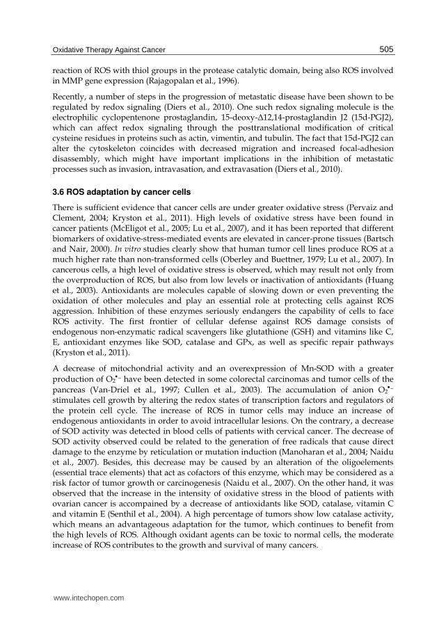
In this sense, the high concentration of ROS could function as a double-edged sword. A moderate increase of ROS could trigger both proliferation and differentiation, as well as other tumor characteristics. However, when ROS levels increase up to the lethal threshold it may break through the antioxidant capacity of the cell and trigger its death, by apoptosis or necrosis, depending on the degree of oxidative damage. Under physiological conditions, normal cells maintain redox homeostasis with a low level of basal ROS by controlling the equilibrium between the generation of ROS (pro-oxidant) and their elimination (antioxidant capacity). Exogenous agents that increase the generation of ROS or decrease the antioxidant capacity of cancerous cells will move the redox equilibrium and induce a general increase of ROS levels, which will cause cell death when exceeding the tolerance threshold (Figure 1).
www.intechopen.com
Oxidative Therapy Against Cancer
507
Fig. 1. Biological basis for therapeutic selectivity in oxidative therapy against cancer cells. Adapted from Gupte et al., 2009 and Trachootham et al., 2009.
Anticancer therapies aimed at the mitochondria of tumor cells are being developed in present years, since these organelles are involved in cell death (Daley et al., 2005; Nguyen and Hussain, 2007; Ozben, 2007; Pilkington et al., 2008). Mitochondria are the greatest oxygen consumers in the cell and they are an important source of reactive oxygen mediators. A drug that damages these organelles would cause an increase in ROS production and cell death. Currently, there is an anticancer strategy at its peak known as “oxidative therapy”, which consists of inducing high ROS steady state levels in tumor cells. This therapy may be carried out in two different ways: causing the generation of high levels of ROS in solid tumors and inhibiting the antioxidant system of tumor cells (Fang et al., 2007; Trachootham et al., 2009). It is well established that high levels of ROS, like H2O2 and O2
−, induce apoptosis in a wide variety of tumor cells activating the caspase cascade (Matsura et al., 1999; Yamakawa et al., 2000).
4.2 Cell death by ROS increment
Neoplastic cells are metabolically more active and require a high supply of ATP in order to keep cell growth and proliferation under control. This high energy demand in the MRC leads to the increase of ROS generation (Behrend et al., 2003). The excessive production of ROS may damage several cellular components like DNA, proteins, lipids and cell membranes. The oxidation of mitochondrial lipids and proteins causes the permeabilization of the mitochondrial membrane, which leads to an alteration of the coupling efficiency of the electron transport chain, resulting in the generation of more free radicals, and the release of cytochrome c, activating the process of programmed cell death (apoptosis) which depends on caspases (Conklin, 2004). There is evidence that the main mechanism by which oxidant agents may kill cells is the activation of apoptosis. In some cases, the high levels of ROS generated may inhibit apoptosis at a caspase level and divert the process toward
www.intechopen.com

Oxidative Stress and Diseases
508
necrosis (Chandra et al., 2000). The change from apoptosis to necrosis is critical in solid tumors and requires considerable amounts of ROS, a decrease of ATP and alterations in the mitochondrial electron-transport chain (Lee et al., 1999). The harmful consequences of this change lie in the inflammation caused by the rupture of necrotic cells and later release of enzymes that degrade the tissues. Thereby, death by apoptosis is preferred in antineoplastic therapies. Apoptotic cells cause the least damage to nearby tissues, since they do not release their content and are phagocytosed by macrophages.
Cancerous cells evolve with the mediation of endogenous and exogenous oxidative agents, depending on the cell type and the evolution state of the tumor. Tumors adapt to these conditions through the development of powerful antioxidant mechanisms and even by the use of endogenous ROS for proliferation. When ROS levels increase above the tolerance threshold, death of tumor cells is induced. Therefore, the fact that an excess of ROS causes cell damage and even death by apoptosis provides us with a strategy for eliminating cancerous cells, which are more sensitive to exogenous oxidative stress than normal cells, through the generation of free radicals, induced by oxidant pharmacological agents. In some cases, tumor cells attacked with antineoplastic therapies may gain resistance to oxidative stress, which is why combined therapies are promising, and are intended to converge toward improving the oxidative action above the critical threshold, or gathering different cytotoxic mechanisms.
Unfortunately, antitumor therapies may exert harmful effects on normal tissues, partially caused by ROS, which limits the application dose and its antitumor activity. Overcoming these secondary effects, without altering the therapy efficiency, is a priority and a challenge in biomedical research. In this sense, great importance is given to targeted therapies, using vehicles (liposomes, nanoparticles) that recognize specific molecules expressed in tumor cells.
4.3 Antioxidants in oxidative therapy against cancer
Antioxidants play an essential role in cell protection against ROS. The oxidation of antioxidant enzymes reduces the capacity of cells to eliminate free radicals. An important approach in the antitumor therapeutic strategies is to inhibit the antioxidant systems, like catalase, SOD and GPx, which are the main defense lines of the cell. The inhibitors of different antioxidant enzymes have been characterized by their capacity for eliminating neoplastic cells, alone or combined. It has also been described that many pharmacological agents may have more than one mechanism of action and affect multiple biological processes. There are agents that induce apoptosis which are oxidant and others that stimulate cell metabolism. On the other hand, there are apoptosis inhibitors that have antioxidant activities. In the absence of adequate antioxidant defenses, the damage from oxidative stress leads to the activation of the genes responsible for apoptosis.
There are conflicting data in the results obtained by different researchers regarding the levels of antioxidants in tumor tissue and in blood from cancer patients. In some cases, there are differences between different antioxidants in the same patient; some increase and others decrease (Ray et al., 2000). In breast cancer, for example, several studies describe an increase of lipid peroxidation and a decrease of antioxidants (Khanzode et al., 2004; Sener et al., 2007). However, other studies performed in neoplastic tissues have shown a greater presence of ROS and a high expression of antioxidants (Oltra et al., 2001; Gönenç et al.,
www.intechopen.com

Oxidative Therapy Against Cancer
509
2006). So, for instance, an increase in the expression of Mn-SOD has been observed in breast cancer and in blood samples from patients with different types of leukemia (Nishiura et al., 1992; Devi et al., 2000; Ray et al., 2000). This fact may reflect an adaptive mechanism by which cancerous cells respond to an increase of ROS levels produced in mitochondria. ROS can induce the over-regulation of Mn-SOD through the modulation of the redox states of the transcription factors (AP- 1, NF-kappaB). Due to its high expression in certain types of cancer, Mn-SOD has been considered as a tumor marker (Schadendorf et al., 1995). This expression of SOD protects tumor cells against a lethal increase of ROS levels. In fact, it has been demonstrated that the inhibition of SOD with 2-methoxyestradiol would induce apoptosis in leukemia cells through a mechanism mediated by free radicals, without showing significant toxicity in normal lymphocytes (Zhou et al., 2003).
Antitumor therapies mediated by ROS show a promising therapeutic activity in clinical studies (Trachootham et al., 2009). However, some tumor cells, especially in advanced stages of the disease, have adapted to oxidative stress due to their antioxidant capacity. This redox adaptation does not only allow tumor cells surviving under high levels of ROS, it also provides an increase of survival molecules and a greater capacity for drug inactivation. Moreover, it has been suggested that resistance to the agents that induce intracellular ROS production, such as paclitaxel, doxorubicin or other drugs, is correlated to the increase of antioxidants (Glorieux et al., 2011). Thereby, the capability of certain drugs to inhibit or reduce the antioxidant machinery is very useful in oxidative therapy. These drugs could be used in combination with oxidant agents for greater efficiency in antitumor therapies.
5. Amitriptyline as an anti-cancer agent
Amitriptyline is a commonly prescribed tricyclic antidepressant drug that is well known to death investigators, forensic pathologists, and toxicologists. Amitriptyline has sedative effects and is frequently prescribed for patients experiencing symptoms of depression. Amitriptyline, have also been used for therapeutic treatment of neuropathic and inflammatory diseases such as fibromyalgia, chronic fatigue syndrome, migraine, irritable bowel syndrome, and atypical facial pain (Gruber et al., 1996). Besides its anxiolytic properties, amitriptyline has central anticholinergic effects. Amitriptyline inhibits serotonin and noradrenaline uptake in presynaptic nerve ending (Maubach et al., 1999). However, toxicity of amitriptyline has been observed during standard treatments, and frequently during suicidal or accidental overdosage. Tricyclic antidepressant overdosage has toxic effects over cardiovascular, autonomous nervous, and central nervous systems, and may result in cardiotoxicity, cardiac conduction delays, dysrhythmia, hypotension, altered mental status, and seizures (Thanacoody and Thomas, 2005; Kiyan et al., 2006).
In vitro administration of amitriptyline to cell cultures induces several signs of toxicity. Amitriptyline treatment induces alteration of cellular permeability based on its detergent nature (Kitagawa et al., 2006). Furthermore, amitriptyline causes alterations in the glucidic metabolism of neurons resulting in a decrease of both uptake and transport of glucose (Mannerstrom and Tahti, 2004). Additionally, amitriptyline provokes an increase of intracellular lipid peroxidation in mouse 3T3 fibroblasts (Viola et al., 2000) and some mouse tissues (Bautista-Ferrufino et al., 2011), and many of these toxic effects are prevented by antioxidants (Slamon and Pentreath, 2000). Recently, our group has shown that
www.intechopen.com
Oxidative Stress and Diseases
510
amitriptyline induced toxicity is caused through a mitochondrial dysfunction, and increased ROS level (Moreno-Fernandez et al., 2008; Cordero et al., 2009). Amitriptyline reduced significantly the number of cultured cells; enhanced the production of stimulated lipid peroxidation, inverting the lipid reduced/oxidized ratio; decreased catalase protein levels, cytochrome c, ΔΨm, and citrate synthase activity; revealing mitochondrial damage. So, amitriptyline-induced toxicity is caused through mitochondrial dysfunction, and increased mitochondrial ROS production. Moreover, CoQ level was decreased by amitriptyline treatment and CoQ and alpha-tocopherol supplementation ameliorated amitriptyline-induced toxicity in both cultured human primary fibroblasts and zebrafish embryos (Cordero et al., 2009).
Other toxic effects attributed to amitriptyline lie in the alteration of neuron carbohydrate metabolism, which results in a decrease of glucose absorption and transport; causing a total loss of neuron viability in a cell line of neuroblastoma (Mannestrom et al., 2004).
Recent studies have shown that some antidepressants can kill cancerous cells. In fact, tricyclic antidepressants have shown to cause cell death in human normal lymphocytes (Karlson et al., 1998), Hodgkin´s lymphoma cells (Serafeim et al., 2003), neurons (Lirk et al., 2006), glioma cells (Xia et al., 1999; Daley et al., 2005; Levkovitz et al., 2005) and colorectal cancer cells (Arimochi y Morita, 2006). Chlorimipramine exerts its effect via the inhibition of complex III of the MRC (Daley et al., 2005). The same is valid for amitriptyline, as we have already reported (Cordero et al., 2009). We showed in fibroblasts treated with amitriptyline a decrease of expression level of proteins of complex I, complex III, cytochrome c, and reduced CoQ10 levels. Deficient mitochondrial protein expression levels and reduced levels of CoQ10 may impair normal mitochondrial electron flow and proton pumping, inducing a drop in respiratory complexes activity, and mitochondrial membrane potential. Our data showed that amitriptyline-treated fibroblasts have reduced NADH:cytochrome c reductase (complex I+III) activity, and lower mitochondrial membrane potential, which may contribute to impaired mitochondrial protein import and aggravate mitochondrial dysfunction, ROS production, and oxidative stress. It has been proposed that ROS damage can induce the mitochondrial permeability transition (MPT) by the opening of non-specific high conductance permeability transition (PT) pores in the mitochondrial inner membrane (England and Cotter, 2005). This, in turn, leads to a simultaneous collapse of mitochondrial membrane potential. The activation of MPTcauses mitochondria to become permeable to all solutes up to a molecular mass of about 1500 Da (Forte and Bernardi, 2005). After MPT, mitochondria undergo a dramatic swelling driven by colloid osmotic forces, which culminates in the rupture of the outer membrane and release of proapoptotic mitochondrial intermembrane proteins into the cytosol, such as cytochrome c, apoptosis inducing factor, Smac/Diablo, and others (Cordero et al.,2009).
We have also studied the effect of amitriptyline on tumor cell lines (Cordero et al., 2010). We observed that this drug induced important mitochondrial damage in tumor cell lines (H460: non-small cell lung cancer, HeLa: epithelial cervical cancer, and HepG2: hepatoma), generating high amounts of ROS and provoking apoptotic cell death. Moreover, amitriptyline effects have been compared with three antitumor drugs frequently used in cancer therapy: camptothecin (CPT), doxorubicin (Doxo), and methotrexate (Metho). Interestingly, amitriptyline induced significantly higher ROS generation in comparison with the other drugs, producing a dose-dependent increase of apoptosis in human cancer cells
www.intechopen.com

Oxidative Therapy Against Cancer
511
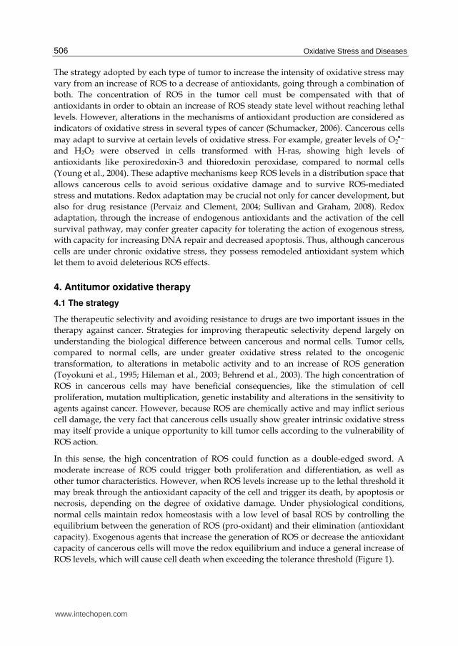
through a mechanism dependent on caspase-3 activation. Apoptosis percentage was significally higher in those cells treated with amitriptyline than in cells treated with CPT, Doxo or Metho (Figure 2A). Moreover, when the cell cycle of synchronized cultures was stopped at the G0/G1 phase by depriving cells from serum, the difference of apoptosis percentage among amitriptyline and the remaining drugs was significantly higher than in normal cultures (Figure 2B). These results suggest that the effect of amitriptyline does not depend on cell cycle stage, whereas CPT, Doxo, and Metho are more harmful in dividing cells, as most chemotherapeutic drugs. These data are of special interest for cancer treatment during the nongrowing phases of certain tumors.
Fig. 2. Comparative study of amitriptyline and different chemotherapeutic drugs for the evaluation of apoptosis. (A) Percentages of apoptotic cells in H460 cell cultures 24 h after administration of drugs at different concentration. (B) Apoptosis assessment in synchronized cultures stopped at the G0/G1 phase. Adapted from Cordero et al., 2010.
After treating cancer cells with amitriptyline, we have found increased ROS level and several signs of mitochondrial damage, as attenuated complex I+III activity, decreased protein levels of complex III, decreased membrane potential, and a significant reduction of the number of this organelle, shown by cytochrome c and citrate synthase determination, and electron microscopy (Figure 3). So, this tricyclic compound provokes oxidative stress in cancer cells, being mitochondria the target of its toxicity. None of the chemotherapeutic drugs tested seemed to damage mitochondria seriously. However, the chemotherapeutic drugs induced apoptosis and increased ROS production in tumor cells, although not with the intensity of amitriptyline.
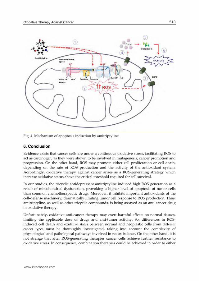
According to our data, amitriptyline induces a mitochondrial damage characterized by a decrease of the expression levels of complexes I and III of the MRC as well as of cytochrome c and of CoQ levels, which suggests an alteration in the activity, organization and assembly of the mitochondrial complexes, being this reflected in a decrease of the electron flow as well as a decrease of the mitochondrial membrane potential and, therefore, an increase of intramitochondrial ROS prodution. The damage caused by the increase of mitochondrial ROS induces the opening of the mitochondrial permeability transition pore (MPT), thus increasing mitochondrial permeability with the consequent release of proapoptotic proteins to the cytosol such as cytochrome c, Smac/Diablo, etc., initiating the intrinsic pathway of apoptosis dependent on caspase-3 (Figure 4).
www.intechopen.com

Oxidative Stress and Diseases
512
Fig. 3. Transmission electron microscopy showing damage and fewer mitochondria in H460 cells treated with 50 mmol/l of amitriptyline. Degenerating mitochondria (arrows) are observed in treated tumor cells. (A) Nontreated tumor cells. (B) Amitriptyline-treated tumor cells (Cordero et al., 2010).
In general, the increase of ROS production causes, as a response, an increase of the antioxidants activities. However, under the high input rate of ROS, enzyme inactivation prevails, which leads to the reduction of antioxidant enzymes activity and to the process of oxidative damage. Thus, tumor cells frequently possess very little antioxidative enzymes, such as catalase, SOD, and glutathione peroxidase, which are known to play a protective role against ROS in normal cells. We have observed in normal fibroblast treated with amitriptyline a decrease in protein expression of antioxidant enzymes (catalase and MnSOD) 16 h after the treatment, followed by restored levels after 24h, as a mechanism of antioxidant defense (Moreno-Fernández et al., 2008). Interestingly, in cancer cells, the same concentration of amitriptyline provoked an unrestorable decrease of catalase (Cordero et al., 2010). The difference of the antioxidant status observed in cancer cells, in comparison with healthy fibroblasts, may be caused by the lower antioxidant level present in the cancer cell lines used. Besides the decrease of catalase and MnSOD, amitriptyline also produces a significant decrease of CoQ level in tumor cells (Cordero et al., 2010). CoQ plays a critical protective role by either acting as an antioxidant or by the noncompetitive inhibition of the neutral sphingomyelinase of plasma membrane, preventing ceramide production (Mates et al., 1999). Most chemotherapeutic drugs do not provoke any decrement of antioxidants. Instead, they frequently induce an increase of antioxidants as a protecting mechanism against ROS generation, leading to lower cell death (Brea-Calvo et al., 2006). The fact that amitriptyline downregulates both catalase and CoQ activity is very interesting since it destroys the already decreased antioxidant defenses present in cancer cells, making the oxidative stress produced by the amitriptyline-induced ROS generation a more effective weapon.
Thus, amitriptyline promotes enhanced oxidative damage to cancer cells as this drug attacks cells by two different mechanisms: by the production of a high amount of ROS, provoking apoptosis; and by a significant decrease in antioxidant levels, seriously limiting cell reaction to oxidative stress. Therefore, amitriptyline could be used for anticancer oxidant therapy against tumors that present significant oxidative stress and/or low antioxidant defenses. For anticancer therapeutics on those tumors with a similar redox status than normal cells, a drug delivery vehicle should be used.
www.intechopen.com
Oxidative Therapy Against Cancer
513
Fig. 4. Mechanism of apoptosis induction by amitriptyline.
6. Conclusion
Evidence exists that cancer cells are under a continuous oxidative stress, facilitating ROS to act as carcinogen, as they were shown to be involved in mutagenesis, cancer promotion and progression. On the other hand, ROS may promote either cell proliferation or cell death, depending on the rate of ROS production and the activity of the antioxidant system. Accordingly, oxidative therapy against cancer arises as a ROS-generating strategy which increase oxidative status above the critical threshold required for cell survival.
In our studies, the tricyclic antidepressant amitriptyline induced high ROS generation as a result of mitochondrial dysfunction, provoking a higher level of apoptosis of tumor cells than common chemotherapeutic drugs. Moreover, it inhibits important antioxidants of the cell-defense machinery, dramatically limiting tumor cell response to ROS production. Thus, amitriptyline, as well as other tricyclic compounds, is being assayed as an anti-cancer drug in oxidative therapy.
Unfortunately, oxidative anti-cancer therapy may exert harmful effects on normal tissues, limiting the applicable dose of drugs and anti-tumor activity. So, differences in ROS-induced cell death and oxidative status between normal and neoplastic cells from different
cancer types must be thoroughly investigated, taking into account the complexity of physiological and pathological pathways involved in redox balance. On the other hand, it is not strange that after ROS-generating therapies cancer cells achieve further resistance to oxidative stress. In consequence, combination therapies could be achieved in order to either
www.intechopen.com

Oxidative Stress and Diseases
514
increase the intensity of oxidative stress above the critical threshold, or to perform different cytotoxic mechanisms.
Nevertheless, although greater efforts must be made, oxidative therapy against cancer is a promising strategy that is worthy of being investigated.
7. References
Arbiser, J.L.; Petros, J.; Klafter R.; Govindajaran, B.; McLaughlin E.R.; Brown L.F.; et al. (2002). Reactive oxygen generated by Nox1 triggers the angiogenic switch. Proc Natl Acad Sci USA. 99 (Jan) (715-720), ISSN 0027-8424.
Arimochi, H. & Morita, K. (2006). Characterization of cytotoxic actions of tricyclic antidepressants on human HT29 colon carcinoma cells. Eur J Pharmacol. 10,541, (Jul) (17- 23), ISSN 0014-2999.
Attardi, L. D. & Donehower, L. A. (2005). Probing p53 biological functions through the use of genetically engineered mouse models. Mutat. Res. 576 (Aug) (4-21), ISSN 0027- 5107.
Bartsch, H. & Nair, J. (2000). New DNA-based biomarkers for oxidative stress and cancer chemoprevention studies. Eur J. Cancer 36,10, (Jun) (1229-1234), ISSN 0959-8049.
Bautista-Ferrufino, M.R.; Cordero M.D.; Sánchez-Alcázar, J.A.; Illanes, M.; Fernández-Rodríguez, A.; Navas, P. & de Miguel, M. (2011). Amitriptyline induces coenzyme Q deficiency and oxidative damage in mouse lung and liver. Toxicol Lett. 204 (Jul) (32-37), ISSN 0378-4274.
Behrend, L.; Henderson, G. & Zwacka, R. M. (2003). Reactive oxygen species in oncogenic transformation. Biochem. Soc. Trans. 31, (Dec.) (1441–1444), ISSN 0300-512.
Bourdon, J. C. (2007). p53 and its isoforms in cancer. Cancer 97 (Aug) (277–282), ISSN 0007-0920.
Brea-Calvo, G.; Rodriguez-Hernandez, A.; Fernandez-Ayala, D. J.; Navas, P.; & Sanchez-Alcazar, J.A. (2006). Chemotherapy induces an increase in coenzyme Q10 levels in cancer cell lines. Free Radic Biol Med 40 (Apr) (1293–1302), ISSN 0891- 5849.
Chandra, J.; Samali, A.; & Orrenius, S. (2000). Triggering and modulaton of apoptosis by oxidative stress. Free Radic Biol Med; 29 (Aug) (323–333), ISSN 0891-5849.
Chang, E.F.; Wong, R.J.; Vreman, H.J.; Igarashi, T.; Galo, E.; Sharp, F.R.; Stevenson, D.K.; &Noble-Haeusslein, L.J. (2003). Heme oxygenase-2 protects against lipid peroxidation-mediated cell loss and impaired motor recovery after traumatic brain injury. J Neurosci. 23, 9, (May) (3689-3696), ISSN 0270-6474.
Conklin, K. A. (2004). Free radicals: the pros and cons of antioxidants. Cancer chemotherapy and antioxidants J. Nutr; 134 (Nov) (3201-3204), ISSN 0022-3166.
Cordero, M.D.; Moreno-Fernández, A.M.; Gomez-Skarmeta, J.L.; De Miguel, M.; Garrido-Maraver, J.; Oropesa-Ávila, M.; et al. (2009). Coenzyme Q10 and alpha tocopherol protect against amitryptyline toxicity. Toxicol Appl Pharmacol 15 (Mar) (329-337), ISSN 00410-008X.
Cordero, M.D.; Sánchez- Alcázar, J.A.; Bautista-Ferrufino, M.R.; Carmona-Lopez, M.I.; Illanes, M.; Rios, M. J.; et al. (2010). Acute oxidant damage promoted on cancer cells by amitriptyline in comparison with some common chemotherapeutic drugs. Anticancer drugs 10 (Nov) (932-944), ISSN 0959-4973.
www.intechopen.com
Oxidative Therapy Against Cancer
515
Cotan, D.; Cordero, M.D.; Garrido-Maraver, J.; Oropesa-Ávila, M.; Rodríguez-Hernández, A.; Gómez Izquierdo, L.; et al. (2011). Secondary coenzyme Q10 deficiency triggers mitochondria degradation by mitophagy in MELAS fibroblasts. FASEB J. 25 (Aug) (2669-2687), ISSN 0892-6638.
Cuezva, J.M.; Krajewska, M.; de Heredia M.L.; Krajewski, S.; Santamaría, G.; Kim, H.; et al. (2002). The bioenergetic signature of cancer: a marker of tumor progression. Cancer Res; 62 (6674-6681), ISSN 0008- 5472.
Cullen, J.J.; Mitros, F.A.; & Oberley, L.W. (2003). Expression of antioxidant enzymes in diseases of the human pancreas: another link between chronic pancreatitis and pancreatic cancer. Pancreas 26, 1, (Jan) (23-27), ISSN 0885-3177.
Daley, E.; Wilkie, D.; Loesch, A.; Hargreaves, I.P.; Kendall, D.A.; & Pilkington G. J, et al. (2005); Clomipramine: a novel anticancer agent with a mitochondrial target. Biochem Biophys Res Commun 328 (Mar) (623–632), ISSN 0006-291X.
Davies, K. (1995). Breast cancer genes. Further enigmatic variations. Nature 378 (Dec) (362-363), ISSN 0028-0836.
Denko, N.C.; Fontana, L.A. Hudson, K.M. et al. (2003). Investigating hypoxic tumor physiology through gene expression patterns. Oncogene 22 (Sep) (5907-14), ISSN 0950-9232.
Devi, G.S.; Prasad, M.H.; Saraswathi, I.; Raghu, D.; Rao, D.N. & Reddy, P.P. (2000). Free radicals, antioxidant enzymes and lipid peroxidation in different types of leukemias. Clin Chim Acta 293 (Marz) (53-62), ISSN 0009-8981.
Diers, A.R.; Higdon, A.N.; Ricart, K.C.; Johnson, M.S.; Agarwal, A.; Kayanaraman, B.; et al. (2010). Mitochondrial targeting of the electrophilic lipid 15-deoxy-Delta prostaglandin J2 increases apoptotic efficacy via redox cell signalling mechanisms. Biochem J. 426, (Jan) (31-41), ISSN 0264-6021.
Dreher, D.; & Junod, A.F. (1996). Role of oxygen free radicals in cancer development. European Journal of Cancer, 32A (Jan) (30-38), ISSN 0959-8049.
England, K. & Cotter, T.G. (2005). Direct oxidative modifications of signalling proteins in mammalian cells and their effects on apoptosis. Redox Rep 10 (237-245), ISSN 1351-0002.
Fang, J.; Nakamura, H.; & Iyer, A. K. (2007). Tumor-targeted induction of oxystress for cancer therapy. J. Drug Target. 15 (Aug) (475-486), ISSN 1061-186X.
Ferraro, D.; Corso, S.; Fasano, E.; Panieri, E.; Santangelo, R.; Borrello, S.; et al. (2006). Pro-metastatic signaling by c-Met through RAC- 1 and reactive oxygen species (ROS). Oncogene 25 (3689-3698), ISSN 0950-9232.
Filomeno, G.; Rotilio, G. & Ciriolo, M.R. (2005) Disulfide relays and phosphorylative cascades: partners in redox-mediated signalling patways. Cell Death Differ 12 (Dic) (1555-1563), ISSN 1350-9047.
Floyd, R.A. (1990). Role of oxygen free radicals in carcinogenesis and brain ischemia. FASEB J. 4 (Jun) (2587–2597), ISSN 0892-6638.
Forte, M. & Bernardi, P. (2005). Genetic dissection of the permeability transition pore. J. Bioenerg. Biomembr. 37, 3 (Jun) (121- 128), ISSN 0145-479X.
Glorieux C, Dejeans N, Sid B, Beck R, Calderon PB, Verrax J. (2011). Catalase overexpression in mammary cancer cells leads to a less aggressive phenotype and an altered response to chemotherapy. Biochem Pharmacol. Jun 13. PMID: 21689642, ISSN 0006-2952.
www.intechopen.com
Oxidative Stress and Diseases
516
Gönenç, A.; Erten, D.; Aslan, S.; Akinci, M.; Simşek, B.; & Torun, M. (2006). Lipid peroxidation and antioxidant status in blood and tissue of malignant breast tumor and benign breast disease. Cell Biol Int. 30, 4, (Apr) (376-380), ISSN 1065-6995.
Gruber, A.J.; Hudson, J.I. & Pope, G. (1996). The management of treatment-resistant depression in disorders on the interface of psychiatry and medicine. Fibromyalgia, chronic fatigue syndrome, migrañe, irritable bowel syndrome, atypical facial pain, and premenstrual dysphoric disorder. Psychiatr Clin North Am, 19,2, (351–359), ISSN 0193-953X.
Gupte, A. & Mumper, R.J. (2009). Elevated copper and oxidative stress in cancer cells as a target for cancer treatment. Cancer Treat rev. 35(Feb) (32-46), ISSN 0305-7372.
Haas, R.H.; Parikh, S.; Falk, M.J.; Saneto, R.P.; Wolf, N.I.; Darin, N. et al. (2008). The in-depth evaluation of suspected mitochondrial disease. Mol Genet Metab. 94, (May) (16-37), ISSN 1096-7193.
Hanahan, D. & Weinberg, R.A. (2011).The hallmarks of cancer. Cell. 144, (Mar) (209-219), ISSN 0092-8674.
Harris, A.L. (2002). Hypoxia- A key regulatory factor in tumor growth. Nat Rev Cancer. 2, 1, (Jan) (38-47), ISSN 1474-175X.
Hasegawa, Y.; Takano, T.; Miyauchi, A.; Matsuzuka, F.; Yoshida, H. & Kuma K. (2002). Decreased expression of glutathione peroxidase mRNA in thyroid anaplastic carcinoma. Cancer Lett; 182, (Aug) (69–74) ISSN 0304-3835.
Hileman, E.O.; Liu, J.; Albitar, M.; Keating, M.J. & Huang, P. (2003). Intrinsic oxidative stress in cancer cells a biochemical basis for therapeutic selectivity. Cancer Chemother. Pharmacol. 53, 3 (Mar) (209-219), ISSN 0344-5704.
Huang, P.; Feng, L.; Oldham, E.A.; Keating, M.J. & Plunkett, W. (2003). Superoxide dismutase as a target for the selective killing of cancer cells. Nature 407,5, (Dic) (390-395), ISSN 0028-0836.
Indo, H.P.; Davidson, M.; et al. (2007). Evidence of ROS generation by mitochondria in cells with impaired electron transport chain and mitochondrial DNA damage. Mitochondrion. 7, 2, (106-118), ISSN 1567-7249.
Ishikawa, K. et al. (2008). ROS-generating mitochondrial DNA mutations can regulate tumor cell metástasis. Science. 320, (May) (661–664), ISSN 0036-8075.
Kamp, D.W.; Shacter E. & Weitzman, S.A. (2011). Chronic inflammation and cancer: The role of the mitochondria. Oncology. 25 (Apr) (400-410), ISSN 0890-9091.
Karlsson, H.; Gu, Y.; DePierre, J.; Nassberger, L. (1998). Induction of apoptosis in proliferating lymphocytes by tricyclic antidepressants. Apoptosis. 3, 4 (Sep) (255–260), ISSN 1360-8185.
Kennedy, C. H.; Cueto, R.; Belinsky, S.A.; Lechner, J.F. & Pryor, W. A. (1998). Overexpression of hMTH1 mRNA: a molecular marker of oxidative stress in lung cancer cells. FEBS Lett. 429, 1, (Jun) (17-20), ISSN 0014-5793.
Khanzode, S.S.; Muddeshwar, M.G. & Dakhale, G.N. (2004). Antioxidant enzymes and lipid peroxidation in different stages of breast cancer. Free Radic. Res. 38, 1, (Jan) (81-85), ISSN 1071-5762.
Kitagawa, N., Oda, M., Nobutaka, I., Satoh, H., Totoki, T. & Morimoto, M., (2006). A proposed mechanism for amitriptyline neurotoxicity based on its detergent nature. Toxicol. Appl. Pharmacol. 15, 217, (Nov) (100–106), ISSN 0041-008X.
www.intechopen.com
Oxidative Therapy Against Cancer
517
Kiyan, S.; Aksay, E.; Yanturali, S.; Atilla, R.; & Ersel, M.; (2006). Acute myocardial infarction associated with amitriptyline overdose. Basic Clin. Pharmacol. Toxicol. 98, 45, (May) (462–466), ISSN 1742-7835.
Klauning, J.E. & Kamendulis, L.M. (2004). The role of oxidative stress in carcinogenesis. Annu Rev Pharmacol Toxicol. 44, (239-267), ISSN 0362- 1642.
Kryston, T.B.; Georgiev, A.B.; Pissis, P. & Georgakilas, A.G. (2011). Role of oxidative stress and DNA damage in human carcinogenesis. Mutat Res 3, 711, (Jun) (193-201), ISSN 0027-5107.
Kundu, N.; Zhang, S. & Fulton, A. M. (1995). Sublethal oxidative stress inhibits tumor cell adhesion and enhances experimental metastasis of murine mammary carcinoma. Clin. Exp. Metastasis 13, 1, (Nov) (16–22), ISSN 0262-0898.
Lee, Y.J.; Galaforo, S.S.; Sim, J.E.; et al. (1999). Dominant negative Jun Nterminal protein kinase (JNK-1) inhibits metabolic oxidative stress during glucose deprivation in a human breast carcinoma cell line. Free Radic Biol Med. 274, 28, (Feb) (575-584) ISSN 0891-0895.
Levkovitz, Y.; Gil-Adl.; Zeldich, E.; Dayag, M. & Weizman, A. (2005). Differential induction of apoptosis by antidepressants in glioma and neuroblastoma cell lines: evidence for p-c-Jun, cytochrome c, and caspase-3 involvement. J. Mol Neurosci. 27, 3, (29- 42), ISSN 0895-8696.
Lirk, P.; Haller, I.; Hausott, B.; Ingorokva, S.; Deibl, M.; Gerner, P. & Klimaschewski, L. (2006). The neurotoxic effects of amitriptyline are mediated by apoptosis and are effectively blocked by inhibition of caspase activity. Anesth Analg. 102, (Jun) (1728-33), ISSN 0003-2999.
Lu, W.; Ogasawara, M. A. & Huang, P. (2007). Models of reactive oxygen species in cancer. Drug Discov. 4, (67–73), ISSN 1740-6757.
Mannerstrom, M. & Tahti, H. (2004). Modulation of glucose uptake in glial and neuronal cell lines by selected neurological drugs. Toxicol. Lett. 15, 151, (Jun) (87-97), ISSN 0378-4274.
Manoharan, S.; Klanjiappan, K. & Kayalvizi, M. (2004). Enhanced lipid peroxidation and impaired enzimatic antioxidant activities in the erythrocytes of the patients with cervical carcinoma. Cell Mol Biol Lett. 9, 4 A (699-707), ISSN 1425-8153.
Mantovani, G.; Maccio, A.; Madeddu, C. & Massa, E. (2003). Cancer-related cachexia and oxidative stress: beyond current therapeutic options. Expert Rev Anticancer Ther. 3,3 (Jun) (381-392), ISSN 1473- 7140.
Mates, J.M.; Perez-Gómez, C. & Nuñez de Castro, I. (1999). Antioxidant enzymes and Human diseases. Clin Biochem; 32 (Nov) (595–603), ISSN 0009-9120.
Matsura, T.; Kai, M.; Fujii, Y.; Ito H. & Yamada, K. (1999). Hydrogen peroxide-induced apoptosis in HL-60 cells requires caspase-3 activation. Free Radic Res. 30 (Jan) (73-83), ISSN 1071-5762.
Maubach, K.A.; Rupniak, N.M.; Kramer, M.S. & Hill, R.G. (1999). Novel strategies for pharmacotherapy of depression. Curr. Opin. Chem. Biol. 3, 4 (Aug) (481–488), ISSN 1367-5931.
McEligot, A. J., Yang, S. & Meyskens, F. L. Jr. (2005). Redox regulation by intrinsic species and extrinsic nutrients in normal and cancer cells. Annu. Rev. Nutr. 25 (261–295), ISSN 0199-9885.
www.intechopen.com

Oxidative Stress and Diseases
518
Moreno-Fernández, A.M.; Cordero, M.D.; De Miguel, M.; Delgado-Rufino, M.D; Sanchez-Alcazar, J.A.; Navas, P. (2008). Cytotoxic effects of amitriptyline in human fibroblasts. Toxicology; 243 (Jan) (51-58), ISSN 0300-483X.
Naidu, M.S.K.; Suryakar, A.N.; Sanjay, C.; Swami, S.C.; Katkam. R.V. & Kumbar, K.M. (2007). Oxidative stress and antioxidant status in cervical cancer patients. Indian J Clin Biochem. 22, 5 (140-144), ISSN 0200-423.
Nguyen, D.M.; Hussain, M. (2007). The role of the mitochondria in mediating cytotoxicity of anti-cancer therapies. J Bioenerg Biomembr. 39, 1, (Feb) (13–21), ISSN 0145-479X.
Nishiura, T.; Suzuki, K. & Kawaguchi, T. et al. (1992). Elevated serum manganese superoxide dismutase in acute leukemias. Cancer Lett. 15, 62 (Mar) (211-215), ISSN 0304-3835.
Oberley, L. W. & Buettner, G. R. (1979). Role of superoxide dismutase in cancer: a review. Cancer Res. 39, 4, (Apr) (1141–1149), ISSN 0008-5472.
Okamoto, K.; Kondo-Okamoto, N. & Ohsumi, Y. (2009). Mitochondria-anchored receptor Atg32 mediates degradation of mitochondria via selective autophagy. Dev. Cell. 17,1 (Jul) (87-97), ISSN 1534-5807.
Oltra, A. M.; Carbonell, F.; Tormos, C.; Iradi, A. & Saez, G. T. (2001). Antioxidant enzyme activities and the production of MDA and 8-oxo-dG in chronic lymphocytic leukemia. Free Radic. Biol. Med. 30 (Jun) (1286–1292), ISSN 0891-5849.
Ozben, T. (2008). Oxidative stress and apoptosis: Impact on cancer therapy. J Pharm Sci. 96, 9, (Sep) (2181-2196), ISSN 0022-3549.
Pelicano, H.; Feng, L.; Zhou, Y.; Carew, J.S.; Hileman, E.O.; Plunkett, W.; et al. (2003). Inhibition of mitochondrial respiration: a novel strategy to enhance drug-induced apoptosis in human leukemia cells by a reactive oxygen species-mediated mechanism. J. Biol. Chem. 278, (Sep) (37832–37839), ISSN 0021-9258.
Pelicano, H.; Carney, D.; & Huang, P. (2004). Ros stress in cancer cells and therapeutic implications. Drug Resist Updat. 7, 2, (Apr) (97-110), ISSN 1368-7646.
Pervaiz, S. & Clement, M. V. (2004). Tumor intracellular redox status and drug resistance serendipity or a causal relationship? Curr. Pharm. Des. 10, (1969–1977), ISSN 1381-6128.
Pieczenik, S.R. & Neustadt, J. (2007). Mitochondrial dysfunction and molecular pathways of disease. Exp Mol Pathol. 83, 1, (Aug) (84-92), ISSN 0014-4800.
Pilkington, G.J.; Parker, K. & Murray, S.A. (2008). Approaches to mitochondrially mediated cancer therapy Semin Cancer Biol. 18, (Jun) (226-235), ISSN 1368-7646.
Rajagopalan, S.; Meng, X.P.; Ramasamy, S.; Harrison D.G.; & Galis, Z. S. (1996). Reactive oxygen species produced by macrophage-derived foam cells regulate the activity of vascular matrix metalloproteinases in vitro. Implications for atherosclerotic plaque stability. J Clin Invest. 98, (Dec) (2572-2579), ISSN 0021-9738.
Ramsey, M. R. & Sharpless, N. E. (2006). ROS as a tumour suppressor? Nature Cell Biol. 8,11, (1213–1215), ISSN 1465-7392.
Ray, G.; Batra, S.; Shukla, N.K.; Deo, S.; Raina, V.; Ashok, S.; Husain, S.A. (2000). Lipid peroxidation, free radical production and antioxidant status in breast cancer. Breast Cancer Res. Treat. 59, 2, (Jan) (163–170) ISSN 0167-6806.
Retel, J.; Hoebee, B. Braun, J.E.; et al (1993). Mutational specificity of oxidative DNA damage. Mutat Res. 299, (May) (165-182), ISSN 0027-5107.
www.intechopen.com

Oxidative Therapy Against Cancer
519
Reuter, S.; Gupta, S.C.; Chaturvedi, M.M.; & Aggarwal, B.B. (2010). Oxidative stress, inflammation, and cancer: how are they linked? Free Radic Biol Med. 49, 11, (Dic) (1603-1616), ISSN 0891-5849.
Sandhu, C. & Slingerland, J. (2000). Desregulación of the cell cycle in cancer. Cancer Detection and Prevention. 24, (107-118), ISSN 0361-090X.
Sato, K.; Ito, K.; Kohara, H.; Yamaguchi, Y.; Adachi, K. & Endo, H. (1992). Negative regulation of catalase gene expression in hepatoma cells. Mol Cell Biol. 12, (Jun) (2525–2533), ISSN 0270-7306.
Schadendorf, D.; Zuberbier, T.; Diehl, S.; Schadendorf, C.; & Czarnetzki, B.M. (1995). Serum manganese superoxide dismutase is a new tumour marker for malignant melanoma. Melanoma Res. 5, (Oct) (351-353), ISSN 8960-8981.
Schumacker, P.T. (2006). Reactive oxygen species in cancer cells: live by the sword, die by the sword. Cancer cell. 10, (Sep.) (175-176), ISSN 1535-6108.
Sener, D.E.; Gonenc, A.; Akmer, M. & Torun, M. (2007). Lipid peroxidation and total antioxidant status in patients with breast cancer. Cell Biochem Func. 25, (Jul) (377–382), ISSN 0263-6484.
Senthil, K.; Aranganathan, S. & Nalini, N. (2004). Evidence of oxidative stress in the circulation of ovarian cancer patients. Clin Chim Acta. 339, (Jan) (27-32), ISSN 0009-8981.
Serafeim, A.; Holder, M.J.; Grafton, G.; Chamba, A.; Dravson, M.T. & Luonq, Q.T, et al. (2003). Selective serotonin reuptake inhibitors directly signal for apoptosis in biopsy-like Burkitt lymphoma cells. Blood. 101, (Apr) (3212–3219), ISSN 0006-4971.
Slamon, N.D. & Pentreath, V.W. (2000). Antioxidant defense against antidepressants in C6 and 1321N1 cells. Chem Biol Interact. 14, 127, (Jul) (181–199), ISSN 0009-2797.
Spitz, D.R.; Sim, J.E.; Ridnour, L.A.; Galoforo, S.S. & Lee, Y.J. (2000). Glucose deprivation-induced oxidative stress in human tumor cells. A fundamental defect in metabolism? Ann N Y Acad Sci. 899, (349-362), ISSN 0077-8923.
Sullivan, R. & Graham, C. H. (2008). Chemosensitization of cancer by nitric oxide. Curr. Pharm. Des. 14, (1113–1123), ISSN 1381-6128.
Takahashi, A.; Ohtani, N.; Yamakoshi, K.; Iida, S.; Tahara, H.; Nakayama, K.; et al. (2006). Mitogenic signalling and the p16INK4a-Rb pathway cooperate to enforce irreversible cellular senescence. Nature Cell Biol. 8, (Nov) (1291– 1297), ISSN 1465-7392.
Thanacoody, H.K. & Thomas, S.H. (2005). Tricyclic antidepressant poisoning: cardiovascular toxicity. Toxicol. 24, 3, (205–214), ISSN 1176-2551.
Toyokuni, S.; Okamoto, K.; Yodoi, J.; & Hiai, H. (1995). Persistent oxidative stress in cancer. FEBS Lett. 358, (Jan) (1–3), ISSN 0014-5793.
Trachootham, D.; Alexandre, J. & Huang, P. (2009). Targeting cancer cells by ROS mediated mechanisms: a radical therapeutic approach? Nat Rev. 8, (Jul) (579–591), ISSN 1474-1776.
Turrens, J.F. (2003). Mitochondrial formation of reactive oxygen species. J Physiol. 552, 2, (Oct) (335-44), ISSN 0022-3751.
Turunen, M.; Olsson, J. & Dallner, G. (2004). Metabolism and function of coenzyme Q. Biochim Biophys Acta. 1660, 1, (Jan) (171-99), ISSN 0006-3002.
Ushio-Fukai, M. & Nakamura, Y. (2008). Reactive oxygen species and angiogenesis: NADPH oxidase as target for cancer therapy. Cancer Lett. 266, (Jul) (37–52), ISSN 0304-3835.
www.intechopen.com
Oxidative Stress and Diseases
520
Van-Driel, B.E.; Lyon, H.; Hoogenraad, D.C.; Anten, S. & Hansen, U. (1997). Expression of CuZn- and Mn-superoxide dismutase in human colorectal neoplasms. Free Radic Biol Med. 23, (435- 344), ISSN 0891-5849.
Verrax, J.; Taper, H. & Calderon, P.B. (2008). Targeting cancer cells by an oxidant based-therapy. Curr Mol Pharmacol. 1, (Jan) (80-92), ISSN 1874-4672.
Viola, G.; Miolo, G. Vedaldi, D. Dall’Acqua F. (2000). In vitro studies of the phototoxic potential of the antidepressant drugs amitriptyline and imipramine. Il Farmaco. 55, 3, (Mar) (211–218), ISSN 0014-827X.
Vousden, K. H. & Lane, D. P. (2007). p53 in health and disease. Nature Rev. Mol. Cell Biol. 8 (Apr) (275–283), ISSN 1471-0072.
Westermarck, J.; Kahari, V.M. (1999). Regulation of matrix metalloproteinase expression in tumor invasion. FASEB J. 13, 8, (May) (781-792), ISSN 0892-6638.
Xia, Z.; Bergstrand, A.; DePierre, J.W. & Nassberger, L., (1999). The antidepressants imipramine, clomipramine, and citalopram induce apoptosis in human acute myeloid leukemia HL-60 cells via caspase-3 activation. J. Biochem biol Toxicol. 13, 6, (338-347), ISSN 1095-6670.
Xu, R.; Pelicano, H.; Zhou, Y.; Carew, J.S.; Feng, L.; Bhalla, K.N.; et al. (2005). Inhibition of glycolysis in cancer cells: a novel strategy to overcome drug-resistance associated with mitochondrial respiratory defect and hypoxia. Cancer Res. 65, 2, (Jan) (613-621), ISSN 0008-5472.
Yamakawa, H.; Ito, Y.; Naganawa, T.; Banno, Y.; Nakashima. S. & Yoshimura, S. et al. (2000) Activation of caspase 9 and 3 during H2O2-induced apoptosis of PC12 cells independent of ceramide formation. Neurol Res. 22, 6, (Sept) (556–564), ISSN 0161-6412.
Yano, M.; Ikea, M.; Abe, K.; Kawai, Y.; Kuroki, M.; Mori, K.; et al. (2009).Oxidative stress induces anti-hepatitis C virus status via the activation of extracellular signal-regulated kinase. Hepathology. 50, 3, (Sep) (556-564), ISSN 0270-9139.
Young, T. W.; Mei, F.C.; Yang, G.; Thompson-Lanza, J.A.; Liu, J.; & Cheng, X. (2004). Activation of antioxidant pathways in ras-mediated oncogenic transformation of human surface ovarian epithelial cells revealed by Functional proteomics and mass spectrometry. Cancer Res. 1, 64, (4577–4584), ISSN 0008-5472.
Zhou, Y.; Hileman, E.O.; Plunkett. W.; Keating, M.J. & Huang, P. (2003). Free radical stress in chronic lymphocytic leukemia cells and its role in cellular sensitivity to ROS generating anticancer agents. Blood. 15, 101, (May) (4098-104), ISSN 0006-4971.
Zorov,D.B.; Juhaszova, M.; & Sollott, S.J. (2006). Mitochondrial ROS-induced ROS release: An update and review. Biochimica et Biophysica Acta (BBA)–Bioenergetics. 1757, (May) (509-517), ISSN 0006-4971.
www.intechopen.com
Oxidative Stress and DiseasesEdited by Dr. Volodymyr Lushchak
ISBN 978-953-51-0552-7Hard cover, 610 pagesPublisher InTechPublished online 25, April, 2012Published in print edition April, 2012
InTech EuropeUniversity Campus STeP Ri Slavka Krautzeka 83/A 51000 Rijeka, Croatia Phone: +385 (51) 770 447 Fax: +385 (51) 686 166www.intechopen.com
InTech ChinaUnit 405, Office Block, Hotel Equatorial Shanghai No.65, Yan An Road (West), Shanghai, 200040, China
Phone: +86-21-62489820 Fax: +86-21-62489821
The development of hypothesis of oxidative stress in the 1980s stimulated the interest of biological andbiomedical sciences that extends to this day. The contributions in this book provide the reader with theknowledge accumulated to date on the involvement of reactive oxygen species in different pathologies inhumans and animals. The chapters are organized into sections based on specific groups of pathologies suchas cardiovascular diseases, diabetes, cancer, neuronal, hormonal, and systemic ones. A special sectionhighlights potential of antioxidants to protect organisms against deleterious effects of reactive species. Thisbook should appeal to many researchers, who should find its information useful for advancing their fields.
How to referenceIn order to correctly reference this scholarly work, feel free to copy and paste the following:
Manuel de Miguel and Mario D. Cordero (2012). Oxidative Therapy Against Cancer, Oxidative Stress andDiseases, Dr. Volodymyr Lushchak (Ed.), ISBN: 978-953-51-0552-7, InTech, Available from:http://www.intechopen.com/books/oxidative-stress-and-diseases/oxidative-therapy-against-cancer

© 2012 The Author(s). Licensee IntechOpen. This is an open access articledistributed under the terms of the Creative Commons Attribution 3.0License, which permits unrestricted use, distribution, and reproduction inany medium, provided the original work is properly cited.