Embed Size (px)
Citation preview

Overview of Anatomy and Physiology
WHAT TO EXPECT THIS YEAR
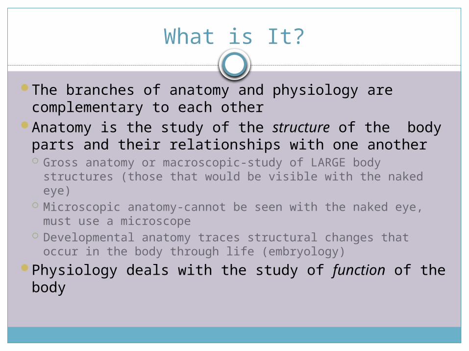
What is It?
The branches of anatomy and physiology are complementary to each other
Anatomy is the study of the structure of the body parts and their relationships with one another Gross anatomy or macroscopic-study of LARGE body
structures (those that would be visible with the naked eye) Microscopic anatomy-cannot be seen with the naked eye,
must use a microscope Developmental anatomy traces structural changes that
occur in the body through life (embryology)Physiology deals with the study of function of the
body
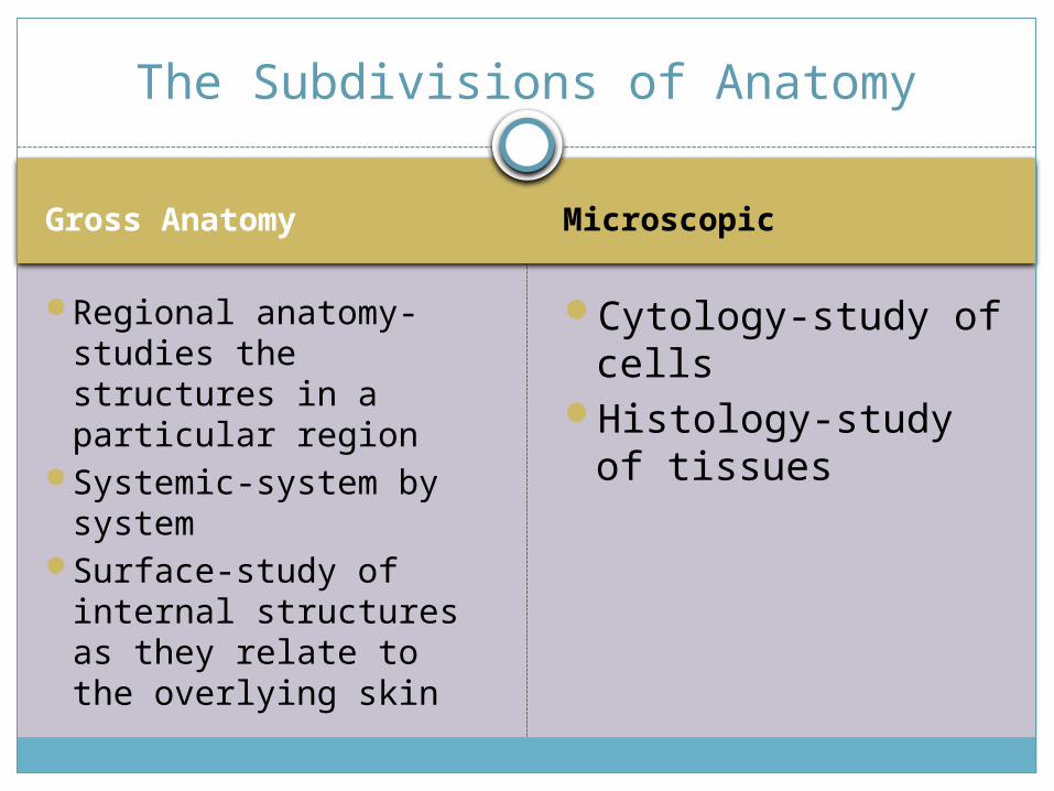
Gross Anatomy Microscopic
Regional anatomy-studies the structures in a particular region
Systemic-system by system
Surface-study of internal structures as they relate to the overlying skin
Cytology-study of cells
Histology-study of tissues
The Subdivisions of Anatomy

Levels of Organization
The human body has many levels of organization
The most basic is the chemical level (chapter 2)-at this level atoms combine to form molecules which will combine to make organelles
Organelles make cells (chapter 3)Cells make tissues (chapter 4)Tissues make organs which in turn make
organ systems (chapters 5-27)
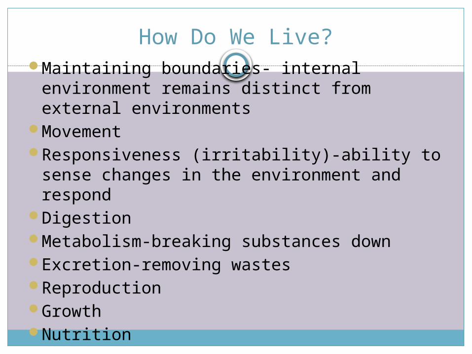
How Do We Live?Maintaining boundaries- internal
environment remains distinct from external environments
MovementResponsiveness (irritability)-ability to sense
changes in the environment and respondDigestionMetabolism-breaking substances downExcretion-removing wastesReproductionGrowthNutrition

Pause-Activity 1
Break into 9 groupsEach group will have a different topic of how we
live and will create a posterYou will only get ONE piece of construction paperYou must include the following:
What is your topic What does it mean to you Examples Pictures Which body systems help Anything relevant

HomeostasisThe ability to maintain a relatively stable
internal environment in an ever-changing outside world
The internal environment of the body is in a dynamic state of equilibrium
Chemical, thermal, and neural factors interact to maintain homeostasis
There are two types of feedback mechanisms: positive and negative
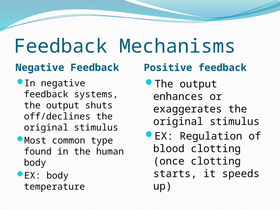
Feedback MechanismsNegative Feedback Positive feedbackIn negative feedback
systems, the output shuts off/declines the original stimulus
Most common type found in the human body
EX: body temperature
The output enhances or exaggerates the original stimulus
EX: Regulation of blood clotting (once clotting starts, it speeds up)

Homeostatic Control MechanismsThere are three essential components of
control mechanisms: Control Center, Receptor and Effector
Control Center: analyzes the input, determines the appropriate response and activities the effector
Receptor: senses changes in the environment and responds by sending information to the control center
Effector: makes the changes needed to maintain homeostasis

Pathways for HomeostasisAfferent: the nerve structures through which
an impulse, especially a sensory impression, is conducted to the cerebral cortex.
Efferent: the nerve structures through which an impulse passes between groups of nerve cells or between the central nervous system and an organ or muscle

The Language of Anatomy
These MUST be memorized!!
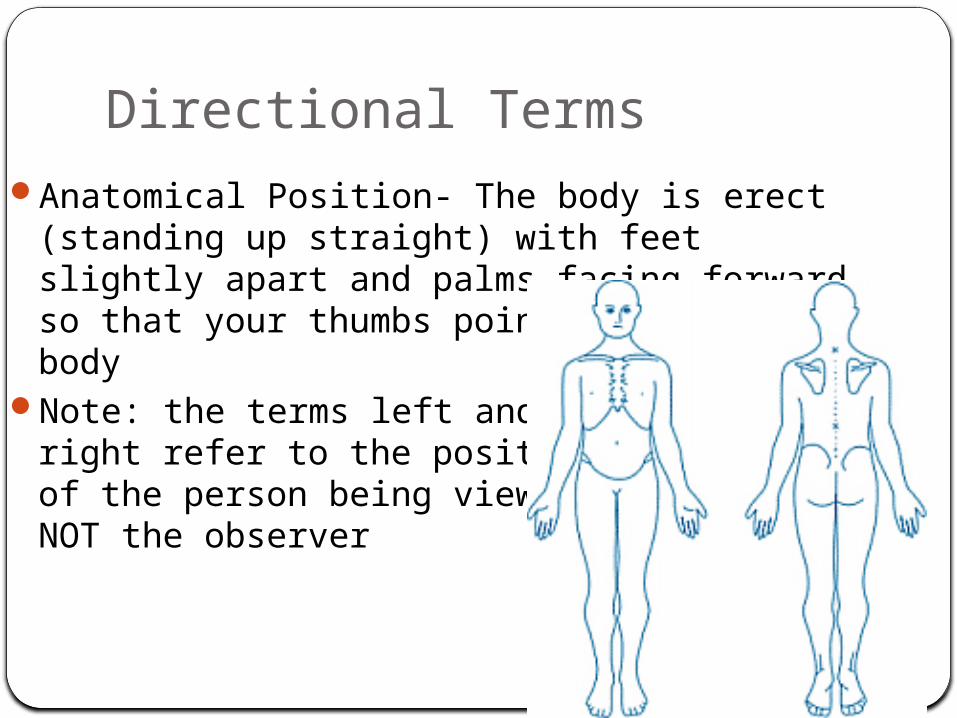
Directional Terms Anatomical Position- The body is erect
(standing up straight) with feet slightly apart and palms facing forward so that your thumbs point away from the body
Note: the terms left and right refer to the positionof the person being viewedNOT the observer

UP and DOWNSuperior (Cranial)
Inferior (Caudal)
Toward the head or upper body (above)
Away from the head or lower body (below)

FRONT and BACK
Anterior (ventral)
Posterior (dorsal)
Toward or at the front of the body
Toward or at the back of the body

In Between
Medial
Lateral
Toward or at the midline of the body (inner side of)
Away from the midline of the body (outer side of)Intermediate• Between a more
medial and more lateral structure

Extremities-UP and Down
Proximal
Distal
Closer to the origin of the body part or point of attachment of a limb to the trunk
Farther from the origin of a body part of the point of attachment of a limb to the body trunk

IN and OUTSuperficial (external)
Deep (internal)
Toward or at the body surface
Away from the body surface

Activity 2-in pairs
• Create one set of labels for the anatomical directions we’ve just learned (EXCEPT superficial and deep)
• Have one person in the group lay in the CORRECT anatomical position and have the other partner label the directions
• When finished, switch and let the other person try
Activity 3
• Create a foldable using a different color for each pair; put the pairs across from one another (for example: hot would be on one side, cold on the other-both would be written in the same color)
• Fold paper into hot dog shape, and make six tabs (five cuts) on each side
• The top two tabs, cut completely off your paper this will be the title of the foldable: directional terms
More on Activity 3
• For the rest of the tabs label as follows: Superior, Inferior, Anterior, Posterior, Medial, Lateral, Proximal, Distal, Superficial, Deep
• Inside the tab you need to define what each is AND provide an example; use BASIC anatomy parts in which you know such as arm, shoulder, chest, leg, foot, etc.

Axial Appendicular
Makes up the main axis of our body
Includes head, neck and truck
Consists of appendages (limbs)
These are attached to the axis
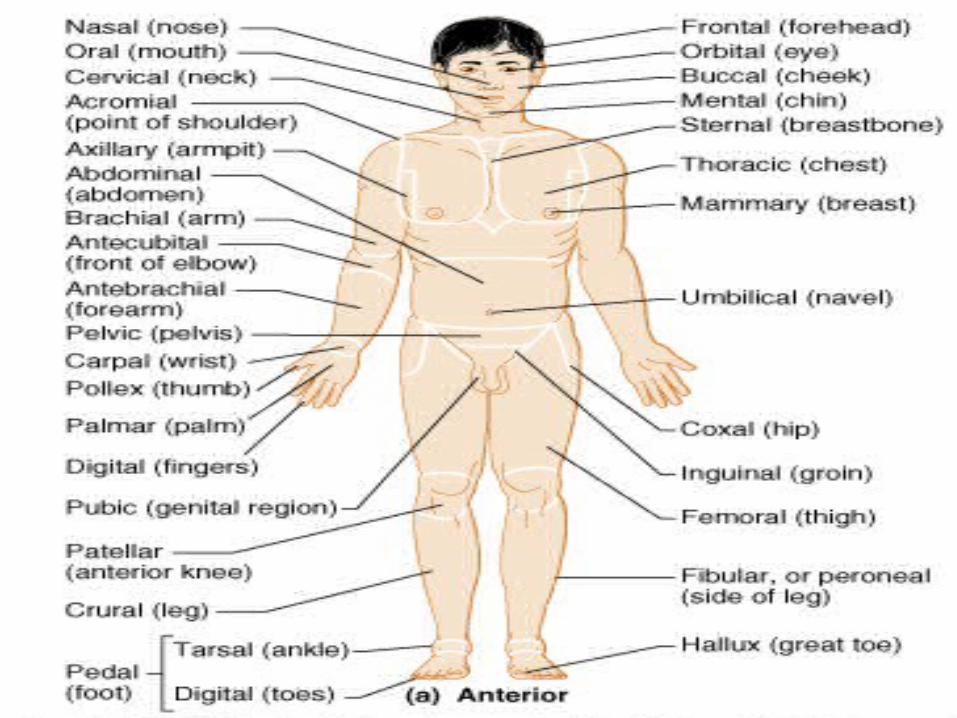
Regional Terms

IN YOUR TEXTBOOK, PG. 14 FIGURE 1.7 LISTS ALL
THE REGIONAL TERMS USED TO DESIGNATE
SPECIFIC BODY AREAS-YOU MUST KNOW THESE
AS WELL!!!
More Regional Terms

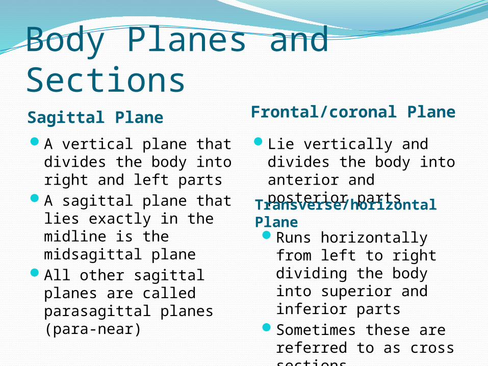
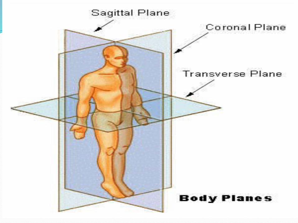
Body Planes and SectionsSagittal Plane Frontal/coronal Plane
A vertical plane that divides the body into right and left parts
A sagittal plane that lies exactly in the midline is the midsagittal plane
All other sagittal planes are called parasagittal planes (para-near)
Lie vertically and divides the body into anterior and posterior parts
Transverse/horizontal Plane Runs horizontally from
left to right dividing the body into superior and inferior parts
Sometimes these are referred to as cross sections

Body Cavities
Dorsal Cavity Protects the organs of
the nervous system Has TWO subdivisions:
Cranial (skull) and Vertebral (spinal cord)
Because both of these are essentially part of the same organ, the two cavities are continuous with one another
Ventral Body Cavity More anterior and larger
cavity Houses internal organs
collectively called the viscera
Has TWO major subdivisions: Thoracic (surrounded by ribs) and abdominopelvic (abdomen and pelvic regions)
The two major divisions are separated by the diaphragm

Other Cavities
Oral and digestiveNasalOrbital (eyes)Middle earSynovial (joint)

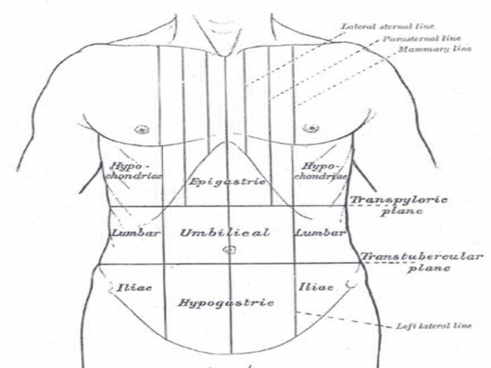
Subdivisions of Abdominopelvic Region
Left/Right hypochondriac region: Part of the liver, gallbladder, & part of diaphragm in right; part of diaphragm and part of stomach in left
Left/Right lumbar region: Ascending colon of large intestine in right; Descending colon of large intestine in left
Left/Right iliac/inguinal region: Cecum of large intestine in right; Part of sigmoid colon of large intestine in left
Epigastric Region: Most of the stomachUmbilical Region: Transverse colon of large
intestine, small intestineHypogastric/pubic Region: Bladder

Activity 4-pairs
Again make labels, this time for the body cavities AND their subdivisions (use chart paper for the abdominal subdivisions)
Repeat the same procedure as we did in activity 1