Embed Size (px)
Citation preview
Proc. Nati. Acad. Sci. USAVol. 89, pp. 9267-9271, October 1992Medical Sciences
AKT2, a putative oncogene encoding a member of a subfamily ofprotein-serine/threonine kinases, is amplified in humanovarian carcinomas
(Src homology 2-like domain/gene amplification/chromosome alterations)
JIN QUAN CHENG, ANDREW K. GODWIN, ALFONSO BELLACOSA, TAKAHIRO TAGUCHI, THOMAS F. FRANKE,THOMAS C. HAMILTON, PHILIP N. TsICHLIS, AND JOSEPH R. TESTA*Department of Medical Oncology, Fox Chase Cancer Center, Philadelphia, PA 19111
Communicated by Irwin A. Rose, July 13, 1992 (received for review May 26, 1992)
ABSTRACT We isolated cDNA clones containing the en-tire coding region of the putative oncogene AKT2. Sequenceanalysis and in vitro translation demonstrated that AKT2encodes a 56-kDa protein with homology to serine/threoninekinases; moreover, this protein contains a Src homology 2-likedomain. AKT2 was shown to be amplified and overexpressed in2 of 8 ovarian carcinoma cell lines and 2 of 15 primary ovariantumors. AKT2 was mapped to chromosome region 19q13.1-q13.2 by fluorescence in situ hybridization. In the two ovariancarcinoma cell lines exhibiting amplification of AKT2, theamplified sequences were localized within homogeneouslystaining regions. We conclude that AKT2 belongs to a distinctsubfamily of protein-serine/threonine kinases containing Srchomology 2-like domains and that alterations of AKT2 maycontribute to the pathogenesis of ovarian carcinomas.
akt, the protooncogene transduced by the acute transformingretrovirus AKT8 (1, 2), encodes a protein-serine/threoninekinase containing a Src homology 2-like (SH2-like) domain(3). Two putative human cellular homologs, AKTI and AK12,were cloned by screening a human genomic DNA library witha v-akt probe under conditions of reduced stringency (4).Recently, we obtained AKTI clones from a normal humanthymus cDNA library by using an AKTI genomic probe.Sequence analysis of portions of AKTI cDNA clones re-vealed that AKTI is the true human homolog of v-akt and isidentical to the recently cloned RAC gene, which has beenshown to encode a kinase related to members of the proteinkinase C (PKC) family and the cyclic adenosine monophos-phate-dependent protein kinase (cAMP-PK) (5). AKTI hasbeen mapped to human chromosome band 14q32 (6), proxi-mal to the immunoglobulin heavy-chain locus, and has beenshown to be amplified in a gastric adenocarcinoma (4).
In this communication, we report the cDNA cloning,sequence analysis,t and chromosomal mapping ofAKT2 anddemonstrate that this putative oncogene encodes a proteinbelonging to a subfamily of serine/threonine kinases con-taining SH2-like domains. Moreover, we show that AKT2 isamplified and overexpressed in some human ovarian carci-noma cell lines and primary tumors, suggesting that it con-tributes to the development of common epithelial tumors ofthe ovary.
MATERIALS AND METHODSOvarian Carcinoma Cell Lines and Primary Tumors. The
ovarian carcinoma cell lines examined in this study have beendescribed (7-9). Primary tumor specimens were obtained
from patients who underwent surgery at Fox Chase CancerCenter.cDNA Cloning and Sequencing. A human cDNA library in
Agtll derived from normal thymus cells (Clontech) wasscreened with a 2.0-kilobase (kb) Sma I-BamHI AKT2 ge-nomic probe (4). The hybridization was carried out underconditions of high stringency; the final wash was at 650C for30 min in 0.1x SSC/0.1% SDS (lx SSC is 0.15 M NaCI/15mM sodium citrate, pH 7.0). Positive clones were plaque-purified, and the EcoRI inserts were subcloned into pBlue-script SK(-) (Stratagene) for further analysis. A 1.8-kb DNAfragment from the largest clone, AHTakt-6, was subjected tonested deletions in both directions, using the exonucleaseIII-mung bean nuclease method (Stratagene). Plasmid cloneswith overlapping deletions were sequenced. The sequencingreactions were performed on alkali-denatured double-stranded DNA by using the dideoxynucleotide chain-termination method (10) and the Sequenase version 2.0system (United States Biochemical). The sequence of the1.8-kb fragment was determined for both strands. Sequenceanalysis was carried out with the University of WisconsinGenetics Computer Group software package (11).
In Vitro Transcription and Translation. The EcoRI insertfrom AHTakt-6 was subcloned into pBluescript SK(-); thelinearized plasmid was used as a template for in vitro tran-scription by T3 RNA polymerase. The capped RNA (2 tkg)was translated in vitro by using rabbit reticulocyte lysate(Stratagene) for 1 hr at 30'C in the presence of 20 ,.Ci (740kBq) of [35S]methionine. The samples were analyzed bySDS/15% PAGE followed by autoradiography for 12 hr atroom temperature.
Southern and Northern Analysis. DNA from normal tis-sues, tumor specimens, and cell lines was isolated by stan-dard techniques (12). Digested DNA, after careful spectro-photometric measurement of DNA concentration, was frac-tionated by 1.0%o agarose gel electrophoresis, and transferredto nylon membranes (GeneScreenPlus; NEN/DuPont) (12).The membranes were hybridized overnight with 32P-labeledprobes and washed at high stringency. Chromosome-mapping-panel blots containing human, hamster, and hu-man-hamster somatic cell hybrid DNAs (Bios, New Haven,CT) were hybridized according to the manufacturer's instruc-tions.
Total cellular RNA was obtained by a one-step guanidin-ium isothiocyanate/phenol/chloroform extraction procedure(13). For Northern analysis, 15 ug of total RNA per lane was
Abbreviations: SH2-like, Src homology 2-like; PKC, protein kinaseC; cAMP-PK, cyclic adenosine monophosphate-dependent proteinkinase; FISH, fluorescence in situ hybridization.*To whom reprint requests should be addressed.tThe sequence reported in this paper has been deposited in theGenBank data base (accession no. M95936).
9267
The publication costs of this article were defrayed in part by page chargepayment. This article must therefore be hereby marked "advertisement"in accordance with 18 U.S.C. §1734 solely to indicate this fact.
9268 Medical Sciences: Cheng et al.
electrophoresed in a 1% agarose/2.2 M formaldehyde gel,blotted onto Magna NT membrane filters (Micron Separa-tions, Westborough, MA), and hybridized overnight with32P-labeled probes.Autoradiograms exposed within the linear range ofthe film
were quantified by scanning densitometry (UltraScan XLlaser densitometer; Pharmacia LKB) to assess gene amplifi-cation and levels of RNA expression.
Fluorescence in Situ Hybridization (FISH). Metaphasespreads were prepared from normal lymphocyte cultures andfrom ovarian carcinoma cell lines OVCAR-3 (passage 51) andOVCAR-8 (passage 8), following cell cycle synchronizationwith 5-bromodeoxyuridine (14). FISH was carried out basi-cally according to Fan et al. (14), with a nonisotopicallylabeled 2.0-kb genomic AK72 fragment as probe. The probewas prepared by nick-translation (BRL) using biotin-11-dUTP (Enzo Diagnostics, New York). Hybridization wasdetected with fluorescein isothiocyanate-conjugated avidin.Chromosomes were counterstained with propidium iodideand 4',6-diamidino-2-phenylindole dihydrochloride and ob-served under fluorescence microscopy. Metaphase spreadswere photographed on Kodak Ektachrome 400 film.
RESULTSCloning and Sequence Analysis of AKT2 cDNA. Three
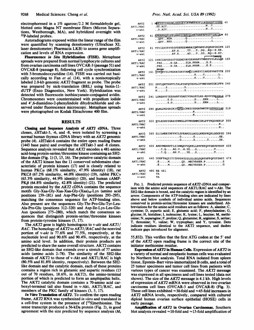
clones, AHTakt-3, -6, and -8, were isolated by screening anormal human thymus cDNA library with an AKT2 genomicprobe (4). AHTakt-6 contains the entire open reading frame(1443 base pairs) and overlaps the AHTakt-3 and -8 clones.Sequence analysis revealed that AKT2 encodes a 481-aminoacid-long protein-serine/threonine kinase containing an SH2-like domain (Fig. 1) (3, 15, 16). The putative catalytic domainof the AKT2 kinase has the 11 conserved subdomains char-acteristic of protein kinases (17) and is closely related tohuman PKCa (68.1% similarity, 47.9%o identity) (18), ratPKC8 (67.2% similarity, 44.01% identity) (19), rabbit PKCy(65.3% similarity, 44.9o identity) (20), and human cAMP-PK,8 (64.8% similarity, 42.8% identity) (21). The predictedprotein encoded by the AKT2 cDNA contains the sequencemotifs Gly-Xaa-Gly-Xaa-Xaa-Gly-(Xaa)16-Lys (amino acidpositions 159-181) and Asp-Phe-Gly (positions 293-295),matching the consensus sequence for ATP-binding sites.Also present are the sequences Gly-Thr-Pro-Glu-Tyr-Leu-Ala-Pro-Glu (positions 312-320) and Asp-Ile-Lys-Leu-Glu-Asn (positions 275-280), which match the consensus se-quences that distinguish protein-serine/threonine kinasesfrom protein-tyrosine kinases (5, 17).The AKT2 gene is highly homologous to v-akt and AKTJ/
RAC. The homology ofAKT2 toAKTJ/RAC and the nonviralportion of v-akt is 77.6% and 77.5%, respectively, at thenucleotide level and 90.6% and 90.4%, respectively, at theamino acid level. In addition, their protein products arepredicted to share the same overall structure. AKT2 containsan SH2-like domain that extends over a stretch of 77 aminoacids (amino acids 2-78). The similarity of the SH2-likedomain of AKT2 to those of v-Akt and AKT1/RAC is high(80.5% and 81.8% identity, respectively). Between the SH2-like domain and the catalytic domain, each of these proteinscontains a region rich in glutamic and aspartic residues (13out of 70 residues, 18.6%, in AKT2), the amino-terminalportion of which is predicted to form an amphipathic helix.The AKT2 catalytic domain contains a 70-amino acid car-boxyl-terminal tail also found in v-Akt, AKT1/RAC, andmembers of the PKC family of kinases (3, 5, 17).In Vitro Translation ofAK12. To confirm the open reading
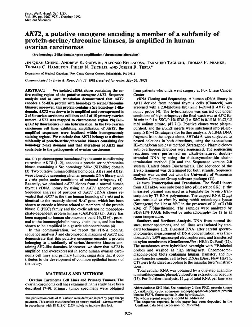
frame, AKT2 RNA was synthesized in vitro and translated ina cell-free system in the presence of [35S]methionine. Thesense transcript produced a 56-kDa protein (Fig. 2), in goodagreement with the size predicted by sequence analysis (Mr
AKT2AKT 1/RAC
v-akt
AKT2AKT1/RAC
v-akt
AKT2AKT1/RAC
v-akt
AKT2AKT 1/RAC
v-akt
AKT2AKT1 /RAC
v-akt
AKT2AKT 1/RAC
v-akt
AKT2AKT 1 /RAC
v-akt
AKT2AKT 1/RAC
v-akt
1 NEVSVIjKEGWLHKRGEYIKTWRPRYFLLKSDGSFIGYKE.SD.AIV.....N.T...D.AIV.....N.T.
40
41 RPEAPDQTLPPLNNFSVAECQLMKTERPRPNTFVIRC W 80..QDV ..REA. Q. I...QDV ..RES. Q.I.
81 TTVIERTFHVDSPDEREEWMRAIQMVANSLKQRAPGEDPM 120. ET.E. ....TT .T. .DG. .KQ--E.EE.. ET.E.. AT.. T. .DG. .RQ--E.ET.
121 DYKCGSPSDSSTTEEMEVAVSKARAKVTMNDFDYLKLLDK 160.FRS... N.GA... SLA.PKHR .... E.E..FRS... N.GA... SLA.PKHR .... E.E.doman
161 ZTF:KVILVREKATGRYYAMKILRKEVIIAKDEVAHTVTE 200.K . V. L..
. K... V.. L..
201 SRVLQNTRHPFLTALKYAFQTHDRLCFVMEYANGGELFFH 240N. S. S.N. S. S.
241 LSRERVFTEERARFYGAEIVSALEYLHS-RDVVYR&.ILL 279. S.D..D.N .. EKN. L.... S.D..D.N .. . EKN. L...
280 NLMLDKDGHIKITQFGLCKEGISDGATMKTFCGTPEYLAP 319......................K.......................K.
AKT2 320 EVLEDNDYGRAVDWWGLGVVMYEMMCGRLPFYNQDHERLF 359AKT1/RAC K..
v-akt ...K..
AKT2 360 ELILMEEIRFPRTLSPEAKSLLAGLLKKDPKQRLGGGPSD 399AKT1/RAC .............. . .............. SE.
v-akt .............. S..............T SE.
AKT2 400 AKEVMEHRFFLSINWQDVVQKKLLPPFKPQVTSEVDTRYF 439AKT1/RAC .. .I.Q ... AG.V. ....... HS.TE...ST
v-akt .. I.Q.... AN.V ... YE...S. T.
AKT2 440 DDEFTAQSITITPPDRYDSLGLLELDQRTHFPQFSYSASI 479AKT1/RAC .E. M. QD. .MECVDSER.P .......... S
v-akt .E. M. QD. .MECVDSER.P .......... G
AKT2 480 RE 481AKT1/RAC TA
v-akt TA
FIG. 1. Predicted protein sequence ofAKT2 cDNA and compar-ison with the amino acid sequences of AKT1/RAC and v-Akt. TheSH2-like domain is boxed, and the catalytic region is identified by anarrow. Components of the ATP-binding sites are indicated by linesabove and below symbols of individual amino acids. Sequencesconserved in protein-serine/threonine kinases are underlined. Ab-breviations for the amino acid residues are as follows: A, alanine; C,cysteine; D, aspartic acid; E, glutamic acid; F, phenylalanine; G,glycine; H, histidine; I, isoleucine; K, lysine; L, leucine; M, methi-onine; N, asparagine; P, proline; Q, glutamine; R, arginine; S, serine;T, threonine; V, valine; W, tryptophan; and Y, tyrosine. Dotsindicate residues identical to the AKT2 sequence, and dashesindicate gaps introduced for alignment.
55,832). This verifies that the first ATG codon at the 5' endof the AKT2 open reading frame is the correct site of theinitiator methionine residue.
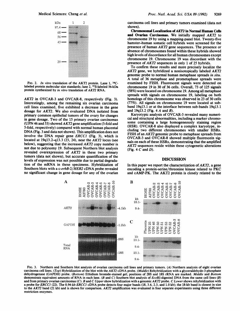
Expression ofAKI2 in Human Cells. Expression ofAK12 ina variety of normal and neoplastic human cells was evaluatedby Northern blot analysis. Total RNA isolated from spleentissue, Epstein-Barr virus-immortalized B cells, and a total of25 tumor specimens and tumor cell lines from patients withvarious types of cancer was examined. The AKT2 messagewas expressed in all specimens and cell lines tested (data notshown). The size of the AKT2 message is 4.1 kb. High levelsof expression ofAKT2 mRNA were observed in two ovariancarcinoma cell lines (OVCAR-3 and OVCAR-8) (Fig. 3).These cell lines exhibited -30-fold and -45-fold increases inAKT2 mRNA levels, respectively, compared with cultureddiploid human ovarian surface epithelial (HOSE) cells inearly passage.
Amplification of AKT2 in Ovarian Carcinomas. Southernblot analysis revealed '-10-fold and -15-fold amplification of
Proc. Natl. Acad. Sci. USA 89 (1992)
Proc. Natl. Acad. Sci. USA 89 (1992) 9269
kDa 1 297.4--
68--56-
43-
29--
FIG. 2. In vitro translation of the AKT2 protein. Lane 1, 14C-labeled protein molecular size standards; lane 2, 35S-labeled 56-kDaprotein synthesized by in vitro translation of AKT2 RNA.
AK72 in OVCAR-3 and OVCAR-8, respectively (Fig. 3).Interestingly, among the remaining six ovarian carcinomacell lines examined, five exhibited a decrease in the genedosage for AKT2. We also evaluated DNA isolated fromprimary common epithelial tumors of the ovary for changesin gene dosage. Two of the 15 primary ovarian carcinomas(UPN 46 and 53) showed AK72 gene amplification (3-fold and5-fold, respectively) compared with normal human placentalDNA (Fig. 3 and data not shown). This amplification does notinvolve the DNA repair gene ERCCI (Fig. 3), which islocated at 19q13.2-q13.3 (23, 24), near the AKT2 locus (seebelow), suggesting that the increased AKT2 copy number isnot due to polysomy 19. Subsequent Northern blot analysisrevealed overexpression of AKT2 in these two primarytumors (data not shown), but accurate quantification of thelevels of expression was not possible due to partial degrada-tion of the mRNA in these specimens. Hybridization ofSouthern blots with a c-erbB-2/HER2 cDNA probe revealedno significant change in gene dosage for any of the ovarian
A Nsc mPI:Loc- oo °-
co uuuQu u
=¢0000000
carcinoma cell lines and primary tumors examined (data notshown).Chromosomal Localization ofAKT2 in Normal Human Cells
and Ovarian Carcinomas. We initially mapped AKT2 tochromosome 19 by using a mapping-panel blot. Twenty-fivehamster-human somatic cell hybrids were screened for thepresence of human AKT2 gene sequences. The presence orabsence ofchromosomes found within these hybrids showedhigh levels ofdiscordance for all human chromosomes exceptchromosome 19. Chromosome 19 was discordant with thepresence of AKT2 sequences in only 1 of 25 hybrids.To confirm these results and more precisely localize the
AK72 gene, we hybridized a nonisotopically labeled AKT2genomic probe to normal human metaphase spreads in situ.A total of 36 metaphase and prometaphase spreads wereexamined by FISH. Fluorescent signals were detected onchromosome 19 in 30 of 36 cells. Overall, 73 of 125 signals(58%) were located on chromosome 19. Among all metaphasespreads with signals on chromosome 19, labeling on bothhomologs of this chromosome was observed in 23 of 30 cells(77%). All signals on chromosome 19 were located at sub-band 19q13.1 or at the interface between sub-bands 19q13.1and 19q13.2 (Fig. 4 A and B).
Karyotypic analysis of OVCAR-3 revealed many numeri-cal and structural abnormalities, including a marker chromo-some containing a large homogeneously staining region(HSR). OVCAR-8 also displayed a complex karyotype, in-cluding two different chromosomes with smaller HSRs.FISH of an AK72 genomic probe to metaphase spreads fromOVCAR-3 and OVCAR-8 showed multiple fluorescein sig-nals on each of these HSRs, demonstrating that the amplifiedAKT2 sequences reside within these cytogenetic alterations(Fig. 4 C and D).
DISCUSSIONIn this paper we report the characterization ofAK72, a geneencoding a protein-serine/threonine kinase related to PKCand cAMP-PK. The AKT2 protein is closely related to the
B
kb23.1-
Ct2 <DQ 00UO0>>>0= -40 00
to r- co -
00 0 0
AKT2 "..u -4.1kb
-1.2kb
9.4-
C 4j U': Co _r-- N3"= N '4 to t Lo-oC ZZ Zz ttzuZCO a4 X,0. 4 oa > >kb D nD DD O O
Ikb-28S 23.1-
9.4-
-18S 23.1-
9.4-
FIG. 3. Northern and Southern blot analysis of ovarian carcinoma cell lines and primary tumors. (A) Northern analysis of eight ovariancarcinoma cell lines. (Top) Hybridization of the blot with the AKT2 cDNA probe. (Middle) Rehybridization with a glyceraldehyde-3-phosphatedehydrogenase (GAPDH) probe. (Bottom) Ethidium bromide-stained gel; positions of 28S and 18S rRNA are marked. Middle and Bottomdemonstrate equivalent amounts of RNA in each lane. (B and C) Southern blot analysis of EcoRI-digested DNA from the same cell lines (B)and from primary ovarian carcinomas (C). B and C Upper show hybridization with a genomic AKT2 probe. C Lower shows rehybridization witha probe for ERCCI (22). The 0.94-kb ERCCI cDNA probe detects four major bands (18, 3.6, 2.3, and 1.0 kb); the 18-kb band is closest in sizeto the AKT2 band (21 kb) and is shown for comparison. AKT2 amplification was evaluated in four separate experiments using three differentrestriction enzymes.
GAPDH
TotalRNA
.., --m- ON 1'- -T 7-1-
Medical Sciences: Cheng et al.

9270 Medical Sciences: Cheng et al.
FIG. 4. FISH ofAK72 to human metaphase chromosomes. (A) Localization offluorescein-labeled probe on propidium iodide-stained normallymphocyte metaphase spread. Fluorescent hybridization signals are indicated by arrows at 19q13.1-q13.2. (B) 4',6-Diamidino-2-phenylindolefluorescence of the same spread, showing a Giemsa-like banding pattern useful to confirm the identity of individual chromosomes (14). (C andD) Localization of AKT2 on propidium iodide-stained chromosomes from ovarian carcinoma cell lines OVCAR-3 (C) and OVCAR-8 (D).Fluorescent hybridization signals are indicated at 19q13.1-q13.2 (shorter arrows) and within homogeneously staining regions (longer arrows).
v-Akt and AKT1/RAC kinases, which also contain an SH2-like region amino-terminal to the kinase domain (3). TheSH2-like regions of AKT2, AKT1/RAC, and v-Akt/c-Aktdisplay similarities to the SH2 domains characteristic ofcytoplasmic tyrosine kinases and other signaling proteins (3,15). This suggests that the SH2-like domains may representregions of protein-protein interactions that take placethrough the recognition of phospho amino acid residues.However, the similarity between SH2 and SH2-like domainsis very distant, and the function of the SH2-like region mayhave diverged from that of SH2. The detection of this domainin both AKT1 and AKT2 places them into a distinct subfamilyofprotein-serine/threonine kinases. This subfamily may con-tain multiple genes, as suggested by Southern blot analysis ofhuman genomic DNA hybridized to a c-akt SH2-like probe atlow stringency (data not shown).The chromosomal location ofAK72 at 19q13.1-q13.2 is in
close proximity to the map location of genes encodingtransforming growth factor P1, carcinoembryonic antigen,the zinc finger protein ZFP36, and several proteins involvedin DNA repair (22, 23). BCL3, a putative oncogene that isrearranged in some B-cell chronic lymphocytic leukemiasdisplaying a 14;19 translocation (24), is also located at19q13.1-q13.2 (23). In addition to the t(14;19) disruptingBLC3, the same 19q13 chromosomal region is the target ofother cytogenetic abnormalities found in ovarian carcinoma,glioma, and non-small-cell lung carcinoma (25-27). It will beimportant to determine whether the t(14;19) observed inB-cell chronic lymphocytic leukemia or the other aberrationsof the 19q13 region seen in other neoplasms affect theexpression of AKT2.Gene amplification is an important mechanism for in-
creased expression of genes involved in tumorigenesis. Am-plification of various oncogenes has been observed in manyprimary tumors and tumor cell lines (28). In some types ofcancer, amplification of specific oncogenes has been corre-lated with advanced disease (29-31). The amplification andoverexpression ofAKT2 in some ovarian carcinoma cell linesand primary tumors suggest that AKT2, in cooperation withother protooncogenes and tumor-suppressor genes (32-36),contributes to the pathogenesis of ovarian cancer. Furtherstudies are needed to determine whether amplification of
AKT2 occurs in a significant proportion of other humanneoplasms and whether such amplification represents a use-ful prognostic marker.
Note Added in Proof. Jones et al. (37) recently cloned a gene, racprotein kinase ,, which encodes a protein highly related to PKC andcAMP-PK. A portion of the rac protein kinase (3 DNA sequence isidentical toAK72, but the regulatory region and the 3' end ofthe openreading frame show considerable differences from those of AKT2.
We thank Drs. Alfred G. Knudson, Robert P. Perry, and Anna M.Skalka for valuable comments; Dr. Stephen P. Staal for AKTI andAKT2 genomic probes; Dr. Marcel van Duin for an ERCCI cDNAprobe; and Dr. Ze Min Liu for preparing karyotypes. This researchwas supported in part by Public Health Service Grants RR05895,CA06927, CA38047, and CA52181 and by an appropriation from theCommonwealth ofPennsylvania. A.B. was a Fellow ofthe LawrenceGreenwald Foundation for Leukemia and Lymphoma Research andis currently a Fellow of the Italian Association for Cancer Research.
1. Staal, S. P., Hartley, J. W. & Rowe, W. P. (1977) Proc. Natl.Acad. Sci. USA 74, 3065-3067.
2. Staal, S. P. & Hartley, J. W. (1988) J. Exp. Med. 167, 1259-1264.
3. Bellacosa, A., Testa, J. R., Staal, S. P. & Tsichlis, P. N. (1991)Science 254, 274-277.
4. Staal, S. P. (1987) Proc. Natl. Acad. Sci. USA 84, 5034-5037.5. Jones, P. F., Jakubowicz, T., Pitossi, F. J., Maurer, F. &
Hemmings, B. A. (1991) Proc. Natl. Acad. Sci. USA 88,4171-4175.
6. Staal, S. P., Huebner, K., Croce, C. M., Parsa, N. Z. & Testa,J. R. (1988) Genomics 2, %-98.
7. Hamilton, T. C., Lai, G.-M., Rothenberg, M. L., Fojo, A. T.,Young, R. C. & Ozols, R. F. (1989) in Drug Resistance inCancer Therapy, ed. Ozols, R. F. (Kluwer, Boston), pp. 151-169.
8. Hamilton, T. C., Young, R. C., McKoy, W. M., Grotzinger,K. R., Green, J. A., Chu, E. W., Whang-Peng, J., Rogan,A. M., Green, W. R. & Ozols, R. F. (1983) Cancer Res. 43,5379-5389.
9. Eva, A., Robbins, K. C., Anderson, P. R., Srinivasan, A.,Tronick, S. R., Reddy, E. P., Ellmore, N. W., Galen, A. T.,Lautenberger, J. A., Papas, T. S., Westin, E. S., Wong-Staal,F., Gallo, R. C. & Aaronson, S. A. (1982) Nature (London)295, 116-119.
Proc. Nad. Acad Sci. USA 89 (1992)

Proc. Natl. Acad. Sci. USA 89 (1992) 9271
10. Sanger, F., Nicklen, S. & Coulson, A. R. (1977) Proc. Natl.Acad. Sci. USA 74, 5463-5467.
11. Devereux, J., Haeberli, P. & Smithies, 0. (1984) Nucleic AcidsRes. 12, 387-395.
12. Sambrook, J., Fritsch, E. F. & Maniatis, T. (1989) MolecularCloning:A Laboratory Manual (Cold Spring Harbor Lab., ColdSpring Harbor, NY), 2nd Ed.
13. Chomczynski, P. & Sacchi, N. (1987) Anal. Biochem. 162,156-159.
14. Fan, Y., Davis, L. M. & Shows, T. B. (1990) Proc. Natl. Acad.Sci. USA 87, 6223-6227.
15. Koch, C. A., Anderson, D., Moran, M. F., Ellis, C. & Pawson,T. (1991) Science 252, 668-674.
16. Pawson, T. (1988) Oncogene 3, 491-495.17. Hanks, S. K., Quinn, A. M. & Hunter, T. (1988) Science 241,
42-52.18. Finkenzeller, G., Marme, D. & Hug, H. (1990) Nucleic Acids
Res. 18, 2183.19. Ono, Y., Fujii, T., Ogita, K., Kikkawa, U., Igarashi, K. &
Nishizuka, Y. (1988) J. Biol. Chem. 263, 6927-6932.20. Ohno, S., Kawasaki, H., Konno, Y., Inagaki, M., Hidaka, H.
& Suzuki, K. (1988) Biochemistry 27, 2083-2087.21. Beebe, S. J., Oyen, O., Sandberg, M., Froysa, A., Hansson, V.
& Jahnsen, T. (1990) Mol. Endocrinol. 4, 465-475.22. Mohrenweiser, H. W., Carrano, A. V., Fertitta, A., Perry, B.,
Thompson, L. H., Tucker, J. D. & Weber, C. A. (1989) Cyto-genet. Cell Genet. 52, 11-14.
23. Ropers, H. H. & Pericak-Vance, M. A. (1991) Cytogenet. CellGenet. 58, 751-784.
24. Ohno, H., Takimoto, G. & McKeithan, T. W. (1990) Cell 60,991-997.
25. Pejovic, T., Heim, S., Mandahl, N., Baldetorp, B., Elmfors,B., Floderus, U.-M., Furgyik, S., Helm, G., Himmelmann, A.,Willen, H. & Mitelman, F. (1992) Genes Chromosomes Cancer4, 58-68.
26. Mitelman, F., Kaneko, Y. & Trent, J. M. (1990) Cytogenet.Cell Genet. 55, 358-386.
27. Miura, I., Siegfried, J. M., Resau, J., Keller, S. M., Zhou, J. &Testa, J. R. (1990) Genes Chromosomes Cancer 2, 328-338.
28. Bishop, J. M. (1987) Science 235, 305-311.29. Brodeur, G. M., Seeger, R. C., Schwab, M., Varmus, H. E. &
Bishop, J. M. (1984) Science 224, 1121-1124.30. Yokota, J., Tsunetsugu-Yokota, Y., Battifora, H., Le Fevre, C.
& Cline, M. J. (1986) Science 231, 261-265.31. Slamon, D. J., Clark, G. M., Wong, S. G., Levin, W. J.,
Ullrich, A. & McGuire, W. L. (1987) Science 235, 177-182.32. Fukumoto, M., Estensen, R. D., Sha, L., Oakley, G. J.,
Twiggs, L. B., Adcock, L. L., Carson, L. F. & Roninson, I. B.(1989) Cancer Res. 49, 1693-1697.
33. Filmus, J. E. & Buick, R. N. (1985) Cancer Res. 45, 4468-4472.
34. Van't Veer, L. J., Hermens, R., van den Berg-Bakker,L. A. M., Cheng, N. C., Fleuren, G.-J., Bos, J. L., Cleton,F. J. & Schrier, P. I. (1988) Oncogene 2, 157-165.
35. Masuda, H., Battifora, H., Yokota, J., Meltzer, S. & Cline,M. J. (1987) Mol. Biol. Med. 4, 213-227.
36. Godwin, A. K., Hamilton, T. C. & Knudson, A. G. (1992) inGynecologic Oncology: Principles and Practice, eds. Hoskins,W. J., Perez, C. A. & Young, R. C. (Lippincott, Philadelphia),pp. 87-116.
37. Jones, P. F., Jakubowicz, T. & Hemmings, B. A. (1991) CellRegul. 2, 1001-1009.
Medical Sciences: Cheng et al.


![Diet, nutrition, physical activity and ovarian cancer · genes, produces high-grade carcinomas, with a poorer prognosis [7]. 8 OVARIAN CANCER REPORT 214 4. Other established causes](https://img.dokumen.tips/doc/110x75/5d20cbd988c993a5378d8e60/diet-nutrition-physical-activity-and-ovarian-cancer-genes-produces-high-grade.jpg)