Embed Size (px)
Citation preview

2016 www.kce.fgov.be
KCE REPORT 268S
OVARIAN CANCER: DIAGNOSIS, TREATMENT AND FOLLOW-UP APPENDIX

2016 www.kce.fgov.be
KCE REPORT 268S GOOD CLINICAL PRACTICE
OVARIAN CANCER: DIAGNOSIS, TREATMENT AND FOLLOW-UP APPENDIX IGNACE VERGOTE, JOAN VLAYEN, PAULINE HEUS, JACOB P. HOOGENDAM, JOHANNA A.A.G DAMEN, FLEUR T. VAN DE WETERING, FREDERIEKE H. VAN DER BAAN, CLAIRE BOURGAIN, JACQUES DE GRÈVE, DAVID DEBRUYNE, MAXIME FASTREZ, FRÉDÉRIC GOFFIN, MANON HUIZING, JOSEPH KERGER, FRÉDÉRIC KRIDELKA, SIGRID STROOBANTS, WIEBREN TJALMA, PETER VAN DAM, VINCENT VAN DE CAVEYE, GEERT VILLEIRS, PETER VUYLSTEKE, NICOLAS FAIRON, RONALD P. ZWEEMER, LOTTY HOOFT, ROB J.P.M. SCHOLTEN, LEEN VERLEYE

COLOPHON Title : Ovarian cancer: diagnosis, treatment and follow-up – Appendix
Authors : Ignace Vergote (UZ Leuven), Joan Vlayen (KCE), Pauline Heus (Dutch Cochrane Centre), Jacob P. Hoogendam (Dutch Cochrane Centre), Johanna A.A.G. Damen (Dutch Cochrane Centre), Fleur T. van de Wetering (Dutch Cochrane Centre), Frederieke H. van der Baan (Dutch Cochrane Centre), Claire Bourgain (Imelda ziekenhuis, Bonheiden), Jacques De Grève (UZ Brussel), David Debruyne (AZ Groeninge, Kortrijk), Maxime Fastrez (CHU Saint-Pierre, Bruxelles), Frédéric Goffin (Centre Hospitalier Régional de la Citadelle, Liège), Manon Huizing (UZ Antwerpen), Joseph Kerger (Institut Jules Bordet, Bruxelles), Frédéric Kridelka (Centre Hospitalier Universitaire de Liège), Sigrid Stroobants (UZ Antwerpen), Wiebren Tjalma (UZ Antwerpen), Peter Van Dam (UZ Antwerpen), Vincent Van de Caveye (UZ Leuven), Geert Villeirs (UZ Gent), Peter Vuylsteke (Clinique et Maternité Sainte Elisabeth, Namur), Nicolas Fairon (KCE), Ronald P. Zweemer (Dutch Cochrane Centre), Lotty Hooft (Dutch Cochrane Centre), Rob J.P.M. Scholten (Dutch Cochrane Centre), Leen Verleye (KCE)
Project coordinator and senior supervisor:
Sabine Stordeur (KCE)
Reviewers : Anja Desomer (KCE), Raf Mertens (KCE), Hans Van Brabandt (KCE)
Stakeholders: Jean-François Baurain (Cliniques universitaires Saint-Luc), Wim Ceelen (Royal Belgian Society of Surgery), An Claes (Kom op Tegen Kanker), Donald Claeys (Koninklijk Belgisch Genootschap Heelkunde), Cecile Colpaert (Belgian Society of Pathology), Lionel Dhondt (Belgian Society of Medical Oncology), Frederic Forget (Belgian Society of Medical Oncology), Brigitte Honhon (Belgian Society of Medical Oncology), Gerd Jacomen (Belgian Society of Pathology), Etienne Marbaix (Belgian Society of Pathology), Guy Orye (Vlaamse Vereniging voor Obstetrie en Gynaecologie), Elizabeth Van Eycken (Stichting Kankerregister), Maria Carina (Lotgenotengroep Esperanza), Erik Van Limbergen (Radiotherapie UZ Leuven)
External Validators : Isabelle Ray-Coquard (CLCC Léon Bérard, Lyon), Nicholas Reed (Beatson Oncology Centre, UK), Per Olav Vandvik (Department of Health Management and Health Economics, Norway)
Acknowledgements:
Other reported interests: Membership of a stakeholder group on which the results of this report could have an impact: Etienne Marbaix (Cliniques universitaires Saint-Luc; Union Professionnelle des médecins spécialistes en anatomie pathologique), Elisabeth Van Eycken (BVRO; VBS Radiotherapie), Philippe Tummers (VWOG; BGOG), Manon Huizing (BGOG), Wiebren Tjalma (VWOG; VVOG; BGOG)

Participation in scientific or experimental research as an initiator, principal investigator or researcher: Brigitte Honhon (Principal Investigator clinical study BGOG), Frederic Forget (Trinova II AGO-OVAR II, Trinova III), Manon Huizing (Fase II en III studie plaatselijke of nationale coordinatie op gebied van Ovarian, Cervix, hersen en baarmoederhals tumoren), Lionel D’Hondt (clinical studies ovarian cancer), Wiebren Tjalma (studies UZA), Jacob Pieter Hoogendam (coordinerend onderzoeker DETECT studie) Grants, fees or funds for a member of staff or another form of compensation for the execution of research: Jean-François Baurain (Academic study sponsored by Fondation contre le cancer) Consultancy or employment for a company, an association or an organisation that may gain or lose financially due to the results of this report: Elisabeth Van Eycken (Stichting Kankerregister) Payments to speak, training remuneration, subsidised travel or payment for participation at a conference: Jean-François Baurain (Sponsored by Roche for ‘perspectives in gynaecology oncology January2016’) Presidency or accountable function within an institution, association, department or other entity on which the results of this report could have an impact: Elisabeth Van Eycken (Stichting Kankerregister), Frédéric Kridelka (lid en voorzitter OHCOGF), Wiebren Tjalma (voorzitter BIG pelviene oncologie – VWOG/VVOG)
Layout : Joyce Grijseels
Disclaimer : • The external experts were consulted about a (preliminary) version of the scientific report. Their comments were discussed during meetings. They did not co-author the scientific report and did not necessarily agree with its content.
• Subsequently, a (final) version was submitted to the validators. The validation of the report results from a consensus or a voting process between the validators. The validators did not co-author the scientific report and did not necessarily all three agree with its content.
• Finally, this report has been approved by common assent by the Executive Board. • Only the KCE is responsible for errors or omissions that could persist. The policy recommendations
are also under the full responsibility of the KCE
Publication date 29 april 2016
Domain: Good Clinical Practice (GCP)
MeSH : Ovarian Neoplasms; Frozen Sections; Lymph Node Excision; Laparoscopy; ca-125 antigen; Practice Guideline
NLM Classification : WP 322
Language : English
Format : Adobe® PDF™ (A4)
Legal depot : D/2016/10.273/50
Copyright : KCE reports are published under a “by/nc/nd” Creative Commons Licence http://kce.fgov.be/content/about-copyrights-for-kce-publications.
How to refer to this document? Vergote I, Vlayen J, Heus P, Hoogendam J.P, Damen J.A.A.G, van de Wetering F t, van der Baan F.H, Bourgain C, De Grève J, Debruyne D, Fastrez M, Goffin F, Huizing M, Kerger J, Kridelka F, Stroobants S, Tjalma W, Van Dam P, Van de Caveye V, Villeirs G, Vuylsteke P, Fairon N, Zweemer R.P, Hooft L, Scholten R.J.P.M, Verleye L. Ovarian cancer: diagnosis, treatment and follow-up – Appendix. Good Clinical Practice (GCP) Brussels: Belgian Health Care Knowledge Centre (KCE). 2016. KCE Reports 268S. D/2016/10.273/50.
This document is available on the website of the Belgian Health Care Knowledge Centre

KCE Report 268S Ovarian cancer: diagnosis, treatment and follow-up 1
APPENDIX REPORT TABLE OF CONTENTS
APPENDIX REPORT ............................................................................................................................ 1 1. COMPOSITION OF THE GUIDELINE DEVELOPMENT GROUP ..................................................... 12 1.1. COMPOSITION OF THE GUIDELINE DEVELOPMENT GROUP ...................................................... 12 1.2. COMPOSITION OF THE KCE EXPERT TEAM .................................................................................. 13 1.3. EXTERNAL RESEARCHERS INVOLVED IN THE GUIDELINE DEVELOPMENT ............................ 13 2. PICO RESEARCH QUESTIONS ........................................................................................................ 14 2.1. PRE-OPERATIVE ASSESSMENT PELVIC MASS ............................................................................ 14 2.2. INTRA-OPERATIVE FROZEN SECTION ........................................................................................... 15 2.3. LYMPHADENECTOMY ....................................................................................................................... 16 2.4. ADJUVANT CHEMOTHERAPY .......................................................................................................... 16 2.5. LAPAROSCOPIC SURGERY ............................................................................................................. 17 2.6. LAPAROSCOPY, PET-CT AND MRI TO PREDICT END RESULT OF CYTOREDUCTIVE
SURGERY ........................................................................................................................................... 17 2.7. AIM OF CYTOREDUCTIVE SURGERY: NO MACROSCOPIC DISEASE? ....................................... 18 2.8. NEOADJUVANT CHEMOTHERAPY .................................................................................................. 19 2.9. INTRA-PERITONEAL CHEMOTHERAPY .......................................................................................... 19 2.10. FIRST-LINE WEEKLY (DOSE DENSE) CHEMOTHERAPY .............................................................. 19 2.11. ROUTINE CA 125 MEASUREMENT DURING FOLLOW-UP ............................................................ 20 3. SEARCH STRATEGIES ..................................................................................................................... 21 3.1. PRE-OPERATIVE ASSESSMENT PELVIC MASS ............................................................................ 21 3.2. INTRA-OPERATIVE FROZEN SECTION ........................................................................................... 24
3.2.1. Systematic reviews ............................................................................................................... 24 3.2.2. Randomized controlled trials ................................................................................................. 26 3.2.3. Diagnostic test accuracy studies ........................................................................................... 28
3.3. LYMPHADENECTOMY ....................................................................................................................... 30
2 Ovarian cancer: diagnosis, treatment and follow-up KCE Report 268S
3.3.1. Systematic reviews ............................................................................................................... 30 3.3.2. Primary studies ..................................................................................................................... 31
3.4. ADJUVANT CHEMOTHERAPY .......................................................................................................... 33 3.4.1. RCTs and non-randomized studies ...................................................................................... 33
3.5. LAPAROSCOPIC SURGERY ............................................................................................................. 41 3.5.1. Systematic reviews ............................................................................................................... 41 3.5.2. Primary studies ..................................................................................................................... 44
3.6. LAPAROSCOPY, PET-CT AND MRI TO PREDICT END RESULT OF CYTOREDUCTIVE SURGERY ........................................................................................................................................... 47 3.6.1. Systematic reviews ............................................................................................................... 47 3.6.2. RCTs ..................................................................................................................................... 49 3.6.3. Diagnostic accuracy studies.................................................................................................. 51
3.7. AIM OF CYTOREDUCTIVE SURGERY: NO MACROSCOPIC DISEASE? ....................................... 53 3.7.1. RCTs and comparative observational studies – update Elattar 2011 ................................... 53 3.7.2. RCTs and comparative observational studies – Update Ang et al. ...................................... 56
3.8. NEO-ADJUVANT CHEMOTHERAPY AND INTERVAL DEBULKING VERSUS UPFRONT SURGERY ........................................................................................................................................... 62 3.8.1. Systematic reviews ............................................................................................................... 62 3.8.2. RCTs ..................................................................................................................................... 65
3.9. INTRA-PERITONEAL CHEMOTHERAPY .......................................................................................... 69 3.10. FIRST-LINE WEEKLY (DOSE DENSE) CHEMOTHERAPY .............................................................. 75 3.11. ROUTINE CA 125 MEASUREMENT DURING FOLLOW-UP ............................................................ 80
3.11.1. Systematic reviews ............................................................................................................... 80 3.11.2. Randomized controlled trials ................................................................................................. 82
3.12. PATIENT PREFERENCES ................................................................................................................. 86 4. STUDY SELECTION AND QUALITY APPRAISAL ........................................................................... 95 4.1. QUALITY APPRAISAL TOOLS ........................................................................................................... 95
4.1.1. Guidelines ............................................................................................................................. 95 4.1.2. Systematic reviews ............................................................................................................... 96 4.1.3. Diagnostic accuracy studies.................................................................................................. 98

KCE Report 268S Ovarian cancer: diagnosis, treatment and follow-up 3
4.1.4. Primary studies for therapeutic interventions ........................................................................ 99 4.2. STUDY SELECTION AND QUALITY APPRAISAL ........................................................................... 101
4.2.1. Pre-operative assessment pelvic mass .............................................................................. 101 4.2.2. Intra-operative frozen section ............................................................................................. 103 4.2.3. Lymphadenectomy .............................................................................................................. 110 4.2.4. Adjuvant chemotherapy ...................................................................................................... 118 4.2.5. Laparoscopic surgery .......................................................................................................... 126 4.2.6. Laparoscopy, PET-CT and MRI to predict end result of cytoreductive surgery .................. 131 4.2.7. Aim of cytoreductive surgery: no macroscopic disease? .................................................... 139 4.2.8. Neo-adjuvant chemotherapy and interval debulking versus upfront surgery ...................... 150 4.2.9. Intra-peritoneal chemotherapy ............................................................................................ 154 4.2.10. First-line weekly (dose dense) chemotherapy .................................................................... 159 4.2.11. Routine CA125 measurement during follow-up .................................................................. 164
5. EVIDENCE TABLES BY CLINICAL QUESTION ............................................................................. 169 5.1. PRE-OPERATIVE ASSESSMENT PELVIC MASS .......................................................................... 169 5.2. INTRA-OPERATIVE FROZEN SECTION ......................................................................................... 171 5.3. LYMPHADENECTOMY ..................................................................................................................... 189 5.4. ADJUVANT CHEMOTHERAPY ........................................................................................................ 215 5.5. LAPAROSCOPIC SURGERY IN EARLY STAGE OVARIAN CANCER ........................................... 219
5.5.1. Systematic reviews ............................................................................................................. 219 5.5.2. Primary studies ................................................................................................................... 222
5.6. LAPAROSCOPY, PET-CT AND MRI TO PREDICT END RESULT OF CYTOREDUCTIVE SURGERY ......................................................................................................................................... 232
5.7. AIM OF CYTOREDUCTIVE SURGERY: NO MACROSCOPIC DISEASE? ..................................... 246 5.7.1. Prognostic value end result of surgery ................................................................................ 246 5.7.2. Effect of (ultra)radical surgery ............................................................................................. 266
5.8. NEO-ADJUVANT CHEMOTHERAPY AND INTERVAL DEBULKING VERSUS UPFRONT SURGERY ......................................................................................................................................... 271 5.8.1. Systematic reviews ............................................................................................................. 271 5.8.2. RCTs ................................................................................................................................... 272

4 Ovarian cancer: diagnosis, treatment and follow-up KCE Report 268S
5.9. INTRAPERITONEAL CHEMOTHERAPY ......................................................................................... 277 5.10. FIRST-LINE WEEKLY (DOSE DENSE) CHEMOTHERAPY ............................................................ 280 5.11. ROUTINE CA125 MEASUREMENT DURING FOLLOW-UP ........................................................... 283 6. SUMMARY OF FINDINGS TABLES AND GRADE PROFILES ...................................................... 286 6.1. PRE-OPERATIVE ASSESSMENT PELVIC MASS .......................................................................... 286 6.2. INTRA-OPERATIVE FROZEN SECTION ......................................................................................... 289 6.3. ADJUVANT CHEMOTHERAPY ........................................................................................................ 291 6.4. LAPAROSCOPIC SURGERY IN EARLY STAGE OVARIAN CANCER ........................................... 294 6.5. LAPAROSCOPY, PET-CT AND MRI TO PREDICT END RESULT OF CYTOREDUCTIVE
SURGERY ......................................................................................................................................... 294 6.5.1. Laparoscopy ........................................................................................................................ 294 6.5.2. MRI or CT ............................................................................................................................ 296 6.5.3. DW-MRI ............................................................................................................................... 298 6.5.4. PET-CT ............................................................................................................................... 299
6.6. AIM OF CYTOREDUCTIVE SURGERY: NO MACROSCOPIC DISEASE? ..................................... 301 6.6.1. Prognostic value end result of surgery ................................................................................ 301 6.6.2. Effect of (ultra)radical surgery ............................................................................................. 304
6.7. NEO-ADJUVANT CHEMOTHERAPY AND INTERVAL DEBULKING VERSUS UPFRONT SURGERY ......................................................................................................................................... 307
6.8. INTRAPERITONEAL CHEMOTHERAPY ......................................................................................... 314 6.9. FIRST-LINE WEEKLY (DOSE DENSE) CHEMOTHERAPY ............................................................ 316 6.10. ROUTINE CA125 MEASUREMENT DURING FOLLOW-UP ........................................................... 317 7. FOREST PLOTS ............................................................................................................................... 318 7.1. INTRA-OPERATIVE FROZEN SECTION ......................................................................................... 318 7.2. AIM OF CYTOREDUCTIVE SURGERY: NO MACROSCOPIC DISEASE? ..................................... 322 7.3. NEOADJUVANT CHEMOTHERAPY AND INTERVAL DEBULKING VERSUS UPFRONT
SURGERY ......................................................................................................................................... 327 7.4. FIRST-LINE WEEKLY (DOSE DENSE) CHEMOTHERAPY ............................................................ 330 8. EXTERNAL REVIEW ........................................................................................................................ 331 8.1. STAKEHOLDER REVIEW ................................................................................................................. 331
KCE Report 268S Ovarian cancer: diagnosis, treatment and follow-up 5
REFERENCES .................................................................................................................................. 333
LIST OF FIGURES Figure 1 – PRISMA flowchart selection of primary studies Risk of Malignancy index..................................... 102 Figure 2 – Study flow of selection of RCTs frozen section .............................................................................. 104 Figure 3 – Study flow of selection of DTA studies frozen section ................................................................... 104 Figure 4 – Frozen section: summary QUADAS assessment of the 11 newly included diagnostic test accuracy studies .............................................................................................................................................................. 107 Figure 5 – Frozen section: overview critical appraisal all included DTA studies* (1) ...................................... 108 Figure 6 – Frozen section: overview critical appraisal all included DTA studies* (2) ...................................... 109 Figure 7 – PRISMA flowchart SRs lymphadenectomy .................................................................................... 111 Figure 8 – PRISMA flowchart primary studies lymphadenectomy................................................................... 113 Figure 9 – PRISMA flowchart selection of RCTs adjuvant chemotherapy for borderline ovarian tumours ..... 119 Figure 10 – PRISMA flowchart selection of RCTs and non-randomized studies adjuvant chemotherapy micro-invasive ovarian tumours ................................................................................................................................. 121 Figure 11 – PRISMA flowchart selection of RCTs adjuvant chemotherapy for invasive early stage ovarian cancer ......................................................................................................................................................................... 124 Figure 12 – Study flow of selection of SRs ...................................................................................................... 127 Figure 13 – Study flow of selection of primary studies .................................................................................... 130 Figure 14 – PRISMA flowchart selection of SRs prediction end result of surgery ........................................... 132 Figure 15 – PRISMA flowchart selection of RCTs prediction end result of surgery ........................................ 134 Figure 16 – PRISMA flowchart selection of DTA prediction end result of surgery .......................................... 135 Figure 17 – Summary of the QUADAS assessments of included DTA studies ............................................... 138 Figure 18 – Prognostic value end result of surgery: study flow of selection of primary studies ...................... 140 Figure 19 – Prognostic value end result of surgery: risk of bias assessment of observational studies .......... 144 Figure 20 – PRISMA flowchart selection effect of (ultra)radical surgery ......................................................... 146 Figure 21 – Effect of (ultra)radical surgery: risk of bias assessment of observational studies ........................ 149 Figure 22 – Study flow of selection of SRs ...................................................................................................... 150 Figure 23 – Study flow of selection of primary studies .................................................................................... 152 Figure 24 – Risk of bias summary of RCTs ..................................................................................................... 154

6 Ovarian cancer: diagnosis, treatment and follow-up KCE Report 268S
Figure 25 – PRISMA flowchart selection of SRs intraperitoneal chemotherapy ............................................. 155 Figure 26 – PRISMA flowchart selection of RCTs intraperitoneal chemotherapy ........................................... 157 Figure 27 – PRISMA flowchart selection of SRs weekly (dose dense) chemotherapy ................................... 160 Figure 28 – PRISMA flowchart selection of RCTs weekly (dose dense) chemotherapy ................................. 162 Figure 29 – Weekly (dose-dense) chemotherapy: risk of bias summary of RCTs .......................................... 164 Figure 30 – CA125 measurements during FU: PRISMA flowchart selection of SRs ...................................... 165 Figure 31 – Routine CA125 measurements during FU: PRISMA flowchart selection of RCTs ...................... 167 Figure 32 – Routine CA125 measurement during follow-up: Risk of bias summary of RCTs ......................... 168 Figure 33 – Paired forest plot of sensitivity and specificity of frozen section: malignant versus borderline or benign ovarian tumours ................................................................................................................................... 318 Figure 34 – ROC plot of individual and pooled sensitivity and specificity of frozen section: malignant versus borderline or benign ovarian tumours .............................................................................................................. 319 Figure 35 – Paired forest plot of sensitivity and specificity of frozen section: malignant or borderline ovarian tumours versus benign tumours ...................................................................................................................... 320 Figure 36 – ROC plot of individual and pooled sensitivity and specificity of frozen section: malignant or borderline versus benign ovarian tumours ........................................................................................................................ 321 Figure 37 – Prognostic value end result surgery: forest plot overall survival RD 0.1-1.0 cm vs. microscopic RD .................................................................................................................................................................... 322 Figure 38 – Prognostic value end result surgery: forest plot overall survival RD >1.0 cm vs. microscopic RD .................................................................................................................................................................... 323 Figure 39 – Prognostic value end result surgery: forest plot overall survival RD >0 cm vs. microscopic RD (0 cm) ................................................................................................................................................................... 324 Figure 40 – Prognostic value end result surgery: forest plot progression-free survival RD 0.1-1.0 cm vs. microscopic RD ................................................................................................................................................ 325 Figure 41 – Prognostic value end result surgery: forest plot progression-free survival RD >1.0 cm vs. microscopic RD ................................................................................................................................................ 326 Figure 42 – Neoadjuvant chemotherapy and interval debulking versus upfront surgery: forest plot overall survival ............................................................................................................................................................. 327 Figure 43 – Neoadjuvant chemotherapy and interval debulking versus upfront surgery: forest plot progression-free survival ...................................................................................................................................................... 327 Figure 44 – Neoadjuvant chemotherapy and interval debulking versus upfront surgery: forest plot QLQ-C30, global health/QOL at 6 months ........................................................................................................................ 327

KCE Report 268S Ovarian cancer: diagnosis, treatment and follow-up 7
Figure 45 – Neoadjuvant chemotherapy and interval debulking versus upfront surgery: forest plot QLQ-C30, global health/QOL at 12 months ...................................................................................................................... 328 Figure 46 – Neoadjuvant chemotherapy and interval debulking versus upfront surgery: forest plot postoperative death ................................................................................................................................................................ 328 Figure 47 – Neoadjuvant chemotherapy and interval debulking versus upfront surgery: forest plot postoperative haemorrhage grade 3-4 ................................................................................................................................... 328 Figure 48 – Neoadjuvant chemotherapy and interval debulking versus upfront surgery: forest plot postoperative venous complications grade 3-4 ...................................................................................................................... 329 Figure 49 – Neoadjuvant chemotherapy and interval debulking versus upfront surgery: forest plot postoperative infections grade 3-4 ......................................................................................................................................... 329 Figure 50 – Neoadjuvant chemotherapy and interval debulking versus upfront surgery: forest plot gastrointestinal fistula ...................................................................................................................................... 329 Figure 51 – Neoadjuvant chemotherapy and interval debulking versus upfront surgery: forest plot urinary fistula ......................................................................................................................................................................... 330 Figure 52 – Dose-dense chemotherapy: forest plot progression-free survival ................................................ 330 Figure 53 – Dose-dense chemotherapy: forest plot overall survival ............................................................... 330
LIST OF TABLES Table 1 – AGREE II instrument ......................................................................................................................... 95 Table 2 – AMSTAR checklist ............................................................................................................................. 96 Table 3 – The QUADAS tool .............................................................................................................................. 98 Table 4 – Cochrane Collaboration’s tool for assessing risk of bias ................................................................... 99 Table 5 – Methodological quality of the included systematic review (AMSTAR) regarding diagnostic accuracy studies pre-operative assessment pelvic mass ............................................................................................... 101 Table 6 – Excluded references Risk of Malignancy index (n=15) ................................................................... 103 Table 7 – Excluded DTA studies frozen section .............................................................................................. 105 Table 8 – Methodological quality of included systematic reviews (AMSTAR) regarding frozen section ......... 106 Table 9 – Excluded SRs lymphadenectomy .................................................................................................... 112 Table 10 – Excluded primary studies lymphadenectomy ................................................................................ 114 Table 11 – Methodological quality systematic review (AMSTAR) lymphadenectomy ..................................... 118 Table 12 – Excluded primary studies adjuvant chemotherapy for borderline ovarian tumours (n=6) ............. 120

8 Ovarian cancer: diagnosis, treatment and follow-up KCE Report 268S
Table 13 – Excluded RCTs and non-randomized studies adjuvant chemotherapy for micro-invasive disease (n=37) ............................................................................................................................................................... 122 Table 14 – Excluded RCTs adjuvant chemotherapy for invasive early stage ovarian cancer ........................ 125 Table 15 – Methodological quality of the included systematic reviews (AMSTAR) regarding adjuvant chemotherapy in patients with borderline or (micro-) invasive early-stage ovarian cancer ............................. 125 Table 16 – Excluded SRs based on full-text evaluation .................................................................................. 128 Table 17 – Methodological quality of the included systematic review (AMSTAR) ........................................... 129 Table 18 – Laparoscopic surgery early-stage ovarian cancer: excluded studies based on full text evaluation ......................................................................................................................................................... 131 Table 19 – Prediction of end result of cytoreductive surgery: excluded SRs based on full-text evaluation .... 133 Table 20 – Prediction end result of surgery: critical appraisal of included SR ................................................ 133 Table 21 – Prediction of end result of surgery: excluded DTA studies based on full text selection ................ 136 Table 22 – Prognostic value end result of surgery: quality appraisal of the included SR ............................... 139 Table 23 – Prognostic value end result of surgery: excluded studies based on full text selection .................. 141 Table 24 – Effect of (ultra)radical surgery: quality appraisal of the included SR ............................................. 145 Table 25 – Effect of (ultra)radical surgery: excluded studies based on full text selection ............................... 147 Table 26 – Excluded SRs based on full-text evaluation .................................................................................. 151 Table 27 – Methodological quality of the included SRs (AMSTAR) ................................................................ 151 Table 28 – Excluded RCTs based on full-text evaluation: search January 2015 ............................................ 153 Table 29 – Excluded RCTs based on full-text evaluation: search September 2015. ...................................... 153 Table 30 – Intraperitoneal chemotherapy: excluded SRs based on full text selection .................................... 156 Table 31 – Intraperitoneal chemotherapy: methodological quality of the included SR (AMSTAR) ................. 156 Table 32 – Intraperitoneal chemotherapy: excluded RCTs based on full text selection ................................. 158 Table 33 – Weekly (dose-dense) chemotherapy: excluded SRs based on full text selection ......................... 161 Table 34 – Weekly (dose-dense) chemotherapy: excluded RCTs based on full text selection ...................... 163 Table 35 – Routine CA125 measurement during follow-up: excluded SRs based on full-text evaluation ...... 166 Table 36 – Routine CA125 measurement during follow-up: methodological quality of the included SR (AMSTAR) ......................................................................................................................................................................... 166 Table 37 – Evidence table: SR Pre-operative assessment pelvic mass ......................................................... 169 Table 38 – Evidence table (1): DTA study frozen section ............................................................................... 171 Table 39 – Evidence table (2): DTA study frozen section ............................................................................... 172
KCE Report 268S Ovarian cancer: diagnosis, treatment and follow-up 9
Table 40 – Evidence table (3): DTA study frozen section ............................................................................... 174 Table 41 – Evidence table (4): DTA study frozen section ............................................................................... 176 Table 42 – Evidence table (5): DTA study frozen section ............................................................................... 178 Table 43 – Evidence table (6): DTA study frozen section ............................................................................... 179 Table 44 – Evidence table (7): DTA study frozen section ............................................................................... 181 Table 45 – Evidence table (8): DTA study frozen section ............................................................................... 183 Table 46 – Evidence table (9): DTA study frozen section ............................................................................... 184 Table 47 – Evidence table (10): DTA study frozen section ............................................................................. 186 Table 48 – Evidence table (11): DTA study frozen section ............................................................................. 187 Table 49 – Evidence table: SR lymphadenectomy .......................................................................................... 189 Table 50 – Evidence table (1): lymphadenectomy .......................................................................................... 192 Table 51 – Evidence table (2): lymphadenectomy .......................................................................................... 194 Table 52 – Evidence table (3): lymphadenectomy .......................................................................................... 197 Table 53 – Evidence table (4): lymphadenectomy .......................................................................................... 200 Table 54 – Evidence table (5): lymphadenectomy .......................................................................................... 202 Table 55 – Evidence table (6): lymphadenectomy .......................................................................................... 204 Table 56 – Evidence table (7): lymphadenectomy .......................................................................................... 206 Table 57 – Evidence table (8): lymphadenectomy .......................................................................................... 209 Table 58 – Evidence table (9): lymphadenectomy .......................................................................................... 211 Table 59 – Evidence table (10): lymphadenectomy ........................................................................................ 213 Table 60 – Evidence table: SR adjuvant chemotherapy in patients with borderline or (micro-) invasive early-stage ovarian cancer ........................................................................................................................................ 215 Table 61 – Evidence table: SR adjuvant chemotherapy (post-surgery) chemotherapy for invasive early stage ovarian cancer ................................................................................................................................................. 217 Table 62 – Evidence table of systematic reviews: laparoscopy for early stage ovarian cancer (1) ................ 219 Table 63 – Evidence table of systematic reviews: laparoscopy for early stage ovarian cancer (2) ................ 221 Table 64 – Evidence table: Primary study laparoscopy in early-stage ovarian cancer (1) .............................. 222 Table 65 – Evidence table: Primary study laparoscopy in early stage ovarian cancer (2) .............................. 224 Table 66 – Evidence table: Primary study laparoscopy in early stage ovarian cancer (3) .............................. 225 Table 67 – Evidence table: Primary study laparoscopy in early stage ovarian cancer (4) .............................. 227

10 Ovarian cancer: diagnosis, treatment and follow-up KCE Report 268S
Table 68 – Evidence table: Primary study laparoscopy in early stage ovarian cancer (5) .............................. 228 Table 69 – Evidence table: Primary study laparoscopy in early-stage ovarian cancer (6) .............................. 230 Table 70 – Evidence table: Primary study laparoscopy in early stage ovarian cancer (7) .............................. 231 Table 71 – Evidence table: SRs prediction of end result of surgery ................................................................ 232 Table 72 – Evidence table (1): DTA study prediction of end result of surgery ................................................ 235 Table 73 – Evidence table (2): DTA study prediction of end result of surgery ................................................ 237 Table 74 – Evidence table (3): DTA study prediction of end result of surgery ................................................ 241 Table 75 – Evidence table (4): DTA study prediction of end result of surgery ................................................ 244 Table 76 – Evidence table: SRs prognostic value end result of surgery ......................................................... 246 Table 77 – Evidence table (1): observational study prognostic value end result of surgery ........................... 248 Table 78 – Evidence table (2): observational study prognostic value end result of surgery ........................... 250 Table 79 – Evidence table (3): observational study prognostic value end result of surgery ........................... 251 Table 80 – Evidence table (4): observational study prognostic value end result of surgery ........................... 253 Table 81 – Evidence table (5): observational study prognostic value end result of surgery ........................... 255 Table 82 – Evidence table (6): observational study prognostic value end result of surgery ........................... 256 Table 83 – Evidence table (7): observational study prognostic value end result of surgery ........................... 258 Table 84 – Evidence table (8): observational study prognostic value end result of surgery ........................... 260 Table 85 – Evidence table (9): observational study prognostic value end result of surgery ........................... 262 Table 86 – Evidence table (10): observational study prognostic value end result of surgery ......................... 263 Table 87 – Evidence table: SR effect of (ultra)radical surgery ........................................................................ 266 Table 88 – Evidence table (1): Observational study effect of (ultra)radical surgery ........................................ 267 Table 89 – Evidence table (2): Observational study effect of (ultra)radical surgery ........................................ 269 Table 90 – Evidence table: SRs neo-adjuvant chemotherapy and interval debulking versus upfront debulking followed by chemotherapy ............................................................................................................................... 271 Table 91 – Evidence table (1): RCTs neo-adjuvant chemotherapy and interval debulking versus upfront debulking followed by chemotherapy ............................................................................................................... 272 Table 92 – Evidence table (2): RCTs neo-adjuvant chemotherapy and interval debulking versus upfront debulking followed by chemotherapy ............................................................................................................... 275 Table 93 – Evidence table: SR intraperitoneal versus intravenous chemotherapy ......................................... 277 Table 94 – Evidence table: SR routine CA125 measurement during follow-up .............................................. 283

KCE Report 268S Ovarian cancer: diagnosis, treatment and follow-up 11
Table 95 – Evidence table: RCT routine CA125 measurement during follow-up ............................................ 284 Table 96 – GRADE evidence profile: RMI 1 .................................................................................................... 286 Table 97 – GRADE evidence profile: RMI 2 .................................................................................................... 287 Table 98 – GRADE evidence profile: IOTA simple rules ................................................................................. 288 Table 99 – GRADE evidence profile: IOTA LR2 .............................................................................................. 289 Table 100 – GRADE evidence profile: intraoperative frozen section .............................................................. 290 Table 101 – GRADE evidence profile: adjuvant chemotherapy for patients with a (presumed) early stage borderline ovarian tumour ................................................................................................................................ 291 Table 102 – GRADE evidence profile: adjuvant chemotherapy for patients with invasive (presumed) early stage ovarian cancer ................................................................................................................................................. 292 Table 103 – GRADE evidence profile: prognostic value end result of surgery RD 0.1-1.0 cm compared to microscopic RD ................................................................................................................................................ 301 Table 104 – GRADE evidence profile: prognostic value end result of surgery RD >1 cm compared to microscopic RD .................................................................................................................................................................... 302 Table 105 – GRADE evidence profile: prognostic value end result of surgery any RD compared to microscopic RD .................................................................................................................................................................... 303 Table 106 – GRADE evidence profile: effect (ultra)radical surgery ................................................................. 304 Table 107 – GRADE evidence profile: primary debulking versus neo-adjuvant chemotherapy ...................... 307 Table 108 – GRADE evidence profile: routine CA125 measurements during follow-up ................................. 317

12 Ovarian cancer: diagnosis, treatment and follow-up KCE Report 268S
1. COMPOSITION OF THE GUIDELINE DEVELOPMENT GROUP 1.1. Composition of the Guideline Development Group Clinicians Affiliations
Ignace Vergote, President of the GDG Gynaecological Oncologist, Universitair ziekenhuis Gasthuisberg, Leuven
Claire Bourgain Pathologist, Imelda ziekenhuis, Bonheiden
Jacques De Grève Medical oncologist, Universitair ziekenhuis Brussel, Brussel
David Debruyne Gynaecological Oncologist, AZ Groeninge, Kortrijk
Maxime Fastrez Gynaecological Oncologist, CHU Saint-Pierre, Bruxelles
Frédéric Goffin Gynaecological Oncologist, Centre Hospitalier Régional de la Citadelle, Liège
Manon Huizing Medical oncologist, Universitair ziekenhuis Antwerpen, Antwerpen
Joseph Kerger Medical oncologist, Institut Jules Bordet, Bruxelles
Frédéric Kridelka Gynaecological Oncologist, Centre Hospitalier Universitaire de Liège, Liège
Sigrid Stroobants Specialist nuclear medicine, Universitair ziekenhuis Antwerpen, Antwerpen
Wiebren Tjalma Gynaecological Oncologist, Universitair ziekenhuis Antwerpen, Antwerpen Chairman Flemish working group pelvic oncology
Peter Van Dam Gynaecological Oncologist, Universitair ziekenhuis Antwerpen, Antwerpen
Vincent Van de Caveye Radiologist, Universitair ziekenhuis Gasthuisberg, Leuven
Geert Villeirs Radiologist, Universitair ziekenhuis Gent, Gent
Peter Vuylsteke Medical oncologist, Clinique et Maternité Sainte Elisabeth, Namur

KCE Report 268S Ovarian cancer: diagnosis, treatment and follow-up 13
1.2. Composition of the KCE expert team KCE member Specific role
Leen Verleye Principal Investigator
Joan Vlayen Senior Researcher
Nicolas Fairon Information Specialist
Sabine Stordeur Project Coordinator and Senior supervisor
1.3. External researchers involved in the guideline development Subcontractor Specific role
Rob Scholten Senior clinical epidemiologist
Lotty Hooft Senior clinical epidemiologist
Fleur van de Wetering Researcher
Pauline Heus Researcher
Jaap Hoogendam Researcher
Johanna Damen Researcher
Frederieke van der Baan Researcher
Ronald Zweemer Gynaecological oncologist

14 Ovarian cancer: diagnosis, treatment and follow-up KCE Report 268S
2. PICO RESEARCH QUESTIONS 2.1. Pre-operative assessment pelvic mass PICO
Population Adult patients (≥18 years of age) with a (complex) pelvic mass without clear signs of advanced disease
Intervention RMI or other diagnostic tests/models
Comparator No formal test/lodel, subjective assessment
Outcomes Clinical outcomes Proportion of patients undergoing unnecessary laparotomy and/or staging (7.8) Overall survival (7.5) Disease-free survival (7.3) Quality-of-Life (5.8) Proportion of patients treated by a gynaecological oncologist (6.8) Proportion of patients who need two surgical procedures (5.1) Proportion of patients who need adjuvant chemotherapy (4.6) Diagnostic accuracy outcomes False negatives (9.0) True negatives (7.8) True positives (7.3) False positives (7.3) Adverse events associated with diagnostic intervention (5.5) Inconclusive results (5.2)

KCE Report 268S Ovarian cancer: diagnosis, treatment and follow-up 15
2.2. Intra-operative frozen section PICO
Population Adult patients (≥18 years of age) with (presumed) early-stage ovarian cancer
Intervention Use of intraoperative frozen section
Comparator No use of intraoperative frozen section
Outcomes Clinical outcomes Proportion of patients undergoing unnecessary staging/lymphadenectomy (7.5) Proportion of patients who need two surgical procedures (7.4) Perioperative morbidity (6.9) Quality-of-Life (6.6) Overall survival (5.0) Disease-free survival (5.3) Diagnostic accuracy outcomes False positives (7.8) False negatives (7.3) Inconclusive results (6.8) True positives (6.3) True negatives (5.5) Adverse events associated with diagnostic intervention (5.5)

16 Ovarian cancer: diagnosis, treatment and follow-up KCE Report 268S
2.3. Lymphadenectomy PICO
Population Adult patients (≥18 years of age) with a) borderline, b) micro- invasive and c) invasive (presumed) early-stage ovarian cancer who underwent systematic pelvic and para-aortic lymphadenectomy
Intervention NA
Comparator NA
Outcomes Prevalence of malignant disease in pelvic and para-aortic lymph nodes
2.4. Adjuvant chemotherapy PICO
Population Adult patients (≥18 years of age) with a) borderline, b) micro- invasive and c) invasive (presumed) early-stage ovarian cancer; subgroups according to patient, tumour or staging characteristics
Intervention Adnexectomy + surgical staging with adjuvant chemotherapy
Comparator Adnexectomy + surgical staging without adjuvant chemotherapy
Outcomes Overall survival (8.0) Side-effects of treatment (7.8) Disease-free survival (7.5) Quality of life (7.4)

KCE Report 268S Ovarian cancer: diagnosis, treatment and follow-up 17
2.5. Laparoscopic surgery PICO
Population Adult patients (≥18 years of age) with presumed borderline or invasive early-stage ovarian cancer who undergo surgery, including comprehensive staging
Intervention Bilateral salpingo-oophorectomy + comprehensive staging via laparoscopy (included in intervention group if conversion to laparotomy needed)
Comparator Bilateral salpingo-oophorectomy + comprehensive staging via laparotomy (vertical incision)
Outcomes Overall survival (7.5) Treatment-related morbidity (7.4) Quality of life (6.8) Disease-free survival (6.6) Proportion of patients who need adjuvant chemotherapy (6.1)
2.6. Laparoscopy, PET-CT and MRI to predict end result of cytoreductive surgery PICO
Population Adult patients (≥18 years of age) with advanced stage ovarian cancer (stage IIIc-IV), possibly eligible for debulking surgery based on CT-scan
Intervention PET-CT, (diffusion) MRI or laparoscopy as add-on test
Comparator CT alone
Outcomes Clinical outcomes Overall survival (7.3) Quality-of-Life (6.6) Treatment morbidity/adverse events (6.2) Proportion of debulking procedures with end result > 1 cm (7.3) Diagnostic accuracy outcomes False negatives (8.0) True positives (7.4)

18 Ovarian cancer: diagnosis, treatment and follow-up KCE Report 268S
PICO
Inconclusive results (7.2) False positives (7.1) True negatives (7.0) Adverse events associated with diagnostic intervention (6.5)
2.7. Aim of cytoreductive surgery: no macroscopic disease? PICO 1
Population Adult patients (≥18 years of age) with advanced stage ovarian cancer (stage IIIc-IV)
Intervention Complete debulking (no macroscopic disease left in situ)
Comparator Debulking with end result a) macroscopic disease < 1 cm ('optimal') or b) macroscopic disease > 1 cm ('incomplete')
Outcomes Overall survival (8.6) Quality of life (7.5) Peri-operative morbidity (6.9)
PICO 2
Population Adult patients (≥18 years of age) with advanced stage ovarian cancer (stage IIIc-IV)
Intervention Ultra-radical (extensive) surgery
Comparator Standard surgery
Outcomes Overall survival (8.6) Quality of life (7.5) Peri-operative morbidity (6.9)

KCE Report 268S Ovarian cancer: diagnosis, treatment and follow-up 19
2.8. Neoadjuvant chemotherapy PICO
Population Adult women with advanced stage (IIIc/IV) ovarian cancer
Intervention Primary debulking followed by chemotherapy
Comparator Neoadjuvant chemotherapy followed by interval debulking
Outcomes Overall survival Progression-free survival Adverse events Quality of life
2.9. Intra-peritoneal chemotherapy PICO
Population Women with newly diagnosed stge III-IV ovarian cancer who had cytoreductive surgery to residuel disease < 1cm. Women who received preoperative neoadjuvant (IV) chemotherapy were not excluded.
Intervention First-line chemotherapy that was at least partially administered intraperitoneally
Comparator First-line chemotherapy that was administered exclusively intravenously
Outcomes Treatment morbidity/adverse events (6.4) Quality-of-Life (6.3) Overall survival (5.8) Progression-free survival (5.4)
2.10. First-line weekly (dose dense) chemotherapy PICO First-line dose-dense chemotherapy
Population Included: women with newly diagnosed stage II-IV epithelial ovarian cancer who underwent debulking surgery (primary debulking) or who are planned for interval debulking surgery. Studies who also include women with high risk early-stage ovarian EOC who underwent surgical staging will also be included.
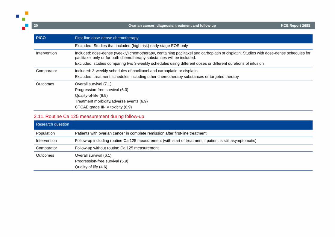
20 Ovarian cancer: diagnosis, treatment and follow-up KCE Report 268S
PICO First-line dose-dense chemotherapy
Excluded: Studies that included (high risk) early-stage EOS only
Intervention Included: dose-dense (weekly) chemotherapy, containing paclitaxel and carboplatin or cisplatin. Studies with dose-dense schedules for paclitaxel only or for both chemotherapy substances will be included. Excluded: studies comparing two 3-weekly schedules using different doses or different durations of infusion
Comparator Included: 3-weekly schedules of paclitaxel and carboplatin or cisplatin. Excluded: treatment schedules including other chemotherapy substances or targeted therapy
Outcomes Overall survival (7.1) Progression-free survival (6.0) Quality-of-life (6.9) Treatment morbidity/adverse events (6.9) CTCAE grade III-IV toxicity (6.9)
2.11. Routine Ca 125 measurement during follow-up Research question
Population Patients with ovarian cancer in complete remission after first-line treatment
Intervention Follow-up including routine Ca 125 measurement (with start of treatment if patient is still asymptomatic)
Comparator Follow-up without routine Ca 125 measurement
Outcomes Overall survival (6.1) Progression-free survival (5.9) Quality of life (4.6)
KCE Report 268S Ovarian cancer: diagnosis, treatment and follow-up 21
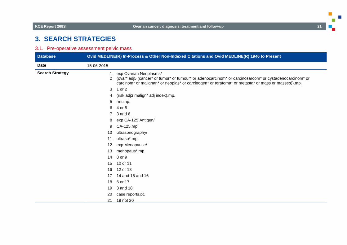
3. SEARCH STRATEGIES 3.1. Pre-operative assessment pelvic mass Database Ovid MEDLINE(R) In-Process & Other Non-Indexed Citations and Ovid MEDLINE(R) 1946 to Present
Date 15-06-2015
Search Strategy 1 exp Ovarian Neoplasms/ 2 (ovar* adj5 (cancer* or tumor* or tumour* or adenocarcinom* or carcinosarcom* or cystadenocarcinom* or
carcinom* or malignan* or neoplas* or carcinogen* or teratoma* or metasta* or mass or masses)).mp. 3 1 or 2 4 (risk adj3 malign* adj index).mp. 5 rmi.mp. 6 4 or 5 7 3 and 6 8 exp CA-125 Antigen/ 9 CA-125.mp.
10 ultrasonography/ 11 ultraso*.mp. 12 exp Menopause/ 13 menopaus*.mp. 14 8 or 9 15 10 or 11 16 12 or 13 17 14 and 15 and 16 18 6 or 17 19 3 and 18 20 case reports.pt. 21 19 not 20

22 Ovarian cancer: diagnosis, treatment and follow-up KCE Report 268S
Database Ovid: Embase Classic+Embase 1947 to 2015 June 15
Date 15-06-2015
Search Strategy 1 exp ovary tumor/ 2 (ovar* adj5 (cancer* or tumor* or tumour* or adenocarcinom* or carcinosarcom* or cystadenocarcinom* or
carcinom* or malignan* or neoplas* or carcinogen* or teratoma* or metasta* or mass or masses)).mp. 3 1 or 2 4 (risk adj3 malign* adj index).mp. 5 rmi.mp. 6 4 or 5 7 exp CA 125 antigen/ 8 ca-125.mp. 9 exp echography/
10 ultraso*.mp. 11 exp menopause/ 12 menopaus*.mp. 13 7 or 8 14 9 or 10 15 11 or 12 16 13 and 14 and 15 17 6 or 16 18 3 and 17 19 case report/ 20 limit 18 to (conference abstract or conference paper or conference proceeding or "conference review") 21 19 or 20 22 18 not 21
KCE Report 268S Ovarian cancer: diagnosis, treatment and follow-up 23
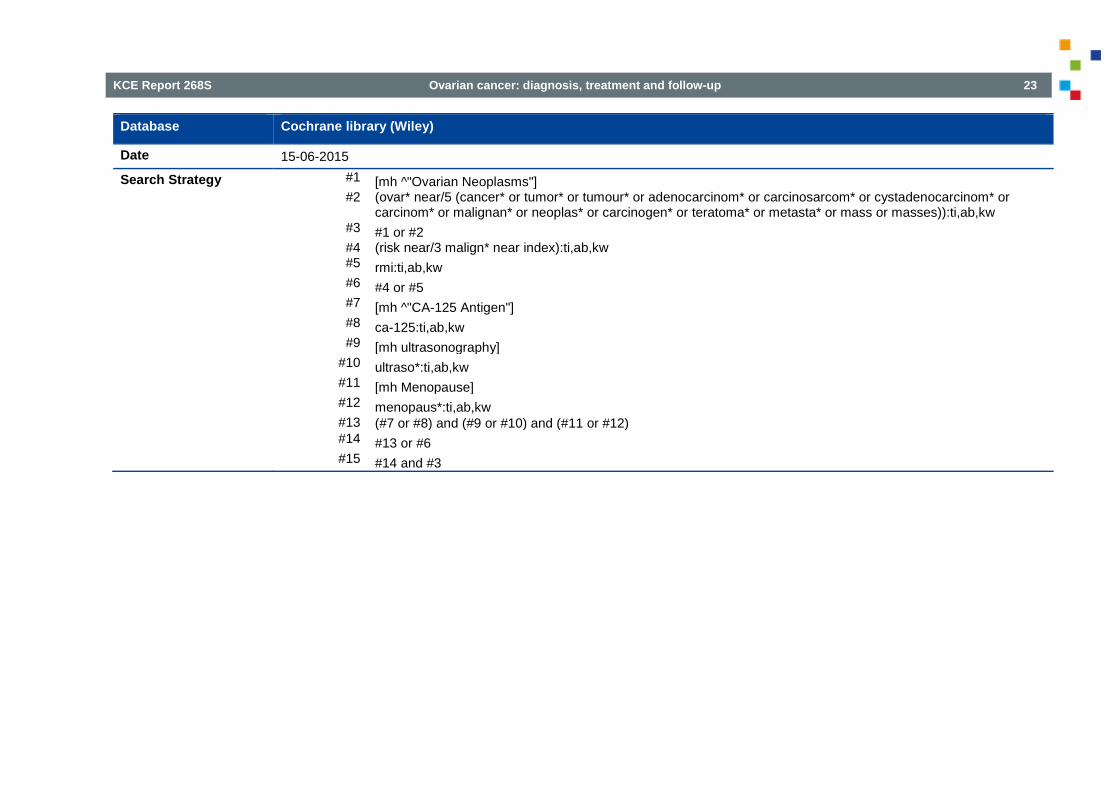
Database Cochrane library (Wiley)
Date 15-06-2015
Search Strategy #1 [mh ^"Ovarian Neoplasms"] #2 (ovar* near/5 (cancer* or tumor* or tumour* or adenocarcinom* or carcinosarcom* or cystadenocarcinom* or
carcinom* or malignan* or neoplas* or carcinogen* or teratoma* or metasta* or mass or masses)):ti,ab,kw #3 #1 or #2 #4 (risk near/3 malign* near index):ti,ab,kw #5 rmi:ti,ab,kw #6 #4 or #5 #7 [mh ^"CA-125 Antigen"] #8 ca-125:ti,ab,kw #9 [mh ultrasonography]
#10 ultraso*:ti,ab,kw #11 [mh Menopause] #12 menopaus*:ti,ab,kw #13 (#7 or #8) and (#9 or #10) and (#11 or #12) #14 #13 or #6 #15 #14 and #3

24 Ovarian cancer: diagnosis, treatment and follow-up KCE Report 268S
3.2. Intra-operative frozen section 3.2.1. Systematic reviews
Database OvidSP Ovid MEDLINE(R) In-Process & Other Non-Indexed Citations and Ovid MEDLINE(R) 1946 to Present
Date 28-04-2015
Search Strategy 1 exp Ovarian Neoplasms/ 2 (ovar* adj5 (cancer* or neoplasm* or carcinoma* or cystadenocarcinoma* or tumo?r*)).ti,ab,kw. 3 1 or 2 4 exp Frozen Sections/ 5 (FS or FSA or IFS or IFSA).ti,ab,kw. 6 ((intraoperative or intra-operative) adj5 (consultation* or microscopy or immunohistochemistry or histolog* or diagnos* or patholog*)).ti,ab,kw. 7 (cryosection* or cryogenic*).ti,ab,kw. 8 ((frozen or fresh or quick) adj5 (section* or tissue* or specimen*)).ti,ab,kw. 9 4 or 5 or 6 or 7 or 8 10 3 and 9 11 animals/ not humans/ 12 10 not 11 13 (MEDLINE or systematic review or (literature adj2 review)).tw. or (search* adj12 (literature or database?)).ti,ab. or intervention$.ti. 14 12 and 13
Note SR filter based on healthcanada.ca article (PMID: 22512835)
Database OvidSP Embase Classic+Embase 1947 to 2015 April 27
Date 28-04-2015 Search Strategy 1 exp ovary tumor/
2 (ovar* adj5 (cancer* or neoplasm* or carcinoma* or cystadenocarcinoma* or tumo?r*)).ti,ab,kw. 3 1 or 2 4 frozen section/ 5 (FS or FSA or IFS or IFSA).ti,ab,kw.

KCE Report 268S Ovarian cancer: diagnosis, treatment and follow-up 25
6 ((intraoperative or intra-operative) adj5 (consultation* or microscopy or immunohistochemistry or histolog* or diagnos* or patholog*)).ti,ab,kw. 7 (cryosection* or cryogenic*).ti,ab,kw. 8 ((frozen or fresh or cryostat* or quick) adj5 (section* or tissue* or specimen*)).ti,ab,kw. 9 4 or 5 or 6 or 7 or 8 10 3 and 9 11 (exp animal/ or animal.hw. or nonhuman/) not (exp human/ or human cell/ or (human or humans).ti.) 12 10 not 11 13 MEDLINE.tw. or exp systematic review/ or systematic review.tw. or (literature adj2 review).tw. or meta-analysis/ or (search* adj12 (literature or database?)).ti,ab. 14 12 and 13 15 limit 14 to (conference abstract or conference paper or conference proceeding) 16 14 not 15
Note SR filter based on healthcanada.ca article (PMID: 22512835)
Database Thecochranelibrary.com
*Cochrane database of systematic reviews
*Database of Abstracts of Reviews of Effects
Date 28-04-2015 Search Strategy #1 MeSH descriptor: [Ovarian Neoplasms] explode all trees
#2 (ovar* near/5 (cancer* or neoplasm* or carcinoma* or cystadenocarcinoma* or tumo?r*)):ti,ab,kw #3 #1 or #2 #4 MeSH descriptor: [Frozen Sections] explode all trees #5 (FS or FSA or IFS or IFSA):ti,ab,kw #6 ((intraoperative or intra-operative) near/5 (consultation* or microscopy or immunohistochemistry or histolog* or diagnos* or patholog*)):ti,ab,kw #7 (cryosection* or cryogenic*):ti,ab,kw #8 ((frozen or fresh or cryostat* or quick) near/5 (section* or tissue* or specimen*)):ti,ab,kw #9 #4 or #5 or #6 or #7 or #8

26 Ovarian cancer: diagnosis, treatment and follow-up KCE Report 268S
#10 #3 and #9 Note
3.2.2. Randomized controlled trials
Database OvidSP Ovid MEDLINE(R) In-Process & Other Non-Indexed Citations and Ovid MEDLINE(R) 1946 to Present
Date 29-04-2015 Search Strategy 1 exp Ovarian Neoplasms/
2 (ovar* adj5 (cancer* or neoplasm* or carcinoma* or cystadenocarcinoma* or tumo?r*)).ti,ab,kw. 3 1 or 2 4 exp Frozen Sections/ 5 (FS or FSA or IFS or IFSA).ti,ab,kw. 6 ((intraoperative or intra-operative) adj5 (consultation* or microscopy or immunohistochemistry or histolog* or diagnos* or patholog*)).ti,ab,kw. 7 (cryosection* or cryogenic*).ti,ab,kw. 8 ((frozen or fresh or quick) adj5 (section* or tissue* or specimen*)).ti,ab,kw. 9 4 or 5 or 6 or 7 or 8 10 3 and 9 11 animals/ not humans/ 12 10 not 11 13 (randomized controlled trial or controlled clinical trial).pt. or random*.ab. or placebo.ab. or trial.ab. or groups.ab. 14 12 and 13
Note Adapted Cochrane highly sensitive, specific RCT filter
Database OvidSP Embase Classic+Embase 1947 to 2015 April 28
Date 29-04-2015 Search Strategy 1 exp ovary tumor/
2 (ovar* adj5 (cancer* or neoplasm* or carcinoma* or cystadenocarcinoma* or tumo?r*)).ti,ab,kw. 3 1 or 2 4 frozen section/

KCE Report 268S Ovarian cancer: diagnosis, treatment and follow-up 27
5 (FS or FSA or IFS or IFSA).ti,ab,kw. 6 ((intraoperative or intra-operative) adj5 (consultation* or microscopy or immunohistochemistry or histolog* or diagnos* or patholog*)).ti,ab,kw. 7 (cryosection* or cryogenic*).ti,ab,kw. 8 ((frozen or fresh or cryostat* or quick) adj5 (section* or tissue* or specimen*)).ti,ab,kw. 9 4 or 5 or 6 or 7 or 8 10 3 and 9 11 (exp animal/ or animal.hw. or nonhuman/) not (exp human/ or human cell/ or (human or humans).ti.) 12 10 not 11 13 limit 12 to (conference abstract or conference paper or conference proceeding or "conference review") 14 12 not 13 15 crossover procedure/ or double-blind procedure/ or single-blind procedure/ or randomized controlled trial/ or crossover$.ti,ab,ot. or cross over$.ti,ab,ot. or placebo$.ti,ab,ot. or (doubl$ adj blind$).ti,ab,ot. or allocat$.ti,ab,ot. or random$.ti,ab,ab. or trial$.ti. 16 14 and 15
Note Cochrane filter for RCTs
Database Cochrane central registry of studies (via CRSO gateway, crso.cochrane.org)
Date 29-04-2015
Search Strategy #1 MeSH descriptor: [Ovarian Neoplasms] explode all trees #2 (ovar* near/5 (cancer* or neoplasm* or carcinoma* or cystadenocarcinoma* or tumo?r*)):ti,ab,kw #3 #1 or #2 #4 MeSH descriptor: [Frozen Sections] explode all trees #5 (FS or FSA or IFS or IFSA):ti,ab,kw #6 ((intraoperative or intra-operative) near/5 (consultation* or microscopy or immunohistochemistry or histolog* or diagnos* or patholog*)):ti,ab,kw #7 (cryosection* or cryogenic*):ti,ab,kw #8 ((frozen or fresh or cryostat* or quick) near/5 (section* or tissue* or specimen*)):ti,ab,kw #9 #4 or #5 or #6 or #7 or #8 #10 #3 and #9
Note
28 Ovarian cancer: diagnosis, treatment and follow-up KCE Report 268S
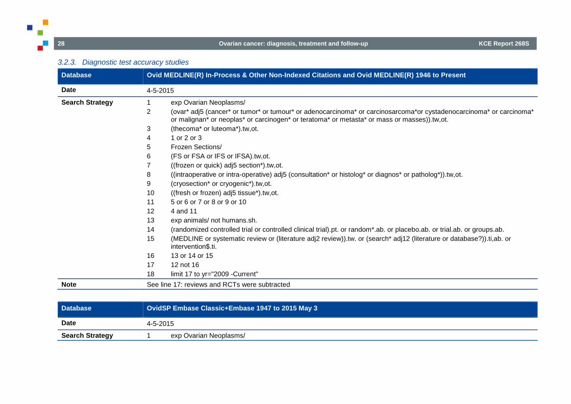
3.2.3. Diagnostic test accuracy studies
Database Ovid MEDLINE(R) In-Process & Other Non-Indexed Citations and Ovid MEDLINE(R) 1946 to Present
Date 4-5-2015 Search Strategy 1 exp Ovarian Neoplasms/
2 (ovar* adj5 (cancer* or tumor* or tumour* or adenocarcinoma* or carcinosarcoma*or cystadenocarcinoma* or carcinoma* or malignan* or neoplas* or carcinogen* or teratoma* or metasta* or mass or masses)).tw,ot. 3 (thecoma* or luteoma*).tw,ot. 4 1 or 2 or 3 5 Frozen Sections/ 6 (FS or FSA or IFS or IFSA).tw,ot. 7 ((frozen or quick) adj5 section*).tw,ot. 8 ((intraoperative or intra-operative) adj5 (consultation* or histolog* or diagnos* or patholog*)).tw,ot. 9 (cryosection* or cryogenic*).tw,ot. 10 ((fresh or frozen) adj5 tissue*).tw,ot. 11 5 or 6 or 7 or 8 or 9 or 10 12 4 and 11 13 exp animals/ not humans.sh. 14 (randomized controlled trial or controlled clinical trial).pt. or random*.ab. or placebo.ab. or trial.ab. or groups.ab. 15 (MEDLINE or systematic review or (literature adj2 review)).tw. or (search* adj12 (literature or database?)).ti,ab. or intervention$.ti. 16 13 or 14 or 15 17 12 not 16 18 limit 17 to yr="2009 -Current"
Note See line 17: reviews and RCTs were subtracted
Database OvidSP Embase Classic+Embase 1947 to 2015 May 3
Date 4-5-2015 Search Strategy 1 exp Ovarian Neoplasms/

KCE Report 268S Ovarian cancer: diagnosis, treatment and follow-up 29
2 (ovar* adj5 (cancer* or tumor* or tumour* or adenocarcinoma* or carcinosarcoma*or cystadenocarcinoma* or carcinoma* or malignan* or neoplas* or carcinogen* or teratoma* or metasta* or mass or masses)).tw,ot. 3 (thecoma* or luteoma*).tw,ot. 4 1 or 2 or 3 5 Frozen Sections/ 6 (FS or FSA or IFS or IFSA).tw,ot. 7 ((frozen or quick) adj5 section*).tw,ot. 8 ((intraoperative or intra-operative) adj5 (consultation* or histolog* or diagnos* or patholog*)).tw,ot. 9 (cryosection* or cryogenic*).tw,ot. 10 ((fresh or frozen) adj5 tissue*).tw,ot. 11 5 or 6 or 7 or 8 or 9 or 10 12 4 and 11 13 (exp animal/ or animal.hw. or nonhuman/) not (exp human/ or human cell/ or (human or humans).ti.) 14 crossover procedure/ or double-blind procedure/ or single-blind procedure/ or randomized controlled trial/ or crossover$.ti,ab,ot. or cross over$.ti,ab,ot. or placebo$.ti,ab,ot. or (doubl$ adj blind$).ti,ab,ot. or allocat$.ti,ab,ot. or random$.ti,ab,ab. or trial$.ti. 15 MEDLINE.tw. or exp systematic review/ or systematic review.tw. or (literature adj2 review).tw. or meta-analysis/ or (search* adj12 (literature or database?)).ti,ab. 16 13 or 14 or 15 17 12 not 16 18 limit 17 to (conference abstract or conference paper or conference proceeding or "conference review") 19 17 not 18 20 limit 19 to yr="2009 -Current"
Note See line 19: reviews and RCTs were subtracted

30 Ovarian cancer: diagnosis, treatment and follow-up KCE Report 268S
3.3. Lymphadenectomy 3.3.1. Systematic reviews
Database Ovid MEDLINE(R) In-Process & Other Non-Indexed Citations and Ovid MEDLINE(R) 1946 to Present
Date 15-06-2015
Search Strategy 1 exp Ovarian Neoplasms/ 2 (ovar* adj5 (cancer* or neoplasm* or carcinom* or malignan* or tumor* or tumour*)).mp. 3 1 or 2 4 exp Lymph Node Excision/ 5 (lymph adj5 node* adj5 (excis* or dissect* or surg*)).mp. 6 lymphadenectomy.mp. 7 4 or 5 or 6 8 3 and 7 9 case reports.pt.
10 8 not 9 11 (MEDLINE or systematic review or (literature adj2 review)).tw. or (search* adj12 (literature or database?)).ti,ab. or
intervention$.ti. 12 10 and 11
Note SR filter health-evidence Canada (Lee E et al 2012 BMC medical research methodology)
Database Ovid: Embase Classic+Embase 1947 to 2015 June 12
Date 15-06-2015
Search Strategy 1 exp ovary tumor/ 2 (ovar* adj5 (cancer* or neoplasm* or carcinom* or malignan* or tumor* or tumour*)).mp. 3 1 or 2 4 exp lymph node dissection/
5 (lymph adj5 node* adj5 (excis* or dissect* or surg*)).mp. 6 lymphadenectomy.mp.

KCE Report 268S Ovarian cancer: diagnosis, treatment and follow-up 31
7 4 or 5 or 6 8 3 and 7 9 MEDLINE.tw. or exp systematic review/ or systematic review.tw. or meta-analysis/ or (search* adj12 (literature or
database?)).ti,ab. 10 8 and 9
11 limit 10 to (conference abstract or conference paper or conference proceeding or "conference review") 12 10 not 11
Note SR filter health-evidence Canada (Lee E et al 2012 BMC medical research methodology)
Database Cochrane library (Wiley)
Date 15-06-2015
Search Strategy #1 [mh ^"Ovarian Neoplasms"] #2 (ovar* near/5 (cancer* or neoplasm* or carcinom* or malignan* or tumor* or tumour*)):ti,ab,kw #3 #1 or #2 #4 [mh ^" Lymph Node Excision"] #5 (lymph near/5 node* near/5 (excis* or dissect* or surg*)):ti,ab,kw #6 lymphadenectomy:ti,ab,kw #7 #4 or #5 or #6 #8 #7 and #3
Note
3.3.2. Primary studies
Database Ovid MEDLINE(R) In-Process & Other Non-Indexed Citations and Ovid MEDLINE(R) 1946 to Present
Date 22-06-2015
Search Strategy 1 exp Ovarian Neoplasms/ 2 (ovar* adj5 (cancer* or neoplasm* or carcinom* or malignan* or tumor* or tumour*)).mp. 3 1 or 2 4 exp Lymph Node Excision/

32 Ovarian cancer: diagnosis, treatment and follow-up KCE Report 268S
5 (lymph adj5 node* adj5 (excis* or dissect* or surg*)).mp. 6 lymphadenectomy.mp. 7 4 or 5 or 6 8 3 and 7 9 case reports.pt.
10 8 not 9
Note
Database Ovid: Embase Classic+Embase 1947 to 2015 June 18
Date 22-06-2015
Search Strategy 1 exp ovary tumor/ 2 (ovar* adj5 (cancer* or neoplasm* or carcinom* or malignan* or tumor* or tumour*)).mp. 3 1 or 2 4 exp lymph node dissection/
5 (lymph adj5 node* adj5 (excis* or dissect* or surg*)).mp. 6 lymphadenectomy.mp. 7 4 or 5 or 6 8 3 and 7 9 case report/
10 limit 8 to (conference abstract or conference paper or conference proceeding or "conference review") 11 9 or 10 12 8 not 11
Note
Date Cochrane library (Wiley)
Database 22-06-2015
KCE Report 268S Ovarian cancer: diagnosis, treatment and follow-up 33
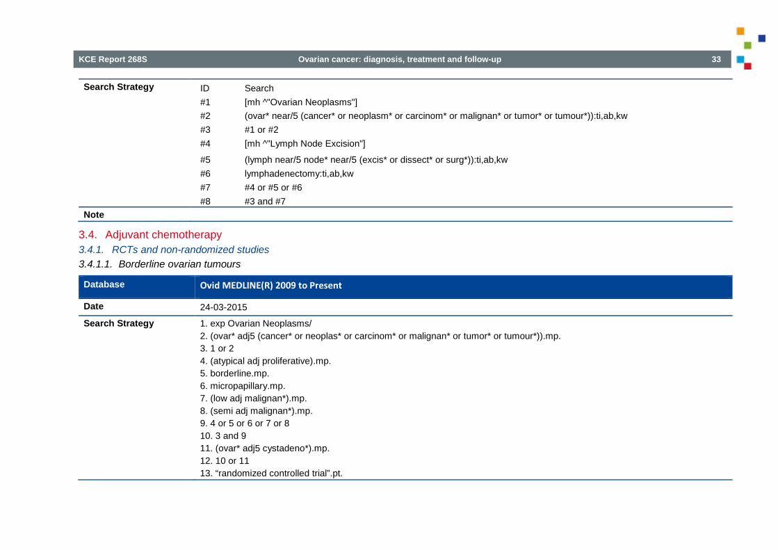
Search Strategy ID Search #1 [mh ^"Ovarian Neoplasms"] #2 (ovar* near/5 (cancer* or neoplasm* or carcinom* or malignan* or tumor* or tumour*)):ti,ab,kw #3 #1 or #2 #4 [mh ^"Lymph Node Excision"]
#5 (lymph near/5 node* near/5 (excis* or dissect* or surg*)):ti,ab,kw #6 lymphadenectomy:ti,ab,kw #7 #4 or #5 or #6 #8 #3 and #7
Note
3.4. Adjuvant chemotherapy 3.4.1. RCTs and non-randomized studies 3.4.1.1. Borderline ovarian tumours
Database Ovid MEDLINE(R) 2009 to Present
Date 24-03-2015 Search Strategy 1. exp Ovarian Neoplasms/
2. (ovar* adj5 (cancer* or neoplas* or carcinom* or malignan* or tumor* or tumour*)).mp. 3. 1 or 2 4. (atypical adj proliferative).mp. 5. borderline.mp. 6. micropapillary.mp. 7. (low adj malignan*).mp. 8. (semi adj malignan*).mp. 9. 4 or 5 or 6 or 7 or 8 10. 3 and 9 11. (ovar* adj5 cystadeno*).mp. 12. 10 or 11 13. “randomized controlled trial”.pt.

34 Ovarian cancer: diagnosis, treatment and follow-up KCE Report 268S
14. “controlled clinical trial”.pt. 15. randomized.ab. 16. placebo.ab. 17. “drug therapy”.fs. 18. “surgery”.fs. 19. “therapy”.fs. 20. “radiotherapy”.fs. 21. randomly.ab. 22. trial.ab. 23. groups.ab. 24. exp Cohort Studies/ 25. cohort*.mp. 26. (case adj series).mp. 27. 13 or 14 or 15 or 16 or 17 or 18 or 19 or 20 or 21 or 22 or 23 or 24 or 25 or 26 28. 12 and 27 29. Animals/ 30. Humans/ 31. 29 not (29 and 30) 32. 28 not 31
Note Search for RCTs regarding RQ5a (Faluyi 2010: Interventions for the treatment of borderline ovarian tumours)
Database Ovid Embase 2009 to current
Date 24-03-2015 Search Strategy 1. Ovary Tumor/
2. (ovar* adj5 (cancer* or neoplas* or carcinom* or malignan* or tumor* or tumour*)).mp. 3. 1 or 2 4. (atypical adj proliferative).mp. 5. borderline.mp. 6. micropapillary.mp. 7. (low adj malignan*).mp.]
KCE Report 268S Ovarian cancer: diagnosis, treatment and follow-up 35
8. (semi adj malignan*).mp. 9. 4 or 5 or 6 or 7 or 8 10. 3 and 9 11. (ovar* adj5 cystadeno*).mp. 12. 10 or 11 13. exp Controlled Clinical Trial/ 14. randomized.ab. 15. placebo.ab. 16. dt.fs. 17. su.fs. 18. th.fs. 19. rt.fs. 20. randomly.ab. 21. trial.ab. 22. groups.ab. 23. exp Cohort Analysis/ 24. cohort*.mp. 25. (case adj series).mp. 26. 13 or 14 or 15 or 16 or 17 or 18 or 19 or 20 or 21 or 22 or 23 or 24 or 25 27. 12 and 26 28. exp Animal/ 29. Human/ 30. 28 not (28 and 29) 31. 27 not 30
Note Search for RCTs regarding RQ5a (Faluyi 2010: Interventions for the treatment of borderline ovarian tumours)
Database Cochrane Central (Issue 2, 2015)
Date 24-03-2015
Search Strategy 1. MeSH descriptor Ovarian Neoplasms explode all trees 2. ovar* near/5 (cancer* or neoplas* or carcinom* or malignan* or tumor* or tumour*)

36 Ovarian cancer: diagnosis, treatment and follow-up KCE Report 268S
3. (#1 OR #2) 4. atypical adj proliferative 5. borderline 6. micropapillary 7. low adj malignan* 8. semi adj malignan* 9. (#4 OR #5 OR #6 OR #7 OR #8) 10. (#3 AND #9) 11. ovar* near/5 cystadeno* 12. (#10 OR #11)
Note Search for RCTs regarding RQ5a (Faluyi 2010: Interventions for the treatment of borderline ovarian tumours)
3.4.1.2. Micro-invasive disease
Database Ovid MEDLINE(R) In-Process & Other Non-Indexed Citations and Ovid MEDLINE(R) 1946 to Present
Date 08-04-2015 Search Strategy 1 exp Ovarian Neoplasms/
2 (ovar* adj5 (cancer* or neoplas* or carcinom* or malignan* or tumor* or tumour*)).ti,ab,kw. 3 or/1-2 [ovarian cancer] 4 borderline.ti,ab,kw. 5 (low adj malignan*).ti,ab,kw. 6 lmp.ti,ab,kw. 7 or/4-6 [borderline] 8 microinvasi*.ti,ab,kw. 9 micro-invasi*.ti,ab,kw. 10 or/8-9 [microinvasive] 11 (ovar* adj5 cystadeno*).ti,ab,kw. 12 7 or 10 13 3 and 12 14 11 or 13 15 exp Chemotherapy, Adjuvant/ or exp Chemoradiotherapy, Adjuvant/ or exp Radiotherapy, Adjuvant/ 16 adjuvant.ti,ab,kw.
KCE Report 268S Ovarian cancer: diagnosis, treatment and follow-up 37
17 15 or 16 18 14 and 17
Note Search for RCTs and non-randomized studies regarding RQ5b
Database Embase Classic+Embase 1947 to 2015 April 07
Date 08-04-2015 Ovid
Search Strategy 1 Ovary Tumor/ 2 (ovar* adj5 (cancer* or neoplas* or carcinom* or malignan* or tumor* or tumour*)).ti,ab,kw. 3 or/1-2 [ovarian cancer] 4 borderline.ti,ab,kw. 5 (low adj malignan*).ti,ab,kw. 6 lmp.ti,ab,kw. 7 or/4-6 [borderline] 8 microinvasi*.ti,ab,kw. 9 micro-invasi*.ti,ab,kw. 10 or/8-9 [microinvasive] 11 (ovar* adj5 cystadeno*).ti,ab,kw. 12 7 or 10 13 3 and 12 14 11 or 13 15 exp adjuvant therapy/ or exp adjuvant/ or exp adjuvant chemotherapy/ or exp adjuvant chemoradiotherapy/ or exp cancer adjuvant therapy/ 16 adjuvant.ti,ab,kw. 17 15 or 16 18 14 and 17 19 limit 18 to (conference abstract or conference paper or conference proceeding or "conference review") 20 18 not 19
Note Search for RCTs and non-randomized studies regarding RQ5b

38 Ovarian cancer: diagnosis, treatment and follow-up KCE Report 268S
Database PubMed Central
Date 08-04-2015 Search Strategy (microinvasive[TW] OR micro-invasive[tw] OR microinvasion[TW] OR micro-invasion[tw]) AND (ovarian cancer[TI] OR ovarian
neoplasm*[TI] OR ovarian cancer[AB] OR ovarian neoplasm*[AB] OR "ovarian neoplasms"[MeSH Terms]) AND ("Chemoradiotherapy, Adjuvant"[mesh] OR "Radiotherapy, Adjuvant"[mesh] OR "Chemoradiotherapy, Adjuvant"[mesh] OR adjuvant[TW])
Note Search for RCTs and non-randomized studies regarding RQ5b
3.4.1.3. Invasive early stage epithelial ovarian cancer
Database Ovid MEDLINE(R) 2011 to Present
Date 24-03-2015 Search Strategy 1. exp Ovarian Neoplasms/
2. (ovar* adj5 (cancer* or tumor* or tumour* or neoplas* or carcinoma* or malignan*)).mp. 3. 1 or 2 4. drug therapy.fs. 5. exp Antineoplastic Agents/ 6. Antineoplastic Combined Chemotherapy Protocols/ 7. chemotherap*.mp. 8. 4 or 5 or 6 or 7 9. surgery.fs. 10. exp Surgical Procedures, Operative/ 11. (surg* or procedure* or intervention*).mp. 12. 9 or 10 or 11 13. 8 and 12 14. Chemotherapy, Adjuvant/ 15. (chemotherap* and adjuvant).mp. 16. 14 or 15 17. 13 or 16 18. 3 and 17 19. randomized controlled trial.pt.
KCE Report 268S Ovarian cancer: diagnosis, treatment and follow-up 39
20. controlled clinical trial.pt. 21. randomized.ab. 22. placebo.ab. 23. clinical trials as topic.sh. 24. randomly.ab. 25. trial.ti. 26. 19 or 20 or 21 or 22 or 23 or 24 or 25 27. 18 and 26
Note Search for RCTs regarding RQ5c (Winter-Roach 2012: Adjuvant (post-surgery) chemotherapy for early stage epithelial ovarian cancer)
Database Ovid Embase 2011 to current
Date 24-03-2015 Search Strategy 1 exp ovary tumor/
2 (ovar* adj5 (cancer* or tumor* or tumour* or malignan* or carcinoma* or neoplasm*)).mp. 3 1 or 2 4 exp chemotherapy/ 5 exp antineoplastic agent/ 6 chemotherap*.mp. 7 4 or 5 or 6 8 exp gynecologic surgery/ 9 (surgery or surgical* or procedure* or intervention*).mp. 10 8 or 9 11 7 and 10 12 adjuvant chemotherapy/ 13 (adjuvant adj5 chemotherap*).mp. 14 12 or 13 15 11 or 14 16 3 and 15 17 crossover procedure/

40 Ovarian cancer: diagnosis, treatment and follow-up KCE Report 268S
18 randomized controlled trial/ 19 single blind procedure/ 20 random*.mp. 21 factorial*.mp. 22 (crossover* or cross over* or cross-over).mp. 23 placebo*.mp. 24 (doubl* adj blind*).mp. 25 (singl* adj blind*).mp. 26 assign*.mp. 27 allocat*.mp. 28 volunteer*.mp. 29 17 or 18 or 19 or 20 or 21 or 22 or 23 or 24 or 25 or 26 or 27 or 28 30 16 and 29
Note Search for RCTs regarding RQ5c (Winter-Roach 2012: Adjuvant (post-surgery) chemotherapy for early stage epithelial ovarian cancer)
Database Cochrane Central (Issue 2, 2015)
Date 24-03-2015
Search Strategy 1 MeSH descriptor Ovarian Neoplasms explode all trees 2 ovar* near/5 (cancer* or tumor* or tumour* or neoplas* or carcinoma*or malignan*) 3 (#1 OR #2) 4 Any MeSH descriptor with qualifier: DT 5 MeSH descriptor Antineoplastic Agents explode all trees 6 MeSH descriptor Antineoplastic Combined Chemotherapy Protocols, this term only 7 chemotherap* 8 (#4 OR #5 OR #6 OR #7) 9 Any MeSH descriptor with qualifier: SU 10 MeSH descriptor Surgical Procedures, Operative explode all trees 11 surg* or procedure* or intervention* 12 (#9 OR #10 OR #11)
KCE Report 268S Ovarian cancer: diagnosis, treatment and follow-up 41
13 (#8 AND #12) 14 MeSH descriptor Chemotherapy, Adjuvant explode all trees 15 chemotherap* and adjuvant 16 (#14 OR #15) 17 (#13 OR #16) 18 (#3 AND #17)
Note Search for RCTs regarding (Winter-Roach 2012: Adjuvant (post-surgery) chemotherapy for early stage epithelial ovarian cancer)
3.5. Laparoscopic surgery 3.5.1. Systematic reviews Database Medline OvidSP
Date 2015-03-09
Search strategy
1 exp Ovarian Neoplasms/ 65326
2 (ovar* adj5 (cancer* or neoplas* or malignan* or tumor* or tumour* or carcin* or adenocarcin* or metasta*)).tw. 68272
3 1 or 2 85995
4 exp Laparoscopy/ 71161
5 laparoscop*.ab,ti. 86822
6 peritoneoscop*.ab,ti. 720
7 celioscop*.ab,ti. 548
8 Laparotomy/ 15757
9 (endoscop* adj5 abdom*).ab,ti. 1336
10 laparotom*.ab,ti. 39237
11 (abdom* adj5 (surg* or incision)).ab,ti. 25235
12 4 or 5 or 6 or 7 or 8 or 9 or 10 or 11 156391
13 3 and 12 5036

42 Ovarian cancer: diagnosis, treatment and follow-up KCE Report 268S
14 limit 13 to systematic reviews 68
15 randomized controlled trial.pt. 387168
16 controlled clinical trial.pt. 88869
17 randomized.ti,ab. 333894
18 placebo.ti,ab. 163360
19 clinical trials as topic/ 171370
20 randomly.ti,ab. 226003
21 trial?.ti. 182508
22 15 or 16 or 17 or 18 or 19 or 20 or 21 963148
23 exp animal/ not humans/ 4000190
24 22 not 23 888683
25 13 and 24 299
26 limit 25 to yr="2006 -Current" 86
Notes
Database Embase
Date 2015-03-09
Search strategy
1 'ovary tumor'/exp 105023
2 (ovar* NEAR/5 (cancer* OR tumor* OR tumour* OR neoplas* OR carcin* OR adenocarcin* OR malignan* OR metasta*)):ab,ti
88199
3 #1 OR #2 121355
4 'laparoscopy'/exp 106372
5 'laparotomy'/exp 56282
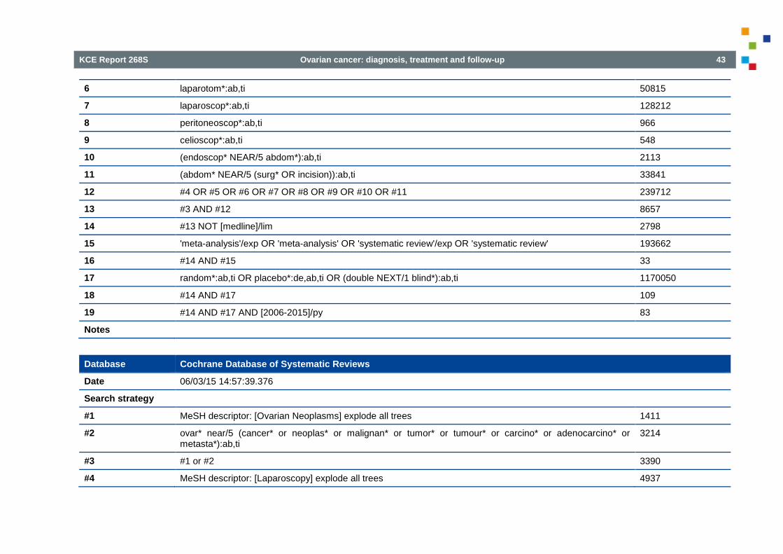
KCE Report 268S Ovarian cancer: diagnosis, treatment and follow-up 43
6 laparotom*:ab,ti 50815
7 laparoscop*:ab,ti 128212
8 peritoneoscop*:ab,ti 966
9 celioscop*:ab,ti 548
10 (endoscop* NEAR/5 abdom*):ab,ti 2113
11 (abdom* NEAR/5 (surg* OR incision)):ab,ti 33841
12 #4 OR #5 OR #6 OR #7 OR #8 OR #9 OR #10 OR #11 239712
13 #3 AND #12 8657
14 #13 NOT [medline]/lim 2798
15 'meta-analysis'/exp OR 'meta-analysis' OR 'systematic review'/exp OR 'systematic review' 193662
16 #14 AND #15 33
17 random*:ab,ti OR placebo*:de,ab,ti OR (double NEXT/1 blind*):ab,ti 1170050
18 #14 AND #17 109
19 #14 AND #17 AND [2006-2015]/py 83
Notes
Database Cochrane Database of Systematic Reviews
Date 06/03/15 14:57:39.376
Search strategy
#1 MeSH descriptor: [Ovarian Neoplasms] explode all trees 1411
#2 ovar* near/5 (cancer* or neoplas* or malignan* or tumor* or tumour* or carcino* or adenocarcino* or metasta*):ab,ti
3214
#3 #1 or #2 3390
#4 MeSH descriptor: [Laparoscopy] explode all trees 4937
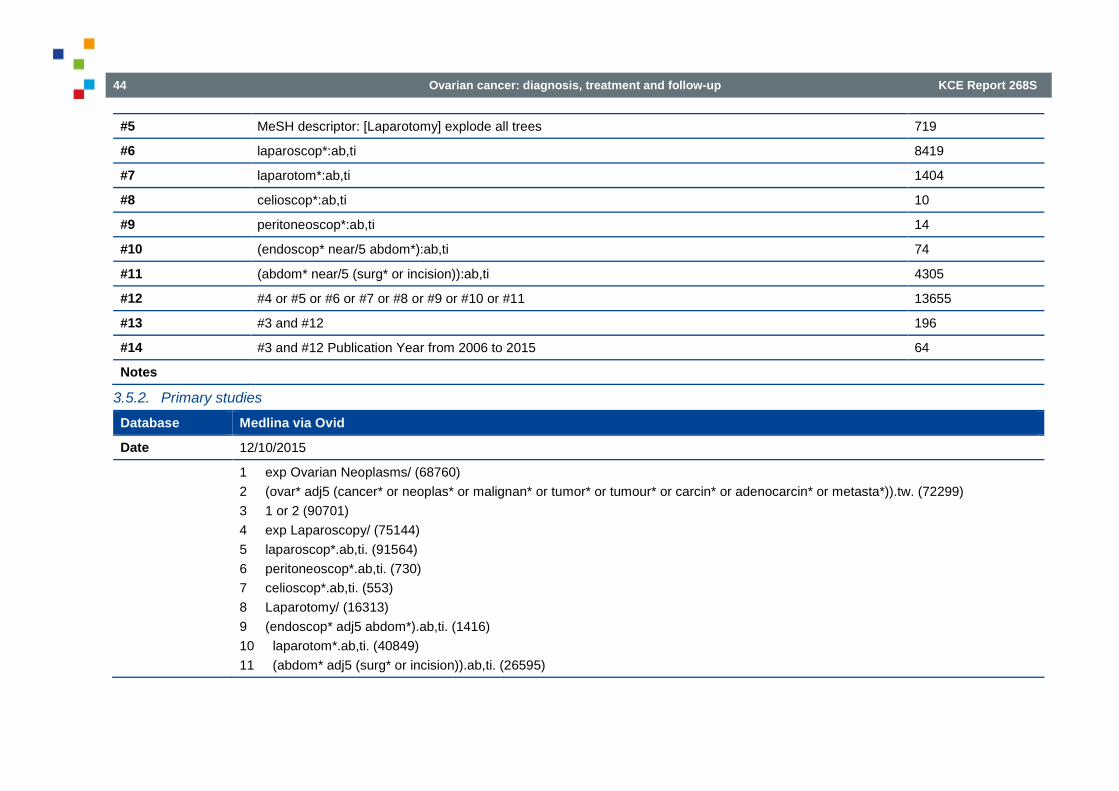
44 Ovarian cancer: diagnosis, treatment and follow-up KCE Report 268S
#5 MeSH descriptor: [Laparotomy] explode all trees 719
#6 laparoscop*:ab,ti 8419
#7 laparotom*:ab,ti 1404
#8 celioscop*:ab,ti 10
#9 peritoneoscop*:ab,ti 14
#10 (endoscop* near/5 abdom*):ab,ti 74
#11 (abdom* near/5 (surg* or incision)):ab,ti 4305
#12 #4 or #5 or #6 or #7 or #8 or #9 or #10 or #11 13655
#13 #3 and #12 196
#14 #3 and #12 Publication Year from 2006 to 2015 64
Notes
3.5.2. Primary studies
Database Medlina via Ovid
Date 12/10/2015
1 exp Ovarian Neoplasms/ (68760) 2 (ovar* adj5 (cancer* or neoplas* or malignan* or tumor* or tumour* or carcin* or adenocarcin* or metasta*)).tw. (72299) 3 1 or 2 (90701) 4 exp Laparoscopy/ (75144) 5 laparoscop*.ab,ti. (91564) 6 peritoneoscop*.ab,ti. (730) 7 celioscop*.ab,ti. (553) 8 Laparotomy/ (16313) 9 (endoscop* adj5 abdom*).ab,ti. (1416) 10 laparotom*.ab,ti. (40849) 11 (abdom* adj5 (surg* or incision)).ab,ti. (26595)
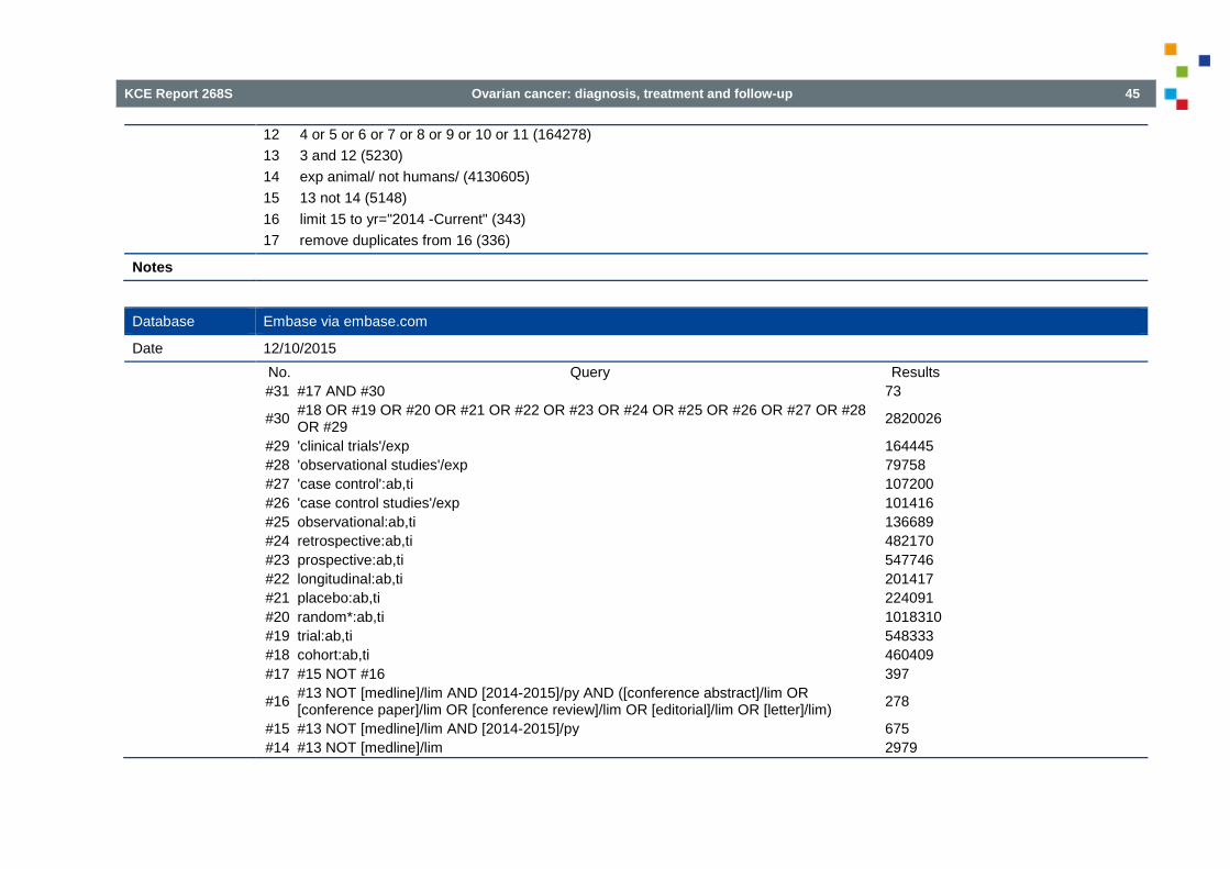
KCE Report 268S Ovarian cancer: diagnosis, treatment and follow-up 45
12 4 or 5 or 6 or 7 or 8 or 9 or 10 or 11 (164278) 13 3 and 12 (5230) 14 exp animal/ not humans/ (4130605) 15 13 not 14 (5148) 16 limit 15 to yr="2014 -Current" (343) 17 remove duplicates from 16 (336)
Notes
Database Embase via embase.com
Date 12/10/2015
No. Query Results #31 #17 AND #30 73
#30 #18 OR #19 OR #20 OR #21 OR #22 OR #23 OR #24 OR #25 OR #26 OR #27 OR #28 OR #29 2820026
#29 'clinical trials'/exp 164445 #28 'observational studies'/exp 79758 #27 'case control':ab,ti 107200 #26 'case control studies'/exp 101416 #25 observational:ab,ti 136689 #24 retrospective:ab,ti 482170 #23 prospective:ab,ti 547746 #22 longitudinal:ab,ti 201417 #21 placebo:ab,ti 224091 #20 random*:ab,ti 1018310 #19 trial:ab,ti 548333 #18 cohort:ab,ti 460409 #17 #15 NOT #16 397
#16 #13 NOT [medline]/lim AND [2014-2015]/py AND ([conference abstract]/lim OR [conference paper]/lim OR [conference review]/lim OR [editorial]/lim OR [letter]/lim) 278
#15 #13 NOT [medline]/lim AND [2014-2015]/py 675 #14 #13 NOT [medline]/lim 2979

46 Ovarian cancer: diagnosis, treatment and follow-up KCE Report 268S
#13 #3 AND #12 9048 #12 #4 OR #5 OR #6 OR #7 OR #8 OR #9 OR #10 OR #11 254203 #11 (abdom* NEAR/5 (surg* OR incision)):ab,ti 35769 #10 (endoscop* NEAR/5 abdom*):ab,ti 2240 #9 celioscop*:ab,ti 554 #8 peritoneoscop*:ab,ti 987 #7 laparoscop*:ab,ti 137734 #6 laparotom*:ab,ti 52873 #5 'laparotomy'/exp 59121 #4 'laparoscopy'/exp 112826 #3 #1 OR #2 127662
#2 (ovar* NEAR/5 (cancer* OR tumor* OR tumour* OR neoplas* OR carcin* OR adenocarcin* OR malignan* OR metasta*)):ab,ti 93171
#1 'ovary tumor'/exp 109809
Notes
Database Cochrane
Date 12/10/2015
#1 [mh "ovarian neoplasms"] 1433 #2 ovar* near/5 (cancer* or neoplas* or malignan* or tumor* or tumour* or carcino* or adenocarcino* or metasta*):ab,ti 3417 #3 #1 or #2 3594 #4 [mh laparoscopy] 5052 #5 [mh laparotomy] 725 #6 laparoscop*:ab,ti 9249 #7 laparotom*:ab,ti 1482 #8 celioscop*:ab,ti 10 #9 peritoneoscop*:ab,ti 15 #10 (endoscop* near/5 abdom*):ab,ti 84 #11 (abdom* near/5 (surg* or incision)):ab,ti 4655 #12 #4 or #5 or #6 or #7 or #8 or #9 or #10 or #11 14818

KCE Report 268S Ovarian cancer: diagnosis, treatment and follow-up 47
#13 #3 and #12 203 #14 #3 and #12 Publication Year from 2014 to 2015 13
Notes
3.6. Laparoscopy, PET-CT and MRI to predict end result of cytoreductive surgery 3.6.1. Systematic reviews
Database OvidSP Ovid MEDLINE(R) In-Process & Other Non-Indexed Citations and Ovid MEDLINE(R) 1946 to Present
Date 22-01-2015 Search Strategy 1 exp Ovarian Neoplasms/
2 (ovar* adj5 (cancer or neoplasm or carcinoma or cystadenocarcinoma or tumo?r)).ti,ab,kw. 3 1 or 2 4 exp Magnetic Resonance Imaging/ 5 (magn* adj3 imag*).ti,ab,kw. 6 mri.ti,ab,kw. 7 exp Positron-Emission Tomography/ 8 (pet adj3 scan*).ti,ab,kw. 9 (positr* adj4 tomogr*).ti,ab,kw. 10 exp Laparoscopy/ 11 laparoscop*.ti,ab,kw. 12 or/4-11 13 3 and 12 14 (MEDLINE or systematic review).tw. or (search* adj12 (literature or database?)).ti,ab. or intervention$.ti. 15 13 and 14
Note SR filter based on healthcanada.ca article (PMID: 22512835)
Database OvidSP Embase Classic+Embase 1947 to 2015 January 20
Date 22-01-2015 Search Strategy 1 exp ovary cancer/

48 Ovarian cancer: diagnosis, treatment and follow-up KCE Report 268S
2 (ovar* adj5 (cancer or neoplasm or carcinoma or cystadenocarcinoma or tumo?r)).ti,ab,kw. 3 1 or 2 4 exp nuclear magnetic resonance imaging/ 5 (magn* adj3 imag*).ti,ab,kw. 6 mri.ti,ab,kw. 7 exp positron emission tomography/ 8 (pet adj3 scan*).ti,ab,kw. 9 (positr* adj4 tomogr*).ti,ab,kw. 10 exp laparoscopy/ 11 laparoscop*.ti,ab,kw. 12 or/4-11 13 MEDLINE.tw. or exp systematic review/ or systematic review.tw. or meta-analysis/ or (search* adj12 (literature or database?)).ti,ab. 14 3 and 12 and 13
Note SR filter based on healthcanada.ca article (PMID: 22512835)
Database Thecochranelibrary.com *Cochrane database of systematic reviews *Database of Abstracts of Reviews of Effects
Date 22-01-2015
Search Strategy #1 MeSH descriptor: [Ovarian Neoplasms] explode all trees #2 (ovar* next/5 (cancer or neoplasm or carcinoma or cystadenocarcinoma or tumo?r)):ti,ab,kw #3 #1 or #2 #4 [mh ^"Magnetic Resonance Imaging"] #5 (magn* next/3 imag*):ti,ab,kw #6 mri:ti,ab,kw #7 [mh ^"Positron-Emission Tomography"] #8 (pet near/3 scan*):ti,ab,kw #9 (positr* near/4 tomogr*):ti,ab,kw

KCE Report 268S Ovarian cancer: diagnosis, treatment and follow-up 49
#10 [mh ^Laparoscopy] #11 laparoscop*:ti,ab,kw #12 #4 or #5 or #6 or #7 or #8 or #9 or #10 or #11 #13 #3 and #12
3.6.2. RCTs
Database OvidSP Ovid MEDLINE(R) In-Process & Other Non-Indexed Citations and Ovid MEDLINE(R) 1946 to Present
Date 16-02-2015 Search Strategy 1 exp Ovarian Neoplasms/
2 (ovar* adj5 (cancer or neoplasm or carcinoma or cystadenocarcinoma or malign* or tumo?r)).ti,ab,kw. 3 1 or 2 4 exp Magnetic Resonance Imaging/ 5 MRi.ti,ab,kw. 6 (magn* adj3 imag*).ti,ab,kw. 7 (Diffusion adj3 Imaging).ti,ab,kw. 8 Diffusion-weighted.ti,ab,kw. 9 or/4-8 10 exp Positron-Emission Tomography/ 11 (pet adj3 scan*).ti,ab,kw. 12 (positr* adj4 tomogr*).ti,ab,kw. 13 ("Positron emission tomography" adj3 "computed tomography").ti,ab,kw. 14 (pet-ct or "pet/ct" or fdg-pet).ti,ab,kw. 15 or/10-14 16 9 or 15 17 3 and 16 18 exp Ovarian Neoplasms/ri 19 17 or 18 20 (randomized controlled trial or controlled clinical trial).pt. or clinical trials as topic.sh. or (random* or placebo).ti,ab,kw,hw. or trial.ti. 21 19 and 20
Note Adapted Cochrane highly sensitive, specific RCT filter

50 Ovarian cancer: diagnosis, treatment and follow-up KCE Report 268S
Database OvidSP Embase Classic+Embase 1947 to 2015 February 13
Date 16-02-2015
Search Strategy 1 exp *ovary cancer/ 2 (ovar* adj5 (cancer or neoplasm or carcinoma or cystadenocarcinoma or malign* or tumo?r)).ti,ab,kw. 3 1 or 2 4 exp nuclear magnetic resonance imaging/ 5 MRi.ti,ab,kw. 6 (magn* adj3 imag*).ti,ab,kw. 7 (Diffusion adj3 Imaging).ti,ab,kw. 8 Diffusion-weighted.ti,ab,kw. 9 4 or 5 or 6 or 7 or 8 10 exp positron emission tomography/ 11 (pet adj3 scan*).ti,ab,kw. 12 (positr* adj4 tomogr*).ti,ab,kw. 13 ("Positron emission tomography" adj3 "computed tomography").ti,ab,kw. 14 (pet-ct or "pet/ct" or fdg-pet).ti,ab,kw. 15 or/10-14 16 9 or 15 17 3 and 16 18 crossover procedure/ or double-blind procedure/ or single-blind procedure/ or randomized controlled trial/ or crossover$.ti,ab,ot. or cross over$.ti,ab,ot. or placebo$.ti,ab,ot. or (doubl$ adj blind$).ti,ab,ot. or allocat$.ti,ab,ot. or random$.ti,ab,ab. or trial$.ti. 19 17 and 18
Note Cochrane filter for RCTs
Database Cochrane central registry of studies (via CRSO gateway, crso.cochrane.org)
Date 16-02-2015
Search Strategy #1 Ovarian Neoplasms:MH #2 ovary cancer:EH #3 (ovar* adj5 (cancer or neoplasm or carcinoma or cystadenocarcinoma or malign* or tumo?r)):ti,ab,kw #4 #1 OR #2 OR #3

KCE Report 268S Ovarian cancer: diagnosis, treatment and follow-up 51
#5 Magnetic Resonance Imaging:MH #6 nuclear magnetic resonance imaging:EH #7 MRi:ti,ab,kw #8 (magn* adj3 imag*):ti,ab,kw #9 (Diffusion adj3 Imaging):ti,ab,kw #10 Diffusion-weighted:ti,ab,kw #11 #5 OR #6 OR #7 OR #8 OR #9 OR #10 #12 positron emission tomography:EH #13 Positron-Emission Tomography:MH #14 (pet adj3 scan*):ti,ab,kw #15 (positr* adj4 tomogr*):ti,ab,kw #16 ("Positron emission tomography" adj3 "computed tomography"):ti,ab,kw #17 (pet-ct or "pet/ct" or fdg-pet):ti,ab,kw #18 #12 OR #13 OR #14 OR #15 OR #16 OR #17 #19 #11 OR #18 #20 #4 AND #19 #21 MESH DESCRIPTOR Ovarian Neoplasms EXPLODE ALL TREES WITH QUALIFIERS RI #22 #20 OR #21
3.6.3. Diagnostic accuracy studies
Database Ovid MEDLINE(R) In-Process & Other Non-Indexed Citations and Ovid MEDLINE(R) 1946 to Present
Date 7-2-2015 Search Strategy 1 exp Ovarian Neoplasms/
2 (ovar* adj5 (cancer or neoplasm or carcinoma or cystadenocarcinoma or malign* or tumo?r)).ti,ab,kw. 3 1 or 2 4 exp Magnetic Resonance Imaging/ 5 MRi.ti,ab,kw. 6 (magn* adj3 imag*).ti,ab,kw. 7 (Diffusion adj3 Imaging).ti,ab,kw. 8 Diffusion-weighted.ti,ab,kw. 9 or/4-8

52 Ovarian cancer: diagnosis, treatment and follow-up KCE Report 268S
10 exp Positron-Emission Tomography/ 11 (pet adj3 scan*).ti,ab,kw. 12 (positr* adj4 tomogr*).ti,ab,kw. 13 ("Positron emission tomography" adj3 "computed tomography").ti,ab,kw. 14 (pet-ct or "pet/ct" or fdg-pet).ti,ab,kw. 15 or/10-14 16 9 or 15 17 3 and 16 18 exp Ovarian Neoplasms/ri 19 17 or 18 20 (randomized controlled trial or controlled clinical trial).pt. or clinical trials as topic.sh. or (random* or placebo).ti,ab,kw,hw. or trial.ti. 21 case reports.pt. 22 20 or 21 23 19 not 22
Note See line 23: case reports and RCTs were subtracted
Database OvidSP Embase Classic+Embase 1947 to 2015 February 17
Date 7-2-2015 Search Strategy 1 exp *ovary cancer/
2 (ovar* adj5 (cancer or neoplasm or carcinoma or cystadenocarcinoma or malign* or tumo?r)).ti,ab,kw. 3 1 or 2 4 exp nuclear magnetic resonance imaging/ 5 MRi.ti,ab,kw. 6 (magn* adj3 imag*).ti,ab,kw. 7 (Diffusion adj3 Imaging).ti,ab,kw. 8 Diffusion-weighted.ti,ab,kw. 9 4 or 5 or 6 or 7 or 8 10 exp positron emission tomography/ 11 (pet adj3 scan*).ti,ab,kw.
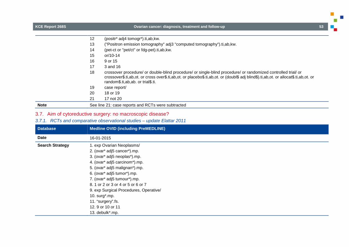
KCE Report 268S Ovarian cancer: diagnosis, treatment and follow-up 53
12 (positr* adj4 tomogr*).ti,ab,kw. 13 ("Positron emission tomography" adj3 "computed tomography").ti,ab,kw. 14 (pet-ct or "pet/ct" or fdg-pet).ti,ab,kw. 15 or/10-14 16 9 or 15 17 3 and 16 18 crossover procedure/ or double-blind procedure/ or single-blind procedure/ or randomized controlled trial/ or crossover$.ti,ab,ot. or cross over$.ti,ab,ot. or placebo$.ti,ab,ot. or (doubl$ adj blind$).ti,ab,ot. or allocat$.ti,ab,ot. or random$.ti,ab,ab. or trial$.ti. 19 case report/ 20 18 or 19 21 17 not 20
Note See line 21: case reports and RCTs were subtracted
3.7. Aim of cytoreductive surgery: no macroscopic disease? 3.7.1. RCTs and comparative observational studies – update Elattar 2011
Database Medline OVID (including PreMEDLINE)
Date 16-01-2015 Search Strategy 1. exp Ovarian Neoplasms/
2. (ovar* adj5 cancer*).mp. 3. (ovar* adj5 neoplas*).mp. 4. (ovar* adj5 carcinom*).mp. 5. (ovar* adj5 malignan*).mp. 6. (ovar* adj5 tumor*).mp. 7. (ovar* adj5 tumour*).mp. 8. 1 or 2 or 3 or 4 or 5 or 6 or 7 9. exp Surgical Procedures, Operative/ 10. surg*.mp. 11. “surgery”.fs. 12. 9 or 10 or 11 13. debulk*.mp.
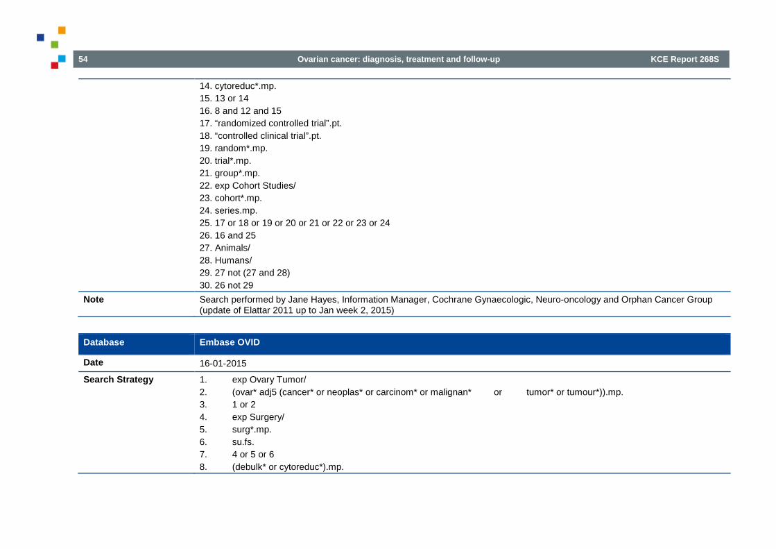
54 Ovarian cancer: diagnosis, treatment and follow-up KCE Report 268S
14. cytoreduc*.mp. 15. 13 or 14 16. 8 and 12 and 15 17. “randomized controlled trial”.pt. 18. “controlled clinical trial”.pt. 19. random*.mp. 20. trial*.mp. 21. group*.mp. 22. exp Cohort Studies/ 23. cohort*.mp. 24. series.mp. 25. 17 or 18 or 19 or 20 or 21 or 22 or 23 or 24 26. 16 and 25 27. Animals/ 28. Humans/ 29. 27 not (27 and 28) 30. 26 not 29
Note Search performed by Jane Hayes, Information Manager, Cochrane Gynaecologic, Neuro-oncology and Orphan Cancer Group (update of Elattar 2011 up to Jan week 2, 2015)
Database Embase OVID
Date 16-01-2015 Search Strategy 1. exp Ovary Tumor/
2. (ovar* adj5 (cancer* or neoplas* or carcinom* or malignan* or tumor* or tumour*)).mp. 3. 1 or 2 4. exp Surgery/ 5. surg*.mp. 6. su.fs. 7. 4 or 5 or 6 8. (debulk* or cytoreduc*).mp.

KCE Report 268S Ovarian cancer: diagnosis, treatment and follow-up 55
9. 3 and 7 and 8 10. exp Controlled Clinical Trial/ 11. crossover procedure/ 12. double-blind procedure/ 13. randomized controlled trial/ 14. single-blind procedure/ 15. random*.mp. 16. factorial*.mp. 17. (crossover* or cross over* or cross-over*).mp. 18. placebo*.mp. 19. (double* adj blind*).mp. 20. (singl* adj blind*).mp. 21. assign*.mp. 22. allocat*.mp. 23. volunteer*.mp. 24. exp cohort analysis/ 25. cohort*.mp. 26. series.mp. 27. 10 or 11 or 12 or 13 or 14 or 15 or 16 or 17 or 18 or 19 or 20 or 21 or 22 or 23 or 24 or 25 or 26 28. 9 and 27
Note Search performed by Jane Hayes, Information Manager, Cochrane Gynaecologic, Neuro-oncology and Orphan Cancer Group (update of Elattar 2011 up to 2015 Jan week 2, 2015; search amended)
Database CENTRAL
Date 16-01-2015
Search Strategy 1. MeSH descriptor Ovarian Neoplasms explode all trees 2. ovar* near/5 cancer* 3. ovar* near/5 neoplas* 4. ovar* near/5 carcinom* 5. ovar* near/5 malignan*

56 Ovarian cancer: diagnosis, treatment and follow-up KCE Report 268S
6. ovar* near/5 tumor* 7. ovar* near/5 tumour* 8. (#1 OR #2 OR #3 OR #4 OR #5 OR #6 OR #7) 9. MeSH descriptor Surgical Procedures, Operative explode all trees 10. surg* 11. Any MeSH descriptor with qualifier: SU 12. (#9 OR #10 OR #11) 13. debulk* 14. cytoreduc* 15. (#13 OR #14) 16. (#8 AND #12 AND #15)
Note Search performed by Jane Hayes, Information Manager, Cochrane Gynaecologic, Neuro-oncology and Orphan Cancer Group (update of Elattar 2011 up to Issue 1, 2015)
3.7.2. RCTs and comparative observational studies – Update Ang et al.
Database Medline OVID (including PreMEDLINE)
Date 16-01-2015 Search Strategy 1. exp Ovarian Neoplasms/
2. (ovar* adj5 (cancer* or neoplas* or carcinom* or malignan* or tumor* or tumour*)).mp. 3. 1 or 2 4. exp Surgical Procedures, Operative/ 5. surg*.mp. 6. surgery.fs. 7. 4 or 5 or 6 8. debulk*.mp. 9. cytoreduc*.mp. 10. (ultraradical or ultra-radical or ultra radical).mp. 11. exp Omentum/ 12. omentum.mp. 13. bowel.mp. 14. abdom*.mp.

KCE Report 268S Ovarian cancer: diagnosis, treatment and follow-up 57
15. exp Spleen/ 16. spleen.mp. 17. exp Liver/ 18. liver.mp. 19. exp Diaphragm/ 20. diaphragm*.mp. 21. exp Lymph Nodes/ 22. (lymph adj node*).mp. 23. exp Peritoneum/ 24. peritone*.mp. 25. exp Urinary Tract/ 26. (urinary adj tract).mp. 27. 8 or 9 or 10 or 11 or 12 or 13 or 14 or 15 or 16 or 17 or 18 or 19 or 20 or 21 or 22 or 23 or 24 or 25 or 26 28. 7 and 27 29. exp Splenectomy/ 30. splenectomy.mp. 31. exp Hysterectomy/ 32. (abdom* adj5 hysterectomy).mp. 33. abdominohysterectomy.mp. 34. exp Lymph Node Excision/ 35. (lymph adj node adj excision).mp. 36. (bilateral adj salpingo adj oophorectomy).mp. 37. omentectomy.mp. 38. exp Surgical Stomas/ 39. stoma.mp. 40. 29 or 30 or 31 or 32 or 33 or 34 or 35 or 36 or 37 or 38 or 39 41. 28 or 40 42. 3 and 41 43. “randomized controlled trial”.pt. 44. “controlled clinical trial”.pt. 45. randomized.ab. 46. randomly.ab.

58 Ovarian cancer: diagnosis, treatment and follow-up KCE Report 268S
47. trial.ab. 48. groups.ab. 49. exp Cohort Studies/ 50. cohort*.mp. 51. (case adj series).mp. 52. 43 or 44 or 45 or 46 or 47 or 48 or 49 or 50 or 51 53. 42 and 52 54. Animals/ 55. Humans/ 56. 54 not (54 and 55) 57. 53 not 56
Note Search performed by Jane Hayes, Information Manager, Cochrane Gynaecologic, Neuro-oncology and Orphan Cancer Group (update of Ang 2011 up to Jan week 2, 2015)
Database Embase OVID
Date 16-01-2015
Search Strategy 1. exp Ovary Tumor/ 2. (ovar* adj5 (cancer* or neoplas* or carcinom* or malignan* or tumor* or tumour*)).mp. 3. 1 or 2 4. exp Surgery/ 5. surg*.mp. 6. su.fs. 7. 4 or 5 or 6 8. debulk*.mp. 9. cytoreduc*.mp. 10. (ultraradical or ultra-radical or ultra radical).mp. 11. exp Omentum/ 12. omentum.mp. 13. bowel.mp.

KCE Report 268S Ovarian cancer: diagnosis, treatment and follow-up 59
14. abdom*.mp. 15. exp Spleen/ 16. spleen.mp. 17. exp Liver/ 18. liver.mp. 19. exp Diaphragm/ 20. diaphragm*.mp. 21. exp Lymph Node/ 22. (lymph adj node).mp. 23. exp Peritoneum/ 24. peritone*.mp. 25. exp Urinary Tract/ 26. (urinary adj tract).mp. 27. 8 or 9 or 10 or 11 or 12 or 13 or 14 or 15 or 16 or 17 or 18 or 19 or 20 or 21 or 22 or 23 or 24 or 25 or 26 28. 7 and 27 29. exp Splenectomy/ 30. splenectomy.mp. 31. exp Hysterectomy/ 32. (abdom* adj5 hysterectomy).mp. 33. abdominohysterectomy.mp. 34. exp Lymphadenectomy/ 35. (lymph adj node adj excision).mp. 36. (bilateral adj salpingo adj oophorectomy).mp. 37. omentectomy.mp. 38. exp Stoma/ 39. stoma.mp. 40. 29 or 30 or 31 or 32 or 33 or 34 or 35 or 36 or 37 or 38 or 39 41. 28 or 40 42. 3 and 41 43. exp Controlled Clinical Trial/ 44. randomized.ab. 45. randomly.ab.

60 Ovarian cancer: diagnosis, treatment and follow-up KCE Report 268S
46. trial.ab. 47. groups.ab. 48. exp Cohort Analysis/ 49. cohort*.mp. 50. (case adj series).mp. 51. 50 or 49 or 46 or 45 or 43 or 44 or 48 or 47 52. 42 and 51 53. exp Animal/ 54. Human/ 55. 53 not (53 and 54) 56. 52 not 55
Note Search performed by Jane Hayes, Information Manager, Cochrane Gynaecologic, Neuro-oncology and Orphan Cancer Group (update of Ang 2011 up to 2015 Jan week 2)
Database CENTRAL
Date 16-01-2015
Search Strategy 1. MeSH descriptor Ovarian Neoplasms explode all trees 2. ovar* near/5 (cancer* or neoplas* or carcinom* or malignan* or tumor* or tumour*) 3. (#1 OR #2) 4. MeSH descriptor Surgical Procedures, Operative explode all trees 5. surg* 6. Any MeSH descriptor with qualifier: SU 7. (#4 OR #5 OR #6) 8. debulk* 9. cytoreduc* 10. ultradical or ultra-radical or ultra radical 11. MeSH descriptor Omentum explode all trees 12. omentum 13. bowel

KCE Report 268S Ovarian cancer: diagnosis, treatment and follow-up 61
14. abdom* 15. MeSH descriptor Spleen explode all trees 16. spleen 17. MeSH descriptor Liver explode all trees 18. liver 19. MeSH descriptor Diaphragm explode all trees 20. diaphragm* 21. MeSH descriptor Lymph Nodes explode all trees 22. lymph next node* 23. MeSH descriptor Peritoneum explode all trees 24. peritone* 25. MeSH descriptor Urinary Tract explode all trees 26. urinary next tract 27. (#8 OR #9 OR #10 OR #11 OR #12 OR #13 OR #14 OR #15 OR #16 OR #17 OR #18 OR #19 OR #20 OR #21 OR #22 OR 23 OR #24 OR #25 OR #26) 28. (#7 AND #27) 29. MeSH descriptor Splenectomy explode all trees 30. splenectomy 31. MeSH descriptor Hysterectomy explode all trees 32. abdom* near/5 hysterectomy 33. abdominohysterectomy 34. MeSH descriptor Lymph Node Excision explode all trees 35. lymph next node next excision 36. bilateral next salpingo next oophorectomy 37. omentectomy 38. MeSH descriptor Surgical Stomas explode all trees 39. stoma 40. (#29 OR #30 OR #31 OR #32 OR #33 OR #34 OR #35 OR #36 OR #37 OR #38 OR #39) 41. (#28 OR #40) 42. (#3 AND #41)

62 Ovarian cancer: diagnosis, treatment and follow-up KCE Report 268S
Note Search performed by Jane Hayes, Information Manager, Cochrane Gynaecologic, Neuro-oncology and Orphan Cancer Group (update of Ang 2011 up to Issue 1, 2015)
3.8. Neo-adjuvant chemotherapy and interval debulking versus upfront surgery 3.8.1. Systematic reviews
Database Medline
Date 13-01-2015 Search Strategy 11 meta-analysis.mp,pt. or review.pt. or search:.tw. (2070030)
12 (ovar$ adj5 neoplas$).tw. (2788) 13 (ovar$ adj5 cancer$).tw. (38175) 14 (ovar$ adj5 carcin$).tw. (16733) 15 (ovar$ adj5 tumo$).tw. (19875) 16 (ovar$ adj5 metasta$).tw. (3787) 17 (ovar$ adj5 malig$).tw. (6363) 18 exp Ovarian Neoplasms/ (64515) 19 or/12-18 (79952) 20 exp Surgical Procedures, Operative/ (2416197) 21 surg*.mp. (1479510) 22 surgery.fs. (1594742) 23 20 or 21 or 22 (3370799) 24 debulk*.mp. (4471) 25 cytoreduc*.mp. (5478) 26 24 or 25 (9393) 27 23 and 26 (7986) 28 19 and 27 (2920) 29 exp Chemotherapy, Adjuvant/ (30217) 30 exp Antineoplastic Combined Chemotherapy Protocols/ (106512) 31 exp Antineoplastic Agents/ (828285) 32 chemother*.mp. (316565) 33 exp Drug Therapy/ (1058527) 34 exp Drug Therapy, Combination/ (260471)
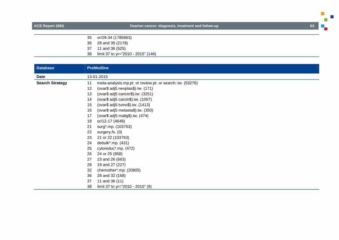
KCE Report 268S Ovarian cancer: diagnosis, treatment and follow-up 63
35 or/29-34 (1785863) 36 28 and 35 (2178) 37 11 and 36 (525) 38 limit 37 to yr="2010 - 2015" (146)
Database PreMedline
Date 13-01-2015 Search Strategy 11 meta-analysis.mp,pt. or review.pt. or search:.tw. (53276)
12 (ovar$ adj5 neoplas$).tw. (171) 13 (ovar$ adj5 cancer$).tw. (3251) 14 (ovar$ adj5 carcin$).tw. (1007) 15 (ovar$ adj5 tumo$).tw. (1413) 16 (ovar$ adj5 metasta$).tw. (350) 17 (ovar$ adj5 malig$).tw. (474) 19 or/12-17 (4648) 21 surg*.mp. (103763) 22 surgery.fs. (0) 23 21 or 22 (103763) 24 debulk*.mp. (431) 25 cytoreduc*.mp. (472) 26 24 or 25 (858) 27 23 and 26 (663) 28 19 and 27 (227) 32 chemother*.mp. (20805) 36 28 and 32 (168) 37 11 and 36 (11) 38 limit 37 to yr="2010 - 2015" (9)

64 Ovarian cancer: diagnosis, treatment and follow-up KCE Report 268S
Database Embase
Date 13-01-2015 Search Strategy #1. (ovar* NEAR/5 neoplas*):ab,ti OR (ovar* NEAR/5 cancer*):ab,ti OR (ovar* NEAR/5 carcin*):ab,ti OR (ovar* NEAR/5
tumo*):ab,ti OR (ovar* NEAR/5 metasta*):ab,ti OR (ovar* NEAR/5 malig*):ab,ti (85836) #2. 'ovary cancer'/exp (79565) #3. #1 OR #2 (109571) #4. 'antineoplastic agent'/exp OR 'cancer chemotherapy'/exp OR 'cancer combination chemotherapy'/exp OR 'combination
chemotherapy'/exp (1584539) #5. 'cytoreductive surgery'/exp (7174) #6. debulk*:ab,ti (7145) #7. #5 or #6 (12234) #8. #3 AND #4 AND #7 (2939) #9. #8 AND AND ([cochrane review]/lim OR [systematic review]/lim OR [meta analysis]/lim) AND ([article]/lim OR [article in
press]/lim OR [review]/lim) AND ([dutch]/lim OR [english]/lim OR [french]/lim) AND [2010-2015]/py (22)
Database Cochrane Library
Date 13-01-2015 Search Strategy #1 (ovar* and (neoplas* or cancer$ or tumo* or carcin* or metasta* or malig*)):ti,ab
#2 MeSH descriptor: [Ovarian Neoplasms] 1 tree(s) exploded #3 #1 or #2 #4 MeSH descriptor: [Surgical Procedures, Operative] 1 tree(s) exploded #5 surg*:ti,ab #6 #4 or #5 #7 (debulk* or cytoreduc*):ti,ab #8 #6 and #7 #9 MeSH descriptor: [Chemotherapy, Adjuvant] 1 tree(s) exploded #10 MeSH descriptor: [Antineoplastic Combined Chemotherapy Protocols] 1 tree(s) exploded #11 MeSH descriptor: [Antineoplastic Agents] 1 tree(s) exploded #12 MeSH descriptor: [Drug Therapy] 1 tree(s) exploded #13 MeSH descriptor: [Drug Therapy, Combination] 1 tree(s) exploded

KCE Report 268S Ovarian cancer: diagnosis, treatment and follow-up 65
#14 chemother*:ti,ab #15 #9 or #10 or #11 or #12 or #13 or #14 #16 #3 and #8 and #15
3.8.2. RCTs
Database Medline
Date 13-01-2015 Search Strategy 1 randomized controlled trial.pt. (380225)
2 controlled clinical trial.pt. (88313) 3 randomized.ab. (279149) 4 placebo.ab. (147367) 5 clinical trials as topic.sh. (170168) 6 randomly.ab. (198174) 7 trial.ti. (120096) 8 1 or 2 or 3 or 4 or 5 or 6 or 7 (871418) 9 exp animals/ not humans.sh. (3967499) 10 8 not 9 (799334) 12 (ovar$ adj5 neoplas$).tw. (2788) 13 (ovar$ adj5 cancer$).tw. (38175) 14 (ovar$ adj5 carcin$).tw. (16733) 15 (ovar$ adj5 tumo$).tw. (19875) 16 (ovar$ adj5 metasta$).tw. (3787) 17 (ovar$ adj5 malig$).tw. (6363) 18 exp Ovarian Neoplasms/ (64515) 19 or/12-18 (79952) 20 exp Surgical Procedures, Operative/ (2416197) 21 surg*.mp. (1479510) 22 surgery.fs. (1594742) 23 20 or 21 or 22 (3370799) 24 debulk*.mp. (4471) 25 cytoreduc*.mp. (5478)

66 Ovarian cancer: diagnosis, treatment and follow-up KCE Report 268S
26 24 or 25 (9393) 27 23 and 26 (7986) 28 19 and 27 (2920) 29 exp Chemotherapy, Adjuvant/ (30217) 30 exp Antineoplastic Combined Chemotherapy Protocols/ (106512) 31 exp Antineoplastic Agents/ (828285) 32 chemother*.mp. (316565) 33 exp Drug Therapy/ (1058527) 34 exp Drug Therapy, Combination/ (260471) 35 or/29-34 (1785863) 36 28 and 35 (2178) 39 10 and 36 (341) 41 limit 39 to yr="2011 - 2015" (61)
Comment The search was updated on September 1st 2015 and yielded 10 new hits.
Database PreMedline
Date 13-01-2015 Search Strategy 1 randomized controlled trial.pt. (940)
2 controlled clinical trial.pt. (99) 3 randomized.ab. (26074) 4 placebo.ab. (9424) 6 randomly.ab. (23362) 7 trial.ti. (10972) 8 1 or 2 or 3 or 4 or 6 or 7 (55274) 12 (ovar$ adj5 neoplas$).tw. (171) 13 (ovar$ adj5 cancer$).tw. (3251) 14 (ovar$ adj5 carcin$).tw. (1007) 15 (ovar$ adj5 tumo$).tw. (1413) 16 (ovar$ adj5 metasta$).tw. (350) 17 (ovar$ adj5 malig$).tw. (474)

KCE Report 268S Ovarian cancer: diagnosis, treatment and follow-up 67
19 or/12-17 (4648) 21 surg*.mp. (103763) 22 surgery.fs. (0) 23 21 or 22 (103763) 24 debulk*.mp. (431) 25 cytoreduc*.mp. (472) 26 24 or 25 (858) 27 23 and 26 (663) 28 19 and 27 (227) 32 chemother*.mp. (20805) 36 28 and 32 (168) 39 8 and 36 (22) 41 limit 39 to yr="2011 - 2015" (12)
Comment The search was updated on September 1st 2015 and yielded 11 new hits.
Database Embase
Date 13-01-2015 Search Strategy #1. (ovar* NEAR/5 neoplas*):ab,ti OR (ovar* NEAR/5 cancer*):ab,ti OR (ovar* NEAR/5 carcin*):ab,ti OR (ovar* NEAR/5
tumo*):ab,ti OR (ovar* NEAR/5 metasta*):ab,ti OR (ovar* NEAR/5 malig*):ab,ti (85836) #2. 'ovary cancer'/exp (79565) #3. #1 OR #2 (109571) #4. 'antineoplastic agent'/exp OR 'cancer chemotherapy'/exp OR 'cancer combination chemotherapy'/exp OR 'combination
chemotherapy'/exp (1584539) #5. 'cytoreductive surgery'/exp (7174) #6. debulk*:ab,ti (7145) #7. #5 or #6 (12234) #8. #3 AND #4 AND #7 (2939) #9. #8 AND ([controlled clinical trial]/lim OR [randomized controlled trial]/lim) AND ([article]/lim OR [article in press]/lim OR [review]/lim) AND ([dutch]/lim OR [english]/lim OR [french]/lim) AND [2011-2015]/py (31)
Comment The search was updated on September 1st 2015 and yielded 3 new hits.
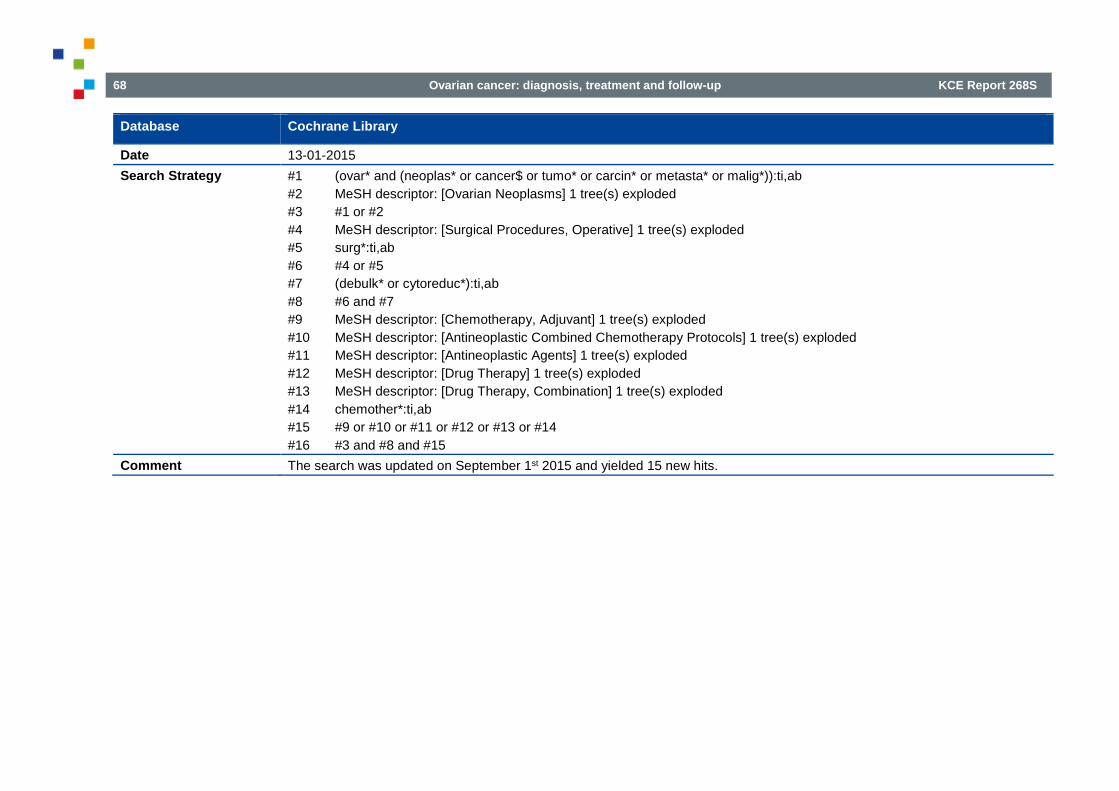
68 Ovarian cancer: diagnosis, treatment and follow-up KCE Report 268S
Database Cochrane Library
Date 13-01-2015 Search Strategy #1 (ovar* and (neoplas* or cancer$ or tumo* or carcin* or metasta* or malig*)):ti,ab
#2 MeSH descriptor: [Ovarian Neoplasms] 1 tree(s) exploded #3 #1 or #2 #4 MeSH descriptor: [Surgical Procedures, Operative] 1 tree(s) exploded #5 surg*:ti,ab #6 #4 or #5 #7 (debulk* or cytoreduc*):ti,ab #8 #6 and #7 #9 MeSH descriptor: [Chemotherapy, Adjuvant] 1 tree(s) exploded #10 MeSH descriptor: [Antineoplastic Combined Chemotherapy Protocols] 1 tree(s) exploded #11 MeSH descriptor: [Antineoplastic Agents] 1 tree(s) exploded #12 MeSH descriptor: [Drug Therapy] 1 tree(s) exploded #13 MeSH descriptor: [Drug Therapy, Combination] 1 tree(s) exploded #14 chemother*:ti,ab #15 #9 or #10 or #11 or #12 or #13 or #14 #16 #3 and #8 and #15
Comment The search was updated on September 1st 2015 and yielded 15 new hits.
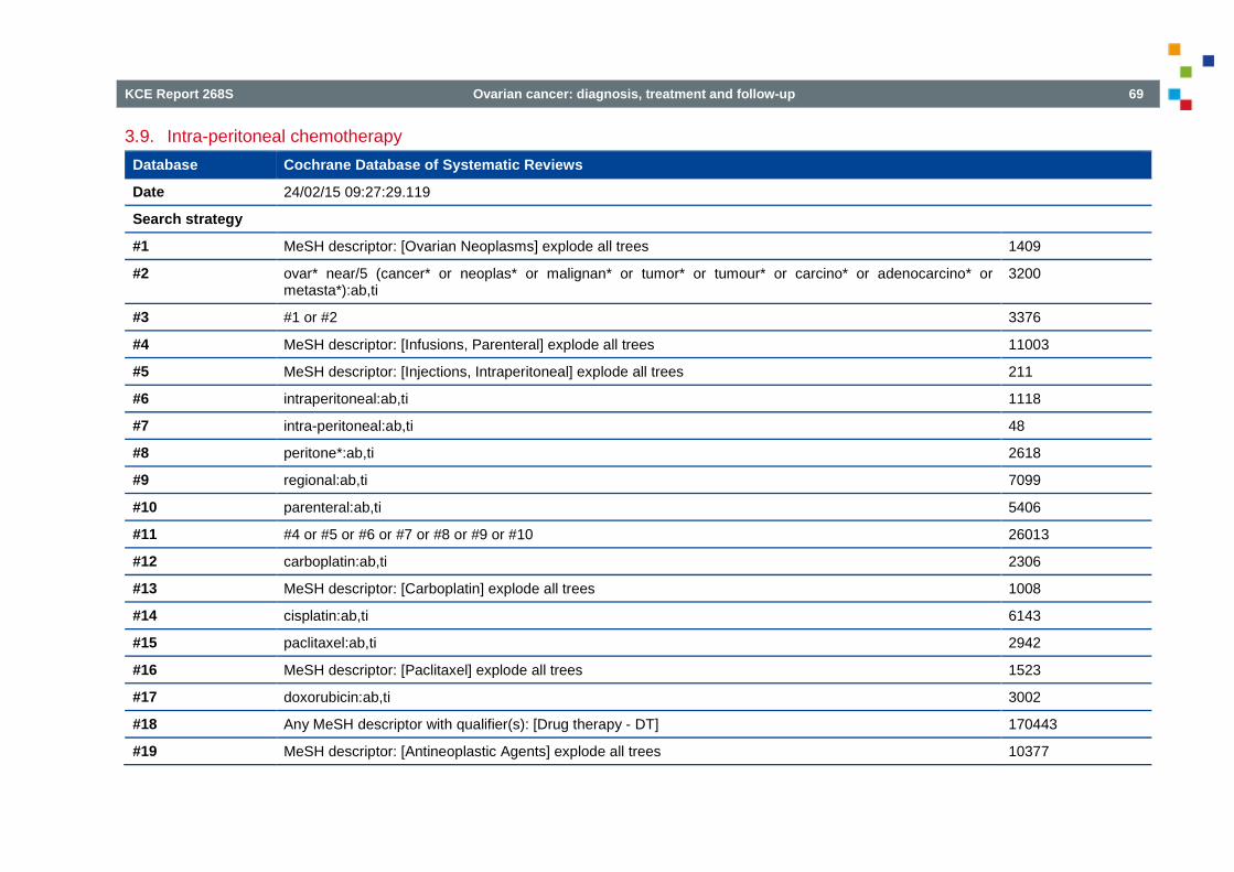
KCE Report 268S Ovarian cancer: diagnosis, treatment and follow-up 69
3.9. Intra-peritoneal chemotherapy Database Cochrane Database of Systematic Reviews
Date 24/02/15 09:27:29.119
Search strategy
#1 MeSH descriptor: [Ovarian Neoplasms] explode all trees 1409
#2 ovar* near/5 (cancer* or neoplas* or malignan* or tumor* or tumour* or carcino* or adenocarcino* or metasta*):ab,ti
3200
#3 #1 or #2 3376
#4 MeSH descriptor: [Infusions, Parenteral] explode all trees 11003
#5 MeSH descriptor: [Injections, Intraperitoneal] explode all trees 211
#6 intraperitoneal:ab,ti 1118
#7 intra-peritoneal:ab,ti 48
#8 peritone*:ab,ti 2618
#9 regional:ab,ti 7099
#10 parenteral:ab,ti 5406
#11 #4 or #5 or #6 or #7 or #8 or #9 or #10 26013
#12 carboplatin:ab,ti 2306
#13 MeSH descriptor: [Carboplatin] explode all trees 1008
#14 cisplatin:ab,ti 6143
#15 paclitaxel:ab,ti 2942
#16 MeSH descriptor: [Paclitaxel] explode all trees 1523
#17 doxorubicin:ab,ti 3002
#18 Any MeSH descriptor with qualifier(s): [Drug therapy - DT] 170443
#19 MeSH descriptor: [Antineoplastic Agents] explode all trees 10377

70 Ovarian cancer: diagnosis, treatment and follow-up KCE Report 268S
#20 MeSH descriptor: [Antineoplastic Combined Chemotherapy Protocols] explode all trees 11117
#21 MeSH descriptor: [Chemotherapy, Adjuvant] explode all trees 3379
#22 chemotherap*:ab,ti 26623
#23 #12 or #13 or #14 or #15 or #17 or #18 or #19 or #20 or #21 or #22 192066
#24 #11 and #23 8832
#25 #3 and #24 336
#26 #3 and #24 Publication Year from 2011 to 2014 90
Notes Details results among databases: SR: 10 CENTRAL: 74 HTA: 5 DARE:1
Database Medline OvidSP
Date 2015-02-23
Search strategy
1 exp Ovarian Neoplasms/ 65067
2 (ovar* adj5 (cancer* or neoplas* or malignan* or tumor* or tumour* or carcin* or adenocarcin* or metasta*)).tw. 67987
3 1 or 2 85673
4 Injections, Intraperitoneal/ 28470
5 (intraperitoneal or intra-peritoneal).tw. 60125
6 peritone*.tw. 94623
7 regional.tw. 183004
8 parenteral.tw. 42061

KCE Report 268S Ovarian cancer: diagnosis, treatment and follow-up 71
9 exp Infusions, Parenteral/ 82561
10 intraabdominal.tw. 6951
11 intra-abdominal.tw. 16189
12 4 or 5 or 6 or 7 or 8 or 9 or 10 or 11 478577
13 carboplatin*.ab,ti. 11002
14 exp carboplatin/ 9242
15 paclitaxel*.ab,ti. 19027
16 exp paclitaxel/ 19862
17 dt.fs. 1737201
18 exp Antineoplastic Agents/ 833941
19 exp chemotherapy/ 1065821
20 chemotherap*.ab,ti. 282468
21 cisplatin.ab,ti. 42948
22 doxorubicin.ab,ti. 28932
23 13 or 14 or 15 or 16 or 17 or 18 or 19 or 20 or 21 or 22 2848347
24 12 and 23 174927
25 3 and 24 4454
26 limit 25 to yr="2011 -Current" 970
27 limit 26 to systematic reviews 23
28 randomized controlled trial.pt. 384788
29 controlled clinical trial.pt. 88618
30 randomized.ti,ab. 331574
31 placebo.ti,ab. 162525

72 Ovarian cancer: diagnosis, treatment and follow-up KCE Report 268S
32 clinical trials as topic/ 170815
33 randomly.ti,ab. 224770
34 trial?.ti. 180986
35 28 or 29 or 30 or 31 or 32 or 33 or 34 957861
36 exp animal/ not humans/ 3986356
37 35 not 36 883692
38 26 and 37 124
Notes 23 systematic reviews 124 RCT Filter for systematic reviews: NLM http://www.nlm.nih.gov/bsd/pubmed_subsets/sysreviews_strategy.html Filter for RCT: Cochrane Highly Sensitive Search Strategy for identifying randomized trials in MEDLINE: sensitivity- and precision-maximizing version (2008 revision) http://handbook.cochrane.org/chapter_6/box_6.4.d_cochrane_hsss_2008_sensprec_ovid.htm

KCE Report 268S Ovarian cancer: diagnosis, treatment and follow-up 73
Database Embase
Date 2015-02-24
Search strategy
1 'ovary tumor'/exp 104815
2 (ovar* NEAR/5 (cancer* OR tumor* OR tumour* OR neoplas* OR carcin* OR adenocarcin* OR malignan* OR metasta*)):ab,ti
88010
3 #1 OR #2 121107
4 'infusion'/exp 88565
5 'intraperitoneal drug administration'/de 110454
6 intraperitoneal:ab,ti 71843
7 'intra peritoneal':ab,ti 2210
8 peritone*:ab,ti 117098
9 regional:ab,ti 238405
10 parenteral:ab,ti 55368
11 #4 OR #5 OR #6 OR #7 OR #8 OR #9 OR #10 638229
12 carboplatin*:ab,ti 17046
13 'carboplatin'/exp 46667
14 paclitaxel:ab,ti 28435
15 'paclitaxel'/exp 70605
16 'paclitaxel derivative'/exp 718
17 'paclitaxel trevatide'/exp 0
18 'paclitaxel tocosol'/exp 24
19 'paclitaxel poliglumex'/exp 306
20 'antineoplastic agent'/exp 1544121

74 Ovarian cancer: diagnosis, treatment and follow-up KCE Report 268S
21 'antineoplastic agent'/exp 1544121
22 chemotherap*:ab,ti 408112
23 cisplatin:ab,ti 57442
24 doxorubicin:ab,ti 37472
25 'drug therapy':lnk 3018373
26 'chemotherapy'/exp 424221
27 #12 OR #13 OR #14 OR #15 OR #16 OR #17 OR #18 OR #19 OR #20 OR #21 OR #22 OR #23 OR #24 OR #25 OR #26
3971314
28 #11 AND #27 152724
29 #3 AND #28 6995
30 #3 AND #28 AND [2011-2015]/py 2355
31 [medline]/lim 21664157
32 #30 NOT #31 1427
33 'meta-analysis'/exp OR 'meta-analysis' OR 'systematic review'/exp OR 'systematic review' 192672
34 #32 AND #33 20
35 random*:ab,ti OR placebo*:de,ab,ti OR (double NEXT/1 blind*):ab,ti 1167015
36 #32 AND #35 182
Notes 20 systematic reviews 182 RCT Overlap with Medline excluded. SR search filter: RCT search filter: Wong SS, Wilczynski NL, Haynes RB. Developing optimal search strategies for detecting clinically sound treatment studies in EMBASE. Journal of the Medical Library Association : JMLA. 2006 Jan;94(1):41-7

KCE Report 268S Ovarian cancer: diagnosis, treatment and follow-up 75
3.10. First-line weekly (dose dense) chemotherapy Database Ovid MEDLINE(R) In-Process & Other Non-Indexed Citations and Ovid MEDLINE(R) <1946 to Present>
Date 2015-04-16
Search strategy 1 exp ovarian neoplasms/ 65867
2 (ovar* adj5 (cancer* or neoplas* or malignan* or tumor* or tumour* or carcin* or adenocarcin* or metasta*)).ab,ti.
69007
3 1 or 2 86816
4 carboplatin*.ab,ti. 11164
5 exp carboplatin/ 9376
6 paclitaxel*.ab,ti. 19411
7 exp paclitaxel/ 20174
8 dt.fs. 1758489
9 exp Antineoplastic Agents/ 843443
10 exp chemotherapy/ 1077341
11 chemotherap*.ab,ti. 286974
12 cisplatin.ab,ti. 43688
13 doxorubicin.ab,ti. 29393
14 4 or 5 or 6 or 7 or 8 or 9 or 10 or 11 or 12 or 13 2881614
15 3 and 14 28923
16 dose-dense.ab,ti. 657
17 weekly.ab,ti. 76686
18 "dose dense".ab,ti. 657

76 Ovarian cancer: diagnosis, treatment and follow-up KCE Report 268S
19 "dose density".ab,ti. 136
20 (every adj3 week?).ab,ti. 25203
21 ((sequential or reccurent or interval or aggressive or scheduled or regimen? or cycle? or weekly) adj3 (therapy or therapies or chemotherapy or chemotherapies or treatment or dose?)).ab,ti.
109038
22 "once a week".ab,ti. 7039
23 monthly.ab,ti. 38991
24 bimonthly.ab,ti. 1118
25 (each adj3 week?).ab,ti. 12584
26 pk.fs. 243125
27 ad.fs. 1141350
28 16 or 17 or 18 or 19 or 20 or 21 or 22 or 23 or 24 or 25 or 26 or 27 1482266
29 15 and 28 10252
30 exp animal/ not humans/ 4021057
31 29 not 30 9896
32 limit 31 to systematic reviews 153
33 randomized controlled trial.pt. 391172
34 controlled clinical trial.pt. 89152
35 randomized.ti,ab. 338664
36 placebo.ti,ab. 165344
37 clinical trials as topic/ 172108
38 randomly.ti,ab. 228633
39 trial?.ti. 185032
40 33 or 34 or 35 or 36 or 37 or 38 or 39 973161
41 exp animal/ not humans/ 4021057
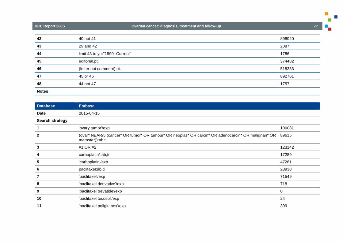
KCE Report 268S Ovarian cancer: diagnosis, treatment and follow-up 77
42 40 not 41 898020
43 29 and 42 2087
44 limit 43 to yr="1990 -Current" 1786
45 editorial.pt. 374482
46 (letter not comment).pt. 518333
47 45 or 46 892761
48 44 not 47 1757
Notes
Database Embase
Date 2015-04-15
Search strategy
1 'ovary tumor'/exp 106031
2 (ovar* NEAR/5 (cancer* OR tumor* OR tumour* OR neoplas* OR carcin* OR adenocarcin* OR malignan* OR metasta*)):ab,ti
89615
3 #1 OR #2 123142
4 carboplatin*:ab,ti 17289
5 'carboplatin'/exp 47261
6 paclitaxel:ab,ti 28938
7 'paclitaxel'/exp 71549
8 'paclitaxel derivative'/exp 718
9 'paclitaxel trevatide'/exp 0
10 'paclitaxel tocosol'/exp 24
11 'paclitaxel poliglumex'/exp 309
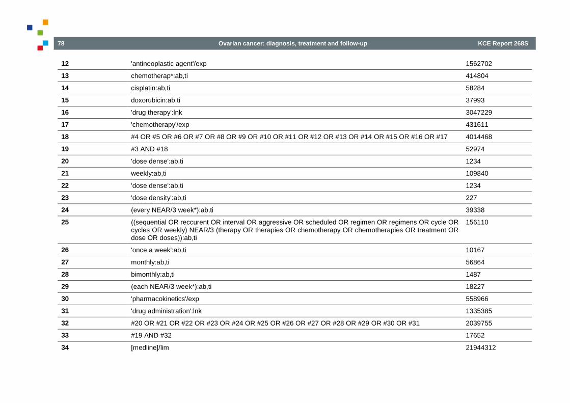
78 Ovarian cancer: diagnosis, treatment and follow-up KCE Report 268S
12 'antineoplastic agent'/exp 1562702
13 chemotherap*:ab,ti 414804
14 cisplatin:ab,ti 58284
15 doxorubicin:ab,ti 37993
16 'drug therapy':lnk 3047229
17 'chemotherapy'/exp 431611
18 #4 OR #5 OR #6 OR #7 OR #8 OR #9 OR #10 OR #11 OR #12 OR #13 OR #14 OR #15 OR #16 OR #17 4014468
19 #3 AND #18 52974
20 'dose dense':ab,ti 1234
21 weekly:ab,ti 109840
22 'dose dense':ab,ti 1234
23 'dose density':ab,ti 227
24 (every NEAR/3 week*):ab,ti 39338
25 ((sequential OR reccurent OR interval OR aggressive OR scheduled OR regimen OR regimens OR cycle OR cycles OR weekly) NEAR/3 (therapy OR therapies OR chemotherapy OR chemotherapies OR treatment OR dose OR doses)):ab,ti
156110
26 'once a week':ab,ti 10167
27 monthly:ab,ti 56864
28 bimonthly:ab,ti 1487
29 (each NEAR/3 week*):ab,ti 18227
30 'pharmacokinetics'/exp 558966
31 'drug administration':lnk 1335385
32 #20 OR #21 OR #22 OR #23 OR #24 OR #25 OR #26 OR #27 OR #28 OR #29 OR #30 OR #31 2039755
33 #19 AND #32 17652
34 [medline]/lim 21944312
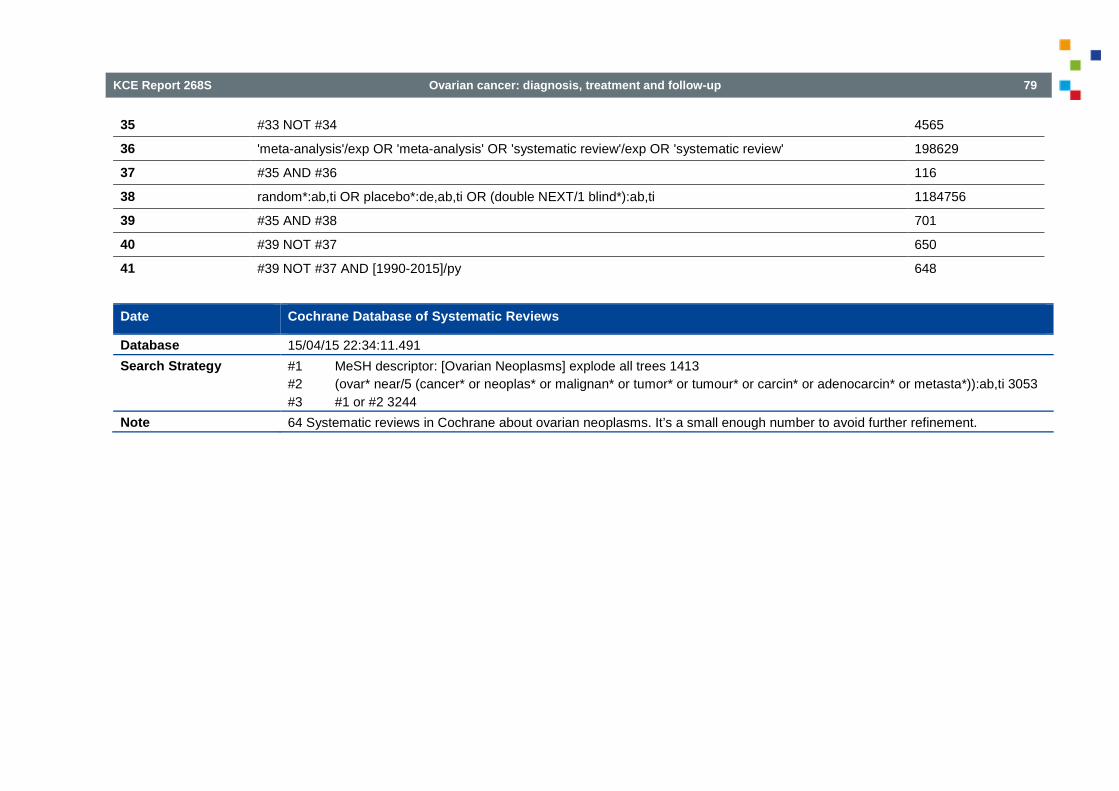
KCE Report 268S Ovarian cancer: diagnosis, treatment and follow-up 79
35 #33 NOT #34 4565
36 'meta-analysis'/exp OR 'meta-analysis' OR 'systematic review'/exp OR 'systematic review' 198629
37 #35 AND #36 116
38 random*:ab,ti OR placebo*:de,ab,ti OR (double NEXT/1 blind*):ab,ti 1184756
39 #35 AND #38 701
40 #39 NOT #37 650
41 #39 NOT #37 AND [1990-2015]/py 648
Date Cochrane Database of Systematic Reviews
Database 15/04/15 22:34:11.491 Search Strategy #1 MeSH descriptor: [Ovarian Neoplasms] explode all trees 1413
#2 (ovar* near/5 (cancer* or neoplas* or malignan* or tumor* or tumour* or carcin* or adenocarcin* or metasta*)):ab,ti 3053 #3 #1 or #2 3244
Note 64 Systematic reviews in Cochrane about ovarian neoplasms. It’s a small enough number to avoid further refinement.
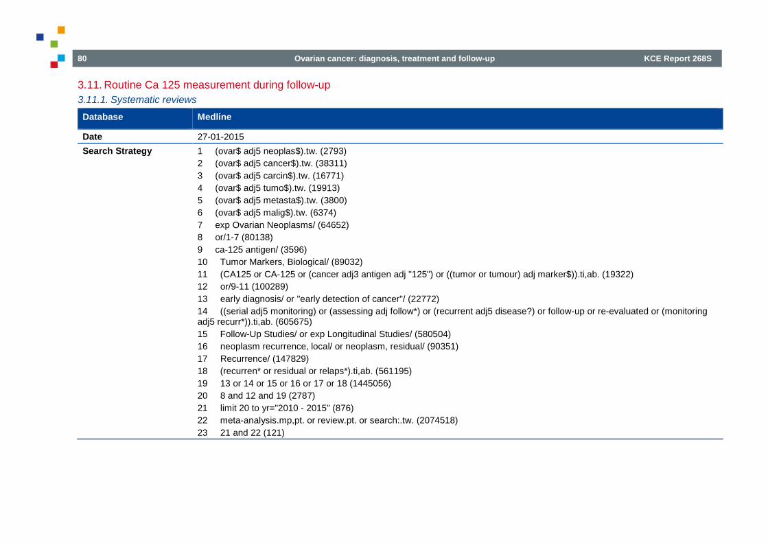
80 Ovarian cancer: diagnosis, treatment and follow-up KCE Report 268S
3.11. Routine Ca 125 measurement during follow-up 3.11.1. Systematic reviews
Database Medline
Date 27-01-2015 Search Strategy 1 (ovar$ adj5 neoplas$).tw. (2793)
2 (ovar$ adj5 cancer$).tw. (38311) 3 (ovar$ adj5 carcin$).tw. (16771) 4 (ovar$ adj5 tumo$).tw. (19913) 5 (ovar$ adj5 metasta$).tw. (3800) 6 (ovar$ adj5 malig$).tw. (6374) 7 exp Ovarian Neoplasms/ (64652) 8 or/1-7 (80138) 9 ca-125 antigen/ (3596) 10 Tumor Markers, Biological/ (89032) 11 (CA125 or CA-125 or (cancer adj3 antigen adj "125") or ((tumor or tumour) adj marker$)).ti,ab. (19322) 12 or/9-11 (100289) 13 early diagnosis/ or "early detection of cancer"/ (22772) 14 ((serial adj5 monitoring) or (assessing adj follow*) or (recurrent adj5 disease?) or follow-up or re-evaluated or (monitoring adj5 recurr*)).ti,ab. (605675) 15 Follow-Up Studies/ or exp Longitudinal Studies/ (580504) 16 neoplasm recurrence, local/ or neoplasm, residual/ (90351) 17 Recurrence/ (147829) 18 (recurren* or residual or relaps*).ti,ab. (561195) 19 13 or 14 or 15 or 16 or 17 or 18 (1445056) 20 8 and 12 and 19 (2787) 21 limit 20 to yr="2010 - 2015" (876) 22 meta-analysis.mp,pt. or review.pt. or search:.tw. (2074518) 23 21 and 22 (121)

KCE Report 268S Ovarian cancer: diagnosis, treatment and follow-up 81
Database PreMedline
Date 27-01-2015 Search Strategy 1 (ovar$ adj5 neoplas$).tw. (176)
2 (ovar$ adj5 cancer$).tw. (3227) 3 (ovar$ adj5 carcin$).tw. (996) 4 (ovar$ adj5 tumo$).tw. (1416) 5 (ovar$ adj5 metasta$).tw. (342) 6 (ovar$ adj5 malig$).tw. (479) 8 or/1-6 (4621) 11 (CA125 or CA-125 or (cancer adj3 antigen adj "125") or ((tumor or tumour) adj marker$)).ti,ab. (1396) 14 ((serial adj5 monitoring) or (assessing adj follow*) or (recurrent adj5 disease?) or follow-up or re-evaluated or (monitoring adj5 recurr*)).ti,ab. (49622) 18 (recurren* or residual or relaps*).ti,ab. (47248) 19 14 or 18 (86576) 20 8 and 11 and 19 (113) 21 limit 20 to yr="2010 - 2015" (87) 22 meta-analysis.mp,pt. or review.pt. or search:.tw. (53038) 23 21 and 22 (5)
Database Embase
Date 27-01-2015 Search Strategy #1. (ovar* NEAR/5 neoplas*):ab,ti OR (ovar* NEAR/5 cancer*):ab,ti OR (ovar* NEAR/5 carcin*):ab,ti OR (ovar* NEAR/5
tumo*):ab,ti OR (ovar* NEAR/5 metasta*):ab,ti OR (ovar* NEAR/5 malig*):ab,ti OR 'ovary cancer'/exp (109792) #2. 'ca 125 antigen'/exp OR 'tumor antigen'/de (38399) #3. ca125:ab,ti OR 'ca 125':ab,ti OR ((cancer NEAR/3 antigen):ab,ti AND (antigen NEAR/1 '125'):ab,ti) OR (tum??r NEAR/2
marker?):ab,ti (11819) #4. (serial NEAR/5 monitoring):ab,ti OR (assessing NEAR/1 follow*):ab,ti OR (recurrent NEAR/5 disease?):ab,ti OR 'follow
up':ab,ti OR 're evaluated':ab,ti OR (monitoring NEAR/5 recurr*):ab,ti OR 'minimal residual disease'/exp OR 'cancer recurrence'/exp OR 'recurrent disease'/exp OR 'tumor recurrence'/exp OR recurren*:ab,ti OR residual:ab,ti OR relaps*:ab,ti OR 'follow up'/exp OR 'longitudinal study'/exp (1915994)
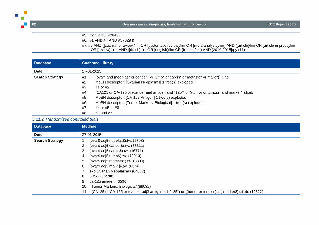
82 Ovarian cancer: diagnosis, treatment and follow-up KCE Report 268S
#5. #2 OR #3 (42843) #6. #1 AND #4 AND #5 (3294) #7. #6 AND ([cochrane review]/lim OR [systematic review]/lim OR [meta analysis]/lim) AND ([article]/lim OR [article in press]/lim
OR [review]/lim) AND ([dutch]/lim OR [english]/lim OR [french]/lim) AND [2010-2015]/py (11)
Database Cochrane Library
Date 27-01-2015 Search Strategy #1 (ovar* and (neoplas* or cancer$ or tumo* or carcin* or metasta* or malig*)):ti,ab
#2 MeSH descriptor: [Ovarian Neoplasms] 1 tree(s) exploded #3 #1 or #2 #4 (CA125 or CA-125 or (cancer and antigen and "125") or ((tumor or tumour) and marker*)):ti,ab #5 MeSH descriptor: [CA-125 Antigen] 1 tree(s) exploded #6 MeSH descriptor: [Tumor Markers, Biological] 1 tree(s) exploded #7 #4 or #5 or #6 #8 #3 and #7
3.11.2. Randomized controlled trials
Database Medline
Date 27-01-2015 Search Strategy 1 (ovar$ adj5 neoplas$).tw. (2793)
2 (ovar$ adj5 cancer$).tw. (38311) 3 (ovar$ adj5 carcin$).tw. (16771) 4 (ovar$ adj5 tumo$).tw. (19913) 5 (ovar$ adj5 metasta$).tw. (3800) 6 (ovar$ adj5 malig$).tw. (6374) 7 exp Ovarian Neoplasms/ (64652) 8 or/1-7 (80138) 9 ca-125 antigen/ (3596) 10 Tumor Markers, Biological/ (89032) 11 (CA125 or CA-125 or (cancer adj3 antigen adj "125") or ((tumor or tumour) adj marker$)).ti,ab. (19322)
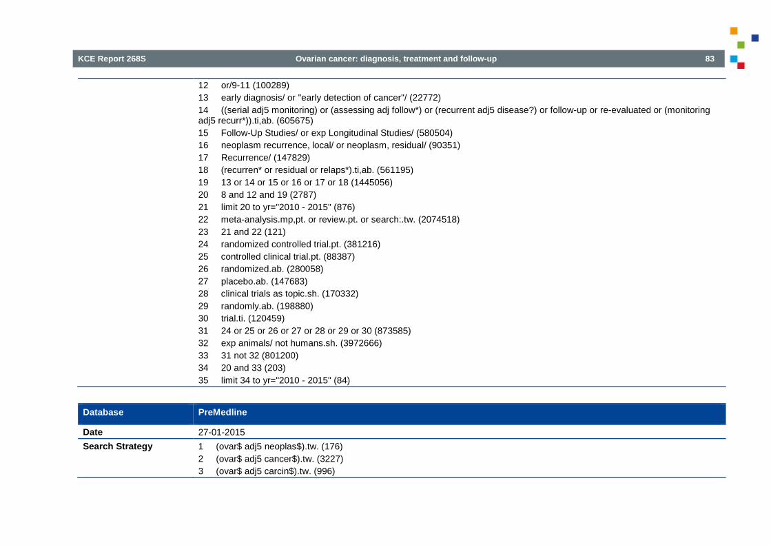
KCE Report 268S Ovarian cancer: diagnosis, treatment and follow-up 83
12 or/9-11 (100289) 13 early diagnosis/ or "early detection of cancer"/ (22772) 14 ((serial adj5 monitoring) or (assessing adj follow*) or (recurrent adj5 disease?) or follow-up or re-evaluated or (monitoring adj5 recurr*)).ti,ab. (605675) 15 Follow-Up Studies/ or exp Longitudinal Studies/ (580504) 16 neoplasm recurrence, local/ or neoplasm, residual/ (90351) 17 Recurrence/ (147829) 18 (recurren* or residual or relaps*).ti,ab. (561195) 19 13 or 14 or 15 or 16 or 17 or 18 (1445056) 20 8 and 12 and 19 (2787) 21 limit 20 to yr="2010 - 2015" (876) 22 meta-analysis.mp,pt. or review.pt. or search:.tw. (2074518) 23 21 and 22 (121) 24 randomized controlled trial.pt. (381216) 25 controlled clinical trial.pt. (88387) 26 randomized.ab. (280058) 27 placebo.ab. (147683) 28 clinical trials as topic.sh. (170332) 29 randomly.ab. (198880) 30 trial.ti. (120459) 31 24 or 25 or 26 or 27 or 28 or 29 or 30 (873585) 32 exp animals/ not humans.sh. (3972666) 33 31 not 32 (801200) 34 20 and 33 (203) 35 limit 34 to yr="2010 - 2015" (84)
Database PreMedline
Date 27-01-2015 Search Strategy 1 (ovar$ adj5 neoplas$).tw. (176)
2 (ovar$ adj5 cancer$).tw. (3227) 3 (ovar$ adj5 carcin$).tw. (996)

84 Ovarian cancer: diagnosis, treatment and follow-up KCE Report 268S
4 (ovar$ adj5 tumo$).tw. (1416) 5 (ovar$ adj5 metasta$).tw. (342) 6 (ovar$ adj5 malig$).tw. (479) 8 or/1-6 (4621) 11 (CA125 or CA-125 or (cancer adj3 antigen adj "125") or ((tumor or tumour) adj marker$)).ti,ab. (1396) 14 ((serial adj5 monitoring) or (assessing adj follow*) or (recurrent adj5 disease?) or follow-up or re-evaluated or (monitoring adj5 recurr*)).ti,ab. (49622) 18 (recurren* or residual or relaps*).ti,ab. (47248) 19 14 or 18 (86576) 20 8 and 11 and 19 (113) 21 limit 20 to yr="2010 - 2015" (87) 24 randomized controlled trial.pt. (692) 25 controlled clinical trial.pt. (81) 26 randomized.ab. (25987) 27 placebo.ab. (9342) 29 randomly.ab. (23154) 30 trial.ti. (10928) 31 24 or 25 or 26 or 27 or 29 or 30 (54893) 34 20 and 31 (8) 35 limit 34 to yr="2010 - 2015" (5)
Database Embase
Date 27-01-2015 Search Strategy #1. (ovar* NEAR/5 neoplas*):ab,ti OR (ovar* NEAR/5 cancer*):ab,ti OR (ovar* NEAR/5 carcin*):ab,ti OR (ovar* NEAR/5
tumo*):ab,ti OR (ovar* NEAR/5 metasta*):ab,ti OR (ovar* NEAR/5 malig*):ab,ti OR 'ovary cancer'/exp (109792) #2. 'ca 125 antigen'/exp OR 'tumor antigen'/de (38399) #3. ca125:ab,ti OR 'ca 125':ab,ti OR ((cancer NEAR/3 antigen):ab,ti AND (antigen NEAR/1 '125'):ab,ti) OR (tum??r NEAR/2
marker?):ab,ti (11819)

KCE Report 268S Ovarian cancer: diagnosis, treatment and follow-up 85
#4. (serial NEAR/5 monitoring):ab,ti OR (assessing NEAR/1 follow*):ab,ti OR (recurrent NEAR/5 disease?):ab,ti OR 'follow up':ab,ti OR 're evaluated':ab,ti OR (monitoring NEAR/5 recurr*):ab,ti OR 'minimal residual disease'/exp OR 'cancer recurrence'/exp OR 'recurrent disease'/exp OR 'tumor recurrence'/exp OR recurren*:ab,ti OR residual:ab,ti OR relaps*:ab,ti OR 'follow up'/exp OR 'longitudinal study'/exp (1915994)
#5. #2 OR #3 (42843) #6. #1 AND #4 AND #5 (3294) #7. #6 AND [randomized controlled trial]/lim AND ([article]/lim OR [article in press]/lim OR [review]/lim) AND ([dutch]/lim OR [english]/lim OR [french]/lim) AND [2013-2015]/py (15)
Database Cochrane Library
Date 27-01-2015 Search Strategy #1 (ovar* and (neoplas* or cancer$ or tumo* or carcin* or metasta* or malig*)):ti,ab
#2 MeSH descriptor: [Ovarian Neoplasms] 1 tree(s) exploded #3 #1 or #2 #4 (CA125 or CA-125 or (cancer and antigen and "125") or ((tumor or tumour) and marker*)):ti,ab #5 MeSH descriptor: [CA-125 Antigen] 1 tree(s) exploded #6 MeSH descriptor: [Tumor Markers, Biological] 1 tree(s) exploded #7 #4 or #5 or #6 #8 #3 and #7

86 Ovarian cancer: diagnosis, treatment and follow-up KCE Report 268S
3.12. Patient preferences Database Ovid MEDLINE(R) In-Process & Other Non-Indexed Citations and Ovid MEDLINE(R) 1946 to Present
Date 2014-01-07
Search Strategy
# Query Results
1 exp ovarian neoplasms/ 67267
2 (ovar* adj3 (cancer? or neoplasm? or tumo?r* or carcinoma? or adenocarcinoma?)).ab,ti.
64301
3 1 or 2 84809
4 Patient Preference/ 3618
5 Patient Satisfaction/ 63207
6 limit 5 to yr="1997 - 2009" 37917
7 preference*.mp. 113390
8 choice*.mp. 267461
9 preferred.mp. 91792
10 chosen.mp. 84902
11 prefer.mp. 14996
12 prefers.mp. 2918
13 choose.mp. 28177
14 chooses.mp. 1254
15 decided.mp. 20266
16 decide.mp. 16016
17 decides.mp. 1555
18 desire*.mp. 73532
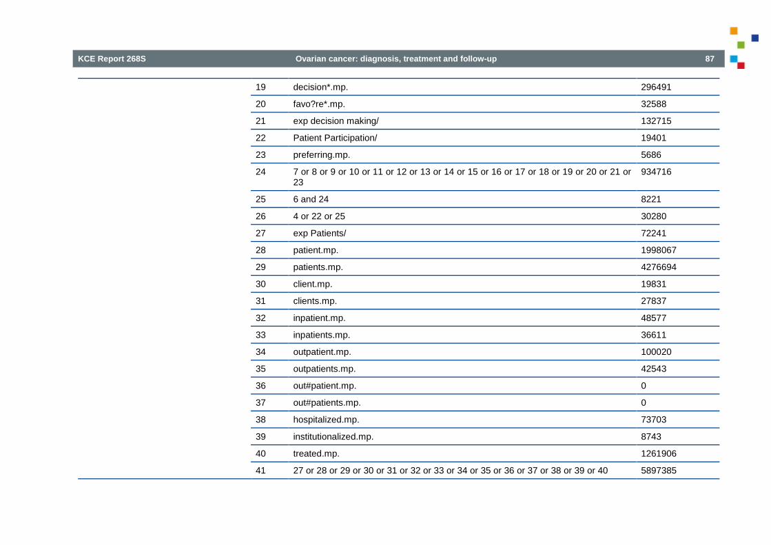
KCE Report 268S Ovarian cancer: diagnosis, treatment and follow-up 87
19 decision*.mp. 296491
20 favo?re*.mp. 32588
21 exp decision making/ 132715
22 Patient Participation/ 19401
23 preferring.mp. 5686
24 7 or 8 or 9 or 10 or 11 or 12 or 13 or 14 or 15 or 16 or 17 or 18 or 19 or 20 or 21 or 23
934716
25 6 and 24 8221
26 4 or 22 or 25 30280
27 exp Patients/ 72241
28 patient.mp. 1998067
29 patients.mp. 4276694
30 client.mp. 19831
31 clients.mp. 27837
32 inpatient.mp. 48577
33 inpatients.mp. 36611
34 outpatient.mp. 100020
35 outpatients.mp. 42543
36 out#patient.mp. 0
37 out#patients.mp. 0
38 hospitalized.mp. 73703
39 institutionalized.mp. 8743
40 treated.mp. 1261906
41 27 or 28 or 29 or 30 or 31 or 32 or 33 or 34 or 35 or 36 or 37 or 38 or 39 or 40 5897385
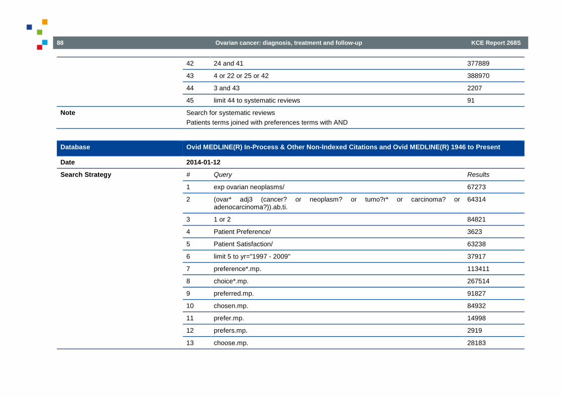
88 Ovarian cancer: diagnosis, treatment and follow-up KCE Report 268S
42 24 and 41 377889
43 4 or 22 or 25 or 42 388970
44 3 and 43 2207
45 limit 44 to systematic reviews 91
Note Search for systematic reviews Patients terms joined with preferences terms with AND
Database Ovid MEDLINE(R) In-Process & Other Non-Indexed Citations and Ovid MEDLINE(R) 1946 to Present
Date 2014-01-12
Search Strategy
# Query Results
1 exp ovarian neoplasms/ 67273
2 (ovar* adj3 (cancer? or neoplasm? or tumo?r* or carcinoma? or adenocarcinoma?)).ab,ti.
64314
3 1 or 2 84821
4 Patient Preference/ 3623
5 Patient Satisfaction/ 63238
6 limit 5 to yr="1997 - 2009" 37917
7 preference*.mp. 113411
8 choice*.mp. 267514
9 preferred.mp. 91827
10 chosen.mp. 84932
11 prefer.mp. 14998
12 prefers.mp. 2919
13 choose.mp. 28183

KCE Report 268S Ovarian cancer: diagnosis, treatment and follow-up 89
14 chooses.mp. 1253
15 decided.mp. 20273
16 decide.mp. 16021
17 decides.mp. 1556
18 desire*.mp. 73566
19 decision*.mp. 296621
20 favo?re*.mp. 32609
21 exp decision making/ 132706
22 Patient Participation/ 19395
23 preferring.mp. 5681
24 7 or 8 or 9 or 10 or 11 or 12 or 13 or 14 or 15 or 16 or 17 or 18 or 19 or 20 or 21 or 23
935040
25 6 and 24 8221
26 patient.mp. 1998898
27 patients.mp. 4278177
28 client.mp. 19836
29 clients.mp. 27845
30 inpatient.mp. 48601
31 inpatients.mp. 36623
32 outpatient.mp. 100063
33 outpatients.mp. 42561
34 out#patient.mp. 0
35 out#patients.mp. 0
36 hospitalized.mp. 73726

90 Ovarian cancer: diagnosis, treatment and follow-up KCE Report 268S
37 institutionalized.mp. 8740
38 treated.mp. 1262260
39 ((preference* or choice* or preferred or chosen or prefer or prefers or choose or chooses or decided or decide or decides or desire* or decision* or favo?re* or preferring) adj3 (patient or patients or client or clients or inpatient or inpatients or outpatient or outpatients or out#patient or out#patients or hospitalized or institutionalized or treated)).mp.
49103
40 4 or 22 or 25 or 39 70668
41 3 and 40 328
Note Search for all studies Patients terms joined with preferences terms with proximity operator
Database Embase (Embase.com)
Date 2015-01-07
Search Strategy
# Query Results
#1 'ovary tumor'/exp 103,433
#2 (ovar* NEAR/3 (cancer* OR neoplasm* OR tumor* OR tumour* OR carcinoma* OR adenocarcinoma*)):ab,ti
78,998
#3 #1 OR #2 115,133
#4 'patient participation'/de OR 'patient preference'/de OR (choice* OR chosen OR prefer* OR choos* OR decid* OR desire* OR decision* OR favore* OR favoure* OR 'decision making'/exp AND ('patient'/exp OR patient OR patients OR client OR clients OR inpatient OR inpatients OR outpatient OR outpatients OR out?patient OR out?patients OR hospitalized OR institutionalized OR treated))
608,786
#5 #3 AND #4 4,367
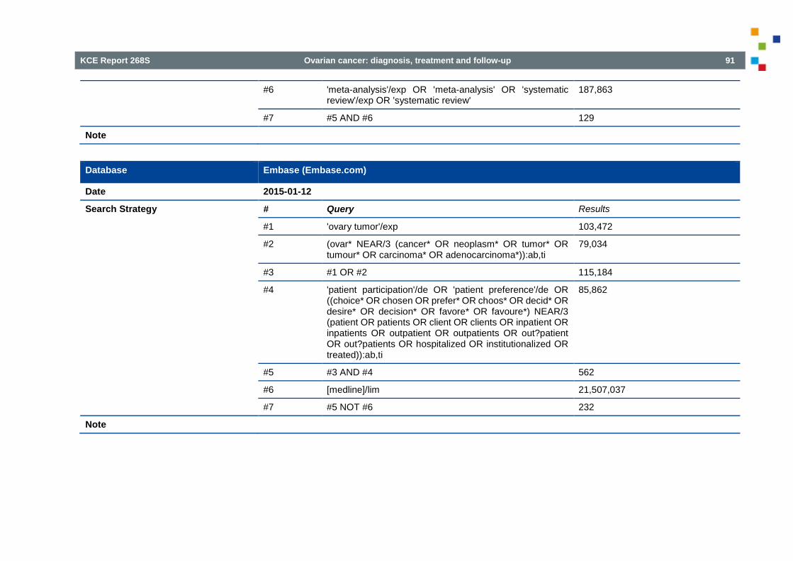
KCE Report 268S Ovarian cancer: diagnosis, treatment and follow-up 91
#6 'meta-analysis'/exp OR 'meta-analysis' OR 'systematic review'/exp OR 'systematic review'
187,863
#7 #5 AND #6 129
Note
Database Embase (Embase.com)
Date 2015-01-12
Search Strategy
# Query Results
#1 'ovary tumor'/exp 103,472
#2 (ovar* NEAR/3 (cancer* OR neoplasm* OR tumor* OR tumour* OR carcinoma* OR adenocarcinoma*)):ab,ti
79,034
#3 #1 OR #2 115,184
#4 'patient participation'/de OR 'patient preference'/de OR ((choice* OR chosen OR prefer* OR choos* OR decid* OR desire* OR decision* OR favore* OR favoure*) NEAR/3 (patient OR patients OR client OR clients OR inpatient OR inpatients OR outpatient OR outpatients OR out?patient OR out?patients OR hospitalized OR institutionalized OR treated)):ab,ti
85,862
#5 #3 AND #4 562
#6 [medline]/lim 21,507,037
#7 #5 NOT #6 232
Note
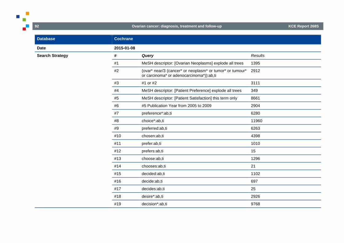
92 Ovarian cancer: diagnosis, treatment and follow-up KCE Report 268S
Database Cochrane
Date 2015-01-08
Search Strategy
# Query Results
#1 MeSH descriptor: [Ovarian Neoplasms] explode all trees 1395
#2 (ovar* near/3 (cancer* or neoplasm* or tumor* or tumour* or carcinoma* or adenocarcinoma*)):ab,ti
2912
#3 #1 or #2 3111
#4 MeSH descriptor: [Patient Preference] explode all trees 349
#5 MeSH descriptor: [Patient Satisfaction] this term only 8661
#6 #5 Publication Year from 2005 to 2009 2904
#7 preference*:ab,ti 6280
#8 choice*:ab,ti 11960
#9 preferred:ab,ti 6263
#10 chosen:ab,ti 4398
#11 prefer:ab,ti 1010
#12 prefers:ab,ti 15
#13 choose:ab,ti 1296
#14 chooses:ab,ti 21
#15 decided:ab,ti 1102
#16 decide:ab,ti 697
#17 decides:ab,ti 25
#18 desire*:ab,ti 2926
#19 decision*:ab,ti 9768
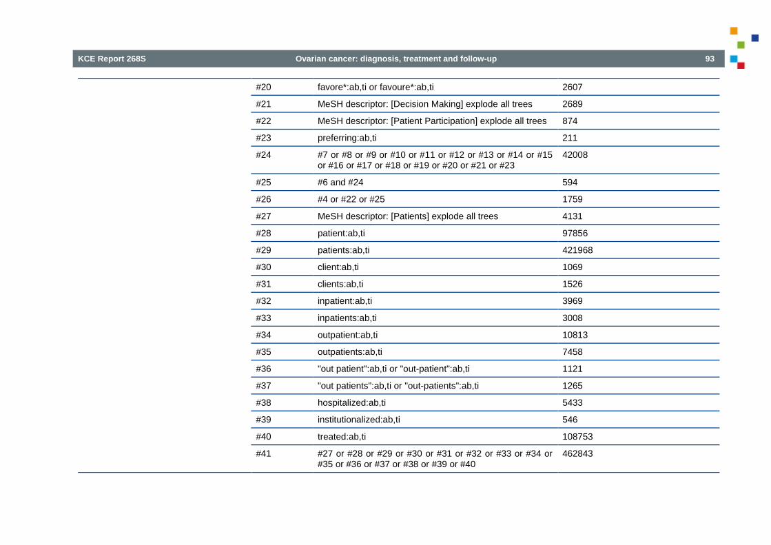
KCE Report 268S Ovarian cancer: diagnosis, treatment and follow-up 93
#20 favore*:ab,ti or favoure*:ab,ti 2607
#21 MeSH descriptor: [Decision Making] explode all trees 2689
#22 MeSH descriptor: [Patient Participation] explode all trees 874
#23 preferring:ab,ti 211
#24 #7 or #8 or #9 or #10 or #11 or #12 or #13 or #14 or #15 or #16 or #17 or #18 or #19 or #20 or #21 or #23
42008
#25 #6 and #24 594
#26 #4 or #22 or #25 1759
#27 MeSH descriptor: [Patients] explode all trees 4131
#28 patient:ab,ti 97856
#29 patients:ab,ti 421968
#30 client:ab,ti 1069
#31 clients:ab,ti 1526
#32 inpatient:ab,ti 3969
#33 inpatients:ab,ti 3008
#34 outpatient:ab,ti 10813
#35 outpatients:ab,ti 7458
#36 "out patient":ab,ti or "out-patient":ab,ti 1121
#37 "out patients":ab,ti or "out-patients":ab,ti 1265
#38 hospitalized:ab,ti 5433
#39 institutionalized:ab,ti 546
#40 treated:ab,ti 108753
#41 #27 or #28 or #29 or #30 or #31 or #32 or #33 or #34 or #35 or #36 or #37 or #38 or #39 or #40
462843

94 Ovarian cancer: diagnosis, treatment and follow-up KCE Report 268S
#42 #24 and #41 27451
#43 #4 or #22 or #25 or #42 28240
#44 #3 and #43 154
Note Details: CDSR: 11 Central: 137 Methods: 5 Economic evaluations: 1
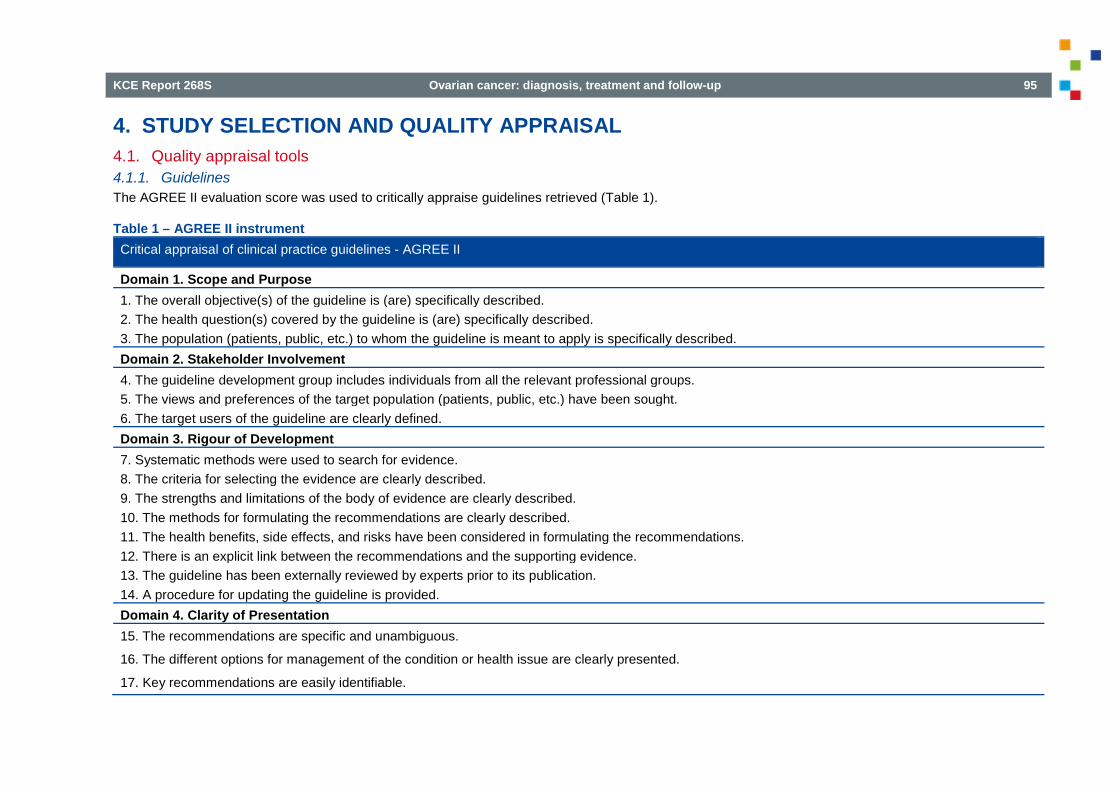
KCE Report 268S Ovarian cancer: diagnosis, treatment and follow-up 95
4. STUDY SELECTION AND QUALITY APPRAISAL 4.1. Quality appraisal tools 4.1.1. Guidelines The AGREE II evaluation score was used to critically appraise guidelines retrieved (Table 1).
Table 1 – AGREE II instrument Critical appraisal of clinical practice guidelines - AGREE II
Domain 1. Scope and Purpose 1. The overall objective(s) of the guideline is (are) specifically described. 2. The health question(s) covered by the guideline is (are) specifically described. 3. The population (patients, public, etc.) to whom the guideline is meant to apply is specifically described. Domain 2. Stakeholder Involvement 4. The guideline development group includes individuals from all the relevant professional groups. 5. The views and preferences of the target population (patients, public, etc.) have been sought. 6. The target users of the guideline are clearly defined. Domain 3. Rigour of Development 7. Systematic methods were used to search for evidence. 8. The criteria for selecting the evidence are clearly described. 9. The strengths and limitations of the body of evidence are clearly described. 10. The methods for formulating the recommendations are clearly described. 11. The health benefits, side effects, and risks have been considered in formulating the recommendations. 12. There is an explicit link between the recommendations and the supporting evidence. 13. The guideline has been externally reviewed by experts prior to its publication. 14. A procedure for updating the guideline is provided. Domain 4. Clarity of Presentation 15. The recommendations are specific and unambiguous.
16. The different options for management of the condition or health issue are clearly presented.
17. Key recommendations are easily identifiable.

96 Ovarian cancer: diagnosis, treatment and follow-up KCE Report 268S
Critical appraisal of clinical practice guidelines - AGREE II
Domain 5. Applicability 18. The guideline describes facilitators and barriers to its application. 19. The guideline provides advice and/or tools on how the recommendations can be put into practice. 20. The potential resource implications of applying the recommendations have been considered. 21. The guideline presents monitoring and/ or auditing criteria. Domain 6. Editorial Independence 22. The views of the funding body have not influenced the content of the guideline. 23. Competing interests of guideline development group members have been recorded and addressed.
4.1.2. Systematic reviews The AMSTAR checklist was used to assess systematic reviews (Table 2). Table 2 – AMSTAR checklist Question Answer
1. Was an ‘a priori’ design provided? The research question and inclusion criteria should be established before the conduct of the review.
Ye s No Ca n’t a ns we r Not a pplica ble
2. Was there duplicate study selection and data extraction? There should be at least two independent data extractors and a consensus procedure for disagreements should be in place.
Ye s No Ca n’t a ns we r Not a pplica ble
3. Was a comprehensive literature search performed? At least two electronic sources should be searched. The report must include years and databases used (e.g. Central, EMBASE, and MEDLINE). Key words and/or MESH terms must be stated and where feasible the search strategy should be provided. All searches should be supplemented by consulting current contents, reviews, textbooks, specialized registers, or experts in the particular field of study, and by reviewing the references in the studies found.
Ye s No Ca n’t a ns we r Not a pplica ble
4. Was the status of publication (i.e. grey literature) used as an inclusion criterion? Ye s No
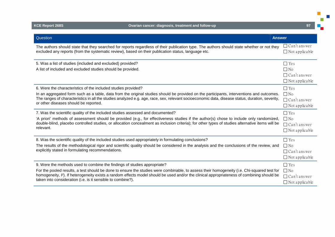
KCE Report 268S Ovarian cancer: diagnosis, treatment and follow-up 97
Question Answer
The authors should state that they searched for reports regardless of their publication type. The authors should state whether or not they excluded any reports (from the systematic review), based on their publication status, language etc.
Ca n’t a ns we r Not a pplica ble
5. Was a list of studies (included and excluded) provided? A list of included and excluded studies should be provided.
Ye s No Ca n’t a ns we r Not a pplica ble
6. Were the characteristics of the included studies provided? In an aggregated form such as a table, data from the original studies should be provided on the participants, interventions and outcomes. The ranges of characteristics in all the studies analyzed e.g. age, race, sex, relevant socioeconomic data, disease status, duration, severity, or other diseases should be reported.
Ye s No Ca n’t a ns we r Not a pplica ble
7. Was the scientific quality of the included studies assessed and documented? ‘A priori’ methods of assessment should be provided (e.g., for effectiveness studies if the author(s) chose to include only randomized, double-blind, placebo controlled studies, or allocation concealment as inclusion criteria); for other types of studies alternative items will be relevant.
Ye s No Ca n’t a ns we r Not a pplica ble
8. Was the scientific quality of the included studies used appropriately in formulating conclusions? The results of the methodological rigor and scientific quality should be considered in the analysis and the conclusions of the review, and explicitly stated in formulating recommendations.
Ye s No Ca n’t a ns we r Not a pplica ble
9. Were the methods used to combine the findings of studies appropriate? For the pooled results, a test should be done to ensure the studies were combinable, to assess their homogeneity (i.e. Chi-squared test for homogeneity, I²). If heterogeneity exists a random effects model should be used and/or the clinical appropriateness of combining should be taken into consideration (i.e. is it sensible to combine?).
Ye s No Ca n’t a ns we r Not a pplica ble

98 Ovarian cancer: diagnosis, treatment and follow-up KCE Report 268S
Question Answer
10. Was the likelihood of publication bias assessed? An assessment of publication bias should include a combination of graphical aids (e.g., funnel plot, other available tests) and/or statistical tests (e.g., Egger regression test).
Ye s No Ca n’t a ns we r Not a pplica ble
11. Was the conflict of interest stated? Potential sources of support should be clearly acknowledged in both the systematic review and the included studies.
Ye s No Ca n’t a ns we r Not applicable
4.1.3. Diagnostic accuracy studies For the quality assessment of diagnostic accuracy studies, we used the QUADAS Tool (Table 3). Table 3 – The QUADAS tool Item Label Yes No Unclear Not
applicable
1. Was the spectrum of patients representative of the patients who will receive the test in practice?
2. Were selection criteria clearly described?
3. Is the reference standard likely to correctly classify the target condition?
4. Is the time period between reference standard and index test short enough to be reasonably sure that the target condition did not change between the two tests?
5. Did the whole sample or a random selection of the sample, receive verification using a reference standard of diagnosis?
6. Did patients receive the same reference standard regardless of the index test result?
7. Was the reference standard independent of the index test (i.e. the index test did not form part of the reference standard)?
8. Was the execution of the index test described in sufficient detail to permit replication of the test?
9. Was the execution of the reference standard described in sufficient detail to permit its replication?
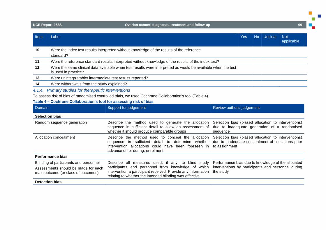
KCE Report 268S Ovarian cancer: diagnosis, treatment and follow-up 99
Item Label Yes No Unclear Not applicable
10. Were the index test results interpreted without knowledge of the results of the reference standard?
11. Were the reference standard results interpreted without knowledge of the results of the index test? 12. Were the same clinical data available when test results were interpreted as would be available when the test
is used in practice?
13. Were uninterpretable/ intermediate test results reported? 14. Were withdrawals from the study explained?
4.1.4. Primary studies for therapeutic interventions To assess risk of bias of randomised controlled trials, we used Cochrane Collaboration’s tool (Table 4). Table 4 – Cochrane Collaboration’s tool for assessing risk of bias Domain Support for judgement Review authors’ judgement
Selection bias Random sequence generation Describe the method used to generate the allocation
sequence in sufficient detail to allow an assessment of whether it should produce comparable groups
Selection bias (biased allocation to interventions) due to inadequate generation of a randomised sequence
Allocation concealment Describe the method used to conceal the allocation sequence in sufficient detail to determine whether intervention allocations could have been foreseen in advance of, or during, enrolment
Selection bias (biased allocation to interventions) due to inadequate concealment of allocations prior to assignment
Performance bias Blinding of participants and personnel Assessments should be made for each main outcome (or class of outcomes)
Describe all measures used, if any, to blind study participants and personnel from knowledge of which intervention a participant received. Provide any information relating to whether the intended blinding was effective
Performance bias due to knowledge of the allocated interventions by participants and personnel during the study
Detection bias
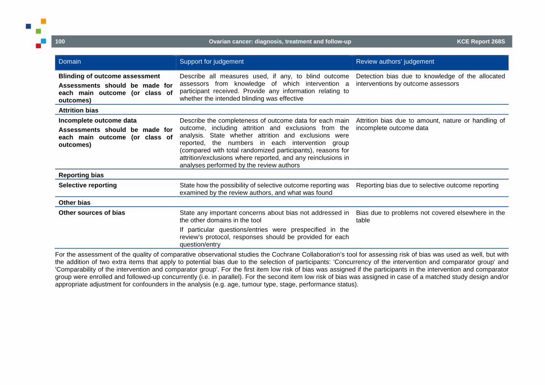
100 Ovarian cancer: diagnosis, treatment and follow-up KCE Report 268S
Domain Support for judgement Review authors’ judgement
Blinding of outcome assessment Assessments should be made for each main outcome (or class of outcomes)
Describe all measures used, if any, to blind outcome assessors from knowledge of which intervention a participant received. Provide any information relating to whether the intended blinding was effective
Detection bias due to knowledge of the allocated interventions by outcome assessors
Attrition bias Incomplete outcome data Assessments should be made for each main outcome (or class of outcomes)
Describe the completeness of outcome data for each main outcome, including attrition and exclusions from the analysis. State whether attrition and exclusions were reported, the numbers in each intervention group (compared with total randomized participants), reasons for attrition/exclusions where reported, and any reinclusions in analyses performed by the review authors
Attrition bias due to amount, nature or handling of incomplete outcome data
Reporting bias Selective reporting State how the possibility of selective outcome reporting was
examined by the review authors, and what was found Reporting bias due to selective outcome reporting
Other bias Other sources of bias State any important concerns about bias not addressed in
the other domains in the tool If particular questions/entries were prespecified in the review’s protocol, responses should be provided for each question/entry
Bias due to problems not covered elsewhere in the table
For the assessment of the quality of comparative observational studies the Cochrane Collaboration’s tool for assessing risk of bias was used as well, but with the addition of two extra items that apply to potential bias due to the selection of participants: 'Concurrency of the intervention and comparator group' and 'Comparability of the intervention and comparator group'. For the first item low risk of bias was assigned if the participants in the intervention and comparator group were enrolled and followed-up concurrently (i.e. in parallel). For the second item low risk of bias was assigned in case of a matched study design and/or appropriate adjustment for confounders in the analysis (e.g. age, tumour type, stage, performance status).
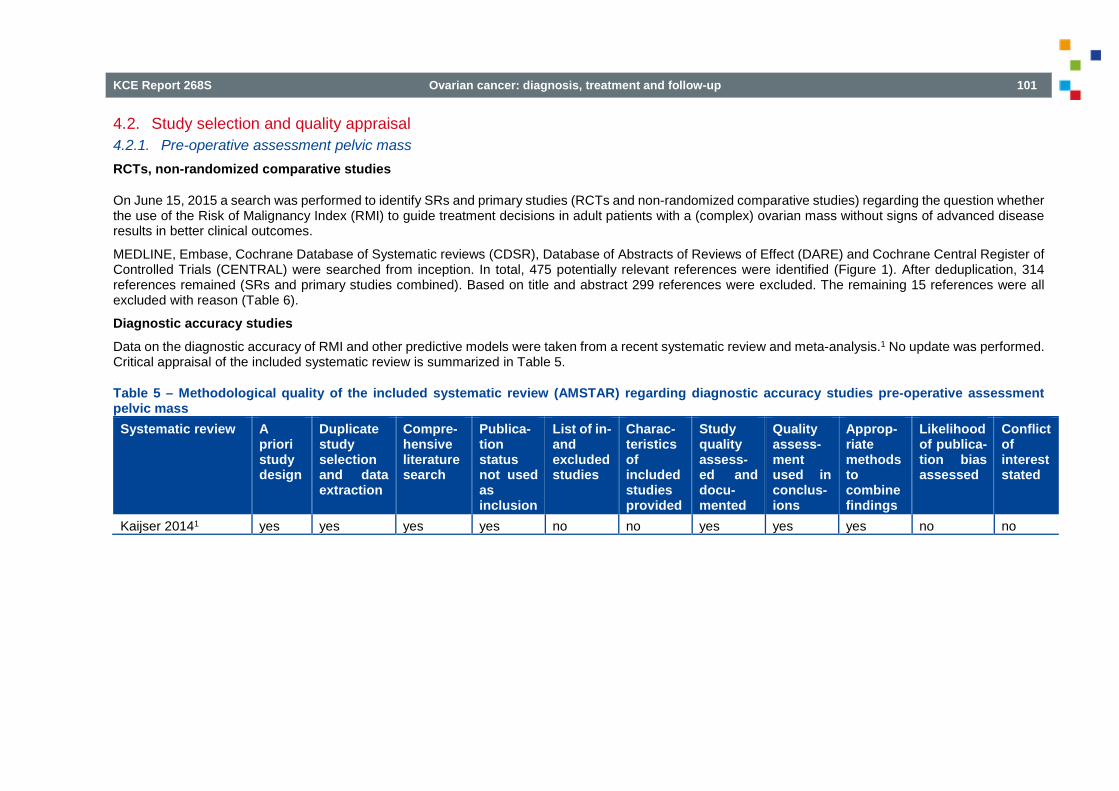
KCE Report 268S Ovarian cancer: diagnosis, treatment and follow-up 101
4.2. Study selection and quality appraisal 4.2.1. Pre-operative assessment pelvic mass RCTs, non-randomized comparative studies
On June 15, 2015 a search was performed to identify SRs and primary studies (RCTs and non-randomized comparative studies) regarding the question whether the use of the Risk of Malignancy Index (RMI) to guide treatment decisions in adult patients with a (complex) ovarian mass without signs of advanced disease results in better clinical outcomes.
MEDLINE, Embase, Cochrane Database of Systematic reviews (CDSR), Database of Abstracts of Reviews of Effect (DARE) and Cochrane Central Register of Controlled Trials (CENTRAL) were searched from inception. In total, 475 potentially relevant references were identified (Figure 1). After deduplication, 314 references remained (SRs and primary studies combined). Based on title and abstract 299 references were excluded. The remaining 15 references were all excluded with reason (Table 6).
Diagnostic accuracy studies
Data on the diagnostic accuracy of RMI and other predictive models were taken from a recent systematic review and meta-analysis.1 No update was performed. Critical appraisal of the included systematic review is summarized in Table 5.
Table 5 – Methodological quality of the included systematic review (AMSTAR) regarding diagnostic accuracy studies pre-operative assessment pelvic mass Systematic review A
priori study design
Duplicate study selection and data extraction
Compre-hensive literature search
Publica-tion status not used as inclusion
List of in- and excluded studies
Charac-teristics of included studies provided
Study quality assess-ed and docu-mented
Quality assess-ment used in conclus-ions
Approp-riate methods to combine findings
Likelihood of publica-tion bias assessed
Conflict of interest stated
Kaijser 20141 yes yes yes yes no no yes yes yes no no

102 Ovarian cancer: diagnosis, treatment and follow-up KCE Report 268S
Figure 1 – PRISMA flowchart selection of primary studies Risk of Malignancy index
Potentially relevant references identified after
deduplicationN=314
Full text evaluationN=15
SRs: N=5RCTs: N=6
Observational studies: N=4
N=299Excluded on the basis of title and abstract
Included N=0
N=15Excluded
Population N=1 (SR)Intervention N=4 (N=1 SR; N=2 RCTs; N=1 Obs)Comparison N=1 (SR)Outcome N=4 (N=2 RCTs; N=2 Obs)Non-systematic review N=1Model-based Obs.study N=1Protocol N=1 (SR)Ongoing RCT N=2
Potentially relevant references identified from
databasesN=475
Medline N=219Embase N=243
CDSR N=0DARE N=0
CENTRAL N=13
N=161Duplicates
Potenially relevant references identified from
other sourcesN=0

KCE Report 268S Ovarian cancer: diagnosis, treatment and follow-up 103
Table 6 – Excluded references Risk of Malignancy index (n=15) Reference Study design Reason
Chan 20062 SR Excluded on intervention (RMI not studied as an intervention) Fung 20043 SR Excluded on population (asymptomatic postmenopausal women from general population) Geomini 20114 Observational study Excluded on design (model based study) Jacobs 19995 RCT Excluded on intervention (no RMI) Jeyarajah 19996 Observational study Excluded on intervention (no RMI) Kobayashi 20087 RCT Excluded on intervention (no RMI) Lockwood 20138 SR Protocol Menon 20059 RCT Excluded on outcome Menon 200910 RCT Excluded on outcome Myers 200611 SR Excluded comparison Nunes 201212 RCT Protocol / ongoing study Ozols 200413 SR Excluded on design (non-systematic review) Sayasneh 201314 RCT Ongoing study Timmerman 200715 Observational study Excluded on outcome (accuracy measures, predictive values) Van Calster 201216 Observational study Excluded on outcome (only accuracy outcomes)
4.2.2. Intra-operative frozen section 4.2.2.1. Selection of RCTs On April 29, 2015 a search was performed to identify RCTs regarding the use of intraoperative frozen section to guide treatment decisions for patients with (presumed) early-stage ovarian cancer. MEDLINE, Embase and CENTRAL were searched from inception. In total, 94 potentially relevant references were identified (Figure 2). After deduplication 76 references remained. Based on title and abstract all references were excluded.
4.2.2.2. Selection of diagnostic test accuracy studies
On May 5, 2015 a search was performed to identify diagnostic test accuracy studies regarding the use of intraoperative frozen section to guide treatment decisions for patients with (presumed) early-stage ovarian cancer. MEDLINE and Embase were searched from 2009. In total, 706 potentially relevant references were identified (Figure 3). After deduplication, 509 references remained. Based on title and abstract 488 references were excluded. Of the remaining 21 references, 11 were included, two were already assessed in already included SRs and eight studies were excluded with reason (Table 7).

104 Ovarian cancer: diagnosis, treatment and follow-up KCE Report 268S
Figure 2 – Study flow of selection of RCTs frozen section
Potentially relevant RCTs identified after deduplication
N=76
Full text evaluationN=0
N=76Excluded on the basis of title and
abstract
Included N=0
N=0Excluded on full text
Potentially relevant RCTs identified from databases
N= 94MEDLINE N=66EMBASE N=19CENTRAL N=9
N=18Duplicates
Figure 3 – Study flow of selection of DTA studies frozen section
Potentially relevant studies identified after deduplication
N=509
Full text evaluationN=21
N=488Excluded on the basis of title and
abstract
Included N=11
N=8Excluded on full text
No DTA study N=1Biased selection N=4
No 3*3 Table N=1No PDF N=2
Already assessed in included SR N=2
Potentially relevant studies identified from databases
N=706MEDLINE N=270EMBASE N=436
N=197Duplicates

KCE Report 268S Ovarian cancer: diagnosis, treatment and follow-up 105
Table 7 – Excluded DTA studies frozen section Reference Reasons
Abbasi 201117 No PDF available Aslam 201018 Selection was based on first 400 frozen section positives and 400 frozen section negatives. Sampling fraction
must be known to allow unbiased estimates of sensitivity and specificity Basaran 201419 (Posthoc) selection of only borderline ovarian tumours Gultekin 201120 No PDF available / (posthoc) selection of only borderline ovarian tumours Ouladsahebmadare 201521 (Posthoc) exclusion of borderline ovarian tumours Pongsuvareeyakul 201222 (Posthoc) selection of only mucinous tumours Stewart 201023 3 by 3 Tables for frozen section not retrievable Ureyen 201424 No DTA study / only women with borderline ovarian tumours included
4.2.2.3. Critical appraisal Systematic reviews
Of the included reviews, quality appraisal through the AMSTAR criteria was performed. Three reviews scored positively on the majority of the items25-27, although in two meta-analysis was performed according to methods that are now considered obsolete.26, 27 Because meta-analyses can be replicated with state-of-the-art methods, these reviews were further processed. One review scored negatively on almost all AMSTAR items and was excluded.28

106 Ovarian cancer: diagnosis, treatment and follow-up KCE Report 268S
Table 8 – Methodological quality of included systematic reviews (AMSTAR) regarding frozen section Systematic review A
priori study design
Duplicate study selection and data extraction
Compre-hensive literature search
Publica-tion status not used as inclusion
List of in- and excluded studies
Charac-teristics of included studies provided
Study quality assess-ed and docu-mented
Quality assess-ment used in conclus-ions
Approp-riate methods to combine findings
Likelihood of publica-tion bias assessed
Conflict of interest stated
Covens 201225 ? + (+) - +/- - +* - + - - Geomini 200526 ? + + - + + +* + +** + - Heatley 201228 ? ? - - - - - NA - - - Medeiros 200527 ? + + - +*** + + + +** + -
NA=not applicable; * Globally described ** Statistical methods used are now considered obsolete (but primary study results will be used for a replication of a state-of-the art random effects meta-analysis) *** Complete list available from the authors
Diagnostic test accuracy studies
Of the 11 studies identified in the search update, one scored high risk of bias for Patient Selection and two low risk (Figure 4). Unclear risk of bias was present in four studies for Index Test, in all studies for Reference Standard and in two studies for Flow and Timing. All other studies were considered low risk of bias for those domains (Figure 4). Two studies scored high applicability concerns for Patient Selection, four studies had unclear concerns for one or more applicability domains and the remainder scored low concern.

KCE Report 268S Ovarian cancer: diagnosis, treatment and follow-up 107
Figure 4 – Frozen section: summary QUADAS assessment of the 11 newly included diagnostic test accuracy studies

108 Ovarian cancer: diagnosis, treatment and follow-up KCE Report 268S
Figure 5 – Frozen section: overview critical appraisal all included DTA studies* (1)
*For three studies, critical appraisal is lacking due language or single critical appraisal only.

KCE Report 268S Ovarian cancer: diagnosis, treatment and follow-up 109
Figure 6 – Frozen section: overview critical appraisal all included DTA studies* (2)

110 Ovarian cancer: diagnosis, treatment and follow-up KCE Report 268S
4.2.3. Lymphadenectomy 4.2.3.1. Selection of systematic reviews
On June 15, 2015 a search was performed to identify SRs regarding systematic pelvic and para-aortic lymphadenectomy in patients with borderline or (micro)invasive (presumed) early-stage ovarian cancer. MEDLINE, Embase, CDSR and DARE were searched from inception. In total, 91 potentially relevant references were identified (Figure 7). After deduplication, 76 references remained. Based on title and abstract 57 references were excluded. Of the remaining nine references, one was included and eight were excluded with reason (Table 9).

KCE Report 268S Ovarian cancer: diagnosis, treatment and follow-up 111
Figure 7 – PRISMA flowchart SRs lymphadenectomy
Potentially relevant SRs identified after deduplication
N=76
Full text evaluationN=19
N=57Excluded on the basis of title and abstract
Included N=1
N=18Excluded
Population N=2Comparison N=2Outcome N=3Non-systematic review N=3No quality assessment N= 3Protocol N=1Language N=1No full text N=3
Potentially relevant SRs identified from databases
N=91
Medline N=30Embase N=56
Cochrane Database SRs N=3DARE N=2
N=15Duplicates
Potenially relevant SRs identified from other
sourcesN=0

112 Ovarian cancer: diagnosis, treatment and follow-up KCE Report 268S
Table 9 – Excluded SRs lymphadenectomy Reference Reasons
Atienza-Amores 201429 Outcome Bois 200930 No full text Dodge 201231 Outcome, one database searched and no methodological quality assessed in included studies Faluyi 200932 Outcome Gao 201533 Comparison Gurumurthy 201434 Outcome Hackethal 200835 Population Haritwal 200436 One database searched, no methodological quality assessed in included studies Kim 201037 Comparison Kleppe 201438 Excluded on population Kuhn 199139 No full text Lecuru 199840 One database searched, no methodological quality assessed in included studies Munstedt 200441 Non-systematic review Papadia 200442 Non-systematic review Qin 201243 Language Roy 199844 No full text Shyamala 201045 Non-systematic review Smits 201546 Protocol of systematic review
4.2.3.2. Selection of primary studies On June 22, 2015 a search was performed to identify primary studies (any study design) that reported on prevalence of malignant disease in pelvic and para-aortic lymph nodes in patients with borderline or (micro)invasive (presumed) early-stage ovarian cancer undergoing systematic pelvic and para-aortic lymphadenectomy. MEDLINE, Embase and CENTRAL were searched from 2011 (search date of included systematic review). In total, 2587 potentially relevant references were identified (Figure 8). After deduplication 1848 remained. Of those, 610 references published from 2011 to date were screened. Based on title and abstract 503 references were excluded. Of the remaining 107 references, after full-text screening, nine were included and 98 were excluded with reason (Table 10).

KCE Report 268S Ovarian cancer: diagnosis, treatment and follow-up 113
Figure 8 – PRISMA flowchart primary studies lymphadenectomy
Potentially relevant studies 2011-2015
N=610
Full text evaluationN=107
N=503Excluded on the basis of title and abstract
Included studies N=9
Potentially relevant studies identified from databases
N= 2587
MEDLINE N=1050EMBASE N=1484CENTRAL N=53
N= 739Duplicates
Potentially relevant studies identified after deduplication
N=1848
N= 1238<2011
N=98Excluded
Ongoing study N=1Language N=2Study design N=6Population N=35Intervention N=3Outcome N=41No full tekst N=7Duplicate N=3

114 Ovarian cancer: diagnosis, treatment and follow-up KCE Report 268S
Table 10 – Excluded primary studies lymphadenectomy Reference Reason
Akbayir 201247 Excluded on population AlHilli 201348 Excluded on population Aoki 201449 Excluded on study design Ataseven 201450 Excluded on outcome Azuar 201351 Excluded on outcome Bachmann201452 No full text available Bae 201253 Excluded on outcome Bennett 201554 Excluded on outcome Berretta 201455 Excluded on population Boren 201256 Excluded on population Bostanci 201457 Excluded on language Braicu 201358 Excluded on population Brown 201459 Excluded on outcome Capilna 201160 Excluded on population Chang 201361 Duplicate, included Chang 201362 Excluded on population Chatchotikawong 201563 Excluded on population Chereau 201264 Excluded on study design Cusido 201165 Excluded on population Dell 201266 Excluded on intervention
Ditto 201467 Excluded on outcome Eltabbakh 201168 No full text available Ertas 201469 Excluded on population Ertas 201469 Duplicate, excluded on population Ertas 201470 Excluded on population

KCE Report 268S Ovarian cancer: diagnosis, treatment and follow-up 115
Reference Reason
Euscher 201171 Excluded on population Gmyrek 201172 Excluded on outcome Grabowski 201273 Excluded on outcome Guvenal 201374 Excluded on outcome Gyimadu 201275 Excluded on outcome Haller 201176 Duplicate, included Han 201277 Excluded on outcome Hareyama 201278 Excluded on outcome Hareyama 201579 Excluded on outcome Hu 201380 Excluded on population Ibeanu 201381 Excluded on outcome Kanat-Pektas 201182 Excluded on outcome Karalok 201583 Excluded on population Kiran 201584 Excluded on outcome Lambaudie 201285 Excluded on population Lazarou 201486 No full text available Li 201487 Excluded on outcome Magazzino 201188 Excluded on outcome Mahdi 201189 Excluded on population Mahdi 201190 Excluded on population Mahdi 201391 Excluded on outcome Marpeau 201292 Excluded on study design Matsuo 201293 Excluded on population Matsuo 201494 Excluded on outcome Matsuo 201495 Excluded on population Matsuo 201596 Excluded on outcome

116 Ovarian cancer: diagnosis, treatment and follow-up KCE Report 268S
Reference Reason
Mbarki 201197 Excluded on outcome McNally 201598 Excluded on outcome Meinhold-Heerlein 201499 Excluded on study design Miyamoto 2011100 Excluded on language Miyamoto 2012101 Excluded on outcome Momeni 2013102 Excluded on population Moore 2011103 Excluded on population Morales 2013104 Excluded on intervention Moreau-Claeys 2011105 Excluded on population Mury 2011106 Excluded on outcome Oh 2014107 Excluded on population Ohba 2012108 Excluded on outcome Ozalp 2014109 No full text available Pan 2011110 Excluded on population Park 2011111 Excluded on population Park 2012112 Excluded on outcome Park 2012113 Excluded on population Paulsson 2013114 No full text available Pereira 2013115 Excluded on population Pereira 2014116 Excluded on outcome Petry 2012117 Excluded on study design Rauh-Hain 2013118 Excluded on outcome Rauh-Hain 2013119 Excluded on outcome Rauh-Hain 2015120 Excluded on outcome Rizzuto 2015121 Excluded on outcome Romeo 2013122 Excluded on population
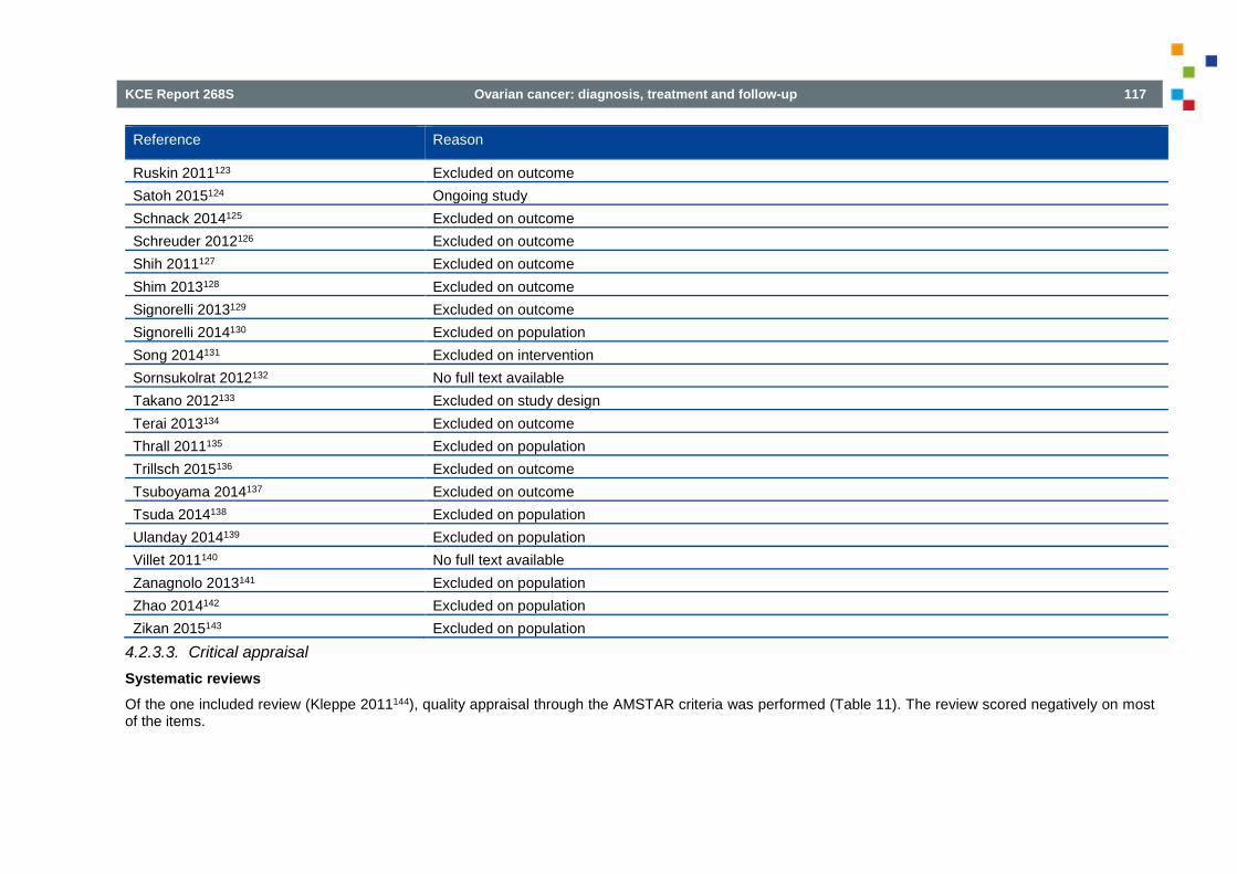
KCE Report 268S Ovarian cancer: diagnosis, treatment and follow-up 117
Reference Reason
Ruskin 2011123 Excluded on outcome Satoh 2015124 Ongoing study Schnack 2014125 Excluded on outcome Schreuder 2012126 Excluded on outcome Shih 2011127 Excluded on outcome Shim 2013128 Excluded on outcome Signorelli 2013129 Excluded on outcome Signorelli 2014130 Excluded on population Song 2014131 Excluded on intervention Sornsukolrat 2012132 No full text available Takano 2012133 Excluded on study design Terai 2013134 Excluded on outcome Thrall 2011135 Excluded on population Trillsch 2015136 Excluded on outcome Tsuboyama 2014137 Excluded on outcome Tsuda 2014138 Excluded on population Ulanday 2014139 Excluded on population Villet 2011140 No full text available Zanagnolo 2013141 Excluded on population Zhao 2014142 Excluded on population Zikan 2015143 Excluded on population
4.2.3.3. Critical appraisal Systematic reviews
Of the one included review (Kleppe 2011144), quality appraisal through the AMSTAR criteria was performed (Table 11). The review scored negatively on most of the items.
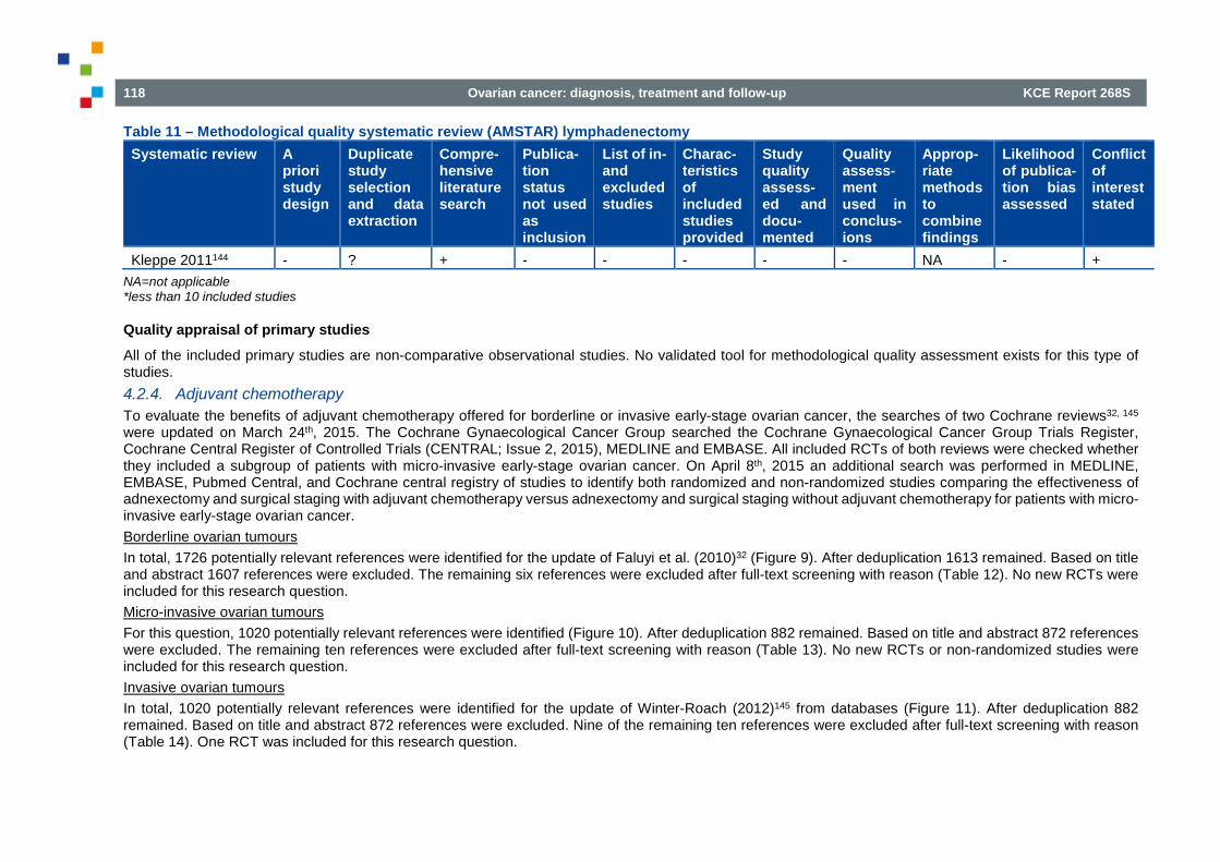
118 Ovarian cancer: diagnosis, treatment and follow-up KCE Report 268S
Table 11 – Methodological quality systematic review (AMSTAR) lymphadenectomy Systematic review A
priori study design
Duplicate study selection and data extraction
Compre-hensive literature search
Publica-tion status not used as inclusion
List of in- and excluded studies
Charac-teristics of included studies provided
Study quality assess-ed and docu-mented
Quality assess-ment used in conclus-ions
Approp-riate methods to combine findings
Likelihood of publica-tion bias assessed
Conflict of interest stated
Kleppe 2011144 - ? + - - - - - NA - + NA=not applicable *less than 10 included studies
Quality appraisal of primary studies
All of the included primary studies are non-comparative observational studies. No validated tool for methodological quality assessment exists for this type of studies. 4.2.4. Adjuvant chemotherapy To evaluate the benefits of adjuvant chemotherapy offered for borderline or invasive early-stage ovarian cancer, the searches of two Cochrane reviews32, 145 were updated on March 24th, 2015. The Cochrane Gynaecological Cancer Group searched the Cochrane Gynaecological Cancer Group Trials Register, Cochrane Central Register of Controlled Trials (CENTRAL; Issue 2, 2015), MEDLINE and EMBASE. All included RCTs of both reviews were checked whether they included a subgroup of patients with micro-invasive early-stage ovarian cancer. On April 8th, 2015 an additional search was performed in MEDLINE, EMBASE, Pubmed Central, and Cochrane central registry of studies to identify both randomized and non-randomized studies comparing the effectiveness of adnexectomy and surgical staging with adjuvant chemotherapy versus adnexectomy and surgical staging without adjuvant chemotherapy for patients with micro-invasive early-stage ovarian cancer. Borderline ovarian tumours In total, 1726 potentially relevant references were identified for the update of Faluyi et al. (2010)32 (Figure 9). After deduplication 1613 remained. Based on title and abstract 1607 references were excluded. The remaining six references were excluded after full-text screening with reason (Table 12). No new RCTs were included for this research question. Micro-invasive ovarian tumours For this question, 1020 potentially relevant references were identified (Figure 10). After deduplication 882 remained. Based on title and abstract 872 references were excluded. The remaining ten references were excluded after full-text screening with reason (Table 13). No new RCTs or non-randomized studies were included for this research question. Invasive ovarian tumours In total, 1020 potentially relevant references were identified for the update of Winter-Roach (2012)145 from databases (Figure 11). After deduplication 882 remained. Based on title and abstract 872 references were excluded. Nine of the remaining ten references were excluded after full-text screening with reason (Table 14). One RCT was included for this research question.

KCE Report 268S Ovarian cancer: diagnosis, treatment and follow-up 119
Figure 9 – PRISMA flowchart selection of RCTs adjuvant chemotherapy for borderline ovarian tumours
Potentially relevant RCTs identified after deduplication
N=1613
Full text evaluationN=6
N=1607Excluded on the basis of title and abstract
Included RCTs N=0
Potentially relevant RCTs identified from databases
N= 1726
MEDLINE N=569EMBASE N=1113CENTRAL N=20
Special Register = 24
N= 113Duplicates
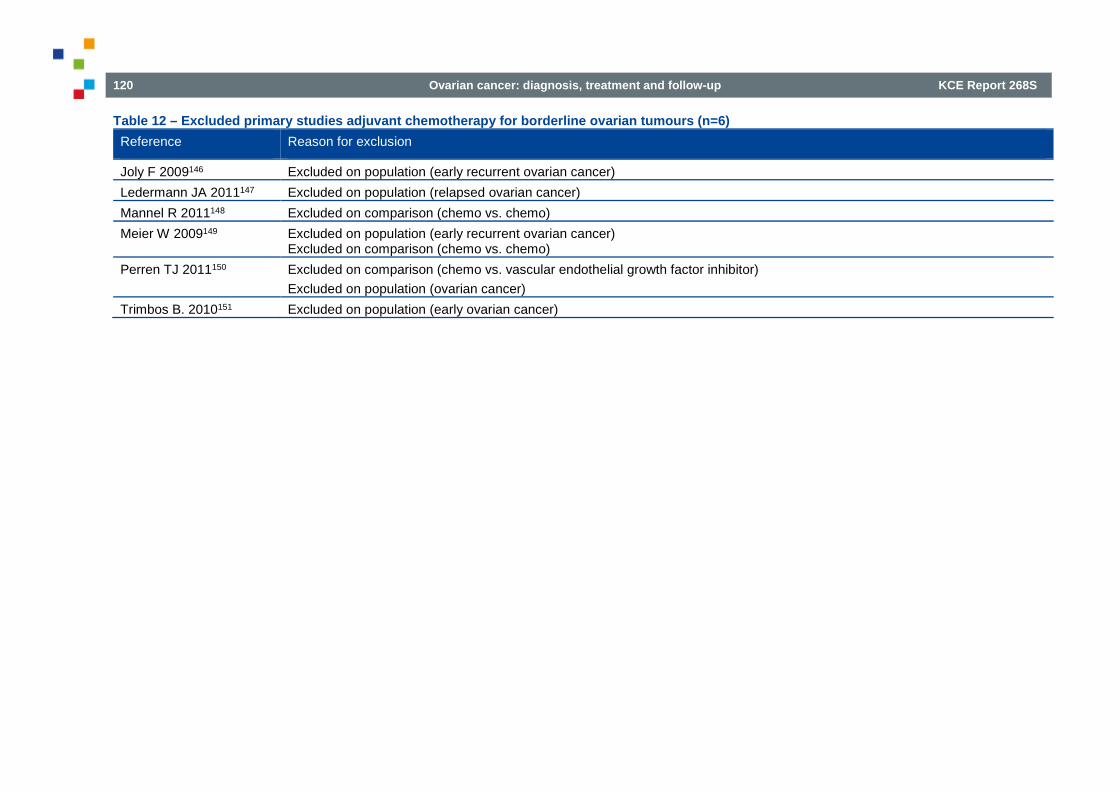
120 Ovarian cancer: diagnosis, treatment and follow-up KCE Report 268S
Table 12 – Excluded primary studies adjuvant chemotherapy for borderline ovarian tumours (n=6) Reference Reason for exclusion
Joly F 2009146 Excluded on population (early recurrent ovarian cancer) Ledermann JA 2011147 Excluded on population (relapsed ovarian cancer) Mannel R 2011148 Excluded on comparison (chemo vs. chemo) Meier W 2009149 Excluded on population (early recurrent ovarian cancer)
Excluded on comparison (chemo vs. chemo) Perren TJ 2011150 Excluded on comparison (chemo vs. vascular endothelial growth factor inhibitor)
Excluded on population (ovarian cancer) Trimbos B. 2010151 Excluded on population (early ovarian cancer)

KCE Report 268S Ovarian cancer: diagnosis, treatment and follow-up 121
Figure 10 – PRISMA flowchart selection of RCTs and non-randomized studies adjuvant chemotherapy micro-invasive ovarian tumours
Potentially relevant studies identified after deduplication
N=342
Full text evaluationN=37
N=305Excluded on the basis of title and abstract
Included studies N=0
Potentially relevant studiess identified from databases
N= 520
MEDLINE N=222EMBASE N=258
Pubmed Central N=29Cochrane CR of studies N=11
N= 178Duplicates

122 Ovarian cancer: diagnosis, treatment and follow-up KCE Report 268S
Table 13 – Excluded RCTs and non-randomized studies adjuvant chemotherapy for micro-invasive disease (n=37) Reference Reason for exclusion
Ceballos KM 2006152 Excluded on intervention Cusido M 1999153 Excluded on language Damak T 2014154 Excluded on population Dgani R 1990155 Excluded on population Du Bois A 2013156 Excluded on design Eltabbakh GH 2000157 Excluded on design Gilks CB 2003158 Excluded on design Haritwal A 201436 Excluded on design Hogberg T 2001159 Excluded on design Kaern J 1993a160 Excluded on population Kaern J 1993b161 Excluded on population Kane A 2009162 Excluded on design Kennedy AW 1996163 Excluded on comparison Laurent I 2008164 Excluded on intervention Leary A 2014165 Excluded on population Leiserowitz G 2005166 Excluded on design Leitao MM 2004167 Excluded on population Leitao MM 2011168 Excluded on design Li Y 2003169 Excluded on language Li ZX 1994170 Excluded on language Linasmita V 1990171 No PDF available Manchul LA 1992172 No PDF available Massad LS 1991173 Excluded on population Morice P 2001174 No PDF available Park JY 2011111 Excluded on population Raymond E 1997175 Excluded on population

KCE Report 268S Ovarian cancer: diagnosis, treatment and follow-up 123
Reference Reason for exclusion
Rettenmaier MA 2010176 Excluded on population Shih KK 2010177 Excluded on population Shih KK 2011127 Excluded on design Trimble CL 2002178 Excluded on population Tulpin L 2008179 No PDF available Uzan C 2011180 Excluded on population Vasconcelos I 2015a181 Excluded on population Vasconcelos I 2015b182 Excluded on design Vergote IB 1992183 Excluded on comparison Yokoyama Y 2006184 Excluded on population Zhang Z 1998185 Excluded on language

124 Ovarian cancer: diagnosis, treatment and follow-up KCE Report 268S
Figure 11 – PRISMA flowchart selection of RCTs adjuvant chemotherapy for invasive early stage ovarian cancer
Potentially relevant RCTs identified after deduplication
N=882
Full text evaluationN=10
N=872Excluded on the basis of title and abstract
Included RCTs N=1
Potentially relevant RCTs identified from databases
N= 1020
MEDLINE N=130EMBASE N=774CENTRAL N=116
N= 138Duplicates
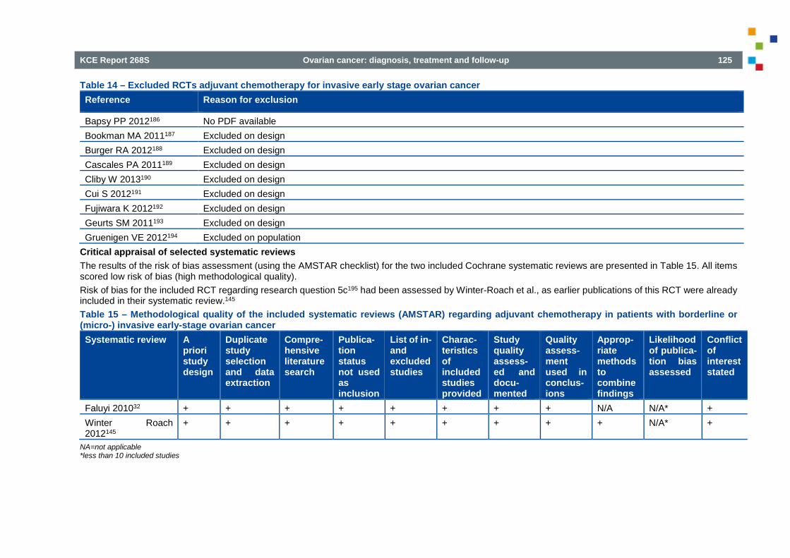
KCE Report 268S Ovarian cancer: diagnosis, treatment and follow-up 125
Table 14 – Excluded RCTs adjuvant chemotherapy for invasive early stage ovarian cancer Reference Reason for exclusion
Bapsy PP 2012186 No PDF available Bookman MA 2011187 Excluded on design Burger RA 2012188 Excluded on design Cascales PA 2011189 Excluded on design Cliby W 2013190 Excluded on design Cui S 2012191 Excluded on design Fujiwara K 2012192 Excluded on design Geurts SM 2011193 Excluded on design Gruenigen VE 2012194 Excluded on population
Critical appraisal of selected systematic reviews The results of the risk of bias assessment (using the AMSTAR checklist) for the two included Cochrane systematic reviews are presented in Table 15. All items scored low risk of bias (high methodological quality). Risk of bias for the included RCT regarding research question 5c195 had been assessed by Winter-Roach et al., as earlier publications of this RCT were already included in their systematic review.145 Table 15 – Methodological quality of the included systematic reviews (AMSTAR) regarding adjuvant chemotherapy in patients with borderline or (micro-) invasive early-stage ovarian cancer Systematic review A
priori study design
Duplicate study selection and data extraction
Compre-hensive literature search
Publica-tion status not used as inclusion
List of in- and excluded studies
Charac-teristics of included studies provided
Study quality assess-ed and docu-mented
Quality assess-ment used in conclus-ions
Approp-riate methods to combine findings
Likelihood of publica-tion bias assessed
Conflict of interest stated
Faluyi 201032 + + + + + + + + N/A N/A* + Winter Roach 2012145
+ + + + + + + + + N/A* +
NA=not applicable *less than 10 included studies
126 Ovarian cancer: diagnosis, treatment and follow-up KCE Report 268S
4.2.5. Laparoscopic surgery Selection of systematic reviews
On March 09, 2015 a search was performed to identify SRs evaluating laparoscopic surgery for presumed early stage ovarian cancer. MEDLINE (including PreMedline), Embase and the Cochrane Database of Systematic Reviews (CDSR), the Cochrane Library Health Technology Assessment Database (CLIB HTA), and the Database of Abstracts of Reviews of Effects (DaRe) were searched. In total 233 potential relevant references were identified (Figure 12). After de-duplication, 112 unique references remained. Based on title and abstract 100 reviews were excluded. Twelve reviews were included for full-text evaluation. Of these, eight were excluded with reasons (Table 16). Two further reviews were excluded based on critical appraisal (Table 17).196, 197

KCE Report 268S Ovarian cancer: diagnosis, treatment and follow-up 127
Figure 12 – Study flow of selection of SRs
Potentially relevant SR identified from databases:233
Potenially relevant SRs after deduplication:112
Duplicates: 121
Excluded based on title and abstract: 100
Full text evaluation: 12
Included SRs: 2
Excluded based on full text evaluation: 10

128 Ovarian cancer: diagnosis, treatment and follow-up KCE Report 268S
Table 16 – Excluded SRs based on full-text evaluation Reference Reason for exclusion Lu 2015198 Included Matsushita 2014199 Case-series, no comparison group Lu 2014200 abstract Bogani 2014197 Included Park 2013196 Included Mandic 2013201 Narrative review Lu 2013202 Full text not available Lawrie 2013203 Included Brockbank 2013204 No comparison with laparotomy Dodge 201231 Guideline, insufficient reporting of the systematic review Covens 201225 Medline search only, from 2004 to 2009 Leblanc 2006205 Narrative review, no report of systematic search
Quality appraisal of selected systematic reviews
Table 17 shows the results of the risk of bias assessment for the included systematic review, using AMSTAR criteria.

KCE Report 268S Ovarian cancer: diagnosis, treatment and follow-up 129
Table 17 – Methodological quality of the included systematic review (AMSTAR) Systematic review A priori
study design
Duplicate study selection and data extraction
Compre-hensive literature search
Publica-tion status not used as inclusion
List of in- and excluded studies
Charac-teristics of included studies provided
Study quality assessed and docu-mented
Quality assess-ment used in conclus-ions
Appropriate methods to combine findings
Likelihood of publica-tion bias assessed
Conflict of interest stated
Lu 2015198 Y Y Y ? N Y Y Y N Y N Lawrie 2013203 Y Y Y Y Y NA NA NA NA NA Y Park 2013196 Y Y Y N N Y N N N Y N Bogani 2014197 ? ? Y N N Y N N N N N Zhang 2015206 Y Y Y Y N Y N N N N N
Selection of primary studies
On October 12, 2015 a search was performed to identify RCTs and non-randomized comparative studies evaluating laparoscopic surgery for presumed early stage ovarian cancer. The search date of Lu 2015198 was used as starting point. MEDLINE (including PreMedline), Embase and CENTRAL were searched, limited from 2013 onwards. In total, 422 potential relevant references were identified (Figure 13). After de-duplication, 418 unique references remained. Based on title and abstract 412 studies were excluded. After full text evaluation (Table 18), three papers were included, one systematic review with search date May 2014206 and two primary non-randomized studies.207,
208 The two primary studies were already included in the systematic review of Zhang et al (Table 17). A third study that was included in the review of Zhang et al., but not in the review of Lu et al. was excluded as patients who were treated with laparoscopy converted to laparotomy were analysed separately and not all patients underwent comprehensive staging.209 Both newly included primary studies were non-randomized studies with a high risk of bias as no case-mix adjustment was performed.

130 Ovarian cancer: diagnosis, treatment and follow-up KCE Report 268S
Figure 13 – Study flow of selection of primary studies
Potentially relevant primary studies identified from databases: 422
Duplicates:4
Potentially relevant primary studies after deduplication: 418
Excluded based on title and abstract: 412
Full text evaluation: 6
Included: 1 SR, 2 primary studies
Excluded based on full text evaluation: 3

KCE Report 268S Ovarian cancer: diagnosis, treatment and follow-up 131
Table 18 – Laparoscopic surgery early-stage ovarian cancer: excluded studies based on full text evaluation Reference Reason for exclusion Zhang Y. 2015206 Systematic review, included Zhang Z.-M. 2014210 Chinese Liu 2014208 Included in Zhang et al. 2015 Koo 2014207 Included in Zhang et al. 2015 Gallotta 2014211 No comparator group with laparotomy Aoki 2014212 Opinion paper, narrative
4.2.6. Laparoscopy, PET-CT and MRI to predict end result of cytoreductive surgery 4.2.6.1. Systematic reviews
On January 22, 2015 a search was performed to identify SRs regarding PET-CT, laparoscopy and (diffusion) MRI to predict complete/optimal resection of advanced stage disease in patients with advanced stage ovarian cancer considered eligible for debulking surgery based on CT-scan. MEDLINE, Embase, CDSR and DARE were searched from inception. In total, 233 potentially relevant references were identified (Figure 14). After deduplication, 168 references remained. Based on title and abstract 159 references were excluded. Of the remaining nine references, one was included213 and eight were excluded with reason (Table 19).
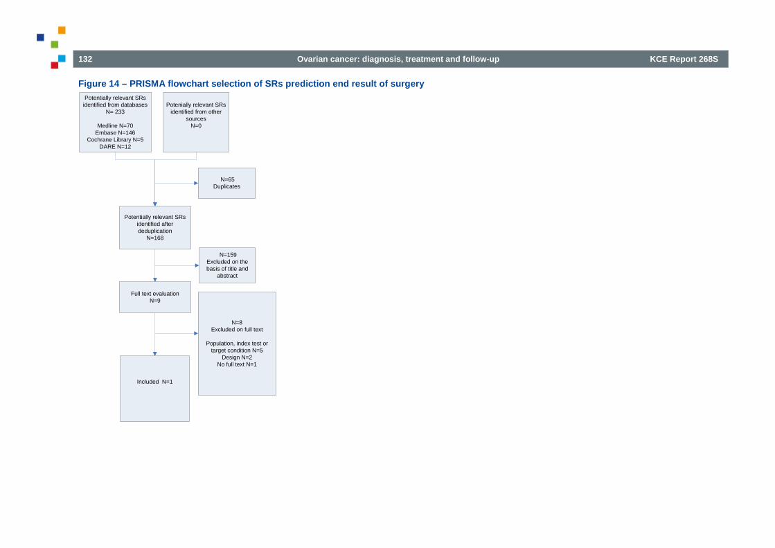
132 Ovarian cancer: diagnosis, treatment and follow-up KCE Report 268S
Figure 14 – PRISMA flowchart selection of SRs prediction end result of surgery
Potentially relevant SRs identified after deduplication
N=168
Full text evaluationN=9
N=159Excluded on the basis of title and
abstract
Included N=1
N=8Excluded on full text
Population, index test or target condition N=5
Design N=2No full text N=1
Potentially relevant SRs identified from databases
N= 233
Medline N=70Embase N=146
Cochrane Library N=5DARE N=12
N=65Duplicates
Potenially relevant SRs identified from other
sourcesN=0

KCE Report 268S Ovarian cancer: diagnosis, treatment and follow-up 133
Table 19 – Prediction of end result of cytoreductive surgery: excluded SRs based on full-text evaluation Reference Reasons
Bertagna 2013 214 Index test and population not of interest Chang 2013 215 Target condition not of interest Havrilesky 2005 216 Target condition not of interest Hoh 1998 217 No PDF Ibeanu 2010 218 No systematic review Lai 2014 219 Unclear index test and population Musto 2011 220 No SR Yuan 2012 221 Target condition not of interest; mixed population
Quality appraisal of selected systematic reviews
Of the one included review (Rutten 2014),213 quality appraisal through the AMSTAR criteria was performed. The review scored positively on the majority of the items. However, the SR failed to address whether there was a conflict of interest for both the review and the included studies. Overall, the SR is considered as having a ‘low risk’ of bias (Table 20).
Table 20 – Prediction end result of surgery: critical appraisal of included SR Systematic review A
priori study design
Duplicate study selection and data extraction
Compre-hensive literature search
Publica-tion status not used as inclusion
List of in- and excluded studies
Charac-teristics of included studies provided
Study quality assess-ed and docu-mented
Quality assess-ment used in conclus-ions
Approp-riate methods to combine findings
Likelihood of publica-tion bias assessed
Conflict of interest stated
Rutten 2014 213 + + + + + + + + + NA* - NA=not applicable *less than 10 included studies

134 Ovarian cancer: diagnosis, treatment and follow-up KCE Report 268S
4.2.6.2. Randomized controlled trials
On February 16, 2015 a search was performed to identify RCTs regarding the effect on patient important outcomes of using PET-CT, laparoscopy or (diffusion) MRI to predict complete/optimal resection of advanced stage disease in patients with advanced stage ovarian cancer considered eligible for debulking surgery based on CT-scan. MEDLINE, Embase and CENTRAL were searched from inception. In total, 206 potentially relevant references were identified (Figure 15). After deduplication 153 references remained. Based on title and abstract all references were excluded.
Figure 15 – PRISMA flowchart selection of RCTs prediction end result of surgery
Potentially relevant RCTs identified after deduplication
N=153
Full text evaluationN=0
N=153Excluded on the basis of title and
abstract
Included N=0
N=0Excluded on full text
Potentially relevant RCTs identified from databases
N= 206MEDLINE N=66EMBASE N=106CENTRAL N=34
N=53Duplicates
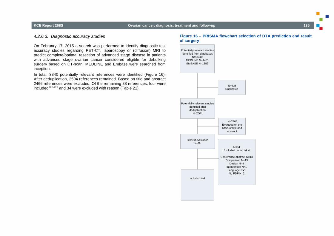
KCE Report 268S Ovarian cancer: diagnosis, treatment and follow-up 135
4.2.6.3. Diagnostic accuracy studies
On February 17, 2015 a search was performed to identify diagnostic test accuracy studies regarding PET-CT, laparoscopy or (diffusion) MRI to predict complete/optimal resection of advanced stage disease in patients with advanced stage ovarian cancer considered eligible for debulking surgery based on CT-scan. MEDLINE and Embase were searched from inception. In total, 3340 potentially relevant references were identified (Figure 16). After deduplication, 2504 references remained. Based on title and abstract 2466 references were excluded. Of the remaining 38 references, four were included222-225 and 34 were excluded with reason (Table 21).
Figure 16 – PRISMA flowchart selection of DTA prediction end result of surgery
Potentially relevant studies identified after deduplication
N=2504
Full text evaluationN=38
N=2466Excluded on the basis of title and
abstract
Included N=4
N=34Excluded on full tekst
Conference abstract N=13Comparison N=13
Design N=4Intervention N=1Language N=1No PDF N=2
Potentially relevant studies identified from databases
N= 3340MEDLINE N=1481EMBASE N=1859
N=836Duplicates
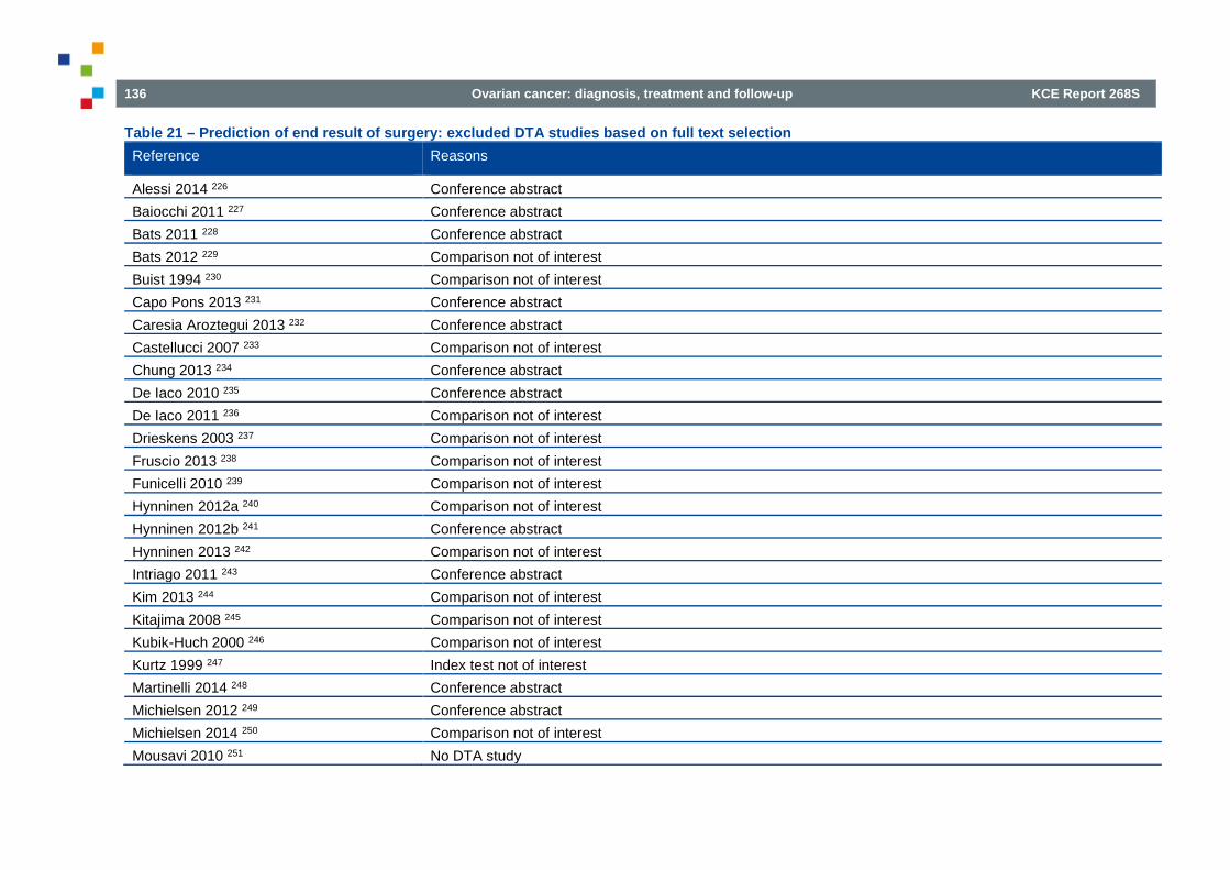
136 Ovarian cancer: diagnosis, treatment and follow-up KCE Report 268S
Table 21 – Prediction of end result of surgery: excluded DTA studies based on full text selection Reference Reasons
Alessi 2014 226 Conference abstract Baiocchi 2011 227 Conference abstract Bats 2011 228 Conference abstract Bats 2012 229 Comparison not of interest Buist 1994 230 Comparison not of interest Capo Pons 2013 231 Conference abstract Caresia Aroztegui 2013 232 Conference abstract Castellucci 2007 233 Comparison not of interest Chung 2013 234 Conference abstract De Iaco 2010 235 Conference abstract De Iaco 2011 236 Comparison not of interest Drieskens 2003 237 Comparison not of interest Fruscio 2013 238 Comparison not of interest Funicelli 2010 239 Comparison not of interest Hynninen 2012a 240 Comparison not of interest Hynninen 2012b 241 Conference abstract Hynninen 2013 242 Comparison not of interest Intriago 2011 243 Conference abstract Kim 2013 244 Comparison not of interest Kitajima 2008 245 Comparison not of interest Kubik-Huch 2000 246 Comparison not of interest Kurtz 1999 247 Index test not of interest Martinelli 2014 248 Conference abstract Michielsen 2012 249 Conference abstract Michielsen 2014 250 Comparison not of interest Mousavi 2010 251 No DTA study

KCE Report 268S Ovarian cancer: diagnosis, treatment and follow-up 137
Reference Reasons
Risum 2008 252 DTA data could not be retrieved Risum 2011 253 No DTA study Semelka 1993 254 Comparison not of interest Shim 2013a 255 Conference abstract Shim 2013b 255 Conference abstract Stark 2010 256 No DTA study Torpy 2011 257 No DTA study Zhu 1999 258 Language (Chinese)
Quality appraisal of selected DTA studies
Three of the four included studies had high risk of bias (due to patient selection and no blinded assessment of the reference test, amongst others). All studies had applicability concerns (spectrum of patients not applicable; index test not used as add-on test in women in whom CT indicates a resectable tumour; threshold for resectability undefined) (Figure 17).

138 Ovarian cancer: diagnosis, treatment and follow-up KCE Report 268S
Figure 17 – Summary of the QUADAS assessments of included DTA studies

KCE Report 268S Ovarian cancer: diagnosis, treatment and follow-up 139
4.2.7. Aim of cytoreductive surgery: no macroscopic disease? 4.2.7.1. Prognostic value end result of surgery
The starting point was a Cochrane systematic review (Table 22).259 On January 16, 2015 a search was performed in MEDLINE, Embase and CENTRAL to identify RCTs and comparative observational studies regarding the effect of the extent of debulking on various outcomes in patients with stage IIIc-IV ovarian cancer. In total, 1 315 potentially relevant references were identified (Figure 18). After deduplication, 1 085 references remained. Based on title and abstract 1 010 references were excluded. Of the remaining 75 references, 10 were included260-269 and 65 were excluded with reason (Table 23). Table 22 – Prognostic value end result of surgery: quality appraisal of the included SR Systematic review A
priori study design
Duplicate study selection and data extraction
Compre-hensive literature search
Publica-tion status not used as inclusion
List of in- and excluded studies
Charac-teristics of included studies provided
Study quality assess-ed and docu-mented
Quality assess-ment used in conclus-ions
Approp-riate methods to combine findings
Likelihood of publica-tion bias assessed
Conflict of interest stated
Elattar 2011259 + + + + + + + + + NA +
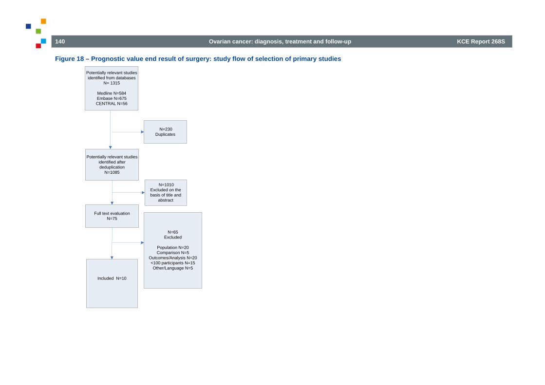
140 Ovarian cancer: diagnosis, treatment and follow-up KCE Report 268S
Figure 18 – Prognostic value end result of surgery: study flow of selection of primary studies
Potentially relevant studies identified after deduplication
N=1085
Full text evaluationN=75
N=1010Excluded on the basis of title and
abstract
Included N=10
N=65Excluded
Population N=20Comparison N=5
Outcomes/Analysis N=20<100 participants N=15Other/Language N=5
Potentially relevant studies identified from databases
N= 1315
Medline N=584Embase N=675CENTRAL N=56
N=230Duplicates

KCE Report 268S Ovarian cancer: diagnosis, treatment and follow-up 141
Table 23 – Prognostic value end result of surgery: excluded studies based on full text selection Reference Reasons
Abaid 2011270 <100 participants Altman 2012271 also included patients undergoing IDS; not analysed separately Bacic 2013272 <100 participants Bakrin 2013273 <100 participants Barlin 2012274 no multivariate analysis Barlin 2013275 only debulking status 0.1-1.0 cm Barriuso 2010276 <100 participants Bats 2012229 <100 participants Bengrine-Lefevre 2011277 also included patients undergoing IDS; not analysed separately Bereder 2009278 included also recurrent cancer Braicu 2011279 no results according to debulking status Burger 2013280 no results according to debulking status Chao 2013281 <100 participants Chereau 2011282 <100 participants Chi 2010283 no results according to debulking status Chi 2012284 no multivariate analysis` Chua 2011285 majority not ovarian cancer David 2013286 <100 participants du Bois 2010 287 also stage <IIIC included Fanfani 2012288 also included patients undergoing IDS; not analysed separately Fu 2014289 also stage <III included Gerestein 2011290 predictors of suboptimal debulking Gonzalez 2013291 no results according to debulking status / Spanish Green 2006292 only debulking status >1 cm Greimel 2013293 no results according to debulking status Hoskins 2010294 also stage <IIIC included and patients undergoing IDS
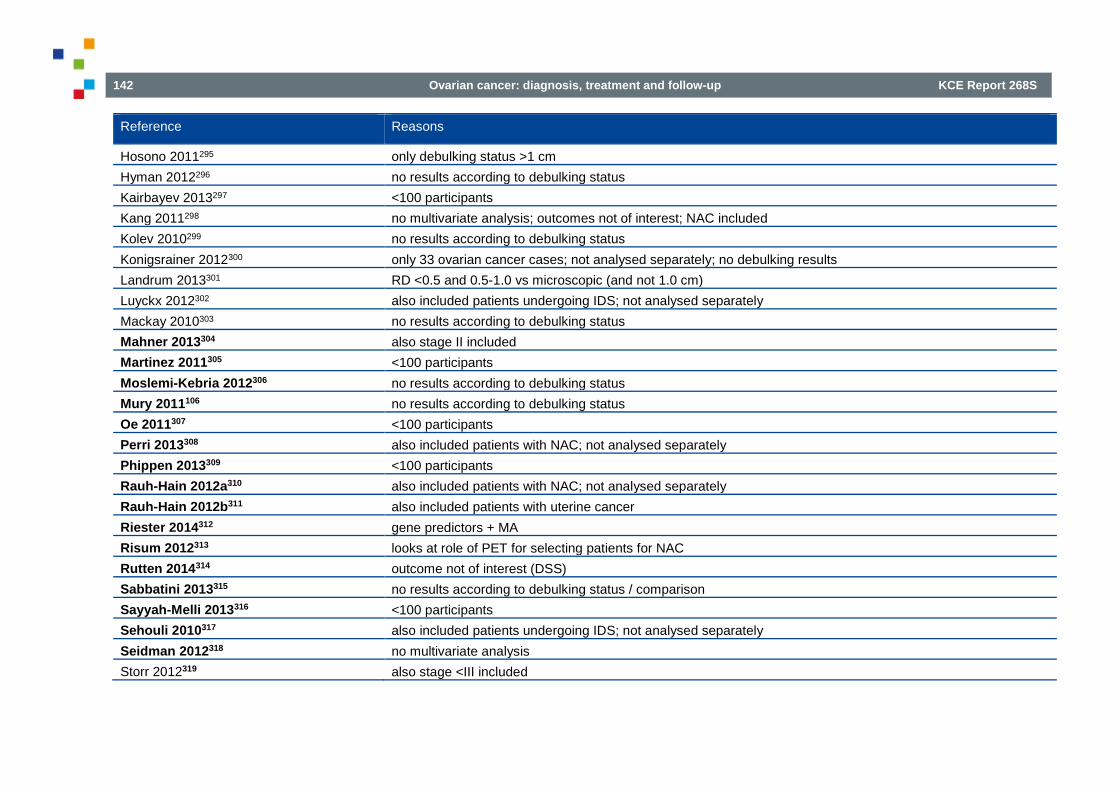
142 Ovarian cancer: diagnosis, treatment and follow-up KCE Report 268S
Reference Reasons
Hosono 2011295 only debulking status >1 cm Hyman 2012296 no results according to debulking status Kairbayev 2013297 <100 participants Kang 2011298 no multivariate analysis; outcomes not of interest; NAC included Kolev 2010299 no results according to debulking status Konigsrainer 2012300 only 33 ovarian cancer cases; not analysed separately; no debulking results Landrum 2013301 RD <0.5 and 0.5-1.0 vs microscopic (and not 1.0 cm) Luyckx 2012302 also included patients undergoing IDS; not analysed separately Mackay 2010303 no results according to debulking status Mahner 2013304 also stage II included Martinez 2011305 <100 participants Moslemi-Kebria 2012306 no results according to debulking status Mury 2011106 no results according to debulking status Oe 2011307 <100 participants Perri 2013308 also included patients with NAC; not analysed separately Phippen 2013309 <100 participants Rauh-Hain 2012a310 also included patients with NAC; not analysed separately Rauh-Hain 2012b311 also included patients with uterine cancer Riester 2014312 gene predictors + MA Risum 2012313 looks at role of PET for selecting patients for NAC Rutten 2014314 outcome not of interest (DSS) Sabbatini 2013315 no results according to debulking status / comparison Sayyah-Melli 2013316 <100 participants Sehouli 2010317 also included patients undergoing IDS; not analysed separately Seidman 2012318 no multivariate analysis Storr 2012319 also stage <III included

KCE Report 268S Ovarian cancer: diagnosis, treatment and follow-up 143
Reference Reasons
Tang 2008320 <100 participants Taskin 2013321 no results according to debulking status (nor in PDS arm) Teo 2013322 mixed population (only 39 with ovarian cancer) Terauchi 2011323 Japanese Tiuliandin 2011324 included also recurrent cancer Trope 2012325 RD 0.1-2.0 and >2.0 vs microscopic (and not 1.0 cm) Tsolakidis 2011326 <100 participants van Altena 2012327 also stage <IIIC included Van De Laar 2014328 included also recurrent cancer / secondary debulking van Meurs 2013329 predictors of benefit from NACT; no survival data according to debulking status Vergote 2010330 no multivariate analysis (see suppl materials page 24) Walters 2013331 outcomes not of interest Zapardiel 2011332 no multivariate analysis according to debulking status
The risk of bias assessments of the ten included comparative observational studies and the eleven studies that were already included in Elattar’s review are summarised in Figure 19. All studies scored high risk for selection bias. In all studies blinding was not reported. Blinding of personnel and patients for the debulking results, however, is not possible and blinding of the outcome assessors is not relevant for objective outcomes. All studies but two studies scored low risk of bias for completeness of follow-up (which may be due to the selection process: only patients with complete follow-up might have been selected). The risk of selective reporting was not clear in all studies. Only two studies scored low risk of other bias and seven scored high risk, due to adjusted HRs coming from prognostic models. No details were presented on how modelling was performed, but this seems to have been done based on significance testing (and not on including putative confounders in the analysis, irrespective of statistical significance). Eight studies scored low risk of bias for representative sample and 11 studies high risk bias (because the population did not comprise of women with stage IIIC/IV).

144 Ovarian cancer: diagnosis, treatment and follow-up KCE Report 268S
Figure 19 – Prognostic value end result of surgery: risk of bias assessment of observational studies

KCE Report 268S Ovarian cancer: diagnosis, treatment and follow-up 145
4.2.7.2. Effect of ultra-radical surgery
The starting point was a high-quality Cochrane systematic review (Table 24).333 On January 16, 2015 a search was performed in MEDLINE, Embase and CENTRAL to identify RCTs and comparative observational studies regarding the effectiveness of ultra-radical (extensive) surgery in patients with stage IIIc-IV ovarian cancer. In total, 2,381 potentially relevant references were identified (Figure 20). After deduplication, 2,043 references remained. Based on title and abstract 1,985 references were excluded. Of the remaining 58 references, two were included261, 302 and 56 were excluded with reason (Table 25). Table 24 – Effect of (ultra)radical surgery: quality appraisal of the included SR Systematic review A
priori study design
Duplicate study selection and data extraction
Compre-hensive literature search
Publica-tion status not used as inclusion
List of in- and excluded studies
Charac-teristics of included studies provided
Study quality assess-ed and docu-mented
Quality assess-ment used in conclus-ions
Approp-riate methods to combine findings
Likelihood of publica-tion bias assessed
Conflict of interest stated
Ang 2011333 + + + + + + + + NA NA* + NA=not applicable *less than 10 included studies

146 Ovarian cancer: diagnosis, treatment and follow-up KCE Report 268S
Figure 20 – PRISMA flowchart selection effect of (ultra)radical surgery
Potentially relevant studies identified after deduplication
N=2,043
Full text evaluationN=58
N=1,985Excluded on the basis of title and
abstract
Included N=2
N=56Excluded
Population N=10Comparison N=25
Outcomes/Analysis N=6<100 participants N=8Other/Language N=7
Potentially relevant studies identified from databases
N= 2,381
Medline N=723Embase N=1,535CENTRAL N=123
N=338Duplicates
Potenially relevant studies identified from
other sourcesN=0

KCE Report 268S Ovarian cancer: diagnosis, treatment and follow-up 147
Table 25 – Effect of (ultra)radical surgery: excluded studies based on full text selection Reference Reasons
Angioli 2013334 <100 participants Barlin 2013275 no multivariate analysis Butler 2012335 <100 participants; conference abstract Cascales 2014336 intervention HIPEC Chang 2012262 no ultra-radical surgery Chereau 2011337 mixed FIGO stages; no multivariate analysis Chua 2011285 comparison not of interest Clark 2012338 outcomes not of interest; conference abstract Clark 2014339 comparison not of interest Cormier 2012340 no ultra-radical surgery; conference abstract Favero 2014341 comparison not of interest / conference abstract Ferrero 2014342 comparison not of interest Fotopoulou 2012343 comparison not of interest; conference abstract Fotopoulou 2013344 no comparator Gremeau 2014345 comparison not of interest Guyon 2014346 comparison not of interest Hamilton 2011347 comparison not of interest Hudry 2013348 comparison not of interest Hwang 2014349 mixed population; comparison not of interest Janda 2014350 comparison not of interest Jiang 2013351 included, but conference abstract Kato 2013a352 population not of interest Kato 2013b353 comparison not of interest Kehoe 2013354 comparison not of interest Kim 2011355 comparison not of interest; <100 participants Kolev 2014356 recurrent cancer
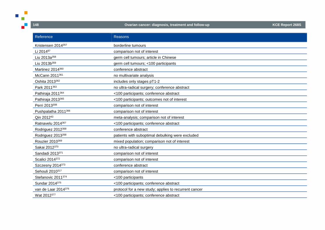
148 Ovarian cancer: diagnosis, treatment and follow-up KCE Report 268S
Reference Reasons
Kristensen 2014357 borderline tumours Li 201487 comparison not of interest Liu 2013a358 germ cell tumours; article in Chinese Liu 2013b359 germ cell tumours; <100 participants Martinez 2014360 conference abstract McCann 2011361 no multivariate analysis Oshita 2013362 includes only stages pT1-2 Park 2011363 no ultra-radical surgery; conference abstract Pathiraja 2011364 <100 participants; conference abstract Pathiraja 2013365 <100 participants; outcomes not of interest Perri 2013308 comparison not of interest Pushpalatha 2011366 comparison not of interest Qin 201243 meta-analysis; comparison not of interest Ratnavelu 2014367 <100 participants; conference abstract Rodriguez 2012368 conference abstract Rodriguez 2013268 patients with suboptimal debulking were excluded Rouzier 2010369 mixed population; comparison not of interest Sakai 2012370 no ultra-radical surgery Sandadi 2013371 comparison not of interest Scalici 2014372 comparison not of interest Szczesny 2014373 conference abstract Sehouli 2010317 comparison not of interest Stefanovic 2011374 <100 participants Sundar 2014375 <100 participants; conference abstract van de Laar 2014376 protocol for a new study; applies to recurrent cancer Wat 2012377 <100 participants; conference abstract
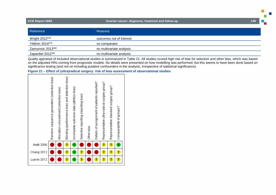
KCE Report 268S Ovarian cancer: diagnosis, treatment and follow-up 149
Reference Reasons
Wright 2012378 outcomes not of interest Yildirim 2014379 no comparator Zamurovic 2013380 no multivariate analysis Zapardiel 2012381 no multivariate analysis
Quality appraisal of included observational studies is summarized in Table 21. All studies scored high risk of bias for selection and other bias, which was based on the adjusted HRs coming from prognostic models. No details were presented on how modelling was performed, but this seems to have been done based on significance testing (and not on including putative confounders in the analysis, irrespective of statistical significance). Figure 21 – Effect of (ultra)radical surgery: risk of bias assessment of observational studies
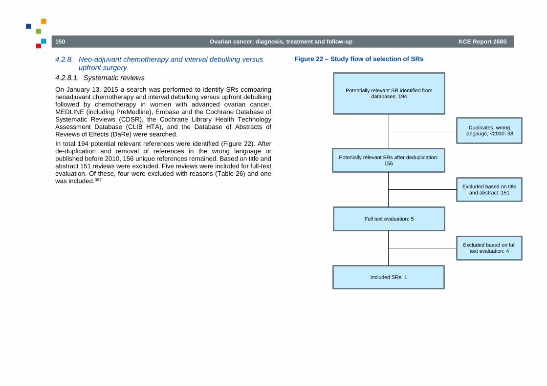
150 Ovarian cancer: diagnosis, treatment and follow-up KCE Report 268S
4.2.8. Neo-adjuvant chemotherapy and interval debulking versus upfront surgery
4.2.8.1. Systematic reviews On January 13, 2015 a search was performed to identify SRs comparing neoadjuvant chemotherapy and interval debulking versus upfront debulking followed by chemotherapy in women with advanced ovarian cancer. MEDLINE (including PreMedline), Embase and the Cochrane Database of Systematic Reviews (CDSR), the Cochrane Library Health Technology Assessment Database (CLIB HTA), and the Database of Abstracts of Reviews of Effects (DaRe) were searched. In total 194 potential relevant references were identified (Figure 22). After de-duplication and removal of references in the wrong language or published before 2010, 156 unique references remained. Based on title and abstract 151 reviews were excluded. Five reviews were included for full-text evaluation. Of these, four were excluded with reasons (Table 26) and one was included.382
Figure 22 – Study flow of selection of SRs
Potentially relevant SR identified from databases: 194
Potenially relevant SRs after deduplication: 156
Duplicates, wrong langauge, <2010: 38
Excluded based on title and abstract: 151
Full text evaluation: 5
Included SRs: 1
Excluded based on full text evaluation: 4

KCE Report 268S Ovarian cancer: diagnosis, treatment and follow-up 151
Table 26 – Excluded SRs based on full-text evaluation Reference Reason for exclusion
Dai-yuan M, Bang-xian T, Xian-fu L, Ye-qin Z, Hong-Wei C. A meta-analysis: neoadjuvant chemotherapy versus primary surgery in ovarian carcinoma FIGO stageIII and IV. World Journal of Surgical Oncology. 2013;11(267)
One of the included RCTs evaluated irrelevant comparison
Tangjitgamol S, Manusirivithaya S, Laopaiboon M, Lumbiganon P, Bryant A. Interval debulking surgery for advanced epithelial ovarian cancer. Cochrane Database Syst Rev. 2010(10):CD006014
Updated by Tangjitgamol S 2013
Tangjitgamol S, Manusirivithaya S, Laopaiboon M, Lumbiganon P, Bryant A. Interval debulking surgery for advanced epithelial ovarian cancer. Cochrane Database of Systematic Reviews. 2013;4
Wrong comparison
Vitale SG, Marilli I, Lodato M, Tropea A, Cianci A. The role of cytoreductive surgery in advanced-stage ovarian cancer: a systematic review. Updates Surg. 2013;65(4):265-70
Narrative review
Quality appraisal of selected systematic reviews
Table 27 shows the results of the risk of bias assessment for the included systematic review, using AMSTAR criteria. Table 27 – Methodological quality of the included SRs (AMSTAR) Systematic review A priori
study design
Duplicate study selection and data extraction
Compre-hensive literature search
Publica-tion status not used as inclusion
List of in- and excluded studies
Charac-teristics of included studies provided
Study quality assessed and docu-mented
Quality assess-ment used in conclus-ions
Appropriate methods to combine findings
Likelihood of publica-tion bias assessed
Conflict of interest stated
Morrison J 2012 Y Y Y Y Y Y Y Y NA NA N 4.2.8.2. RCTs On January 13, 2015 a search was performed to identify RCTs comparing neoadjuvant chemotherapy and interval debulking versus upfront debulking followed by chemotherapy in women with advanced ovarian cancer. The search date of Morrison 2012 was used as starting point. MEDLINE (including PreMedline), Embase and CENTRAL were searched, limited from 2011 onwards. In total, 249 potential relevant references were identified (Figure 23). After de-duplication and removal of references in the wrong language or published before 2011, 106 unique references remained. Based on title and abstract 101 studies were excluded. Of the remaining five studies, one study was included and four studies were excluded with reason (Table 28).293 On September 1, 2015 the search was rerun, and yielded 39 new potential relevant references. After de-duplication, 37 unique references remained. Based on title and abstract 33 studies were excluded. Of the remaining four studies, one study was included and three studies were excluded with reason (Table 29).
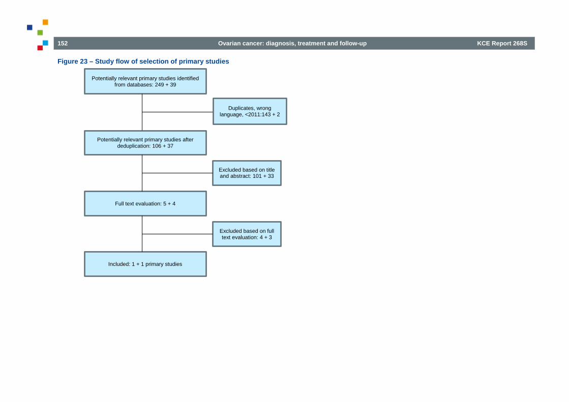
152 Ovarian cancer: diagnosis, treatment and follow-up KCE Report 268S
Figure 23 – Study flow of selection of primary studies
Potentially relevant primary studies identified from databases: 249 + 39
Duplicates, wrong language, <2011:143 + 2
Potentially relevant primary studies after deduplication: 106 + 37
Excluded based on title and abstract: 101 + 33
Full text evaluation: 5 + 4
Included: 1 + 1 primary studies
Excluded based on full text evaluation: 4 + 3

KCE Report 268S Ovarian cancer: diagnosis, treatment and follow-up 153
Table 28 – Excluded RCTs based on full-text evaluation: search January 2015 Reference Reason for exclusion
Greimel E, Kristensen G, Vergote I, Hoskins P, Burg MEL, Casado HA, et al. Quality of life in advanced ovarian cancer patients: A randomized phase III study comparing upfront debulking surgery versus neo-adjuvant chemotherapy. International journal of gynecological cancer. 2011;21(12 SUPPL. 3):S620.
Poster
Rowland M, Farris C, Lesnock J, Krivak T. Neoadjuvant chemotherapy is less costly than primary debulking surgery for treatment of advanced stage ovarian cancer in patients > 65 years old. Gynecologic oncology. 2013;131(1):278-9.
Abstract of cost-effectiveness study
van de Laar R, Zusterzeel PL, Van Gorp T, Buist MR, van Driel WJ, Gaarenstroom KN, et al. Cytoreductive surgery followed by chemotherapy versus chemotherapy alone for recurrent platinum-sensitive epithelial ovarian cancer (SOCceR trial): a multicenter randomised controlled study. BMC Cancer. 2014;14(22).
Wrong comparison
Van Meurs H.S, Tajik P, Hof M.H.P, Vergote I, Kenter G.G, Mol B.W.J, et al. Which patients benefit most from primary surgery or neoadjuvant chemotherapy in stage IIIC or IV ovarian cancer? An exploratory analysis of the European Organisation for Research and Treatment of Cancer 55971 randomised trial. Eur. J. Cancer. 2013;49(15):3191-201.
Post-hoc analysis of EORTC study
Table 29 – Excluded RCTs based on full-text evaluation: search September 2015. Reference Reason for exclusion
Onda T, Yoshikawa H, Shibata T, Nakamura K, Satoh T, Saito T, et al. Comparison of treatment invasiveness between upfront debulking surgery versus interval debulking surgery following neoadjuvant chemotherapy for stage III/IV ovarian, tubal, and peritoneal cancers in phase III randomized trial: JCOG0602. Journal of clinical oncology. 2014;32(15 SUPPL. 1).
Abstract
Rowland MR, Lesnock JL, Farris C, Kelley JL, Krivak TC. Cost-utility comparison of neoadjuvant chemotherapy versus primary debulking surgery for treatment of advanced-stage ovarian cancer in patients 65 years old or older. Am J Obstet Gynecol. 2015;212(6):763.e1-8.
Cost-utility study
Forde GK, Chang J, Ziogas A, Tewari KS, Bristow RE. Primary debulking surgery and neo-adjuvant chemotherapy in the Medicare population: An analysis of cost of care. Gynecologic oncology. 2015;137:109-10.
Abstract of cost of illness study

154 Ovarian cancer: diagnosis, treatment and follow-up KCE Report 268S
Quality appraisal of selected RCTs
Figure 24 shows the results of the risk of bias assessment for the included studies.
Figure 24 – Risk of bias summary of RCTs
4.2.9. Intra-peritoneal chemotherapy 4.2.9.1. Systematic reviews On February 23, 2015 a search was performed to identify SRs comparing postoperative intra-peritoneal chemotherapy with standard intravenous chemotherapy in women with advanced ovarian cancer. MEDLINE (including PreMedline), Embase and the Cochrane Database of Systematic Reviews (CDSR), the Cochrane Library Health Technology Assessment Database (CLIB HTA), and the Database of Abstracts of Reviews of Effects (DaRe) were searched. In total 59 potential relevant references were identified (Figure 25). After de-duplication 52 unique references remained. Based on title and abstract 46 reviews were excluded. Six reviews were included for full-text evaluation. Of these, five were excluded with reasons (Table 30) and one was included.383

KCE Report 268S Ovarian cancer: diagnosis, treatment and follow-up 155
Figure 25 – PRISMA flowchart selection of SRs intraperitoneal chemotherapy
Potentially relevant SRs identified from databases: 59
Duplicates: 7
Potentially relevant SRs after deduplication: 52
Full text evaluation: 6
Excluded based on title and abstract: 46
Excluded based on full text evaluation: 5
Included SRs: 1
156 Ovarian cancer: diagnosis, treatment and follow-up KCE Report 268S
Table 30 – Intraperitoneal chemotherapy: excluded SRs based on full text selection Reference Reason for exclusion
Han 2014 Abstract Francis 2014 Abstract Bharaswadkar 2014 Abstract Morgan 2013 Guideline without report of systematic review/critical appraisal Hodeib 2013 Abstract Jaaback 2011 Included
Table 31 shows the results of the risk of bias assessment for the included systematic review, using AMSTAR criteria.
Table 31 – Intraperitoneal chemotherapy: methodological quality of the included SR (AMSTAR) Systematic review A priori
study design
Duplicate study selection and data extraction
Compre-hensive literature search
Publica-tion status not used as inclusion
List of in- and excluded studies
Charac-teristics of included studies provided
Study quality assessed and docu-mented
Quality assess-ment used in conclus-ions
Appropriate methods to combine findings
Likelihood of publica-tion bias assessed
Conflict of interest stated
Jaaback K 2011 Y Y Y Y Y Y Y Y Y (Y) N 4.2.9.2. Randomized controlled trials On February 23, 2015 a search was performed to identify RCTs comparing postoperative intra-peritoneal chemotherapy with standard intravenous chemotherapy in women with advanced ovarian cancer. The search date of Jaaback 2011 was used as starting point. MEDLINE (including PreMedline), Embase and CENTRAL were searched, limited from 2011 onwards. In total, 380 potential relevant references were identified (Figure 26). After de-duplication, 335 unique references remained. Based on title and abstract 301 studies were excluded. Of the remaining 34 studies, 1 study was included and 33 studies were excluded with reason (Table 32).384

KCE Report 268S Ovarian cancer: diagnosis, treatment and follow-up 157
Figure 26 – PRISMA flowchart selection of RCTs intraperitoneal chemotherapy
Potentially relevant RCTs identified from databases: 380
Duplicates: 45
Potentially relevant RCTs after deduplication: 335
Excluded based on title and abstract: 301
Full text evaluation: 34
Excluded based on full text evaludation: 33
Included RCTs: 1

158 Ovarian cancer: diagnosis, treatment and follow-up KCE Report 268S
Table 32 – Intraperitoneal chemotherapy: excluded RCTs based on full text selection Reference Reason for exclusion
Zhao 2015 No surgery, both study arms receive IP chemotherapy (cisplatin vs cisplatin + bevacizumab) Wright 2014 Abstract, no RCT (cohort study) Schmalfeldt 2014 Abstract, no RCT, HIPEC Mousavi 2014 Arabic Massari 2014 HIPEC, cohort study Kwa 2014 Narrative review Francis 2014 Abstract of review Chiva 2014 Review of retrospective data re: HIPEC Chang 2014 Abstract of SR Bowles 2014 No RCT (cohort study) Zhao 2013 Conference abstract Zhang 2013 Conference abstract, identical to Zhao 2013 Ubago-Pérez 2013 Compares HIPEC with IV/IP chemotherapy Tsilimparis 2013 No RCT (cohort study) Simkens 2013 No RCT (case report) Marth 2013 Abstract of narrative review Markman 2013 Narrative review Grosso 2013 Abstract, RCT including HIPEC, no results reported Fujiwara 2013 Narrative review von Gruenigen 2012 Included. Additional results QoL GOG 172 Rubino 2012 Narrative review Fujiwara 2012 Editorial De Bree 2012 Narrative review Chan 2012 SR, included Barlin 2012 No RCT (cohort study)

KCE Report 268S Ovarian cancer: diagnosis, treatment and follow-up 159
Reference Reason for exclusion
Ansaloni 2012 Letter Sharma 2011 Abstract, no relevant outcomes reported Mackay 2011 Narrative review Le 2011 No RCT (retrospective non-randomized comparative study) Landrum 2011 No RCT (single-arm phase II study) Fujiwara 2011 Protocol only Deraco 2011 No RCT, (cohort study) Cascales 2011 Letter Armstrong 2011 Discussion paper
4.2.10. First-line weekly (dose dense) chemotherapy 4.2.10.1. Systematic reviews On April 15, 2015 a search was performed to identify SRs comparing postoperative intra-peritoneal chemotherapy with standard intravenous chemotherapy in women with advanced ovarian cancer. MEDLINE (including PreMedline), Embase and the Cochrane Database of Systematic Reviews (CDSR), the Cochrane Library Health Technology Assessment Database (CLIB HTA), and the Database of Abstracts of Reviews of Effects (DaRe) were searched. In total 333 potential relevant references were identified (Figure 27). After de-duplication 317 unique references remained. Based on title and abstract 311 reviews were excluded. Six reviews were included for full-text evaluation. Of these, five were excluded with reasons (Table 33) and one was included.385 The review of Covens et al. performed a comprehensive search for randomized controlled trials comparing different chemotherapy schedules for first-line treatment of advanced ovarian cancer. No RCT comparing dose dense chemotherapy with 3-weekly administration was identified. The search date of Covens et al. (2001) was used as a starting point for the selection of RCTs.

160 Ovarian cancer: diagnosis, treatment and follow-up KCE Report 268S
Figure 27 – PRISMA flowchart selection of SRs weekly (dose dense) chemotherapy
Potentially relevant SRs identified from databases: 333
Duplicates: 16
Potentially relevant SRs after deduplication: 317
Full text evaluation: 6
Excluded based on title and abstract: 311
Excluded based on full tekst evaluation: 5
SRs included: 1

KCE Report 268S Ovarian cancer: diagnosis, treatment and follow-up 161
Table 33 – Weekly (dose-dense) chemotherapy: excluded SRs based on full text selection Reference Reason for exclusion
Fizazi 2000 Not in epithelial ovarian cancer Covens 2002 Included as starting point for text selection Kyrigou 2006 Data on different regimens/schedules only reported in narrative summary Torri 1993 Not comparison of interest, reference list checked Ben-David 1995 Not comparison of interest, reference list checked Stewart 1999 Comparison of interest not included
4.2.10.2. Randomized controlled trials On April 15, 2015 a search was performed to identify RCTs comparing postoperative intra-peritoneal chemotherapy with standard intravenous chemotherapy in women with advanced ovarian cancer. MEDLINE (including PreMedline), Embase and CENTRAL were searched from inception. In total, 3276 potential relevant references were identified (Figure 28). After de-duplication, 2831 unique references remained. For study selection, the search date of Covens et al. was used as a starting point. Based on publication date, title and abstract 2802 studies were excluded. Of the remaining 29 studies, 4 papers386-389 were included and 25 studies were excluded with reason (Table 34). Three386-388 of the four publications reported on the same trial, so finally, only two studies (but four publications) were included.

162 Ovarian cancer: diagnosis, treatment and follow-up KCE Report 268S
Figure 28 – PRISMA flowchart selection of RCTs weekly (dose dense) chemotherapy
Potentially relevant RCTs identified from databases: 3276
Duplicates: 445
Excluded based on title and abstract: 2802
Potentially relevant RCTs after deduplication: 2831
Full text evaluation: 29
Excluded based on full text evaluation: 25
Included RCTs: 4

KCE Report 268S Ovarian cancer: diagnosis, treatment and follow-up 163
Table 34 – Weekly (dose-dense) chemotherapy: excluded RCTs based on full text selection Reference Reason for exclusion
Wu 2001 Excluded, weekly versus monthly schedules, continued until intolerable toxicity De Jongh 2002 Excluded, prior platinum-based chemotherapy allowed, dose-escalation study Samson 2004 Narrative summary of conference Shen 2005 Excluded, Chinese, possibly duplicate of Shen 2006 Van der Burg 2005 Excluded, Narrative review Vasey 2005 Excluded, Narrative review Shen 2006 Excluded, abstract Spriggs 2007 Excluded, compares two 3-weekly schedules with different infusion durations Sehouli 2008 Excluded, non-randomized phase I study Van der Burg 2009 abstract Katsumata 2009 Included Fruscio 2011 Excluded, cisplatin monotherapy in both arms Van der Burg 2011 Excluded, Narrative review Williams 2011 Excluded, not comparison of interest (long versus short duration of infusion) Dalton 2012 Cost-effectiveness study, no new data Gonzalez-Martin 2012 Excluded, Abstract, includes bevacizumab Katsumata 2012 Excluded, Abstract JGOG 3016 trial Chan 2013 Excluded, Abstract Gonzalez-Martin 2013 Excluded, Abstract, includes bevacizumab Hook 2013 Excluded, Abstract Katsumata 2013 Included Pignata 2013 Excluded, Abstract Harano 2014 Included Hook 2014 Excluded, Abstract ICON 8 Monk 2014 Excluded, Abstract GOG 262 Pignata 2014 Included

164 Ovarian cancer: diagnosis, treatment and follow-up KCE Report 268S
Reference Reason for exclusion
Slaughter 2014 Narrative review Van der Burg 2014 Excluded, Only chemotherapy before surgery compares weekly vs 3-weekly, after IDS second randomization to 3 or 6 cycles
of 3-weekly chemotherapy Kumar 2015 Narrative review
Figure 29 shows the results of the risk of bias assessment for the included RCTs
Figure 29 – Weekly (dose-dense) chemotherapy: risk of bias summary of RCTs
4.2.11. Routine CA125 measurement during follow-up 4.2.11.1. Systematic reviews On January 27, 2015 a search was performed to identify SRs evaluating routine CA125 measurements during follow-up in women with ovarian cancer. MEDLINE (including PreMedline), Embase and the Cochrane Database of Systematic Reviews (CDSR), the Cochrane Library Health Technology Assessment Database (CLIB HTA), and the Database of Abstracts of Reviews of Effects (DaRe) were searched. In total 171 potential relevant references were identified (Figure 30). After de-duplication and removal of references in the wrong language or published before 2010, 140 unique references remained. Based on title and abstract 137 reviews were excluded. Three reviews were included for full-text evaluation. Of these, two were excluded with reasons (Table 35) and one was included.390

KCE Report 268S Ovarian cancer: diagnosis, treatment and follow-up 165
Figure 30 – CA125 measurements during FU: PRISMA flowchart selection of SRs
Potentially relevant SRs identified from databases: 171
Duplicates, wrong language, <2010: 31
Potentially relevant SRs after deduplication: 140
Full text evaluation: 3
Excluded based on title and abstract: 137
Excluded based on full text evaluation: 2
Included SRs: 1

166 Ovarian cancer: diagnosis, treatment and follow-up KCE Report 268S
Table 35 – Routine CA125 measurement during follow-up: excluded SRs based on full-text evaluation Reference Reason for exclusion
Kew F, Galaal K, Bryant A, Naik R. Evaluation of follow-up strategies for patients with epithelial ovarian cancer following completion of primary treatment. Cochrane Database Syst Rev. 2011(6):CD006119
Updated by Clarke T 2014
Geurts SM, de Vegt F, van Altena AM, van Dijck JA, Tjan-Heijnen VC, Verbeek AL, et al. Considering early detection of relapsed ovarian cancer: a review of the literature. Int J Gynecol Cancer. 2011;21(5):837-45
No quality appraisal of included studies
Table 36 shows the results of the risk of bias assessment for the included systematic review, using AMSTAR criteria. Table 36 – Routine CA125 measurement during follow-up: methodological quality of the included SR (AMSTAR) Systematic review A priori
study design
Duplicate study selection and data extraction
Compre-hensive literature search
Publica-tion status not used as inclusion
List of in- and excluded studies
Charac-teristics of included studies provided
Study quality assessed and docu-mented
Quality assess-ment used in conclus-ions
Appropriate methods to combine findings
Likelihood of publica-tion bias assessed
Conflict of interest stated
Clarke T 2014 Y Y Y Y Y Y Y Y NA NA N 4.2.11.2. Randomized controlled trials On January 27, 2015 a search was performed to identify RCTs evaluating routine CA125 measurements during follow-up in women with ovarian cancer. The search date of Clarke 2014 was used as starting point. MEDLINE (including PreMedline), Embase and CENTRAL were searched, limited from 2013 onwards. In total, 371 potential relevant references were identified (Figure 31). After de-duplication and removal of references in the wrong language or published before 2013, 61 unique references remained. Based on title and abstract all 61 studies were excluded. Figure 32 shows the results of the risk of bias assessment for the single RCT391 included in Clarke 2014.
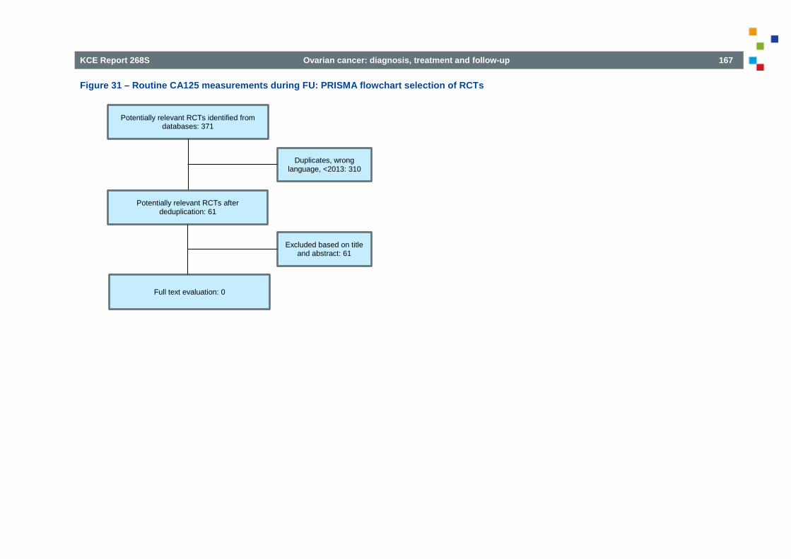
KCE Report 268S Ovarian cancer: diagnosis, treatment and follow-up 167
Figure 31 – Routine CA125 measurements during FU: PRISMA flowchart selection of RCTs
Potentially relevant RCTs identified from databases: 371
Duplicates, wrong language, <2013: 310
Potentially relevant RCTs after deduplication: 61
Excluded based on title and abstract: 61
Full text evaluation: 0

168 Ovarian cancer: diagnosis, treatment and follow-up KCE Report 268S
Figure 32 – Routine CA125 measurement during follow-up: Risk of bias summary of RCTs
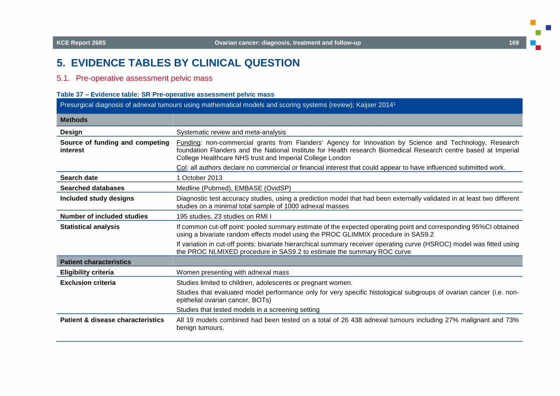
KCE Report 268S Ovarian cancer: diagnosis, treatment and follow-up 169
5. EVIDENCE TABLES BY CLINICAL QUESTION 5.1. Pre-operative assessment pelvic mass
Table 37 – Evidence table: SR Pre-operative assessment pelvic mass Presurgical diagnosis of adnexal tumours using mathematical models and scoring systems (review); Kaijser 20141
Methods
Design Systematic review and meta-analysis Source of funding and competing interest
Funding: non-commercial grants from Flanders’ Agency for Innovation by Science and Technology, Research foundation Flanders and the National Institute for Health research Biomedical Research centre based at Imperial College Healthcare NHS trust and Imperial College London CoI: all authors declare no commercial or financial interest that could appear to have influenced submitted work.
Search date 1 October 2013 Searched databases Medline (Pubmed), EMBASE (OvidSP) Included study designs Diagnostic test accuracy studies, using a prediction model that had been externally validated in at least two different
studies on a minimal total sample of 1000 adnexal masses Number of included studies 195 studies, 23 studies on RMI I Statistical analysis If common cut-off point: pooled summary estimate of the expected operating point and corresponding 95%CI obtained
using a bivariate random effects model using the PROC GLIMMIX procedure in SAS9.2 If variation in cut-off points: bivariate hierarchical summary receiver operating curve (HSROC) model was fitted using the PROC NLMIXED procedure in SAS9.2 to estimate the summary ROC curve
Patient characteristics Eligibility criteria Women presenting with adnexal mass Exclusion criteria Studies limited to children, adolescents or pregnant women.
Studies that evaluated model performance only for very specific histological subgroups of ovarian cancer (i.e. non-epithelial ovarian cancer, BOTs) Studies that tested models in a screening setting
Patient & disease characteristics All 19 models combined had been tested on a total of 26 438 adnexal tumours including 27% malignant and 73% benign tumours.

170 Ovarian cancer: diagnosis, treatment and follow-up KCE Report 268S
Presurgical diagnosis of adnexal tumours using mathematical models and scoring systems (review); Kaijser 20141
Interventions Index test Risk of malignancy index I or other risk model Target condition Ovarian borderline or invasive cancer Reference standard histopathology Results Sensitivity RMI 1: 0.72 [95%CI 0.67-0.76]
RMI 2: 0.75 [95%CI 0.75-0.80]] IOTA simple rules: 0.93 [0.89-0.95]
Specificity RMI 1: 0.92 [95%CI 0.89-0.93] RMI 2: 0.87 [95%CI 0.84-0.90] IOTA simple rules: 0.81 [95%CI 0.76-0.85]
Notes Limitations Comparative accuracy between considered tests not restricted to studies that have evaluated the tests/models in the
same individuals. 41% of studies were retrospective in design.
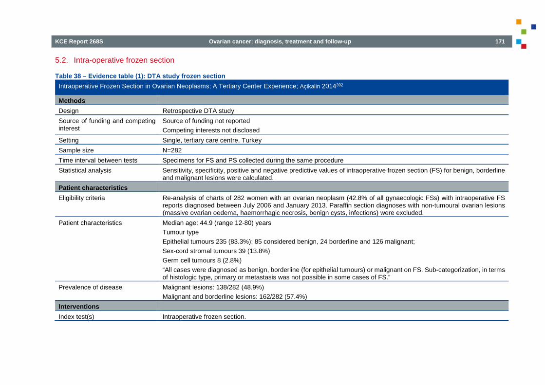
KCE Report 268S Ovarian cancer: diagnosis, treatment and follow-up 171
5.2. Intra-operative frozen section
Table 38 – Evidence table (1): DTA study frozen section Intraoperative Frozen Section in Ovarian Neoplasms; A Tertiary Center Experience; Açikalin 2014392
Methods Design Retrospective DTA study Source of funding and competing interest
Source of funding not reported Competing interests not disclosed
Setting Single, tertiary care centre, Turkey Sample size N=282 Time interval between tests Specimens for FS and PS collected during the same procedure Statistical analysis Sensitivity, specificity, positive and negative predictive values of intraoperative frozen section (FS) for benign, borderline
and malignant lesions were calculated. Patient characteristics Eligibility criteria Re-analysis of charts of 282 women with an ovarian neoplasm (42.8% of all gynaecologic FSs) with intraoperative FS
reports diagnosed between July 2006 and January 2013. Paraffin section diagnoses with non-tumoural ovarian lesions (massive ovarian oedema, haemorrhagic necrosis, benign cysts, infections) were excluded.
Patient characteristics Median age: 44.9 (range 12-80) years Tumour type Epithelial tumours 235 (83.3%); 85 considered benign, 24 borderline and 126 malignant; Sex-cord stromal tumours 39 (13.8%) Germ cell tumours 8 (2.8%) “All cases were diagnosed as benign, borderline (for epithelial tumours) or malignant on FS. Sub-categorization, in terms of histologic type, primary or metastasis was not possible in some cases of FS.”
Prevalence of disease Malignant lesions: 138/282 (48.9%) Malignant and borderline lesions: 162/282 (57.4%)
Interventions Index test(s) Intraoperative frozen section.
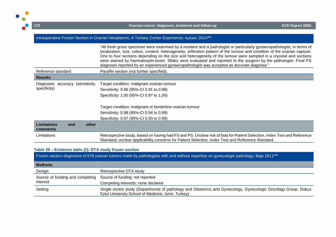
172 Ovarian cancer: diagnosis, treatment and follow-up KCE Report 268S
Intraoperative Frozen Section in Ovarian Neoplasms; A Tertiary Center Experience; Açikalin 2014392
“All fresh gross specimen were examined by a resident and a pathologist or particularly gynaecopathologist, in terms of localization, size, colour, content, heterogeneity, infiltration pattern of the tumour and condition of the ovarian capsule. One to four sections depending on the size and heterogeneity of the tumour were sampled in a cryostat and sections were stained by haematoxylin-eosin. Slides were evaluated and reported to the surgeon by the pathologist. Final PS diagnosis reported by an experienced gynaecopathologist was accepted as accurate diagnose.”
Reference standard Paraffin section (not further specified). Results Diagnostic accuracy (sensitivity, specificity)
Target condition: malignant ovarian tumour Sensitivity: 0.96 (95%-CI 0.91 to 0.98) Specificity: 1.00 (95%-CI 0.97 to 1.00) Target condition: malignant or borderline ovarian tumour Sensitivity: 0.98 (95%-CI 0.94 to 0.99) Specificity: 0.97 (95%-CI 0.93 to 0.99)
Limitations and other comments
Limitations Retrospective study, based on having had FS and PS. Unclear risk of bias for Patient Selection, Index Test and Reference Standard; unclear applicability concerns for Patient Selection, Index Test and Reference Standard.
Table 39 – Evidence table (2): DTA study frozen section Frozen section diagnoses of 578 ovarian tumors made by pathologists with and without expertise on gynecologic pathology; Bige 2011393
Methods Design Retrospective DTA study Source of funding and competing interest
Source of funding: not reported Competing interests: none declared
Setting Single centre study (Departments of pathology and Obstetrics and Gynecology, Gynecologic Oncology Group, Dokuz Eylul University School of Medicine, Izmir, Turkey)

KCE Report 268S Ovarian cancer: diagnosis, treatment and follow-up 173
Frozen section diagnoses of 578 ovarian tumors made by pathologists with and without expertise on gynecologic pathology; Bige 2011393
Sample size N=578 women operated with the suspicion of ovarian neoplasms; 519 analysed (definitive diagnosis of 14 patients (2.4%) could not be obtained; in 23 patients, no ovarian issue could be identified; in 22 cases, metastasis to the ovaries was detected “but there were no discordance between the pathologists”).
Time interval between tests Specimens for FS and PS collected during the same procedure Statistical analysis Sensitivity, specificity, positive and negative predictive values of intraoperative FS for benign, borderline and malignant
lesions were calculated. Subgroups of results of frozen section diagnosis by gynaecological pathologists (Group 1) and non-gynaecological pathologists (Group 2) were also compared.
Patient characteristics Eligibility criteria Retrospective analysis of reports of frozen section diagnosis of 578 patients who underwent surgery for ovarian tumours
between January 2002 and December 2010. Indications of frozen section diagnosis were radiologically or macroscopically benign appearing ovarian masses with high Ca125 values, history of malignancy other than ovary and fertility preserving surgery for young women.
Patient characteristics Mean age (range) in years: benign 41 (13–82), borderline 45 (25–63) and malignant 53 (24–74) Tumour type: not reported. Deferred cases: 14/578 (2.4%)
Prevalence of disease Malignant lesions: 121/519 (23.3%) Malignant and borderline lesions: 148/519 (28.5%)
Interventions Index test(s) Intraoperative frozen section (categories: primary epithelial ovarian tumour (benign, borderline and malignant); primary
ovarian germ cell tumour, ovarian metastatic carcinoma, benign non-neoplastic lesions and no definitive diagnosis). “Following incision to the abdomen either by laparoscopy or by laparotomy, the unfixed fresh masses were delivered immediately to the pathologist with a detailed form including all of the clinical findings of the patient. After macroscopic examination, two to five sections especially from solid areas were obtained according to the type and size of the mass. Frozen procedures were performed through cryostat. The sections were 7–8 μm in thickness and were stained with haematoxylin-eosin. Meanwhile, the imprint was conducted. All of the stained sections were examined under the light microscope by at least two pathologists who were consultants of that month in question.”
Reference standard Paraffin block. “Paraffin blocks were prepared for the final diagnosis from the sections obtained from each 1 cm of the greatest diameter of the mass. Sections 4-5 μm in thickness were obtained from these paraffin blocks. Thereafter these sections were stained with haematoxylin-eosin and examined.”
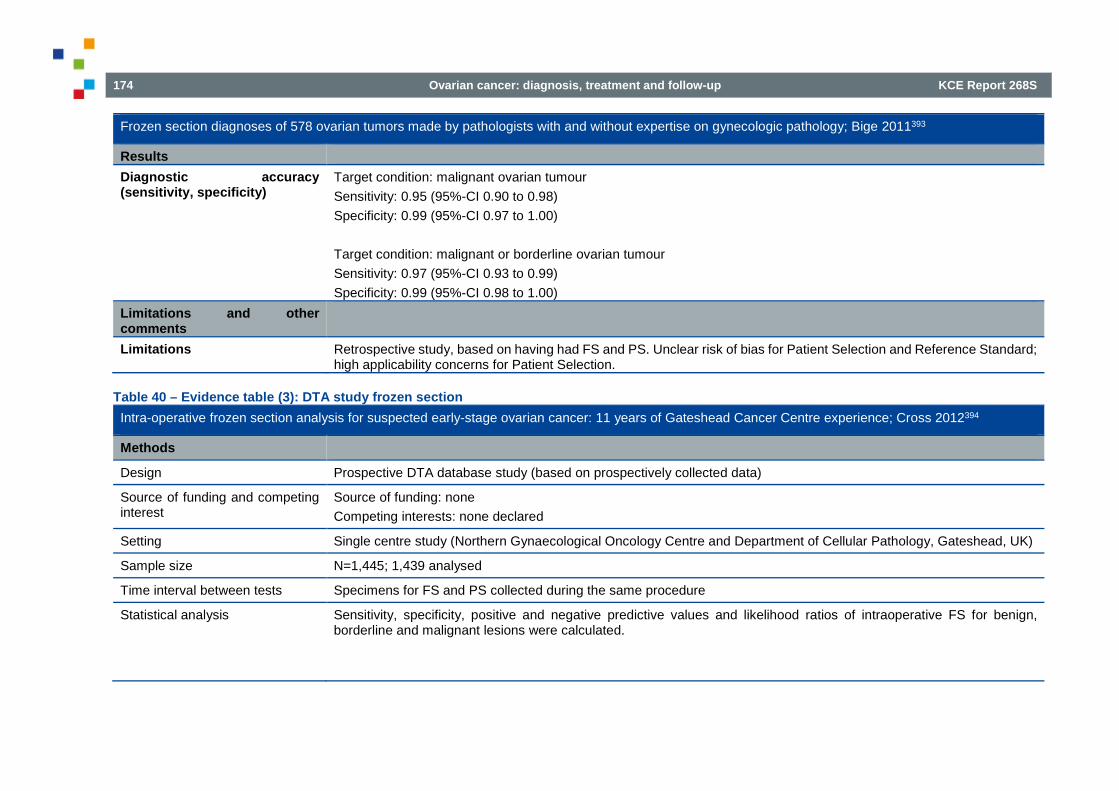
174 Ovarian cancer: diagnosis, treatment and follow-up KCE Report 268S
Frozen section diagnoses of 578 ovarian tumors made by pathologists with and without expertise on gynecologic pathology; Bige 2011393
Results Diagnostic accuracy (sensitivity, specificity)
Target condition: malignant ovarian tumour Sensitivity: 0.95 (95%-CI 0.90 to 0.98) Specificity: 0.99 (95%-CI 0.97 to 1.00) Target condition: malignant or borderline ovarian tumour Sensitivity: 0.97 (95%-CI 0.93 to 0.99) Specificity: 0.99 (95%-CI 0.98 to 1.00)
Limitations and other comments
Limitations Retrospective study, based on having had FS and PS. Unclear risk of bias for Patient Selection and Reference Standard; high applicability concerns for Patient Selection.
Table 40 – Evidence table (3): DTA study frozen section Intra-operative frozen section analysis for suspected early-stage ovarian cancer: 11 years of Gateshead Cancer Centre experience; Cross 2012394
Methods
Design Prospective DTA database study (based on prospectively collected data)
Source of funding and competing interest
Source of funding: none Competing interests: none declared
Setting Single centre study (Northern Gynaecological Oncology Centre and Department of Cellular Pathology, Gateshead, UK)
Sample size N=1,445; 1,439 analysed
Time interval between tests Specimens for FS and PS collected during the same procedure
Statistical analysis Sensitivity, specificity, positive and negative predictive values and likelihood ratios of intraoperative FS for benign, borderline and malignant lesions were calculated.

KCE Report 268S Ovarian cancer: diagnosis, treatment and follow-up 175
Intra-operative frozen section analysis for suspected early-stage ovarian cancer: 11 years of Gateshead Cancer Centre experience; Cross 2012394
Patient characteristics
Eligibility criteria Ovarian lesions sent for routine intra-operative FS between January 2000 and December 2010 for suspected ovarian cancer. Any other non-ovarian FSs (e.g. lymph nodes, non-ovarian masses) were excluded. Six deferred cases were excluded from the overall analysis.
Patient characteristics Mean age: not reported Tumour type Serous 486 (33.8%) Mucinous 267 (18.6%) Endometrioid 151 (10.5%) Clear cell 58 (4.0%) Urothelial/Brenner 20 (1.4%) Germ cell 78 (5.4%) Sex-cord stromal 138 (9.6%) Secondary tumour diagnoses 69 (4.8%) “Benign frozen section ovarian mass diagnoses” 169 (11.7%) Not mentioned: 3 Deferred cases: 6/1,445 (0.4%)
Prevalence of disease Malignant lesions: 516/1,439 (35.9%) Malignant and borderline lesions: 659/1,439 (45.8%)
Interventions Index test(s) Intraoperative frozen section.
“The specimen, once removed, is transported by a hospital porter direct to the histology laboratory and handed over to the laboratory staff. The pathologist on duty inspects the specimen and, after describing it, routinely takes up to two pieces of tissue for FS analysis. These are then processed and, after hand staining, are given to the duty pathologist for reporting. The result is then telephoned to the surgeon involved in the operating theatre. The FS (and subsequent paraffin section) is reported by one of five experienced consultant histopathologists. Two of these act as leads for gynaecological pathology reporting, but all five take part in the routine reporting of the FS on a rota basis.”
Reference standard Paraffin block (not further specified).

176 Ovarian cancer: diagnosis, treatment and follow-up KCE Report 268S
Intra-operative frozen section analysis for suspected early-stage ovarian cancer: 11 years of Gateshead Cancer Centre experience; Cross 2012394
Results
Diagnostic accuracy (sensitivity, specificity)
Target condition: malignant ovarian tumour Sensitivity: 0.80 (95%-CI 0.77 to 0.84) Specificity: 0.99 (95%-CI 0.99 to 1.00) Target condition: malignant or borderline ovarian tumour Sensitivity: 0.91 (95%-CI 0.89 to 0.93) Specificity: 0.99 (95%-CI 0.97 to 0.99)
Limitations and other comments
Limitations Unclear risk of bias for Reference Standard.
Table 41 – Evidence table (4): DTA study frozen section Accuracy of Intra-operative Frozen Section Analysis of Ovarian Tumours; Gorisek 2009395
Methods Design Retrospective DTA study Source of funding and competing interest
Source of funding: not reported Competing interests: none declared
Setting Single centre study (Maribor Teaching Hospital, Department of Gynaecologic Oncology, Slovenia) Sample size N=131 Time interval between tests Specimens for FS and PS collected during the same procedure Statistical analysis Sensitivity, specificity, positive and negative predictive values of intraoperative FS for benign, borderline and malignant
lesions were calculated Patient characteristics Eligibility criteria Women treated for benign, borderline and malignant ovarian tumours between 1 January 1993 and 31 December 2001 Patient characteristics Mean age: not reported.
Tumour type
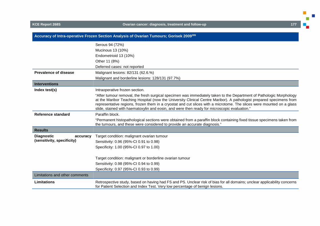
KCE Report 268S Ovarian cancer: diagnosis, treatment and follow-up 177
Accuracy of Intra-operative Frozen Section Analysis of Ovarian Tumours; Gorisek 2009395
Serous 94 (72%) Mucinous 13 (10%) Endometrioid 13 (10%) Other 11 (8%) Deferred cases: not reported
Prevalence of disease Malignant lesions: 82/131 (62.6.%) Malignant and borderline lesions: 128/131 (97.7%)
Interventions Index test(s) Intraoperative frozen section.
“After tumour removal, the fresh surgical specimen was immediately taken to the Department of Pathologic Morphology at the Maribor Teaching Hospital (now the University Clinical Centre Maribor). A pathologist prepared specimens from representative regions, frozen them in a cryostat and cut slices with a microtome. The slices were mounted on a glass slide, stained with haematoxylin and eosin, and were then ready for microscopic evaluation.”
Reference standard Paraffin block. “Permanent histopathological sections were obtained from a paraffin block containing fixed tissue specimens taken from the tumours, and these were considered to provide an accurate diagnosis.”
Results Diagnostic accuracy (sensitivity, specificity)
Target condition: malignant ovarian tumour Sensitivity: 0.96 (95%-CI 0.91 to 0.98) Specificity: 1.00 (95%-CI 0.97 to 1.00) Target condition: malignant or borderline ovarian tumour Sensitivity: 0.98 (95%-CI 0.94 to 0.99) Specificity: 0.97 (95%-CI 0.93 to 0.99)
Limitations and other comments
Limitations Retrospective study, based on having had FS and PS. Unclear risk of bias for all domains; unclear applicability concerns for Patient Selection and Index Test. Very low percentage of benign lesions.

178 Ovarian cancer: diagnosis, treatment and follow-up KCE Report 268S
Table 42 – Evidence table (5): DTA study frozen section Accuracy of intra-operative frozen section in the diagnosis of ovarian tumours; Ilker 2011396
Methods
Design Retrospective DTA study Source of funding and competing interest
Source of funding: not reported Competing interests: not reported
Setting Single centre study (Zonguldak Maternity Hospital, Zonguldak, Turkey) Sample size N=278; N=266 analysed Time interval between tests Specimens for FS and PS collected during the same procedure Statistical analysis Sensitivity, specificity, positive and negative predictive values of intraoperative FS for benign, borderline and malignant
lesions were calculated. Patient characteristics Eligibility criteria Women who underwent surgery for ovarian masses and on whom frozen section was performed between January 2002
and December 2008. Twelve patients (4.3%) for whom a definitive result was not obtained from FS were excluded.. Patient characteristics Mean age: 42.5 (± 13.5) years
Tumour type Epithelial tumours 16 (6.0%) Germ cell tumours 10 (3.8%) Other malignant tumours 2 (0.8%) Borderline tumours 9 (3.4%) Benign tumours 229 (86.1%) Deferred cases: 12/278 (4.3%)
Prevalence of disease Malignant lesions: 28/266 (10.5%) Malignant and borderline lesions: 37/266 (13.9%)
Interventions Index test(s) Intraoperative frozen section.
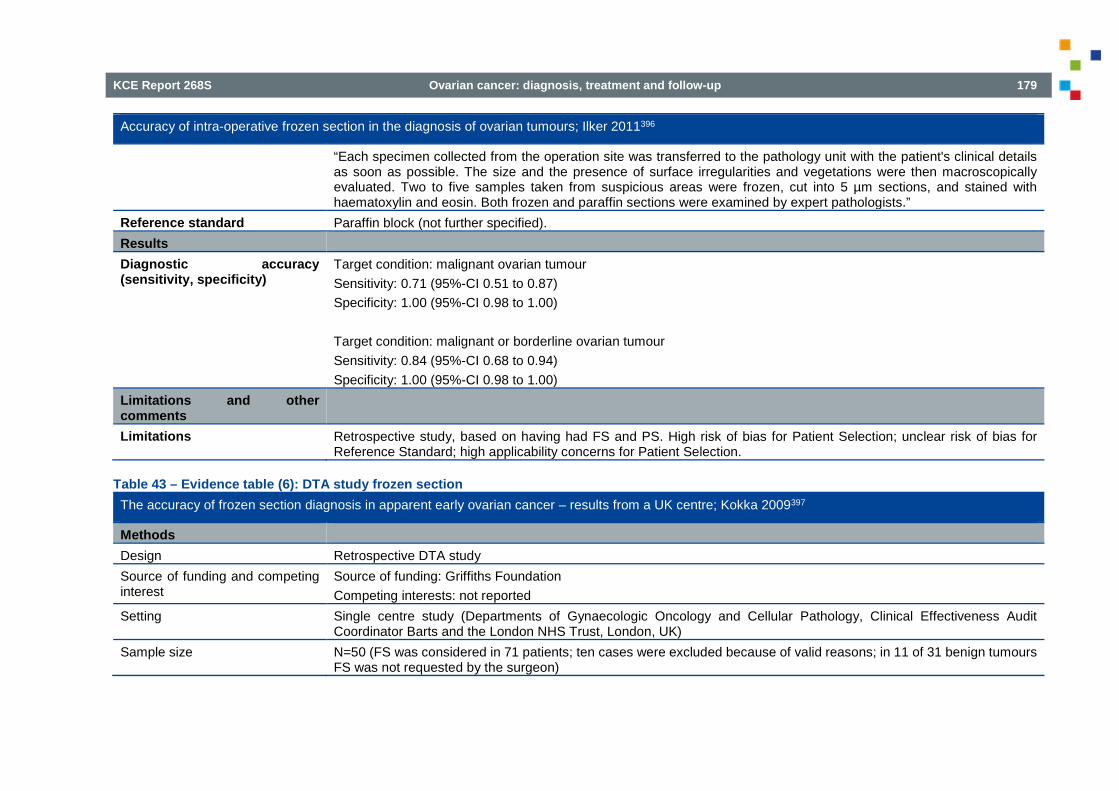
KCE Report 268S Ovarian cancer: diagnosis, treatment and follow-up 179
Accuracy of intra-operative frozen section in the diagnosis of ovarian tumours; Ilker 2011396
“Each specimen collected from the operation site was transferred to the pathology unit with the patient's clinical details as soon as possible. The size and the presence of surface irregularities and vegetations were then macroscopically evaluated. Two to five samples taken from suspicious areas were frozen, cut into 5 µm sections, and stained with haematoxylin and eosin. Both frozen and paraffin sections were examined by expert pathologists.”
Reference standard Paraffin block (not further specified). Results Diagnostic accuracy (sensitivity, specificity)
Target condition: malignant ovarian tumour Sensitivity: 0.71 (95%-CI 0.51 to 0.87) Specificity: 1.00 (95%-CI 0.98 to 1.00) Target condition: malignant or borderline ovarian tumour Sensitivity: 0.84 (95%-CI 0.68 to 0.94) Specificity: 1.00 (95%-CI 0.98 to 1.00)
Limitations and other comments
Limitations Retrospective study, based on having had FS and PS. High risk of bias for Patient Selection; unclear risk of bias for Reference Standard; high applicability concerns for Patient Selection.
Table 43 – Evidence table (6): DTA study frozen section The accuracy of frozen section diagnosis in apparent early ovarian cancer – results from a UK centre; Kokka 2009397
Methods Design Retrospective DTA study Source of funding and competing interest
Source of funding: Griffiths Foundation Competing interests: not reported
Setting Single centre study (Departments of Gynaecologic Oncology and Cellular Pathology, Clinical Effectiveness Audit Coordinator Barts and the London NHS Trust, London, UK)
Sample size N=50 (FS was considered in 71 patients; ten cases were excluded because of valid reasons; in 11 of 31 benign tumours FS was not requested by the surgeon)

180 Ovarian cancer: diagnosis, treatment and follow-up KCE Report 268S
The accuracy of frozen section diagnosis in apparent early ovarian cancer – results from a UK centre; Kokka 2009397
Time interval between tests Specimens for FS and PS collected during the same procedure Statistical analysis Sensitivity, specificity, positive and negative predictive values of intraoperative FS for benign, borderline and malignant
lesions were calculated. Patient characteristics Eligibility criteria “Between October 2006 and May 2008 FS was considered in 71 patients with suspected early ovarian cancer. Ten cases
were excluded: three because of extensive ovarian disease at laparotomy that had not been detected on preoperative imaging and seven because of non-ovarian neoplasms diagnosed through operative findings (three cases) or FS (four cases).” “Furthermore, an experienced surgeon is likely to identify certain types of benign ovarian masses on inspection alone, without using FS. Accordingly, in 11/31 benign tumours FS was not requested.”
Patient characteristics Mean age: 54 years (range not reported) Tumour type Serous 25 (40.9%) Mucinous 17 (27.9%) Clear cell 4 (6.6%) Endometrioid 1 (1.6%) Sarcoma 1 (1.6%) Other 13 (21.3%) RMI: 10 RMI <25, 35 RMI 25-250, 16 RMI >250 Deferred cases: not reported
Prevalence of disease Malignant lesions: 20/50 (40.0%) Malignant and borderline lesions: 30/50 (60.0%)
Interventions Index test(s) Intraoperative frozen section (not specified). Reference standard Paraffin block (not further specified). Results Diagnostic accuracy (sensitivity, specificity)
NB: accuracy parameters calculated for 50 out of 61 cases in which FS was deemed necessary

KCE Report 268S Ovarian cancer: diagnosis, treatment and follow-up 181
The accuracy of frozen section diagnosis in apparent early ovarian cancer – results from a UK centre; Kokka 2009397
Target condition: malignant ovarian tumour Sensitivity: 0.95 (95%-CI 0.75 to 1.00) Specificity: 1.00 (95%-CI 0.88 to 1.00) Target condition: malignant or borderline ovarian tumour Sensitivity: 0.93 (95%-CI 0.78 to 0.99) Specificity: 0.90 (95%-CI 0.68 to 0.99)
Limitations and other comments
Limitations Retrospective study, based on having had FS and PS (letter to the editor). Unclear risk of bias for Index Test and Reference Standard; no applicability concerns.
Table 44 – Evidence table (7): DTA study frozen section How accurate is intraoperative frozen section in the diagnosis of ovarian tumors? Malipatil 2013398
Methods Design Retrospective DTA study Source of funding and competing interest
Source of funding: none Competing interests: none declared
Setting Single centre study (Department of Pathology, St. John’s Medical College, Bangalore, India) Sample size N= 218 (N=377 referrals for diagnosis; intraoperative diagnosis sought in 233 (apparently 223?) cases; diagnosis deferred
in five cases due to extensive areas of haemorrhage and necrosis and was excluded from further analysis; 218 cases analysed)
Time interval between tests Specimens for FS and PS collected during the same procedure Statistical analysis Sensitivity, specificity, positive and negative predictive values of intraoperative FS for benign, borderline and malignant
lesions were calculated. Patient characteristics Eligibility criteria Not specified. “Frozen and permanent section reports of ovarian tumours referred to the department over a period of 10
years from 1999 to 2008 were retrieved.” Patient characteristics Mean age: 42.8 (range 11 to 75) years
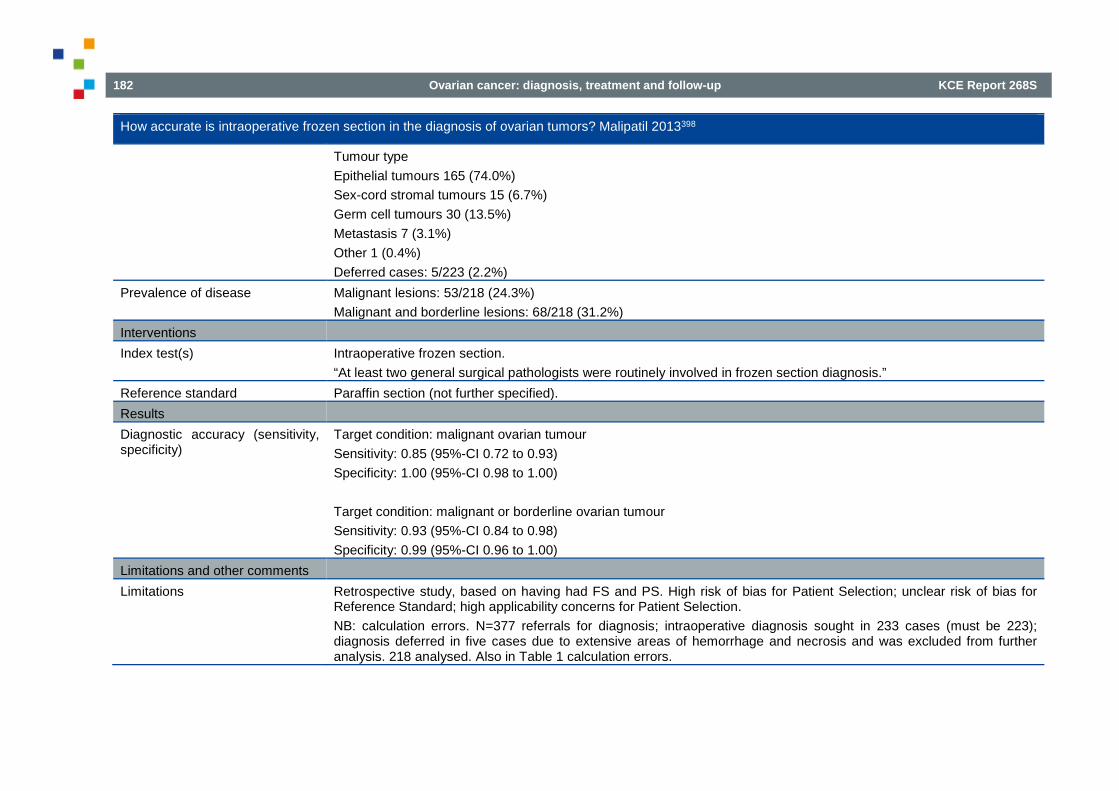
182 Ovarian cancer: diagnosis, treatment and follow-up KCE Report 268S
How accurate is intraoperative frozen section in the diagnosis of ovarian tumors? Malipatil 2013398
Tumour type Epithelial tumours 165 (74.0%) Sex-cord stromal tumours 15 (6.7%) Germ cell tumours 30 (13.5%) Metastasis 7 (3.1%) Other 1 (0.4%) Deferred cases: 5/223 (2.2%)
Prevalence of disease Malignant lesions: 53/218 (24.3%) Malignant and borderline lesions: 68/218 (31.2%)
Interventions Index test(s) Intraoperative frozen section.
“At least two general surgical pathologists were routinely involved in frozen section diagnosis.” Reference standard Paraffin section (not further specified). Results Diagnostic accuracy (sensitivity, specificity)
Target condition: malignant ovarian tumour Sensitivity: 0.85 (95%-CI 0.72 to 0.93) Specificity: 1.00 (95%-CI 0.98 to 1.00) Target condition: malignant or borderline ovarian tumour Sensitivity: 0.93 (95%-CI 0.84 to 0.98) Specificity: 0.99 (95%-CI 0.96 to 1.00)
Limitations and other comments Limitations Retrospective study, based on having had FS and PS. High risk of bias for Patient Selection; unclear risk of bias for
Reference Standard; high applicability concerns for Patient Selection. NB: calculation errors. N=377 referrals for diagnosis; intraoperative diagnosis sought in 233 cases (must be 223); diagnosis deferred in five cases due to extensive areas of hemorrhage and necrosis and was excluded from further analysis. 218 analysed. Also in Table 1 calculation errors.

KCE Report 268S Ovarian cancer: diagnosis, treatment and follow-up 183
Table 45 – Evidence table (8): DTA study frozen section Accuracy of frozen section diagnosis in ovarian masses: experience at a tertiary oncology center; Rakhshan 2009399
Methods
Design Retrospective DTA study Source of funding and competing interest
Source of funding: not reported Competing interests: none declared
Setting Single centre study (Department of Pathology, Shohada Hospital, Shaheed Beheshti University of Medical Sciences, Tehran, Iran)
Sample size N=282 Time interval between tests Specimens for FS and PS collected during the same procedure Statistical analysis Sensitivity, specificity, positive and negative predictive values of intraoperative FS for benign, borderline and malignant
lesions were calculated. Patient characteristics Eligibility criteria Ovarian masses submitted for frozen section from March 1994 to May 2008. Patient characteristics Mean age: 44 (range 13 to 78) years
Tumour type Epithelial tumours 158 (56.0%) Sex-cord stromal tumours 19 (6.8%) Germ cell tumours 39 (13.8%) Non-neoplastic lesions 53 (18.8%) Metastasis: 7 (2.5%) Other 6 (2.1%) Deferred cases: 0/282 (0%)
Prevalence of disease Malignant lesions: 65/282 (23.0%) Malignant and borderline lesions: 80/282 (28.4%)
Interventions Index test(s) Intraoperative frozen section.

184 Ovarian cancer: diagnosis, treatment and follow-up KCE Report 268S
Accuracy of frozen section diagnosis in ovarian masses: experience at a tertiary oncology center; Rakhshan 2009399
“In all specimens, after gross examination, one to five sections depending on the tumour size and the degree of suspicion were frozen in a cryostat. Then 5 µm sections were stained with haematoxylin and eosin, and were interpreted by one of the five attending general pathologists.” “All frozen section specimens were reviewed by a pathology resident and an attending surgical pathologist expert in gynaecologic pathology.”
Reference standard Paraffin sections (not further specified). Results Diagnostic accuracy (sensitivity, specificity)
Target condition: malignant ovarian tumour Sensitivity: 0.92 (95%-CI 0.83 to 0.97) Specificity: 1.00 (95%-CI 0.97 to 1.00) Target condition: malignant or borderline ovarian tumour Sensitivity: 0.91 (95%-CI 0.83 to 0.96) Specificity: 1.00 (95%-CI 0.97 to 1.00)
Limitations and other comments
Limitations Retrospective study, based on having had FS and PS. Unclear risk of bias for Patient Selection and Reference Standard; unclear applicability concerns for Patient Selection.
Table 46 – Evidence table (9): DTA study frozen section Accuracy rate of frozen section studies in ovarian cancers: A regional cancer institute experience; Subbian 2013400
Methods
Design Retrospective DTA study Source of funding and competing interest
Source of funding: none Competing interests: none declared
Setting Single centre study (Department of Gynecologic Oncology, Kidwai Memorial Institute of Oncology, Bangalore, Karnataka, India)
Sample size N=135

KCE Report 268S Ovarian cancer: diagnosis, treatment and follow-up 185
Accuracy rate of frozen section studies in ovarian cancers: A regional cancer institute experience; Subbian 2013400
Time interval between tests Specimens for FS and PS collected during the same procedure Statistical analysis Sensitivity, specificity, positive and negative predictive values of intraoperative FS for benign, borderline and malignant
lesions were calculated. Patient characteristics Eligibility criteria Retrospective analysis of reports of frozen section and paraffin block diagnoses of patients undergoing surgery as primary
line of therapy for suspected ovarian neoplasms, from March 2004 to January 2006. Patient characteristics Mean age: 43.7 (range 13 to 86) years
Tumour type Epithelial tumours 77 (60.6%) Sex-cord stromal tumours 7 (5.5%) Germ cell tumours 16 (12.5%) Other 27 (21.2%) of which 11 non-neoplastic Deferred cases: 8/135 (5.9%)
Prevalence of disease Malignant lesions: 59/117 (50.4%) Malignant and borderline lesions: 75/117 (64.1%)
Interventions Index test(s) Intraoperative frozen section.
“All the frozen section diagnoses were made by a team of expert oncopathologists at the institute. Before sectioning, gross examination of the tumour was carried out and frozen section samples were taken from solid or suspicious areas. The number of bits sampled varied from one to three (average of two). The frozen section and the permanent section reports of each patient were compared. The frozen section results were divided into the following groups: Deferred, benign, borderline and malignant. Reports mentioned as ‘suggestive of ’, ‘suspicious of’ or ‘compatible with’ were included in the diagnoses mentioned.”
Reference standard Paraffin block (not further specified). Results Diagnostic accuracy (sensitivity, specificity)
2*2 Tables reconstructed! Target condition: malignant ovarian tumour Sensitivity: 0.92 (95%-CI 0.81 to 0.97) Specificity: 0.98 (95%-CI 0.91 to 1.00)

186 Ovarian cancer: diagnosis, treatment and follow-up KCE Report 268S
Accuracy rate of frozen section studies in ovarian cancers: A regional cancer institute experience; Subbian 2013400
Target condition: malignant or borderline ovarian tumour Sensitivity: 0.83 (95%-CI 0.72 to 0.90) Specificity: 0.90 (95%-CI 0.77 to 0.97)
Limitations and other comments
Limitations Retrospective study, based on having had FS and PS. Unclear risk of bias for Patient Selection, Reference Standard and Flow and Timing; no applicability concerns.
Table 47 – Evidence table (10): DTA study frozen section Role of Frozen Section in Intraoperative Assessment of Ovarian Masses: a Tertiary Oncology Center Experience; Sukumaran 2014401
Methods
Design Retrospective DTA study Source of funding and competing interest
Source of funding: not reported Competing interests: not reported
Setting Single centre study (Divisions of Pathology and Surgical Oncology, Regional Cancer Centre, Trivandrum, Kerala, India) Sample size N=233 Time interval between tests Specimens for FS and PS collected during the same procedure Statistical analysis Sensitivity, specificity, positive and negative predictive values of intraoperative FS for benign, borderline and malignant
lesions were calculated. Patient characteristics Eligibility criteria Women with ovarian masses in which intraoperative frozen section examination was performed between 2009 and 2012.
Cases suffering from torsion with extensive haemorrhage and infarction were excluded. Patient characteristics Mean age: 46 (range 14 to 86) years
Tumour type Epithelial tumours 153 (11.6%) Sex-cord stromal tumours 16 (6.9%) Germ cell tumours 30 (12.9%)
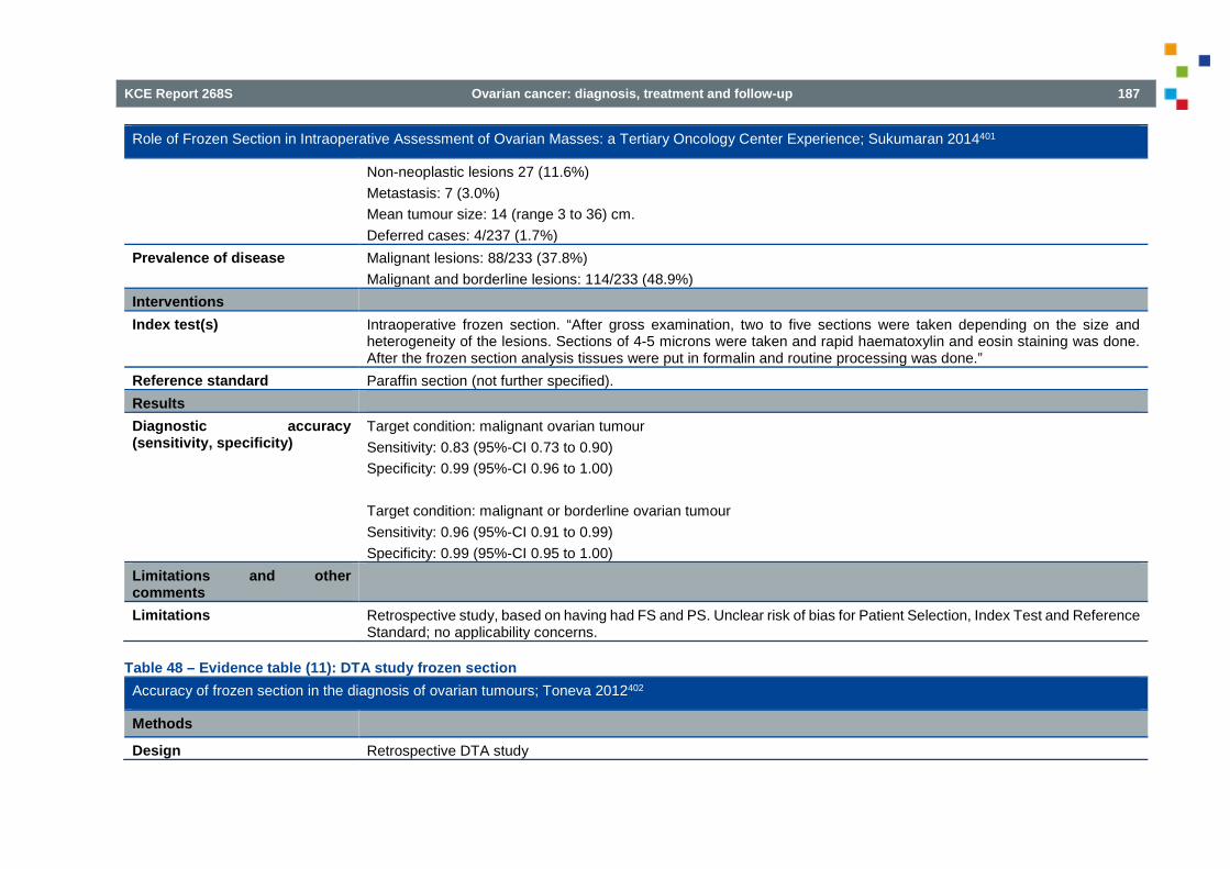
KCE Report 268S Ovarian cancer: diagnosis, treatment and follow-up 187
Role of Frozen Section in Intraoperative Assessment of Ovarian Masses: a Tertiary Oncology Center Experience; Sukumaran 2014401
Non-neoplastic lesions 27 (11.6%) Metastasis: 7 (3.0%) Mean tumour size: 14 (range 3 to 36) cm. Deferred cases: 4/237 (1.7%)
Prevalence of disease Malignant lesions: 88/233 (37.8%) Malignant and borderline lesions: 114/233 (48.9%)
Interventions Index test(s) Intraoperative frozen section. “After gross examination, two to five sections were taken depending on the size and
heterogeneity of the lesions. Sections of 4-5 microns were taken and rapid haematoxylin and eosin staining was done. After the frozen section analysis tissues were put in formalin and routine processing was done.”
Reference standard Paraffin section (not further specified). Results Diagnostic accuracy (sensitivity, specificity)
Target condition: malignant ovarian tumour Sensitivity: 0.83 (95%-CI 0.73 to 0.90) Specificity: 0.99 (95%-CI 0.96 to 1.00) Target condition: malignant or borderline ovarian tumour Sensitivity: 0.96 (95%-CI 0.91 to 0.99) Specificity: 0.99 (95%-CI 0.95 to 1.00)
Limitations and other comments
Limitations Retrospective study, based on having had FS and PS. Unclear risk of bias for Patient Selection, Index Test and Reference Standard; no applicability concerns.
Table 48 – Evidence table (11): DTA study frozen section Accuracy of frozen section in the diagnosis of ovarian tumours; Toneva 2012402
Methods
Design Retrospective DTA study

188 Ovarian cancer: diagnosis, treatment and follow-up KCE Report 268S
Accuracy of frozen section in the diagnosis of ovarian tumours; Toneva 2012402
Source of funding and competing interest
Source of funding: not reported Competing interests: none declared
Setting Single centre study (Department of Obstetrics and Gynaecology, South Essex Gynaecological Oncology Centre, Southend University Hospital NHS Foundation Trust, Westcliff on Sea, Essex, UK)
Sample size N=66 Time interval between tests Specimens for FS and PS collected during the same procedure Statistical analysis Sensitivity, specificity, positive and negative predictive values of intraoperative FS for benign, borderline and malignant
lesions were calculated. Patient characteristics Eligibility criteria Inclusion from October 2005 to September 2008. Indications for intraoperative frozen section: high RMI; presence of
necrotic areas; fragile papillary areas within the cyst; ascites and solid components, which were all thought to represent an increased chance of malignancy.
Patient characteristics Mean age: 61.4 (±14.8) years Tumour type Serous 48% Mucinous 18.7% Endometriomas 8% Fibromas 5.3% Other 20% Mean tumour size: 14 cm (range 3.5 – 36 cm) Average overall RMI: 719.6 (4 RMI <25, 30 RMI 25-250, 32 RMI >250) Deferred cases: not reported
Prevalence of disease Malignant lesions: 28/66 (42.4%) Malignant and borderline lesions: 48/66 (72.7%)
Interventions Index test(s) Intraoperative frozen section.

KCE Report 268S Ovarian cancer: diagnosis, treatment and follow-up 189
Accuracy of frozen section in the diagnosis of ovarian tumours; Toneva 2012402
“During the operation, the surgeon completed a request form with relevant clinical information and sent the whole ovary/ovaries to be examined for frozen section with no markings attached. The frozen section was performed by the on-call pathologist who was normally a general histopathologist, however, there was always an expert in gynaecological disease available for consultation in difficult cases. On receipt of the specimen, the pathologist initially performed a gross examination of the tumour and identified any solid or suspicious areas. The pathologist then made three to five cuts, depending on the size of the tumour. The specimens were frozen, sliced and stained with haematoxylin and eosin (H&E) and interpreted. The diagnosis of ‘benign’, ‘borderline’ or ‘malignant’ was then communicated to the surgeon via telephone or in person. At our centre, the average time elapsed between sampling and diagnosis was 30-60 min.”
Reference standard Paraffin block (not further specified). Results Diagnostic accuracy (sensitivity, specificity)
Target condition: malignant ovarian tumour Sensitivity: 0.89 (95%-CI 0.72 to 0.98) Specificity: 1.00 (95%-CI 0.91 to 1.00) Target condition: malignant or borderline ovarian tumour Sensitivity: 0.88 (95%-CI 0.75 to 0.95) Specificity: 1.00 (95%-CI 0.81 to 1.00)
Limitations and other comments
Limitations Retrospective study, based on having had FS and PS. Unclear risk of bias for Patient Selection and Reference Standard; no applicability concerns.
5.3. Lymphadenectomy
Table 49 – Evidence table: SR lymphadenectomy Lymph node metastasis in stages I and II ovarian cancer: a review; Kleppe 2011144
Methods
Design Systematic review Source of funding and competing interest
No information about source of funding. Authors declare that there are no conflicts of interest.

190 Ovarian cancer: diagnosis, treatment and follow-up KCE Report 268S
Lymph node metastasis in stages I and II ovarian cancer: a review; Kleppe 2011144
Search date Not reported; the most recent included study is from August 2010 Searched databases MEDLINE and EMBASE; additional literature was searched through cross-references of the retrieved articles. Included study designs Randomized controlled trials, and prospective and retrospective cohort studies. Number of included studies N=14; Authors state that there are two RCTs and 12 retrospective cohort studies included. However, we identified two
prospective observational studies instead of the two RCTs. Statistical analysis Not applicable as only incidences of lymph node metastases are determined. Patient characteristics Eligibility criteria RCTs, and prospective and retrospective cohort studies that included patients with clinically FIGO stage I or II epithelial
ovarium carcinoma or a subset of these specific groups; in all patients within the trial or cohort a complete staging laparotomy including a systematic pelvic and para-aortic lymph node dissection (LND) had to be performed.
Exclusion criteria Not specified, except that it is stated that searches were limited to “full text”, “English” and “human”. Patient & disease characteristics In the 14 included studies there were 1247 patients with FIGO stage I-II.
The mean number of pelvic and para-aortic lymph nodes removed ranged from 20 to 78. Five studies described the number of removed lymph nodes from the para-aortic and pelvic region separately: the mean number of para-aortic and pelvic lymph nodes ranged from 15 to 46 and 14 to 74, respectively
Interventions Surgical procedures “A complete staging laparotomy including a systematic pelvic and para-aortic lymph node dissection (LND) had to be
performed in all patients within the trial or cohort.” Outcome assessment No information on outcome assessment in included studies. Results Prevalence of malignant disease in pelvic and para-aortic lymph nodes
Clinical FIGO stage I and II Overall incidence of lymph node metastases (14 studies, n=1247): 177/1247 (14.2%; range 6.1–29.6%) Only para-aortic region: 88/1247 (7.1%; range 3.0–13.0%) Only pelvic region: 36/1247 (2.9%; range 0.0–11.1%) Both para-aortic and pelvic region: 53/1247 (4.3%; range 0.0–14.8%) Incidence of lymph node metastases according to differentiation grade (6 studies, n=361, missing data n=66)

KCE Report 268S Ovarian cancer: diagnosis, treatment and follow-up 191
Lymph node metastasis in stages I and II ovarian cancer: a review; Kleppe 2011144
Grade 1: 6/149 (4.0%; range 0.0-6.3%) Grade 2: 17/103 (16.5%; range 0.0-27.3%) Grade 3: 10/50 (20.0%; range 0.0-100.0%) Incidence of lymph node metastases according to histological subtype (8 studies, n=574). Serous: 35/150 (23.3%; range 8.7-50.0%) Mucinous: 4/155 (2.6%; range 0.0-6.7%) Endometrioid: 6/92 (6.5%; range 0.0-20.0%) Clear cell: 20/139 (14.4%; range 0.0-31.3%) Undifferentiated / others: 11/38 (28.9%; range 0.0-64.3%) Clinical FIGO stage I Incidence of lymph node metastases according to differentiation grade (4 studies, n=188, missing data n=20) Grade 1: 2/68 (2.9%; range 0.0-6.3%) Grade 2: 9/65 (13.8%; range 0.0-27.3%) Grade 3: 7/35 (20.0%; range 0.0-100.0%) Incidence of lymph node metastases according to histological subtype (5 studies, n=336) Serous: 19.3% Mucinous: 1.9% Endometrioid: not reported Clear cell: not reported Undifferentiated / others: not reported Unilateral clinical FIGO stages I-II (7 studies) “In case of lymph node metastases, they were localized only at the contralateral pelvic and para-aortic site in respectively 16.1% and 18.0%.”

192 Ovarian cancer: diagnosis, treatment and follow-up KCE Report 268S
Lymph node metastasis in stages I and II ovarian cancer: a review; Kleppe 2011144
Limitations and other comments
Limitations All but two of included studies were retrospective studies, no details of included studies, no quality assessment of included studies.
Table 50 – Evidence table (1): lymphadenectomy Analysis of para-aortic lymphadenectomy up to the level of the renal vessels in apparent early-stage ovarian cancer; Chang 201361
Methods
Design Retrospective analysis of medical records Source of funding and competing interest
Source of funding: one of the authors was supported by the Queen of Hearts Foundation Authors report no potential conflict of interest relevant to this article
Setting Single centre: Ajou University Hospital, Korea Sample size N=124 Duration Medical records from January 1, 2000 through December 31, 2011 were retrospectively reviewed. Statistical analysis Patients who underwent systematic pelvic and para-aortic lymphadenectomy were compared to those who did not.
The chi-square or Fisher's exact test were used for comparison of observed frequencies. Student t-test or Mann-Whitney U-test were applied for comparison of continuous variables. Kaplan-Meier method was used to estimate progression-free and overall survival rates and the log-rank to compare survival functions. A logistic regression model was performed for multivariate analysis and used in estimating the odds ratios of various parameters which were found to be significant in the univariate analysis. Backward stepwise model-selection methods, using a cut-off p-value of 0.05, were used to select factors that were included in the multivariate analysis. Statistical significance was defined as p<0.05.
Patient characteristics Eligibility criteria Ovarian cancer that was thought to be confined to the ovary without any extra-ovarian metastatic lesions at the time
of preoperative imaging studies Exclusion criteria Not specified Patient & disease characteristics Median age: 46 yrs (range 19 to 74)
FIGO stage: I: n=87 (70.2%); IA: n=54, IB: n=2, IC: n=31

KCE Report 268S Ovarian cancer: diagnosis, treatment and follow-up 193
Analysis of para-aortic lymphadenectomy up to the level of the renal vessels in apparent early-stage ovarian cancer; Chang 201361
II: n=19 (15.3%); IIA: n=4, IIB: n=6, IIIC: n=9 III: n=18 (14.5%); IIIA: n=1, IIIB n=0, IIIC: n=17 Patients with systematic lymphadenectomy (n=86, of which n=69 pelvic and para aortic lymphadenectomy) vs. patients without systematic lymphadenectomy (n=38) Mean/median* age (range): 47.5 yrs (19-74) vs. 45 yrs (19-72) Mean/median parity (range): 2 (0-7) vs. 1 (0-4) Mean/median BMI: 23.1 kg/m2 (14.4-35.2) vs. 25.4 kg/m2 (7.9-28.3) FIGO stage: IA: 33 (38.4%) vs. 21 (55.3%); IB: 2 (2.3%) vs. 0; IC: 20 (23.3%) vs. 11 (28.9%); IIA: 3 (3.5%) vs. 1 (2.6%); IIB: 3 (3.5%) vs. 3 (7.9%); IIC: 7 (8.1%) vs. 2 (5.3%); IIIA: 1 (1.2%) vs. 0; IIIB 0 vs. 0; IIIC: 17 (19.8%) vs. 0 Histology: serous: 35 (40.7%) vs. 7 (18.4%); mucinous: 16 (18.6%) vs. 16 (42.1%); endometrioid: 8 (9.3%) vs. 4 (10.5%); clear cell: 16 (18.6%) vs. 6 (15.8%); mixed: 9 (10.5%) vs. 3 (7.9%); others: 2 (2.4%) vs. 2 (5.2%) Tumour grade: grade 1: 37 (43.0%) vs. 24 (63.2%); grade 2: 19 (22.1%) vs. 5 (13.2%); grade 3: 29 (33.7%) vs. 7 (18.4%); unknown: 1 (1.2%) vs. 2 (5.3%) Mean/median preoperative serum CA-125 (range): 108.9 U/mL (3.5-15,600.0) vs. 87.4 U/mL (6.0-1,722.0) Presence of ascites: 36 (41.9%) vs. 7 (18.4%) “Patients with systematic lymphadenectomy had higher parity (p<0.01), more frequent ascites (p=0.01), and higher numbers of resected lymph nodes (p<0.01). There were no statistically significant differences in demographic features, FIGO stage, tumour histology, tumour grade, and preoperative CA-125 level between the two groups.” *not specified whether mean or median is presented
Interventions Surgical procedures Surgical staging according to the FIGO system. Standard surgical staging procedures included total abdominal
hysterectomy (n=104, 83.9%), unilateral or bilateral salpingo-oophorectomy (n=20, 16.1%), peritoneal washings for cytology (n=124, 100%), infracolic omentectomy (n=120, 96.8%), multiple biopsies of pelvic and abdominal peritoneum (n=117, 94.4%) pelvic lymphadenectomy (n=86, 69.4%), and para-aortic lymphadenectomy (n=69, 55.6%). Pelvic lymphadenectomy included bilateral resection of the common iliac nodes, presacral nodes, external iliac nodes, internal iliac nodes, deep inguinal nodes, and obturator nodes.

194 Ovarian cancer: diagnosis, treatment and follow-up KCE Report 268S
Analysis of para-aortic lymphadenectomy up to the level of the renal vessels in apparent early-stage ovarian cancer; Chang 201361
Para-aortic lymphadenectomy included removal of all nodal tissues over the vena cava and aorta from the aortic bifurcation to the level of the renal vessels. Median number of harvested lymph nodes (range): pelvic: 25 (10-53); para-aortic: 10 (5-29)
Outcome assessment All pelvic lymph nodes were separately sent to pathology with dividing into the right and left pelvic lymph nodes. Most of the para-aortic lymph nodes were separately sent to the intraoperative frozen section or postoperative permanent section for pathologic evaluation with dividing into low and high para-aortic lymph nodes according to the inferior mesenteric artery. However, para-aortic lymph nodes from some patients were sent en bloc to pathology without dividing into low and high nodes.
Results Prevalence of malignant disease in pelvic and para-aortic lymph nodes
Lymph node metastasis in patients with pelvic and para-aortic lymphadenectomy: 17/69 (24.6%) - from para-aortic and pelvic areas 9/69 (13.0%) - from only the pelvic area: 5/69 (7.2%) - from only the para-aortic area: 8/69 (11.6%) “On multivariate analysis, grade 3 tumour (odds ratio [OR], 5.42; 95% confidence interval [CI], 1.51-19.52; p=0.01) and positive cytology (OR, 4.22; 95% CI, 1.12-15.96; p=0.03) were independent predictors for lymph node metastasis.”
Limitations and other comments Limitations Relatively small total lymph node count retrieved which may have resulted in underestimation of the incidence of nodal
metastasis. Risk of selection bias due to retrospective study design. The lack of accurate para-aortic lymph node mapping could lead to the potential overestimation or underestimation of para-aortic lymph node metastasis.
Table 51 – Evidence table (2): lymphadenectomy Systematic para-aortic and pelvic lymphadenectomy in early stage epithelial ovarian cancer: a prospective study; Ditto 2012403
Methods
Design Prospective study Source of funding and competing interest
Source of funding: none reported Authors report no potential conflict of interest relevant to this article
Setting Single centre: IRCCS National Cancer Institute, Milan, Italy Sample size Sample size calculated: n=128; n=124 recruited, n=111 analysed
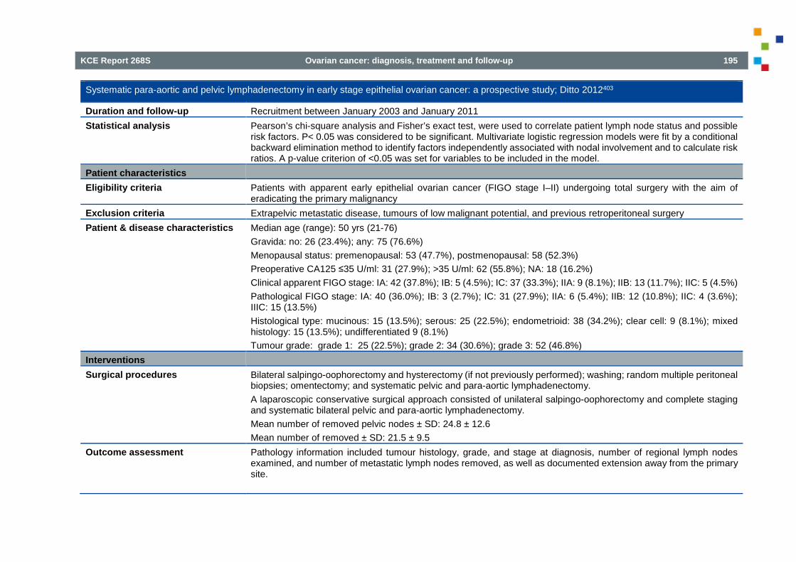
KCE Report 268S Ovarian cancer: diagnosis, treatment and follow-up 195
Systematic para-aortic and pelvic lymphadenectomy in early stage epithelial ovarian cancer: a prospective study; Ditto 2012403
Duration and follow-up Recruitment between January 2003 and January 2011 Statistical analysis Pearson’s chi-square analysis and Fisher’s exact test, were used to correlate patient lymph node status and possible
risk factors. P< 0.05 was considered to be significant. Multivariate logistic regression models were fit by a conditional backward elimination method to identify factors independently associated with nodal involvement and to calculate risk ratios. A p-value criterion of <0.05 was set for variables to be included in the model.
Patient characteristics Eligibility criteria Patients with apparent early epithelial ovarian cancer (FIGO stage I–II) undergoing total surgery with the aim of
eradicating the primary malignancy Exclusion criteria Extrapelvic metastatic disease, tumours of low malignant potential, and previous retroperitoneal surgery Patient & disease characteristics Median age (range): 50 yrs (21-76)
Gravida: no: 26 (23.4%); any: 75 (76.6%) Menopausal status: premenopausal: 53 (47.7%), postmenopausal: 58 (52.3%) Preoperative CA125 ≤35 U/ml: 31 (27.9%); >35 U/ml: 62 (55.8%); NA: 18 (16.2%) Clinical apparent FIGO stage: IA: 42 (37.8%); IB: 5 (4.5%); IC: 37 (33.3%); IIA: 9 (8.1%); IIB: 13 (11.7%); IIC: 5 (4.5%) Pathological FIGO stage: IA: 40 (36.0%); IB: 3 (2.7%); IC: 31 (27.9%); IIA: 6 (5.4%); IIB: 12 (10.8%); IIC: 4 (3.6%); IIIC: 15 (13.5%) Histological type: mucinous: 15 (13.5%); serous: 25 (22.5%); endometrioid: 38 (34.2%); clear cell: 9 (8.1%); mixed histology: 15 (13.5%); undifferentiated 9 (8.1%) Tumour grade: grade 1: 25 (22.5%); grade 2: 34 (30.6%); grade 3: 52 (46.8%)
Interventions Surgical procedures Bilateral salpingo-oophorectomy and hysterectomy (if not previously performed); washing; random multiple peritoneal
biopsies; omentectomy; and systematic pelvic and para-aortic lymphadenectomy. A laparoscopic conservative surgical approach consisted of unilateral salpingo-oophorectomy and complete staging and systematic bilateral pelvic and para-aortic lymphadenectomy. Mean number of removed pelvic nodes ± SD: 24.8 ± 12.6 Mean number of removed ± SD: 21.5 ± 9.5
Outcome assessment Pathology information included tumour histology, grade, and stage at diagnosis, number of regional lymph nodes examined, and number of metastatic lymph nodes removed, as well as documented extension away from the primary site.

196 Ovarian cancer: diagnosis, treatment and follow-up KCE Report 268S
Systematic para-aortic and pelvic lymphadenectomy in early stage epithelial ovarian cancer: a prospective study; Ditto 2012403
Results Prevalence of malignant disease in pelvic and para-aortic lymph nodes
Lymph node metastasis in patients with pelvic and para-aortic lymphadenectomy: 15/111 (13.5%) - from para-aortic and pelvic areas 5/111 (4.5%) - from only the pelvic area: 2/111 (1.8%) - from only the para-aortic area: 8/111 (7.2%) Patients with node metastases by histological type Mucinous: 0/15 (0%) Serous: 7/25 (28%) Endometrioid: 4/38 (10.5%) Clear cell: 0/9 (0%) Mixed histology: 1/15 (6.7%) Undifferentiated: 3/9 (33.3%) “At univariate analysis age, menopause, FIGO stage, grading (G1–2 vs. G3), histological type (serous vs. mucinous vs. other), and bilaterality of the specimens were significant factors for lymph node metastases, while CA125 of >35 U/ml and positive cytology were not. At multivariate analysis, only bilaterality (p = 0.018) and menopause (p = 0.032) maintained a statistically significant association with lymph node metastases, while the grading lost its significance at the final step of logistic regression analysis.” “On the basis of the present series and the data in the literature, omitting a systematic lymphadenectomy can be considered for grade I cancers and for mucinous tumours regardless of grade. The rate of positive lymph nodes in the study was 13.5%, with a 95% confidence interval of 7.1–19.9.[…] the logistic regression model presented a good discriminating power measured by the area under the receiver operating characteristic curve of 0.81. This means that the surgeon, using only data on menopausal status and bilaterality of the tumour, has a 19% chance of making the wrong decision regarding the necessity of lymph node dissection.”
Limitations and other comments
Limitations Small number of patients enrolled, which probably hampers significance at multivariate analysis for the variables histological subtype grading.

KCE Report 268S Ovarian cancer: diagnosis, treatment and follow-up 197
Table 52 – Evidence table (3): lymphadenectomy Frequency and distribution of lymph node metastases in epithelial ovarian cancer - significance of serous histology; Haller 201176
Methods
Design Retrospective review of the medical records Source of funding and competing interest
Not reported
Setting Single centre (tertiary care academic hospital); Department of Obstetrics and Gynecology, Clinical Hospital Centre of Rijeka and Department of Pathology, School of Medicine, University of Rijeka, Rijeka, Croatia.
Sample size N=173 included Duration and follow-up Medical records from January 1995 to December 2007 were retrospectively reviewed Statistical analysis “The results are expressed as means, SDs, minimums, maximums, and percentages. Statistical significance was
calculated using χ2 analysis (to determine the relationship between lymph node status and other disease variables) and the independent sample t test (to compare the means). Regression model (Enter method) was applied where appropriate to identify the independent prognostic factor. P values less than 0.05 were considered statistically significant.”
Patient characteristics Eligibility criteria Patients primary epithelial ovarian cancer with complete pelvic and aortic lymphadenectomy up to the level of the left
renal vein at first surgery Exclusion criteria Borderline tumours including micropapillary serous carcinoma, carcinosarcoma (malignant müllerian mixed tumours),
and primary extra-ovarial (peritoneal) carcinomas Patient & disease characteristics Mean age (range): 53 yrs (31-74)
Patients with serous tumour (n=76, 43.9%) vs. nonserous tumour (n=97, 56.1%): Age ≤ 49 yrs: 27 (43.6%) vs. 35 (56.4%); age 50-59 yrs: 24 (42.1%) vs. 33 (57.9%); age ≥60 yrs: 25 (46.3%) vs. 29 (53.7%); Intraperitoneal stage: pT1: 7 (9.2%) vs. 58 (59.8%); pT2: 15 (19.7%) vs. 20 (20.6%); pT3: 54 (71.1%) vs. 19 (19.6%); Grade 1: 8 (10.5%) vs. 27 (27.8%); grade 2: 37 (48.7%) vs. 30 (30.9%); grade 3: 31 (40.8%) vs. 40 (41.3%); Residual tumour 0 cm: 40 (52.6%) vs. 90 (92.8%); residual tumour <1 cm: 23 (30.3%) vs. 5 (5.2%); residual tumour ≥1 cm: 13 (17.1%) vs. 2 (2.0%).

198 Ovarian cancer: diagnosis, treatment and follow-up KCE Report 268S
Frequency and distribution of lymph node metastases in epithelial ovarian cancer - significance of serous histology; Haller 201176
Statistically significant differences between serous and non-serous tumours for intraperitoneal stage, grade and residual tumour size.
Interventions Surgical procedures Lymphadenectomy was a part of primary surgical evaluation, in cases where ovarian malignant lesions are
encountered as determined by frozen histological sections. Additional criteria for lymphadenectomy include age younger than 70 years, relatively good medical condition, and intraoperative reduction of tumour to less than 2 cm. After the year 2000, this value was diminished to less than 1 cm. In these patients, lymphadenectomy was associated with hysterectomy, bilateral salpingo-oophorectomy, infracolic omentectomy, multiple peritoneal biopsies, as well as cytoreductive techniques such as supracolic omentectomy, bowel or splenic resection, pelvic, and/or hemidiaphragmatic deperitonization or other procedures, depending on intra-abdominal spread of the disease. Lymphadenectomy was a part of primary surgery via transperitoneal approach. A pubic-xiphoid incision was made to expose intra-abdominal organs. After the diagnosis was established on the frozen section during surgery in the pelvis, pelvic lymphadenectomy was performed. Aortic lymphadenectomy was the last surgical procedure, performed after completion of intraperitoneal surgery. Median number (range) of lymph nodes removed per patient: 37 (12-69); pelvic and para-aortic region: 25 (9-47) and 12 (4-40), respectively.
Outcome assessment All dissected lymph nodes were histologically evaluated on paraffin-embedded and hematoxylin and eosin-stained sections.
Results Prevalence of malignant disease in pelvic and para-aortic lymph nodes
Lymph node metastasis in patients with pelvic and para-aortic lymphadenectomy: 59/173 (34.1%) Age ≤ 49 yrs: 21/62 (33.9%) age 50-59 yrs: 16/57 (28.1%) age ≥60 yrs: 22/54 (40.7%) Intraperitoneal stage pT1:4/65 (6.2%) pT2: 10/35 (28.6%) pT3: 45/73 (61.6%) Grade grade1: 6/35 (17.1%)
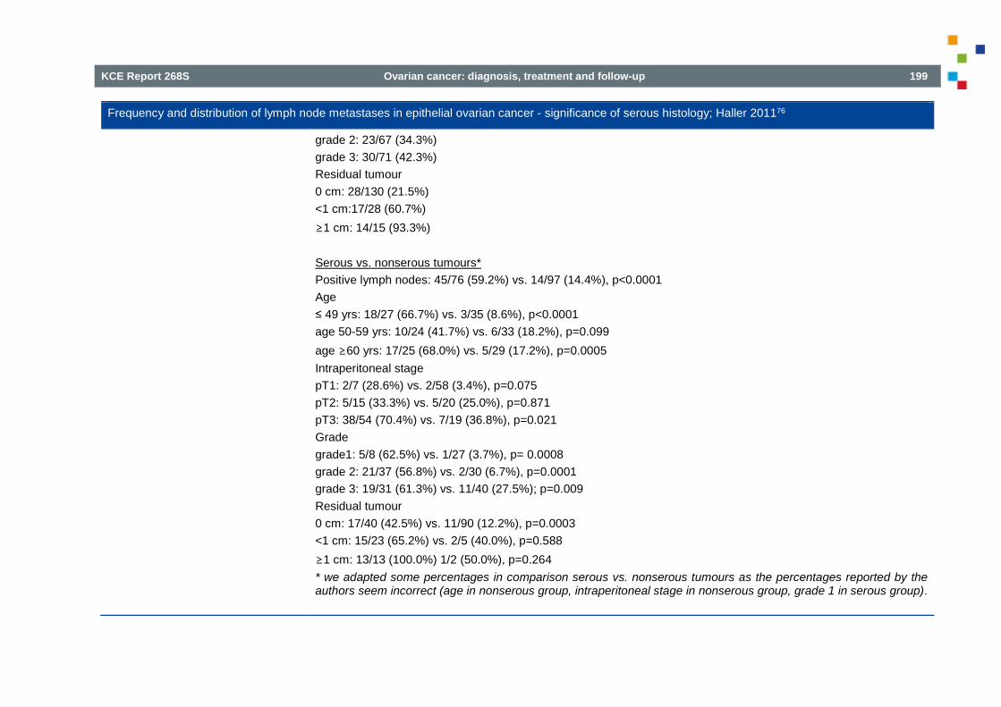
KCE Report 268S Ovarian cancer: diagnosis, treatment and follow-up 199
Frequency and distribution of lymph node metastases in epithelial ovarian cancer - significance of serous histology; Haller 201176
grade 2: 23/67 (34.3%) grade 3: 30/71 (42.3%) Residual tumour 0 cm: 28/130 (21.5%) <1 cm:17/28 (60.7%) ≥1 cm: 14/15 (93.3%) Serous vs. nonserous tumours* Positive lymph nodes: 45/76 (59.2%) vs. 14/97 (14.4%), p<0.0001 Age ≤ 49 yrs: 18/27 (66.7%) vs. 3/35 (8.6%), p<0.0001 age 50-59 yrs: 10/24 (41.7%) vs. 6/33 (18.2%), p=0.099 age ≥60 yrs: 17/25 (68.0%) vs. 5/29 (17.2%), p=0.0005 Intraperitoneal stage pT1: 2/7 (28.6%) vs. 2/58 (3.4%), p=0.075 pT2: 5/15 (33.3%) vs. 5/20 (25.0%), p=0.871 pT3: 38/54 (70.4%) vs. 7/19 (36.8%), p=0.021 Grade grade1: 5/8 (62.5%) vs. 1/27 (3.7%), p= 0.0008 grade 2: 21/37 (56.8%) vs. 2/30 (6.7%), p=0.0001 grade 3: 19/31 (61.3%) vs. 11/40 (27.5%); p=0.009 Residual tumour 0 cm: 17/40 (42.5%) vs. 11/90 (12.2%), p=0.0003 <1 cm: 15/23 (65.2%) vs. 2/5 (40.0%), p=0.588 ≥1 cm: 13/13 (100.0%) 1/2 (50.0%), p=0.264 * we adapted some percentages in comparison serous vs. nonserous tumours as the percentages reported by the authors seem incorrect (age in nonserous group, intraperitoneal stage in nonserous group, grade 1 in serous group).

200 Ovarian cancer: diagnosis, treatment and follow-up KCE Report 268S
Frequency and distribution of lymph node metastases in epithelial ovarian cancer - significance of serous histology; Haller 201176
“Logistic regression (Enter method) identified the following independent variables predicting lymph node metastases among the ovarian carcinoma: serous histology (P = 0.009; 95% confidence interval [CI], 0.13-0.75), residual tumour mass (P = 0.0107; 95% CI, 1.21-4.52), and intraperitoneal stage (P = 0.0096; 95% CI, 1.08-4.12), whereas age and tumour differentiation had no prognostic value.”
Limitations and other comments
Limitations Indication for lymphadenectomy changed after the year 2000: “Additional criteria for lymphadenectomy include age younger than 70 years, relatively good medical condition, and intraoperative reduction of tumour to less than 2 cm. After the year 2000, this value was diminished to less than 1 cm.”
Table 53 – Evidence table (4): lymphadenectomy Prognostic value of lymph node involvement in ovarian serous borderline tumours (Study 1); Lesieur 2011404
Methods
Design Retrospective review of medical records Source of funding and competing interest
Not reported
Setting Single centre: Departments of Gynecologic Surgery, Pathology and Oncology, Institut Gustave Roussy, France
Sample size N = 49 included Duration and follow-up Patient enrolment from January 1973 to February 2006 Statistical analysis NA Patient characteristics Eligibility criteria Patients who had undergone a lymphadenectomy and had a final diagnosis of a borderline tumour of the ovary (BOT). BOT
was defined as an ovarian tumour with the following: (1) a stratified epithelial lining; (2) the formation of microscopic papillary projections; (3) nuclear atypia; and (4) above all, the absence of frank stromal invasion.
Exclusion criteria Not specified Patient & disease characteristics
Mean age: 36.6 y Infertility: 16.7%
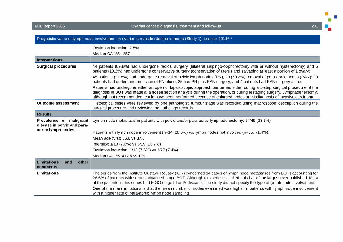
KCE Report 268S Ovarian cancer: diagnosis, treatment and follow-up 201
Prognostic value of lymph node involvement in ovarian serous borderline tumours (Study 1); Lesieur 2011404
Ovulation induction: 7.5% Median CA125: 257
Interventions Surgical procedures 44 patients (89.8%) had undergone radical surgery (bilateral salpingo-oophorectomy with or without hysterectomy) and 5
patients (10.2%) had undergone conservative surgery (conservation of uterus and salvaging at least a portion of 1 ovary). 45 patients (91.8%) had undergone removal of pelvic lymph nodes (PN), 29 (59.2%) removal of para-aortic nodes (PAN): 20 patients had undergone resection of PN alone, 25 had PN plus PAN surgery, and 4 patients had PAN surgery alone. Patients had undergone either an open or laparoscopic approach performed either during a 1-step surgical procedure, if the diagnosis of BOT was made at a frozen section analysis during the operation, or during restaging surgery. Lymphadenectomy, although not recommended, could have been performed because of enlarged nodes or misdiagnosis of invasive carcinoma.
Outcome assessment Histological slides were reviewed by one pathologist, tumour stage was recorded using macroscopic description during the surgical procedure and reviewing the pathology records.
Results Prevalence of malignant disease in pelvic and para-aortic lymph nodes
Lymph node metastasis in patients with pelvic and/or para-aortic lymphadenectomy: 14/49 (28.6%) Patients with lymph node involvement (n=14, 28.6%) vs. lymph nodes not involved (n=35, 71.4%): Mean age (yrs): 35.6 vs 37.0 Infertility: 1/13 (7.6%) vs 6/29 (20.7%) Ovulation induction: 1/13 (7.6%) vs 2/27 (7.4%) Median CA125: 417.5 vs 179
Limitations and other comments
Limitations The series from the Institute Gustave Roussy (IGR) concerned 14 cases of lymph node metastases from BOTs accounting for 28.6% of patients with serous advanced-stage BOT. Although this series is limited, this is 1 of the largest ever published. Most of the patients in this series had FIGO stage III or IV disease. The study did not specify the type of lymph node involvement. One of the main limitations is that the mean number of nodes examined was higher in patients with lymph node involvement with a higher rate of para-aortic lymph node sampling.

202 Ovarian cancer: diagnosis, treatment and follow-up KCE Report 268S
Table 54 – Evidence table (5): lymphadenectomy Prognostic value of lymph node involvement in ovarian serous borderline tumours (Study 2); Lesieur 2011404
Methods
Design Retrospective review of population based registry Source of funding and competing interest
Not reported
Setting SEER Program, with data on cancer incidence and survival from 15 population-based cancer registries that cover one quarter of the US population
Sample size N = 6017 included, N=1503 with lymph node sampling Duration and follow-up Data collection between 1973 and 2004. Patients with lymph node involvement had been diagnosed between 1988
and 2000. Statistical analysis NA Patient characteristics Eligibility criteria Patients with BOT, including ICD codes 8442 (serous cystadenoma of borderline malignancy), 8451 (papillary
cystadenoma of borderline malignancy), 8462 (serous papillary cystic tumour of borderline malignancy), 8472 (mucinous cystic tumour of borderline malignancy), and 8473 (papillary mucinous cystadenoma of borderline malignancy). Staging of BOT was based exclusively on histologically diagnosed extra-ovarian lesions. Only histopathologically diagnosed tumours that represented either the only primary tumour or the first primary tumour diagnosed for each patient were included.
Exclusion criteria Not specified Patient & disease characteristics Mean age: 48.1 years
FIGO stage: I: 4943 (82.2%); II: 351 (5.8%); III: 494 (8.2%); IV: 121 (2.0%); not available: 108 (1.8%) Pathology: serous cystadenoma 1458 (24.2%); papillary cystadenoma: 103 (1.7%); serous papillary cystadenoma: 2240 (37.2%); mucinous cystadenoma: 2076 (34.5%); mucinous papillary cystadenoma: 140 (2.3%) Patients with lymph node sampling (n=1503) vs. no lymph node sampling (n=4514) Mean age: 47.8y vs 48.2y Age <40y: 511 (34.0%) vs. 1669 (37.0%); age 40-60y: 672 (44.7%) vs. 1715 (38.0%); age>60y: 320 (21.3%) vs.1130 (25.0%) FIGO stage: I: 1104 (73.5%) vs. 3839 (85.0%); II: 144 (9.6%) vs. 207 (4.6%); III: 204 (13.6%) vs. 290 (6.4%); IV: 37 (2.5%) vs. 84 (1.9%); not available: 14 (0.9%) vs. 94 (2.1%)
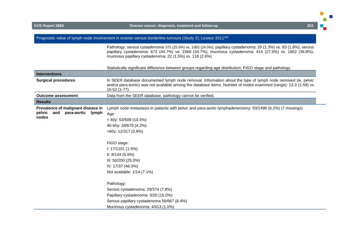
KCE Report 268S Ovarian cancer: diagnosis, treatment and follow-up 203
Prognostic value of lymph node involvement in ovarian serous borderline tumours (Study 2); Lesieur 2011404
Pathology: serous cystadenoma 375 (25.0%) vs. 1083 (24.0%); papillary cystadenoma: 20 (1.3%) vs. 83 (1.8%); serous papillary cystadenoma: 672 (44.7%) vs. 1568 (34.7%); mucinous cystadenoma: 414 (27.5%) vs. 1662 (36.8%); mucinous papillary cystadenoma: 22 (1.5%) vs. 118 (2.6%) Statistically significant difference between groups regarding age distribution, FIGO stage and pathology
Interventions Surgical procedures In SEER database documented lymph node removal. Information about the type of lymph node removed (ie, pelvic
and/or para-aortic) was not available among the database items. Number of nodes examined (range): 13.3 (1-59) vs 10.52 (1-77)
Outcome assessment Data from the SEER database, pathology cannot be verified. Results Prevalence of malignant disease in pelvic and para-aortic lymph nodes
Lymph node metastasis in patients with pelvic and para-aortic lymphadenectomy: 93/1496 (6.2%) (7 missings) Age: < 40y: 53/509 (10.4%) 40-60y: 28/670 (4.2%) >60y: 12/317 (3.8%) FIGO stage: I: 17/1101 (1.5%) II: 8/144 (5.6%) III: 50/200 (25.0%) IV: 17/37 (46.0%) Not available: 1/14 (7.1%) Pathology: Serous cystadenoma: 29/374 (7.8%) Papillary cystadenoma: 3/20 (15.0%) Serous papillary cystadenoma 56/667 (8.4%) Mucinous cystadenoma: 4/413 (1.0%)

204 Ovarian cancer: diagnosis, treatment and follow-up KCE Report 268S
Prognostic value of lymph node involvement in ovarian serous borderline tumours (Study 2); Lesieur 2011404
Mucinous papillary cystadenoma: 1/22 (4.5%) Limitations and other comments
Limitations Use of the SEER database has limitations: histology cannot be verified and the analysis of the type of surgery performed is too complex because of the multiplicity and vagueness of the headings concerning the surgical procedures and changes in codes over time. Many interesting details were not included: micropapillary pattern and presence of peritoneal or lymph node endosalpingiosis. Another main limitation of the database was the potential unreliability of the staging because some patients reported with lymph node involvement were not classified as having stage III or IV disease (as they should have been).
Table 55 – Evidence table (6): lymphadenectomy Clinical impact of systematic pelvic and para-aortic lymphadenectomy for pT1 and pT2 ovarian cancer: a retrospective survey by the Sankai Gynecology Study Group; Oshita (2013)362
Methods Design Retrospective review of the medical records Source of funding and competing interest
Source of funding: none reported Authors report no potential conflict of interest relevant to this article
Setting Multicentre, 16 institutions belonging to the Sankai Gynecology Study Group, Japan Sample size N = 422 included, N=284 with systematic pelvic and para-aortic lymphadenectomy Duration Patient enrolment between 1995 and 2005 Statistical analysis Clinical and pathological characteristics were compared between the patients with and without systematic
lymphadenectomy and between patients with and without lymph node metastasis. Correlation of variables was assessed with Fisher’s exact, chi-squared, and unpaired t tests.
Patient characteristics Eligibility criteria Patients with pT1 and pT2 epithelial ovarian cancer. Exclusion criteria Patients who (1) underwent lymphadenectomy only in the pelvic region (2) underwent lymph node sampling or (3) had
missing details regarding surgery or pathological assessment. Patient & disease characteristics Patients with systematic lymphadenectomy (n=284) vs no lymphadenectomy (n=138)
Mean age (range): 53.5 yrs (17-80) vs 52 yrs (16-91)

KCE Report 268S Ovarian cancer: diagnosis, treatment and follow-up 205
Clinical impact of systematic pelvic and para-aortic lymphadenectomy for pT1 and pT2 ovarian cancer: a retrospective survey by the Sankai Gynecology Study Group; Oshita (2013)362
Histology: serous: 63 (22.2%) vs 22 (15.9%); endometrial: 71 (25.0%) vs 23 (16.7%); clear cell: 83 (29.2%) vs 32 (23.2%); mucinous: 54 (19.0%) vs 50 (36.2%)*; other: 13 (4.6%) vs 11 (8.0%) Stage: pT1a: 62 (21.8%) vs 47 (34.1%)*; pT1b: 9 (3.2%) vs 2 (1.5%); pT1c: 133 (46.8%) vs 68 (49.3%); pT2a: 9 (3.2%) vs 0 (0.0%); pT2b: 11 (3.9%) vs 3 (2.2%); pT2c: 60 (21.1%) vs 18 (13.0%) * The rates of mucinous histology and cases staged at pT1a were significantly higher in the group without systematic lymphadenectomy.
Interventions Surgical procedures Systematic lymphadenectomy meant lymph node dissection extending from pelvic lymph nodes to the para-aortic
lymph nodes at the height of the renal veins. The median number of resected lymph nodes was 34 (10th–90th percentile: 20–52), consisting of 22 (10th–90th percentile: 12–40) in pelvic lesion and 10 (10th–90th percentile: 3–25) in para-aortic lesion.
Outcome assessment Not reported. Results Prevalence of malignant disease in pelvic and para-aortic lymph nodes
Lymph node metastasis in patients with pelvic and para-aortic lymphadenectomy: 23/284 (8.1%) - from para-aortic and pelvic areas 9/284 (3.2%) - from only the pelvic area: 2/284 (0.7%) - from only the para-aortic area: 10/284 (3.5%) (numbers don’t add up: 21 instead of 23 were subdivided into anatomical region) Stage: pT1: 9/204 (4.4%) pT2: 14/80 (17.5%)* Histology: Serous: 10/63 (15.9%)* Endometrial: 5/71 (7.0%) Clear cell: 6/83 (7.2%) Mucinous: 2/54 (3.7%)

206 Ovarian cancer: diagnosis, treatment and follow-up KCE Report 268S
Clinical impact of systematic pelvic and para-aortic lymphadenectomy for pT1 and pT2 ovarian cancer: a retrospective survey by the Sankai Gynecology Study Group; Oshita (2013)362
Other: 0/13 (0.0%) * The rate of lymph node metastasis in pT2 was significantly higher than in pT1 and serous adenocarcinoma was also associated with a higher incidence of lymph node metastasis than other histological subtypes.
Limitations and other comments
Limitations Difference in age distribution between the groups with and without lymphadenectomy: younger patients (under 30 years old) and older patients (over 70 years old) received lymphadenectomy less frequently. Whether systematic pelvic and para-aortic lymphadenectomy was performed completely or not depended on the patient’s background rather than the treatment strategy of each surgeon. The quality of the lymphadenectomies may vary in a multi-institutional study. The extent of a systematic lymphadenectomy may be slightly different depending on the gynaecologist who performed the surgery. Moreover, it is very difficult to evaluate each lymphadenectomy from medical records.
Table 56 – Evidence table (7): lymphadenectomy Risk factors for lymph node metastasis in apparent early-stage epithelial ovarian cancer: Implications for surgical staging; Powless 2011405
Methods
Design Retrospective review of medical records Source of funding and competing interest
Source of funding: none reported Authors report no potential conflict of interest relevant to this article
Setting Single centre, Mayo Clinic, USA Sample size N=190 included, 115 with systematic (comprehensive), bilateral pelvic and para-aortic lymph node assessment Duration Patient enrolment between January 1994 and December 2003 Statistical analysis Pearson's Chi-squared analysis and Fisher's Exact Test when needed were used to estimate the differences between
patients' lymph node status in terms of evaluated variables. A p-value of <0.05 was considered to be significant. Multivariate logistic regression models were fit, using stepwise selection, to identify factors independently associated with nodal involvement and to calculate risk ratios. A p-value criterion of <0.05 was set for variables to be included in the model.

KCE Report 268S Ovarian cancer: diagnosis, treatment and follow-up 207
Risk factors for lymph node metastasis in apparent early-stage epithelial ovarian cancer: Implications for surgical staging; Powless 2011405
Patient characteristics Eligibility criteria All cases of apparent early stage epithelial ovarian cancer that was grossly confined to the pelvis at the time of initial
exploration without visible evidence of abdominal spread. Patients all underwent surgical lymph node assessment (not all comprehensive).
Exclusion criteria Patients were excluded if they had undergone neoadjuvant treatment, had synchronous malignancies, or were found to not have epithelial ovarian cancer.
Patient & disease characteristics Total: n=190 Median age (range): 55 yrs (21-91) Final assigned stage: IA: 54 (28.4%); IB: 10 (5.3%); IC: 51 (26.8%); IIA: 1 (0.5%); IIB: 4 (2.1%); IIC: 37 (19.5%); IIIA: 8 (4.2%); IIIC: 25 (13.2%) Histology: serous: 43 (22.6%); mucinous: 29 (15.3%); endometrioid: 74 (38.9%); clear cell: 26 (13.7%); other: 18 (19.5%) Grade:1: 36 (18.9%); 2: 56 (29.5%); 3: 98 (51.6%) Ascites present: 39 (20.5%) Median CA 125 (range): 99 U/ml (7-4335)
Interventions Surgical procedures All patients underwent surgery by a gynaecologic oncologist, and staging was performed according to existing practice
at the time of surgery. This included total hysterectomy, bilateral adnexectomy, omentectomy, directed biopsies of suspicious peritoneal implants and random biopsies of pelvic and upper abdominal peritoneal surfaces. Frozen section was standardly utilized during the surgical evaluation of biopsies and lymph nodes as per our institutional routine and the information was used for intra-operative decision making. 115 patients (60.5%) underwent systematic bilateral pelvic and para-aortic lymph node assessment, 187 patients (98.4%) underwent any pelvic lymph node assessment (46 of them only ipsilateral), 158 patients (83.2%) underwent any para-aortic lymph node assessment (26 of them only ipsilateral). Mean lymph node counts (SD) were 25.2 ± 14.3 (pelvic) and 10.7 ± 7.7 (para-aortic).
Outcome assessment Data abstracted from medical records, including patient demographics, tumour histology and grade, operative staging procedures, and intraoperative findings.
Results Prevalence of malignant disease in pelvic and para-aortic lymph nodes
Lymph node metastasis in patients with any lymph node assessment: 25/190 (13.2%) Lymph node metastasis in patients with comprehensive pelvic and para-aortic lymphadenectomy: 19/115 (16.5%)

208 Ovarian cancer: diagnosis, treatment and follow-up KCE Report 268S
Risk factors for lymph node metastasis in apparent early-stage epithelial ovarian cancer: Implications for surgical staging; Powless 2011405
- from para-aortic and pelvic areas 4/115 (3.5%) - from only the pelvic area: 5/115 (4.3%) - from only the para-aortic area: 10/115 (8.7%) Laterality of adnexal involvement: Unilateral: 6/73 (8.2%) Bilateral: 13/42 (30.9%) p=0.002 Cytology: Negative/not done: 8/70 (11.4%) Positive: 11/45 (24.4%) p=0.067 Ascites: Absent: 10/88 (11.4%) Present: 9/27 (33.3%) p=0.007 Histology: Serous: 9/30 (30.0%) Mucinous: 0/16 (0.0%) Other: 10/66 (15.1%) p=0.030 Grade grade1: 0/14 (0.0%) grade 2: 1/36 (2.8%) grade 3: 18/65 (27.7%) p=0.001 FIGO stage: IA-IC: 9/73 (12.3%)

KCE Report 268S Ovarian cancer: diagnosis, treatment and follow-up 209
Risk factors for lymph node metastasis in apparent early-stage epithelial ovarian cancer: Implications for surgical staging; Powless 2011405
IIA-IIIA: 10/42 (23.8%) p=0.110 CA 125: ≤ 35 U/ml: 0/12 (0.0%) > 35 U/ml: 13/53 (24.5%) p=0.104 In a multivariate model the independent risk factors for lymph node metastasis among those with comprehensive pelvic and para-aortic lymph node assessment are bilateral adnexal involvement (p=0.029) as compared to unilateral, the presence of ascites (p=0.027), and a higher FIGO grade (p<0.001).
Limitations and other comments Limitations There is no information on the patients who did not undergo any lymph node sampling.
Thoroughness of lymphadenectomy versus lymph node sampling cannot be accurately inferred retrospectively. Frozen section was standardly utilized during the surgical evaluation of biopsies and lymph nodes as per our institutional routine and the information was used for intra-operative decision making. This could impact the extent of staging biopsies and lymphadenectomy performed at the surgeon's discretion.
Table 57 – Evidence table (8): lymphadenectomy Impact of surgical staging in stage i clear cell adenocarcinoma of the ovary; Suzuki 2014406
Methods Design Retrospective review of medical records Source of funding and competing interest
Supported in part by Japan Society for the Promotion of Science KAKENHI (grant 25462616). Authors report no conflicts of interest.
Setting Multicentre: 4 hospitals affiliated to The Jikei University School of Medicine, Tokyo, Japan Sample size N=165 of which n=80 staged with optimal staging surgery Duration Medical records from 2000 through 2009 were retrospectively reviewed Statistical analysis Not applicable

210 Ovarian cancer: diagnosis, treatment and follow-up KCE Report 268S
Impact of surgical staging in stage i clear cell adenocarcinoma of the ovary; Suzuki 2014406
Patient characteristics Eligibility criteria Patients with stage I pure-type clear cell adenocarcinoma of the ovary. Exclusion criteria Not specified Patient & disease characteristics Patients with optimal surgical staging (n=80) vs. with non-optimal surgical staging (n=85)
Median age (range): 52 yrs (33-74) vs. 54 (30-99); Age <50 yrs: 32 vs. 31; ≥50 yrs: 48 vs. 54; FIGO stage: 1A: 13 vs. 29; IC1: 43 vs. 43; IC2: 6 vs. 7; IC3: 18 vs. 6; Statistically significant differences between optimal and non-optimal surgical staging in FIGO stage.
Interventions Surgical procedures 3 types of the surgical staging categories were defined: optimal (including pelvic and para-aortic lymphadenectomy;
n=80), minimal (n=74), and inadequate (n=11). In addition, non-optimal staging surgery was defined as minimal or inadequate staging surgeries. The choice between systematic and selected lymphadenectomy in each patient was determined by the institutional treatment policy in staging surgery for presumed early-stage epithelial ovarian cancer at the time of surgery. The number of lymph nodes that were removed and pathologically examined was not considered for the completion of the lymphadenectomy. Pelvic lymphadenectomy was the removal of the common, external, and internal iliac nodes and the obturator node groups to the level of the inguinal ligament. Para-aortic lymphadenectomy was the removal of node-bearing tissues along aorta and vena cava to the level of the renal veins. Median number (range) of lymph nodes removed per patient: systematic lymphadenectomy: 41 (14-89); selected lymphadenectomy: 11 (1-44).
Outcome assessment Surgical staging was assessed according to FIGO (approved by the FIGO Executive Board in October 2012 and published in January 2014).
Results Prevalence of malignant disease in pelvic and para-aortic lymph nodes
Lymph node metastases were detected in 5 (5.9%) of 85 patients who underwent complete pelvic and para-aortic lymphadenectomy. *Different number of patients undergoing complete pelvic and para-aortic lymphadenectomy reported at results as compared to before: 85 instead of 80. Prevalence of malignancy in lymph nodes: 5/80 (6.3%)

KCE Report 268S Ovarian cancer: diagnosis, treatment and follow-up 211
Impact of surgical staging in stage i clear cell adenocarcinoma of the ovary; Suzuki 2014406
Limitations and other comments Limitations No comparison between patients with and without lymph node metastasis.
“The choice between systematic and selected lymphadenectomy in each patient was determined by the institutional treatment policy in staging surgery for presumed early-stage epithelial ovarian cancer at the time of surgery.”
Table 58 – Evidence table (9): lymphadenectomy Lymphadenectomy in surgical stage I epithelial ovarian cancer; Svolgaard 2014407
Methods Design Retrospective review of nationwide registry Source of funding and competing interest
Source of funding: none reported Authors report no potential conflict of interest relevant to this article
Setting Nationwide: Danish Gynecological Cancer Database (DGCD) (Denmark) Sample size N=627 included Duration Patient enrolment between January 2005 and May 2011 Statistical analysis A two-tailed p-value <0.05 was considered significant. Patient characteristics Eligibility criteria Women presenting with tumour macroscopically confined to the ovary without visible evidence of abdominal spread
at the time of the initial surgical exploration, i.e. surgical stage I. Exclusion criteria None Patient & disease characteristics Median age: 59 y (range 13-90)
Grade 1: n=239 (46.8%); grade 2: n=159 (31.1%), grade 3: n=113 (22.1%) (data for n=511) Histology: serous n=192 (30.6%); mucinous n=131 (20.9%); clear cell n=71 (11.3%); endometrioid n=142 (22.6%); other n=91 (14.7%) Systematic lymphadenectomy (N=216) vs. no lymphadenectomy (N=411) Surgical FIGO stage: Ia: 111 (51.3%) vs 213 (51.8%); Ib: 12 (5.6%) vs 36 (8.8%); Ic: 93 (43.1%) vs 162 (39.4%)
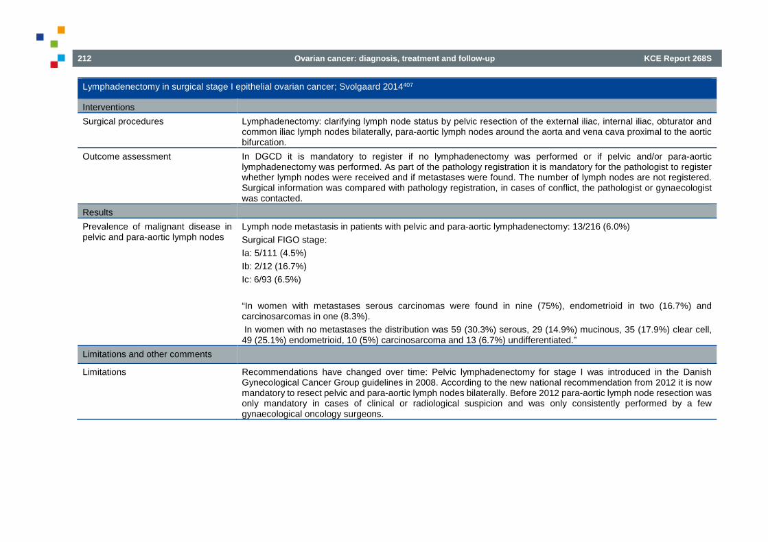
212 Ovarian cancer: diagnosis, treatment and follow-up KCE Report 268S
Lymphadenectomy in surgical stage I epithelial ovarian cancer; Svolgaard 2014407
Interventions Surgical procedures Lymphadenectomy: clarifying lymph node status by pelvic resection of the external iliac, internal iliac, obturator and
common iliac lymph nodes bilaterally, para-aortic lymph nodes around the aorta and vena cava proximal to the aortic bifurcation.
Outcome assessment In DGCD it is mandatory to register if no lymphadenectomy was performed or if pelvic and/or para-aortic lymphadenectomy was performed. As part of the pathology registration it is mandatory for the pathologist to register whether lymph nodes were received and if metastases were found. The number of lymph nodes are not registered. Surgical information was compared with pathology registration, in cases of conflict, the pathologist or gynaecologist was contacted.
Results Prevalence of malignant disease in pelvic and para-aortic lymph nodes
Lymph node metastasis in patients with pelvic and para-aortic lymphadenectomy: 13/216 (6.0%) Surgical FIGO stage: Ia: 5/111 (4.5%) Ib: 2/12 (16.7%) Ic: 6/93 (6.5%) “In women with metastases serous carcinomas were found in nine (75%), endometrioid in two (16.7%) and carcinosarcomas in one (8.3%). In women with no metastases the distribution was 59 (30.3%) serous, 29 (14.9%) mucinous, 35 (17.9%) clear cell, 49 (25.1%) endometrioid, 10 (5%) carcinosarcoma and 13 (6.7%) undifferentiated.”
Limitations and other comments
Limitations Recommendations have changed over time: Pelvic lymphadenectomy for stage I was introduced in the Danish Gynecological Cancer Group guidelines in 2008. According to the new national recommendation from 2012 it is now mandatory to resect pelvic and para-aortic lymph nodes bilaterally. Before 2012 para-aortic lymph node resection was only mandatory in cases of clinical or radiological suspicion and was only consistently performed by a few gynaecological oncology surgeons.
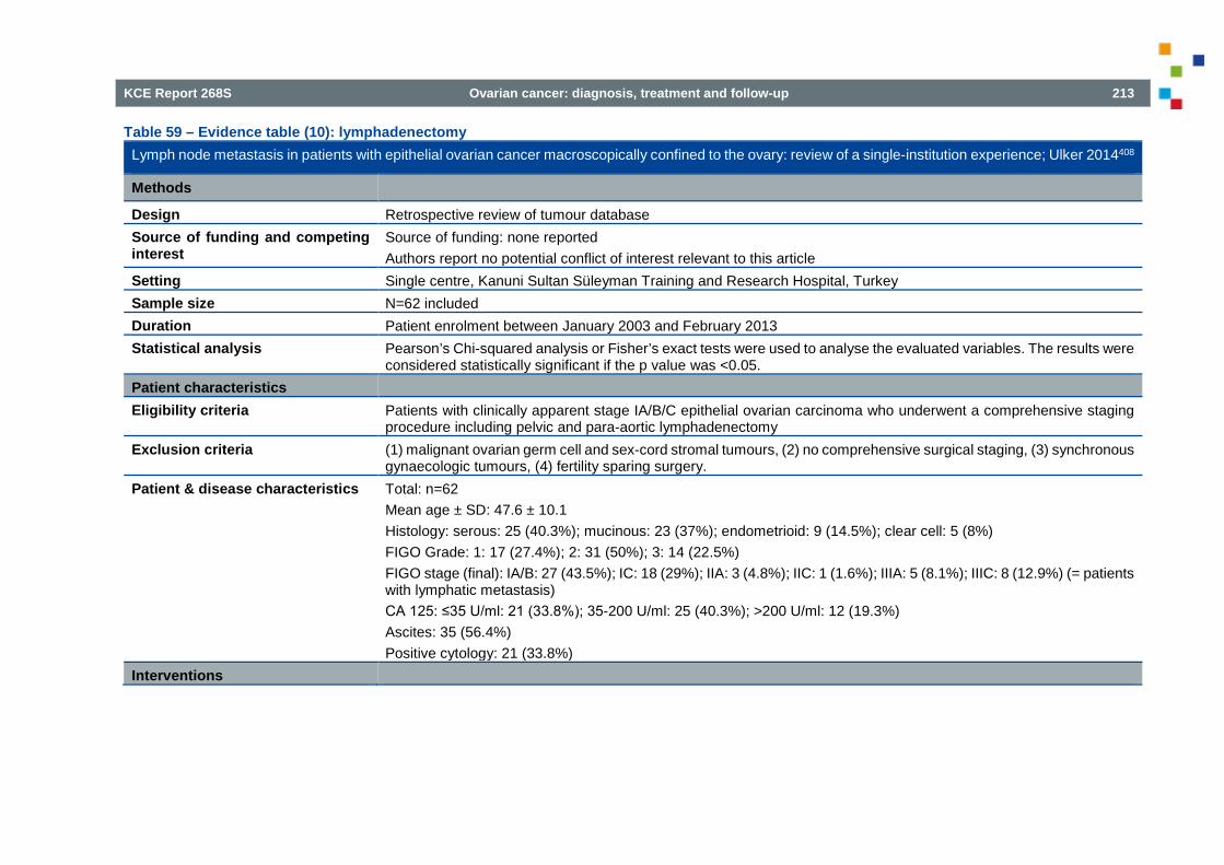
KCE Report 268S Ovarian cancer: diagnosis, treatment and follow-up 213
Table 59 – Evidence table (10): lymphadenectomy Lymph node metastasis in patients with epithelial ovarian cancer macroscopically confined to the ovary: review of a single-institution experience; Ulker 2014408
Methods
Design Retrospective review of tumour database Source of funding and competing interest
Source of funding: none reported Authors report no potential conflict of interest relevant to this article
Setting Single centre, Kanuni Sultan Süleyman Training and Research Hospital, Turkey Sample size N=62 included Duration Patient enrolment between January 2003 and February 2013 Statistical analysis Pearson’s Chi-squared analysis or Fisher’s exact tests were used to analyse the evaluated variables. The results were
considered statistically significant if the p value was <0.05. Patient characteristics Eligibility criteria Patients with clinically apparent stage IA/B/C epithelial ovarian carcinoma who underwent a comprehensive staging
procedure including pelvic and para-aortic lymphadenectomy Exclusion criteria (1) malignant ovarian germ cell and sex-cord stromal tumours, (2) no comprehensive surgical staging, (3) synchronous
gynaecologic tumours, (4) fertility sparing surgery. Patient & disease characteristics Total: n=62
Mean age ± SD: 47.6 ± 10.1 Histology: serous: 25 (40.3%); mucinous: 23 (37%); endometrioid: 9 (14.5%); clear cell: 5 (8%) FIGO Grade: 1: 17 (27.4%); 2: 31 (50%); 3: 14 (22.5%) FIGO stage (final): IA/B: 27 (43.5%); IC: 18 (29%); IIA: 3 (4.8%); IIC: 1 (1.6%); IIIA: 5 (8.1%); IIIC: 8 (12.9%) (= patients with lymphatic metastasis) CA 125: ≤35 U/ml: 21 (33.8%); 35-200 U/ml: 25 (40.3%); >200 U/ml: 12 (19.3%) Ascites: 35 (56.4%) Positive cytology: 21 (33.8%)
Interventions

214 Ovarian cancer: diagnosis, treatment and follow-up KCE Report 268S
Lymph node metastasis in patients with epithelial ovarian cancer macroscopically confined to the ovary: review of a single-institution experience; Ulker 2014408
Surgical procedures The staging process involved obtaining peritoneal washings for cytology and multiple peritoneal biopsies from both suspicious and normal appearing areas, total abdominal hysterectomy and bilateral salpingo-oophorectomy. After the initial step of the surgery, pelvic and para-aortic lymphadenectomy, appendectomy and total omentectomy were also performed. Specifically, pelvic lymphadenectomy included removal of the common, external, internal and obturator node groups to the level of the inguinal ligament. The para-aortic area was exposed just above the bifurcation of the aorta. Para-aortic lymphadenectomy included removal of node bearing tissues along the aorta and vena cava to the level of the renal veins on both sides. Resected pelvic nodes were subdivided as being right or left sided. However, para-aortic nodes could not be subdivided in this way due to the en bloc resection technique that was applied in the para-aortic region. Mean lymph node counts ± SD: 20.2 ± 5.3 (pelvic) and 10.7 ± 2 (para-aortic).
Outcome assessment Data abstracted from medical records Results Prevalence of malignant disease in pelvic and para-aortic lymph nodes
Lymph node metastasis in patients with pelvic and para-aortic lymphadenectomy: 8/62 (12.9%) - from para-aortic and pelvic areas 1/62 (1.6%) - from only the pelvic area: 4/62 (6.5%) - from only the para-aortic area: 3/62 (4.8%) Histology: Serous: 5/25 (20.0%) Mucinous: 1/23 (4.3%) Endometrioid: 1/9 (11.1%) Clear cell: 1/5 (20.0%) FIGO Grade: 1: 0/17 (0.0%) 2: 2/31 (6.4%) 3: 6/14 (42.9%) p=0.001 CA 125: ≤ 35 U/ml: 1/21 (4.8%)

KCE Report 268S Ovarian cancer: diagnosis, treatment and follow-up 215
Lymph node metastasis in patients with epithelial ovarian cancer macroscopically confined to the ovary: review of a single-institution experience; Ulker 2014408
35-200 U/ml: 3/25 (12.0%) >200 U/ml: 4/12 (33.3%) Ascites: 6/35 (17.1%) p=0.24 Positive cytology: 7/21 (33.3%) p=0.001
Limitations and other comments
Limitations There is no information on the patients who did not undergo a comprehensive surgical staging procedure. Data abstracted from medical records.
5.4. Adjuvant chemotherapy
Table 60 – Evidence table: SR adjuvant chemotherapy in patients with borderline or (micro-) invasive early-stage ovarian cancer Interventions for the treatment of borderline ovarian tumours (Review);Faluyi 201032
Methods
Design Systematic review Source of funding and competing interest
Department of Health, UK; NHS Cochrane Collaboration programme Grant Scheme CPG-506
Search date January 2009 Searched databases Cochrane Gynaecological Cancer Group Trials Register, Cochrane Central Register of Controlled Trials (CENTRAL),
MEDLINE and EMBASE. Registers of clinical trials, abstracts of scientific meetings, reference lists of included studies were also searched.
Included study designs RCTs Number of included studies 7 RCTs
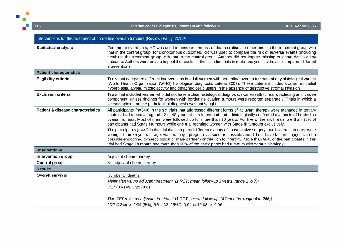
216 Ovarian cancer: diagnosis, treatment and follow-up KCE Report 268S
Interventions for the treatment of borderline ovarian tumours (Review);Faluyi 201032
Statistical analysis For time to event data, HR was used to compare the risk of death or disease recurrence in the treatment group with that in the control group; for dichotomous outcomes, RR was used to compare the risk of adverse events (including death) in the treatment group with that in the control group. Authors did not impute missing outcome data for any outcome. Authors were unable to pool the results of the included trials in meta-analyses as they all compared different interventions.
Patient characteristics Eligibility criteria Trials that compared different interventions in adult women with borderline ovarian tumours of any histological variant
(World Health Organization (WHO) histological diagnostic criteria 2003). These criteria included ovarian epithelial hyperplasia, atypia, mitotic activity and detached cell clusters in the absence of destructive stromal invasion.
Exclusion criteria Trials that included women who did not have a clear histological diagnosis; women with tumours including an invasive component, unless findings for women with borderline ovarian tumours were reported separately. Trials in which a second opinion on the pathological diagnosis was not sought.
Patient & disease characteristics All participants (n=340) in the six trials that addressed different forms of adjuvant therapy were managed in tertiary centres, had a median age of 42 to 48 years at enrolment and had a histologically confirmed diagnosis of borderline ovarian tumour. Most of them were followed up for more than 10 years. For five of the six trials more than 96% of participants had Stage I tumours while one trial recruited women with Stage III tumours exclusively. The participants (n=32) in the trial that compared different extents of conservative surgery, had bilateral tumours, were younger than 35 years of age, wanted to get pregnant as soon as possible and did not have factors suggestive of a possible endocrine, gynaecological or male partner contribution to infertility. More than 90% of the participants in this trial had Stage I tumours and more than 90% of the participants had tumours with serous histology.
Interventions Intervention group Adjuvant chemotherapy Control group No adjuvant chemotherapy Results Overall survival Number of deaths
Melphalan vs. no adjuvant treatment (1 RCT; mean follow-up 3 years, range 1 to 7)) 0/17 (0%) vs. 0/25 (0%) Thio-TEPA vs. no adjuvant treatment (1 RCT ; mean follow up 147 months, range 4 to 246)): 6/27 (22%) vs.2/39 (5%), RR 4.33, 95%CI 0.94 to 19.88, p=0.06
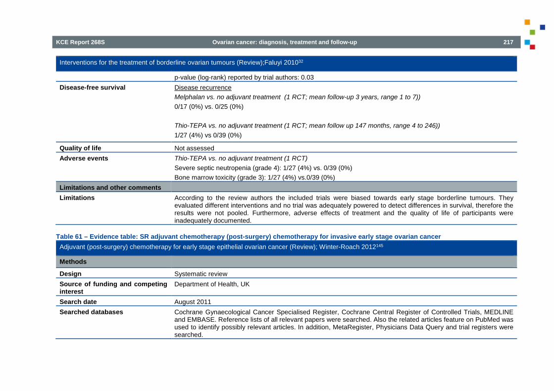
KCE Report 268S Ovarian cancer: diagnosis, treatment and follow-up 217
Interventions for the treatment of borderline ovarian tumours (Review);Faluyi 201032
p-value (log-rank) reported by trial authors: 0.03 Disease-free survival Disease recurrence
Melphalan vs. no adjuvant treatment (1 RCT; mean follow-up 3 years, range 1 to 7)) 0/17 (0%) vs. 0/25 (0%) Thio-TEPA vs. no adjuvant treatment (1 RCT; mean follow up 147 months, range 4 to 246)) 1/27 (4%) vs 0/39 (0%)
Quality of life Not assessed Adverse events Thio-TEPA vs. no adjuvant treatment (1 RCT)
Severe septic neutropenia (grade 4): 1/27 (4%) vs. 0/39 (0%) Bone marrow toxicity (grade 3): 1/27 (4%) vs.0/39 (0%)
Limitations and other comments Limitations According to the review authors the included trials were biased towards early stage borderline tumours. They
evaluated different interventions and no trial was adequately powered to detect differences in survival, therefore the results were not pooled. Furthermore, adverse effects of treatment and the quality of life of participants were inadequately documented.
Table 61 – Evidence table: SR adjuvant chemotherapy (post-surgery) chemotherapy for invasive early stage ovarian cancer Adjuvant (post-surgery) chemotherapy for early stage epithelial ovarian cancer (Review); Winter-Roach 2012145
Methods
Design Systematic review Source of funding and competing interest
Department of Health, UK
Search date August 2011 Searched databases Cochrane Gynaecological Cancer Specialised Register, Cochrane Central Register of Controlled Trials, MEDLINE
and EMBASE. Reference lists of all relevant papers were searched. Also the related articles feature on PubMed was used to identify possibly relevant articles. In addition, MetaRegister, Physicians Data Query and trial registers were searched.

218 Ovarian cancer: diagnosis, treatment and follow-up KCE Report 268S
Adjuvant (post-surgery) chemotherapy for early stage epithelial ovarian cancer (Review); Winter-Roach 2012145
Included study designs RCTs Number of included studies 5 RCTs Statistical analysis Results of studies in a meta-analysis were pooled when clinically similar studies were available. For time-to-event
data, HRs were pooled using the generic inverse variance facility. For any dichotomous outcomes (e.g. adverse events, and numbers of patients who relapsed or died, if it was not possible to treat these outcomes as time-to-event data), RRs were pooled. Random effects models were used for all meta-analyses. If it was inappropriate to pool the data because of clinical heterogeneity, a meta-analysis excluding outlying studies was performed.
Patient characteristics Eligibility criteria RCTs that compared adjuvant chemotherapy with no adjuvant chemotherapy or placebo, that included women with
early stage (I/IIa) epithelial ovarian cancer staged at laparotomy Exclusion criteria Not specified Patient & disease characteristics A total of 1277 patients were enrolled with a median follow-up of 46 to 121 months. All five trials included women with
FIGO stage I ovarian cancer and one trial included women with FIGO stage II as well. One of the trials was flawed by the inclusion of women with borderline ovarian tumours [27/92 (30%)], although they were evenly distributed between the two study groups. In one of the trials there was a predetermined intention to examine, in a subgroup, the effect of staging adequacy in either trial arm. The adequacy of staging in the other trials has not been specified but is assumed to be adequate rather than optimal. For four of the included trials data on histological subtypes could be retrieved from the original publications (n=1179): serous n=350 (29.7%), mucinous n=234 (19.8%), endometrioid n=277 (23.5%), clear-cell n=181 (15.4%), undifferentiated n=34 (2.9%), other n= 58 (4.9%), missing n=45 (3.8%).
Interventions Intervention group Adjuvant chemotherapy (given within three months following surgery, which removed all visible disease.).
Four of the included trials used cisplatin-based chemotherapy, while one used melphalan. Control group No adjuvant chemotherapy or placebo Results Overall survival 5-year overall survival (3 studies, n=1006): HR=0.71 (95% CI 0.53 to 0.93)
Suboptimal staging (2 studies, n=772): HR=0.63 (95% CI 0.46 to 0.85) Optimal staging (2 studies, n=234): HR=1.22 (95% CI 0.63 to 2.37) Tests for subgroup differences: Chi² = 3.14, df = 1 (P= 0.08) and I² = 68.1%

KCE Report 268S Ovarian cancer: diagnosis, treatment and follow-up 219
Adjuvant (post-surgery) chemotherapy for early stage epithelial ovarian cancer (Review); Winter-Roach 2012145
10-year overall survival (2 studies, n=925): HR=0.74 (95% CI 0.58 to 0.95) Low/medium risk patients (1 study, n=219): HR=0.95 (95% CI 0.54 to 1.66) High risk patients (1 study, n=201): HR=0.48 (95% CI 0.32 to 0.72)
Disease-free survival 5-year progression/recurrence/disease free survival (4 studies, n=1170): HR=0.67 (95% CI 0.53 to 0.84) Suboptimal staging (3 studies, n=934): HR=0.64 (95% CI 0.50 to 0.82) Optimal staging (2 studies, n=234): HR=0.67 (95% CI 0.36 to 1.22) Tests for subgroup differences: no differences (P = 0.91; I² = 0%) 10-year progression/recurrence/disease free survival (2 studies, n=925): HR=0.67 (95% CI 0.54 to 0.84) Suboptimal staging (1 study, n=448): HR=0.60 (95% CI 0.41 to 0.87) Optimal staging (1 study, n=448): HR=0.73 (95% CI 0.38 to 1.42) Low/medium risk patients (1 study, n=477): HR=0.96 (95% CI 0.50 to 1.38) High risk patients (1 study, n=477): HR=0.52 (95% CI 0.33 to 0.82)
Quality of life Not assessed by review authors. We checked the full text reports of the included RCTs and none of them presented results for quality of life.
Adverse events A comparison of the risk of adverse events between adjuvant chemotherapy and no chemotherapy was not possible, since none of the included trials reported adverse events among women who did not receive adjuvant chemotherapy.
Limitations and other comments Limitations Post hoc ‘risk’ subgroups; post hoc assignment of studies to ‘staging’ subgroups.
5.5. Laparoscopic surgery in early stage ovarian cancer 5.5.1. Systematic reviews Table 62 – Evidence table of systematic reviews: laparoscopy for early stage ovarian cancer (1) Lawrie 2013203
Methods
Design SR
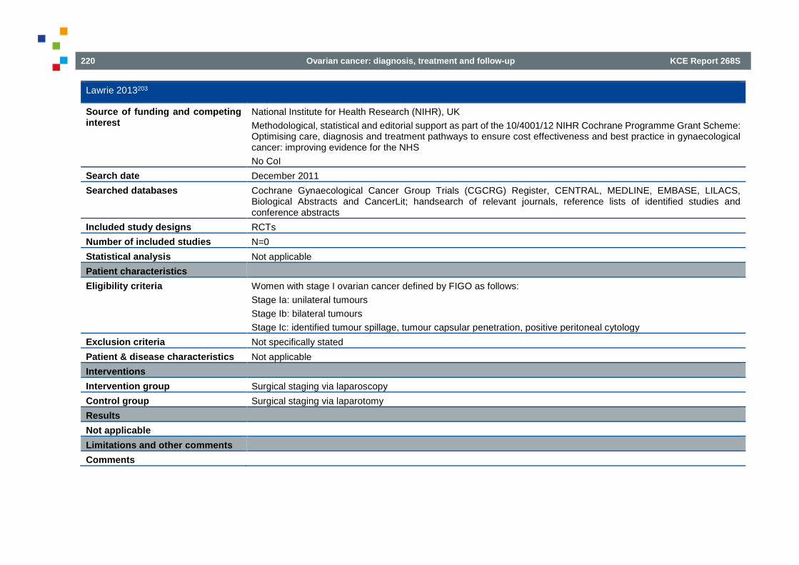
220 Ovarian cancer: diagnosis, treatment and follow-up KCE Report 268S
Lawrie 2013203
Source of funding and competing interest
National Institute for Health Research (NIHR), UK Methodological, statistical and editorial support as part of the 10/4001/12 NIHR Cochrane Programme Grant Scheme: Optimising care, diagnosis and treatment pathways to ensure cost effectiveness and best practice in gynaecological cancer: improving evidence for the NHS No CoI
Search date December 2011 Searched databases Cochrane Gynaecological Cancer Group Trials (CGCRG) Register, CENTRAL, MEDLINE, EMBASE, LILACS,
Biological Abstracts and CancerLit; handsearch of relevant journals, reference lists of identified studies and conference abstracts
Included study designs RCTs Number of included studies N=0 Statistical analysis Not applicable Patient characteristics Eligibility criteria Women with stage I ovarian cancer defined by FIGO as follows:
Stage Ia: unilateral tumours Stage Ib: bilateral tumours Stage Ic: identified tumour spillage, tumour capsular penetration, positive peritoneal cytology
Exclusion criteria Not specifically stated Patient & disease characteristics Not applicable Interventions Intervention group Surgical staging via laparoscopy Control group Surgical staging via laparotomy Results Not applicable Limitations and other comments Comments

KCE Report 268S Ovarian cancer: diagnosis, treatment and follow-up 221
Table 63 – Evidence table of systematic reviews: laparoscopy for early stage ovarian cancer (2) Lu 2015198
Methods Design SR Source of funding and competing interest
Funding not reported No CoI
Search date March 2014 Searched databases MEDLINE, Embase, Cochrane Library, China Biology Medicine, Chinese National Knowledge Infrastructure Included study designs RCTs or well-designed nonrandomized controlled trials with no confinement on allocation concealment, blinding, or
districts Number of included studies N=11 Statistical analysis RevMan version 5.2
I2 statistic to estimate the degree of heterogeneity Dichotomous data presented as relative risks with 95%CI If no heterogeneity was detected, a fixed effects model was used to analyse the data. A random effects model was used if there was any unexplained heterogeneity If a sufficient number of eligible trials (>10 studies) existed, a funnel plot analysis was conducted to assess reporting biases Subgroup analysis to test for differences between results published in English and Chinese
Patient characteristics Eligibility criteria Patients with early stage ovarian cancer Exclusion criteria Studies in which laparoscopic surgery was performed for diagnostic biopsy instead of radical treatment Patient & disease characteristics 235 patients in the laparoscopy group and 356 patients in the laparotomy group
Age (range): 41.9-55y vs. 41.5-61y Epithelial cancer (range): 70-100% vs. 68-100% FIGO stage I (range): 73-100% vs. 68-100%
Interventions Intervention group Surgical staging via laparoscopy Control group Surgical staging via laparotomy

222 Ovarian cancer: diagnosis, treatment and follow-up KCE Report 268S
Lu 2015198
Results (ranges across studies) Operative time MD -54.0 (95%CI -96.99 to -11.01) to 105.00 (61.5 to 148.5) Intraoperative blood loss MD -7.70 (-9.51 to -5.89) to -0.01 (-0.57 to 0.56) Pelvic lymphadenectomy number MD -6.70 (-13.3 to -0.1) to 4.50 (-2.14 to 11.14) Para-aortic lymphadenectomy number
MD -2.50 (-5.44 to 0.44) to 5.10 (2.13 to 8.07)
Time to first postoperative flatus MD -2.60 (-3.07 to -2.13) to -0.45 (-0.79 to -0.11) Intraoperative complications OR 0.28 (0.01 to 6.10) to 5.43 (0.21 to 140.18) Postoperative complications OR 0.13 (0.02 to 0.80) to 0.69 (0.15 to 3.18) Hospital stay MD -9.28 (-10.76 to -7.80) to 0.20 (-3.20 to 3.60) Recurrence rate OR 0.04 (0.01 to 0.38) to 6.29 (0.28 to 140.86) Postoperative mortality OR 0.35 (0.01 to 9.24) to 0.53 (0.07 to 4.01) Limitations and other comments Comments Invalid meta-analysis: pooling of unadjusted data from observational trials
5.5.2. Primary studies
Table 64 – Evidence table: Primary study laparoscopy in early-stage ovarian cancer (1) Chi 2005409
Methods
Design Non-randomized comparative study Source of funding and competing interest
Not reported
Setting Single centre, USA Sample size 50 patients (20 laparoscopy, 30 laparotomy) Duration and follow-up October 2000 – March 2003

KCE Report 268S Ovarian cancer: diagnosis, treatment and follow-up 223
Chi 2005409
Statistical analysis Fisher exact test was used to compare categorical variables. Continuous variables were compared with the Student t test and the Wilcoxon rank sums test. All statistical tests were 2 sided.
Patient characteristics Eligibility criteria Patients who underwent (attempt of) comprehensive staging for apparent stage I ovarian or fallopian tube cancer, by
laparoscopy or laparotomy between October 2000 and March 2003. Exclusion criteria Borderline carcinomas, stromal tumours and germ cell tumours were excluded from the laparotomy arm (they were
included in the laparoscopy group if the surgical intent was comprehensive staging). Patients with macroscopic disease noted at the time of surgery were excluded
Patient & disease characteristics Mean age 49 +/- 11 years. No significant differences between groups in terms of mean age, BMI, primary disease site, histologic type, tumour grade. 65% of the 20 patients in the laparoscopy group had previous adnexal surgery without staging procedure and 23% of the laparotomy group.
Interventions Intervention group Comprehensive laparoscopic staging including cytological washings, peritoneal biopsies, omentectomy and nodal
dissection) Control group Comprehensive staging via open surgery (laparotomy) Results Operating time 321 +/-64 min vs. 276 +/-68 min (p=0.04) Estimated blood loss 235 +/-138 ml vs 367 +/- 208 ml (p=0.003) Hospital stay 3.1 +/- 0.7 days vs 5.8 +/- 2.6 days (p<0.001) Complications 0 (0)% vs 2 (7)% (p=1.00) Limitations and other comments Limitations Data collected retrospectively.
No case-mix adjustment Many patients had adnexectomy before staging surgery, effect on pathological stage unknown, no data on oncological outcomes reported.
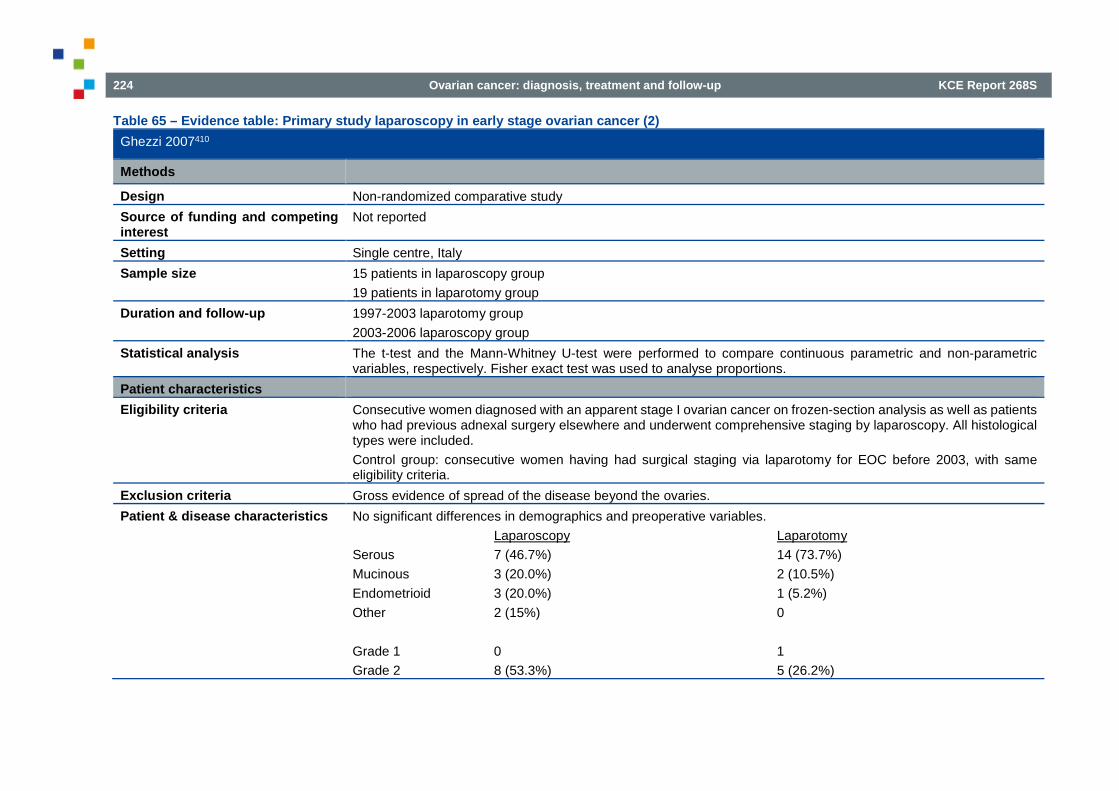
224 Ovarian cancer: diagnosis, treatment and follow-up KCE Report 268S
Table 65 – Evidence table: Primary study laparoscopy in early stage ovarian cancer (2) Ghezzi 2007410
Methods
Design Non-randomized comparative study Source of funding and competing interest
Not reported
Setting Single centre, Italy Sample size 15 patients in laparoscopy group
19 patients in laparotomy group Duration and follow-up 1997-2003 laparotomy group
2003-2006 laparoscopy group Statistical analysis The t-test and the Mann-Whitney U-test were performed to compare continuous parametric and non-parametric
variables, respectively. Fisher exact test was used to analyse proportions. Patient characteristics Eligibility criteria Consecutive women diagnosed with an apparent stage I ovarian cancer on frozen-section analysis as well as patients
who had previous adnexal surgery elsewhere and underwent comprehensive staging by laparoscopy. All histological types were included. Control group: consecutive women having had surgical staging via laparotomy for EOC before 2003, with same eligibility criteria.
Exclusion criteria Gross evidence of spread of the disease beyond the ovaries. Patient & disease characteristics No significant differences in demographics and preoperative variables.
Laparoscopy Laparotomy Serous 7 (46.7%) 14 (73.7%) Mucinous 3 (20.0%) 2 (10.5%) Endometrioid 3 (20.0%) 1 (5.2%) Other 2 (15%) 0 Grade 1 0 1 Grade 2 8 (53.3%) 5 (26.2%)

KCE Report 268S Ovarian cancer: diagnosis, treatment and follow-up 225
Ghezzi 2007410
Grade 3 7 (46.7%) 13 (58.4%) FIGO stage Ic 6 (40%) 5 (26.2%)
Interventions Intervention group Comprehensive stage via laparoscopy Control group Comprehensive stage via laparotomy Results Intraoperative rupture Intraoperative rupture occurred in 3 (20%) cases in the laparoscopy group and 2 (10.5%) cases in the laparotomy
group (p=0.63). Operating time 377 +/- 47min vs 272 +/- 81min (p=0.002) Estimated blood loss 250 (50-1000) ml vs 400 (150-1000) ml (p=0.28) Hospital stay 3 (2-12) days vs 7 (4-14) days (p=0.001) Complications 2 (13.3%) vs 8 (42.1%) (p=0.13) Adjuvant chemotherapy 11 (73.3%) vs 13 (68.4%) Limitations and other comments Limitations Non-contemporary cohorts
No case-mix adjustment Table 66 – Evidence table: Primary study laparoscopy in early stage ovarian cancer (3) Lee 2011411
Methods
Design Non-randomized comparative study Source of funding and competing interest
Grants from the Yonsei University Research Fund 2010, Faculty Research Grant of Yonsei University College of Medicine 2009 and National Research Foundation of Korea Grant funded by the Korean Government
Setting Single centre, South-Korea Sample size 26 patients in laparoscopy group and 87 in the laparotomy group Duration and follow-up 2005-2010 Statistical analysis Student t test was used for parametric variables and Mann-Whitney test was used for nonparametric variables

226 Ovarian cancer: diagnosis, treatment and follow-up KCE Report 268S
Lee 2011411
Patient characteristics Eligibility criteria Newly diagnosed untreated gynaecologic cancers and transferred patients with inadequate staging surgery. Exclusion criteria Evidence of more advanced disease in preoperative work-up
Prior treatment with pelvic radiotherapy or chemotherapy No follow-up data available
Patient & disease characteristics No significant differences in demographics and preoperative variables Laparoscopy Laparotomy Serous 10 (38.5%) 24 (27.6%) Mucinous 8 (30.8%) 22 (25.3%) Endometrioid 2 (7.7%) 7 (8.0%) Clear cell 2 (7.7%) 11 (12.6%) Squamous 0 7 (8.0%) Sex cord 2 (7.7%) 14 (16.1%) Grade 1 6 (23.1%) 25 (28.7%) Grade 2 8 (30.8%) 13 (14.9%) Grade 3 7 (26.9%) 21 (24.1%) Unknown grade 5 (19.2%) 28 (32.2%) FIGO stage IC 14 (53.8%) 35 (39.1%)
Interventions Intervention group Laparoscopic staging including total hysterectomy, bilateral salpingo-oophorectomy, bilateral pelvic and para-aortic
lymphadenectomy, appendectomy, infracolic omentectomy Control group Same procedures by laparotomy Results Intraoperative rupture 0 vs 13 (14.9% (p=0.037) Operating time 227 +/-105.8 min vs 184.6+/-61.4 min (p=0.016) Estimated blood loss 230.4+/-183.6 ml vs 474.8+/-329.2 ml (p<0.001)
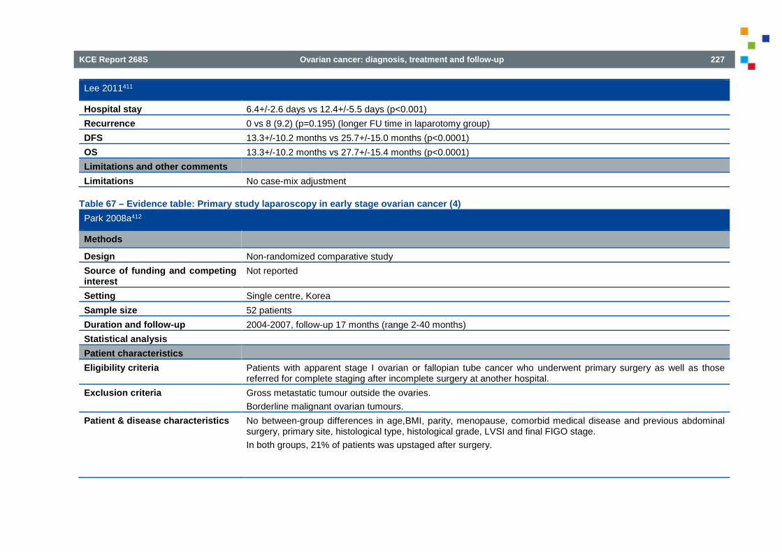
KCE Report 268S Ovarian cancer: diagnosis, treatment and follow-up 227
Lee 2011411
Hospital stay 6.4+/-2.6 days vs 12.4+/-5.5 days (p<0.001) Recurrence 0 vs 8 (9.2) (p=0.195) (longer FU time in laparotomy group) DFS 13.3+/-10.2 months vs 25.7+/-15.0 months (p<0.0001) OS 13.3+/-10.2 months vs 27.7+/-15.4 months (p<0.0001) Limitations and other comments Limitations No case-mix adjustment
Table 67 – Evidence table: Primary study laparoscopy in early stage ovarian cancer (4) Park 2008a412
Methods
Design Non-randomized comparative study Source of funding and competing interest
Not reported
Setting Single centre, Korea Sample size 52 patients Duration and follow-up 2004-2007, follow-up 17 months (range 2-40 months) Statistical analysis Patient characteristics Eligibility criteria Patients with apparent stage I ovarian or fallopian tube cancer who underwent primary surgery as well as those
referred for complete staging after incomplete surgery at another hospital. Exclusion criteria Gross metastatic tumour outside the ovaries.
Borderline malignant ovarian tumours. Patient & disease characteristics No between-group differences in age,BMI, parity, menopause, comorbid medical disease and previous abdominal
surgery, primary site, histological type, histological grade, LVSI and final FIGO stage. In both groups, 21% of patients was upstaged after surgery.

228 Ovarian cancer: diagnosis, treatment and follow-up KCE Report 268S
Park 2008a412
Interventions Intervention group Laparoscopic staging procedure including peritoneal leavages, random periotnela biopsies, total hysterectomy,
bilateral salpingo-oophorectomy, systematic pelvic and para-aortic lymph node dissection, infracolic omentectomy and appendectomy. Ovarian tumours were removed using an endobag.
Control group As for laparoscopy, but a generous midline abdominal incision Results Intraoperative rupture 2 (10.5%) vs 4 (12.1%) (p=1.00) Operating time 220.7 +/- 82.7 min vs 274.7+/-63.2 min (p=0.012) Estimated blood loss 240.0+/-228.3ml vs 568.2+/-451.7ml (p=0.005) Hospital stay 8.9+/-6.1 days vs 14.5+/-5.6 days (p=0.002) Perioperative complications 2 (10.5%) vs 9 (27.3%) (p=0.290) Adjuvant chemotherapy 78.9% vs 78.8% Limitations and other comments Limitations No adjustment for prognostic factors
Patients with secondary surgery also included.
Table 68 – Evidence table: Primary study laparoscopy in early stage ovarian cancer (5) Park 2008b413
Methods
Design Non-randomized comparative study (retrospective chart review) Source of funding and competing interest
Not reported
Setting Single centre, Korea Sample size 36 consecutive patients Duration and follow-up 2001-2006; median follow-up 17 months (range 5-61 months)
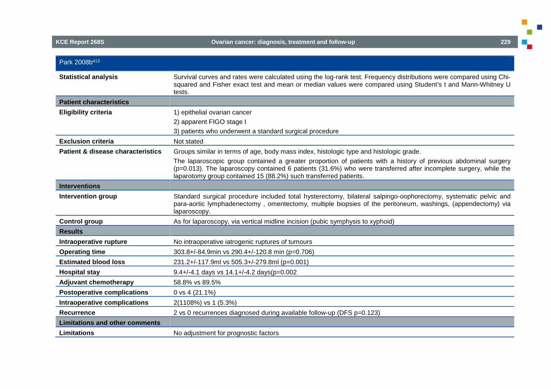
KCE Report 268S Ovarian cancer: diagnosis, treatment and follow-up 229
Park 2008b413
Statistical analysis Survival curves and rates were calculated using the log-rank test. Frequency distributions were compared using Chi-squared and Fisher exact test and mean or median values were compared using Student’s t and Mann-Whitney U tests.
Patient characteristics Eligibility criteria 1) epithelial ovarian cancer
2) apparent FIGO stage I 3) patients who underwent a standard surgical procedure
Exclusion criteria Not stated Patient & disease characteristics Groups similar in terms of age, body mass index, histologic type and histologic grade.
The laparoscopic group contained a greater proportion of patients with a history of previous abdominal surgery (p=0.013). The laparoscopy contained 6 patients (31.6%) who were transferred after incomplete surgery, while the laparotomy group contained 15 (88.2%) such transferred patients.
Interventions Intervention group Standard surgical procedure included total hysterectomy, bilateral salpingo-oophorectomy, systematic pelvic and
para-aortic lymphadenectomy , omentectomy, multiple biopsies of the peritoneum, washings, (appendectomy) via laparoscopy.
Control group As for laparoscopy, via vertical midline incision (pubic symphysis to xyphoid) Results Intraoperative rupture No intraoperative iatrogenic ruptures of tumours Operating time 303.8+/-84.9min vs 290.4+/-120.8 min (p=0.706) Estimated blood loss 231.2+/-117.9ml vs 505.3+/-279.8ml (p=0.001) Hospital stay 9.4+/-4.1 days vs 14.1+/-4.2 days(p=0.002 Adjuvant chemotherapy 58.8% vs 89.5% Postoperative complications 0 vs 4 (21.1%) Intraoperative complications 2(1108%) vs 1 (5.3%) Recurrence 2 vs 0 recurrences diagnosed during available follow-up (DFS p=0.123) Limitations and other comments Limitations No adjustment for prognostic factors

230 Ovarian cancer: diagnosis, treatment and follow-up KCE Report 268S
Table 69 – Evidence table: Primary study laparoscopy in early-stage ovarian cancer (6) Liu 2014208
Methods
Design Non-randomized comparative study (retrospective chart review) Source of funding and competing interest
Not reported
Setting Single centre, China Sample size 35 cases in the laparoscopy group, 40 cases in the laparotomy group Duration and follow-up March 2002-May 2010, FU 36 to 84 months Statistical analysis X2 test was used for categorical data Patient characteristics Eligibility criteria Early-stage ovarian cancer surgically stage after intraoperative pathologic evaluation Exclusion criteria Not specified Patient & disease characteristics No significant differences in pathological stage, histological type and degree of differentiation Interventions Intervention group Staging via laparoscopy, including peritoneal washings, hysterectomy, bilateral salpingo-oophorectomy, (pelvic)
lymphadenectomy, omentectomy, appendectomy Control group Laparotomy, no further details provided Results Intraoperative rupture 3/35 vs 2/40 cases of intraoperative rupture Operating time 209.71+/-17.57 min vs 200.50+/-20.62 min (p>0.05) Estimated blood loss 197.14+/-98.48ml vs 345+/-165.95ml (p>0.05) Hospital stay 16.29+/-6.20 days vs 21.85+/-4.88 days (p<0.05) Adjuvant chemotherapy 85.71% vs 90.00% (p>0.05) Postoperative complications 4/35 (11.43%) vs 5/40(12.50%) (p>0.05) Recurrence rate 5.71% vs 5.00% (p>0.05) Median time to recurren 13 vs 14.5 months Median tumour-free survival time 54.3 months vs 57.2 months

KCE Report 268S Ovarian cancer: diagnosis, treatment and follow-up 231
Liu 2014208
3-year survival rate 97.14% vs 97.50% (p>0.05) 5-year survival rate 94.11% vs 96.30% (p>0.05) Limitations and other comments Limitations No adjustment for prognostic factors
Table 70 – Evidence table: Primary study laparoscopy in early stage ovarian cancer (7) Koo 2014207
Methods Design Non-randomized comparative study (retrospective chart review) Source of funding and competing interest
No potential conflict of interest relevant to this article
Setting Single centre, Korea Sample size 77 patients, 24 in the laparoscopy group and 53 patients in the laparotomy group. Duration and follow-up October 2006-December 2012, mean follow-up 31 months Statistical analysis Continuous variables were compared by using the Student t-test and categorical variables were compared by using
the 2-tailed chi-square test; Survival analysis were conducted by using the Kaplan-Meier method and surviving data were censored at the date of last follow-up.
Patient characteristics Eligibility criteria All patients who underwent comprehensive staging for early-stage ovarian cancer Exclusion criteria Borderline ovarian malignancy
Advanced ovarian cancer FIGO stage III-IV Concurrent malignancy of another organ Patients referred from other hospital after staging surgery Fertility sparing surgery
Patient & disease characteristics No significant difference in mean age, parity, BMI, preoperative Ca 125-level, preoperative haemoglobin level or previous surgical history. Referral for restaging surgery after initial surgical confirmation of ovarian cancer was more common in the laparoscopy group (58.3% vs 17.0%; p<0.001).

232 Ovarian cancer: diagnosis, treatment and follow-up KCE Report 268S
Koo 2014207
Appendectomy was performed more often in the laparotomy group (81.1% vs 41.7%: p=0.001) The mean tumour size was 7.3cm in the laparoscopy group and 11.2cm in the laparotomy group (p=0.001)
Interventions Intervention group Comprehensive staging via laparoscopy including total hysterectomy, bilateral salpingo-oophorectomy, systematic
pelvic and para-aortic lymph node dissection, omentectomy, peritoneal cytology, multiple biopsies from the entire abdominal peritoneum.
Control group As for laparoscopy, laparotomy via midline longitudinal incision Results Intraoperative rupture 13 (54.2%) vs 21 (39.6%) (p=0.465) Operating time 192.9+/-73.5 min vs 224.1+/-85.4 min (p=0.127) Estimated blood loss 697.9+/-396.9 ml vs 972.6+/-827.8 ml (p=0.127) Hospital stay 13.7+/-5.4 days vs 13.1+/-4.1 days (p=0.594) Adjuvant chemotherapy 21 (87.5%) vs 48 (90.6%) (p=0.480) Intraoperative complications 0 vs 3(5.7%) (p=0.548) Postoperative complications Similar between groups Recurrence rate 2 (8.3%) vs 2 (3.8%) (p=0.585) Disease-free survival 59.3 months (95%CI 51.8-66.7 months) vs 66.3 months (95%CI 62.8-69.9 months) (p=0.367) 3-year survival rate 86.1% vs 94.7% Limitations and other comments Limitations No case-mix adjustment
5.6. Laparoscopy, PET-CT and MRI to predict end result of cytoreductive surgery
Table 71 – Evidence table: SRs prediction of end result of surgery Laparoscopy for diagnosing resectability of disease in patients with advanced ovarian cancer; Rutten 2014
Methods
Design Systematic review

KCE Report 268S Ovarian cancer: diagnosis, treatment and follow-up 233
Laparoscopy for diagnosing resectability of disease in patients with advanced ovarian cancer; Rutten 2014
Source of funding and competing interest
None known
Search date February 2013 Searched databases MEDLINE, EMBASE, CENTRAL, the Cochrane Register of DiagnosticTest Accuracy Studies, MEDION and ISI Web
of Science. Included study designs Diagnostic test accuracy studies Number of included studies N=7 (reporting on six cohorts) Statistical analysis Data from each study was summarised in two by two tables of TP, FP, TN, FN and used to calculate sensitivity and
specificity. Studies providing insufficient data were used to present data on negative predicting values (NPV). Investigation for heterogeneity among estimates of NPV and estimates of test positivity was performed using Cochran’s Q-test.
Patient characteristics Eligibility criteria Studies that evaluated the diagnostic accuracy of laparoscopy to determine the resectability of disease in patients
who are suspected of advanced ovarian cancer and planned to receive primary debulking surgery were included. Participants included women suspected of having advanced stage ovarian carcinoma (FIGO stage IIB, IIC, IIIA to C, IV), who were scheduled for primary debulking surgery and did not have contraindications for laparoscopy or laparotomy.
Exclusion criteria Case-control studies were excluded, exclusion criteria not further specified. Patient & disease characteristics A total of 408 patients were included in all studies. Only two studies avoided partial verification bias and provided data
to calculate sensitivity and specificity. One of these included 64 patients undergoing surgery for a suspected advanced ovarian or peritoneal cancer, mean age 57.4 years (SD 12.7). The second study included 113 patients suspected of advanced primary ovarian cancer, mean age 59 years (range 39 to 81).
Interventions Index test Additional open diagnostic laparoscopy, performed when a patient was planned for primary debulking surgery after
conventional diagnostic work-up. Conventional diagnostic work-up consisted of physical and ultrasound examination, serum CA 125 measurement and/or CT or MRI scan.
Target condition Ovarian cancer deposits that could not be resected at laparotomy to at least less than 1 cm in diameter. Reference standard Laparotomy Results Overall survival Not addressed
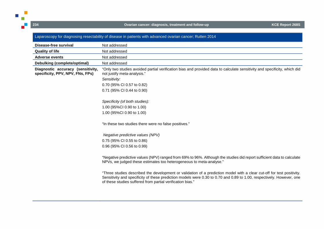
234 Ovarian cancer: diagnosis, treatment and follow-up KCE Report 268S
Laparoscopy for diagnosing resectability of disease in patients with advanced ovarian cancer; Rutten 2014
Disease-free survival Not addressed Quality of life Not addressed Adverse events Not addressed Debulking (complete/optimal) Not addressed Diagnostic accuracy (sensitivity, specificity, PPV, NPV, FNs, FPs)
“Only two studies avoided partial verification bias and provided data to calculate sensitivity and specificity, which did not justify meta-analysis.” Sensitivity: 0.70 (95% CI 0.57 to 0.82) 0.71 (95% CI 0.44 to 0.90) Specificity (of both studies): 1.00 (95%CI 0.90 to 1.00) 1.00 (95%CI 0.90 to 1.00) “In these two studies there were no false positives.” Negative predictive values (NPV) 0.75 (95% CI 0.55 to 0.86) 0.96 (95% CI 0.56 to 0.99) “Negative predictive values (NPV) ranged from 69% to 96%. Although the studies did report sufficient data to calculate NPVs, we judged these estimates too heterogeneous to meta-analyse.” “Three studies described the development or validation of a prediction model with a clear cut-off for test positivity. Sensitivity and specificity of these prediction models were 0.30 to 0.70 and 0.89 to 1.00, respectively. However, one of these studies suffered from partial verification bias.”

KCE Report 268S Ovarian cancer: diagnosis, treatment and follow-up 235
Laparoscopy for diagnosing resectability of disease in patients with advanced ovarian cancer; Rutten 2014
Limitations and comments Limitations Source of funding or support was not reported for each of the included studies. Methodological heterogeneity exists
on the definition of a positive/negative reference test. Three studies used the cut-off of 1.0 cm for residual disease. Heterogeneity exists between the included studies regarding the pre-treatment work-up, the clinical and/or radiological tests performed, and their influence on study/debulking eligibility. Two of seven original studies avoided partial verification bias by referring all patients to laparotomy (i.e. the reference test). Consequently, of the 168 women considered ineligible for a debulking (i.e. positive at laparoscopy), only 52 cases (52/168=31%) were completely verified. Applicability to our research question: the two original studies which avoided the risk of partial verification bias, included a mix of primary debulking, interval debulking and recurrent disease laparotomies, without clear subanalyses.
Table 72 – Evidence table (1): DTA study prediction of end result of surgery Espada 2013; Diffusion-weighted magnetic resonance imaging evaluation of intra-abdominal sites of implants to predict likelihood of suboptimal cytoreductive surgery in patients with ovarian carcinoma.
Methods
Design Prospective DTA study Source of funding and competing interest
Source of funding not reported Competing interests not disclosed
Setting Single centre study, Spain Sample size N=34 Time interval between tests Within 15 days Statistical analysis “Sensitivity, specificity, positive predictive value (PPV), negative predictive value (NPV) and accuracy for each feature for
diffusion-weighted magnetic resonance imaging (DWMRI) predicting affection in exploratory laparotomy (EL); same parameters were calculated for each feature for DWMRI and EL for predicting suboptimal cytoreduction. According to literature, minimum criteria for including a certain anatomical site in the predictive score were: specificity >75%, PPV >50% and NPV >50%. Receiver operating characteristic (ROC) curve analysis was used to assess the ability of the model to predict surgical outcome for DWMRI and EL. Comparison between the areas under ROC curves was performed, using the Hanley and McNeil method. The Fisher exact test was used to examine the strength of statistical association between each predictive score and surgical outcome. Statistical analysis was carried out using the PASW Statistics v. 18.0 software.”

236 Ovarian cancer: diagnosis, treatment and follow-up KCE Report 268S
Espada 2013; Diffusion-weighted magnetic resonance imaging evaluation of intra-abdominal sites of implants to predict likelihood of suboptimal cytoreductive surgery in patients with ovarian carcinoma.
Patient characteristics Eligibility criteria Patients undergoing surgery for a suspected advanced ovarian carcinoma between December 2006 and June 2012. Patient characteristics N=34
Median age (SD): 53.1 (11.9) years Disease stage: stage III: 28 (82.4%); stage IV: 6 (17.7%) Histology: serous adenocarcinoma: 28 (82.4%), endometrioid/clear cell carcinoma: 3 (8.8%), mucinous adenocarcinoma: 2 (5.9%), poorly differentiated carcinoma; 1 (2.9%)
Prevalence of disease Prevalence of residual disease > 1 cm 8/34 (24%) Interventions Index test(s) “All patients underwent a preoperative pelvic and abdominal MRI study for tumour staging at a 3-T system (GE Medical
System, Milwaukee, WI, USA). A phased-array torso surface coil was used, with parallel imaging with an acceleration factor of 2 in the phase direction and 1 in the slice direction.” Diffusion-weighted magnetic resonance imaging (DWMRI). DWMRI of the pelvis study was acquired in the axial plane using a spinecho echo planar imaging acquisition. DWI parameters included a b value of 600 s/mm, TE 2.8, TR 4,800, matrix 128×128, slice thickness 5 mm, interslice gap 0 mm, 6 nex, FOV 38, phase FOV 0.70. DWMRI of the abdomen was also acquired in an axial plane with the same sequence but with a large FOV. A score was constructed that addressed the affection of 9 sites: (1) small and/or large bowel mesentery, (2) hepatic parenchyma , hylium or surface over 2 cm, (3) omental extension (spleen or splenic hylium, stomach or lesser sac), (4) diaphragm, (5) peritoneal thickening, (6) peritoneal macroscopic implants (≥2 cm), (7) miliar visceral peritoneum implants, (8) massive ascites, (9) suprarenal para-aortic lymph nodes (≥1 cm) The score with the highest overall accuracy was selected as threshold: score of ≥6 (accuracy 91.1 %) DWMRI findings were evaluated by the same two radiologists, who were blind to the clinical data.
Reference standard Exploratory laparotomy (EL) “with the aim of performing a maximal tumour cytoreduction to residual disease of 1 cm in maximal diameter” Patients were operated on by the same gynaecological oncologist who was aware of the DWMRI findings.

KCE Report 268S Ovarian cancer: diagnosis, treatment and follow-up 237
Espada 2013; Diffusion-weighted magnetic resonance imaging evaluation of intra-abdominal sites of implants to predict likelihood of suboptimal cytoreductive surgery in patients with ovarian carcinoma.
Results Debulking (complete/optimal) “Twenty-six patients (76.5 %) were considered as being optimally cytoreducted to residual tumour < 1 cm.” Diagnostic accuracy (sensitivity, specificity, PPV, NPV, FNs, FPs)
Target condition: residual tumour size >1 cm Sensitivity: 0.75 (95%-CI 0.35 to 0.97) Specificity: 0.96 (95%-CI 0.80 to 1.00) PPV: 0.86 (95%-CI 0.42 to 1.00) NPV: 0.93 (95%-CI 0.76 to 0.99) In a hypothetical study of 1000 patients and with a prevalence of 24% (= study prevalence) 60 (95%-CI 7 to 156) patients will be incorrectly classified as being resectable (FNs) 30 (95%-CI 0 to 152) patients will be incorrectly classified as not being resectable (FPs)
Limitations and other comments
Limitations High risk of bias for the domains Patient selection, Index test and Reference standard Applicability concerns for the domains Patient selection and Index test DW-MRI not assessed as add-on rest to CT Very small sample size ((very) wide 95%-CIs) Confusion about the reference standard: elective laparotomy (EL) seems to be the reference standard, but in Tables 2 and 3 of the report the diagnostic accuracy of EL is also reported. It's not clear with what other reference standard EL is compared.
Table 73 – Evidence table (2): DTA study prediction of end result of surgery Forstner 1995; Ovarian cancer: staging with CT and MR imaging
Methods
Design Prospective DTA study Source of funding and competing interest
Austrian E. Schroedinger Stipendium (100874- Med) Competing interests not disclosed
Setting Single centre study, USA
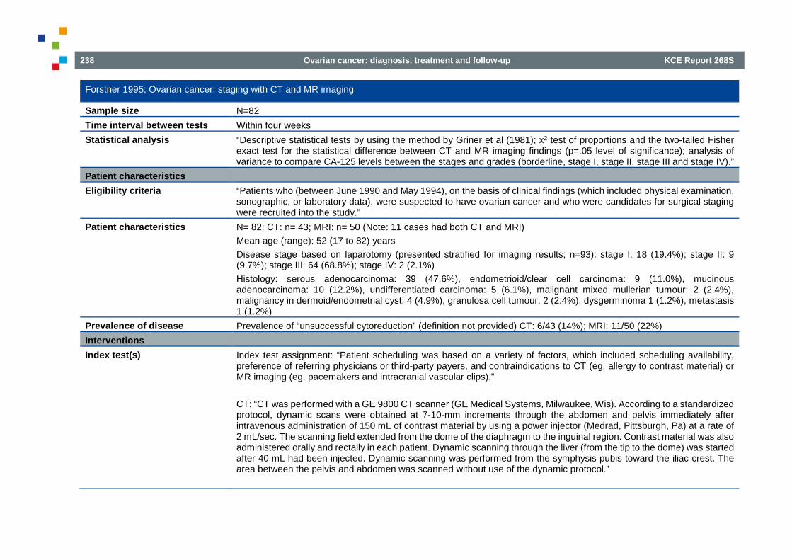
238 Ovarian cancer: diagnosis, treatment and follow-up KCE Report 268S
Forstner 1995; Ovarian cancer: staging with CT and MR imaging
Sample size N=82 Time interval between tests Within four weeks Statistical analysis “Descriptive statistical tests by using the method by Griner et al (1981); x2 test of proportions and the two-tailed Fisher
exact test for the statistical difference between CT and MR imaging findings (p=.05 level of significance); analysis of variance to compare CA-125 levels between the stages and grades (borderline, stage I, stage II, stage III and stage IV).”
Patient characteristics Eligibility criteria “Patients who (between June 1990 and May 1994), on the basis of clinical findings (which included physical examination,
sonographic, or laboratory data), were suspected to have ovarian cancer and who were candidates for surgical staging were recruited into the study.”
Patient characteristics N= 82: CT: n= 43; MRI: n= 50 (Note: 11 cases had both CT and MRI) Mean age (range): 52 (17 to 82) years Disease stage based on laparotomy (presented stratified for imaging results; n=93): stage I: 18 (19.4%); stage II: 9 (9.7%); stage III: 64 (68.8%); stage IV: 2 (2.1%) Histology: serous adenocarcinoma: 39 (47.6%), endometrioid/clear cell carcinoma: 9 (11.0%), mucinous adenocarcinoma: 10 (12.2%), undifferentiated carcinoma: 5 (6.1%), malignant mixed mullerian tumour: 2 (2.4%), malignancy in dermoid/endometrial cyst: 4 (4.9%), granulosa cell tumour: 2 (2.4%), dysgerminoma 1 (1.2%), metastasis 1 (1.2%)
Prevalence of disease Prevalence of “unsuccessful cytoreduction” (definition not provided) CT: 6/43 (14%); MRI: 11/50 (22%) Interventions Index test(s) Index test assignment: “Patient scheduling was based on a variety of factors, which included scheduling availability,
preference of referring physicians or third-party payers, and contraindications to CT (eg, allergy to contrast material) or MR imaging (eg, pacemakers and intracranial vascular clips).” CT: “CT was performed with a GE 9800 CT scanner (GE Medical Systems, Milwaukee, Wis). According to a standardized protocol, dynamic scans were obtained at 7-10-mm increments through the abdomen and pelvis immediately after intravenous administration of 150 mL of contrast material by using a power injector (Medrad, Pittsburgh, Pa) at a rate of 2 mL/sec. The scanning field extended from the dome of the diaphragm to the inguinal region. Contrast material was also administered orally and rectally in each patient. Dynamic scanning through the liver (from the tip to the dome) was started after 40 mL had been injected. Dynamic scanning was performed from the symphysis pubis toward the iliac crest. The area between the pelvis and abdomen was scanned without use of the dynamic protocol.”

KCE Report 268S Ovarian cancer: diagnosis, treatment and follow-up 239
Forstner 1995; Ovarian cancer: staging with CT and MR imaging
MRI: “MRI performed with a 1.5-T system (Signa; GE Medical Systems). The imaging protocol included TI-weighted, T2-weighted, and contrast-enhanced T1-weighted sequences in the transaxial plane. Transaxial images were obtained from the dome of the liver to the inferior pubic ramus (10.0-mm section thickness with a 2.0-mm intersection gap in the abdomen and 5.0-mm section thickness with a 2.5- or 1.0-mm intersection gap in the pelvis) with T1-weighted (500-600/11-15[repetition time msec/echo time msec]; (n=50), T2-weighted (conventional spinecho technique; 2,000/80; n=12), and fast spin-echo (4,000-5,500/85-102; echo train length of eight; n= 38) techniques. Additional sagittal T2-weighted images were obtained in the pelvis with the imaging parameters described above. Transaxial Tl-weighted imaging sequences through the pelvis (imaged first) and abdomen were repeated immediately after intravenous bolus injection of gadopentetate dimeglumine (0.1 mmol/kg; Magnevist; Berlex Laboratories, Wayne, NJ). Other MR imaging parameters were two signals acquired, a 256 x 192 matrix, and a 32- 40-cm field of view for T1-weighted imaging; two signals acquired, a 256 x 192 matrix, and a 32-40-cm field of view for conventional T2-weighted imaging; and four signals acquired, a 256 x 256 matrix, and a 28-40-cm field of view for fast spinecho T2-weighted imaging. Intramuscular glucagon (I mg; Eli Lilly, Indianapolis, lnd) was administered in all patients. Administration of glucagon was not medically contraindicated in any patient. A body coil was used in imaging the abdomen (in all patients) and pelvis (n=18). In 32 patients, the pelvis was imaged by using the phased-array coil.” “CT and MR images were analyzed prospectively by two of the authors (R.F., H.H.); discrepancies were resolved in consultation, and interpretations were rendered in concert. In patients who underwent both CT and MR imaging, images were reviewed randomly and a consensus reading was obtained independently for each type of examination.”
Reference standard “Staging laparotomy (total abdominal hysterectomy: all patients); bilateral salpingo-oophorectomy: n=78; and infracolic omentectomy: n=58 or supracolic omentectomy: n=24). In all patients, resection or cytoreduction of peritoneal implants was performed throughout the abdomen and pelvis” No definition of (un)successful cytoreduction was presented.
Results Debulking (complete/optimal) “Successful cytoreduction was not achieved in six patients who underwent CT and in 11 patients who underwent MR
imaging” Diagnostic accuracy (sensitivity, specificity, PPV, NPV, FNs, FPs)
Target condition: ‘unsuccessful cytoreduction’ (definition not provided) CT Sensitivity: 0.50 (95%-CI 0.12 to 0.88) Specificity: 1.00 (95%-CI 0.91 to 1.00) PPV: 1.00 (95%-CI 0.29 to 1.00)

240 Ovarian cancer: diagnosis, treatment and follow-up KCE Report 268S
Forstner 1995; Ovarian cancer: staging with CT and MR imaging
NPV: 0.93 (95%-CI 0.80 to 0.98) In a hypothetical study of 1000 patients and with a prevalence of 18% (= average study prevalence) 90 (95%-CI 22 to 158) patients will be incorrectly classified as ‘successful cytoreduction’ (FNs) 0 (95%-CI 0 to 74) patients will be incorrectly classified as ‘unsuccessful cytoreduction’ (FPs) MRI Sensitivity: 0.91 (95%-CI 0.59 to 1.00) Specificity: 0.97 (95%-CI 0.87 to 1.00) PPV: 0.91 (95%-CI 0.59 to 1.00) NPV: 0.97 (95%-CI 0.87 to 1.00) In a hypothetical study of 1000 patients and with a prevalence of 18% (= average study prevalence) 16 (95%-CI 0 to 74) patients will be incorrectly classified as ‘successful cytoreduction’ (FNs) 25 (95%-CI 0 to 107) patients will be incorrectly classified as ‘unsuccessful cytoreduction’ (FPs)
Limitations and other comments
Limitations Applicability concerns for the domains Patient selection and Index test Sample also includes stage I and II and non-epithelial carcinomas. Index test no add-on to CT. No definition provided for what constitutes “successful cytoreduction”. Change of methodology / design during the study: “Initially, all patients were to be scheduled to undergo both CT and MR imaging examinations. Because of difficulties in patient recruitment, the study design was changed to require either CT or MR imaging.” “In patients who underwent both CT and MR imaging, images were reviewed randomly and a consensus reading was obtained independently for each type of examination.” Assignment of CT or MR imaging examinations occurred in a non-randomized manner (see above; index test paragraph). Methodology heterogeneity: for MRI, T2w sequence parameters (SE vs. FSE) and the used coil (pelvis vs phased array) were non-uniformly applied.

KCE Report 268S Ovarian cancer: diagnosis, treatment and follow-up 241
Table 74 – Evidence table (3): DTA study prediction of end result of surgery Qayyum 2005; Role of CT and MR imaging in predicting optimal cytoreduction of newly diagnosed primary epithelial ovarian cancer
Methods Design Retrospective DTA study Source of funding and competing interest
Source of funding not reported Competing interests not disclosed
Setting Single centre study, USA Sample size N=137 Time interval between tests Within four weeks Statistical analysis “Statistical analysis was performed using the two-sided Fisher exact test. P values of less than 0.05 were considered
statistically significant.” Patient characteristics Eligibility criteria Women who underwent primary cytoreductive surgery for epithelial ovarian cancer at our institution over a 9-year period.
Seventy women did not undergo preoperative CT or MR imaging and were excluded from the study. Patient characteristics N=137
Mean age,(range): 58 (17 to 83) years Stage I/II/III/IV: 26/6/94/11 Histology: epithelial ovarian cancer, not further specified.
Prevalence of disease Prevalence of residual disease >2 cm: CT 14/91 (15%), MRI 7/46 (15%) Interventions Index test(s) CT or MRI (CT: n=91; MRI: n=46)
CT: “CT was performed with a GE 9800 CT scanner (GE Medical Systems, Milwaukee, WI) (n = 30) or with a GE High Speed Advantage (n = 20). The CT protocol utilized 7–10 mm incremental imaging through the abdomen and pelvis. There was a 70-s delay from onset of intravenous injection of 150 ml of nonionic contrast material using a power injector (Medrad, Pittsburgh, PA) at a rate of 2 ml/s. Oral and rectal contrast materials were also administered. Helical scans were performed with 7-mm collimation and a pitch of 1. Outside CT examinations (n=41) were performed on conventional and helical scanners from several different manufacturers; scans were obtained with both oral and intravenous contrast.”

242 Ovarian cancer: diagnosis, treatment and follow-up KCE Report 268S
Qayyum 2005; Role of CT and MR imaging in predicting optimal cytoreduction of newly diagnosed primary epithelial ovarian cancer
MRI: “MRI was performed with a 1.5-T system (Signa; General Electric Medical Systems). The protocol included pelvic imaging with precontrast, axial T1-weighted spin-echo sequence (TR/TE, 5-700/10-15; slice thickness/gap, 8/2), axial and sagittal T2-weighted fast-spin-echo sequences (TR/TE, 4000/85; slice thickness/gap, 5/1; echo train length of 8), and axial T1-weighted spin-echo sequence (TR/TE, 5-700/ 10-15; slice thickness/gap, 8/2) after intravenous bolus injection of gadopentetate dimeglumine (0.1 mmol/kg; Magnevist; Berlex Laboratories, Wayne, NJ). Pelvic imaging was followed by imaging of the body with axial T1- weighted spoiled-gradient-echo sequence (TR/TE, 90-150/ 4.2; slice thickness/gap, 8/2, flip angle of 708) post gadolinium incremental slices to the diaphragm, axial T2 weighted single-shot-fast-spin-echo sequence (TR/TE, maximum/ 100; slice thickness/gap, 8/2), and axial T1-weighted spin-echo sequence (TR/TE, 5-700/10-15; slice thickness/ gap, 8/2) in incremental slices to above the renal hila. A field of view of 24–28 cm was used for the pelvis and 32–40 cm for the abdomen. Intramuscular glucagon (1 mg; Eli Lilly, Indianapolis, IN) was administered to all patients prior to scanning. The outside MR studies (n=8) were performed on 1.5-T scanners from different manufacturers. The pulse sequences varied but studies included at least one gadolinium enhanced series.” “Two experienced radiologists independently analysed all the CT and MR images. Both readers were aware of the diagnosis of ovarian cancer but blinded to the stage and surgical outcome. Specific criteria for inoperable tumour were used to predict suboptimal cytoreduction. The imaging criteria for inoperable tumour were derived from surgical and imaging literature and supplemented by personal communication with experts in gynaecologic oncology. Patients were considered to have inoperable tumour when lesions measuring greater than 2 cm in diameter were located at any one or more of several critical sites”
Reference standard Surgery (performed by one of three gynaecologic oncology attending surgeons; staging based on the International Federation of Gynaecology and Obstetrics (FIGO) classification) “Reduction of all tumour sites to less than 2 cm in maximum diameter was considered optimal.”
Results Debulking (complete/optimal) Suboptimal (>2 cm) cytoreductive surgery: 21/137 (15%)
“Of the 21 patients with suboptimal cytoreduction, 15 had stage III and 6 had stage IV disease.” “Overall, optimal cytoreduction was achieved in 100% of cases with stage I (26/26) or stage II (6/6), 84% with stage III (79/94), and 45% with stage IV disease (5/11).”
Diagnostic accuracy (sensitivity, specificity, PPV, NPV, FNs, FPs)
Target condition: residual tumour size >2 cm CT (n=91) Sensitivity: 0.79 (95%-CI 0.49 to 0.95)

KCE Report 268S Ovarian cancer: diagnosis, treatment and follow-up 243
Qayyum 2005; Role of CT and MR imaging in predicting optimal cytoreduction of newly diagnosed primary epithelial ovarian cancer
Specificity: 0.99 (95%-CI 0.93 to 1.00) PPV: 0.92 (95%-CI 0.62 to 1.00) NPV: 0.96 (95%-CI 0.89 to 0.99) In a hypothetical study of 1000 patients and with a prevalence of 15% (= study prevalence) 31 (95%-CI 7 to 76) patients will be incorrectly classified as having no residual tumour (FNs) 8 (95%-CI 0 to 59) patients will be incorrectly classified as having residual disease >2 cm (FPs MRI (n=46) Sensitivity: 0.71 (95%-CI 0.29 to 0.96) Specificity: 1.00 (95%-CI 0.91 to 1.00) PPV: 1.00 (95%-CI 0.48 to 1.00) NPV: 0.95 (95%-CI 0.83 to 0.99) In a hypothetical study of 1000 patients and with a prevalence of 15% (= study prevalence) 43 (95%-CI 6 to 106) patients will be incorrectly classified as having no residual tumour (FNs) 0 (95%-CI 0 to 76) patients will be incorrectly classified as having residual disease >2 cm (FPs) “CT and MR imaging were equally effective (p = 1.0) in the detection of inoperable tumour.”
Limitations and other comments Limitations High risk of bias for the domain Reference standard
Applicability concerns for all domains Index test not assessed as add-on test to CT Target condition was defined as residual disease >2 cm “The cross-sectional design may have resulted in a selection bias with respect to which patients underwent preoperative imaging. The direction of this bias is difficult to ascertain.” “Patients underwent either CT or MR imaging. True comparison of the two modalities would involve imaging each patient with both CT and MR imaging.”
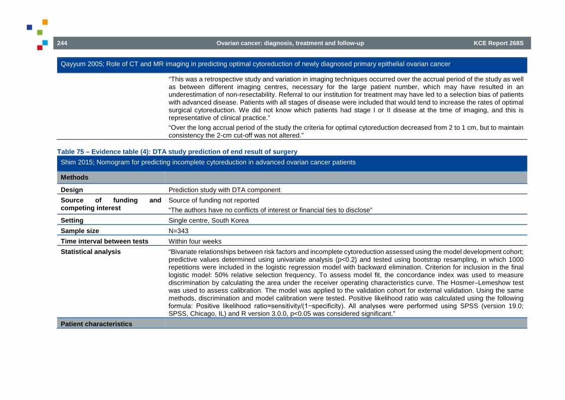
244 Ovarian cancer: diagnosis, treatment and follow-up KCE Report 268S
Qayyum 2005; Role of CT and MR imaging in predicting optimal cytoreduction of newly diagnosed primary epithelial ovarian cancer
“This was a retrospective study and variation in imaging techniques occurred over the accrual period of the study as well as between different imaging centres, necessary for the large patient number, which may have resulted in an underestimation of non-resectability. Referral to our institution for treatment may have led to a selection bias of patients with advanced disease. Patients with all stages of disease were included that would tend to increase the rates of optimal surgical cytoreduction. We did not know which patients had stage I or II disease at the time of imaging, and this is representative of clinical practice.” “Over the long accrual period of the study the criteria for optimal cytoreduction decreased from 2 to 1 cm, but to maintain consistency the 2-cm cut-off was not altered.”
Table 75 – Evidence table (4): DTA study prediction of end result of surgery Shim 2015; Nomogram for predicting incomplete cytoreduction in advanced ovarian cancer patients
Methods
Design Prediction study with DTA component Source of funding and competing interest
Source of funding not reported “The authors have no conflicts of interest or financial ties to disclose”
Setting Single centre, South Korea Sample size N=343 Time interval between tests Within four weeks Statistical analysis “Bivariate relationships between risk factors and incomplete cytoreduction assessed using the model development cohort;
predictive values determined using univariate analysis (p<0.2) and tested using bootstrap resampling, in which 1000 repetitions were included in the logistic regression model with backward elimination. Criterion for inclusion in the final logistic model: 50% relative selection frequency. To assess model fit, the concordance index was used to measure discrimination by calculating the area under the receiver operating characteristics curve. The Hosmer–Lemeshow test was used to assess calibration. The model was applied to the validation cohort for external validation. Using the same methods, discrimination and model calibration were tested. Positive likelihood ratio was calculated using the following formula: Positive likelihood ratio=sensitivity/(1−specificity). All analyses were performed using SPSS (version 19.0; SPSS, Chicago, IL) and R version 3.0.0, p<0.05 was considered significant.”
Patient characteristics

KCE Report 268S Ovarian cancer: diagnosis, treatment and follow-up 245
Shim 2015; Nomogram for predicting incomplete cytoreduction in advanced ovarian cancer patients
Eligibility criteria Age between 18 and 80 years; pathologically confirmed ovarian cancer; positron-emission tomography/computed tomography performed 4 weeks prior to surgery; primary staging and subsequent cytoreductive surgery; postoperative diagnosis of FIGO stages III–IV cancer. Patient enrolment between 2006 and 2012. Patients who did not receive primary treatment at the indicated institution, received neoadjuvant chemotherapy or had a history of other malignancies were excluded.
Patient characteristics Model development cohort: n = 240; validation cohort: n = 103 (Total n = 343) Median age, years (range): 55 (27-80) vs. 54 (20-76) FIGO stage, n (%),IIIA/ IIIB/ IIIC/ IV: 6 (2.5)/ 10 (4.2)/ 173 (72.1)/ 51 (21.3) vs. 2 (1.9)/ 4 (3.9)/ 67 (65.0)/ 30 (29.1) Histology, n (%),serous/ mucinous/ endometrioid/ clear cell/ transitional cell/ carcinosarcoma/ others: 185 (77.1)/ 5 (2.1)/9 (3.8)/19 (7.9)/6 (2.5)/10 (4.2)/6 (2.5) vs. 89 (86.4)/0/4 (3.9)/3 (2.9)/4 (3.9)/ 1 (1.0)/2 (1.9)
Prevalence of disease Prevalence of residual disease > 0 cm 223/343 (65%) Interventions Index test(s) Positron emission tomography/computed tomography (PET/CT)
Furosemide (40-mg tablet) and duspatalin (135-mg tablet) were orally administered just before venous blood glucose measurement. Venous blood glucose levels were maintained b140 mg/dl. All patients were injected with 0.2 mCi/kg 18F-fluorodeoxyglucose and allowed to rest in a sitting or supine position for approximately 60 min prior to scanning. The patients were then positioned in the scanner with their arms above their heads. Positron-emission tomography/computed tomography scans from the base of the skull to the mid-thigh were performed using Discovery STE (GE Healthcare, Waukesha, WI), Biograph Truepoint 16 (Siemens/CTI, Knoxville, TN), or Biograph Truepoint 40 (Siemens/CTI) scanners. The scanners obtained combination multislice computed tomography and positron-emission tomography tomographs. The computed tomography data were used for attenuation correction. A total of five to six bed positions for 2–3 min per position were acquired for emission scanning (3 min/bed with the Discovery STE and Biograph Truepoint 16; 2 min/bedwith the Biograph Truepoint 40). All scans were reconstructed using an ordered-subsets expectation maximization algorithm (20 subsets and two iterations for the Discovery STE; 16 subsets and two iterations for the Biograph Truepoint 16; 21 subsets and three iterations for the Biograph Truepoint 40). Calibration of each scanner against dose calibrators and well counters was routinely performed. The measured standardized uptake value of the phantom was within the acceptable range of 90–110%. The mean standardized uptake value of the liver was also calculated by drawing a three-dimensional region of interest with a 3-cm diameter within the normal inferior right lobe. Further details with regard to the criteria used to interpret positron-emission tomography scans are described in the Supplemental Methods.

246 Ovarian cancer: diagnosis, treatment and follow-up KCE Report 268S
Shim 2015; Nomogram for predicting incomplete cytoreduction in advanced ovarian cancer patients
Reference standard Systematically performed surgical exploration of the abdominal cavity. Complete cytoreduction defined as ‘no gross residual tumour’ (i.e. residual disease = 0 cm)
Results Debulking (complete/optimal) “Complete cytoreduction was achieved in 120 patients (35%).” Diagnostic accuracy (sensitivity, specificity, PPV, NPV, FNs, FPs)
Target condition: residual tumour size > 0 cm Combined development and validation set: Sensitivity: 0.66 (95%-CI 0.60 to 0.73) Specificity: 0.88 (95%-CI 0.80 to 0.93) PPV: 0.91 (95%-CI 0.85 to 0.95) NPV: 0.58 (95%-CI 0.51 to 0.66) In a hypothetical study of 1000 patients and with a prevalence of 65% (= study prevalence) 221 (95%-CI 175 to 260) patients will be incorrectly classified as having no residual tumour (FNs) 42 (95%-CI 24 to 70) patients will be incorrectly classified as having residual disease >0 cm (FPs)
Limitations and other comments
Limitations Applicability concerns for the domains Index test and Reference standard Index test no add-on to CT Index test (predictors summarised in a nomogram) does not solely include PET/CT based determinants, but also a surgical aggressiveness index PET/CT was not performed in all cases: of all advanced stage patients 77% underwent preoperative PET/CT.
5.7. Aim of cytoreductive surgery: no macroscopic disease? 5.7.1. Prognostic value end result of surgery
Table 76 – Evidence table: SRs prognostic value end result of surgery Optimal primary surgical treatment for advanced epithelial ovarian cancer; Elattar 2011259
Methods Design Systematic review

KCE Report 268S Ovarian cancer: diagnosis, treatment and follow-up 247
Optimal primary surgical treatment for advanced epithelial ovarian cancer; Elattar 2011259
Source of funding and competing interest
Department of Health, UK. NHS Cochrane Collaboration programme Grant Scheme CPG-506 Declaration of interest: none
Search date August 2010 Searched databases MEDLINE, EMBASE, CENTRAL, the Cochrane Gynaecological Cancer Review Group Trials Register Included study designs Prospective and retrospective cohort studies (including data from RCTs) and unselected case series of 100 or more
patients which included concurrent comparison groups. Case-control studies, observational studies that did not have concurrent comparison groups, included fewer than 100 patients or that didn’t apply multivariable analyses to correct for baseline differences.
Number of included studies N=11 (only retrospective studies) Statistical analysis Hazard ratios (HRs) were pooled using the generic inverse variance method (random-effect models) Patient characteristics Eligibility criteria Adult women (over 18 years of age) with surgically staged advanced epithelial ovarian cancer (FIGO stage III/IV) who
had confirmed histological diagnoses. Studies defining optimal cytoreduction as surgery leading to residual tumours with a maximum diameter of any threshold up to 2 cm.
Exclusion criteria Chemotherapy prior to surgery or other concurrent malignancies Patient & disease characteristics A total of 4735 patients were included in all studies (3844 stage III and 891 stage IV); median age for patients with
advanced EOC varied between 54 to 64 years (range 16 to 91 years); median duration of follow-up varied from 28 months to 47.5 months (range 1 to 199 months).
Interventions Intervention a) RD >1 cm; b) RD 0.1-1 cm (after primary cytoreductive surgery) Comparator No gross RD Results Overall survival HR (RD < 1 cm vs microscopic disease)= 2.20 (95% CI 1.90 to 2.54) (6 studies)
HR (RD >1 cm vs microscopic disease)= 3.16 (95% CI 2.26 to 4.41) (4 studies) Disease-free survival Not addressed Progression-free survival HR (RD < 1 cm vs microscopic disease)= 1.96 (95% CI 1.72 to 2.23) (2 studies)
HR (RD > 1 cm vs microscopic disease) (stage III)= 2.36 (95% CI 2.06 to 2.71) (1 study) Recurrence rate Not addressed

248 Ovarian cancer: diagnosis, treatment and follow-up KCE Report 268S
Optimal primary surgical treatment for advanced epithelial ovarian cancer; Elattar 2011259
Quality of life “Quality of life outcomes were not reported by treatment arm or to a satisfactory level in any of the studies.” (loco)regional control Not addressed Adverse events “Adverse events were not reported by treatment arm or to a satisfactory level in any of the studies.” Limitations and comments Limitations Source of funding or support was not reported for each of the included studies. Quality of life outcomes and adverse
events were not reported by treatment arm or to a satisfactory level in any of the included studies.
Table 77 – Evidence table (1): observational study prognostic value end result of surgery Analysis of racial disparities in stage IIIC epithelial ovarian cancer care and outcomes in a tertiary gynecologic oncology referral center; Bristow 2011260
Methods Design Retrospective chart review Source of funding and competing interest
Source of funding: the Queen of Hearts Foundation for Ovarian Cancer Research Declaration of interest: none declared
Setting Johns Hopkins Hospital, USA Sample size N=433 Duration Patient enrollment between January 1995 and December 2008 Follow-up Median follow-up: 33.0 months Statistical analysis “Relationships between racial classification and clinic-pathologic variables were compared using Chi2 test of
independence and Pearson's R for categorical variables and t-test for continuous variables. OS was calculated from the date of diagnosis using Kaplan–Meier curves and compared using the log-rank test and Cox proportional hazards model.“
Patient characteristics Eligibility criteria FIGO Stage IIIC epithelial ovarian cancer based on intra-operative findings or radiographic imaging coupled with fine-
needle biopsy diagnosis. All epithelial histological subtypes were included. Exclusion criteria Borderline ovarian tumours of low malignant potential. Patient & disease characteristics Patient characteristics reported as Whites (n=366) vs African-Americans (n=39)
Median age: 59 vs 59 years ASA class, I/II/III/IV: 5/124/232/5 vs 0/4/31/4

KCE Report 268S Ovarian cancer: diagnosis, treatment and follow-up 249
Analysis of racial disparities in stage IIIC epithelial ovarian cancer care and outcomes in a tertiary gynecologic oncology referral center; Bristow 2011260
Histology, serous/non-serous: 314/52 vs 31/8 Tumour grade, 1/2/3: 39/33/294 vs 2/4/33 Optimal RD (≤1 cm)/no gross RD: 267/188 vs 27/21
Interventions Intervention group (1) Optimal debulking (RD 0.1-1.0 cm) Control group (2) Complete debulking (no gross RD) Results Overall survival RD 0.1-1.0 cm vs no gross RD: HR= 2.74 (95% CI 1.98 to 3.71) (HR adjusted for age, race, tumour grade, histology,
ASA score, surgical complexity score, serum albumin, administration of platinum-based chemotherapy, and significant peri-operative morbidity)
Disease-free survival Not assessed Progression-free survival Not assessed Recurrence rate Not assessed Quality of life Not assessed (Loco)regional control Not assessed Adverse events Not assessed according debulking status (N/A) Limitations and other comments Limitations Retrospective non-randomised study. Blinding not reported (but not applicable). Adjusted HRs are derived from a
prognostic model. No details on how modelling was performed, but this seems to have been done based on significance testing (and not on including putative confounders in the analysis, irrespective of statistical significance). Patient and disease characteristics not reported according to debulking status. NB: study only included women with stage IIIC ovarian cancer / possible overlap with Peiretti 2012.

250 Ovarian cancer: diagnosis, treatment and follow-up KCE Report 268S
Table 78 – Evidence table (2): observational study prognostic value end result of surgery Impact of Complete Cytoreduction Leaving No Gross Residual Disease Associated with Radical Cytoreductive Surgical Procedures on Survival in Advanced Ovarian Cancer; Chang 2012a261
Methods Design Retrospective review of medical records. The decision to perform a simple or radical procedure for debulking was
determined by the surgeon’s discretion. Source of funding and competing interest
Source of funding: one author was supported by the Queen of Hearts Foundation. Declaration of interest: not reported
Setting Ajou University Hospital, Republic of Korea Sample size N=203 of whom 84 underwent a radical surgical procedure and 119 a simple procedure Duration Patient enrollment between January 1, 2000 and December 31, 2011 Follow-up Median 43 months (range 1-124) Statistical analysis Chi2 test, Student t and Mann–Whitney U tests. Kaplan–Meier for PFS and OS (including log-rank tests). Univariate
and multivariate analyses by the use of Cox proportional hazard model for assessing the influence of various prognostic factors on survival.
Patient characteristics Eligibility criteria Consecutive patients with stage IIIC and IV primary epithelial ovarian, fallopian tube, or peritoneal cancer who
underwent primary cytoreductive surgery. Exclusion criteria Patients who had neoadjuvant chemotherapy, primary cytoreduction at an outside institution, stage IIIC disease based
on lymph node metastasis only, or tumours of low malignant potential were excluded. Patient & disease characteristics Residual disease: >1 cm/0≤1 cm/no 63/67/63
Median age 54 years (range 30-78). BMI 23.3 (11.7–35.2). FIGO Stage IIIC/IV: 189/14. ASA score 1-2/3-4 114/80 (9 not available). Tumour grade 1/2/3 26/72/100 (5 unknown). Ascites >100 mL: 92. Peritoneal carcinomatosis 149.
Interventions Intervention group (1) RD a) macroscopic disease >1 cm ('incomplete') or b) macroscopic disease ≤1 cm ('optimal') Control group (2) Complete debulking (no macroscopic disease left in situ) Results Overall survival Median unadjusted OS RD >1 cm 37 months; RD 0.1–1 cm 46 months; no gross RD 86 months.
HR (RD >1 cm vs no gross RD)= 3.24 (95% CI 1.90 to 5.53) HR (RD 0.1–1 cm vs no gross RD)= 2.22 (95% CI 1.25 to 3.94)
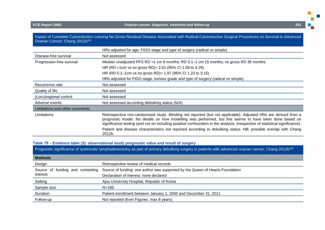
KCE Report 268S Ovarian cancer: diagnosis, treatment and follow-up 251
Impact of Complete Cytoreduction Leaving No Gross Residual Disease Associated with Radical Cytoreductive Surgical Procedures on Survival in Advanced Ovarian Cancer; Chang 2012a261
HRs adjusted for age, FIGO stage and type of surgery (radical vs simple) Disease-free survival Not assessed Progression-free survival Median unadjusted PFS RD >1 cm 9 months; RD 0.1–1 cm 15 months; no gross RD 35 months.
HR (RD >1cm vs no gross RD)= 2.61 (95% CI 1.58 to 4.29) HR (RD 0.1–1cm vs no gross RD)= 1.97 (95% CI 1.23 to 3.15) HRs adjusted for FIGO stage, tumour grade and type of surgery (radical vs simple)
Recurrence rate Not assessed Quality of life Not assessed (Loco)regional control Not assessed Adverse events Not assessed according debulking status (N/A) Limitations and other comments Limitations Retrospective non-randomised study. Blinding not reported (but not applicable). Adjusted HRs are derived from a
prognostic model. No details on how modelling was performed, but this seems to have been done based on significance testing (and not on including putative confounders in the analysis, irrespective of statistical significance). Patient and disease characteristics not reported according to debulking status. NB: possible overlap with Chang 2012b.
Table 79 – Evidence table (3): observational study prognostic value end result of surgery Prognostic significance of systematic lymphadenectomy as part of primary debulking surgery in patients with advanced ovarian cancer; Chang 2012b262
Methods Design Retrospective review of medical records Source of funding and competing interest
Source of funding: one author was supported by the Queen of Hearts Foundation Declaration of interest: none declared
Setting Ajou University Hospital, Republic of Korea Sample size N=189 Duration Patient enrollment between January 1, 2000 and December 31, 2011 Follow-up Not reported (from Figures: max 8 years)
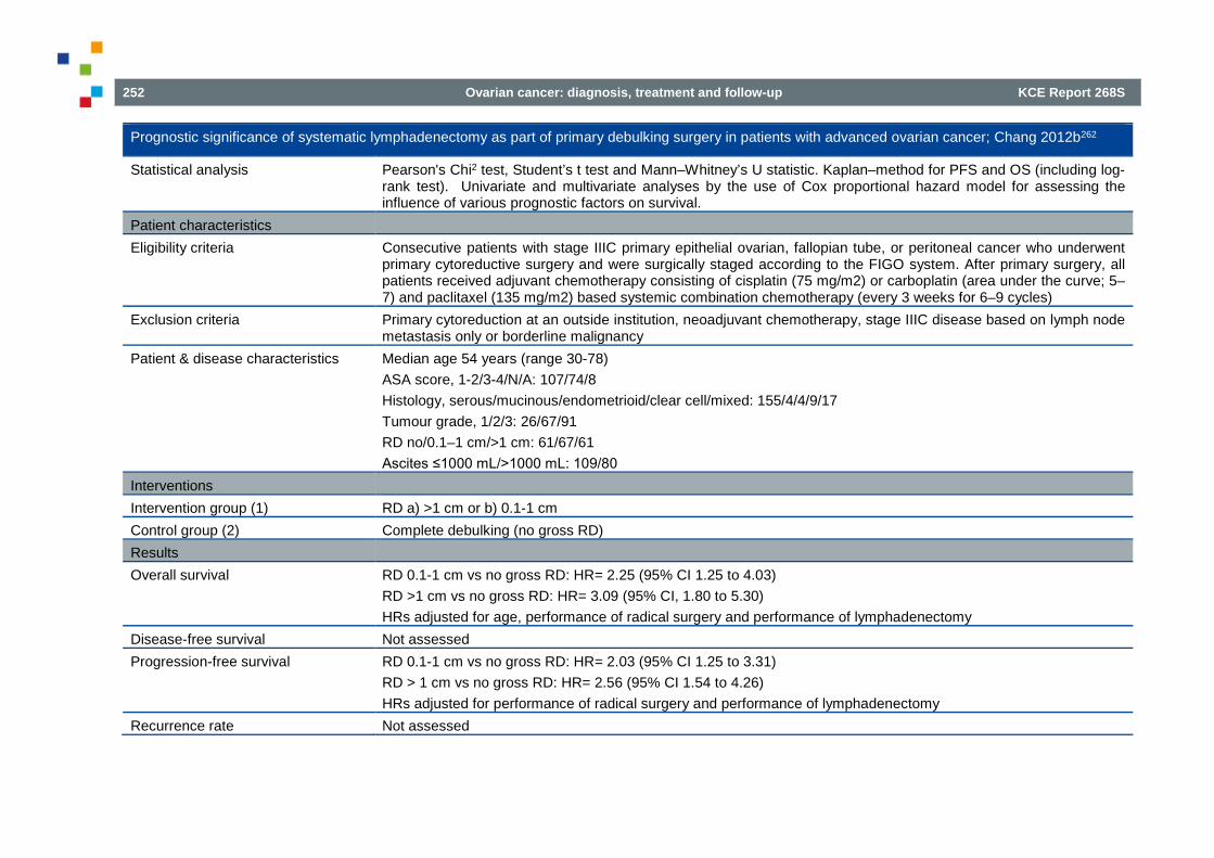
252 Ovarian cancer: diagnosis, treatment and follow-up KCE Report 268S
Prognostic significance of systematic lymphadenectomy as part of primary debulking surgery in patients with advanced ovarian cancer; Chang 2012b262
Statistical analysis Pearson's Chi2 test, Student’s t test and Mann–Whitney’s U statistic. Kaplan–method for PFS and OS (including log-rank test). Univariate and multivariate analyses by the use of Cox proportional hazard model for assessing the influence of various prognostic factors on survival.
Patient characteristics Eligibility criteria Consecutive patients with stage IIIC primary epithelial ovarian, fallopian tube, or peritoneal cancer who underwent
primary cytoreductive surgery and were surgically staged according to the FIGO system. After primary surgery, all patients received adjuvant chemotherapy consisting of cisplatin (75 mg/m2) or carboplatin (area under the curve; 5–7) and paclitaxel (135 mg/m2) based systemic combination chemotherapy (every 3 weeks for 6–9 cycles)
Exclusion criteria Primary cytoreduction at an outside institution, neoadjuvant chemotherapy, stage IIIC disease based on lymph node metastasis only or borderline malignancy
Patient & disease characteristics Median age 54 years (range 30-78) ASA score, 1-2/3-4/N/A: 107/74/8 Histology, serous/mucinous/endometrioid/clear cell/mixed: 155/4/4/9/17 Tumour grade, 1/2/3: 26/67/91 RD no/0.1–1 cm/>1 cm: 61/67/61 Ascites ≤1000 mL/>1000 mL: 109/80
Interventions Intervention group (1) RD a) >1 cm or b) 0.1-1 cm Control group (2) Complete debulking (no gross RD) Results Overall survival RD 0.1-1 cm vs no gross RD: HR= 2.25 (95% CI 1.25 to 4.03)
RD >1 cm vs no gross RD: HR= 3.09 (95% CI, 1.80 to 5.30) HRs adjusted for age, performance of radical surgery and performance of lymphadenectomy
Disease-free survival Not assessed Progression-free survival RD 0.1-1 cm vs no gross RD: HR= 2.03 (95% CI 1.25 to 3.31)
RD > 1 cm vs no gross RD: HR= 2.56 (95% CI 1.54 to 4.26) HRs adjusted for performance of radical surgery and performance of lymphadenectomy
Recurrence rate Not assessed

KCE Report 268S Ovarian cancer: diagnosis, treatment and follow-up 253
Prognostic significance of systematic lymphadenectomy as part of primary debulking surgery in patients with advanced ovarian cancer; Chang 2012b262
Quality of life Not assessed (Loco)regional control Not assessed Adverse events Not assessed according debulking status (N/A) Limitations and other comments Limitations Retrospective non-randomised study. Blinding not reported (but not applicable). Adjusted HRs are derived from a
prognostic model. No details on how modelling was performed, but this seems to have been done based on significance testing (and not on including putative confounders in the analysis, irrespective of statistical significance). Patient and disease characteristics not reported according to debulking status. NB: study only included women with stage IIIC ovarian cancer / possible overlap with Chang 2012a.
Table 80 – Evidence table (4): observational study prognostic value end result of surgery The time interval from surgery to start of chemotherapy significantly impacts prognosis in patients with advanced serous ovarian carcinoma - analysis of patient data in the prospective OVCAD study; Hofstetter 2013263
Methods Design Prospective multicenter study Source of funding and competing interest
Source of funding: the European Union Declaration of interest: none declared
Setting Multicenter study Sample size N=191 Duration Patient enrollment between August 2005 and December 2008 Follow-up Median follow-up: 42 months (range 4 to 69 months) Statistical analysis Kaplan-Meier curves. Cox proportional hazards model for multivariate analysis. Factors that yielded significant results
in the univariate analysis were considered in the multivariate analysis. Patient characteristics Eligibility criteria Patients with stage IIIA-IV primary ovarian cancer Exclusion criteria Neoadjuvant chemotherapy Patient & disease characteristics Age ≤57y/>57y: 98/93
ECOG performance status 0/1/2-3: 113/60/10

254 Ovarian cancer: diagnosis, treatment and follow-up KCE Report 268S
The time interval from surgery to start of chemotherapy significantly impacts prognosis in patients with advanced serous ovarian carcinoma - analysis of patient data in the prospective OVCAD study; Hofstetter 2013263
FIGO stages IIIA/IIIB/IIIC,no lymph node metastasis/IIIC,lymph node metastasis/IIIC,unknown lymph node status/IV: 4/8/28/92/26/33 Tumour grading I-II/III: 51/140 Postoperative RD microscopic/macroscopic: 121/70
Interventions Intervention group (1) Macroscopic RD (residual tumour lesions of any size) = “suboptimal RD” Control group (2) Microscopic RD (no visible residual tumour) Results Overall survival Three year OS: HR of microscopic vs macroscopic RD: 2.95 (95% CI 1.87 to 4.67)
HR adjusted for interval between surgery and start of chemotherapy, tumour stage, age and extent of surgery Disease-free survival Not assessed Progression-free survival Not assessed Recurrence rate Not assessed Quality of life Not assessed (Loco)regional control Not assessed Adverse events Not assessed according debulking status (N/A) Limitations and other comments Limitations Retrospective non-randomised study. Blinding not reported (but not applicable). Adjusted HRs are derived from a
prognostic model. No details on how modelling was performed, but this seems to have been done based on significance testing (and not on including putative confounders in the analysis, irrespective of statistical significance). Patient and disease characteristics not reported according to debulking status. NB: possible overlap with Polterauer 2012
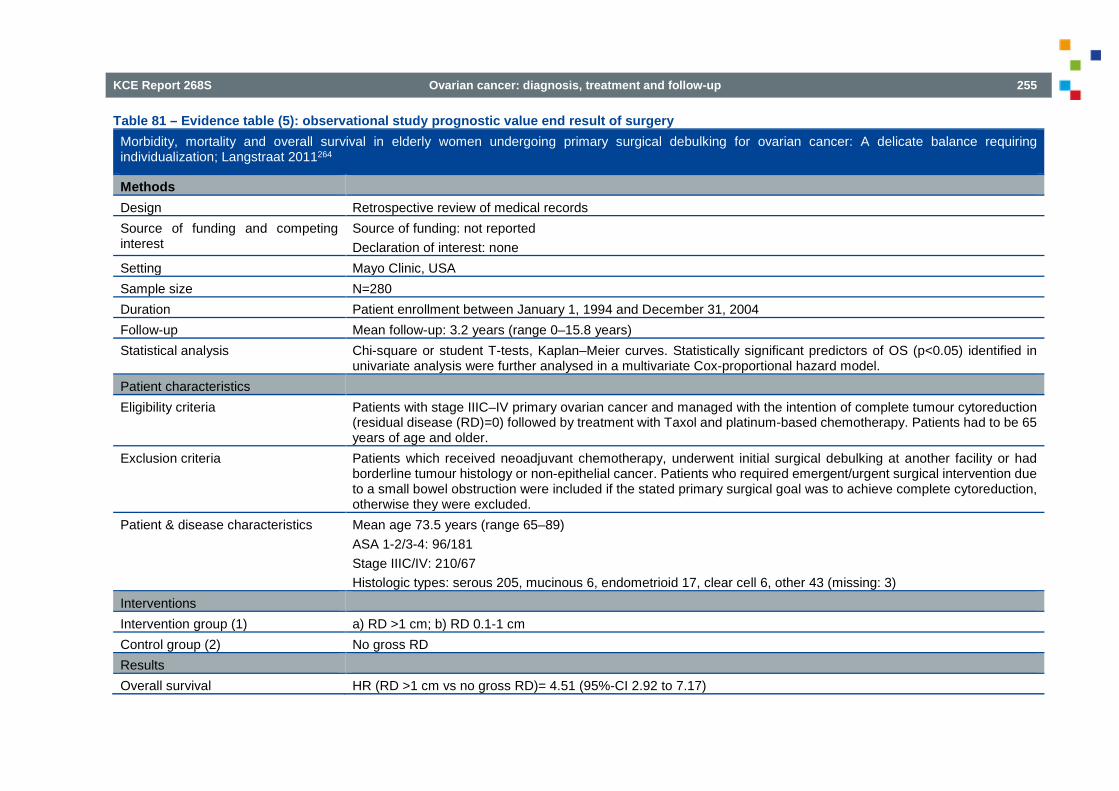
KCE Report 268S Ovarian cancer: diagnosis, treatment and follow-up 255
Table 81 – Evidence table (5): observational study prognostic value end result of surgery Morbidity, mortality and overall survival in elderly women undergoing primary surgical debulking for ovarian cancer: A delicate balance requiring individualization; Langstraat 2011264
Methods Design Retrospective review of medical records Source of funding and competing interest
Source of funding: not reported Declaration of interest: none
Setting Mayo Clinic, USA Sample size N=280 Duration Patient enrollment between January 1, 1994 and December 31, 2004 Follow-up Mean follow-up: 3.2 years (range 0–15.8 years) Statistical analysis Chi-square or student T-tests, Kaplan–Meier curves. Statistically significant predictors of OS (p<0.05) identified in
univariate analysis were further analysed in a multivariate Cox-proportional hazard model. Patient characteristics Eligibility criteria Patients with stage IIIC–IV primary ovarian cancer and managed with the intention of complete tumour cytoreduction
(residual disease (RD)=0) followed by treatment with Taxol and platinum-based chemotherapy. Patients had to be 65 years of age and older.
Exclusion criteria Patients which received neoadjuvant chemotherapy, underwent initial surgical debulking at another facility or had borderline tumour histology or non-epithelial cancer. Patients who required emergent/urgent surgical intervention due to a small bowel obstruction were included if the stated primary surgical goal was to achieve complete cytoreduction, otherwise they were excluded.
Patient & disease characteristics Mean age 73.5 years (range 65–89) ASA 1-2/3-4: 96/181 Stage IIIC/IV: 210/67 Histologic types: serous 205, mucinous 6, endometrioid 17, clear cell 6, other 43 (missing: 3)
Interventions Intervention group (1) a) RD >1 cm; b) RD 0.1-1 cm Control group (2) No gross RD Results Overall survival HR (RD >1 cm vs no gross RD)= 4.51 (95%-CI 2.92 to 7.17)
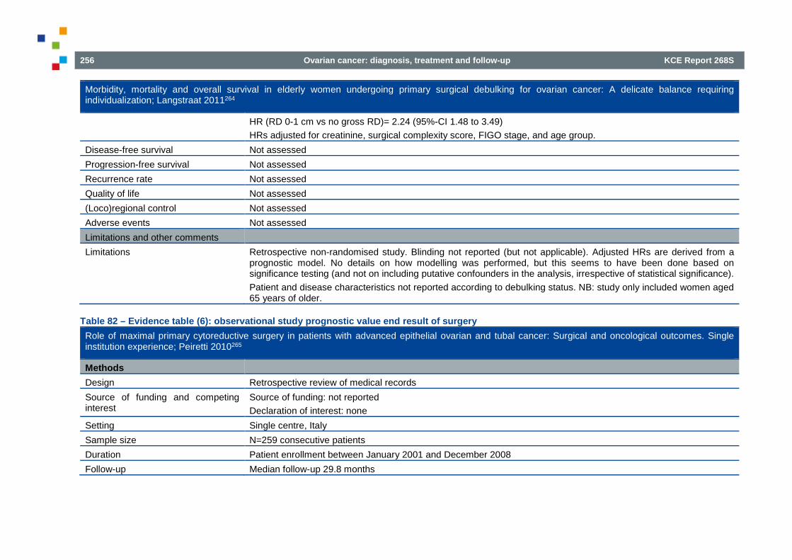
256 Ovarian cancer: diagnosis, treatment and follow-up KCE Report 268S
Morbidity, mortality and overall survival in elderly women undergoing primary surgical debulking for ovarian cancer: A delicate balance requiring individualization; Langstraat 2011264
HR (RD 0-1 cm vs no gross RD)= 2.24 (95%-CI 1.48 to 3.49) HRs adjusted for creatinine, surgical complexity score, FIGO stage, and age group.
Disease-free survival Not assessed Progression-free survival Not assessed Recurrence rate Not assessed Quality of life Not assessed (Loco)regional control Not assessed Adverse events Not assessed Limitations and other comments Limitations Retrospective non-randomised study. Blinding not reported (but not applicable). Adjusted HRs are derived from a
prognostic model. No details on how modelling was performed, but this seems to have been done based on significance testing (and not on including putative confounders in the analysis, irrespective of statistical significance). Patient and disease characteristics not reported according to debulking status. NB: study only included women aged 65 years of older.
Table 82 – Evidence table (6): observational study prognostic value end result of surgery Role of maximal primary cytoreductive surgery in patients with advanced epithelial ovarian and tubal cancer: Surgical and oncological outcomes. Single institution experience; Peiretti 2010265
Methods Design Retrospective review of medical records Source of funding and competing interest
Source of funding: not reported Declaration of interest: none
Setting Single centre, Italy Sample size N=259 consecutive patients Duration Patient enrollment between January 2001 and December 2008 Follow-up Median follow-up 29.8 months

KCE Report 268S Ovarian cancer: diagnosis, treatment and follow-up 257
Role of maximal primary cytoreductive surgery in patients with advanced epithelial ovarian and tubal cancer: Surgical and oncological outcomes. Single institution experience; Peiretti 2010265
Statistical analysis Kaplan–Meier to estimate survival curves; differences in survival were analyzed by the log-rank test. Cox proportional hazards regression to identify independent prognostic variables for OS by univariate and multivariate analyses
Patient characteristics Eligibility criteria Patients with stages IIIC–IV epithelial ovarian and fallopian tube cancer who underwent maximal primary cytoreductive
surgery Exclusion criteria Prior attempt of surgical cytoreduction at another institution, histology consistent with non-epithelial ovarian
malignancies or borderline tumours and neoadjuvant chemotherapy treatment Patient & disease characteristics Median age 58 years (range 22–77)
Primary site: ovary/fallopian tube: 256/3 FIGO stage: IIIC/IV 199/60 Tumour grade 1-2/3/N/A: 53/198/8 Histologic type: serous 184, endometrioid 39, clear cell 8, mixed 26, other 2 Peritoneal carcinomatosis 188
Interventions Intervention group (1) Any RD Control group (2) No grossly visible RD Results Overall survival Unadjusted median OS: not reached for no gross RD; 61.3 months for 1–5 mm; 42.4 months for 6–10 mm; 35.3
months for 10–20 mm; 42.6 months for ≥20 mm No multivariate analysis performed for OS according to debulking status
Progression-free survival Multivariate analysis: “Age greater than 60 years (p=0.025), stage IV vs IIIC (p=0.037) and any residual disease (p=0.032) had an independent association with worse PFS.”
Disease-free survival Not assessed Recurrence rate Not assessed Quality of life Not assessed (Loco)regional control Not assessed Adverse events Not assessed

258 Ovarian cancer: diagnosis, treatment and follow-up KCE Report 268S
Role of maximal primary cytoreductive surgery in patients with advanced epithelial ovarian and tubal cancer: Surgical and oncological outcomes. Single institution experience; Peiretti 2010265
Limitations and other comments Limitations Retrospective non-randomised study. Blinding not reported (but not applicable). Adjusted HRs are derived from a
prognostic model. No details on how modelling was performed, but this seems to have been done based on significance testing (and not on including putative confounders in the analysis, irrespective of statistical significance). Patient and disease characteristics not reported according to debulking status. NB: possible overlap with Peiretti 2012.
Table 83 – Evidence table (7): observational study prognostic value end result of surgery Rectosigmoid resection at the time of primary cytoreduction for advanced ovarian cancer. A multi-center analysis of surgical and oncological outcomes; Peiretti 2012266
Methods Design Retrospective review of medical records Source of funding and competing interest
Source of funding: not reported Declaration of interest: none declared
Setting Gynecologic Oncology at the European Institute of Oncology, Milan, Italy (157 patients) Kelly Gynecologic Oncology Service, Johns Hopkins Medical Institutions, Baltimore, USA (81 patients)
Sample size N=238 consecutive patients Duration Patient enrollment between August 1998 and July 2008 Follow-up Not reported Statistical analysis Kaplan–Meier to estimate survival curves; differences in survival were analyzed by the log-rank test. Cox proportional
hazards regression to identify independent prognostic variables for OS by univariate and multivariate analyses Patient characteristics Eligibility criteria Patients with advanced epithelial ovarian cancer who underwent rectosigmoid colectomy at the time of primary
cytoreductive surgery Exclusion criteria Neoadjuvant treatment Patient & disease characteristics Median age 59.7 years (range 22–85)
Primary site: ovary/fallopian tube/peritoneum: 230/4/4 FIGO stage: IIC/IIIA/IIIB/IIIC/IV 3/1/2/174/58

KCE Report 268S Ovarian cancer: diagnosis, treatment and follow-up 259
Rectosigmoid resection at the time of primary cytoreduction for advanced ovarian cancer. A multi-center analysis of surgical and oncological outcomes; Peiretti 2012266
Tumour grade 1-2/3/N/A: 51/184/3 Histologic type: serous 200, endometrioid 15, clear cell 5, mixed 18
Interventions Intervention group (1) Any other gross RD Control group (2) No grossly visible RD Results Overall survival Unadjusted median OS: any gross RD vs no macroscopic RD: 42 months vs 72 months (p=0.002)
“Presence of any macroscopic residual disease at the end of surgery was the only identified risk factor for OS (p=0.003)”
Progression-free survival Not assessed Disease-free survival Not assessed Recurrence rate Not assessed Quality of life Not assessed (Loco)regional control Not assessed Adverse events Not assessed Limitations and other comments Limitations Retrospective non-randomised study. Blinding not reported (but not applicable). Adjusted HRs are derived from a
prognostic model. No details on how modelling was performed, but this seems to have been done based on significance testing (and not on including putative confounders in the analysis, irrespective of statistical significance). Patient and disease characteristics not reported according to debulking status. NB: only women who underwent rectosigmoid colectomy were included / possible overlap with Bristow 2011 and Peiretti 2010.

260 Ovarian cancer: diagnosis, treatment and follow-up KCE Report 268S
Table 84 – Evidence table (8): observational study prognostic value end result of surgery Prognostic Value of Residual Tumour Size in Patients With Epithelial Ovarian Cancer FIGO Stages IIAYIV; Polterauer 2012267
Methods Design Prospective multicenter study Source of funding and competing interest
Source of funding: the European commission (FP6 Specific Targeted Research or Innovation Project) Declaration of interest: none declared
Setting Multicenter study (5 specialized European centers for gynecologic oncology) Sample size N=226 Duration Patient enrollment between February 2005 and December 2008 Follow-up Median follow-up: 25.0 months (range 1-49) Statistical analysis Chi2 tests. Univariate survival analysis of categorical variables by the log-rank test. Multiple forward stepwise Cox
regression analysis. Patient characteristics Eligibility criteria Patients with epithelial ovarian cancer FIGO Stages IIA-IV in whom radical cytoreductive surgery was performed and
standard chemotherapy with paclitaxel and carboplatin was applied. Exclusion criteria Patients having received neoadjuvant chemotherapy followed by interval debulking. Patient & disease characteristics Mean age 57.5 year (SD 11.9)
FIGO stages II, III and IV: 15 (6.6%), 174 (76.9%), and 37 (16.4%); FIGO stages IIIC and IV: 198 patients (87.6%) Histological type serous/other: 194/32 No macroscopic residual disease: 69.4% Minimal residual disease (≤1 cm): 87.2% (NB: this category also includes the No RD category!)
Interventions Intervention group (1) Any RD (cq minimal RD (≤1 cm) or gross RD (>1 cm)) Control group (2) Complete debulking (no RD) Results Overall survival 3-year OS (unadjusted) with no RD: 72.4%; minimal RD: 65.8%; gross RD: 45.2%
Subgroup analysis of stages IIIC and IV: 3-year OS (unadjusted) with no RD 69.7% (SE= 5.3%); any RD 53.6% (SE= 8.3%) (P = 0.003)
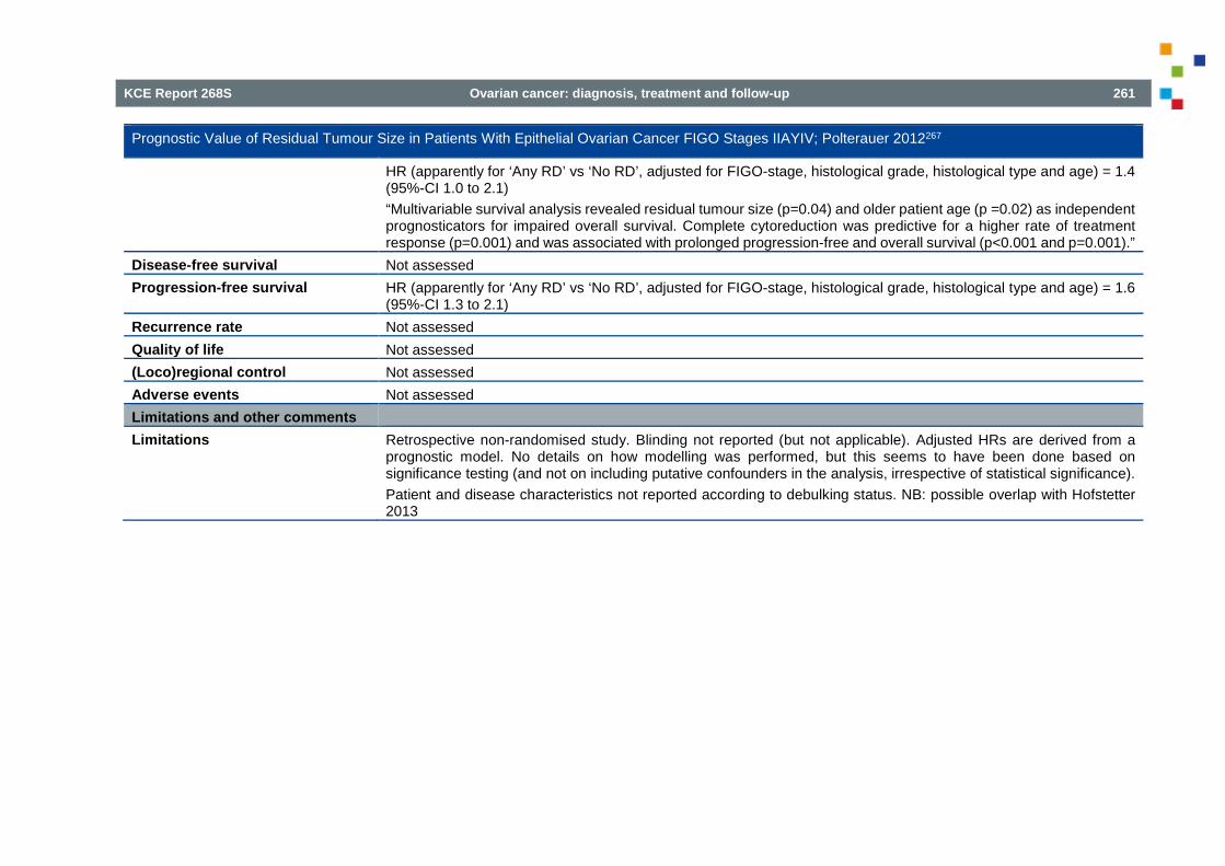
KCE Report 268S Ovarian cancer: diagnosis, treatment and follow-up 261
Prognostic Value of Residual Tumour Size in Patients With Epithelial Ovarian Cancer FIGO Stages IIAYIV; Polterauer 2012267
HR (apparently for ‘Any RD’ vs ‘No RD’, adjusted for FIGO-stage, histological grade, histological type and age) = 1.4 (95%-CI 1.0 to 2.1) “Multivariable survival analysis revealed residual tumour size (p=0.04) and older patient age (p =0.02) as independent prognosticators for impaired overall survival. Complete cytoreduction was predictive for a higher rate of treatment response (p=0.001) and was associated with prolonged progression-free and overall survival (p<0.001 and p=0.001).”
Disease-free survival Not assessed Progression-free survival HR (apparently for ‘Any RD’ vs ‘No RD’, adjusted for FIGO-stage, histological grade, histological type and age) = 1.6
(95%-CI 1.3 to 2.1) Recurrence rate Not assessed Quality of life Not assessed (Loco)regional control Not assessed Adverse events Not assessed Limitations and other comments Limitations Retrospective non-randomised study. Blinding not reported (but not applicable). Adjusted HRs are derived from a
prognostic model. No details on how modelling was performed, but this seems to have been done based on significance testing (and not on including putative confounders in the analysis, irrespective of statistical significance). Patient and disease characteristics not reported according to debulking status. NB: possible overlap with Hofstetter 2013
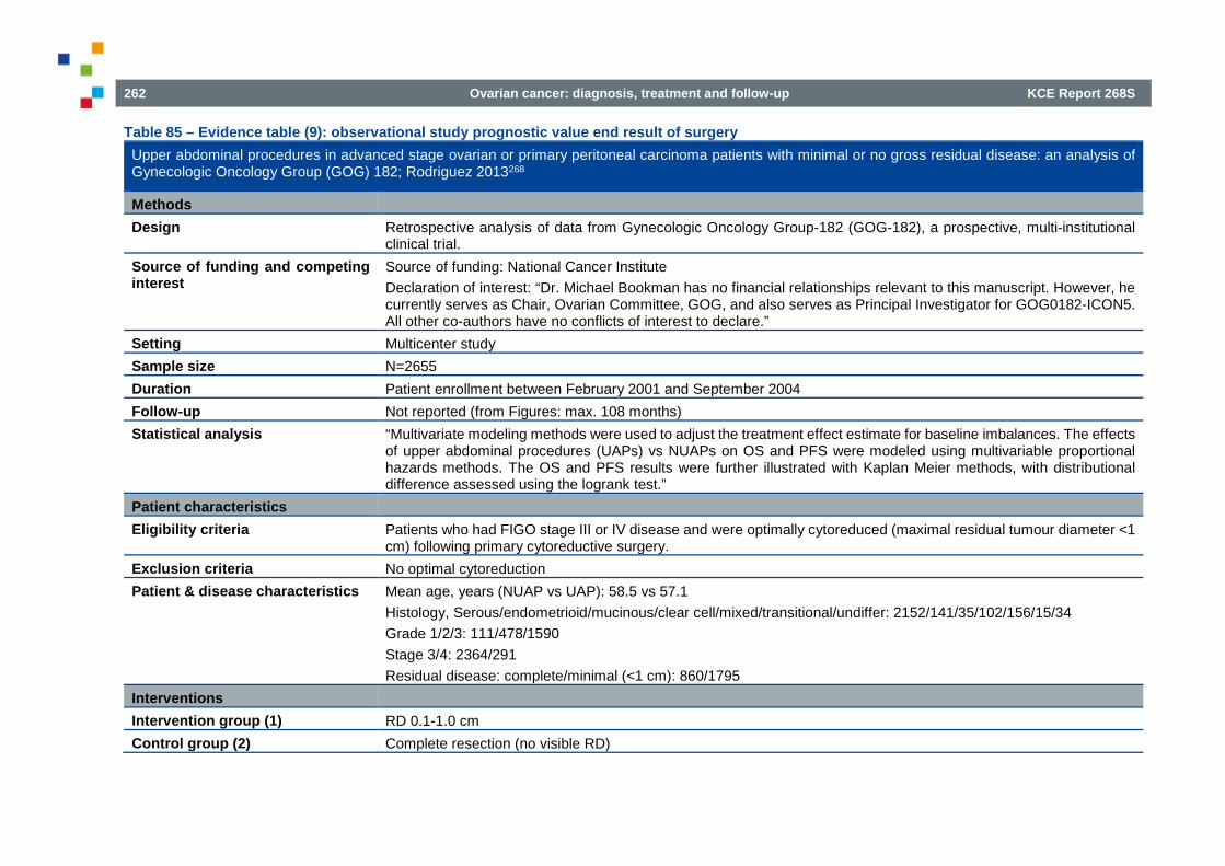
262 Ovarian cancer: diagnosis, treatment and follow-up KCE Report 268S
Table 85 – Evidence table (9): observational study prognostic value end result of surgery Upper abdominal procedures in advanced stage ovarian or primary peritoneal carcinoma patients with minimal or no gross residual disease: an analysis of Gynecologic Oncology Group (GOG) 182; Rodriguez 2013268
Methods Design Retrospective analysis of data from Gynecologic Oncology Group-182 (GOG-182), a prospective, multi-institutional
clinical trial. Source of funding and competing interest
Source of funding: National Cancer Institute Declaration of interest: “Dr. Michael Bookman has no financial relationships relevant to this manuscript. However, he currently serves as Chair, Ovarian Committee, GOG, and also serves as Principal Investigator for GOG0182-ICON5. All other co-authors have no conflicts of interest to declare.”
Setting Multicenter study Sample size N=2655 Duration Patient enrollment between February 2001 and September 2004 Follow-up Not reported (from Figures: max. 108 months) Statistical analysis “Multivariate modeling methods were used to adjust the treatment effect estimate for baseline imbalances. The effects
of upper abdominal procedures (UAPs) vs NUAPs on OS and PFS were modeled using multivariable proportional hazards methods. The OS and PFS results were further illustrated with Kaplan Meier methods, with distributional difference assessed using the logrank test.”
Patient characteristics Eligibility criteria Patients who had FIGO stage III or IV disease and were optimally cytoreduced (maximal residual tumour diameter <1
cm) following primary cytoreductive surgery. Exclusion criteria No optimal cytoreduction Patient & disease characteristics Mean age, years (NUAP vs UAP): 58.5 vs 57.1
Histology, Serous/endometrioid/mucinous/clear cell/mixed/transitional/undiffer: 2152/141/35/102/156/15/34 Grade 1/2/3: 111/478/1590 Stage 3/4: 2364/291 Residual disease: complete/minimal (<1 cm): 860/1795
Interventions Intervention group (1) RD 0.1-1.0 cm Control group (2) Complete resection (no visible RD)

KCE Report 268S Ovarian cancer: diagnosis, treatment and follow-up 263
Upper abdominal procedures in advanced stage ovarian or primary peritoneal carcinoma patients with minimal or no gross residual disease: an analysis of Gynecologic Oncology Group (GOG) 182; Rodriguez 2013268
Results Overall survival “Patients with complete reduction of disease had better prognosis than those with <1 cm (p<0.01). This advantage
showed a trend towards an improved OS in the matched sample of patients with complete reduction with a UAP (HR=0.61, 95% CI: 0.36 to 1.01, p= 0.06), and this was not seen in the full sample of patients (HR=0.80, 95% CI: 0.56 to 1.16, p=0.24).” In patients who underwent an UAP (n= 141): OS of complete reduction (no RD) vs RD <1: 54.6 vs 40.4 months
Disease-free survival Not assessed Progression-free survival In patients who underwent an UAP (n= 141): PFS of complete reduction (no RD) vs RD <1: 20.2 vs 13.7 months
“Patients with completely resected disease had better PFS than those with <1 cm (p < 0.01).” Recurrence rate Not assessed Quality of life Not assessed (Loco)regional control Not assessed Adverse events Not assessed according debulking status (N/A) Limitations and other comments Limitations Retrospective non-randomised study. Blinding not reported (but not applicable). Adjusted HRs derived from a sound
multivariate analysis. Patient and disease characteristics not reported according to debulking status. NB: women with RD >1 cm were excluded.
Table 86 – Evidence table (10): observational study prognostic value end result of surgery Influence of Residual Tumour on Outcome in Ovarian Cancer patients With FIGO Stage IV Disease; Wimberger 2010269
Methods Design Retrospective data set review (retrieved from three prospective, randomized phase III trials: AGO-OVAR (OVAR-3/-
5/-7)) Source of funding and competing interest
Source of funding: grants for the phase III study AGO-OVAR 3 from Bristol-Myers-Squibb, for the AGO-OVAR 5 from Pfizer, and for AGO-OVAR 7 from Glaxo Smith Kline. Declaration of interest: not reported
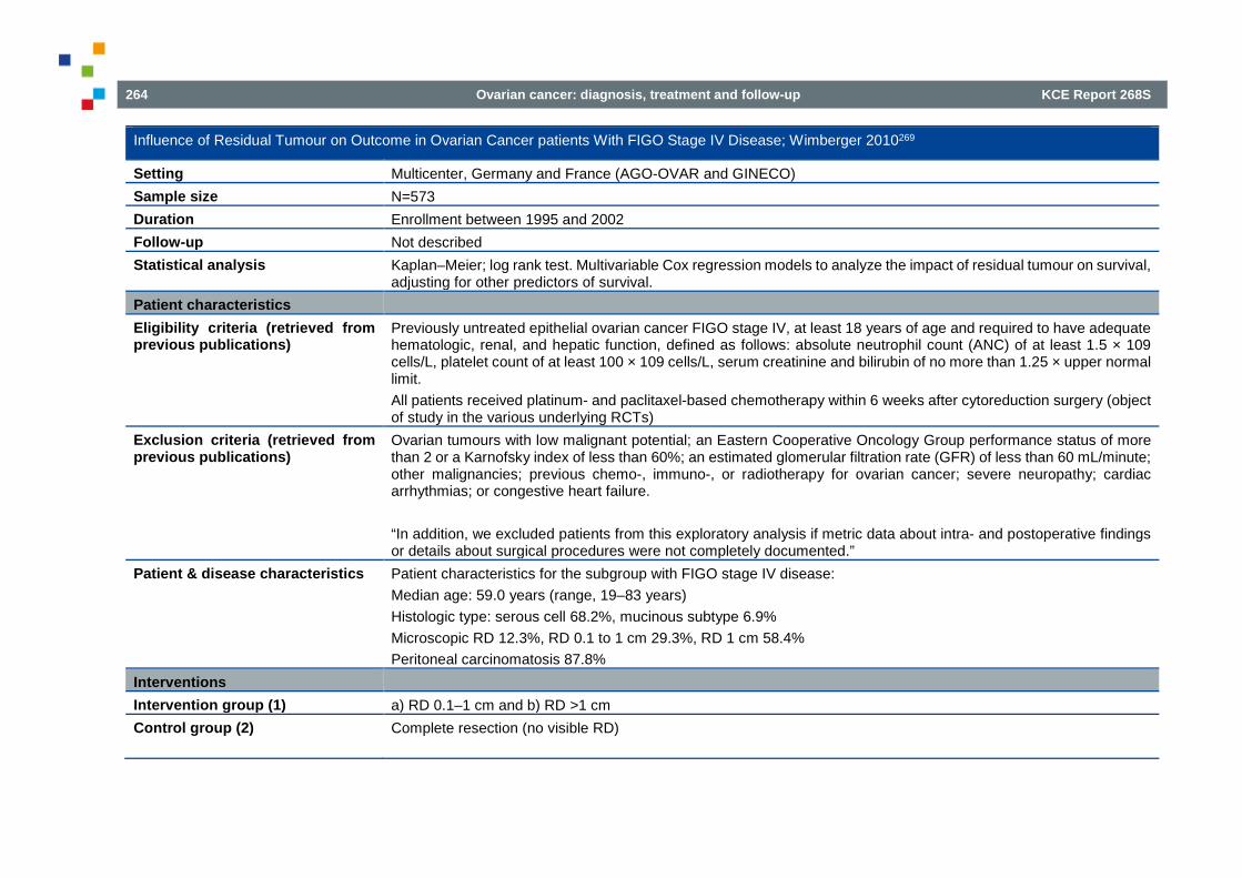
264 Ovarian cancer: diagnosis, treatment and follow-up KCE Report 268S
Influence of Residual Tumour on Outcome in Ovarian Cancer patients With FIGO Stage IV Disease; Wimberger 2010269
Setting Multicenter, Germany and France (AGO-OVAR and GINECO) Sample size N=573 Duration Enrollment between 1995 and 2002 Follow-up Not described Statistical analysis Kaplan–Meier; log rank test. Multivariable Cox regression models to analyze the impact of residual tumour on survival,
adjusting for other predictors of survival. Patient characteristics Eligibility criteria (retrieved from previous publications)
Previously untreated epithelial ovarian cancer FIGO stage IV, at least 18 years of age and required to have adequate hematologic, renal, and hepatic function, defined as follows: absolute neutrophil count (ANC) of at least 1.5 × 10⁹ cells/L, platelet count of at least 100 × 109 cells/L, serum creatinine and bilirubin of no more than 1.25 × upper normal limit. All patients received platinum- and paclitaxel-based chemotherapy within 6 weeks after cytoreduction surgery (object of study in the various underlying RCTs)
Exclusion criteria (retrieved from previous publications)
Ovarian tumours with low malignant potential; an Eastern Cooperative Oncology Group performance status of more than 2 or a Karnofsky index of less than 60%; an estimated glomerular filtration rate (GFR) of less than 60 mL/minute; other malignancies; previous chemo-, immuno-, or radiotherapy for ovarian cancer; severe neuropathy; cardiac arrhythmias; or congestive heart failure. “In addition, we excluded patients from this exploratory analysis if metric data about intra- and postoperative findings or details about surgical procedures were not completely documented.”
Patient & disease characteristics Patient characteristics for the subgroup with FIGO stage IV disease: Median age: 59.0 years (range, 19–83 years) Histologic type: serous cell 68.2%, mucinous subtype 6.9% Microscopic RD 12.3%, RD 0.1 to 1 cm 29.3%, RD 1 cm 58.4% Peritoneal carcinomatosis 87.8%
Interventions Intervention group (1) a) RD 0.1–1 cm and b) RD >1 cm Control group (2) Complete resection (no visible RD)

KCE Report 268S Ovarian cancer: diagnosis, treatment and follow-up 265
Influence of Residual Tumour on Outcome in Ovarian Cancer patients With FIGO Stage IV Disease; Wimberger 2010269
Results Overall survival Patients with stage IV
Median OS (unadjusted) of no RD vs RD 0.1-1.0 cm vs RD >1 cm: 54.6 vs 25.8 vs 23.9 months (p< 0.0001) RD 0.1-1 cm vs no visual RD: HR= 1.87 (95% CI 1.21 to 2.89) RD >1 cm vs no visual RD: HR= 2.13 (95% CI 1.40 to 3.23) HRs adjusted for age, performance status, histological type, presence of peritoneal carcinomatosis and multiple sites (Y/N)
Disease-free survival Not assessed Progression-free survival Patients with stage IV
Median PFS (unadjusted) of no RD vs RD 0.1-1.0 cm vs RD >1 cm: 19.1 vs 13.6 vs 11.3 months (p< 0.0001) RD 0.1-1 cm vs no visual RD: HR= 1.51 (95% CI 1.05 to 2.19) RD >1 cm vs no visual RD: HR= 1.82 (95% CI 1.28 to 2.59) (NB: error in Table 2: lower limit = 1.28 instead of 0.28) HRs adjusted for age, performance status, histological type, presence of peritoneal carcinomatosis and multiple sites (Y/N)
Recurrence rate Not assessed Quality of life Not assessed (Loco)regional control Not assessed Adverse events Not assessed Limitations and other comments Limitations Retrospective non-randomised study. Blinding not reported (but not applicable). Adjusted HRs are derived from a
prognostic model. No details on how modelling was performed, but this seems to have been done based on significance testing (and not on including putative confounders in the analysis, irrespective of statistical significance). Patient and disease characteristics not reported according to debulking status. NB: only women with stage IV were included.

266 Ovarian cancer: diagnosis, treatment and follow-up KCE Report 268S
5.7.2. Effect of (ultra)radical surgery
Table 87 – Evidence table: SR effect of (ultra)radical surgery Ultra-radical (extensive) surgery versus standard surgery for the primary cytoreduction of advanced epithelial ovarian cancer; Ang 2011333
Methods Design Cochrane systematic review Source of funding and competing interest
Department of Health, UKNHS Cochrane Collaboration programme Grant Scheme CPG-506 Declaration of interest: none
Search date November 2010 Searched databases MEDLINE, EMBASE, Cochrane Central Register of Controlled Trials (CENTRAL), Cochrane Gynaecological Cancer
Group Trials Register. Furthermore, registers of clinical trials, abstracts of scientific meetings, reference lists of included studies and contact with experts in the field.
Included study designs RCTs, quasi-randomised trials; non-randomised trials, prospective and retrospective cohort studies, and case series of 100 or more patients, that used statistical adjustment for baseline case mix using multivariate analyses. Case-control studies, uncontrolled observational studies and case series of fewer than 100 patients were excluded.
Number of included studies One retrospective analysis of 194 patients from the Mayo clinic in Rochester Statistical analysis No meta-analysis was performed Patient characteristics Eligibility criteria Adult women with stage III or IV epithelial ovarian cancer, who had ultra-radical surgery as part of interval debulking
surgery Exclusion criteria Women with other concurrent malignancies or women with recurrent disease Patient & disease characteristics* Included: 194 women with stage IIIC ovarian cancer (numbers per type of intervention not reported)
Mean age 64.4 years (range 24 to 87) Histologic types: serous 65%; endometrioid, mixed and seroanaplastic 27%, other 18% Grade III tumour 93% ASA score 2 or 3 90% Mean and median volume of ascites: 2076 ml and 1,000 ml (range: 0 to 12,000 ml)
Interventions* Intervention group Ultra-radical surgery: surgery in which any diaphragmatic surgery, bowel resection, splenectomy, or extensive
abdominal peritoneal stripping or resection was performed

KCE Report 268S Ovarian cancer: diagnosis, treatment and follow-up 267
Ultra-radical (extensive) surgery versus standard surgery for the primary cytoreduction of advanced epithelial ovarian cancer; Ang 2011333
Control group Standard surgery: if none of the above-mentioned procedures were performed, but hysterectomy, complete omentectomy, stripping of pelvic peritoneum, or limited resection of peritoneal-based nodules was carried out
Results* Overall survival Not assessed Disease-free survival Not assessed
Disease-specific survival: HR for death from advanced epithelial ovarian cancer (adjusted for age, ASA score, carcinomatosis, mesenteric involvement, diaphragmatic involvement, ascites, residual disease and operative time): 0.64 (95% CI 0.40 to 1.04) “Women who had ultra-radical surgery had better disease specific survival than those who underwent standard surgery (NS)”
Progression-free survival Not assessed Recurrence rate Not assessed Quality of life Not assessed (Loco)regional control Not assessed Adverse events Perioperative mortality (death within 2 weeks following surgery): 0 vs 3 women (not adjusted for baseline imbalances) Limitations and other comments Limitations No RCTs were identified. Only one comparative observational study could be included. Many of the objectives
of the review could not be met. Main outcomes of interest were not addressed
Table 88 – Evidence table (1): Observational study effect of (ultra)radical surgery Impact of Complete Cytoreduction Leaving No Gross Residual Disease Associated with Radical Cytoreductive Surgical Procedures on Survival in Advanced Ovarian Cancer; Chang 2012261
Methods Design Retrospective review of medical records. The decision to perform simple or radical procedures was determined by the
surgeon’s discretion. Source of funding and competing interest
Source of funding: One author was supported by the Queen of Hearts Foundation. Declaration of interest: not reported
Setting Ajou University Hospital, Republic of Korea

268 Ovarian cancer: diagnosis, treatment and follow-up KCE Report 268S
Impact of Complete Cytoreduction Leaving No Gross Residual Disease Associated with Radical Cytoreductive Surgical Procedures on Survival in Advanced Ovarian Cancer; Chang 2012261
Sample size N=203 of whom 84 underwent a radical surgical procedure and 119 a simple procedure Duration Patient enrollment between January 1, 2000 and December 31, 2011 Follow-up Median 43 months (range 1-124) Statistical analysis Clinical and pathologic factors were compared with Pearson’s Chi2 test. The Student t and Mann–Whitney U tests
were used for continuous data according to normality. Kaplan–Meier for PFS and OS (including log-rank tests). Univariate and multivariate analyses for assessing the influence of various prognostic factors on survival was performed by the Cox proportional hazard model.
Patient characteristics Eligibility criteria Consecutive patients with stage IIIC and IV primary epithelial ovarian, fallopian tube, or peritoneal cancer who
underwent primary cytoreductive surgery. Exclusion criteria Patients who had neoadjuvant chemotherapy, primary cytoreduction at an outside institution, stage IIIC disease based
on lymph node metastasis only, or tumours of low malignant potential were excluded. Patient & disease characteristics Median age 54 years (range 30-78). BMI 23.3 (11.7–35.2). FIGO Stage IIIC/IV: 189/14. ASA score 1-2/3-4 114/80 (9
not available). Tumour grade 1/2/3 26/72/100 (5 unknown). Ascites >100 mL: 92. Peritoneal carcinomatosis 149. Residual disease: no/≤1/1cm 63/67/63
Interventions Intervention group (1) Radical cytoreductive procedures included radical oophorectomy with or without rectosigmoid colectomy, total
omentectomy, multiple bowel resections, diaphragm peritonectomy or resection, liver resection, splenectomy, distal pancreatectomy, and gastric resection. After surgery, all patients received adjuvant platinum-based chemotherapy in combination with paclitaxel for 6–9 cycles.
Control group (2) Simple surgery included total abdominal hysterectomy, bilateral salpingo-oophorectomy, peritoneal biopsies or excisions, infracolic omentectomy, pelvic lymphadenectomy, para-aortic lymphadenectomy, and segmental resection of small bowel. After surgery, all patients received adjuvant platinum-based chemotherapy in combination with paclitaxel for 6–9 cycles.
Results Overall survival Median 66 vs 38 months (p= 0.01; unadjusted)
HR (adjusted for age, FIGO stage and residual disease): 0.56 (95% CI 0.37 to 0.87) Disease-free survival Not assessed

KCE Report 268S Ovarian cancer: diagnosis, treatment and follow-up 269
Impact of Complete Cytoreduction Leaving No Gross Residual Disease Associated with Radical Cytoreductive Surgical Procedures on Survival in Advanced Ovarian Cancer; Chang 2012261
Progression-free survival Median 18 vs 11 months (p= 0.01; unadjusted) HR (adjusted for FIGO stage, tumour grade and residual disease): 0.62 (95% CI 0.42 to 0.92)
Recurrence rate Not assessed Quality of life Not assessed (Loco)regional control Not assessed Adverse events Significant postoperative morbidity 32/84 (38.1%) vs 14/119 (11.8%); RR= 3.24 (95% CI 1.84 to 5.68)
Postoperative death within 30 days 1/84 (1.2%) vs 0/119 (0%); RR= 4.24 (95% CI 0.17 to 102.72) “Patients who underwent radical surgery had significantly longer operative time, larger estimated blood loss, more blood transfusions, longer stay in the intensive care unit, and more lymphocysts than those who underwent simple surgery.”
Limitations and other comments Limitations Retrospective non-randomised study. The decision to perform simple or radical procedures was determined by the
surgeon’s discretion. Confounding by indication can't be excluded. Patient and disease characteristics not reported per type of surgery. Blinding not reported. Adjusted HRs are derived from a prognostic model. No details on how modelling was performed, but this seems to have been done based on significance testing (and not on including putative confounders in the analysis, irrespective of statistical significance).
Table 89 – Evidence table (2): Observational study effect of (ultra)radical surgery Maximal Cytoreduction in Patients With FIGO Stage IIIC to Stage IV Ovarian, Fallopian, and Peritoneal Cancer in Day-to-Day Practice; Luyckx 2012302
Methods Design Retrospective review of medical records of patients treated in 7 French gynaecologic oncology and surgery centers. Source of funding and competing interest
No funding source reported. The authors declared no conflicts of interest.
Setting Seven leading French gynaecologic oncology units
Sample size N=527 of whom 124 underwent a radical surgical procedure and 290 a simple procedure (113 patients ignored for KCE’s RQ)

270 Ovarian cancer: diagnosis, treatment and follow-up KCE Report 268S
Maximal Cytoreduction in Patients With FIGO Stage IIIC to Stage IV Ovarian, Fallopian, and Peritoneal Cancer in Day-to-Day Practice; Luyckx 2012302
Duration Patient enrollment between January 1, 2003 and December 31, 2007 Follow-up Median 49 months Statistical analysis Univariate and multivariate analyses for assessing the influence of various prognostic factors on survival was
performed by the Cox proportional hazard model. Patient characteristics Eligibility criteria Patients with stage IIIC and stage IV (pleural invasion only) ovarian, tubal, or peritoneal epithelial carcinoma who
underwent either primary or interval debulking. All had at least 6 cycles of carboplatin and paclitaxel combination. No bevacizumab was used in any patient.
Exclusion criteria FIGO stage IIIC as defined by lymph nodes invasion only, patients in whom surgical procedures were not detailed enough in the operative reports to allow a retrospective scoring, and patients with nonepithelial histological subtypes or borderline cancer were excluded.
Patient & disease characteristics Median age 59 years (range 24-90). FIGO Stage IIIC/IV: 441/86. Tumour grade 1/2/3 34/138/236 (119 unknown). Primary/Interval debulking 190/268. Ascites: median 50 mL (range 0-8000). Peritoneal cancer index: median 10. Residual disease: 0/≤1/>1cm 374/97/55. Upper abdominal lesion 0/≤2.5/>2.5 cm 175/182/97 (73 unknown).
Interventions Intervention group (1) Ultra-radical surgery involving a combination of digestive tract resections (right colon and cecum, total colectomy, and
others), organ resection (spleen, gallbladder, partial gastrectomy, and others), celiac lymph node dissection, and total abdominal peritoneum stripping in addition to standard surgery (group 2B in the study).
Control group (2) Standard surgery with hysterectomy, bilateral salpingo-oophorectomy, rectosigmoid resection, infragastric omentectomy, pelvic and aortic lymphadenectomy, and, when applicable, appendectomy (group 1 in the study)
Results Overall survival No significant differences between surgical groups in the univariate analysis. Multivariate analysis not performed. Disease-free survival Median 15.9 vs 19.3 months (significant; not adjusted).
HR for relapse or death (adjusted for FIGO stage, tumour grade, presence of upper abdominal disease, amount of residual disease and timing of surgery (primary or interval): 1.66 (95% CI 1.16 to 2.39)
Progression-free survival Not assessed Recurrence rate Not assessed Quality of life Not assessed (Loco)regional control Not assessed
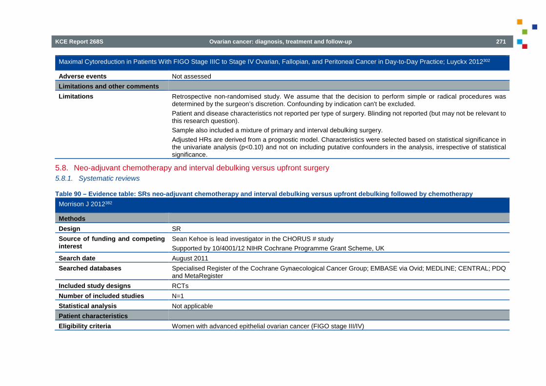
KCE Report 268S Ovarian cancer: diagnosis, treatment and follow-up 271
Maximal Cytoreduction in Patients With FIGO Stage IIIC to Stage IV Ovarian, Fallopian, and Peritoneal Cancer in Day-to-Day Practice; Luyckx 2012302
Adverse events Not assessed Limitations and other comments Limitations Retrospective non-randomised study. We assume that the decision to perform simple or radical procedures was
determined by the surgeon’s discretion. Confounding by indication can't be excluded. Patient and disease characteristics not reported per type of surgery. Blinding not reported (but may not be relevant to this research question). Sample also included a mixture of primary and interval debulking surgery. Adjusted HRs are derived from a prognostic model. Characteristics were selected based on statistical significance in the univariate analysis (p<0.10) and not on including putative confounders in the analysis, irrespective of statistical significance.
5.8. Neo-adjuvant chemotherapy and interval debulking versus upfront surgery 5.8.1. Systematic reviews
Table 90 – Evidence table: SRs neo-adjuvant chemotherapy and interval debulking versus upfront debulking followed by chemotherapy Morrison J 2012382
Methods Design SR Source of funding and competing interest
Sean Kehoe is lead investigator in the CHORUS # study Supported by 10/4001/12 NIHR Cochrane Programme Grant Scheme, UK
Search date August 2011 Searched databases Specialised Register of the Cochrane Gynaecological Cancer Group; EMBASE via Ovid; MEDLINE; CENTRAL; PDQ
and MetaRegister Included study designs RCTs Number of included studies N=1 Statistical analysis Not applicable Patient characteristics Eligibility criteria Women with advanced epithelial ovarian cancer (FIGO stage III/IV)

272 Ovarian cancer: diagnosis, treatment and follow-up KCE Report 268S
Morrison J 2012382
Exclusion criteria Not specifically stated Patient & disease characteristics See Vergote 2010 Interventions Intervention group Primary debulking surgery, with the aim to leave no residual disease > 2 cm, followed by platinum-based
chemotherapy Control group Platinum-based neoadjuvant chemotherapy followed by debulking surgery (with the aim to leave no residual disease
> 2 cm) Results See Vergote 2010 Limitations and other comments Comments Three ongoing studies identified: CHORUS, Kumar et al., Onda et al.
*Characteristics apply to the only included non-randomised study
5.8.2. RCTs
Table 91 – Evidence table (1): RCTs neo-adjuvant chemotherapy and interval debulking versus upfront debulking followed by chemotherapy Vergote I 2010, Greimel E 2013293, 330
Methods Design RCT Source of funding and competing interest
National Cancer Institute, EORTC
Setting Multicentre, worldwide (59 centres) Sample size N=670 Duration and follow-up 9/1998-12/2006 Statistical analysis Overall and progression-free survival rates were estimated by means of the Kaplan-Meier method, and overall survival
rates in the two groups were compared by means of the log-rank test, with a noninferiority ratio of 0.8. Multivariate time-to-event analysis was performed with the use of a Cox proportional-hazards model and univariate screening followed by a stepwise variable-selection procedure.

KCE Report 268S Ovarian cancer: diagnosis, treatment and follow-up 273
Vergote I 2010, Greimel E 2013293, 330
Patient characteristics Eligibility criteria Patients with biopsy-proven stage IIIC or IV invasive epithelial ovarian carcinoma, primary peritoneal carcinoma, or
fallopian-tube carcinoma If a biopsy specimen was not available, a FNA showing an adenocarcinoma was acceptable under the following conditions: the presence of a pelvic (ovarian) mass; the presence of metastases outside the pelvis measuring at least 2 cm in diameter (as noted during diagnostic laparoscopy or laparotomy or on CT); regional lymph-node metastasis or proof of stage IV disease; and a ratio of CA-125 to CEA > 25 If the ration of CA-125 to CEA was 25 or lower, results of a barium enema (or colonoscopy), gastroscopy (or radiologic examination of the stomach), and mammography (performed within 6 weeks before randomization) had to be negative for the presence of a primary tumour Additional criteria: WHO performance status 0-2 Absence of serious disabling diseases that would contraindicate primary cytoreductive surgery or platinum-based chemotherapy
Patient & disease characteristics Median age: 62 vs. 63 years Primary tumour epithelial ovarian: 87.2% vs. 84.7% Serous histology: 66% vs. 58% Stage IIIC: 77% vs. 76%; stage IV: 23% vs. 24%
Interventions Intervention group Primary debulking surgery followed by at least 6 courses of platinum-based chemotherapy (interval debulking surgery
was permitted when surgery was completed without optimal cytoreduction and if stable disease or a response was documented) (PDS group, N=336)
Control group Neo-adjuvant platinum-based chemotherapy followed by interval debulking surgery in all patients with a response or stable disease, followed in turn by at least three courses of platinum-based chemotherapy (NACT group, N=334)
Results Overall survival Median: 29 vs. 30 months
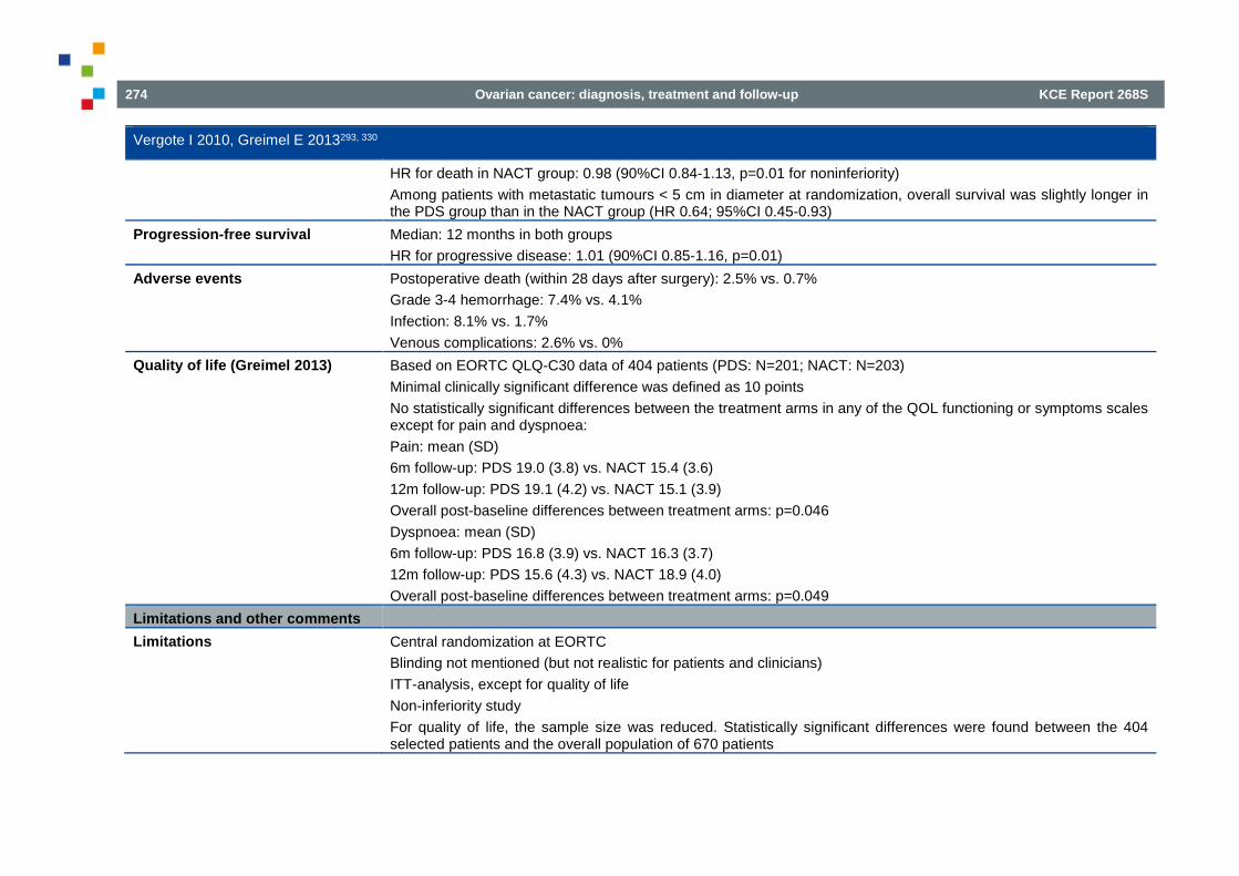
274 Ovarian cancer: diagnosis, treatment and follow-up KCE Report 268S
Vergote I 2010, Greimel E 2013293, 330
HR for death in NACT group: 0.98 (90%CI 0.84-1.13, p=0.01 for noninferiority) Among patients with metastatic tumours < 5 cm in diameter at randomization, overall survival was slightly longer in the PDS group than in the NACT group (HR 0.64; 95%CI 0.45-0.93)
Progression-free survival Median: 12 months in both groups HR for progressive disease: 1.01 (90%CI 0.85-1.16, p=0.01)
Adverse events Postoperative death (within 28 days after surgery): 2.5% vs. 0.7% Grade 3-4 hemorrhage: 7.4% vs. 4.1% Infection: 8.1% vs. 1.7% Venous complications: 2.6% vs. 0%
Quality of life (Greimel 2013) Based on EORTC QLQ-C30 data of 404 patients (PDS: N=201; NACT: N=203) Minimal clinically significant difference was defined as 10 points No statistically significant differences between the treatment arms in any of the QOL functioning or symptoms scales except for pain and dyspnoea: Pain: mean (SD) 6m follow-up: PDS 19.0 (3.8) vs. NACT 15.4 (3.6) 12m follow-up: PDS 19.1 (4.2) vs. NACT 15.1 (3.9) Overall post-baseline differences between treatment arms: p=0.046 Dyspnoea: mean (SD) 6m follow-up: PDS 16.8 (3.9) vs. NACT 16.3 (3.7) 12m follow-up: PDS 15.6 (4.3) vs. NACT 18.9 (4.0) Overall post-baseline differences between treatment arms: p=0.049
Limitations and other comments Limitations Central randomization at EORTC
Blinding not mentioned (but not realistic for patients and clinicians) ITT-analysis, except for quality of life Non-inferiority study For quality of life, the sample size was reduced. Statistically significant differences were found between the 404 selected patients and the overall population of 670 patients

KCE Report 268S Ovarian cancer: diagnosis, treatment and follow-up 275
Table 92 – Evidence table (2): RCTs neo-adjuvant chemotherapy and interval debulking versus upfront debulking followed by chemotherapy Kehoe 2015414
Methods Design RCT Source of funding and competing interest
Cancer Research UK and the Royal College of Obstetricians and Gynaecologists; five authors are employed by the Medical Research Council. The other authors declare no competing interests.
Setting Multicentre, UK and New-Zealand (59 centres) Sample size N=550 Duration and follow-up 9/1998-12/2006 Statistical analysis Non-inferiority study
Overall and progression-free survival survival: Kaplan-Meier method, comparison using stratified log-rank test Quality of life: analysis of covariance with adjustment for baseline scores Stata 12 or later
Patient characteristics Eligibility criteria Patients with biopsy-proven stage IIIC or IV invasive epithelial ovarian carcinoma, primary peritoneal carcinoma, or
fallopian-tube carcinoma If a biopsy specimen was not available, a FNA showing an adenocarcinoma was acceptable under the following conditions: the presence of a pelvic (ovarian) mass; the presence of metastases outside the pelvis measuring at least 2 cm in diameter (as noted during diagnostic laparoscopy or laparotomy or on CT); regional lymph-node metastasis or proof of stage IV disease; and a ratio of CA-125 to CEA > 25 If the ration of CA-125 to CEA was 25 or lower, results of a barium enema (or colonoscopy), gastroscopy (or radiologic examination of the stomach), and mammography (performed within 6 weeks before randomization) had to be negative for the presence of a primary tumour Additional criteria: WHO performance status 0-2 Absence of serious disabling diseases that would contraindicate primary cytoreductive surgery or platinum-based chemotherapy

276 Ovarian cancer: diagnosis, treatment and follow-up KCE Report 268S
Kehoe 2015414
Patient & disease characteristics Median age: 62 vs. 63 years Primary tumour epithelial ovarian: 87.2% vs. 84.7% Serous histology: 66% vs. 58% Stage IIIC: 77% vs. 76%; stage IV: 23% vs. 24%
Interventions Intervention group Primary debulking surgery followed by at least 6 courses of platinum-based chemotherapy (interval debulking surgery
was permitted when surgery was completed without optimal cytoreduction and if stable disease or a response was documented) (PDS group, N=336)
Control group Neoadjuvant platinum-based chemotherapy followed by interval debulking surgery in all patients with a response or stable disease, followed in turn by at least three courses of platinum-based chemotherapy (NACT group, N=334)
Results Overall survival Median: 29 vs. 30 months
HR for death in NACT group: 0.98 (90%CI 0.84-1.13, p=0.01 for noninferiority) Among patients with metastatic tumours < 5 cm in diameter at randomization, overall survival was slightly longer in the PDS group than in the NACT group (HR 0.64; 95%CI 0.45-0.93)
Progression-free survival Median: 12 months in both groups HR for progressive disease: 1.01 (90%CI 0.85-1.16, p=0.01)
Adverse events Postoperative death (within 28 days after surgery): 2.5% vs. 0.7% Grade 3-4 hemorrhage: 7.4% vs. 4.1% Infection: 8.1% vs. 1.7% Venous complications: 2.6% vs. 0%
Quality of life (Greimel 2013) Based on EORTC QLQ-C30 data of 404 patients (PDS: N=201; NACT: N=203) Minimal clinically significant difference was defined as 10 points No statistically significant differences between the treatment arms in any of the QOL functioning or symptoms scales except for pain and dyspnoea: Pain: mean (SD) 6m follow-up: PDS 19.0 (3.8) vs. NACT 15.4 (3.6) 12m follow-up: PDS 19.1 (4.2) vs. NACT 15.1 (3.9) Overall post-baseline differences between treatment arms: p=0.046
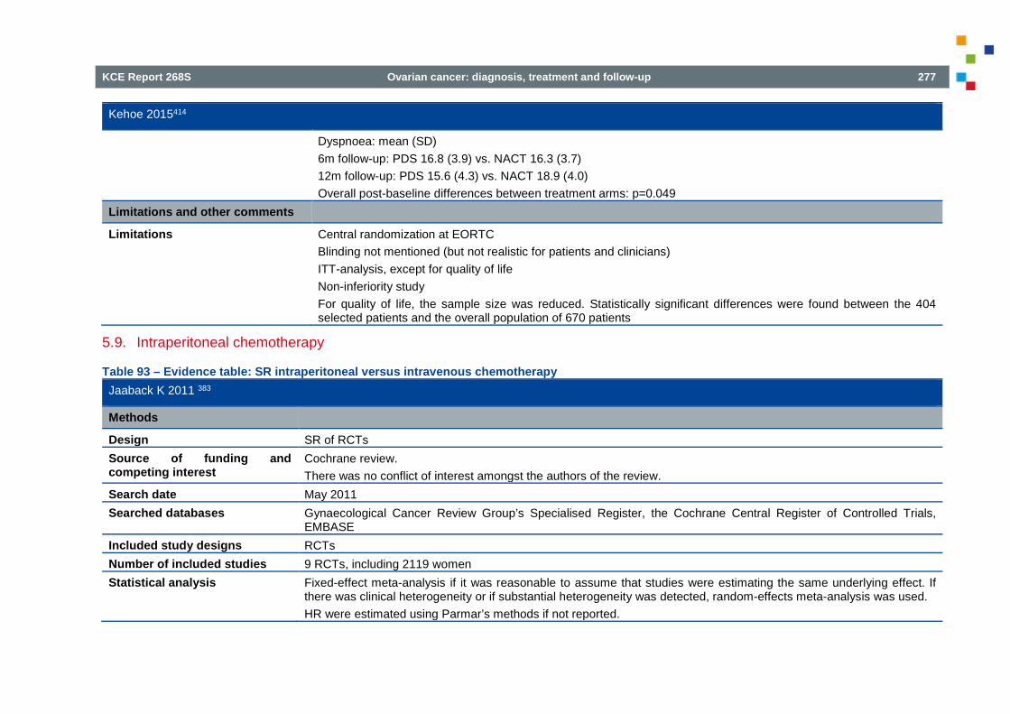
KCE Report 268S Ovarian cancer: diagnosis, treatment and follow-up 277
Kehoe 2015414
Dyspnoea: mean (SD) 6m follow-up: PDS 16.8 (3.9) vs. NACT 16.3 (3.7) 12m follow-up: PDS 15.6 (4.3) vs. NACT 18.9 (4.0) Overall post-baseline differences between treatment arms: p=0.049
Limitations and other comments
Limitations Central randomization at EORTC Blinding not mentioned (but not realistic for patients and clinicians) ITT-analysis, except for quality of life Non-inferiority study For quality of life, the sample size was reduced. Statistically significant differences were found between the 404 selected patients and the overall population of 670 patients
5.9. Intraperitoneal chemotherapy
Table 93 – Evidence table: SR intraperitoneal versus intravenous chemotherapy Jaaback K 2011 383
Methods
Design SR of RCTs Source of funding and competing interest
Cochrane review. There was no conflict of interest amongst the authors of the review.
Search date May 2011 Searched databases Gynaecological Cancer Review Group’s Specialised Register, the Cochrane Central Register of Controlled Trials,
EMBASE Included study designs RCTs Number of included studies 9 RCTs, including 2119 women Statistical analysis Fixed-effect meta-analysis if it was reasonable to assume that studies were estimating the same underlying effect. If
there was clinical heterogeneity or if substantial heterogeneity was detected, random-effects meta-analysis was used. HR were estimated using Parmar’s methods if not reported.

278 Ovarian cancer: diagnosis, treatment and follow-up KCE Report 268S
Jaaback K 2011 383
Patient characteristics Eligibility criteria Standard IV chemotherapy was compared with chemotherapy that included a component of IP administration Exclusion criteria The review excluded the following: radio colloids, gene therapy, biologic therapy, radio-isotopes, vascular growth factors,
immunomodulating drugs, matrix metalloproteinase inhibitors, radiolabelled monoclonal antibodies Patient & disease characteristics
Women with a new diagnosis of primary epithelial ovarian cancer, of any FIGO stage, following primary cytoreductive surgery.
Interventions Intervention group Chemotherapy that included a component of IP administration
Alberts 1996: IV cyclophosphamide (600mg/m2) + IP cisplatin (100mg/m2) repeated every 3 weeks for a total of 6 cycles Gadducci 2000: IV epidox 60mg/m2 + IV CTX 600 mg/m2 + IP cisplatin 50 mg/m2 GOG172: IV paclitaxel 135mg/m2 + IP cisplatin 100 mg/m2 + IP paclitaxel 60mg/m2, repeated every 3 weeks for a total of 6 cycles Kirmani 1994: IP cisplatin 200mg/m2 + IP etoposide 350 mg/m2; repeated every 4 weeks for a total of 6 cycles Markman 2001; IV carboplatin (AUC9) for two courses every 28 days, followed 4 weeks later by IV paclitaxel 135mg/m2 + IP cisplatin 100mg/m2 repeated every three weeks for a total of six cycles Polyzos 1999: IP carboplatin 350 mg/m2 + IV cyclophosphamide 600 mg/m2; repeated every 3 to 4 weeks for a total of 6 cycles Yen 2001: IV cyclophosphamide 500mg/m2 + IV Adriamycin or epirubicin 50mg/m2 + IP cisplatin 100mg/m2; repeated every 3 weeks for a total of 6 cycles Yen 2009: paclitaxel on day 1 with cisplatin or carboplatin IP on day 2; repeated every 3 weeks for 6 cycles Zylberg 1986: IV Adriamycin 20mg X 2 + fluorouracil 500mg X 2 + cisplatin 50mg + vincaleucoblastine 10mg + ifosfamide 1g X 2 + IP bleomycin 15mg + cisplatin 50mg + fluorouracil 500mg + Adriamycin 30mg
Control group Standard IV chemotherapy
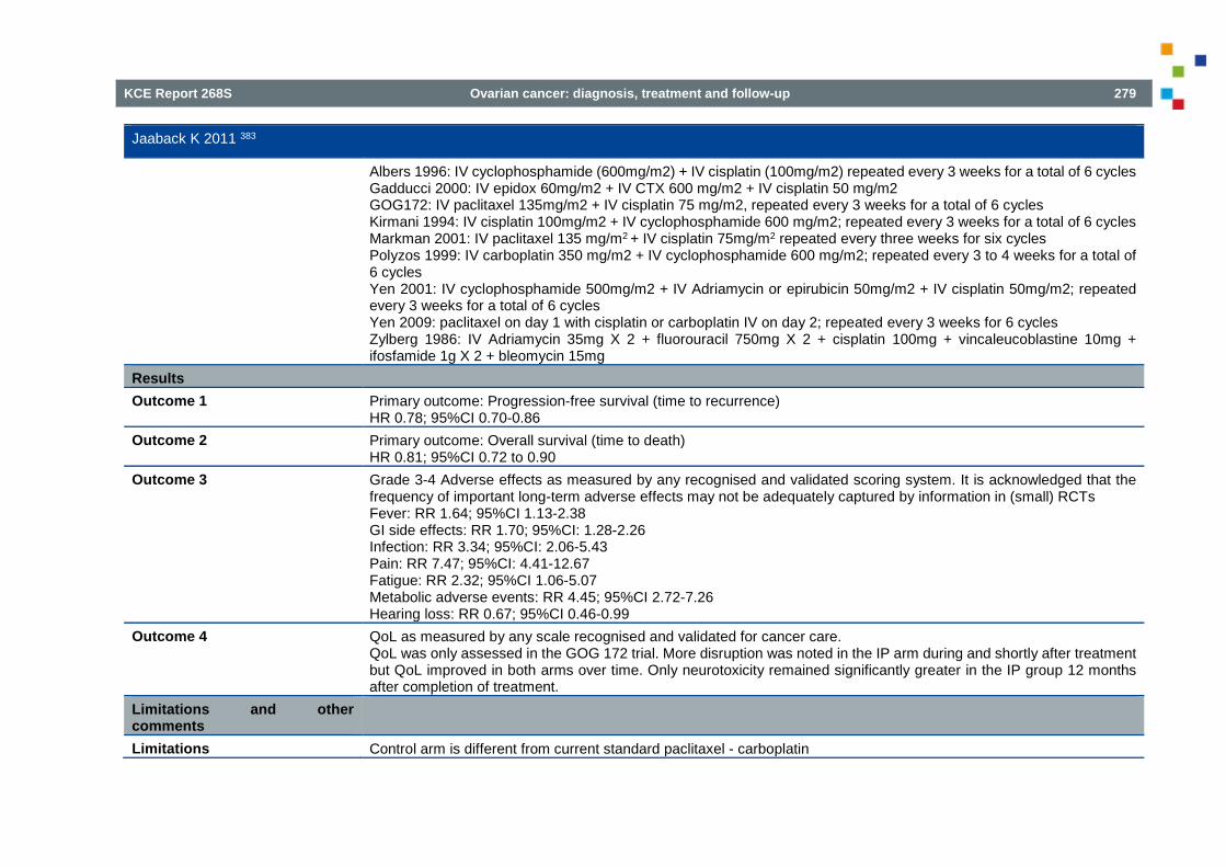
KCE Report 268S Ovarian cancer: diagnosis, treatment and follow-up 279
Jaaback K 2011 383
Albers 1996: IV cyclophosphamide (600mg/m2) + IV cisplatin (100mg/m2) repeated every 3 weeks for a total of 6 cycles Gadducci 2000: IV epidox 60mg/m2 + IV CTX 600 mg/m2 + IV cisplatin 50 mg/m2 GOG172: IV paclitaxel 135mg/m2 + IV cisplatin 75 mg/m2, repeated every 3 weeks for a total of 6 cycles Kirmani 1994: IV cisplatin 100mg/m2 + IV cyclophosphamide 600 mg/m2; repeated every 3 weeks for a total of 6 cycles Markman 2001: IV paclitaxel 135 mg/m2 + IV cisplatin 75mg/m2 repeated every three weeks for six cycles Polyzos 1999: IV carboplatin 350 mg/m2 + IV cyclophosphamide 600 mg/m2; repeated every 3 to 4 weeks for a total of 6 cycles Yen 2001: IV cyclophosphamide 500mg/m2 + IV Adriamycin or epirubicin 50mg/m2 + IV cisplatin 50mg/m2; repeated every 3 weeks for a total of 6 cycles Yen 2009: paclitaxel on day 1 with cisplatin or carboplatin IV on day 2; repeated every 3 weeks for 6 cycles Zylberg 1986: IV Adriamycin 35mg X 2 + fluorouracil 750mg X 2 + cisplatin 100mg + vincaleucoblastine 10mg + ifosfamide 1g X 2 + bleomycin 15mg
Results Outcome 1 Primary outcome: Progression-free survival (time to recurrence)
HR 0.78; 95%CI 0.70-0.86 Outcome 2 Primary outcome: Overall survival (time to death)
HR 0.81; 95%CI 0.72 to 0.90 Outcome 3 Grade 3-4 Adverse effects as measured by any recognised and validated scoring system. It is acknowledged that the
frequency of important long-term adverse effects may not be adequately captured by information in (small) RCTs Fever: RR 1.64; 95%CI 1.13-2.38 GI side effects: RR 1.70; 95%CI: 1.28-2.26 Infection: RR 3.34; 95%CI: 2.06-5.43 Pain: RR 7.47; 95%CI: 4.41-12.67 Fatigue: RR 2.32; 95%CI 1.06-5.07 Metabolic adverse events: RR 4.45; 95%CI 2.72-7.26 Hearing loss: RR 0.67; 95%CI 0.46-0.99
Outcome 4 QoL as measured by any scale recognised and validated for cancer care. QoL was only assessed in the GOG 172 trial. More disruption was noted in the IP arm during and shortly after treatment but QoL improved in both arms over time. Only neurotoxicity remained significantly greater in the IP group 12 months after completion of treatment.
Limitations and other comments
Limitations Control arm is different from current standard paclitaxel - carboplatin

280 Ovarian cancer: diagnosis, treatment and follow-up KCE Report 268S
5.10. First-line weekly (dose dense) chemotherapy Katsumata 2009, Katsumata 2013, Harano 2014386-388
Methods Design RCT Source of funding and competing interest
Bristol-Myers Squibb
Setting Multicentre study, Japan Sample size 637 patients enrolled, 631 eligible Duration and follow-up Final publications after median follow-up of 76.8 months (IQR 68.9-85.6 months) Statistical analysis The cumulative survival curve and median progression-free survival time were estimated by use of the Kaplan-Meier
method. Proportions of adverse events were compared between the groups by the use of two-sided χ2 tests or two-sided Fisher’s exact test.
Patient characteristics Eligibility criteria Histologically or cytologically proven stage II to IV epithelial ovarian cancer
If only cytology available, the following criteria needed to be fullfiled: Diagnosis of adenocarcinoma Abdominal mass mor than 2cm Ca 125/CEA ratio of more than 25 or no evidence of GI cancer Previous chemotherapy was not allowed ECOG preformance status 0-3 Adequate organ function
Patient & disease characteristics Median age 57 vs 57 years old ECOG PS 0-1: 91 vs 90% Residual disease ≤ 1cm 46% vs 45%
Interventions Intervention group Carboplatin AUC 6 on day 1, paclitaxel 80 mg/m2 1-h infusion on day 1, 8 and 15, repeated every 3 weeks Control group Carboplatin AUC 6 on day 1, paclitaxel 180 mg/m2 3-h infusion on day 1, repeated every 3 weeks
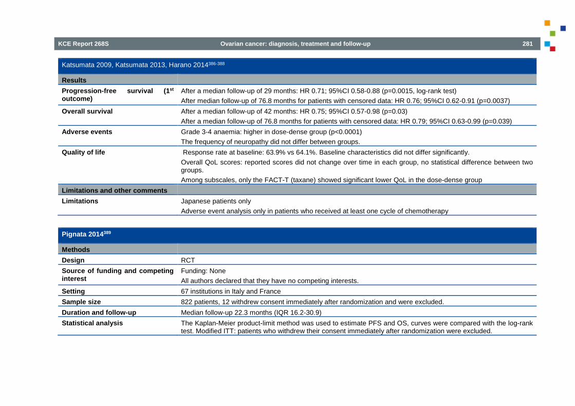
KCE Report 268S Ovarian cancer: diagnosis, treatment and follow-up 281
Katsumata 2009, Katsumata 2013, Harano 2014386-388
Results Progression-free survival (1st outcome)
After a median follow-up of 29 months: HR 0.71; 95%CI 0.58-0.88 (p=0.0015, log-rank test) After median follow-up of 76.8 months for patients with censored data: HR 0.76; 95%CI 0.62-0.91 (p=0.0037)
Overall survival After a median follow-up of 42 months: HR 0.75; 95%CI 0.57-0.98 (p=0.03) After a median follow-up of 76.8 months for patients with censored data: HR 0.79; 95%CI 0.63-0.99 (p=0.039)
Adverse events Grade 3-4 anaemia: higher in dose-dense group (p<0.0001) The frequency of neuropathy did not differ between groups.
Quality of life Response rate at baseline: 63.9% vs 64.1%. Baseline characteristics did not differ significantly. Overall QoL scores: reported scores did not change over time in each group, no statistical difference between two groups. Among subscales, only the FACT-T (taxane) showed significant lower QoL in the dose-dense group
Limitations and other comments Limitations Japanese patients only
Adverse event analysis only in patients who received at least one cycle of chemotherapy
Pignata 2014389
Methods Design RCT Source of funding and competing interest
Funding: None All authors declared that they have no competing interests.
Setting 67 institutions in Italy and France Sample size 822 patients, 12 withdrew consent immediately after randomization and were excluded. Duration and follow-up Median follow-up 22.3 months (IQR 16.2-30.9) Statistical analysis The Kaplan-Meier product-limit method was used to estimate PFS and OS, curves were compared with the log-rank
test. Modified ITT: patients who withdrew their consent immediately after randomization were excluded.

282 Ovarian cancer: diagnosis, treatment and follow-up KCE Report 268S
Pignata 2014389
All patients who received chemotherapy at least once were analyzed for adverse events. The worst toxicity grade for every patient was calculated and grades were compared for every type of toxic effect with the exact Wilcoxon-Mann-Whitney test. The effect of each treatment schedule on every QoL scale was assessed by fitting a linear mixed-effects model, using no within-patient correlation structure among repeated QoL measures
Patient characteristics Eligibility criteria Cytological or histological diagnosis of epithelial ovarian, fallopian tube or peritoneal cancer, FIGO stage IC-IV
ECOG PS 0-2 Previous chemotherapy excluded
Patient & disease characteristics Median age: 59 vs 60 years old ECOG PS 0 75% vs 74% No residual disease: 41% vs 41% Serous disease: 72% vs 67%
Interventions Intervention group Carboplatin AUC 2 and paclitaxel 60mg/m2 every week for 18 weeks consecutively Control group Carboplatin AUC 6 and paclitaxel 175/m2 on day 1, every 21 days for 6cycles Results Progression-free survival HR 0.96; 95%CI 0.80-1.16 (p=0.66)
Adjusted HR 0.94; 95%CI 0.78-1.14 Overall survival HR 1.20; 95%CI 0.90-1.61 (p=0.22)
Adjusted HR 1.21; 95%CI 0.90-1.62 Adverse events Considering all grades of toxicity, the dose-dense group showed a more favourable pattern for neutropenia,
thrombocytopenia, vomiting, hair loss and neuropathy, but a worse pattern for pulmonary toxic effects. Proportion of women with grade 3-4 toxicity (3-weekly vs once a week): neutropenia: 50% vs 42% febrile neutropenia: 3% vs 0.5% thrombocytopenia: 7% vs 1% grade 2 or higher neuropathy: 17% vs 6%

KCE Report 268S Ovarian cancer: diagnosis, treatment and follow-up 283
Pignata 2014389
grade 2 hair loss: 59% vs 29% Quality of life Baseline FACT-O/TOI scores were similar btween treatment assignments
In all analyses, the treatment-by-time interaction favoured chemotherapy every week (p<0.0001) Limitations and other comments Limitations
5.11. Routine CA125 measurement during follow-up
Table 94 – Evidence table: SR routine CA125 measurement during follow-up Clarke T 2014390
Methods Design SR Source of funding and competing interest
Department of Health, UK. NHS Cochrane Collaboration Programme. Grant Scheme CPG-506
Search date July 2013 Searched databases Specialised Register of the Cochrane Gynaecological Cancer Group; EMBASE; MEDLINE; CENTRAL Included study designs RCTs Number of included studies N=1 Statistical analysis Not applicable Patient characteristics Eligibility criteria Women of any age diagnosed with primary ovarian cancer of epithelial histological sub-type who have completed
primary treatment Exclusion criteria Not specifically stated Patient & disease characteristics See Rustin 2010 Interventions Intervention group Follow-up strategy following completion of primary treatment

284 Ovarian cancer: diagnosis, treatment and follow-up KCE Report 268S
Clarke T 2014390
Control group Other or no follow-up strategy Results See Rustin 2010 Limitations and other comments Comments
Table 95 – Evidence table: RCT routine CA125 measurement during follow-up Rustin GJ 2010391
Methods Design RCT Source of funding and competing interest
MRC, EORTC
Setting Multicentre, worldwide (59 centres) Sample size N=529 Duration and follow-up Recruitment: 2/1997-3/2008
Median follow-up from randomisation: 56.9 months Statistical analysis The primary analysis was the log-rank test, stratified by MRC versus EORTC, applied to compare the Kaplan-Meier
survival curves for all time-to-event outcome measures Cox model sensitivity analyses estimating the treatment effect and adjusting for stratification and prognostic factors were done for overall survival All p values are two-sided To adjust for imbalances in follow-up between the two groups, curtailing was used for main analyses of time-to-event outcome measures, by censoring data at 5 years from randomisation for MRC OV05 and 3 years for EORTC 55955. Additional sensitivity analyses were done for uncurtailed data
Patient characteristics Eligibility criteria Women with histologically confirmed epithelial ovarian, fallopian tube, or serous primary peritoneal cancer (based on
local pathology) in complete clinical remission after completion of first-line platinum-based chemotherapy with a normal CA125 concentration

KCE Report 268S Ovarian cancer: diagnosis, treatment and follow-up 285
Rustin GJ 2010391
Patient & disease characteristics Median age: 60 vs. 61 years Histology: serous 65% vs. 58% Stage III: 68% vs. 69%
Interventions Intervention group Early treatment on the basis of increased CA125 concentrations (N=265) Control group Delayed treatment on the basis of clinical recurrence (N=264) Results Overall survival HR 0.98 (95%CI 0.80-1.20; p=0.85)
Median survival from randomization: 25.7 (23.0-27.9) vs. 27.1 (22.8-30.9) months 2-year survival: 53.7% vs. 54.7% Cox models adjusted for stratification and prognostic factors did not change the overall result
Quality of life Median time spent with good global health score: 7.2 (95%CI 5.3-9.3) vs. 9.2 months (6.4-10.5) Median time from randomisation to first deterioration in global health score or death: 3.2 (95%CI 2.4-4.3) vs. 5.8 months (4.4-8.5), HR 0.71 (95%CI 0.58-0.88; p=0.002) Subgroup analyses of individual components of the QLQ-C30 subscales showed deterioration in score sooner in the early group than in the delayed group for almost all subscales There was evidence of significant disadvantages for role, emotional, social, and fatigue subscales with early treatment
Time to second-line chemotherapy Women assigned to early treatment started chemotherapy 4.8 months (95%CI 3.6-5.3) earlier than those allocated delayed treatment
Limitations and other comments Limitations Central randomization at EORTC and MRC
Randomisation by minimisation and stratification Blinding of sites and patients ITT-analysis Since the QLQ-C30 questionnaire asks about symptoms only in the previous week, and the forms were completed just before each course of chemotherapy, this method could underestimate any reduction in quality of life due to chemotherapy

286 Ovarian cancer: diagnosis, treatment and follow-up KCE Report 268S
6. SUMMARY OF FINDINGS TABLES AND GRADE PROFILES 6.1. Pre-operative assessment pelvic mass
Table 96 – GRADE evidence profile: RMI 1 Outcome № of studies
(№ of patients)
Study design Factors that may decrease quality of evidence Effect per 1000 patients
Test accuracy QoE
Risk of bias
Indirectness Inconsistency Imprecision Publication bias
pre-test probability of 27%
True positives (patients with malignant ovarian tumours)
23 studies 5626 patients
cross-sectional (cohort type accuracy study)
not serious
not serious not serious 2 not serious none 194 (181 to 205)
⨁⨁⨁⨁ HIGH 1
False negatives (patients incorrectly classified as not having malignant ovarian tumours)
76 (65 to 89)
True negatives (patients without malignant ovarian tumours)
23 studies 5626 patients
cross-sectional (cohort type accuracy study)
not serious
not serious not serious not serious none 672 (650 to 679)
⨁⨁⨁⨁ HIGH 1
False positives (patients incorrectly classified as having malignant ovarian tumours)
58 (51 to 80)

KCE Report 268S Ovarian cancer: diagnosis, treatment and follow-up 287
Table 97 – GRADE evidence profile: RMI 2 Outcome № of
studies (№ of patients)
Study design Factors that may decrease quality of evidence Effect per 1000 patients
Test accuracy QoE
Risk of bias
Indirectness Inconsistency Imprecision Publication bias
pre-test probability of 27%
True positives (patients with malignant ovarian tumours)
15 studies 4293 patients
cross-sectional (cohort type accuracy study)
not serious
not serious not serious 2 not serious none 203 (186 to 216)
⨁⨁⨁⨁ HIGH 1
False negatives (patients incorrectly classified as not having malignant ovarian tumours)
68 (54 to 84)
True negatives (patients without malignant ovarian tumours)
15 studies 4293 patients
cross-sectional (cohort type accuracy study)
not serious
not serious not serious not serious none 635 (613 to 657)
⨁⨁⨁⨁ HIGH 1
False positives (patients incorrectly classified as having malignant ovarian tumours)
95 (73 to 117)

288 Ovarian cancer: diagnosis, treatment and follow-up KCE Report 268S
Table 98 – GRADE evidence profile: IOTA simple rules Outcome № of
studies (№ of patients)
Study design Factors that may decrease quality of evidence Effect per 1000 patients
Test accuracy QoE Risk of
bias Indirectness Inconsistency Imprecision Publication
bias pre-test probability of 27%
True positives (patients with malignant ovarian tumours)
5 studies 1783 patients
cross-sectional (cohort type accuracy study)
not serious
not serious not serious 2 not serious none 251 (240 to 257)
⨁⨁⨁⨁ HIGH 1
False negatives (patients incorrectly classified as not having malignant ovarian tumours)
19 (14 to 30)
True negatives (patients without malignant ovarian tumours)
5 studies 1783 patients
cross-sectional (cohort type accuracy study)
not serious
not serious not serious not serious none 591 (555 to 621)
⨁⨁⨁⨁ HIGH 1
False positives (patients incorrectly classified as having malignant ovarian tumours)
139 (110 to 175)

KCE Report 268S Ovarian cancer: diagnosis, treatment and follow-up 289
Table 99 – GRADE evidence profile: IOTA LR2 Outcome № of
studies (№ of patients)
Study design Factors that may decrease quality of evidence Effect per 1000 patients
Test accuracy QoE
Risk of bias
Indirectness Inconsistency Imprecision Publication bias
pre-test probability of 27%
True positives (patients with malignant ovarian tumours)
3 studies 1335 patients
cross-sectional (cohort type accuracy study)
not serious
not serious not serious 2 not serious none 248 (238 to 257)
⨁⨁⨁⨁ HIGH 1
False negatives (patients incorrectly classified as not having malignant ovarian tumours)
22 (14 to 32)
True negatives (patients without malignant ovarian tumours)
3 studies 1335 patients
cross-sectional (cohort type accuracy study)
not serious
not serious not serious not serious none 606 (562 to 642)
⨁⨁⨁⨁ HIGH 1
False positives (patients incorrectly classified as having malignant ovarian tumours)
124 (88 to 168)
6.2. Intra-operative frozen section
Results Sensitivity 0.90 (95% CI: 0.88 to 0.92) Specificity 0.99 (95% CI: 0.99 to 1.00)
Results Prevalence 25th percentile:
23.3% Median: 29.0% 75th percentile:
37.8%

290 Ovarian cancer: diagnosis, treatment and follow-up KCE Report 268S
Table 100 – GRADE evidence profile: intraoperative frozen section Outcome № of
studies (№ of patients)
Study design
Factors that may decrease quality of evidence Effect per 1000 patients Test accuracy QoE Risk of
bias Indirectness Inconsistency Imprecision Publication
bias pre-test probability of 23.3%
pre-test probability of 29.0%
pre-test probability of 37.8%
True positives (patients with malignant ovarian tumours)
37 studies 3096 patients
cross-sectional (cohort type accuracy study)
serious 1 not serious not serious 2 not serious none 210 (205 to 215)
262 (255 to 267)
341 (333 to 349)
⨁⨁⨁◯ MODERATE 1
False negatives (patients incorrectly classified as not having malignant ovarian tumours)
23 (18 to 28)
28 (23 to 35)
37 (29 to 45)
True negatives (patients without malignant ovarian tumours)
37 studies 7431 patients
cross-sectional (cohort type accuracy study)
serious 1 not serious not serious not serious none 763 (760 to 765)
706 (704 to 708)
619 (616 to 620)
⨁⨁⨁◯ MODERATE 1

KCE Report 268S Ovarian cancer: diagnosis, treatment and follow-up 291
Outcome № of studies (№ of patients)
Study design
Factors that may decrease quality of evidence Effect per 1000 patients Test accuracy QoE Risk of
bias Indirectness Inconsistency Imprecision Publication
bias pre-test probability of 23.3%
pre-test probability of 29.0%
pre-test probability of 37.8%
False positives (patients incorrectly classified as having malignant ovarian tumours)
4 (2 to 7)
4 (2 to 6)
3 (2 to 6)
1Unclear risk of bias in the majority of studies; most studies retrospective. 2Although there is no perfect overlap of confidence intervals, it was decided not to downgrade, because the various point estimates did not differ substantially
6.3. Adjuvant chemotherapy
Table 101 – GRADE evidence profile: adjuvant chemotherapy for patients with a (presumed) early stage borderline ovarian tumour Quality assessment № of patients Effect Quality Importance № of studies
Study design
Risk of bias
Inconsistency
Indirectness
Imprecision
Other considerations
with adjuvant chemo-therapy
without adjuvant chemo-therapy
Relative (95% CI)
Absolute (95% CI)
Overall survival (follow up: 1 RCT mean 36 (range: 12-84) and other RCT median 147 (range 4 -246) months) 2 randomis
ed trials serious 1
not serious serious2 serious 3 none 17 25 Creasman 1982: 0/17 (0%) vs. 0/25 (0%)
⨁◯◯
◯ VERY LOW
CRITICAL
27 39 Tropé 1993 (study 3): 6/27 (22%) vs. 2/39 (5%), RR 4.33, 95%CI 0.94 to 19.88
Disease-free survival (follow up: 1 RCT mean 36 (range: 12-84) and other RCT median 147 (range 4 -246) months)

292 Ovarian cancer: diagnosis, treatment and follow-up KCE Report 268S
2 randomised trials
serious 1
not serious serious2 serious 3 none 17 25 Creasman 1982: 0/17 (0%) vs. 0/25 (0%)
⨁◯◯
◯ VERY LOW
CRITICAL
27 39 Tropé 1993 (study 3): 1/27 (4%) vs 0/39 (0%)
Quality of life - not reported - - - - - - - - - - - - CRITICAL Adverse events (follow up: median 147 (range 4 -246) months) 1 randomised
trials serious 1 not serious serious2 serious 3 none 27 39 -Severe septic neutropenia
(grade 4): 1/27 (4%) vs. 0/39 (0%) Bone marrow toxicity (grade 3): 1/27 (4%) vs.0/39 (0%)
⨁◯◯◯ VERY LOW
IMPORTANT
1Unclear risk of bias for most items 2Chemotherapy regimens used in both RCTs differ from currently used chemotherapy regimens 3Optimal information size not reached
Table 102 – GRADE evidence profile: adjuvant chemotherapy for patients with invasive (presumed) early stage ovarian cancer Quality assessment № of patients Effect Quality Importance № of studies
Study design
Risk of bias
Inconsistency
Indirectness
Imprecision
Other considerations
adjuvant chemo-therapy
without adjuvant chemo-therapy
Relative (95% CI)
Absolute (95% CI)
5-year overall survival 3 randomise
d trials not serious
not serious not serious
serious1 none 506 502 HR 0.71 (0.53 to 0.93)
⨁⨁⨁◯ MODERATE
CRITICAL
5-year overall survival - subgroup 'optimal staging' 2 randomise
d trials Serious4
not serious not serious
very serious 2
none 117 117 HR 1.22 (0.63 to 2.37)
⨁◯◯
◯ VERY LOW
CRITICAL
5-year overall survival - subgroup 'suboptimal staging' 2 randomise
d trials not serious
not serious not serious
serious1 none 389 383 HR 0.63 (0.46 to 0.85)
⨁⨁⨁◯ MODERATE
CRITICAL
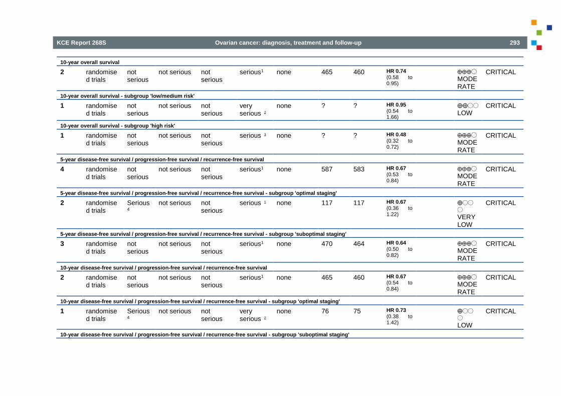
KCE Report 268S Ovarian cancer: diagnosis, treatment and follow-up 293
10-year overall survival 2 randomise
d trials not serious
not serious not serious
serious1 none 465 460 HR 0.74 (0.58 to 0.95)
⨁⨁⨁◯ MODERATE
CRITICAL
10-year overall survival - subgroup 'low/medium risk' 1 randomise
d trials not serious
not serious not serious
very serious 2
none ? ? HR 0.95 (0.54 to 1.66)
⨁⨁◯◯ LOW
CRITICAL
10-year overall survival - subgroup 'high risk' 1 randomise
d trials not serious
not serious not serious
serious 3 none ? ? HR 0.48 (0.32 to 0.72)
⨁⨁⨁◯ MODERATE
CRITICAL
5-year disease-free survival / progression-free survival / recurrence-free survival 4 randomise
d trials not serious
not serious not serious
serious1 none 587 583 HR 0.67 (0.53 to 0.84)
⨁⨁⨁◯ MODERATE
CRITICAL
5-year disease-free survival / progression-free survival / recurrence-free survival - subgroup 'optimal staging' 2 randomise
d trials Serious4
not serious not serious
serious 1 none 117 117 HR 0.67 (0.36 to 1.22)
⨁◯◯
◯ VERY LOW
CRITICAL
5-year disease-free survival / progression-free survival / recurrence-free survival - subgroup 'suboptimal staging' 3 randomise
d trials not serious
not serious not serious
serious1 none 470 464 HR 0.64 (0.50 to 0.82)
⨁⨁⨁◯ MODERATE
CRITICAL
10-year disease-free survival / progression-free survival / recurrence-free survival 2 randomise
d trials not serious
not serious not serious
serious1 none 465 460 HR 0.67 (0.54 to 0.84)
⨁⨁⨁◯ MODERATE
CRITICAL
10-year disease-free survival / progression-free survival / recurrence-free survival - subgroup 'optimal staging' 1 randomise
d trials Serious4
not serious not serious
very serious 2
none 76 75 HR 0.73 (0.38 to 1.42)
⨁◯◯
◯ LOW
CRITICAL
10-year disease-free survival / progression-free survival / recurrence-free survival - subgroup 'suboptimal staging'

294 Ovarian cancer: diagnosis, treatment and follow-up KCE Report 268S
1 randomised trials
not serious
not serious not serious
serious 1 none 148 147 HR 0.60 (0.41 to 0.87)
⨁⨁⨁◯ MODERATE
CRITICAL
10-year disease-free survival / progression-free survival / recurrence-free survival - subgroup 'low/medium risk' 1 randomise
d trials not serious
not serious not serious
very serious 1
none ? ? HR 0.96 (0.58 to 1.59)
⨁⨁◯◯ LOW
CRITICAL
10-year disease-free survival / progression-free survival / recurrence-free survival - subgroup 'high risk' 1 randomise
d trials not serious
not serious not serious
serious 1 none ? ? HR 0.52 (0.33 to 0.82)
⨁⨁⨁◯ MODERATE
CRITICAL
Quality of life - not measured - - - - - - - - - - - - CRITICAL Adverse events - not reported - - - - - - - - - - - - IMPORTA
NT MD – mean difference, RR – relative risk 1Confidence interval crosses clinical decision threshold. 2Both limits of confidence interval cross clinical decision threshold. 3Optimal information size not reached 4for one trial only subgroup analysis included (no stratification factor), for the other trial optimal staging status is unsure (no detailed information, post-hoc consideration) 6.4. Laparoscopic surgery in early stage ovarian cancer As all evidence is derived from non-randomized comparative studies with methodological limitations, we judged to level of evidence to be very low for all outcomes. No formal GRADE profile was drawn up.
6.5. Laparoscopy, PET-CT and MRI to predict end result of cytoreductive surgery 6.5.1. Laparoscopy
Result Result Sensitivity Laparoscopy (study 1)
0.71 (95% CI: 0.44 to 0.9) Sensitivity Laparoscopy (study 2)
0.7 (95% CI: 0.57 to 0.82)
Specificity Laparoscopy (study 1)
1 (95% CI: 0.9 to 1) Specificity Laparoscopy (study 2)
1 (95% CI: 0.94 to 1)
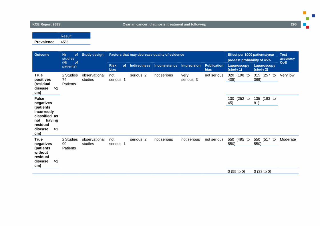
KCE Report 268S Ovarian cancer: diagnosis, treatment and follow-up 295
Result Prevalence 45%
Outcome № of studies (№ of patients)
Study design Factors that may decrease quality of evidence Effect per 1000 patients/year Test accuracy QoE
pre-test probability of 45% Risk of bias
Indirectness Inconsistency Imprecision Publication bias
Laparoscopy (study 1)
Laparoscopy (study 2)
True positives (residual disease >1 cm)
2 Studies 74 Patients
observational studies
not serious 1
serious 2 not serious very serious 3
not serious 320 (198 to 405)
315 (257 to 369)
Very low
False negatives (patients incorrectly classified as not having residual disease >1 cm)
130 (252 to 45)
135 (193 to 81)
True negatives (patients without residual disease >1 cm)
2 Studies 90 Patients
observational studies
not serious 1
serious 2 not serious not serious not serious 550 (495 to 550)
550 (517 to 550)
Moderate
0 (55 to 0) 0 (33 to 0)

296 Ovarian cancer: diagnosis, treatment and follow-up KCE Report 268S
False positives (patients incorrectly classified as having residual disease >1 cm)
1. Based on judgements of the authors of the original review (Rutten 2014) 2. Applicability concerns regarding patient selection 3. Very wide 95%-Cis
6.5.2. MRI or CT
Results MRI Results CT Sensitivity MRI 0.91 (95% CI: 0.59 to 1) Sensitivity CT 0.5 (95% CI: 0.12 to 0.88) Specificity MRI 0.97 (95% CI: 0.87 to 1) Specificity CT 1 (95% CI: 0.91 to 1)

KCE Report 268S Ovarian cancer: diagnosis, treatment and follow-up 297
Result Prevalence 18%
Outcome № of studies (№ of patients)
Study design Factors that may decrease quality of evidence Effect per 1000 patients/year
Test accuracy QoE
pre-test probability of 18%
Risk of bias
Indirectness Inconsistency Imprecision Publication bias
MRI CT
True positives (patients with "unsuccessful cytoreduction")
1 Study 17 Patients
observational studies
not serious
serious 1 not serious very serious 2
not serious 164 (106 to 180)
90 (22 to 158)
Very low
74 more TP in MRI
False negatives (patients incorrectly classified as not having "unsuccessful cytoreduction")
16 (74 to 0)
90 (158 to 22)
74 fewer FN in MRI
True negatives (patients without "unsuccessful cytoreduction")
1 Study 76 Patients
observational studies
not serious
serious 1 not serious serious 3 not serious 795 (713 to 820)
820 (746 to 820)
Low
25 fewer TN in MRI
False positives (patients incorrectly classified as having "unsuccessful cytoreduction")
25 (107 to 0)
0 (74 to 0)
25 more FP in MRI

298 Ovarian cancer: diagnosis, treatment and follow-up KCE Report 268S
1. High risk of bias due to Patient selection and Reference standard 2. Applicability concerns in all domains. Cut-off for incomplete = 2 cm 3. Very wide 95%-CI
6.5.3. DW-MRI
Results Sensitivity 0.75 (95% CI: 0.35 to 0.97) Specificity 0.96 (95% CI: 0.8 to 1)
Results Prevalence 24%
Outcome № of studies (№ of patients)
Study design Factors that may decrease quality of evidence Effect per 1000 patients/year
Test accuracy QoE Risk of bias Indirectness Inconsistency Imprecision Publication
bias pre-test probability of 24%
True positives (patients with residual disease >1 cm)
1 Study 8 Patients
observational studies
very serious 1 2
very serious 3
not serious very serious 4
not serious
180 (84 to 233) Very low
False negatives (patients incorrectly classified as not having residual disease >1 cm)
60 (156 to 7)
True negatives (patients without residual disease >1 cm)
1 Study 26 Patients
observational studies
very serious 1 2
very serious 3
not serious very serious 4
not serious
730 (608 to 760)
Very low

KCE Report 268S Ovarian cancer: diagnosis, treatment and follow-up 299
False positives (patients incorrectly classified as having residual disease >1 cm)
30 (152 to 0)
1. High risk of bias for Patient selection, Index test and Reference standard 2. Post-hoc assessment of optimal index test threshold 3. High applicability concerns regarding Patient selection and Index test. Indextest not used as add-on to CT. 4. Very wide confidence interval
6.5.4. PET-CT
Results Sensitivity 0.66 (95% CI: 0.6 to 0.73) Specificity 0.88 (95% CI: 0.8 to 0.93)
Result Prevalence 65%
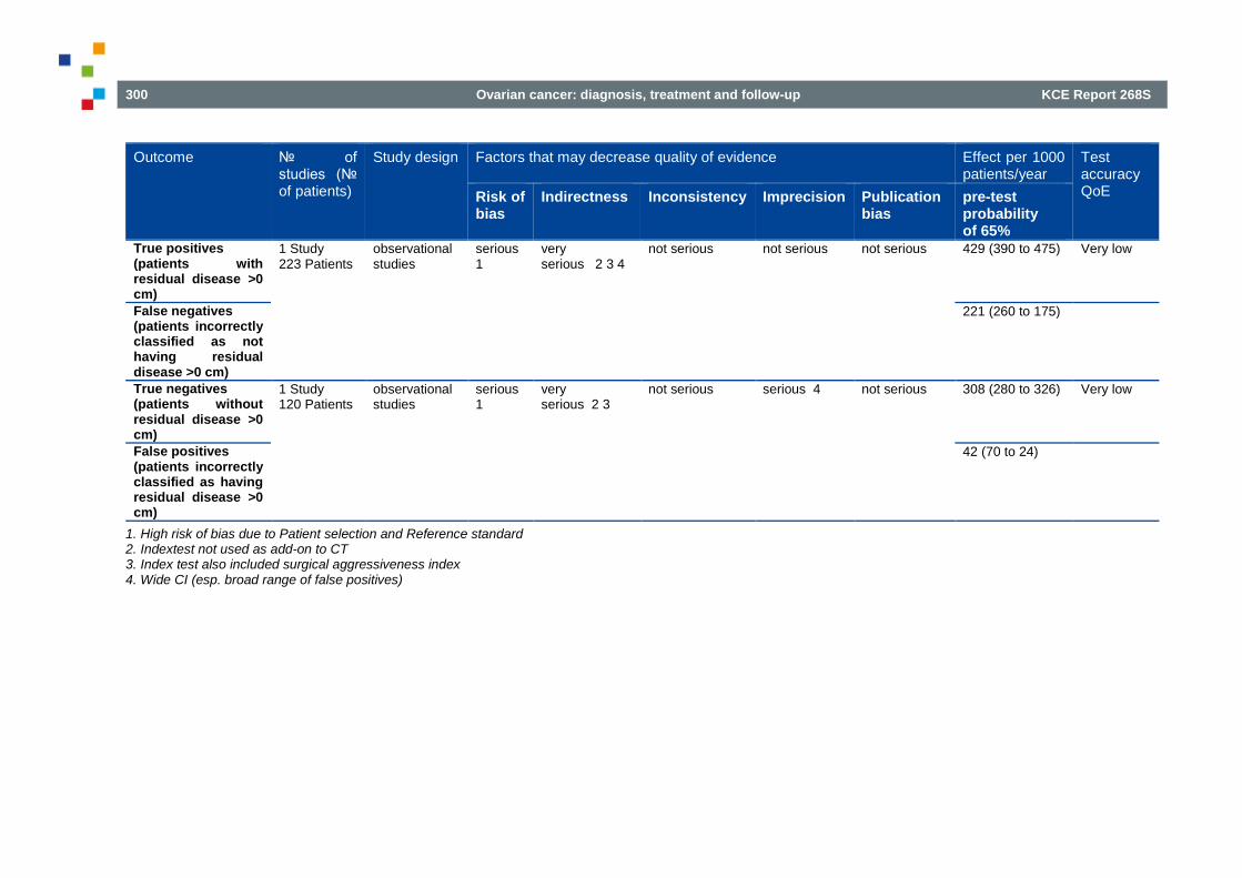
300 Ovarian cancer: diagnosis, treatment and follow-up KCE Report 268S
Outcome № of
studies (№ of patients)
Study design Factors that may decrease quality of evidence Effect per 1000 patients/year
Test accuracy QoE Risk of
bias Indirectness Inconsistency Imprecision Publication
bias pre-test probability of 65%
True positives (patients with residual disease >0 cm)
1 Study 223 Patients
observational studies
serious 1
very serious 2 3 4
not serious not serious not serious 429 (390 to 475) Very low
False negatives (patients incorrectly classified as not having residual disease >0 cm)
221 (260 to 175)
True negatives (patients without residual disease >0 cm)
1 Study 120 Patients
observational studies
serious 1
very serious 2 3
not serious serious 4 not serious 308 (280 to 326) Very low
False positives (patients incorrectly classified as having residual disease >0 cm)
42 (70 to 24)
1. High risk of bias due to Patient selection and Reference standard 2. Indextest not used as add-on to CT 3. Index test also included surgical aggressiveness index 4. Wide CI (esp. broad range of false positives)

KCE Report 268S Ovarian cancer: diagnosis, treatment and follow-up 301
6.6. Aim of cytoreductive surgery: no macroscopic disease? 6.6.1. Prognostic value end result of surgery Table 103 – GRADE evidence profile: prognostic value end result of surgery RD 0.1-1.0 cm compared to microscopic RD
Quality assessment № of patients Effect Quality Importance
№ of studies
Study design Risk of bias
Inconsistency Indirectness Imprecision Other considerations
RD 0.1-1.0 cm
microscopic RD
Relative (95% CI)
Absolute (95% CI)
Overall survival (HR for death) 12 observation
al studies Not serious 1
not serious not serious not serious
publication bias strongly suspected 2
? ? HR 2.21 (1.97 to 2.47)
- ⨁⨁⨁◯ MODERATE
CRITICAL
Progression-free survival (HR for progression) 5 3 observation
al studies Not serious 1
not serious not serious not serious
publication bias strongly suspected 2
? ? HR 1.91 (1.70 to 2.15)
- ⨁⨁⨁◯ MODERATE
CRITICAL
1. Adjusted HRs derived from a prognostic model; probably based on significance testing (and not on including putative confounders in the analysis, irrespective of statistical significance) However, as findings are consistently confirmed in RCTs (that also included high-risk stage II-IIIA-B), we decided not to downgrade. 2. Non-reporting of non-significant associations very plausible 3. A sixth study reported that in patients who underwent an UAP those with completely resected disease had better PFS than those with <1 cm (p < 0.01)
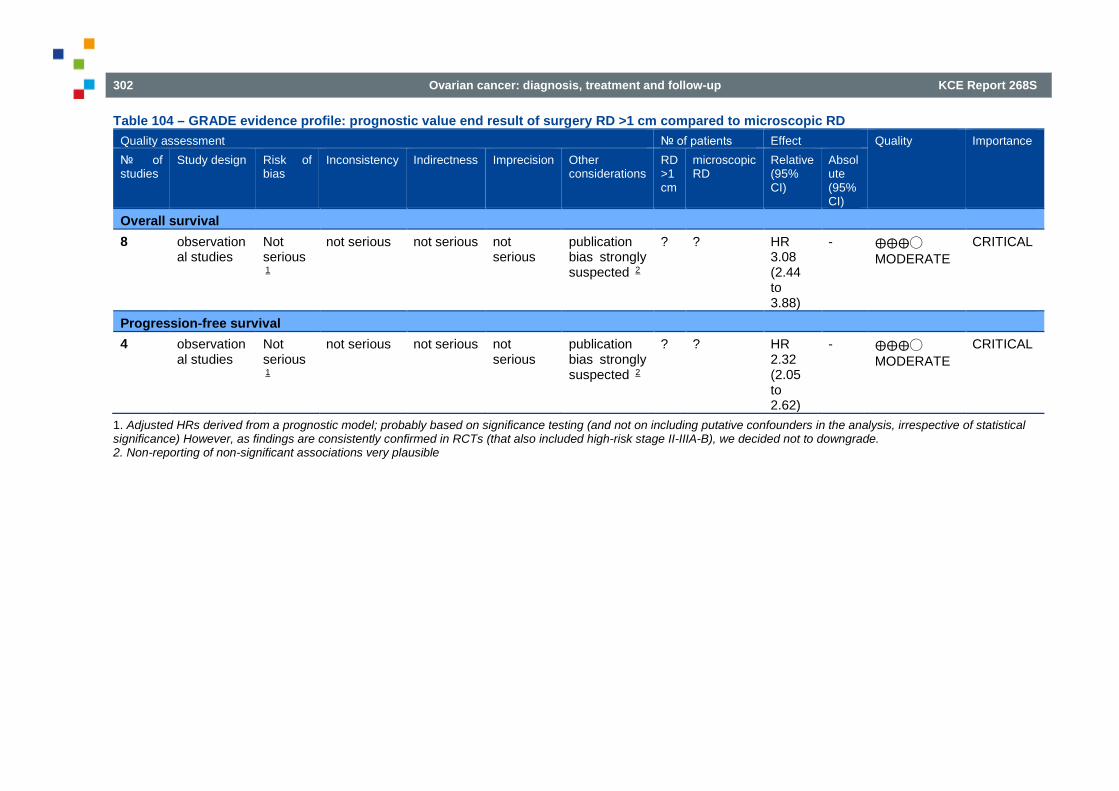
302 Ovarian cancer: diagnosis, treatment and follow-up KCE Report 268S
Table 104 – GRADE evidence profile: prognostic value end result of surgery RD >1 cm compared to microscopic RD Quality assessment № of patients Effect Quality Importance № of studies
Study design Risk of bias
Inconsistency Indirectness Imprecision Other considerations
RD >1 cm
microscopic RD
Relative (95% CI)
Absolute (95% CI)
Overall survival 8 observation
al studies Not serious 1
not serious not serious not serious
publication bias strongly suspected 2
? ? HR 3.08 (2.44 to 3.88)
- ⨁⨁⨁◯ MODERATE
CRITICAL
Progression-free survival 4 observation
al studies Not serious 1
not serious not serious not serious
publication bias strongly suspected 2
? ? HR 2.32 (2.05 to 2.62)
- ⨁⨁⨁◯ MODERATE
CRITICAL
1. Adjusted HRs derived from a prognostic model; probably based on significance testing (and not on including putative confounders in the analysis, irrespective of statistical significance) However, as findings are consistently confirmed in RCTs (that also included high-risk stage II-IIIA-B), we decided not to downgrade. 2. Non-reporting of non-significant associations very plausible

KCE Report 268S Ovarian cancer: diagnosis, treatment and follow-up 303
Table 105 – GRADE evidence profile: prognostic value end result of surgery any RD compared to microscopic RD Quality assessment № of patients Effect Quality Importance № of studies
Study design Risk of bias
Inconsistency Indirectness Imprecision Other considerations
any RD
microscopic RD
Relative (95% CI)
Absolute (95% CI)
3-year overall survival 2 1 observation
al studies Not serious 2
not serious not serious serious 3 publication bias strongly suspected 4
? ? HR 2.03 (1.01 to 4.10)
- ⨁⨁◯◯ LOW
CRITICAL
3-year progression-free survival 1 5 observation
al studies Not serious 2
not serious not serious not serious
publication bias strongly suspected 4
? ? HR 1.6 (1.3 to 2.1)
- ⨁⨁⨁◯ MODERATE
CRITICAL
1. A third study reported that the presence of any macroscopic residual disease after rectosigmoid colectomy was the only significant risk factor for OS (p=0.003) 2. Adjusted HRs derived from prognostic models by selecting characteristics on significance, not on including putative confounders in the analysis, irrespective of statistical significance. However, as findings are consistently confirmed in RCTs (that also included high-risk stage II-IIIA-B), we decided not to downgrade. 3. Wide CI that includes no effect and strong effect 4. Non-reporting of non-significant associations very plausible 5. A second study reported that age greater than 60 years (p=0.025), stage IV vs IIIC (p=0.037) and any residual disease (p=0.032) had an independent association with worse PFS
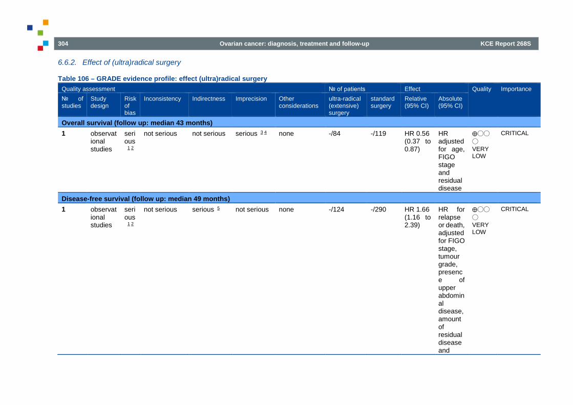
304 Ovarian cancer: diagnosis, treatment and follow-up KCE Report 268S
6.6.2. Effect of (ultra)radical surgery
Table 106 – GRADE evidence profile: effect (ultra)radical surgery Quality assessment № of patients Effect Quality Importance № of studies
Study design
Risk of bias
Inconsistency Indirectness Imprecision Other considerations
ultra-radical (extensive) surgery
standard surgery
Relative (95% CI)
Absolute (95% CI)
Overall survival (follow up: median 43 months) 1 observat
ional studies
serious 1 2
not serious not serious serious 3 4 none -/84 -/119 HR 0.56 (0.37 to 0.87)
HR adjusted for age, FIGO stage and residual disease
⨁◯◯
◯ VERY LOW
CRITICAL
Disease-free survival (follow up: median 49 months) 1 observat
ional studies
serious 1 2
not serious serious 5 not serious none -/124 -/290 HR 1.66 (1.16 to 2.39)
HR for relapse or death, adjusted for FIGO stage, tumour grade, presence of upper abdominal disease, amount of residual disease and
⨁◯◯
◯ VERY LOW
CRITICAL
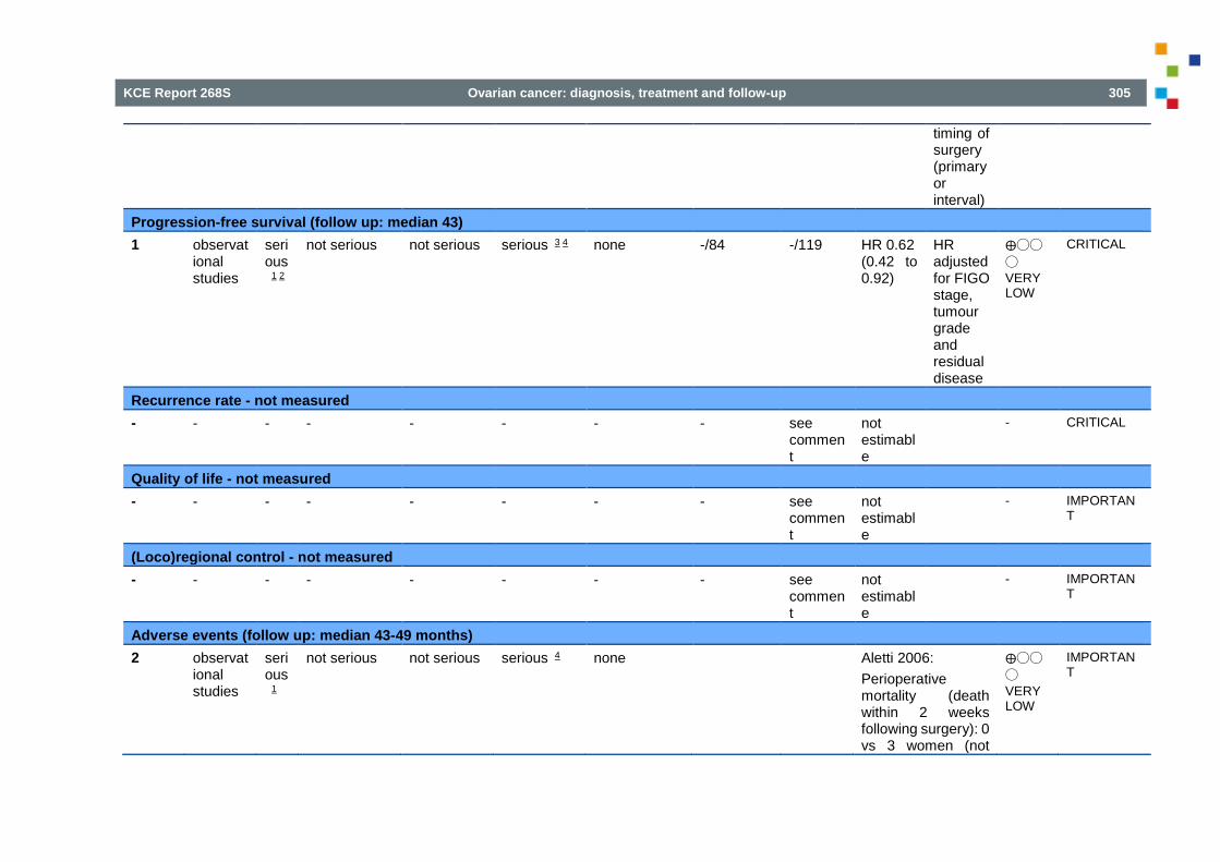
KCE Report 268S Ovarian cancer: diagnosis, treatment and follow-up 305
timing of surgery (primary or interval)
Progression-free survival (follow up: median 43) 1 observat
ional studies
serious 1 2
not serious not serious serious 3 4 none -/84 -/119 HR 0.62 (0.42 to 0.92)
HR adjusted for FIGO stage, tumour grade and residual disease
⨁◯◯
◯ VERY LOW
CRITICAL
Recurrence rate - not measured - - - - - - - - see
comment
not estimable
- CRITICAL
Quality of life - not measured - - - - - - - - see
comment
not estimable
- IMPORTANT
(Loco)regional control - not measured - - - - - - - - see
comment
not estimable
- IMPORTANT
Adverse events (follow up: median 43-49 months) 2 observat
ional studies
serious 1
not serious not serious serious 4 none Aletti 2006: Perioperative mortality (death within 2 weeks following surgery): 0 vs 3 women (not
⨁◯◯
◯ VERY LOW
IMPORTANT

306 Ovarian cancer: diagnosis, treatment and follow-up KCE Report 268S
adjusted for baseline imbalances) Significant postoperative morbidity 32/84 (38.1%) vs 14/119 (11.8%) Luyckx 2012: Postoperative death within 30 days 1/84 (1.2%) vs 0/119 (0%) “Patients who underwent radical surgery had significantly longer operative time, larger estimated blood loss, more blood transfusions, longer stay in the intensive care unit, and more lymphocysts than those who underwent simple surgery.”
MD – mean difference, RR – relative risk 1. Confounding by indication 2. Adjusted HRs derived from a prognostic model; probably based on significance testing (and not on including putative confounders in the analysis, irrespective of statistical significance). 3. Wide confidence interval 4. OIS not reached 5. Sample included a mixture of primary and interval debulking surgery

KCE Report 268S Ovarian cancer: diagnosis, treatment and follow-up 307
6.7. Neo-adjuvant chemotherapy and interval debulking versus upfront surgery
Table 107 – GRADE evidence profile: primary debulking versus neo-adjuvant chemotherapy Quality assessment No of events/patients Effect Quality Importance
No of studies
Design Risk of bias
Inconsistency Indirectness Imprecision Other considerations
Primary debulking
Neo-adjuvant platinum-based chemotherapy
Relative (95% CI)
Absolute
Death (overall survival) 2 randomis
ed trials no serious risk of bias
no serious inconsistency
no serious indirectness
no serious imprecision
none - - HR 0.93 (0.81 to 1.06)
- ⊕⊕⊕⊕ HIGH
Critical
Death (overall survival): subanalysis stage III 9 2 randomis
ed trials no serious risk of bias
serious10 no serious indirectness
no serious imprecision
none - - HR 0.97 (0.78 to 1.20)
- ⊕⊕⊕Ο MODERATE
Critical
Death (overall survival): subanalysis stage IV 9 2 randomis
ed trials no serious risk of bias
no serious inconsistency
no serious indirectness
no serious imprecision
none - - HR 0.80 (0.77 to 1.04)
- ⊕⊕⊕⊕ HIGH
Critical
Death (overall survival): subanalysis metastatic tumour load 0-5 cm 9
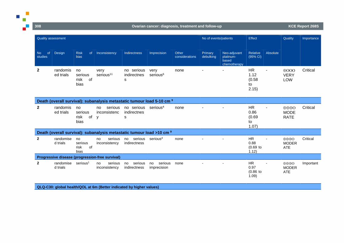
308 Ovarian cancer: diagnosis, treatment and follow-up KCE Report 268S
Quality assessment No of events/patients Effect Quality Importance
No of studies
Design Risk of bias
Inconsistency Indirectness Imprecision Other considerations
Primary debulking
Neo-adjuvant platinum-based chemotherapy
Relative (95% CI)
Absolute
2 randomised trials
no serious risk of bias
very serious11
no serious indirectness
very serious6
none - - HR 1.12 (0.58 to 2.15)
- ⊕ΟΟΟ VERY LOW
Critical
Death (overall survival): subanalysis metastatic tumour load 5-10 cm 9 2 randomis
ed trials no serious risk of bias
no serious inconsistency
no serious indirectness
serious4 none - - HR 0.86 (0.69 to 1.07)
- ⊕⊕⊕Ο MODERATE
Critical
Death (overall survival): subanalysis metastatic tumour load >10 cm 9 2 randomise
d trials no serious risk of bias
no serious inconsistency
no serious indirectness
serious4 none - - HR 0.88 (0.69 to 1.12)
- ⊕⊕⊕Ο MODERATE
Critical
Progressive disease (progression-free survival) 2 randomise
d trials serious1 no serious
inconsistency no serious indirectness
no serious imprecision
none - - HR 0.97 (0.86 to 1.09)
- ⊕⊕⊕Ο MODERATE
Important
QLQ-C30: global health/QOL at 6m (Better indicated by higher values)
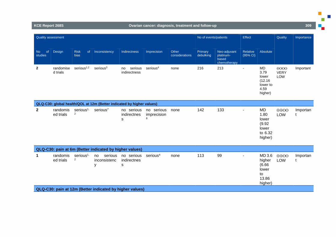
KCE Report 268S Ovarian cancer: diagnosis, treatment and follow-up 309
Quality assessment No of events/patients Effect Quality Importance
No of studies
Design Risk of bias
Inconsistency Indirectness Imprecision Other considerations
Primary debulking
Neo-adjuvant platinum-based chemotherapy
Relative (95% CI)
Absolute
2 randomised trials
serious1,2 serious3 no serious indirectness
serious4 none 216 213 - MD 3.79 lower (12.16 lower to 4.59 higher)
⊕ΟΟΟ VERY LOW
Important
QLQ-C30: global health/QOL at 12m (Better indicated by higher values) 2 randomis
ed trials serious1,
2 serious7 no serious
indirectness
no serious imprecision8
none 142 133 - MD 1.80 lower (9.92 lower to 6.32 higher)
⊕⊕ΟΟ LOW
Important
QLQ-C30: pain at 6m (Better indicated by higher values) 1 randomis
ed trials serious1,
2 no serious inconsistency
no serious indirectness
serious4 none 113 99 - MD 3.6 higher (6.66 lower to 13.86 higher)
⊕⊕ΟΟ LOW
Important
QLQ-C30: pain at 12m (Better indicated by higher values)

310 Ovarian cancer: diagnosis, treatment and follow-up KCE Report 268S
Quality assessment No of events/patients Effect Quality Importance
No of studies
Design Risk of bias
Inconsistency Indirectness Imprecision Other considerations
Primary debulking
Neo-adjuvant platinum-based chemotherapy
Relative (95% CI)
Absolute
1 randomised trials
serious1,
2 no serious inconsistency
no serious indirectness
serious4 none 78 64 - MD 4 higher (7.23 lower to 15.23 higher)
⊕⊕ΟΟ LOW
Important
QLQ-C30: dyspnoea at 6m (Better indicated by higher values) 1 randomis
ed trials serious1,
2 no serious inconsistency
no serious indirectness
serious4 none 113 99 - MD 0.5 higher (9.76 lower to 10.76 higher)
⊕⊕ΟΟ LOW
Important
QLQ-C30: dyspnoea at 12m (Better indicated by higher values) 1 randomis
ed trials serious1,
2 no serious inconsistency
no serious indirectness
serious4 none 78 64 - MD 3.3 lower (14.81 lower to 8.21 higher)
⊕⊕ΟΟ LOW
Important
Postoperative death

KCE Report 268S Ovarian cancer: diagnosis, treatment and follow-up 311
Quality assessment No of events/patients Effect Quality Importance
No of studies
Design Risk of bias
Inconsistency Indirectness Imprecision Other considerations
Primary debulking
Neo-adjuvant platinum-based chemotherapy
Relative (95% CI)
Absolute
2 randomised trials
no serious risk of bias
no serious inconsistency
no serious indirectness
no serious imprecision
none 22/565 (3.9%)
3/541 (0.55%)
RR 6.94 (2.06 to 23.44)
33 more per 1000 (from 6 more to 124 more)
⊕⊕⊕⊕ HIGH
Important
Postoperative haemorrhage grade 3-4 2 randomis
ed trials serious1 serious5 no serious
indirectness
very serious6
none 31/565 (5.5%)
26/541 (3.7%)
RR 1.01 (0.26 to 3.99)
0 more per 1000 (from 36 fewer to 144 more)
⊕ΟΟΟ VERY LOW
Important
Postoperative venous AE grade 3-4 2 randomis
ed trials serious1 no serious
inconsistency
no serious indirectness
no serious imprecision
none 13/565 (2.3%)
0/541 (0.0%)
RR 13.37 (1.79 to 100.07)
- ⊕⊕⊕Ο MODERATE
Important
Postoperative infection grade 3-4
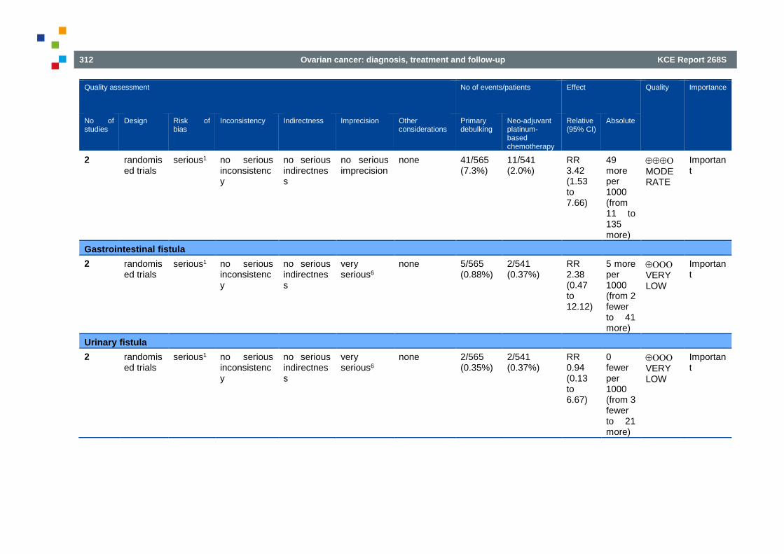
312 Ovarian cancer: diagnosis, treatment and follow-up KCE Report 268S
Quality assessment No of events/patients Effect Quality Importance
No of studies
Design Risk of bias
Inconsistency Indirectness Imprecision Other considerations
Primary debulking
Neo-adjuvant platinum-based chemotherapy
Relative (95% CI)
Absolute
2 randomised trials
serious1 no serious inconsistency
no serious indirectness
no serious imprecision
none 41/565 (7.3%)
11/541 (2.0%)
RR 3.42 (1.53 to 7.66)
49 more per 1000 (from 11 to 135 more)
⊕⊕⊕Ο MODERATE
Important
Gastrointestinal fistula 2 randomis
ed trials serious1 no serious
inconsistency
no serious indirectness
very serious6
none 5/565 (0.88%)
2/541 (0.37%)
RR 2.38 (0.47 to 12.12)
5 more per 1000 (from 2 fewer to 41 more)
⊕ΟΟΟ VERY LOW
Important
Urinary fistula 2 randomis
ed trials serious1 no serious
inconsistency
no serious indirectness
very serious6
none 2/565 (0.35%)
2/541 (0.37%)
RR 0.94 (0.13 to 6.67)
0 fewer per 1000 (from 3 fewer to 21 more)
⊕ΟΟΟ VERY LOW
Important

KCE Report 268S Ovarian cancer: diagnosis, treatment and follow-up 313
1 No blinding. 2 No intention-to-treat analysis. 3 I² = 66%. 4 CI includes MID. 5 I² = 84%. 6 Broad CI that includes appreciable benefit and harm. 7 I² = 46%. 8 CI includes 0, but does not cross MID of 10 points. 9 Predefined groups. 10 I² 53%, not only IIIC in Kehoe 2015.. 11 I² 84%, opposite results.

314 Ovarian cancer: diagnosis, treatment and follow-up KCE Report 268S
6.8. Intraperitoneal chemotherapy Quality assessment No of patients Effect Quality Importance
No of studies
Design Risk of bias
Inconsistency Indirectness Imprecision Other conside- rations
Intra- peritoneal chemotherapy
Intravenous chemo- therapy
Relative (95%CI)
Absolute
Progression-free survival 5 RCTs no
serious risk of bias
no serious inconsistency
serious1 serious2 none - HR 0.78 (0.70-0.86)
⊕⊕ΟΟ LOW
CRITICAL
Overall survival 8 RCTs no
serious risk of bias
no serious inconsistency
serious1 serious2 none - - HR 0.81 (0.72-0.90)
- ⊕⊕ΟΟ LOW
IMPORTANT -
Grade 3-4 gastrointestinal side effects 5 RCTs no
serious risk of bias
no serious inconsistency
serious1 no serious imprecision
none 230/660 131/679 1.70 (1.28-2.26)
170 more per 1000 (from 68 more to 307 more)-
⊕⊕⊕Ο MODERATE
IMPORTANT (34.8%) (19.3%)
Grade 3-4 fatigue 3 RCTs no
serious risk of bias
no serious inconsistency
serious1 serious2 none 88/582 44/589 2.32 (1.06-5.07
57 more per 1000 (from 3 more to 175 more)
⊕⊕ΟΟ LOW
IMPORTANT (15.1%) (7.5%)

KCE Report 268S Ovarian cancer: diagnosis, treatment and follow-up 315
Grade 3-4 neurologic adverse events 6 RCTs no
serious risk of bias
serious3 serious1 serious4 none 114/910 106/955 1.15 (0.67-1.97)
13 more per 1000 (from 28 fewer to 82 more)
IMPORTANT (12.5%) (11.01%)
Quality of Life 1 RCTs no
serious risk of bias
no serious inconsistency
serious1 serious2 none - - - ⊕⊕ΟΟ LOW
IMPORTANT
1 Comparator not current standard chemotherapy 2 Confidence interval includes minimal clinical decision threshold 3 I2=69%, visual heterogeneity on forest plot 4 Confidence interval includes both benefit and harm
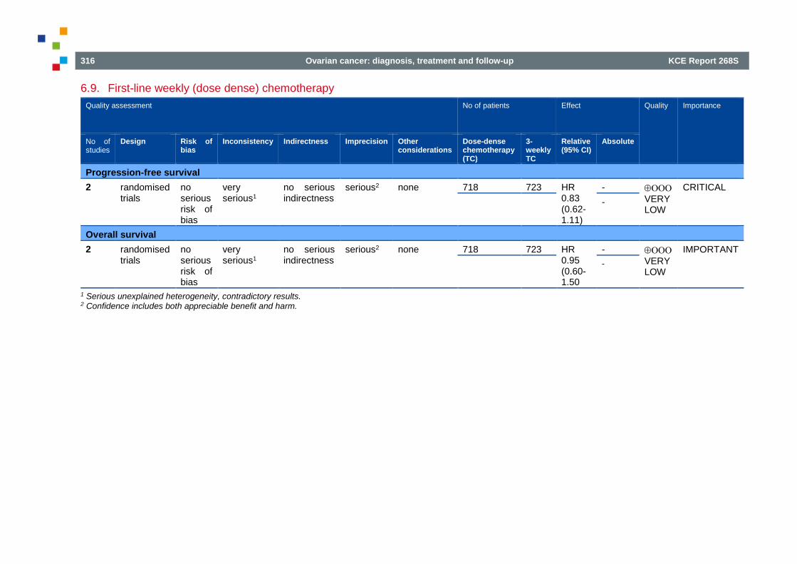
316 Ovarian cancer: diagnosis, treatment and follow-up KCE Report 268S
6.9. First-line weekly (dose dense) chemotherapy Quality assessment No of patients Effect Quality Importance
No of studies
Design Risk of bias
Inconsistency Indirectness Imprecision Other considerations
Dose-dense chemotherapy (TC)
3-weekly TC
Relative (95% CI)
Absolute
Progression-free survival 2 randomised
trials no serious risk of bias
very serious1
no serious indirectness
serious2 none 718 723 HR 0.83 (0.62-1.11)
- ⊕ΟΟΟ VERY LOW
CRITICAL -
Overall survival 2 randomised
trials no serious risk of bias
very serious1
no serious indirectness
serious2 none 718 723 HR 0.95 (0.60-1.50
- ⊕ΟΟΟ VERY LOW
IMPORTANT -
1 Serious unexplained heterogeneity, contradictory results. 2 Confidence includes both appreciable benefit and harm.

KCE Report 268S Ovarian cancer: diagnosis, treatment and follow-up 317
6.10. Routine CA125 measurement during follow-up Table 108 – GRADE evidence profile: routine CA125 measurements during follow-up
Quality assessment No of patients Effect Quality Importance
No of studies
Design Risk of bias
Inconsistency Indirectness Imprecision Other considerations
Early treatment
Delayed treatment
Relative (95% CI)
Absolute
Overall survival (follow-up median 56.9 months) 1 randomised
trials no serious risk of bias
no serious inconsistency
no serious indirectness
very serious1
none 79/265 (29.8%)2
80/264 (30.3%)2
HR 0.98 (0.80 to 1.20)
- ⊕⊕ΟΟ LOW
Time to second-line chemotherapy (follow-up median 56.9 months) 1 randomised
trials no serious risk of bias
no serious inconsistency
no serious indirectness
no serious imprecision
none 11/265 (4.2%)3
31/264 (11.7%)3
HR 0.29 (0.24 to 0.35)
- ⊕⊕⊕⊕ HIGH
Time to third-line treatment or death (follow-up median 56.9 months) 1 randomised
trials no serious risk of bias
no serious inconsistency
no serious indirectness
no serious imprecision
none Median 12.5 months
Median 17.1 months
HR 0.69 (0.57 to 0.83)
- ⊕⊕⊕⊕ HIGH
Time to first deterioration in good global health score or death (follow-up median 56.9 months) 1 randomised
trials serious4 no serious
inconsistency no serious indirectness
no serious imprecision
none Median 3.2 months
Median 5.8 months
HR 0.71 (0.58 to 0.88)
- ⊕⊕⊕Ο MODERATE
1 CI includes benefit and harm. 2 Proportion survivors at data locking. 3 Proportion of patients not started with second-line chemotherapy at data locking. 4 Point estimates and 95% CIs (i.e. adjusted hazard ratios) did not tally with corresponding P values (calculated with Kaplan-Meier, thus unadjusted) for time to first deterioration in QoL score or death for many of the individual sub-scales of EORTCQLQ-C30 questionnaire (Table 4 in trial report). For example, for the emotional sub-scale in the functional QoL category the upper 95% CI was 1.02 and the P value was 0.02. Similarly, several sub-scale factors appeared to have a vastly decreased P value from what might be expected given the point and CI estimates. These discrepancies at least suggest prognostic imbalances between the treatment groups.

318 Ovarian cancer: diagnosis, treatment and follow-up KCE Report 268S
7. FOREST PLOTS 7.1. Intra-operative frozen section
Figure 33 – Paired forest plot of sensitivity and specificity of frozen section: malignant versus borderline or benign ovarian tumours
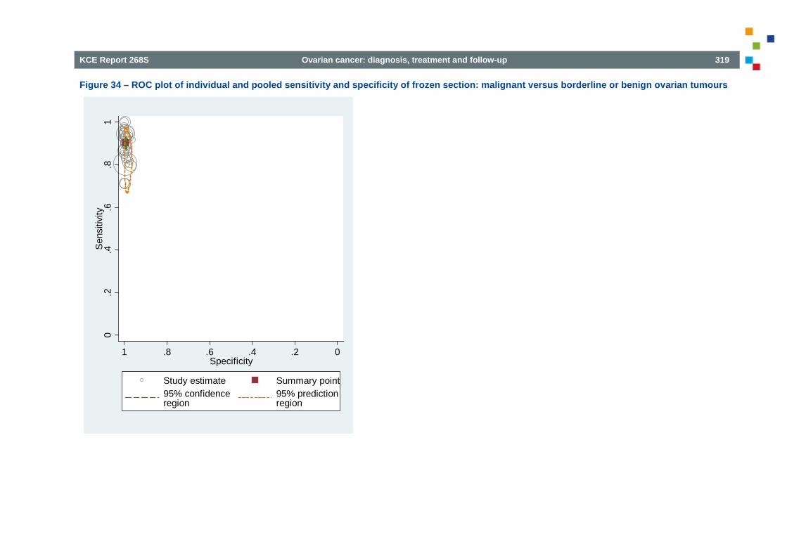
KCE Report 268S Ovarian cancer: diagnosis, treatment and follow-up 319
Figure 34 – ROC plot of individual and pooled sensitivity and specificity of frozen section: malignant versus borderline or benign ovarian tumours
0.2
.4.6
.81
Sen
sitiv
ity
0.2.4.6.81Specificity
Study estimate Summary point95% confidenceregion
95% predictionregion

320 Ovarian cancer: diagnosis, treatment and follow-up KCE Report 268S
Figure 35 – Paired forest plot of sensitivity and specificity of frozen section: malignant or borderline ovarian tumours versus benign tumours

KCE Report 268S Ovarian cancer: diagnosis, treatment and follow-up 321
Figure 36 – ROC plot of individual and pooled sensitivity and specificity of frozen section: malignant or borderline versus benign ovarian tumours
0.2
.4.6
.81
Sen
sitiv
ity
0.2.4.6.81Specificity
Study estimate Summary point95% confidenceregion
95% predictionregion
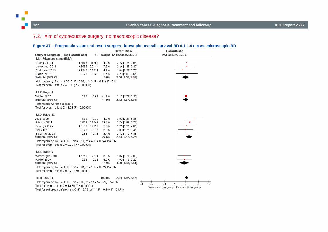
322 Ovarian cancer: diagnosis, treatment and follow-up KCE Report 268S
7.2. Aim of cytoreductive surgery: no macroscopic disease?
Figure 37 – Prognostic value end result surgery: forest plot overall survival RD 0.1-1.0 cm vs. microscopic RD
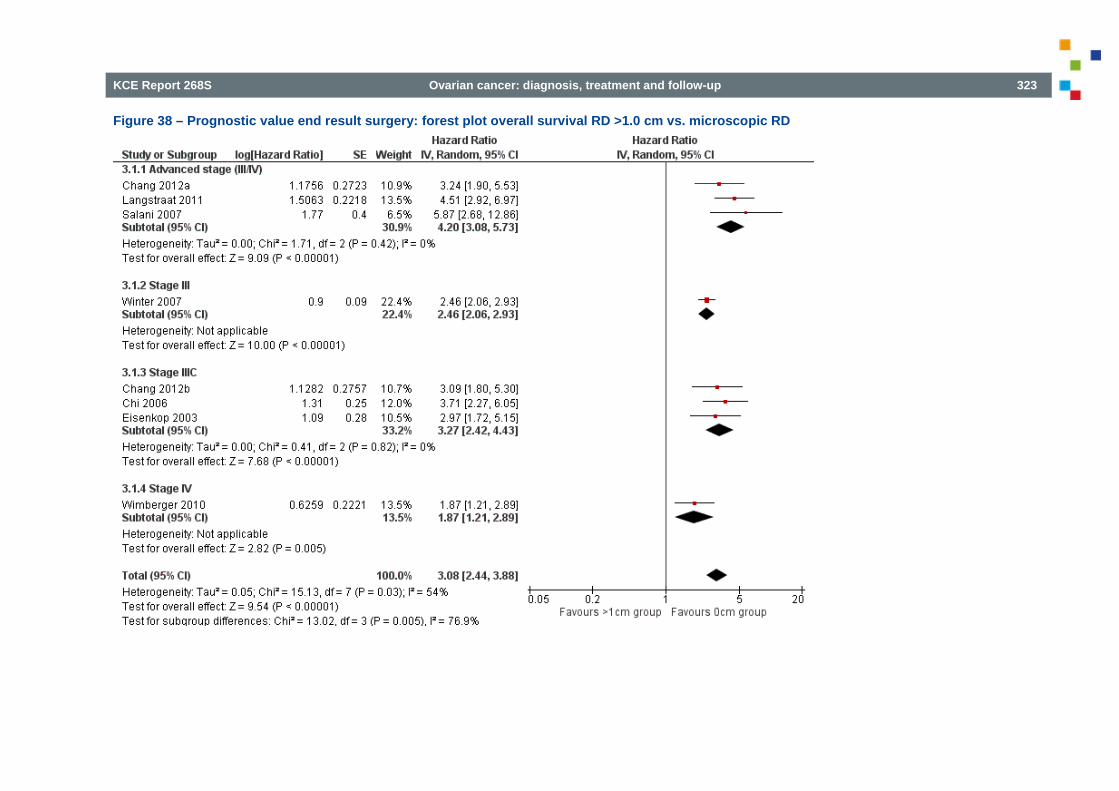
KCE Report 268S Ovarian cancer: diagnosis, treatment and follow-up 323
Figure 38 – Prognostic value end result surgery: forest plot overall survival RD >1.0 cm vs. microscopic RD

324 Ovarian cancer: diagnosis, treatment and follow-up KCE Report 268S
Figure 39 – Prognostic value end result surgery: forest plot overall survival RD >0 cm vs. microscopic RD (0 cm)

KCE Report 268S Ovarian cancer: diagnosis, treatment and follow-up 325
Figure 40 – Prognostic value end result surgery: forest plot progression-free survival RD 0.1-1.0 cm vs. microscopic RD

326 Ovarian cancer: diagnosis, treatment and follow-up KCE Report 268S
Figure 41 – Prognostic value end result surgery: forest plot progression-free survival RD >1.0 cm vs. microscopic RD
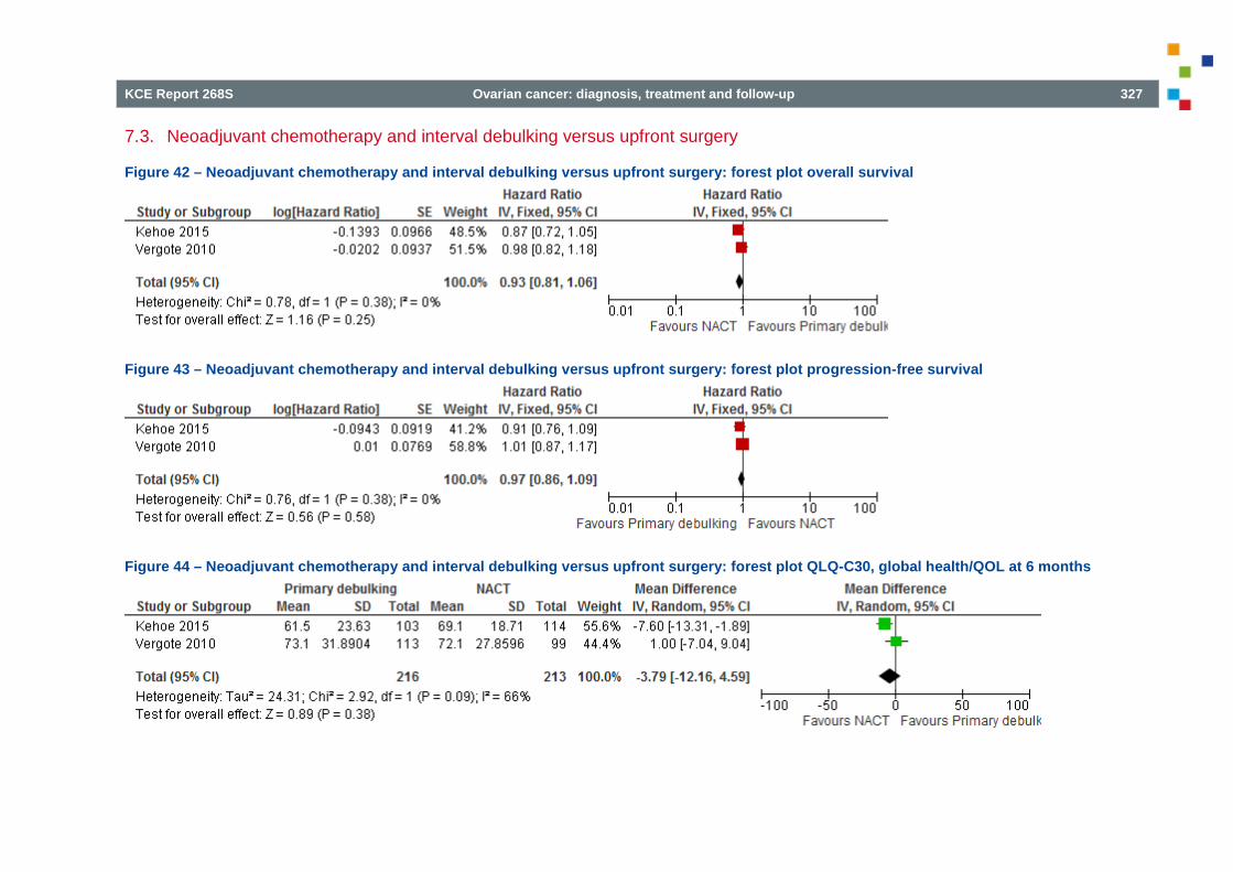
KCE Report 268S Ovarian cancer: diagnosis, treatment and follow-up 327
7.3. Neoadjuvant chemotherapy and interval debulking versus upfront surgery
Figure 42 – Neoadjuvant chemotherapy and interval debulking versus upfront surgery: forest plot overall survival
Figure 43 – Neoadjuvant chemotherapy and interval debulking versus upfront surgery: forest plot progression-free survival
Figure 44 – Neoadjuvant chemotherapy and interval debulking versus upfront surgery: forest plot QLQ-C30, global health/QOL at 6 months

328 Ovarian cancer: diagnosis, treatment and follow-up KCE Report 268S
Figure 45 – Neoadjuvant chemotherapy and interval debulking versus upfront surgery: forest plot QLQ-C30, global health/QOL at 12 months
Figure 46 – Neoadjuvant chemotherapy and interval debulking versus upfront surgery: forest plot postoperative death
Figure 47 – Neoadjuvant chemotherapy and interval debulking versus upfront surgery: forest plot postoperative haemorrhage grade 3-4
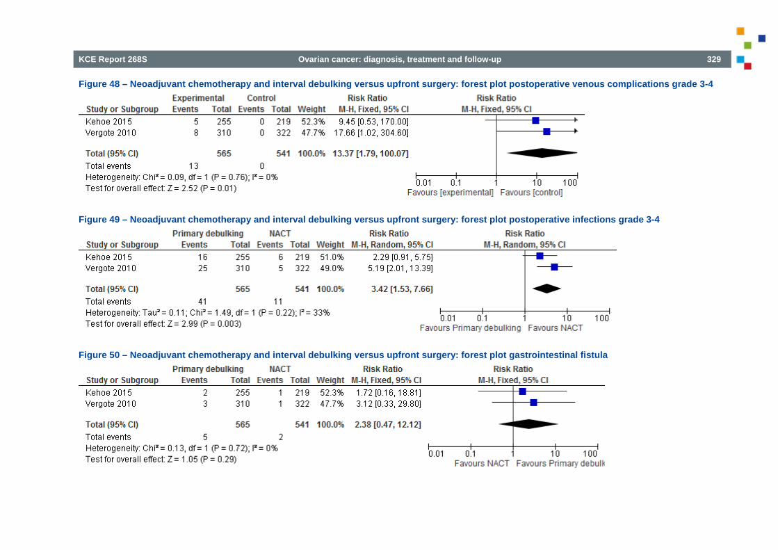
KCE Report 268S Ovarian cancer: diagnosis, treatment and follow-up 329
Figure 48 – Neoadjuvant chemotherapy and interval debulking versus upfront surgery: forest plot postoperative venous complications grade 3-4
Figure 49 – Neoadjuvant chemotherapy and interval debulking versus upfront surgery: forest plot postoperative infections grade 3-4
Figure 50 – Neoadjuvant chemotherapy and interval debulking versus upfront surgery: forest plot gastrointestinal fistula
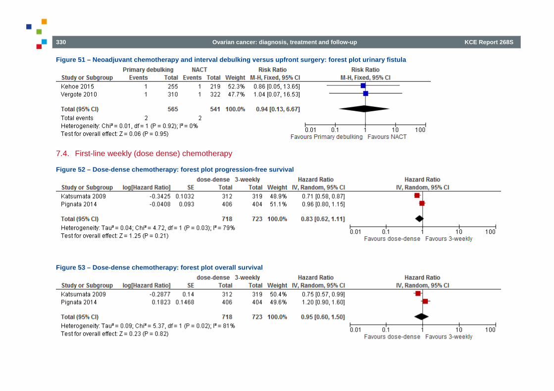
330 Ovarian cancer: diagnosis, treatment and follow-up KCE Report 268S
Figure 51 – Neoadjuvant chemotherapy and interval debulking versus upfront surgery: forest plot urinary fistula
7.4. First-line weekly (dose dense) chemotherapy
Figure 52 – Dose-dense chemotherapy: forest plot progression-free survival
Figure 53 – Dose-dense chemotherapy: forest plot overall survival

KCE Report 268S Ovarian cancer: diagnosis, treatment and follow-up 331
8. EXTERNAL REVIEW 8.1. Stakeholder review

332 Ovarian cancer: diagnosis, treatment and follow-up KCE Report 268S

KCE Report 268S Ovarian cancer: diagnosis, treatment and follow-up 333
REFERENCES 1. Kaijser J, Sayasneh A, Van Hoorde K, Ghaem-Maghami S, Bourne
T, Timmerman D, et al. Presurgical diagnosis of adnexal tumours using mathematical models and scoring systems: a systematic review and meta-analysis. Hum Reprod Update. 2014;20(3):449-62.
2. Chan KK, Selman TJ. Testing for ovarian cancer. Best Practice & Research in Clinical Obstetrics & Gynaecology. 2006;20:977-83.
3. Fung MF, Bryson P, Johnston M, Chambers A, Cancer Care Ontario Practice Guidelines Initiative G, Group. Screening postmenopausal women for ovarian cancer: a systematic review. Journal of Obstetrics & Gynaecology Canada: JOGC. 2004;26:717-28.
4. Geomini PM, Kruitwagen RF, Bremer GL, Massuger L, Mol BW. Should we centralise care for the patient suspected of having ovarian malignancy? Gynecologic Oncology. 2011;122:95-9.
5. Jacobs IJ, Skates SJ, MacDonald N, Menon U, Rosenthal AN, Davies AP, et al. Screening for ovarian cancer: a pilot randomised controlled trial. Lancet. 1999;353:1207-10.
6. Jeyarajah AR, Ind TE, MacDonald N, Skates S, Oram DH, Jacobs IJ. Increased mortality in postmenopausal women with serum CA125 elevation. Gynecologic Oncology. 1999;73:242-6.
7. Kobayashi H, Yamada Y, Sado T, Sakata M, Yoshida S, Kawaguchi R, et al. A randomized study of screening for ovarian cancer: a multicenter study in Japan. International Journal of Gynecological Cancer. 2008;18:414-20.
8. Lockwood S, Ritzert B. Cost-effectiveness of serum CA125 compared to transvaginal ultrasound as a screening test for ovarian cancer: A systematic review protocol. JBI Library of Systematic Reviews. 2013;11:89-106.
9. Menon U, Skates SJ, Lewis S, Rosenthal AN, Rufford B, Sibley K, et al. Prospective study using the risk of ovarian cancer algorithm to screen for ovarian cancer. Journal of Clinical Oncology. 2005;23:7919-26.

334 Ovarian cancer: diagnosis, treatment and follow-up KCE Report 268S
10. Menon U, Gentry-Maharaj A, Hallett R, Ryan A, Burnell M, Sharma A, et al. Sensitivity and specificity of multimodal and ultrasound screening for ovarian cancer, and stage distribution of detected cancers: results of the prevalence screen of the UK Collaborative Trial of Ovarian Cancer Screening (UKCTOCS). Lancet Oncology. 2009;10:327-40.
11. Myers ER, Bastian LA, Havrilesky LJ, Kulasingam SL, Terplan MS, Cline KE, et al. Management of adnexal mass. Evidence Report/Technology Assessment. 2006:1-145.
12. Nunes N, Foo X, Widschwendter M, Jurkovic D. A randomised controlled trial comparing surgical intervention rates between two protocols for the management of asymptomatic adnexal tumours in postmenopausal women. BMJ Open. 2012;2.
13. Ozols RF, Bookman MA, Connolly DC, Daly MB, Godwin AK, Schilder RJ, et al. Focus on epithelial ovarian cancer. Cancer Cell. 2004;5:19-24.
14. Sayasneh A, Preisler J, Stlader C, Husicka R, Naji O, Kaijser J, et al. A randomised controlled trial to compare the clinical impact of RMI versus LR2 to characterise adnexal masses: Interim analysis of phase 4 IOTA study. Bjog. 2013;120:357-8.
15. Timmerman D, Van C, Jurkovic D, Valentin L, Testa AC, Bernard JP, et al. Inclusion of CA-125 does not improve mathematical models developed to distinguish between benign and malignant adnexal tumors. Journal of Clinical Oncology. 2007;25:4194-200.
16. Van Calster, Timmerman D, Valentin L, McIndoe A, Ghaem-Maghami S, Testa AC, et al. Triaging women with ovarian masses for surgery: observational diagnostic study to compare RCOG guidelines with an International Ovarian Tumour Analysis (IOTA) group protocol. BJOG: An International Journal of Obstetrics & Gynaecology. 2012;119:662-71.
17. Abbasi F, Naji S, Yekta Z, Ayatollahi H, Khalifezadeh B. The accuracy of intraoperative aspiration cytology in diagnosis of ovarian tumors. Pakistan Journal of Medical Sciences. 2011;27:591-4.
18. Aslam MF, Ghayoori R, Khulpateea N. Adnexal masses: Relative accuracy of sonography and frozen section in predicting final pathology. Journal of Obstetrics and Gynaecology. 2010;30:187-9.
19. Basaran D, Salman MC, Calis P, Ozek A, Ozgul N, Usubutun A, et al. Diagnostic accuracy of intraoperative consultation (frozen section) in borderline ovarian tumours and factors associated with misdiagnosis. Journal of Obstetrics and Gynaecology. 2014;34:429-34.
20. Gultekin E, Gultekin OE, Cingillioglu B, Sayhan S, Sanci M, Yildirim Y. The value of frozen section evaluation in the management of borderline ovarian tumors. Journal of Cancer Research and Therapeutics. 2011;7:416-20.
21. Ouladsahebmadarek E, Tabrizi AD, Sayyah-Melli M, Jafari-Shobeiri M, Mostafa-Garabaghi P, Nazari F. Comparison of intra operative cytology and frozen section with permanent pathologic results in ovarian masses. International Journal of Women's Health and Reproduction Sciences. 2015;3:99-102.
22. Pongsuvareeyakul T, Khunamornpong S, Settakorn J, Sukpan K, Suprasert P, Siriaunkgul S. Accuracy of frozen-section diagnosis of ovarian mucinous tumors. International Journal of Gynecological Cancer. 2012;22:400-6.
23. Stewart CJ, Brennan BA, Koay E, Naran A, Ruba S. Value of cytology in the intraoperative assessment of ovarian tumors: a review of 402 cases and comparison with frozen section diagnosis. Cancer cytopathology. 2010;118:127-36.
24. Ureyen I, Turan T, Cirik DA, Tasci T, Boran N, Bulbul D, et al. Frozen section in borderline ovarian tumors: Is it reliable? European Journal of Obstetrics Gynecology and Reproductive Biology. 2014;181:115-8.
25. Covens AL, Dodge JE, Lacchetti C, Elit LM, Le T, Devries-Aboud M, et al. Surgical management of a suspicious adnexal mass: a systematic review. Gynecologic Oncology. 2012;126(1):149-56.
26. Geomini P, Bremer G, Kruitwagen R, Mol BW. Diagnostic accuracy of frozen section diagnosis of the adnexal mass: a metaanalysis. Gynecologic Oncology. 2005;96(1):1-9.
KCE Report 268S Ovarian cancer: diagnosis, treatment and follow-up 335
27. Medeiros LR, Rosa DD, Edelweiss MI, Stein AT, Bozzetti MC, Zelmanowicz A, et al. Accuracy of frozen-section analysis in the diagnosis of ovarian tumors: a systematic quantitative review. International Journal of Gynecological Cancer. 2005;15(2):192-202.
28. Heatley MK. A systematic review of papers examining the use of intraoperative frozen section in predicting the final diagnosis of ovarian lesions. International Journal of Gynecological Pathology. 2012;31(2):111-5.
29. Atienza-Amores M, Guerini-Rocco E, Soslow RA, Park KJ, Weigelt B. Small cell carcinoma of the gynecologic tract: A multifaceted spectrum of lesions. Gynecologic Oncology. 2014;134:410-8.
30. Bois AD, Ewald-Riegler N, Bois OD, Harter P. Borderline tumors of the ovary - A systematic review. [German]. Geburtshilfe und Frauenheilkunde. 2009;69:807-33.
31. Dodge JE, Covens AL, Lacchetti C, Elit LM, Le T, Devries-Aboud M, et al. Management of a suspicious adnexal mass: a clinical practice guideline. Current Oncology. 2012;19:e244-57.
32. Faluyi O, Mackean M, Gourley C, Bryant A, Dickinson HO. Interventions for the treatment of borderline ovarian tumours. [Review] [54 refs]. Cochrane Database of Systematic Reviews. 2010:CD007696.
33. Gao J, Yang X, Zhang Y. Systematic lymphadenectomy in the treatment of epithelial ovarian cancer: A meta-analysis of multiple epidemiology studies. Japanese Journal of Clinical Oncology. 2015;45:49-60.
34. Gurumurthy M, Bryant A, Shanbhag S. Effectiveness of different treatment modalities for the management of adult-onset granulosa cell tumours of the ovary (primary and recurrent). Cochrane Database of Systematic Reviews. 2014.
35. Hackethal A, Brueggmann D, Bohlmann MK, Franke FE, Tinneberg HR, Munstedt K. Squamous-cell carcinoma in mature cystic teratoma of the ovary: systematic review and analysis of published data. The Lancet Oncology. 2008;9:1173-80.
36. Haritwal A, Makhija B, Arora M, Agrawal D, Goyal P. Management dilemmas in borderline ovarian tumor. International Journal of Women's Health and Reproduction Sciences. 2014;2:219-24.
37. Kim HS, Ju W, Jee BC, Kim YB, Park NH, Song YS, et al. Systematic lymphadenectomy for survival in epithelial ovarian cancer: a meta-analysis (Structured abstract). International Journal of Gynecological Cancer. 2010;20:520-8.
38. Kleppe M, Amkreutz LCM, Van G, Slangen BFM, Kruse AJ, Kruitwagen RFPM. Lymph-node metastasis in stage i and II sex cord stromal and malignant germ cell tumours of the ovary: A systematic review. Gynecologic Oncology. 2014;133:124-7.
39. Kuhn W, Janicke F, Holscher M, Schule G, Pache L, Graeff H. [Surgical treatment of ovarian cancer: comparison of primary and secondary tumor reduction interventions]. Gynakologische Rundschau. 1991;31 Suppl 2:276-80.
40. Lecuru F, Taurelle R. Transperitoneal laparoscopic pelvic lymphadenectomy for gynecologic malignancies (II). Indications. Surgical endoscopy. 1998;12:97-100.
41. Munstedt K, Franke FE. Role of primary surgery in advanced ovarian cancer. World Journal of Surgical Oncology. 2004;2.
42. Papadia A, Remorgida V, Salom EM, Ragni N. Laparoscopic pelvic and paraaortic lymphadenectomy in gynecologic oncology. Journal of the American Association of Gynecologic Laparoscopists. 2004;11:297-306.
43. Qin JC, Yang ZJ, Xiong L, Li L. Systematic lymphadenectomy for overall survival in epithelial ovarian cancer: A meta-analysis. [Chinese]. Chinese journal of evidence-based medicine. 2012;12(2):224-30.
44. Roy WJ, Rutherford TJ, Schwartz PE. Current status of intraperitoneal chemotherapy in management of epithelial ovarian carcinoma and the role of lymphadenectomy. CME Journal of Gynecologic Oncology. 1998;3:107-16.
45. Shyamala G. Conservative approach in gynecologic malignancies. Journal of Gynecologic Surgery. 2010;26:151-61.

336 Ovarian cancer: diagnosis, treatment and follow-up KCE Report 268S
46. Smits A, Bryant A, Lopes A, Galaal K. Lymph node dissection (lymphadenectomy) for presumed early stage epithelial ovarian cancer. Cochrane Database of Systematic Reviews. 2015.
47. Akbayir O, Corbacioglu A, Goksedef BP, Numanoglu C, Akca A, Guraslan H, et al. The novel criteria for predicting pelvic lymph node metastasis in endometrioid adenocarcinoma of endometrium. Gynecologic Oncology. 2012;125:400-3.
48. AlHilli MM, Podratz KC, Dowdy SC, Bakkum-Gamez JN, Weaver AL, McGree ME, et al. Risk-scoring system for the individualized prediction of lymphatic dissemination in patients with endometrioid endometrial cancer. Gynecologic Oncology. 2013;131:103-8.
49. Aoki D. Laparoscopic surgery for early ovarian cancer. Journal of Gynecologic Oncology. 2014;25:168-9.
50. Ataseven B, Grimm C, Harter P, Prader S, Traut A, Heitz F, et al. Prognostic value of lymph node ratio in patients with advanced epithelial ovarian cancer. Gynecologic Oncology. 2014;135:435-40.
51. Azuar AS, Bourdel N, Ferrarrese G, Dauplat J, Mage G, Canis M. Laparoscopic restaging of borderline ovarian tumours (BLOT): A retrospective study of 142 cases. European Journal of Obstetrics Gynecology and Reproductive Biology. 2013;168:87-91.
52. Bachmann C, Kramer B, Brucker SY, Stabler A, Fend F, Wallwiener D, et al. Relevance of pelvic and para-aortic node metastases in early-stage ovarian cancer. Anticancer Research. 2014;34:6735-8.
53. Bae JW, Lee JH, Choi JS, Son CE, Jeon SW, Hong JH, et al. Laparoscopic lymphadenectomy for gynecologic malignancies: Evaluation of the surgical approach and outcomes over a sevenyear experience. Archives of Gynecology and Obstetrics. 2012;285:823-9.
54. Bennett JA, Dong F, Young RH, Oliva E. Clear cell carcinoma of the ovary: Evaluation of prognostic parameters based on a clinicopathological analysis of 100 cases. Histopathology. 2015;66:808-15.
55. Berretta R, Patrelli TS, Migliavacca C, Rolla M, Franchi L, Monica M, et al. Assessment of tumor size as a useful marker for the surgical staging of endometrial cancer. Oncology Reports. 2014;31:2407-12.
56. Boren T, Lea J, Kehoe S, Miller DS, Richardson D. Lymph node metastasis in endometrioid adenocarcinomas of the uterine corpus with occult cervical involvement. Gynecologic Oncology. 2012;127:43-6.
57. Bostanci E, Ayas S, Boza A, Gurbuz A, Karateke A. Retrospective analysis of 53 cases with borderline ovarian tumors. [Turkish]. Medical Journal of Bakirkoy. 2014;10:27-32.
58. Braicu EI, Fotopoulou C, Chekerov R, Richter R, Blohmer J, Kummel S, et al. Role of serum concentration of VEGFR1 and TIMP2 on clinical outcome in primary cervical cancer: Results of a companion protocol of the randomized, NOGGO-AGO phase III adjuvant trial of simultaneous cisplatin-based radiochemotherapy vs. carboplatin and paclitaxel containing sequential radiotherapy. Cytokine. 2013;61:755-8.
59. Brown JV, Mendivil AA, Abaid LN, Rettenmaier MA, Micha JP, Wabe MA, et al. The safety and feasibility of robotic-assisted lymph node staging in early-stage ovarian cancer. International Journal of Gynecological Cancer. 2014;24:1493-8.
60. Capilna ME, Szabo B, Toma A, Rusu SC, Birciu S, Puiac CI. Paraaortic lymphadenectomy in oncologic gynecology: Our experience on 62 patients. Gineco.ro. 2011;7:94-9.
61. Chang SJ, Bristow RE, Ryu HS. Analysis of para-aortic lymphadenectomy up to the level of the renal vessels in apparent early-stage ovarian cancer. Journal of Gynecologic Oncology. 2013;24:29-36.
62. Chang JS, Koom WS, Kim SW, Kim S, Kim YB, Kim YT, et al. Risk stratification of abdominopelvic failure for FIGO stage III epithelial ovarian cancer patients: Implications for adjuvant radiotherapy. Journal of Gynecologic Oncology. 2013;24:146-53.
KCE Report 268S Ovarian cancer: diagnosis, treatment and follow-up 337
63. Chatchotikawong U, Ruengkhachorn I, Leelaphatanadit C, Phithakwatchara N. 8-year analysis of the prevalence of lymph nodes metastasis, oncologic and pregnancy outcomes in apparent early-stage malignant ovarian germ cell tumors. Asian Pacific Journal of Cancer Prevention: Apjcp. 2015;16:1609-13.
64. Chereau E, Ballester M, Selle F, Rouzier R, Darai E. Clues for paraaortic lymphadenectomy in patients older than 70 years with ovarian cancer. [French]. Gynecologie Obstetrique Fertilite. 2012;40:327-9.
65. Cusido M, Fargas F, Rodriguez I, Alsina A, Baulies S, Tresserra F, et al. Role of lymphadenectomy in endometrioid endometrial cancer.[Erratum appears in Eur J Gynaecol Oncol. 2011;32(3):252 Note: Martinez, A [corrected to Pascual Martinez, A]]. European Journal of Gynaecological Oncology. 2011;32:49-53.
66. Dell A, Signorelli M, Benedetti-Panici P, Maggioni A, Fossati R, Fruscio R, et al. Systematic lymphadenectomy in ovarian cancer at second-look surgery: A randomised clinical trial. British Journal of Cancer. 2012;107:785-92.
67. Ditto A, Martinelli F, Lorusso D, Haeusler E, Carcangiu M, Raspagliesi F. Fertility sparing surgery in early stage epithelial ovarian cancer. Journal of Gynecologic Oncology. 2014;25:320-7.
68. Eltabbakh GH, Mount SL. Mullerian and mesothelial lymph node involvement in patients with borderline ovarian tumors. Journal of Reproductive Medicine. 2011;56:211-8.
69. Ertas IE, Gungorduk K, Taskin S, Akman L, Ozdemir A, Goklu R, et al. Prognostic predictors and spread patterns in adult ovarian granulosa cell tumors: a multicenter long-term follow-up study of 108 patients. International Journal of Clinical Oncology. 2014;19:912-20.
70. Ertas IE, Taskin S, Goklu R, Bilgin M, Goc G, Yildirim Y, et al. Long-term oncological and reproductive outcomes of fertility-sparing cytoreductive surgery in females aged 25 years and younger with malignant ovarian germ cell tumors. Journal of Obstetrics and Gynaecology Research. 2014;40:797-805.
71. Euscher ED, Bassett R, Malpica A. Lymph node counts in endometrial cancer: expectations versus reality. American Journal of Surgical Pathology. 2011;35:913-8.
72. Gmyrek LJ, Jonska-Gmyrek J, Sobiczewski P, Panek G, Bidzinski M. Evaluation of intraoperative and postoperative complications related to lymphadenectomy in ovarian cancer patients. Oncology Letters. 2011;2:537-41.
73. Grabowski JP, Harter P, Buhrmann C, Lorenz D, Hils R, Kommoss S, et al. Re-operation outcome in patients referred to a gynecologic oncology center with presumed ovarian cancer FIGO I-IIIA after sub-standard initial surgery. Surgical Oncology. 2012;21:31-5.
74. Guvenal T, Dursun P, Hasdemir PS, Hanhan M, Guven S, Yetimalar H, et al. Effect of surgical staging on 539 patients with borderline ovarian tumors: A Turkish Gynecologic Oncology Group study. Gynecologic Oncology. 2013;131:546-50.
75. Gyimadu A, Salman MC, Karcaaltincaba M, Yuce K. Retroperitoneal vascular aberrations increase the risk of vascular injury during lymphadenectomy in gynecologic cancers. Archives of Gynecology and Obstetrics. 2012;286:449-55.
76. Haller H, Mamula O, Krasevic M, Rupcic S, Fischer BA, Eminovic S, et al. Frequency and distribution of lymph node metastases in epithelial ovarian cancer: Significance of serous histology. International Journal of Gynecological Cancer. 2011;21:245-50.
77. Han D, Wu X, Li J, Ke G. Postoperative chylous ascites in patients with gynecologic malignancies. International Journal of Gynecological Cancer. 2012;22:186-90.
78. Hareyama H, Ito K, Watanabe S, Hakoyama M, Uchida A, Oku K, et al. Factors influencing the number of pelvic and para-aortic lymph nodes removed in surgical treatment of endometrial and ovarian cancer. International Journal of Gynecological Cancer. 2012;22:1577-84.
79. Hareyama H, Hada K, Goto K, Watanabe S, Hakoyama M, Oku K, et al. Prevalence, classification, and risk factors for postoperative lower extremity lymphedema in women with gynecologic malignancies. International Journal of Gynecological Cancer. 2015;25:751-7.

338 Ovarian cancer: diagnosis, treatment and follow-up KCE Report 268S
80. Hu T, Wu L, Xing H, Yang R, Li X, Huang K, et al. Development of criteria for ovarian preservation in cervical cancer patients treated with radical surgery with or without neoadjuvant chemotherapy: A multicenter retrospective study and meta-analysis. Annals of Surgical Oncology. 2013;20:881-90.
81. Ibeanu OA, Diaz-Montes TP. Outcomes in Ovarian Cancer among Hispanic Women Living in the United States: A Population-Based Analysis. Pathology Research International. 2013;2013:672710.
82. Kanat-Pektas M, Ozat M, Gungor T, Sahin I, Yalcin H, Ozdal B. Complete lymph node dissection: Is it essential for the treatment of borderline epithelial ovarian tumors? Archives of Gynecology and Obstetrics. 2011;283:879-84.
83. Karalok A, Tasci T, Ureyen I, Turkmen O, Ocalan R, Sahin G, et al. Juvenile granulosa cell ovarian tumor: Clinicopathological evaluation of ten patients. Journal of the Turkish German Gynecology Association. 2015;16:32-4.
84. Kiran G, Canaz E, Serin S. Surgical experience affects harvested lymph-node count but not detection rate of metastatic nodes during endometrial and ovarian cancer surgery. Journal of Gynecologic Surgery. 2015;31:1-7.
85. Lambaudie E, Narducci F, Leblanc E, Bannier M, Jauffret C, Cannone F, et al. Robotically assisted laparoscopy for paraaortic lymphadenectomy: Technical description and results of an initial experience. Surgical Endoscopy and Other Interventional Techniques. 2012;26:2430-5.
86. Lazarou A, Fotopoulou C, Coumbos A, Sehouli J, Vasiljeva J, Braicu I, et al. Long-term follow-up of borderline ovarian tumors clinical outcome and prognostic factors. Anticancer Research. 2014;34:6725-30.
87. Li X, Xing H, Li L, Huang Y, Zhou M, Liu Q, et al. Clinical significance of para-aortic lymph node dissection and prognosis in ovarian cancer. Fronteras en medicina. 2014;8(1):96-100.
88. Magazzino F, Katsaros D, Ottaiano A, Gadducci A, Pisano C, Sorio R, et al. Surgical and medical treatment of clear cell ovarian cancer, results from the multicenter italian trials in ovarian cancer (MITO) 9 retrospective study. International Journal of Gynecological Cancer. 2011;21:1063-70.
89. Mahdi H, Swensen RE, Hanna R, Kumar S, Ali-Fehmi R, Semaan A, et al. Prognostic impact of lymphadenectomy in clinically early stage malignant germ cell tumour of the ovary. British Journal of Cancer. 2011;105:493-7.
90. Mahdi H, Thrall M, Kumar S, Hanna R, Seward S, Lockhart D, et al. The prognostic impact of the ratio of positive lymph nodes on survival of epithelial ovarian cancer patients. Journal of Surgical Oncology. 2011;103:724-9.
91. Mahdi H, Moslemi-Kebria M, Levinson KL, Gojayev A, Lockhart D, Ali-Fehmi R, et al. Prevalence and prognostic impact of lymphadenectomy and lymph node metastasis in clinically early-stage ovarian clear cell carcinoma. International Journal of Gynecological Cancer. 2013;23:1226-30.
92. Marpeau L. Para-aortic lymphadenectomy for ovarian cancer in women over 70: Pros and cons. [French]. Gynecologie Obstetrique Fertilite. 2012;40:326.
93. Matsuo K, Sheridan TB, Yoshino K, Miyake T, Hew KE, Im DD, et al. Significance of lymphovascular space invasion in epithelial ovarian cancer. Cancer Medicine. 2012;1:156-64.
94. Matsuo K, Yoshino K, Hiramatsu K, Banzai C, Hasegawa K, Yasuda M, et al. Effect of lymphovascular space invasion on survival of stage i epithelial ovarian cancer. Obstetrics and Gynecology. 2014;123:957-65.
95. Matsuo K, Sheridan TB, Mabuchi S, Yoshino K, Hasegawa K, Studeman KD, et al. Estrogen receptor expression and increased risk of lymphovascular space invasion in high-grade serous ovarian carcinoma. Gynecologic Oncology. 2014;133:473-9.
96. Matsuo K, Yoshino K, Hasegawa K, Murakami R, Ikeda Y, Adachi S, et al. Survival outcome of stage I ovarian clear cell carcinoma with lympho-vascular space invasion. Gynecologic Oncology. 2015;136:198-204.
KCE Report 268S Ovarian cancer: diagnosis, treatment and follow-up 339
97. Mbarki C, Mrad M, Gharbi I, Ben J, Hsayaoui N, Jnifene A, et al. Borderline tumors of the ovary: A retrospective study of 15 cases. [French]. Journal Africain du Cancer. 2011;3:239-44.
98. McNally L, Teng NNH, Kapp DS, Karam A. Does omentectomy in epithelial ovarian cancer affect survival? An analysis of the surveillance, epidemiology, and end results database. International Journal of Gynecological Cancer. 2015;25:607-15.
99. Meinhold-Heerlein I. Borderline ovarian tumors. [German]. Gynakologe. 2014;47:951-60.
100. Miyamoto M, Takano M, Goto T, Shibutani T, Matsuura H, Soyama H, et al. The impact of systematic lymphadenectomy for early-stage ovarian carcinomas. [Japanese]. Japanese Journal of Cancer and Chemotherapy. 2011;38:1837-40.
101. Miyamoto M, Takano M, Goto T, Kato M, Sasaki N, Tsuda H, et al. Clear cell histology as a poor prognostic factor for advanced epithelial ovarian cancer: A single institutional case series through central pathologic review. Journal of Gynecologic Oncology. 2012;24:37-43.
102. Momeni M, Kolev V, Cardenas-Goicoechea J, Getrajdman J, Fishman D, Chuang L, et al. Does the type of surgery for early-stage endometrial cancer affect the rate of reported lymphovascular space invasion in final pathology specimens? American Journal of Obstetrics & Gynecology. 2013;208:71.e1-6.
103. Moore RG, Miller CM, Brown AK, Robison K, Steinhoff M, Lambert-Messerlian G. Utility of tumor marker HE4 to predict depth of myometrial invasion in endometrioid adenocarcinoma of the uterus. International Journal of Gynecological Cancer. 2011;21:1185-90.
104. Morales S, Zapardiel I, Grabowski JP, Hernandez A, Diestro MD, Gonzalez-Benitez C, et al. Surgical Outcome of Extraperitoneal Paraaortic Lymph Node Dissections Compared With Transperitoneal Approach in Gynecologic Cancer Patients. Journal of Minimally Invasive Gynecology. 2013;20:611-5.
105. Moreau-Claeys MV, Brunaud C, Hoffstetter S, Buchheit I, Peiffert D. [High dose rate vaginal brachytherapy in endometrial cancer after surgery]. Cancer Radiotherapie. 2011;15:169-75.
106. Mury D, Woelber L, Jung S, Eulenburg C, Choschzick M, Witzel I, et al. Prognostic and predictive relevance of CA-125 at primary surgery of ovarian cancer. Journal of cancer research and clinical oncology. 2011;137(7):1131-7.
107. Oh J, Park SH, Lee TS, Oh HK, Choi JH, Choi YS. High expression of epidermal growth factor-like domain 7 is correlated with poor differentiation and poor prognosis in patients with epithelial ovarian cancer. Journal of Gynecologic Oncology. 2014;25:334-41.
108. Ohba Y, Todo Y, Akashi D, Kawaguchi S, Minobe S, Okamoto K, et al. Incidence of metastasis in the circumflex iliac nodes distal to the external iliac nodes in ovarian cancer. Gynecologic Oncology. 2012;126:387-90.
109. Ozalp SS, Yalcin OT, Telli E, Oge T, Kabukcuoglu S. Borderline ovarian tumors: outcomes of fertility sparing surgery. European Journal of Gynaecological Oncology. 2014;35:154-6.
110. Pan XY, Lin H, Wang YN, Zhao YW, Cheng JY, Sun AP. Feasibility of laparoscopic extraperitoneal pelvic lymphadenectomy in gynecologic malignancies. Gynecologic Oncology. 2011;122:281-4.
111. Park JY, Kim DY, Kim JH, Kim YM, Kim KR, Kim YT, et al. Micropapillary pattern in serous borderline ovarian tumors: Does it matter? Gynecologic Oncology. 2011;123:511-6.
112. Park HJ, Nam EJ, Rha SY, Kim S, Kim SW, Kim JW, et al. A new prognostic index model using meta-analysis in early-stage epithelial ovarian cancer. Gynecologic Oncology. 2012;126:357-63.
113. Park JY, Jin KL, Kim DY, Kim JH, Kim YM, Kim KR, et al. Surgical staging and adjuvant chemotherapy in the management of patients with adult granulosa cell tumors of the ovary. Gynecologic Oncology. 2012;125:80-6.
114. Paulsson G, Andersson S, Sorbe B. A population-based series of ovarian carcinosarcomas with long-term follow-up. Anticancer Research. 2013;33:1003-8.
340 Ovarian cancer: diagnosis, treatment and follow-up KCE Report 268S
115. Pereira A, Irishina N, Perez-Medina T, Magrina JF, Magtibay PM, Kovaleva A, et al. Defining the optimal lymphadenectomy cut-off value in epithelial ovarian cancer staging surgery utilizing a mathematical model of validation. European Journal of Surgical Oncology. 2013;39:290-6.
116. Pereira A, Perez-Medina T, Magrina JF, Magtibay PM, Rodriguez-Tapia A, De L, et al. Correlation between the extent of intraperitoneal disease and nodal metastasis in node-positive ovarian cancer patients. European Journal of Surgical Oncology. 2014;40:917-24.
117. Petry KU. Lymphadenectomy for pregnant women with stage I cervical cancer. Lancet. 2012;379:1949; author reply -50.
118. Rauh-Hain JA, Davis M, Clemmer J, Clark RM, Growdon WB, Goodman AK, et al. Prognostic determinants in patients with uterine and ovarian clear cell carcinoma: A SEER analysis. Gynecologic Oncology. 2013;131:404-9.
119. Rauh-Hain JA, Diver EJ, Clemmer JT, Bradford LS, Clark RM, Growdon WB, et al. Carcinosarcoma of the ovary compared to papillary serous ovarian carcinoma: A SEER analysis. Gynecologic Oncology. 2013;131:46-51.
120. Rauh-Hain JA, Foley OW, Winograd D, Andrade C, Clark RM, Vargas RJ, et al. Clinical characteristics and outcomes of patients with stage i epithelial ovarian cancer compared with fallopian tube cancer. American Journal of Obstetrics and Gynecology. 2015;212:600.e1-.e8.
121. Rizzuto I, Stavraka C, Chatterjee J, Borley J, Hopkins TG, Gabra H, et al. Risk of ovarian cancer relapse score: A prognostic algorithm to predict relapse following treatment for advanced ovarian cancer. International Journal of Gynecological Cancer. 2015;25:416-22.
122. Romeo M, Pons F, Barretina P, Radua J. Incomplete staging surgery as a major predictor of relapse of borderline ovarian tumor. World Journal of Surgical Oncology. 2013;11.
123. Ruskin R, Urban RR, Sherman AE, Chen LL, Powell CB, Burkhardt Iii DH, et al. Predictors of intensive care unit utilization in gynecologic oncology surgery. International Journal of Gynecological Cancer. 2011;21:1336-42.
124. Satoh T, Tsuda H, Kanato K, Nakamura K, Shibata T, Takano M, et al. A non-randomized confirmatory study regarding selection of fertility-sparing surgery for patients with epithelial ovarian cancer: Japan Clinical Oncology Group Study (JCOG1203). Japanese Journal of Clinical Oncology. 2015;45:595-9.
125. Schnack TH, Sorensen RD, Nedergaard L, Hogdall C. Demographic clinical and prognostic characteristics of primary ovarian, peritoneal and tubal adenocarcinomas of serous histology - A prospective comparative study. Gynecologic Oncology. 2014;135:278-84.
126. Schreuder HWR, Pattij TOS, Zweemer RP, Van B, M W, Verheijen RHM. Increasing experience in laparoscopic staging of early ovarian cancer. Gynecological Surgery. 2012;9:89-96.
127. Shih KK, Zhou Q, Huh J, Morgan JC, Iasonos A, Aghajanian C, et al. Risk factors for recurrence of ovarian borderline tumors. Gynecologic Oncology. 2011;120:480-4.
128. Shim SH, Kim DY, Lee SW, Park JY, Kim JH, Kim YM, et al. Laparoscopic management of early-stage malignant nonepithelial ovarian tumors surgical and survival outcomes. International Journal of Gynecological Cancer. 2013;23:249-55.
129. Signorelli M, Guerra L, Pirovano C, Crivellaro C, Fruscio R, Buda A, et al. Detection of nodal metastases by 18F-FDG PET/CT in apparent early stage ovarian cancer: A prospective study. Gynecologic Oncology. 2013;131:395-9.
130. Signorelli M, Fruscio R, Ceppi L, Dell'Anna T, Vitobello D, Chiappa V, et al. The role of pelvic and aortic lymphadenectomy at second look surgery in apparent early stage ovarian cancer after inadequate surgical staging followed by adjuvant chemotherapy. Gynecologic Oncology. 2014;132:312-5.
131. Song T, Lee YY, Choi CH, Kim TJ, Lee JW, Bae DS, et al. Risk factors for progression to invasive carcinoma in patients with borderline ovarian tumors. International Journal of Gynecological Cancer. 2014;24:1206-14.
132. Sornsukolrat S, Tuipae S. Prognostic factors and survival of borderline ovarian tumors in rajavithi hospital between 1979-2006 A.D. Journal of the Medical Association of Thailand. 2012;95:1141-8.

KCE Report 268S Ovarian cancer: diagnosis, treatment and follow-up 341
133. Takano M, Tsuda H, Sugiyama T. Clear cell carcinoma of the ovary: Is there a role of histology-specific treatment? Journal of Experimental and Clinical Cancer Research. 2012;31.
134. Terai Y, Sasaki H, Tsunetoh S, Tanaka Y, Yoo S, Fujiwara S, et al. Novel para-aortic lymphadenectomy technique for gynecological malignancies prevents postoperative bowel obstruction. Journal of Obstetrics and Gynaecology Research. 2013;39:849-54.
135. Thrall MM, Paley P, Pizer E, Garcia R, Goff BA. Patterns of spread and recurrence of sex cord-stromal tumors of the ovary. Gynecologic Oncology. 2011;122:242-5.
136. Trillsch F, Mahner S, Vettorazzi E, Woelber L, Reuss A, Baumann K, et al. Surgical staging and prognosis in serous borderline ovarian tumours (BOT): A subanalysis of the AGO ROBOT study. British Journal of Cancer. 2015;112:660-6.
137. Tsuboyama T, Tatsumi M, Onishi H, Nakamoto A, Kim T, Hori M, et al. Assessment of combination of contrast-enhanced magnetic resonance imaging and positron emission tomography/computed tomography for evaluation of ovarian masses. Investigative Radiology. 2014;49:524-31.
138. Tsuda N, Ushijima K, Kawano K, Takemoto S, Nishio S, Sonoda G, et al. Prevention of lymphocele development in gynecologic cancers by the electrothermal bipolar vessel sealing device. Journal of Gynecologic Oncology. 2014;25:229-35.
139. Ulanday KT, Ward KK, MacEra CA, Ji M, Plaxe SC. Regional variation in surgical assessment of lymph nodes for staging among women with early-stage epithelial ovarian cancer. Gynecologic Oncology. 2014;132:411-5.
140. Villet R. Lymphadenectomy in ovary cancer. [French]. Journal Medical Libanais. 2011;59:94-9.
141. Zanagnolo V, Rollo D, Tomaselli T, Rosenberg PG, Bocciolone L, Landoni F, et al. Robotic-assisted transperitoneal aortic lymphadenectomy as part of staging procedure for gynaecological malignancies: single institution experience. Obstetrics & Gynecology International. 2013;2013:931318.
142. Zhao Y, Hu W, Hou X, Zhou Q. Chylous Ascites After Laparoscopic Lymph Node Dissection in Gynecologic Malignancies. Journal of Minimally Invasive Gynecology. 2014;21:90-6.
143. Zikan M, Fischerova D, Pinkavova I, Slama J, Weinberger V, Dusek L, et al. A prospective study examining the incidence of asymptomatic and symptomatic lymphoceles following lymphadenectomy in patients with gynecological cancer. Gynecologic Oncology. 2015;137:291-8.
144. Kleppe M, Wang T, Van G, Slangen BFM, Kruse AJ, Kruitwagen RFPM. Lymph node metastasis in stages i and II ovarian cancer: A review. Gynecologic Oncology. 2011;123:610-4.
145. Winter-Roach BA, Kitchener Hc Fau - Lawrie TA, Lawrie TA. Adjuvant (post-surgery) chemotherapy for early stage epithelial ovarian cancer. Cochrane Database of Systematic Reviews. 2012(3):CD004706.
146. Joly F, Weber B, Pautier P, Fabbro M, Selle F, Krieger S, et al. Combined topotecan and lapatinib in patients with early recurrent ovarian or peritoneal cancer after first line of platinum-based chemotherapy: a French FEDEGYN-FNCLCC phase II trial [abstract]. In: M: Y; also available on www.asco.org; 2009. p. Abstract 5555.
147. Ledermann JA, Hackshaw A, Kaye S, Jayson G, Gabra H, McNeish I, et al. Randomized phase II placebo-controlled trial of maintenance therapy using the oral triple angiokinase inhibitor BIBF 1120 after chemotherapy for relapsed ovarian cancer. In: M; 2011. p. 3798-804.
148. Mannel RS, Brady MF, Kohn EC, et al. A randomized phase 111 trial of IV carboplatin and paclitaxel x 3 courses followed by observation versus weekly maintenance low-dose paclitaxel in patients with early-stage ovarian carcinoma: a Gynecologic Oncology Group Study. In: M; 2011. p. 89-94.
342 Ovarian cancer: diagnosis, treatment and follow-up KCE Report 268S
149. Meier W, du Bois A, Reuss A, Kuhn W, Olbricht S, Gropp M, et al. Topotecan versus treosulfan, an alkylating agent, in patients with epithelial ovarian cancer and relapse within 12 months following 1st-line platinum/paclitaxel chemotherapy. A prospectively randomized phase III trial by the Arbeitsgemeinschaft Gynaekologische Onkologie Ovarian Cancer Study Group (AGO-OVAR). In: M: N; 2009. p. 199-205.
150. Perren TJ, Swart AM, Pfisterer J, Ledermann JA, Pujade Lauraine E, Kristensen G, et al. A phase 3 trial of bevacizumab in ovarian cancer. In: M; 2011. p. 2484-96.
151. Trimbos B, Timmers P, Pecorelli S, Coens C, Ven K, van der Burg M, et al. Surgical staging and treatment of early ovarian cancer: long-term analysis from a randomized trial. In: M; 2010. p. 982-7.
152. Ceballos KM, Shaw D, Daya D. Microinvasive cervical adenocarcinoma (FIGO Stage 1A tumors): Results of surgical staging and outcome analysis. American Journal of Surgical Pathology. 2006;30:370-4.
153. Cusido M, Sas A, Treserras E, Grases P, Labastida R. Borderline epithelial tumor of the ovary: Study of 33 cases. [Spanish]. Progresos en Obstetricia y Ginecologia. 1999;42:447-53.
154. Damak T, Ben H, Chargui R, Gamoudi A, Hechiche M, Dhieb T, et al. Borderline tumors of the ovary. Tunisie Medicale. 2014;92:411-6.
155. Dgani R, Blickstein I, Shoham Z, Czernobilsky B, Ben-Hur H, Shani A, et al. Clinical aspects of ovarian tumors of low malignant potential. European Journal of Obstetrics Gynecology and Reproductive Biology. 1990;35:251-8.
156. Du B, Ewald-Riegler N, De G, Reuss A, Mahner S, Fotopoulou C, et al. Borderline tumours of the ovary: A cohort study of the Arbeitsgmeinschaft Gynakologische Onkologie (AGO) Study Group. European Journal of Cancer. 2013;49:1905-14.
157. Eltabbakh GH, Srivastava A, Garafano LL. Surgical response of advanced-stage borderline ovarian tumors to paclitaxel and cisplatin chemotherapy. Gynecologic Oncology. 2000;77:343-4.
158. Gilks CB, Alkushi A, Yue JJ, Lanvin D, Ehlen TG, Miller DM. Advanced-stage serous borderline tumors of the ovary: a clinicopathological study of 49 cases. International Journal of Gynecological Pathology. 2003;22:29-36.
159. Hogberg T, Glimelius B, Nygren P, Care SBUgSCoTA. A systematic overview of chemotherapy effects in ovarian cancer. Acta Oncologica. 2001;40:340-60.
160. Kaern J, Trope CG, Abeler VM. A retrospective study of 370 borderline tumors of the ovary treated at the Norwegian Radium Hospital from 1970 to 1982: A review of clinicopathologic features and treatment modalities. Cancer. 1993;71:1810-20.
161. Kaern J, Trope CG, Kristensen GB, Abeler VM, Pettersen EO. DNA ploidy; the most important prognostic factor in patients with borderline tumors of the ovary. International Journal of Gynecological Cancer. 1993;3:349-58.
162. Kane A, Uzan C, Rey A, Gouy S, Camatte S, Pautier P, et al. Prognostic factors in patients with ovarian serous low malignant potential (borderline) tumors with peritoneal implants. Oncologist. 2009;14:591-600.
163. Kennedy AW, Hart WR. Ovarian papillary serous tumors of low malignant potential (serous borderline tumors). A long-term follow-up study, including patients with microinvasion, lymph node metastasis, and transformation to invasive serous carcinoma. Cancer. 1996;78:278-86.
164. Laurent I, Uzan C, Gouy S, Pautier P, Duvillard P, Morice P. Results after conservative treatment of serous borderline tumors of the ovary with a micropapillary pattern. Annals of Surgical Oncology. 2008;15:3561-6.
165. Leary A, Petrella MC, Pautier P, Duvillard P, Uzan C, Tazi Y, et al. Adjuvant platinum-based chemotherapy for borderline serous ovarian tumors with invasive implants. Gynecologic Oncology. 2014;132:23-7.
166. Leiserowitz G. Surgical staging of ovarian low malignant potential tumors. Women's Oncology Review. 2005;5:29-30.

KCE Report 268S Ovarian cancer: diagnosis, treatment and follow-up 343
167. Leitao MM, Jr, Boyd J, Hummer A, Olvera N, Arroyo CD, et al. Clinicopathologic analysis of early-stage sporadic ovarian carcinoma. American Journal of Surgical Pathology. 2004;28:147-59.
168. Leitao MM. Micropapillary Pattern in Newly Diagnosed Borderline Tumors of the Ovary: What's in a Name? Oncologist. 2011;16:133-5.
169. Li Y, Cui H, Shen DH, Zhao Y, Wei LH, Qian HN. [Clinical and pathological features of borderline ovarian tumors]. Chung-Hua Fu Chan Ko Tsa Chih [Chinese Journal of Obstetrics & Gynecology]. 2003;38:81-4.
170. Li ZX, Qiu XF. [The treatment of borderline ovarian tumors and its prognosis]. Chung-Hua Fu Chan Ko Tsa Chih [Chinese Journal of Obstetrics & Gynecology]. 1994;29:207-9, 51.
171. Linasmita V, Sakonratanakul P, Bhamarapravati Y. Survival of patients with stage I epithelial ovarian carcinoma, 1981-1985 at Ramathibodi Hospital. Journal of the Medical Association of Thailand = Chotmaihet thangphaet. 1990;73 Suppl 1:7-10.
172. Manchul LA, Simm J, Levin W, Fyles AW, Dembo AJ, Pringle JF, et al. Borderline epithelial ovarian tumors: a review of 81 cases with an assessment of the impact of treatment. International Journal of Radiation Oncology, Biology, Physics. 1992;22:867-74.
173. Massad LS, Jr, Hunter VJ, Szpak CA, Clarke-Pearson DL, Creasman WT. Epithelial ovarian tumors of low malignant potential. Obstetrics & Gynecology. 1991;78:1027-32.
174. Morice P, Camatte S, Pautier P, Lhomme C, Duvillard P, Castaigne D. What is to be done with ovarian tumors of borderline malignancy?. [French]. Oncologie. 2001;3:211-6.
175. Raymond E, Drolet Y, Marpeau L, Louvet C, Cady J, Tessier C, et al. Long-term follow-up after adjuvant chemotherapy in completely resected early stage ovarian carcinoma. European Journal of Obstetrics, Gynecology, & Reproductive Biology. 1997;72:181-90.
176. Rettenmaier MA, Lopez K, Abaid LN, Brown JV, rd, Micha JP, et al. Borderline ovarian tumors and extended patient follow-up: an individual institution's experience. Journal of Surgical Oncology. 2010;101:18-21.
177. Shih KK, Zhou QC, Aghajanian C, Huh J, Soslow RA, Morgan JC, et al. Patterns of recurrence and role of adjuvant chemotherapy in stage II-IV serous ovarian borderline tumors. Gynecologic Oncology. 2010;119:270-3.
178. Trimble CL, Kosary C, Trimble EL. Long-term survival and patterns of care in women with ovarian tumors of low malignant potential. Gynecologic Oncology. 2002;86:34-7.
179. Tulpin L, Akerman G, Morel O, Desfeux P, Malartic C, Barranger E. [Management of borderline tumors of the ovary]. Journal de Gynecologie, Obstetrique et Biologie de la Reproduction. 2008;37 Spec No 2:F69-76.
180. Uzan C, Kane A, Rey A, Gouy S, Camatte S, Pautier P, et al. Prognosis and Prognostic Factors of the Micropapillary Pattern in Patients Treated for Stage II and III Serous Borderline Tumors of the Ovary. Oncologist. 2011;16:189-96.
181. Vasconcelos I, Olschewski J, Braicu I, Sehouli J. Limited efficacy of platinum-based adjuvant treatment on the outcome of borderline ovarian tumors. European Journal of Obstetrics Gynecology and Reproductive Biology. 2015;186:26-33.
182. Vasconcelos I, Olschewski J, Braicu I, Sehouli J. A meta-analysis on the impact of platinum-based adjuvant treatment on the outcome of borderline ovarian tumors with invasive implants. Oncologist. 2015;20:151-8.
183. Vergote IB, Vergote-De V, L N, Abeler VM, Aas M, Lindegaard MW, et al. Randomized trial comparing cisplatin with radioactive phosphorus or whole- abdomen irradiation as adjuvant treatment of ovarian cancer. Cancer. 1992;69:741-9.
184. Yokoyama Y, Moriya T, Takano T, Shoji T, Takahashi O, Nakahara K, et al. Clinical outcome and risk factors for recurrence in borderline ovarian tumours. British Journal of Cancer. 2006;94:1586-91.

344 Ovarian cancer: diagnosis, treatment and follow-up KCE Report 268S
185. Zhang Z, Hong W, Li L. [The clinical features and management of borderline ovarian tumors]. Chung-Hua Fu Chan Ko Tsa Chih [Chinese Journal of Obstetrics & Gynecology]. 1998;33:556-9.
186. Bapsy PP. Ovarian cancer. Journal of the Indian Medical Association. 2012;110:894-7.
187. Bookman MA. First line therapy: Have we made any improvement? European Journal of Cancer. 2011;47:S93-S103.
188. Burger RA. Advances in ovarian cancer disease control. Gynecologic Oncology. 2012;124:5-9.
189. Cascales PA, Gil J, Galindo PJ, Machado F, Frutos IM, Paricio PP. Heterogeneity in patients and methods. A problem for hyperthermic intraoperative intraperitoneal chemotherapy (HIPEC) in ovarian carcinoma. European journal of obstetrics, gynecology, and reproductive biology. 2011;158:361-2.
190. Cliby W. Optimizing management tools for ovarian cancer improves survival. Gynecologic Oncology. 2013;130:1-2.
191. Cui S, Ba M, Tang Y, Liu J, Wu Y, Wang B, et al. B ultrasound-guided hyperthermic intraperitoneal perfusion chemotherapy for the treatment of malignant ascites. Oncology reports. 2012;28:1325-31.
192. Fujiwara K. Three ongoing intraperitoneal chemotherapy trials in ovarian cancer. Journal of gynecologic oncology. 2012;23:75-7.
193. Geurts SME, Altena AM, Vegt F, Tjan-Heijnen VCG, Massuger L, Dijck J, et al. No supportive evidence for clinical benefit of routine follow-up in ovarian cancer: A dutch multicenter study. International Journal of Gynecological Cancer. 2011;21:647-53.
194. Gruenigen VE, Huang HQ, Gil KM, Frasure HE, Armstrong DK, Wenzel LB. The association between quality of life domains and overall survival in ovarian cancer patients during adjuvant chemotherapy: A Gynecologic Oncology Group Study. Gynecologic Oncology. 2012;124:379-82.
195. Collinson F, Qian W, Fossati R, Lissoni A, Williams C, Parmar M, et al. Optimal treatment of early-stage ovarian cancer. Annals of oncology. 2014;25:1165-71.
196. Park HJ, Kim DW, Yim GW, Nam EJ, Kim S, Kim YT. Staging laparoscopy for the management of early-stage ovarian cancer: a metaanalysis. Am J Obstet Gynecol. 2013;209(1):58.e1-8.
197. Bogani G, Cromi A, Serati M, Di Naro E, Casarin J, Pinelli C, et al. Laparoscopic and open abdominal staging for early-stage ovarian cancer: our experience, systematic review, and meta-analysis of comparative studies. Int J Gynecol Cancer. 2014;24(7):1241-9.
198. Lu Y, Yao D.-S, Xu J.-H. Systematic review of laparoscopic comprehensive staging surgery in early stage ovarian cancer: A meta-analysis. Taiwanese J. Obstet. Gynecol. 2015;54(1):29-38.
199. Matsushita H, Watanabe K, Yokoi T, Wakatsuki A. Unexpected ovarian malignancy following laparoscopic excision of adnexal masses. Hum. Reprod. 2014;29(9):1912-7.
200. Lu Q, Zhang Z, Liu C. Comparison of laparoscopy and laparotomy staging of early-stage ovarian cancer: A study with 12-year experience. J. Minimally Invasive Gynecol. 2014;21(6):S16.
201. Mandic A, Golubovic A, Majdevac I. Laparoscopy in gynecologic oncology: a review of literature. Vojnosanit Pregl. 2013;70(9):861-5.
202. Lu Y, Yao D, Xu J. Evaluation of comprehensive laparoscopic surgical staging in early-stage ovarian cancer: A meta-analysis. Chin. J. Clin. Oncol. 2013;40(19):1174-8.
203. Lawrie TA, Medeiros LR, Rosa DD, da Rosa MI, Edelweiss MI, Stein AT, et al. Laparoscopy versus laparotomy for FIGO stage I ovarian cancer. Cochrane Database Syst Rev. 2013;2:CD005344.
204. Brockbank EC, Harry V, Kolomainen D, Mukhopadhyay D, Sohaib A, Bridges JE, et al. Laparoscopic staging for apparent early stage ovarian or fallopian tube cancer. First case series from a UK cancer centre and systematic literature review. Eur J Surg Oncol. 2013;39(8):912-7.
205. Leblanc E, Sonoda Y, Narducci F, Ferron G, Querleu D. Laparoscopic staging of early ovarian carcinoma. Curr Opin Obstet Gynecol. 2006;18(4):407-12.

KCE Report 268S Ovarian cancer: diagnosis, treatment and follow-up 345
206. Zhang Y, Fan S, Xiang Y, Duan H, Sun L. Comparison of the prognosis and recurrence of apparent early-stage ovarian tumors treated with laparoscopy and laparotomy: A meta-analysis of clinical studies. BMC Cancer. 2015;15(1).
207. Koo YJ, Kim JE, Kim YH, Hahn HS, Lee IH, Kim TJ, et al. Comparison of laparoscopy and laparotomy for the management of early-stage ovarian cancer: surgical and oncological outcomes. J. gynecol. oncol. 2014;25(2):111-7.
208. Liu M, Li L, He Y, Peng D, Wang X, Chen W, et al. Comparison of laparoscopy and laparotomy in the surgical management of early-stage ovarian cancer. Int J Gynecol Cancer. 2014;24(2):352-7.
209. Lecuru F, Desfeux P, Camatte S, Bissery A, Blanc B, Querleu D. Impact of initial surgical access on staging and survival of patients with stage I ovarian cancer. Int J Gynecol Cancer. 2006;16(1):87-94.
210. Zhang Z.-M, Liu Y, Li J, Qiu H.-B, Kang S, Shan B.-E. The feasibility of comprehensive staging surgery via laparoscopic operation in patients with ovarian cancer and its clinical significance. Tumor. 2014;34(1):55-9.
211. Gallotta V, Ghezzi F, Vizza E, Chiantera V, Ceccaroni M, Franchi M, et al. Laparoscopic staging of apparent early stage ovarian cancer: results of a large, retrospective, multi-institutional series. Gynecol Oncol. 2014;135(3):428-34.
212. Aoki D. Laparoscopic surgery for early ovarian cancer. J. gynecol. oncol. 2014;25(3):168-9.
213. Rutten MJ, Leeflang MM, Kenter GG, Mol BW, Buist M. Laparoscopy for diagnosing resectability of disease in patients with advanced ovarian cancer. Cochrane Database of Systematic Reviews. 2014;2:CD009786.
214. Bertagna F, Biasiotto G, Giubbini R. The role of F-18-fluorothymidine PET in oncology. Clinical and Translational Imaging. 2013;1(2):77-97.
215. Chang MC, Chen JH, Liang JA, Huang WS, Cheng KY, Kao CH. PET or PET/CT for detection of peritoneal carcinomatosis: A meta-analysis. Clinical Nuclear Medicine. 2013;38(8):623-9.
216. Havrilesky LJ, Kulasingam SL, Matchar DB, Myers ER. FDG-PET for management of cervical and ovarian cancer. Gynecologic Oncology. 2005;97(1):183-91.
217. Hoh CK, Seltzer MA, Ting Au Y, Czernin J, Silverman DHS, Gambhir SS, et al. Positron emission tomography in gynecologic malignancies. Journal of Gynecologic Techniques. 1998;4(1):19-24.
218. Ibeanu OA, Bristow RE. Predicting the outcome of cytoreductive surgery for advanced ovarian cancer: a review. International Journal of Gynecological Cancer. 2010;20 Suppl 1:S1-11.
219. Lai CH, Lin G, Yen TC, Liu FY. Molecular imaging in the management of gynecologic malignancies. Gynecologic Oncology. 2014;135(1):156-62.
220. Musto A, Rampin L, Nanni C, Marzola MC, Fanti S, Rubello D. Present and future of PET and PET/CT in gynaecologic malignancies. European Journal of Radiology. 2011;78(1):12-20.
221. Yuan Y, Gu ZX, Tao XF, Liu SY. Computer tomography, magnetic resonance imaging, and positron emission tomography or positron emission tomography/computer tomography for detection of metastatic lymph nodes in patients with ovarian cancer: a meta-analysis. European Journal of Radiology. 2012;81(5):1002-6.
222. Espada M, Garcia-Flores JR, Jimenez M, Alvarez-Moreno E, De H, Gonzalez-Cortijo L, et al. Diffusion-weighted magnetic resonance imaging evaluation of intra-abdominal sites of implants to predict likelihood of suboptimal cytoreductive surgery in patients with ovarian carcinoma. European Radiology. 2013;23:2636-42.
223. Forstner R, Hricak H, Occhipinti KA, Powell CB, Frankel SD, Stern JL. Ovarian cancer: staging with CT and MR imaging. Radiology. 1995;197:619-26.
224. Qayyum A, Coakley FV, Westphalen AC, Hricak H, Okuno WT, Powell B. Role of CT and MR imaging in predicting optimal cytoreduction of newly diagnosed primary epithelial ovarian cancer. Gynecologic Oncology. 2005;96:301-6.
225. Shim SH, Lee SJ, Kim SO, Kim SN, Kim DY, Lee JJ, et al. Nomogram for predicting incomplete cytoreduction in advanced ovarian cancer patients. Gynecologic Oncology. 2015;136:30-6.

346 Ovarian cancer: diagnosis, treatment and follow-up KCE Report 268S
226. Alessi A, Padovano B, Martinelli F, Ditto A, Serafini G, Raspagliesi F, et al. FDG-PET/CT to predict optimal primary cytoreductive surgery (CS) in patients with ovarian cancer (OC): Preliminary results. European Journal of Nuclear Medicine and Molecular Imaging. 2014;41:S202.
227. Baiocchi G, Silva RGM, Lima ENP, Oliveira RAR, Fukazawa EM, Faloppa CC, et al. Role of PET-CT in predicting surgical findings in ovarian cancer. International Journal of Gynecological Cancer. 2011;3):S375.
228. Bats AS, Hugonnet F, Huchon C, Bensaid C, Pierquet-Ghazzar N, Faraggi M, et al. Prognostic significance of mediastinal PET-CT uptake in advanced ovarian cancer. International Journal of Gynecological Cancer. 2011;3):S753.
229. Bats AS, Hugonnet F, Huchon C, Bensaid C, Pierquet-Ghazzar N, Faraggi M, et al. Prognostic significance of mediastinal 18F-FDG uptake in PET/CT in advanced ovarian cancer. European Journal of Nuclear Medicine & Molecular Imaging. 2012;39:474-80.
230. Buist MR, Golding RP, Burger CW, Vermorken JB, Kenemans P, Schutter EM, et al. Comparative evaluation of diagnostic methods in ovarian carcinoma with emphasis on CT and MRI. Gynecologic Oncology. 1994;52:191-8.
231. Capo P, Caresia A, A P, Marti C, Gil M, Barahona O, et al. Accuracy of PET-CT for extra-abdominal disease and-or intra-parenchymal methastasis (stage IV) in advanced ovarian cancer. Pilot study. International Journal of Gynecological Cancer. 2013;1):454.
232. Caresia A, Gamez C, Capo P, Marti C, Cortes R, Robles B, et al. Impact of FDG-PET/CT in initial staging of primary advanced ovarian cancer: Preliminary results. European Journal of Nuclear Medicine and Molecular Imaging. 2013;40:S472.
233. Castellucci P, Perrone AM, Picchio M, Ghi T, Farsad M, Nanni C, et al. Diagnostic accuracy of 18F-FDG PET/CT in characterizing ovarian lesions and staging ovarian cancer: correlation with transvaginal ultrasonography, computed tomography, and histology. Nuclear Medicine Communications. 2007;28:589-95.
234. Chung H, Kang S, Park N, Song Y, Kim J. Pretreatment assessment of primary ovarian cancer with [18 F]fluorodeoxyglucose positron emission tomography/computed tomography. Gynecologic Oncology. 2013;130 (1):e113.
235. De I, Strada I, Ricci M, Orazi L, Musto A, Pozzati F, et al. Laparoscopic findings in correlation with FDG-PET/CT in advanced ovarian cancer staging: Usefulness and limits. Journal of Minimally Invasive Gynecology. 2010;1):S170.
236. De I, Musto A, Orazi L, Zamagni C, Rosati M, Allegri V, et al. FDG-PET/CT in advanced ovarian cancer staging: value and pitfalls in detecting lesions in different abdominal and pelvic quadrants compared with laparoscopy. European Journal of Radiology. 2011;80:e98-103.
237. Drieskens O, Stroobants S, Gysen M, Vandenbosch G, Mortelmans L, Vergote I. Positron emission tomography with FDG in the detection of peritoneal and retroperitoneal metastases of ovarian cancer. Gynecologic & Obstetric Investigation. 2003;55:130-4.
238. Fruscio R, Sina F, Dolci C, Signorelli M, Crivellaro C, Dell'Anna T, et al. Preoperative 18F-FDG PET/CT in the management of advanced epithelial ovarian cancer. Gynecologic Oncology. 2013;131:689-93.
239. Funicelli L, Travaini LL, Landoni F, Trifiro G, Bonello L, Bellomi M. Peritoneal carcinomatosis from ovarian cancer: the role of CT and [8F]FDG-PET/CT. Abdominal Imaging. 2010;35:701-7.
240. Hynninen J, Auranen A, Carpen O, Dean K, Seppanen M, Kemppainen J, et al. FDG PET/CT in staging of advanced epithelial ovarian cancer: frequency of supradiaphragmatic lymph node metastasis challenges the traditional pattern of disease spread. Gynecologic Oncology. 2012;126:64-8.
241. Hynninen J, Auranen A, Seppanen M, Kemppainen J, Virtanen J, Carpen O, et al. Preoperative evaluation of subdiaphragmatic carcinomatosis in epithelial ovarian cancer: Comparison of contrast-enhanced FDG PET/CT and CT. International Journal of Gynecological Cancer. 2012;22:E363.

KCE Report 268S Ovarian cancer: diagnosis, treatment and follow-up 347
242. Hynninen J, Kemppainen J, Lavonius M, Virtanen J, Matomaki J, Oksa S, et al. A prospective comparison of integrated FDG-PET/contrast-enhanced CT and contrast-enhanced CT for pretreatment imaging of advanced epithelial ovarian cancer. Gynecologic Oncology. 2013;131:389-94.
243. Intriago B, Danus M, Calvo N, Trampal C, Martin de B. Contribution of 18F/FDG-PET/CT in the staging and restaging of ovarian carcinoma. European Journal of Nuclear Medicine and Molecular Imaging. 2011;38:S368.
244. Kim HW, Won KS, Zeon SK, Ahn BC, Gayed IW. Peritoneal carcinomatosis in patients with ovarian cancer: enhanced CT versus 18F-FDG PET/CT. Clinical Nuclear Medicine. 2013;38:93-7.
245. Kitajima K, Murakami K, Yamasaki E, Kaji Y, Fukasawa I, Inaba N, et al. Diagnostic accuracy of integrated FDG-PET/contrast-enhanced CT in staging ovarian cancer: comparison with enhanced CT. European Journal of Nuclear Medicine & Molecular Imaging. 2008;35:1912-20.
246. Kubik-Huch RA, Dorffler W, von S, G K, Marincek B, Kochli OR, et al. Value of (18F)-FDG positron emission tomography, computed tomography, and magnetic resonance imaging in diagnosing primary and recurrent ovarian carcinoma. European Radiology. 2000;10:761-7.
247. Kurtz AB, Tsimikas JV, Tempany CM, Hamper UM, Arger PH, Bree RL, et al. Diagnosis and staging of ovarian cancer: comparative values of Doppler and conventional US, CT, and MR imaging correlated with surgery and histopathologic analysis--report of the Radiology Diagnostic Oncology Group. Radiology. 1999;212:19-27.
248. Martinelli F, Ditto A, Alessi A, Padovano B, Lorusso D, Papadia A, et al. FDG-PET/CT to predict optimal primary cytoreductive surgery in patients with advanced ovarian cancer. International Journal of Gynecological Cancer. 2014;4):183-4.
249. Michielsen K, Vandecaveye V, De K, Grauwen N, Leunen K, Op De B, et al. Whole body diffusion-weighted mri for non-invasive staging and operability assessment in patients with ovarian cancer. International Journal of Gynecological Cancer. 2012;22:E154.
250. Michielsen K, Vergote I, Op de B, Amant F, Leunen K, Moerman P, et al. Whole-body MRI with diffusion-weighted sequence for staging of patients with suspected ovarian cancer: a clinical feasibility study in comparison to CT and FDG-PET/CT. European Radiology. 2014;24:889-901.
251. Mousavi AS, Mazhari MM, Guilani MM, Ghaemmaghami F, Behtash N, Akhavan S. Can primary optimal cytoreduction be predicted in advanced epithelial ovarian cancer preoperatively? World Journal of Surgical Oncology. 2010;8.
252. Risum S, Hogdall C, Loft A, Berthelsen AK, Hogdall E, Nedergaard L, et al. Prediction of suboptimal primary cytoreduction in primary ovarian cancer with combined positron emission tomography/computed tomography--a prospective study. Gynecologic Oncology. 2008;108:265-70.
253. Risum S, Loft A, Hogdall C, Berthelsen AK, Hogdall E, Lundvall L, et al. Standardized FDG uptake as a prognostic variable and as a predictor of incomplete cytoreduction in primary advanced ovarian cancer. Acta Oncologica. 2011;50:415-9.
254. Semelka RC, Lawrence PH, Shoenut JP, Heywood M, Kroeker MA, Lotocki R. Primary ovarian cancer: prospective comparison of contrast-enhanced CT and pre-and postcontrast, fat-suppressed MR imaging, with histologic correlation. Journal of Magnetic Resonance Imaging. 1993;3:99-106.
255. Shim S, Kim D, Jung P, Lee S, Park J, Lee J, et al. 18F-fluorodeoxyglucose positron emission tomography (PET) and computed tomography (CT)-based nomogram for prediction of incomplete cytoreduction in patients with advanced ovarian cancer. Gynecologic Oncology. 2013;130 (1):e36-e7.
256. Stark CG. Ovarian cancer. EJHP Practice. 2010;16:62-4. 257. Torpy JM, Burke AE, Golub RM. Ovarian cancer. JAMA - Journal of
the American Medical Association. 2011;305:2484. 258. Zhu Q, Yoshikawa H, Fujita N. [Preoperative staging of patients with
primary ovarian carcinoma by CT and MRI]. Chung-Hua Chung Liu Tsa Chih [Chinese Journal of Oncology]. 1999;21:296-9.
348 Ovarian cancer: diagnosis, treatment and follow-up KCE Report 268S
259. Elattar A, Bryant A, Winter-Roach BA, Hatem M, Naik R. Optimal primary surgical treatment for advanced epithelial ovarian cancer. [Review]. Cochrane database of systematic reviews (Online). 2011(8):CD007565.
260. Bristow RE, Ueda S, Gerardi MA, Ajiboye OB, Ibeanu OA. Analysis of racial disparities in stage IIIC epithelial ovarian cancer care and outcomes in a tertiary gynecologic oncology referral center. Gynecologic Oncology. 2011;122(2):319-23.
261. Chang SJ, Bristow RE, Ryu HS. Impact of complete cytoreduction leaving no gross residual disease associated with radical cytoreductive surgical procedures on survival in advanced ovarian cancer. Annals of surgical oncology. 2012;19(13):4059-67.
262. Chang SJ, Bristow RE, Ryu HS. Prognostic significance of systematic lymphadenectomy as part of primary debulking surgery in patients with advanced ovarian cancer. Gynecologic Oncology. 2012;126(3):381-6.
263. Hofstetter G, Concin N, Braicu I, Chekerov R, Sehouli J, Cadron I, et al. The time interval from surgery to start of chemotherapy significantly impacts prognosis in patients with advanced serous ovarian carcinoma - analysis of patient data in the prospective OVCAD study. Gynecologic Oncology. 2013;131(1):15-20.
264. Langstraat C, Aletti GD, Cliby WA. Morbidity, mortality and overall survival in elderly women undergoing primary surgical debulking for ovarian cancer: a delicate balance requiring individualization. Gynecologic Oncology. 2011;123(2):187-91.
265. Peiretti M, Zanagnolo V, Aletti GD, Bocciolone L, Colombo N, Landoni F, et al. Role of maximal primary cytoreductive surgery in patients with advanced epithelial ovarian and tubal cancer: Surgical and oncological outcomes. Single institution experience. Gynecologic Oncology. 2010;119(2):259-64.
266. Peiretti M, Bristow RE, Zapardiel I, Gerardi M, Zanagnolo V, Biffi R, et al. Rectosigmoid resection at the time of primary cytoreduction for advanced ovarian cancer. A multi-center analysis of surgical and oncological outcomes. Gynecologic Oncology. 2012;126(2):220-3.
267. Polterauer S, Vergote I, Concin N, Braicu I, Chekerov R, Mahner S, et al. Prognostic value of residual tumor size in patients with epithelial ovarian cancer FIGO stages IIA-IV: analysis of the OVCAD data. International Journal of Gynecological Cancer. 2012;22(3):380-5.
268. Rodriguez N, Miller A, Richard SD, Rungruang B, Hamilton CA, Bookman MA, et al. Upper abdominal procedures in advanced stage ovarian or primary peritoneal carcinoma patients with minimal or no gross residual disease: an analysis of Gynecologic Oncology Group (GOG) 182. Gynecologic Oncology. 2013;130(3):487-92.
269. Wimberger P, Wehling M, Lehmann N, Kimmig R, Schmalfeldt B, Burges A, et al. Influence of residual tumor on outcome in ovarian cancer patients with FIGO stage IV disease: an exploratory analysis of the AGO-OVAR (Arbeitsgemeinschaft Gynaekologische Onkologie Ovarian Cancer Study Group). Annals of surgical oncology. 2010;17(6):1642-8.
270. Abaid LN, Goldstein BH, Lopez KL, Micha JP, Brown JV, III, Rettenmaier MA, et al. The prognostic significance of optimal debulking in the setting of a complete clinical response for advanced ovarian carcinoma patients receiving maintenance chemotherapy. Archives of gynecology and obstetrics. 2011;283(5):1127-31.
271. Altman AD, Nelson G, Chu P, Nation J, Ghatage P. Optimal debulking targets in women with advanced stage ovarian cancer: a retrospective study of immediate versus interval debulking surgery. Journal of obstetrics & gynaecology canada: JOGC. 2012;34(6):558-66.
272. Bacic B, Haller H, Mrklic I, Kosta V, Caric A, Tomic S. Prognostic role of E-cadherin in patients with advanced serous ovarian cancer. Archives of gynecology and obstetrics. 2013;287(6):1219-24.
273. Bakrin N, Bereder JM, Decullier E, Classe JM, Msika S, Lorimier G, et al. Peritoneal carcinomatosis treated with cytoreductive surgery and Hyperthermic Intraperitoneal Chemotherapy (HIPEC) for advanced ovarian carcinoma: a French multicentre retrospective cohort study of 566 patients. European journal of surgical oncology. 2013;39(12):1435-43.

KCE Report 268S Ovarian cancer: diagnosis, treatment and follow-up 349
274. Barlin JN, Dao F, Bou ZN, Ferguson SE, Sabbatini PJ, Hensley ML, et al. Progression-free and overall survival of a modified outpatient regimen of primary intravenous/intraperitoneal paclitaxel and intraperitoneal cisplatin in ovarian, fallopian tube, and primary peritoneal cancer. Gynecologic Oncology. 2012;125(3):621-4.
275. Barlin JN, Long KC, Tanner EJ, Gardner GJ, Leitao MMJ, Levine DA, et al. Optimal (<1 cm) but visible residual disease: is extensive debulking warranted? Gynecologic Oncology. 2013;130(2):284-8.
276. Barriuso J, Mendiola M, Redondo A, Sanchez-Navarro I, Marino-Enriquez A, Fresno-Vara JA, et al. Angiogenesis-related gene profile to predict outcome to multimodal therapy in advanced ovarian carcinoma. Journal of clinical oncology. 2010.
277. Bengrine-Lefevre L, Viel E, Brugirard M, Tournigand C, Perez-Staub N, Chibaudel B, et al. Survival according to residual disease after interval laparotomy in advanced epithelial ovarian cancer. Journal of clinical oncology. 2011.
278. Bereder J, Glehen O, Habre J, Desantis M, Cotte E, Mounier N, et al. Cytoreductive surgery combined with perioperative intraperitoneal chemotherapy for the management of peritoneal carcinomatosis from ovarian cancer: A multiinstitutional study of 246 patients. Journal of clinical oncology. 2009;Conference: 2009 Annual Meeting of the American Society of Clinical Oncology, ASCO Orlando, FL United States. Conference Start: 20090529 Conference End: 20090602. Conference Publication:(var.pagings):5542.
279. Braicu EI, Sehouli J, Richter R, Pietzner K, Denkert C, Fotopoulou C. Role of histological type on surgical outcome and survival following radical primary tumour debulking of epithelial ovarian, fallopian tube and peritoneal cancers. British Journal of Cancer. 2011;105(12):1818-24.
280. Burger RA, Brady MF, Rhee J, Sovak MA, Kong G, Nguyen HP, et al. Independent radiologic review of the Gynecologic Oncology Group Study 0218, a phase III trial of bevacizumab in the primary treatment of advanced epithelial ovarian, primary peritoneal, or fallopian tube cancer. Gynecologic Oncology. 2013;131(1):21-6.
281. Chao KC, Chen YJ, Juang CM, Lau HY, Wen KC, Sung PL, et al. Prognosis for advanced-stage primary peritoneal serous papillary carcinoma and serous ovarian cancer in Taiwan. Taiwanese journal of obstetrics & gynecology. 2013;52(1):81-4.
282. Chereau E, Rouzier R, Gouy S, Ferron G, Narducci F, Bergzoll C, et al. Morbidity of diaphragmatic surgery for advanced ovarian cancer: retrospective study of 148 cases. European journal of surgical oncology. 2011;37(2):175-80.
283. Chi DS, Zivanovic O, Levinson KL, Kolev V, Huh J, Dottino J, et al. The incidence of major complications after the performance of extensive upper abdominal surgical procedures during primary cytoreduction of advanced ovarian, tubal, and peritoneal carcinomas. Gynecologic Oncology. 2010;119(1):38-42.
284. Chi DS, Musa F, Dao F, Zivanovic O, Sonoda Y, Leitao MM, et al. An analysis of patients with bulky advanced stage ovarian, tubal, and peritoneal carcinoma treated with primary debulking surgery (PDS) during an identical time period as the randomized EORTC-NCIC trial of PDS vs neoadjuvant chemotherapy (NACT). Gynecologic Oncology. 2012;124(1):10-4.
285. Chua TC, Liauw W, Saxena A, Al-Mohaimeed K, Fransi S, Zhao J, et al. Evolution of locoregional treatment for peritoneal carcinomatosis: single-center experience of 308 procedures of cytoreductive surgery and perioperative intraperitoneal chemotherapy. American journal of surgery. 2011;201(2):149-56.
286. David L, Dinu I, Vatachki G, Lungu V, Toader C, Popescu I. Surgical treatment of advanced ovarian cancer. [Bulgarian, English]. Khirurgiia. 2013(2):4-11.
287. du Bois A, Reuss A, Harter P, Pujade-Lauraine E, Ray-Coquard I, Pfisterer J. Potential role of lymphadenectomy in advanced ovarian cancer: A combined exploratory analysis of three prospectively randomized phase III multicenter trials. Journal of clinical oncology. 2010;28(10):1733-9.
288. Fanfani F, Fagotti A, Salerno MG, Margariti PA, Gagliardi ML, Gallotta V, et al. Elderly and very elderly advanced ovarian cancer patients: does the age influence the surgical management? European journal of surgical oncology. 2012;38(12):1204-10.

350 Ovarian cancer: diagnosis, treatment and follow-up KCE Report 268S
289. Fu Y, Wang X, Pan Z, Xie X. Clinical outcomes and prognostic factors of patients with epithelial ovarian cancer subjected to first-line treatment: a retrospective study of 251 cases. Fronteras en medicina. 2014;8(1):91-5.
290. Gerestein CG, Eijkemans MJ, Bakker J, Elgersma OE, van der Burg ME, Kooi GS, et al. Nomogram for suboptimal cytoreduction at primary surgery for advanced stage ovarian cancer. Anticancer research. 2011;31(11):4043-9.
291. Gonzalez M, Barrera CA, Castillo JS. Advanced epithelial ovarian cancer in a group of colombian patients. [Spanish]. Revista colombiana de cancerologia. 2013;17(4):142-8.
292. Green JA, Berns EMJJ, Coens C, van LI, Thompson-Hehir J, van DP, et al. Alterations in the p53 pathway and prognosis in advanced ovarian cancer: A multi-factorial analysis of the EORTC Gynaecological Cancer group (study 55865). European Journal of Cancer. 2006;42(15):2539-48.
293. Greimel E, Kristensen GB, van der Burg ME, Coronado P, Rustin G, del Rio AS, et al. Quality of life of advanced ovarian cancer patients in the randomized phase III study comparing primary debulking surgery versus neo-adjuvant chemotherapy. Gynecol Oncol. 2013;131(2):437-44.
294. Hoskins P, Vergote I, Cervantes A, Tu D, Stuart G, Zola P, et al. Advanced ovarian cancer: phase III randomized study of sequential cisplatin-topotecan and carboplatin-paclitaxel vs carboplatin-paclitaxel. Journal of the National Cancer Institute. 2010;102(20):1547-56.
295. Hosono S, Kajiyama H, Mizuno K, Sakakibara K, Matsuzawa K, Takeda A, et al. Comparison between serous and non-serous ovarian cancer as a prognostic factor in advanced epithelial ovarian carcinoma after primary debulking surgery. International journal of clinical oncology. 2011;16(5):524-32.
296. Hyman DM, Long KC, Tanner EJ, Grisham RN, Arnold AG, Bhatia J, et al. Outcomes of primary surgical cytoreduction in patients with BRCA-associated high-grade serous ovarian carcinoma. Gynecologic Oncology. 2012;126(2):224-8.
297. Kairbayev M, Kukubassov E, Chingissova Z, Ivakin M. Surgery for advanced ovarian cancer. International Journal of Gynecological Cancer. 2013;Conference: 18th International Meeting of the European Society of Gynaecological Oncology, ESGO 2013 Liverpool United Kingdom. Conference Start: 20131019 Conference End: 20131022. Conference Publication:(var.pagings):914.
298. Kang S, Jong YH, Hwang JH, Lim MC, Seo SS, Yoo CW, et al. Is neo-adjuvant chemotherapy a "waiver" of extensive upper abdominal surgery in advanced epithelial ovarian cancer? Annals of surgical oncology. 2011;18(13):3824-7.
299. Kolev V, Mironov S, Mironov O, Ishill N, Moskowitz CS, Gardner GJ, et al. Prognostic significance of supradiaphragmatic lymphadenopathy identified on preoperative computed tomography scan in patients undergoing primary cytoreduction for advanced epithelial ovarian cancer. International Journal of Gynecological Cancer. 2010;20(6):979-84.
300. Konigsrainer I, Zieker D, Glatzle J, Lauk O, Klimek J, Symons S, et al. Experience after 100 patients treated with cytoreductive surgery and hyperthermic intraperitoneal chemotherapy. World journal of gastroenterology. 2012;18(17):2061-6.
301. Landrum LM, Java J, Mathews CA, Lanneau GSJ, Copeland LJ, Armstrong DK, et al. Prognostic factors for stage III epithelial ovarian cancer treated with intraperitoneal chemotherapy: a Gynecologic Oncology Group study. Gynecologic Oncology. 2013;130(1):12-8.
302. Luyckx M, Leblanc E, Filleron T, Morice P, Darai E, Classe JM, et al. Maximal cytoreduction in patients with FIGO stage IIIC to stage IV ovarian, fallopian, and peritoneal cancer in day-to-day practice: a Retrospective French Multicentric Study. International Journal of Gynecological Cancer. 2012;22(8):1337-43.
303. Mackay HJ, Brady MF, Oza AM, Reuss A, Pujade-Lauraine E, Swart AM, et al. Prognostic relevance of uncommon ovarian histology in women with stage III/IV epithelial ovarian cancer. International Journal of Gynecological Cancer. 2010;20(6):945-52.

KCE Report 268S Ovarian cancer: diagnosis, treatment and follow-up 351
304. Mahner S, Eulenburg C, Staehle A, Wegscheider K, Reuss A, Pujade-Lauraine E, et al. Prognostic impact of the time interval between surgery and chemotherapy in advanced ovarian cancer: analysis of prospective randomised phase III trials. European Journal of Cancer. 2013;49(1):142-9.
305. Martinez-Serrano MJ, Barranco FJ, Martinez-Roman S, Alonso I, Fuste P, Torne A, et al. Oncologic outcomes of primary debulking surgery in advanced ovarian cancer. International Journal of Gynecological Cancer. 2011;Conference: 17th International Meeting of the European Society of Gynaecological Oncology, ESGO 2011 Milan Italy. Conference Start: 20110911 Conference End: 20110914. Conference Publication:(var.pagings):S557-S8.
306. Moslemi-Kebria M, El-Nashar SA, Aletti GD, Cliby WA. Intraoperative hypothermia during cytoreductive surgery for ovarian cancer and perioperative morbidity. Obstetrics and gynecology. 2012;119(3):590-6.
307. Oe S, Hasegawa K, Ichikawa R, Torii Y, Kato R, Komiyama S, et al. [Treatment outcomes for advanced ovarian cancers with peritoneal dissemination]. [Japanese]. Gan to kagaku ryoho [japanese journal of cancer & chemotherapy]. 2011;38(4):591-7.
308. Perri T, Ben-Baruch G, Kalfon S, Beiner ME, Helpman L, Hogen LB, et al. Abdominopelvic cytoreduction rates and recurrence sites in stage IV ovarian cancer: is there a case for thoracic cytoreduction? Gynecologic Oncology. 2013;131(1):27-31.
309. Phippen NT, Barnett JC, Lowery WJ, Miller CR, Leath CA, III. Surgical outcomes and national comprehensive cancer network compliance in advanced ovarian cancer surgery in a low volume military treatment facility. Gynecologic Oncology. 2013;131(1):158-62.
310. Rauh-Hain JA, Winograd D, Growdon WB, Schorge JO, Goodman AK, Boruta DM, et al. Prognostic determinants in patients with uterine and ovarian clear carcinoma. Gynecologic Oncology. 2012;125(2):376-80.
311. Rauh-Hain JA, Rodriguez N, Growdon WB, Goodman AK, Boruta DM, Horowitz NS, et al. Primary debulking surgery versus neoadjuvant chemotherapy in stage IV ovarian cancer. Annals of surgical oncology. 2012;19(3):959-65.
312. Riester M, Wei W, Waldron L, Culhane AC, Trippa L, Oliva E, et al. Risk prediction for late-stage ovarian cancer by meta-analysis of 1525 patient samples. Journal of the National Cancer Institute. 2014:106.
313. Risum S, Loft A, Engelholm SA, Hogdall E, Berthelsen AK, Nedergaard L, et al. Positron emission tomography/computed tomography predictors of overall survival in stage IIIC/IV ovarian cancer. International Journal of Gynecological Cancer. 2012;22(7):1163-9.
314. Rutten MJ, Boldingh JH, Schuit E, Trum H, Van DW, Mol BW, et al. Development and internal validation of a prognostic model for survival after debulking surgery for epithelial ovarian cancer. Gynecologic Oncology. 2014;135(1):13-8.
315. Sabbatini P, Harter P, Scambia G, Sehouli J, Meier W, Wimberger P, et al. Abagovomab as maintenance therapy in patients with epithelial ovarian cancer: A phase III trial of the AGO OVAR, COGI, GINECO, and GEICO-the MIMOSA study. Journal of clinical oncology. 2013;31(12):1554-61.
316. Sayyah-Melli M, Zonoozi GK, Hashemzadeh S, Esfahani A, Ouladehsahebmadarek E, Shobeiry MJ, et al. Comparison of platinum-based neoadjuvant chemotherapy and primary debulking surgery in patients with advanced ovarian cancer. Journal of obstetrics and gynaecology of India. 2013;63(6):405-9.
317. Sehouli J, Savvatis K, Braicu EI, Schmidt SC, Lichtenegger W, Fotopoulou C. Primary versus interval debulking surgery in advanced ovarian cancer: results from a systematic single-center analysis. International Journal of Gynecological Cancer. 2010;20(8):1331-40.

352 Ovarian cancer: diagnosis, treatment and follow-up KCE Report 268S
318. Seidman JD, Yemelyanova A, Cosin JA, Smith A, Kurman RJ. Survival rates for International Federation of Gynecology and Obstetrics stage III ovarian carcinoma by cell type: A study of 262 unselected patients with uniform pathologic review. International Journal of Gynecological Cancer. 2012;22(3):367-71.
319. Storr SJ, Safuan S, Woolston CM, Abdel-Fatah T, Deen S, Chan SY, et al. Calpain-2 expression is associated with response to platinum based chemotherapy, progression-free and overall survival in ovarian cancer. Journal of cellular and molecular medicine. 2012;16(10):2422-8.
320. Tang L, Zheng M, Xiong Y, Ding H, Liu FY. [Clinical characteristics and prognosis of epithelial ovarian cancer in young women]. [Chinese]. Aizheng. 2008;27(9):951-5.
321. Taskin S, Gungor M, Ortac F, Oztuna D. Neoadjuvant chemotherapy equalizes the optimal cytoreduction rate to primary surgery without improving survival in advanced ovarian cancer. Archives of gynecology and obstetrics. 2013;288(6):1399-403.
322. Teo MC, Tan GH, Tham CK, Lim C, Soo KC. Cytoreductive surgery and hyperthermic intraperitoneal chemotherapy in Asian patients: 100 consecutive patients in a single institution. Annals of surgical oncology. 2013;20(9):2968-74.
323. Terauchi F. [Strategies for marginally resectable tumor in patients with advanced ovarian cancer]. [Japanese]. Gan to kagaku ryoho [japanese journal of cancer & chemotherapy]. 2011;38(8):1261-5.
324. Tiuliandin SA, Pokataev IA, Tiuliandina AS, Kuznetsov VV, Zhordania KI, Stenina MB. [Treatment of advanced ovarian cancer: results of the Department of Clinical Pharmacology and Chemotherapy, N.N. Blokhin RORC from 1993-2010]. [Russian]. Vestnik rossiiskoi akademii meditsinskikh nauk. 2011(12):4-9.
325. Trope CG, Elstrand MB, Sandstad B, Davidson B, Oksefjell H. Neoadjuvant chemotherapy, interval debulking surgery or primary surgery in ovarian carcinoma FIGO stage IV? European Journal of Cancer. 2012;48(14):2146-54.
326. Tsolakidis D, Amant F, Leunen K, Cadron I, Neven P, Vergote I. Comparison of diaphragmatic surgery at primary or interval debulking in advanced ovarian carcinoma: an analysis of 163 patients. European Journal of Cancer. 2011;47(2):191-8.
327. van Altena AM, Karim-Kos HE, de VE, Kruitwagen RF, Massuger LF, Kiemeney LA. Trends in therapy and survival of advanced stage epithelial ovarian cancer patients in the Netherlands. Gynecologic Oncology. 2012;125(3):649-54.
328. Van De Laar R, Kruitwagen RFPM, Zusterzeel PLM, Van GT, Massuger LFAG, Van Der Steen S. Surgery for recurrent ovarian cancer in The Netherlands between 2000-2013: A population based cohort study. International Journal of Gynecological Cancer. 2014;Conference: 15th Biennial Meeting of the International Gynecologic Cancer Society Melbourne, VIC Australia. Conference Start: 20141108 Conference End: 20141111. Conference Publication:(var.pagings):350-1.
329. van Meurs HS, Tajik P, Hof MHP, Vergote I, Kenter GG, Mol BWJ, et al. Which patients benefit most from primary surgery or neoadjuvant chemotherapy in stage IIIC or IV ovarian cancer? An exploratory analysis of the European Organisation for Research and Treatment of Cancer 55971 randomised trial. European Journal of Cancer. 2013;49(15):3191-201.
330. Vergote I, Trope CG, Amant F, Kristensen GB, Ehlen T, Johnson N, et al. Neoadjuvant chemotherapy or primary surgery in stage IIIC or IV ovarian cancer. N Engl J Med. 2010;363(10):943-53.
331. Walters CL, Schneider KE, Whitworth JM, Fauci JM, Smith HJ, Barnes MN, et al. Perioperative morbidity and mortality in octogenarians with ovarian cancer. International Journal of Gynecological Cancer. 2013;23(6):1006-9.
332. Zapardiel I, Peiretti M, Zanagnolo V, Biffi R, Bocciolone L, Landoni F, et al. Diaphragmatic surgery during primary cytoreduction for advanced ovarian cancer: peritoneal stripping versus diaphragmatic resection. International Journal of Gynecological Cancer. 2011;21(9):1698-703.

KCE Report 268S Ovarian cancer: diagnosis, treatment and follow-up 353
333. Ang C, Chan KK, Bryant A, Naik R, Dickinson HO. Ultra-radical (extensive) surgery versus standard surgery for the primary cytoreduction of advanced epithelial ovarian cancer. [Review]. Cochrane database of systematic reviews (Online). 2011(4):CD007697.
334. Angioli R, Plotti F, Aloisi A, Capriglione S, Terranova C, Ricciardi R, et al. Does extensive upper abdomen surgery during primary cytoreduction impact on long-term quality of life? International Journal of Gynecological Cancer. 2013;23(3):442-7.
335. Butler J, Watt J, Brockbank E, Pomel C, Jeyarajah A, Reynolds K, et al. Upper abdominal resections in gynaecological malignancy-the barts experience. International Journal of Gynecological Cancer. 2012;Conference: 14th Biennial Meeting of the International Gynecologic Cancer Society, IGCS 2012 Vancouver, BC Canada. Conference Start: 20121013 Conference End: 20121016. Conference Publication:(var.pagings):E1014.
336. Cascales CP, Gil J, Parrilla P. Morbidity and mortality outcomes of cytoreductive surgery and hyperthermic intraperitoneal chemotherapy in patients with primary and recurrent advanced ovarian cancer. European journal of surgical oncology. 2014;40(8):970-5.
337. Chereau E, Ballester M, Selle F, Rouzier R, Darai E. Ovarian cancer in the elderly: impact of surgery on morbidity and survival. European journal of surgical oncology. 2011;37(6):537-42.
338. Clark RM, Growdon WB, Wiechert A, Del Carmen MG, Goodman A, Boruta DM, et al. Hospital readmission after surgical cytoreduction for epithelial ovarian carcinoma: An assessment of risk. Journal of clinical oncology. 2012.
339. Clark RM, Clemmer JT, Melamed A, Rauh-Hain JA, Joseph N, Boruta DM, et al. Primary debulking surgery in stage IIIC and IV ovarian cancer results in improved survival compared to those undergoing neoadjuvant chemotherapy with interval cytoreduction. Gynecologic Oncology. 2014;Conference: 45th Annual Meeting on Women's Cancer of the Society of Gynecologic Oncology, SGO 2014 Tampa, FL United States. Conference Start: 20140322 Conference End: 20140325. Conference Publication:(var.pagings):91-2.
340. Cormier B, Long K, Ducie J, Tanner E, Wadhawan I, Jewell E, et al. Do patients with complete gross resection of advanced stage ovarian cancer benefit from lymphadenectomy? Gynecologic Oncology. 2012;Conference: 43rd Annual Meeting of the Society of Gynecologic Oncology Austin, TX United States. Conference Start: 20120324 Conference End: 20120327. Conference Publication:(var.pagings):S24.
341. Favero G, Macerox N, Pfiffer T, Ribeiro A, Miranda VC, Del Pillar Estevez DM, et al. Laparoscopic versus laparotomic cytoreduction in patients with advanced ovarian cancer submitted to NACT: Evaluation of oncologic safety. Journal of clinical oncology. 2014.
342. Ferrero A, Ditto A, Giorda G, Gadducci A, Greggi S, Daniele A, et al. Secondary cytoreductive surgery for isolated lymph node recurrence of epithelial ovarian cancer: a multicenter study. European journal of surgical oncology. 2014;40(7):891-8.
343. Fotopoulou C, Braicu I, Vergote IB, Cadron I, Amant F, Chekerov R, et al. Interval versus primary tumor debulking surgery in advanced ovarian cancer: Analysis of the European OVCAD data. Journal of clinical oncology. 2012.
344. Fotopoulou C, Braicu EI, Kwee SL, Kuhberg M, Richter R, Pietzner K, et al. Salvage surgery due to bowel obstruction in advanced or relapsed ovarian cancer resulting in short bowel syndrome and long-life total parenteral nutrition: surgical and clinical outcome. International Journal of Gynecological Cancer. 2013;23(8):1495-500.
354 Ovarian cancer: diagnosis, treatment and follow-up KCE Report 268S
345. Gremeau AS, Bourdel N, Jardon K, Rabischong B, Mage G, Pouly JL, et al. Surgical management of non-epithelial ovarian malignancies: advantages and limitations of laparoscopy. European journal of obstetrics, gynecology, and reproductive biology. 2014;172:106-10.
346. Guyon F, Doublier M, Quincy P, Babin G, Floquet A. Surgical management of advanced ovarian cancer: What does the plasmajet brings? International Journal of Gynecological Cancer. 2014;Conference: 15th Biennial Meeting of the International Gynecologic Cancer Society Melbourne, VIC Australia. Conference Start: 20141108 Conference End: 20141111. Conference Publication:(var.pagings):416.
347. Hamilton CA, Miller A, Miller C, Krivak TC, Farley JH, Chernofsky MR, et al. The impact of disease distribution on survival in patients with stage III epithelial ovarian cancer cytoreduced to microscopic residual: a Gynecologic Oncology Group study. Gynecologic Oncology. 2011;122(3):521-6.
348. Hudry D, Cannone F, Houvenaeghel G, Buttarelli M, Jauffret C, Chereau E, et al. Comparison of single-port laparoscopy and conventional laparoscopy for extraperitoneal para-aortic lymphadenectomy. Surgical endoscopy. 2013;27(11):4319-24.
349. Hwang EC, Hwang I, Jung SI, Kang TW, Kwon DD, Heo SH, et al. Prognostic factors for recurrence-free and overall survival after adrenalectomy for metastatic carcinoma: A retrospective cohort pilot study. BMC urology. 2014.
350. Janda M, Graves N, Bauer J, Baker J, Obermair A. Quality of life and nutritional status after early enteral feeding versus standard care after surgery for advanced epithelial ovarian cancer. International Journal of Gynecological Cancer. 2014;Conference: 15th Biennial Meeting of the International Gynecologic Cancer Society Melbourne, VIC Australia. Conference Start: 20141108 Conference End: 20141111. Conference Publication:(var.pagings):45.
351. Jiang R, Yin S, Liu D, Wu X, Wang H, Li Z, et al. Extensive upper abdominal surgery for bulky stage IIIC and IV ovarian cancer: Is it just a "belief"? International Journal of Gynecological Cancer. 2013;Conference: 18th International Meeting of the European Society of Gynaecological Oncology, ESGO 2013 Liverpool United Kingdom. Conference Start: 20131019 Conference End: 20131022. Conference Publication:(var.pagings):492.
352. Kato K, Tate S, Nishikimi K, Shozu M. Assessment of intraoperative tube thoracostomy after diaphragmatic resection as part of debulking surgery for primary advanced-stage Mullerian cancer. Gynecologic Oncology. 2013;131(1):32-5.
353. Kato K, Tate S, Nishikimi K, Shozu M. Bladder function after modified posterior exenteration for primary gynecological cancer. Gynecologic Oncology. 2013;129(1):229-33.
354. Kehoe S. Chemotherapy or upfront surgery for newly diagnosed advanced ovarian cancer: Results from the MRC chorus trial. International Journal of Gynecological Cancer. 2013;Conference: 18th International Meeting of the European Society of Gynaecological Oncology, ESGO 2013 Liverpool United Kingdom. Conference Start: 20131019 Conference End: 20131022. Conference Publication:(var.pagings):17.
355. Kim HS, Kim EN, Jeong SY, Chung HH, Kim YB, Kim JW, et al. Comparison of the efficacy of low anterior resection with primary anastomosis and Hartmann's procedure in advanced primary or recurrent epithelial ovarian cancer. European journal of obstetrics, gynecology, and reproductive biology. 2011;156(2):194-8.
356. Kolev V, Pereira EB, Schwartz M, Sarpel U, Roayaie S, Labow D, et al. The role of liver resection at the time of secondary cytoreduction in patients with recurrent ovarian cancer. International Journal of Gynecological Cancer. 2014;24(1):70-4.
357. Kristensen GS, Schledermann D, Mogensen O, Jochumsen KM. The value of random biopsies, omentectomy, and hysterectomy in operations for borderline ovarian tumors. International Journal of Gynecological Cancer. 2014;24(5):874-9.

KCE Report 268S Ovarian cancer: diagnosis, treatment and follow-up 355
358. Liu Q, Ding X, Yang J, Cao D, Shen K, Lang J, et al. The significance of comprehensive staging surgery in malignant ovarian germ cell tumors. Gynecologic Oncology. 2013;131(3):551-4.
359. Liu Q, Ding XL, Yang JX, Cao DY, Shen K, Lang JH, et al. [Multicenter randomized controlled clinical study for the operative treatment of malignant ovarian germ cell tumors]. [Chinese]. Chung-Hua fu chan ko tsa chih [chinese journal of obstetrics & gynecology]. 2013;48(3):188-92.
360. Martinez A, Picaud L, Luyckx M, Pomel C, Leblanc E, Morice P, et al. Surgical approach and survival in elderly patients with advanced ovarian cancer. International Journal of Gynecological Cancer. 2014;Conference: 15th Biennial Meeting of the International Gynecologic Cancer Society Melbourne, VIC Australia. Conference Start: 20141108 Conference End: 20141111. Conference Publication:(var.pagings):297-8.
361. McCann CK, Growdon WB, Munro EG, Del Carmen MG, Boruta DM, Schorge JO, et al. Prognostic significance of splenectomy as part of initial cytoreductive surgery in ovarian cancer. Annals of surgical oncology. 2011;18(10):2912-8.
362. Oshita T, Itamochi H, Nishimura R, Numa F, Takehara K, Hiura M, et al. Clinical impact of systematic pelvic and para-aortic lymphadenectomy for pT1 and pT2 ovarian cancer: a retrospective survey by the Sankai Gynecology Study Group. International journal of clinical oncology. 2013;18(6):1107-13.
363. Park NH, Kim MK, Kim JW, Song YS, Kang SB. Effect of lymphadenectomy on survival outcomes in advanced epithelial ovarian cancer. International Journal of Gynecological Cancer. 2011;Conference: 17th International Meeting of the European Society of Gynaecological Oncology, ESGO 2011 Milan Italy. Conference Start: 20110911 Conference End: 20110914. Conference Publication:(var.pagings):S665.
364. Pathiraja PNJ, Eltz S, Garruto R, Spain G, Martinek I, Tozzi R. Evaluation surgical morbidity of diaphragmatic surgery with/without pleurectomy to achieve optimum cytoreduction in ovarian cancer. International Journal of Gynecological Cancer. 2011;Conference: 17th International Meeting of the European Society of Gynaecological Oncology, ESGO 2011 Milan Italy. Conference Start: 20110911 Conference End: 20110914. Conference Publication:(var.pagings):S608.
365. Pathiraja PN, Garruto-Campanile R, Tozzi R. Diaphragmatic peritonectomy versus full thickness diaphragmatic resection and pleurectomy during cytoreduction in patients with ovarian cancer. International journal of surgical oncology print. 2013;2013:876150.
366. Pushpalatha K, Kumar KS, Kumar L, Julka PK, Mathur S. Impact of pelvic lymphadenectomy during primary cytoreductive surgery on survival in epithelial ovarian cancer: A prospective study. Journal of clinical oncology. 2011.
367. Ratnavelu N, Biliatis I, Patel A, Founta C, Kucukmetin A, Naik R. Total colectomy to achieve cytoreduction in advanced ovarian cancer: A three-arm matched cohort study. International Journal of Gynecological Cancer. 2014;Conference: 15th Biennial Meeting of the International Gynecologic Cancer Society Melbourne, VIC Australia. Conference Start: 20141108 Conference End: 20141111. Conference Publication:(var.pagings):450-1.
368. Rodriguez N, Miller A, Richard S, Rungruang B, Hamilton C, Bookman M, et al. Upper abdominal procedures in advanced stage ovarian or primary peritoneal carcinoma patients with minimal or no gross residual disease: An analysis of GOG 182. Gynecologic Oncology. 2012;Conference: 43rd Annual Meeting of the Society of Gynecologic Oncology Austin, TX United States. Conference Start: 20120324 Conference End: 20120327. Conference Publication:(var.pagings):S23-S4.
369. Rouzier R, Bergzoll C, Brun JL, Dubernard G, Selle F, Uzan S, et al. The role of lymph node resection in ovarian cancer: analysis of the Surveillance, Epidemiology, and End Results (SEER) database. BJOG. 2010;117(12):1451-8.

356 Ovarian cancer: diagnosis, treatment and follow-up KCE Report 268S
370. Sakai K, Kajiyama H, Umezu T, Shibata K, Mizuno M, Suzuki S, et al. Is there any association between retroperitoneal lymphadenectomy and survival benefit in advanced stage epithelial ovarian carcinoma patients? Journal of obstetrics and gynaecology research. 2012;38(7):1018-23.
371. Sandadi S, Long K, Andikyan V, Vernon J, Sonoda Y, Barakat R, et al. Postoperative outcomes among patients undergoing thoracostomy tube placement at time of diaphragm peritonectomy or resection during primary cytoreductive surgery for ovarian cancer. Gynecologic Oncology. 2013;Conference: 44th Annual Meeting of the Society of Gynecologic Oncology Los Angeles, CA United States. Conference Start: 20130309 Conference End: 20130312. Conference Publication:(var.pagings):e110.
372. Scalici JM, DeCotis-Smith D, Wang B, Finan MA, Rocconi RP. A comparative analysis of the treatment strategies for advanced ovarian cancer. Gynecologic Oncology. 2014;Conference: 45th Annual Meeting on Women's Cancer of the Society of Gynecologic Oncology, SGO 2014 Tampa, FL United States. Conference Start: 20140322 Conference End: 20140325. Conference Publication:(var.pagings):60-1.
373. Szczesny W, Vistad I, Kaern J, Nakling J, Sandvik L, Trope C, et al. Impact of hospital type and treatment on long-term survival among patients with Figo stage IIIC EOC: Follow-up through two recurrences and three treatment lines. International Journal of Gynecological Cancer. 2014;Conference: 15th Biennial Meeting of the International Gynecologic Cancer Society Melbourne, VIC Australia. Conference Start: 20141108 Conference End: 20141111. Conference Publication:(var.pagings):104.
374. Stefanovic A, Jeremic K, Kadija S, Milincic N, Mircic A, Petkovic S, et al. Intestinal surgery in treatment of advanced ovarian cancer--review of our experience. European journal of gynaecological oncology. 2011;32(4):419-22.
375. Sundar S. Quality of life in patients undergoing extensive (radical/ ultraradical surgery) for ovarian cancer-the socqer-1 study. International Journal of Gynecological Cancer. 2014;Conference: 15th Biennial Meeting of the International Gynecologic Cancer Society Melbourne, VIC Australia. Conference Start: 20141108 Conference End: 20141111. Conference Publication:(var.pagings):15.
376. van de Laar R, Zusterzeel PL, Van GT, Buist MR, van Driel WJ, Gaarenstroom KN, et al. Cytoreductive surgery followed by chemotherapy versus chemotherapy alone for recurrent platinum-sensitive epithelial ovarian cancer (SOCceR trial): a multicenter randomised controlled study. BMC cancer. 2014;14:22.
377. Wat J, Butler J, Brockbank E, Pomel C, Jeyarajah A, Rosenthal A, et al. Upper abdominal resections in gynaecological malignancy - the barts experience. HPB. 2012;Conference: 10th World Congress of the International Hepato-Pancreato-Biliary Association Paris France. Conference Start: 20120701 Conference End: 20120705. Conference Publication:(var.pagings):579-80.
378. Wright JD, Herzog TJ, Neugut AI, Burke WM, Lu YS, Lewin SN, et al. Effect of radical cytoreductive surgery on omission and delay of chemotherapy for advanced-stage ovarian cancer. Obstetrics and gynecology. 2012;120(4):871-81.
379. Yildirim Y, Ertas IE, Nayki U, Ulug P, Nayki C, Yilmaz I, et al. En-bloc pelvic resection with concomitant rectosigmoid colectomy and immediate anastomosis as part of primary cytoreductive surgery for patients with advanced ovarian cancer. European journal of gynaecological oncology. 2014;35(4):400-7.
380. Zamurovic M, Soldo V, Cutura N. Survival rate analysis of patients with advanced ovarian cancer treated according to different protocols. Internet journal of oncology. 2013.
381. Zapardiel I, Peiretti M, Zanagnolo V, Biffi R, Bocciolone L, Landoni F, et al. Splenectomy as part of primary cytoreductive surgery for advanced ovarian cancer: A retrospective cohort study. International Journal of Gynecological Cancer. 2012;22(6):968-73.

KCE Report 268S Ovarian cancer: diagnosis, treatment and follow-up 357
382. Morrison J, Haldar K, Kehoe S, Lawrie TA. Chemotherapy versus surgery for initial treatment in advanced ovarian epithelial cancer. Cochrane Database Syst Rev. 2012;8:CD005343.
383. Jaaback K, Johnson N, Lawrie TA. Intraperitoneal chemotherapy for the initial management of primary epithelial ovarian cancer. Cochrane Database Syst Rev. 2011(11):CD005340.
384. von Gruenigen VE, Huang HQ, Gil KM, Frasure HE, Armstrong DK, Wenzel LB. The association between quality of life domains and overall survival in ovarian cancer patients during adjuvant chemotherapy: a Gynecologic Oncology Group Study. Gynecol Oncol. 2012;124(3):379-82.
385. Covens A, Carey M, Bryson P, Verma S, Fung Kee Fung M, Johnston M. Systematic review of first-line chemotherapy for newly diagnosed postoperative patients with stage II, III, or IV epithelial ovarian cancer. Gynecol Oncol. 2002;85(1):71-80.
386. Harano K, Terauchi F, Katsumata N, Takahashi F, Yasuda M, Takakura S, et al. Quality-of-life outcomes from a randomized phase III trial of dose-dense weekly paclitaxel and carboplatin compared with conventional paclitaxel and carboplatin as a first-line treatment for stage II-IV ovarian cancer: Japanese Gynecologic Oncology Group Trial (JGOG3016). Ann Oncol. 2014;25(1):251-7.
387. Katsumata N, Yasuda M, Isonishi S, Takahashi F, Michimae H, Kimura E, et al. Long-term results of dose-dense paclitaxel and carboplatin versus conventional paclitaxel and carboplatin for treatment of advanced epithelial ovarian, fallopian tube, or primary peritoneal cancer (JGOG 3016): a randomised, controlled, open-label trial. The Lancet. Oncology. 2013;14(10):1020-6.
388. Katsumata N, Yasuda M, Takahashi F, Isonishi S, Jobo T, Aoki D, et al. Dose-dense paclitaxel once a week in combination with carboplatin every 3 weeks for advanced ovarian cancer: a phase 3, open-label, randomised controlled trial. Lancet. 2009;374(9698):1331-8.
389. Pignata S, Scambia G, Katsaros D, Gallo C, Pujade-Lauraine E, Placido S, et al. Carboplatin plus paclitaxel once a week versus every 3 weeks in patients with advanced ovarian cancer (MITO-7): a randomised, multicentre, open-label, phase 3 trial. The Lancet. Oncology. 2014;15(4):396-405.
390. Clarke T, Galaal K, Bryant A, Naik R. Evaluation of follow-up strategies for patients with epithelial ovarian cancer following completion of primary treatment. Cochrane Database Syst Rev. 2014;9:CD006119.
391. Rustin GJ, van der Burg ME, Griffin CL, Guthrie D, Lamont A, Jayson GC, et al. Early versus delayed treatment of relapsed ovarian cancer (MRC OV05/EORTC 55955): a randomised trial. Lancet. 2010;376(9747):1155-63.
392. Acikalin A, Torun G, Bagir E, Bayram F, Zeren H, Gulec U, et al. Intraoperative frozen section in ovarian neoplasms; a tertiary center experience. Turk Patoloji Dergisi/Turkish Journal of Pathology. 2014;30:184-8.
393. Bige O, Demir A, Saygili U, Gode F, Uslu T, Koyuncuoglu M. Frozen section diagnoses of 578 ovarian tumors made by pathologists with and without expertise on gynecologic pathology. Gynecologic Oncology. 2011;123:43-6.
394. Cross PA, Naik R, Patel A, Nayar AGN, Hemming JD, Williamson SLH, et al. Intra-operative frozen section analysis for suspected early-stage ovarian cancer: 11 years of Gateshead Cancer Centre experience. BJOG: An International Journal of Obstetrics and Gynaecology. 2012;119:194-201.
395. Gorisek B, Rebolj S, Krajnc I. Accuracy of intra-operative frozen section analysis of ovarian tumours. Journal of International Medical Research. 2009;37:1173-8.
396. Ilker A, Aykut B, Muge H, Ibrahim HM, Ulku OB, Sener G, et al. Accuracy of intra-operative frozen section in the diagnosis of ovarian tumours. Journal of the Pakistan Medical Association. 2011;61:856-8.

358 Ovarian cancer: diagnosis, treatment and follow-up KCE Report 268S
397. Kokka F, Singh N, Reynolds K, Oram D, Jeyarajah A, Hassan L, et al. The accuracy of frozen section diagnosis in apparent early ovarian cancer - Results from a UK centre. Histopathology. 2009;55:756-8.
398. Malipatil R, Crasta JA. How accurate is intraoperative frozen section in the diagnosis of ovarian tumors. Journal of Obstetrics and Gynaecology Research. 2013;39:710-3.
399. Rakhshan A, Zham H, Kazempour M. Accuracy of frozen section diagnosis in ovarian masses: Experience at a tertiary oncology center. Archives of Gynecology and Obstetrics. 2009;280:223-8.
400. Subbian A, Devi UK, Bafna UD. Accuracy rate of frozen section studies in ovarian cancers: A regional cancer institute experience. Indian Journal of Cancer. 2013;50:302-5.
401. Sukumaran R, Somanathan T, Mathews A, Kattor J, Sambasivan S, Nair RP. Role of Frozen Section in Intraoperative Assessment of Ovarian Masses: A Tertiary Oncology Center Experience. Indian Journal of Surgical Oncology. 2014;5:99-103.
402. Toneva F, Wright H, Razvi K. Accuracy of frozen section in the diagnosis of ovarian tumours. Journal of Obstetrics and Gynaecology. 2012;32:479-82.
403. Ditto A, Martinelli F, Reato C, Kusamura S, Solima E, Fontanelli R, et al. Systematic para-aortic and pelvic lymphadenectomy in early stage epithelial ovarian cancer: A prospective study. Annals of Surgical Oncology. 2012;19:3849-55.
404. Lesieur B, Kane A, Duvillard P, Gouy S, Pautier P, Lhomme C, et al. Prognostic value of lymph node involvement in ovarian serous borderline tumors. American Journal of Obstetrics and Gynecology. 2011;204:438.e1-.e7.
405. Powless CA, Aletti GD, Bakkum-Gamez JN, Cliby WA. Risk factors for lymph node metastasis in apparent early-stage epithelial ovarian cancer: Implications for surgical staging. Gynecologic Oncology. 2011;122:536-40.
406. Suzuki K, Takakura S, Saito M, Morikawa A, Suzuki J, Takahashi K, et al. Impact of surgical staging in stage I clear cell adenocarcinoma of the ovary. International Journal of Gynecological Cancer. 2014;24:1181-9.
407. Svolgaard O, Lidegaard O, Nielsen MLS, Nedergaard L, Mosgaard BJ, Lidang M, et al. Lymphadenectomy in surgical stage i epithelial ovarian cancer. Acta Obstetricia et Gynecologica Scandinavica. 2014;93:256-60.
408. Ulker V, Kuru O, Numanoglu C, Akbayir O, Polat I, Uhri M. Lymph node metastasis in patients with epithelial ovarian cancer macroscopically confined to the ovary: Review of a single-institution experience. Archives of Gynecology and Obstetrics. 2014;289:1087-92.
409. Chi DS, Abu-Rustum NR, Sonoda Y, Ivy J, Rhee E, Moore K, et al. The safety and efficacy of laparoscopic surgical staging of apparent stage I ovarian and fallopian tube cancers. Am J Obstet Gynecol. 2005;192(5):1614-9.
410. Ghezzi F, Cromi A, Uccella S, Bergamini V, Tomera S, Franchi M, et al. Laparoscopy versus laparotomy for the surgical management of apparent early stage ovarian cancer. Gynecol Oncol. 2007;105(2):409-13.
411. Lee M, Kim SW, Paek J, Lee SH, Yim GW, Kim JH, et al. Comparisons of surgical outcomes, complications, and costs between laparotomy and laparoscopy in early-stage ovarian cancer. Int J Gynecol Cancer. 2011;21(2):251-6.
412. Park JY, Kim DY, Suh DS, Kim JH, Kim YM, Kim YT, et al. Comparison of laparoscopy and laparotomy in surgical staging of early-stage ovarian and fallopian tubal cancer. Ann Surg Oncol. 2008;15(7):2012-9.
413. Park JY, Bae J, Lim MC, Lim SY, Seo SS, Kang S, et al. Laparoscopic and laparotomic staging in stage I epithelial ovarian cancer: a comparison of feasibility and safety. Int J Gynecol Cancer. 2008;18(6):1202-9.

KCE Report 268S Ovarian cancer: diagnosis, treatment and follow-up 359
414. Kehoe S, Hook J, Nankivell M, Jayson GC, Kitchener H, Lopes T, et al. Primary chemotherapy versus primary surgery for newly diagnosed advanced ovarian cancer (CHORUS): an open-label, randomised, controlled, non-inferiority trial. Lancet. 2015;386(9990):249-57.