Embed Size (px)
Citation preview

7:1 R26–R37N Qiao Endoscopic vs microsurgical for CD
REVIEW
Outcome of endoscopic vs microsurgical transsphenoidal resection for Cushing’s diseaseNidan Qiao1,2
1Department of Neurosurgery, Huashan Hospital, Fudan University, Shanghai, China2Harvard Medical School, Boston, Massachusetts, USA
Correspondence should be addressed to N Qiao: [email protected] or [email protected]
Abstract
Introduction: It is unclear whether the proportions of remission and the recurrence rates
differ between endoscopic transsphenoidal surgery (TS) and microscopic TS in Cushing’s
disease (CD); thus, we conducted a systematic review and meta-analysis to evaluate
studies of endoscopic TS and microscopic TS.
Methods: We conducted a comprehensive search of PubMed to identify relevant studies.
Remission and recurrence were used as outcome measures following surgical treatment
of CD.
Results: A total of 24 cohort studies involving 1670 adult patients were included in the
comparison. Among these studies, 702 patients across 9 studies underwent endoscopic
TS, and 968 patients across 15 studies underwent microscopic TS. Similar baseline
characteristics were observed in both groups. There was no significant difference in
remission between the two groups: 79.7% (95% CI: 73.1–85.0%) in the endoscopic
group and 76.9% (95% CI: 71.3–81.6%) in the microscopic group (P = 0.485). It appears
that patients who underwent endoscopic surgery experience recurrence less often than
patients who underwent microscopic surgery, with recurrence proportions of 11.0%
and 15.9%, respectively (P = 0.134). However, if follow-up time is taken into account,
both groups had a recurrence rate of approximately 4% per person per year (95% CI:
3.1–5.4% and 3.6–5.1%, P = 0.651).
Conclusions: We found that remission proportion and recurrence rate were the same in
patients who underwent endoscopic TS as in patients who underwent microscopic TS.
The definition of diagnosis, remission and recurrence should always be considered in the
studies assessing therapeutic efficacy in CD.
Introduction
Cushing’s disease (CD) is a subtype of pituitary adenoma with hypercortisolism and presents a particular challenge to neurosurgeons. Transsphenoidal surgery (TS) has long been the standard of care for patients with CD (1, 2, 3, 4). However, even under the most favorable circumstances, previous reports have found recurrence proportions of up to 10–20% after the first TS (5, 6, 7). Recurrent or residual CD is associated with a threefold to fivefold increase in mortality rate (8, 9, 10).
In recent decades, the application of endoscopes in the approach for treating pituitary adenomas has gained considerable popularity (11, 55, 56). The endoscopic technique provides a panoramic surgical view with increased illumination of the anatomic structures and allows for a close-up visual examination of the suspected tumor. Different optical angles can be used to make it possible to reach the suprasellar region as well as lateral extensions (12, 13). Owing to these advantages, increasing
10.1530/EC-17-0312
Key Words
f pituitary adenoma
f surgery
f remission
f recurrence
Endocrine Connections(2018) 7, R26–R37
ID: 17-0312
7 1
This work is licensed under a Creative Commons Attribution-NonCommercial 4.0 International License.
https://doi.org/10.1530/EC-17-0312http://www.endocrineconnections.org © 2018 The authors
Published by Bioscientifica Ltd
Downloaded from Bioscientifica.com at 01/13/2020 11:09:52AMvia free access

N Qiao Endoscopic vs microsurgical for CD
R277:1
numbers of neurosurgeons have started to adopt the endoscopic technique in recent years. Compared with microscopic surgery, endoscopic transsphenoidal tumor resection seems to lead to improved patient outcomes, especially in those patients with cavernous sinus invasion (14, 15, 16, 17, 18, 19, 20). In patients with CD, though a few studies do indicate a lower recurrence rate (15, 21, 22), it is still unclear whether the endoscopic technique has any advantages. Indeed, follow-up time in these studies was relatively short. It is unclear whether the proportions of remission and recurrence rates differ between endoscopic TS and microscopic TS.
The best way to compare clinical outcomes between endoscopic TS and microscopic TS is to execute a clinical trial (23), which is neither feasible nor practical due to limited sample size, variation in surgeons’ experience, institutional differences and ethical considerations. Thus, to gain more insight into the potential advantages of endoscopic TS for patients with CD, especially with respect to endocrine outcomes, we conducted a systematic review and meta-analysis to evaluate studies of endoscopic TS and microscopic TS.
Method
Study search strategy
We conducted a comprehensive search (‘Pituitary ACTH Hypersecretion/surgery’ (Mesh) or ‘CD surgery’) using PubMed to identify relevant studies without limitation on language. Reference lists from studies and systematic reviews identified electronically were manually searched to identify additional eligible studies. When more than one publication shared the same patient population, we included only the most recent report in the meta-analysis.
Inclusion and exclusion criteria
We identified eligible articles based on the following inclusion criteria: (1) publication date (later than 2005); (2) study design (cohort studies); (3) target adult population (microscopic TS or endoscopic TS of CD) and (4) sufficient published data to allow for the estimation of a rate with a 95% confidence interval (CI).
To compare the differences between patients with endoscopic surgery and patients with microscopic surgery, several exclusion criteria were employed as follows: (1) studies without endocrinology outcome or follow-up
data; (2) studies with a specific focus on a particular kind of tumor (e.g., macroadenomas or MRI-negative tumors); (3) studies that included children and/or teenagers; (4) studies with fewer than 20 CD patients and (5) studies that included both procedures or did not mention which procedure was used.
Studies with patients treated prior to 1990 were considered separately because the follow-up period was longer in these studies. We included these studies in this meta-analysis to examine the effect of long-term follow-up on recurrence.
Data extraction
The decision about whether a study should be included was made by the author (N Q). The results were reviewed by two senior physicians (M S and X S). The data extracted included the first author’s name and publication date, as well as patient age, gender composition, MRI feature, endocrine remission, follow-up time and recurrence.
The diagnosis of CD was established by Cushingoid symptoms; endogenous hypercortisolism; dynamic test, inferior petrosal sinus sampling and pituitary MRI in most studies. Positron emission tomography with 18-fluorodeoxyglucose to localize a hypermetabolic focus within the sella was used in one study (22). The term ‘remission’ is defined by hypocortisolism with low serum cortisol (<5 μg/dL) and/or low urinary free cortisol (<20 mg per 24 h), and/or low cortisol (<1.8 mg/dL) level after 1 mg dexamethasone. Most of the studies also defined eucortisolism as remission (15, 17, 18, 25, 28, 29, 30, 31, 32, 33, 34, 36, 37). Several studies also include the need for corticosteroid replacement and significant changes in clinical features as remission criteria (14, 25, 30, 34, 35). The proportion of remission was calculated by dividing the number of patients with remission following surgery by the total number of patients. Half of the publications defined ‘recurrence’ as elevated cortisol serum level and/or elevated midnight salivary cortisol levels and/or elevated 24-h UFC levels associated with clinical symptoms of CD (16, 20, 26, 28, 29, 30, 33, 38). The proportion of recurrence following surgery was calculated by dividing the number of patients with recurrence by the number of patients with remission. We also estimated the recurrence rate, which was the number of patients with recurrence following surgery divided by the follow-up time (in patient-years) for patients with remission.
This work is licensed under a Creative Commons Attribution-NonCommercial 4.0 International License.
https://doi.org/10.1530/EC-17-0312http://www.endocrineconnections.org © 2018 The authors
Published by Bioscientifica Ltd
Downloaded from Bioscientifica.com at 01/13/2020 11:09:52AMvia free access

N Qiao Endoscopic vs microsurgical for CD
R287:1
Statistical methods
Demographic characteristics (age, gender, tumor volume and cavernous sinus invasion) and outcome (remission proportion, recurrence proportion and recurrence rate) between endoscopic and microsurgical approaches were compared using ‘metaprop’ function in R. Whether random-effects or fixed-effects should be used was decided by the I2 tests. ‘Forest’ function in R was used for the forest plot with subgroup analysis. The presence of heterogeneity across trials was evaluated, and a P value ≤0.05 was considered to be significant. Meta-regressions were performed with potential modifiers. The sensitivity analysis was also performed by removing a single study to determine the influence of that individual data set on the pooled proportions or rates. Funnel plots were also constructed to estimate the publication bias of the literature. All the statistical analyses were performed with R Studio, version 1.0.143.
Results
Study characteristics
A total of 1104 citations were identified by our search strategy. After a detailed evaluation of these articles, 80 studies remained for assessment. After applying the selection criteria, 36 cohort studies involving 4326 patients were identified and included in the meta-analysis (14, 15, 16, 17, 18, 19, 20, 21, 22, 24, 25, 26, 27, 28, 29, 30, 31, 32, 33, 34, 35, 36, 37, 38, 39, 40, 41, 42, 43, 44, 45, 46, 47, 48, 49, 50). In these 36 studies, 2656 patients were included in 12 studies (39, 40, 41, 42, 43, 44, 45, 46, 47, 48, 49, 50) with treatments prior to 1990 and long-term follow-up. Among the remaining 24 studies, 702 patients across nine studies underwent endoscopic TS (14, 15, 16, 17, 18, 19, 20, 21, 22), and 968 patients across 15 studies underwent microscopic TS (24, 25, 26, 27, 28, 29, 30, 31, 32, 33, 34, 35, 36, 37, 38). Baseline patient characteristics are summarized in Table 1.
Demographic characteristics
No study directly compared endoscopic and microsurgical approaches. Similar baseline characteristics were observed in both groups. The average patient age in the endoscopic group and the microscopic group was 41.3 years and 41.4 years, respectively (P = 0.981). Females accounted for 79.5% (95% CI: 72.5–85.1%) of 702 patients who underwent endoscopic surgery and 81.4% (95% CI: 78.2–84.1%) of 968 patients who underwent microsurgery (P = 0.583). Ta
ble
1
Ch
arac
teri
stic
of
24 s
tud
ies
of
end
osc
op
ic o
r m
icro
sco
pic
tra
ns-
sph
eno
idal
AC
TH-s
ecre
tin
g a
den
om
a re
sect
ion
.
Stu
dy
Year
Pla
ceM
eth
od
Case
sA
ge
Fem
ale
(%
)M
RI-
PA
s (%
)M
icro
PA
s (%
)M
acr
o P
As
(%)
CS i
nvasi
on
(%
)
Fran
k et
al.
(14)
1998
–200
4B
olo
gn
a, It
aly
End
osc
op
y56
4132
(57
.1%
)0
(0.0
%)
25 (
44.6
%)
31 (
55.4
%)
3 (5
.4%
)D
erd
ash
ti (
15)
2004
–200
7To
ron
to, C
anad
aEn
do
sco
py
2542
19 (
76.0
%)
5 (2
0.0%
)15
(60
.0%
)5
(20.
0%)
7 (2
8.0%
)W
agen
mak
ers
et a
l. (1
6)19
98–2
011
Nijm
egen
, Net
her
lan
dEn
do
sco
py
8642
.362
(72
.1%
)20
(23
.3%
)35
(40
.7%
)31
(36
.0%
)15
(17
.4%
)St
arke
et
al. (
17)
2004
–201
1V
irg
inia
, USA
End
osc
op
y61
4952
(85
.2%
)16
(26
.2%
)30
(49
.2%
)15
(24
.6%
)6
(9.8
%)
Ber
ker
et a
l. (2
1)20
06–2
012
An
kara
, Tu
rkey
End
osc
op
y90
38.7
79 (
87.8
%)
4 (4
.4%
)57
(63
.3%
)29
(32
.2%
)N
AK
uo
et
al. (
18)
2000
–201
4Ta
ipei
, Ch
ina
End
osc
op
y40
4138
(95
.0%
)0
(0.0
%)
22 (
55.0
%)
18 (
45.0
%)
9 (2
2.5%
)Sa
rkar
et
al. (
22)
2009
–201
4V
ello
re, I
nd
iaEn
do
sco
py
6431
.951
(79
.7%
)8
(12.
5%)
45 (
70.3
%)
11 (
17.2
%)
NA
Shin
et
al. (
19)
2002
–201
3Pi
ttsb
urg
h, U
SAEn
do
sco
py
5044
39 (
78.0
%)
10 (
20.0
%)
27 (
54.0
%)
13 (
26.0
%)
6 (1
2.0%
)C
ebu
la e
t al
. (20
)20
08–2
013
Sure
snes
, Fra
nce
End
osc
op
y23
042
188
(81.
7%)
70 (
30.4
%)
106
(46.
1%)
54 (
23.5
%)
59 (
25.7
%)
Esp
osi
to e
t al
. (24
)19
98–2
005
Los
An
gel
es, U
SAM
icro
sco
py
4039
37 (
93.0
%)
8 (2
0.0%
)23
(57
.5%
)9
(22.
5%)
NA
Ace
bes
et
al. (
25)
1997
–200
5B
arce
lon
a, S
pai
nM
icro
sco
py
4441
.539
(88
.6%
)7
(15.
9%)
27 (
61.4
%)
10 (
22.7
%)
4 (9
.1%
)Sa
nto
ro e
t al
. (26
)19
95–2
004
Ro
ma,
Ital
yM
icro
sco
py
3644
28 (
77.8
%)
0 (0
.0%
)22
(61
.1%
)14
(38
.9%
)N
APa
til e
t al
. (27
)19
92–2
006
Vir
gin
ia, U
SAM
icro
sco
py
3640
.326
(72
.2%
)10
(27
.8%
)23
(63
.9%
)3
(8.3
%)
0 (0
.0%
)Pa
til e
t al
. (28
)19
92–2
006
Vir
gin
ia, U
SAM
icro
sco
py
215
39.6
166
(77.
2%)
84 (
39.1
%)
131
(60.
9%)
0 (0
.0%
)N
AC
arra
sco
et
al. (
29)
1996
–200
6Pa
ris,
Fra
nce
Mic
rosc
op
y68
3659
(86
.8%
)0
(0.0
%)
58 (
85.3
%)
10 (
14.7
%)
10 (
14.7
%)
Fom
eko
ng
et
al. (
30)
1996
–200
7B
russ
els,
Bel
giu
mM
icro
sco
py
4043
37 (
92.5
%)
3 (7
.5%
)25
(62
.5%
)12
(30
.0%
)6
(15.
0%)
Alw
ani e
t al
. (31
)19
91–2
006
Ro
tter
dam
, Net
her
lan
ds
Mic
rosc
op
y79
40.8
63 (
79.7
%)
14 (
17.7
%)
44 (
55.7
%)
21 (
26.6
%)
NA
Wit
ek a
nd
Zie
lińsk
i (32
)20
05–2
009
War
saw
, Po
lan
dM
icro
sco
py
3636
.330
(83
.3%
)8
(22.
2%)
22 (
61.1
%)
6 (1
6.7%
)N
AD
imo
po
ulo
u e
t al
. (33
)19
90–2
012
Tueb
ing
en, G
erm
any
Mic
rosc
op
y12
050
96 (
80.0
%)
30 (
25.0
%)
58 (
48.3
%)
32 (
26.7
%)
13 (
10.8
%)
Ham
eed
et
al. (
34)
2006
–201
1O
reg
on
, USA
Mic
rosc
op
y52
4538
(73
.1%
)8
(15.
4%)
28 (
53.8
%)
16 (
30.8
%)
NA
Bar
bo
t et
al.
(35)
2001
–200
9Pa
do
va, I
taly
Mic
rosc
op
y57
3848
(84
.2%
)15
(26
.3%
)34
(59
.6%
)8
(14.
0%)
NA
Lam
pro
po
ulo
s et
al.
(36)
2004
–201
1C
rete
, Gre
ece
Mic
rosc
op
y23
46.6
21 (
91.3
%)
5 (2
1.7%
)9
(39.
1%)
9 (3
9.1%
)N
ASo
lak
et a
l. (3
7)20
07–2
014
Zag
reb
, Cro
atia
Mic
rosc
op
y33
3827
(81
.8%
)0
(0.0
%)
23 (
69.7
%)
10 (
30.3
%)
4 (1
2.1%
)A
mla
shi e
t al
. (38
)20
05–2
014
Bo
sto
n, U
SAM
icro
sco
py
8942
.474
(83
.1%
)0
(0.0
%)
79 (
88.8
%)
10 (
11.2
%)
NA
NA
, no
t av
aila
ble
; PA
s, p
itu
itar
y ad
eno
mas
.
This work is licensed under a Creative Commons Attribution-NonCommercial 4.0 International License.
https://doi.org/10.1530/EC-17-0312http://www.endocrineconnections.org © 2018 The authors
Published by Bioscientifica Ltd
Downloaded from Bioscientifica.com at 01/13/2020 11:09:52AMvia free access
N Qiao Endoscopic vs microsurgical for CD
R297:1
More patients in the microscopic group (62.8%) had micro-adenomas than did patients in the endoscopic group (53.1%, 95% CI: 56.1–69.1% and 46.4–59.7% respectively, P = 0.043). Conversely, more patients in the endoscopic group (30.6%) had macroadenomas than did patients in the microscopic group (22.0%, 95% CI: 23.5–38.7% and 17.0–27.9% respectively, P = 0.066). The proportion of MRI-negative tumors was nearly the same in both groups with 16.2% and 17.6% (95% CI: 10.3–24.4% and 12.3–24.5%, P = 0.769), respectively. We determined that 17.0% of patients treated endoscopically had cavernous sinus invasion compared with 11.9% of patients treated microsurgically (95% CI: 11.6–24.2% and 8.8–16.0%, P = 0.149), though most of the studies did not supply these data.
Outcome assessment
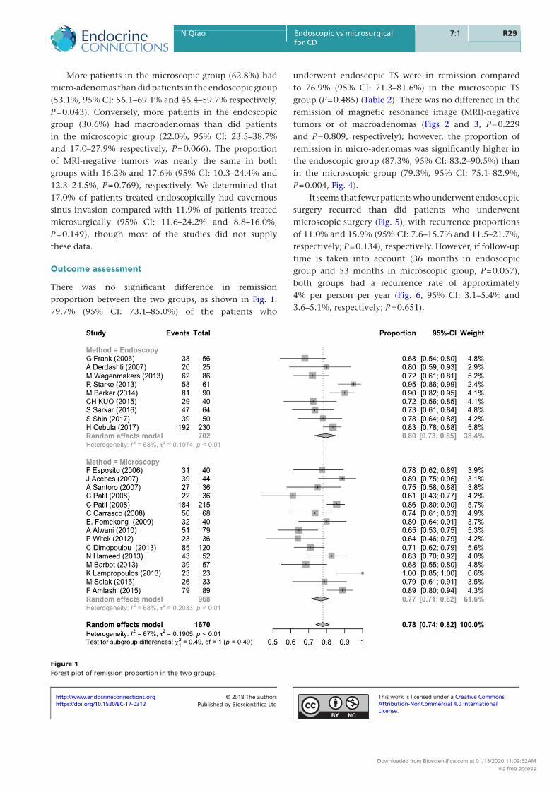
There was no significant difference in remission proportion between the two groups, as shown in Fig. 1: 79.7% (95% CI: 73.1–85.0%) of the patients who
underwent endoscopic TS were in remission compared to 76.9% (95% CI: 71.3–81.6%) in the microscopic TS group (P = 0.485) (Table 2). There was no difference in the remission of magnetic resonance image (MRI)-negative tumors or of macroadenomas (Figs 2 and 3, P = 0.229 and P = 0.809, respectively); however, the proportion of remission in micro-adenomas was significantly higher in the endoscopic group (87.3%, 95% CI: 83.2–90.5%) than in the microscopic group (79.3%, 95% CI: 75.1–82.9%, P = 0.004, Fig. 4).
It seems that fewer patients who underwent endoscopic surgery recurred than did patients who underwent microscopic surgery (Fig. 5), with recurrence proportions of 11.0% and 15.9% (95% CI: 7.6–15.7% and 11.5–21.7%, respectively; P = 0.134), respectively. However, if follow-up time is taken into account (36 months in endoscopic group and 53 months in microscopic group, P = 0.057), both groups had a recurrence rate of approximately 4% per person per year (Fig. 6, 95% CI: 3.1–5.4% and 3.6–5.1%, respectively; P = 0.651).
Figure 1Forest plot of remission proportion in the two groups.
This work is licensed under a Creative Commons Attribution-NonCommercial 4.0 International License.
https://doi.org/10.1530/EC-17-0312http://www.endocrineconnections.org © 2018 The authors
Published by Bioscientifica Ltd
Downloaded from Bioscientifica.com at 01/13/2020 11:09:52AMvia free access

N Qiao Endoscopic vs microsurgical for CD
R307:1
To investigate the effect of follow-up time on recurrence, we also included studies that reported patients treated prior to 1990 and with long-term follow-up (98 months compared to 53 months, respectively; P = 0.010) (Table 3). There was no significant difference in remission between patients with long-term follow-up vs patients with relatively short follow-up times: 75.4% (95% CI: 73.1–85.0%) vs 76.9% (95% CI: 71.3–81.6%, P = 0.849), respectively. Recurrence rate was also comparable between these two groups with 2.7% (95% CI: 2.0–3.8%) and 4.0% (95% CI: 3.2–5.0%) per person per year, respectively.
Quality analysis
Heterogeneity across studies was observed in the proportions of remission (I2 = 67%, P < 0.01) and recurrence (I2 = 67%, P < 0.01). There was no indication of heterogeneity in the recurrence rate (I2 = 17%, P = 0.23). To investigate the source of the heterogeneity, we conducted meta-regressions with several potential modifiers: number of patients, publication year, location in which the study was conducted and remission criteria. Our meta-regression analysis revealed no significant effects on the proportion of remission for publication date (P = 0.362), study location (P = 0.142), number of enrolled cases (P = 0.142) or remission criteria (0.844). Publication date (P = 0.567), study location (P = 0.135) and number of enrolled cases (P = 0.440) did not contribute to the heterogeneity of recurrence proportion.
In the sensitivity analysis, a single study was removed to determine the influence of that individual data set on the pooled proportions or rates; the corresponding proportions and rates were not significantly altered, indicating that our results are statistically robust. Funnel plots were constructed to estimate the publication bias of the literature; the results suggest that any potential publication bias did not substantially influence the results of this meta-analysis.
Discussion
This systematic review and meta-analysis compares outcomes in endoscopic and microsurgical approaches for the treatment of ACTH-secreting pituitary adenomas. In our study, we found that basic characteristics of patients treated endoscopically were comparable to those of patients treated microscopically, except that more patients treated endoscopically had macroadenomas. Similar remission proportions were found for both endoscopic Ta
ble
2
Ou
tco
mes
in e
nd
osc
op
ic o
r m
icro
sco
pic
tra
ns-
sph
eno
idal
AC
TH-s
ecre
tin
g a
den
om
a re
sect
ion
.
Stu
dy
Meth
od
C
ase
s
FU (
mo
nth
s)O
vera
ll
rem
issi
on
(%
)R
em
issi
on
in
M
RI-
PA
s (%
)R
em
issi
on
in
m
icro
PA
s (%
)R
em
issi
on
in
m
acr
o P
As
(%)
Pro
po
rtio
n o
f re
curr
en
ce (
%)
Recu
r ra
te
(/p
erso
n y
ear)
Fran
k et
al.
(14)
End
osc
op
y56
NA
38 (
67.9
%)
0 (N
A)
21 (
84.0
%)
17 (
54.8
%)
NA
NA
Deh
das
hti
an
d G
enti
li (1
5)En
do
sco
py
2517
20 (
80.0
%)
2 (4
0.0%
)13
(86
.7%
)4
(80.
0%)
0 (0
.0%
)0.
0%W
agen
mak
ers
et a
l. (1
6)En
do
sco
py
8671
62 (
72.1
%)
12 (
60.0
%)
29 (
82.9
%)
21 (
67.7
%)
10 (
16.1
%)
2.7%
Star
ke e
t al
. (17
)En
do
sco
py
6128
58 (
95.1
%)
16 (
100.
0%)
29 (
96.7
%)
13 (
86.7
%)
6 (1
2.0%
)5.
1%B
erke
r et
al.
(21)
End
osc
op
y90
3281
(90
.0%
)0
(0.0
%)
53 (
93.0
%)
28 (
96.6
%)
4 (4
.9%
)1.
9%K
uo
et
al. (
18)
End
osc
op
y40
40.2
29 (
72.5
%)
0 (N
A)
18 (
81.8
%)
11 (
61.1
%)
3 (1
0.3%
)3.
1%Sa
rkar
et
al. (
22)
End
osc
op
y64
2947
(79
.7%
)4
(66.
7%)
39 (
86.7
%)
5 (5
5.6%
)4
(8.5
%)
3.5%
Shin
et
al. (
19)
End
osc
op
y50
5039
(78
.0%
)8
(80.
0%)
21 (
77.8
%)
10 (
76.9
%)
9 (2
3.1%
)5.
5%C
ebu
la e
t al
. (20
)En
do
sco
py
230
2118
2 (7
9.1%
)50
(71
.4%
)97
(91
.5%
)35
(64
.8%
)18
(9.
9%)
5.7%
Esp
osi
to e
t al
. (24
)M
icro
sco
py
4033
31 (
77.5
%)
4 (5
0.0%
)21
(91
.3%
)6
(66.
7%)
1 (3
.2%
)1.
2%A
ceb
es e
t al
. (25
)M
icro
sco
py
4449
39 (
88.6
%)
6 (8
5.7%
)N
AN
A3
(7.7
%)
1.9%
San
toro
et
al. (
26)
Mic
rosc
op
y36
58.6
27 (
75.0
%)
0 (N
A)
19 (
86.4
%)
8 (5
7.1%
)4
(14.
8%)
3.0%
Pati
l et
al. (
27)
Mic
rosc
op
y36
3622
(61
.1%
)9
(90.
0%)
13 (
56.5
%)
0 (0
.0%
)2
(9.1
%)
3.0%
Pati
l et
al. (
28)
Mic
rosc
op
y21
545
184
(85.
6%)
NA
NA
NA
37 (
20.1
%)
5.4%
Car
rasc
o e
t al
. (29
)M
icro
sco
py
6845
50 (
73.5
%)
0 (N
A)
41 (
70.7
%)
9 (9
0.0%
)5
(10.
0%)
2.7%
Fom
eko
ng
et
al. (
30)
Mic
rosc
op
y40
8632
(80
.0%
)0
(0.0
%)
21 (
84.0
%)
11 (
91.7
%)
3 (9
.4%
)1.
3%A
lwan
i et
al. (
31)
Mic
rosc
op
y79
8451
(64
.6%
)8
(57.
1%)
37 (
84.1
%)
9 (4
2.9%
)10
(19
.6%
)2.
8%W
itek
an
d Z
ieliń
ski (
32)
Mic
rosc
op
y36
NA
23 (
63.9
%)
3 (3
7.5%
)17
(77
.3%
)3
(50.
0%)
NA
NA
Dim
op
ou
lou
et
al. (
33)
Mic
rosc
op
y12
079
85 (
70.8
%)
17 (
56.7
%)
46 (
79.3
%)
22 (
68.8
%)
29 (
34.1
%)
5.2%
Ham
eed
et
al. (
34)
Mic
rosc
op
y52
23.3
43 (
82.7
%)
6 (7
5.0%
)25
(89
.3%
)12
(75
.0%
)6
(14.
0%)
7.2%
Bar
bo
t et
al.
(35)
Mic
rosc
op
y57
83.6
39 (
68.4
%)
9 (6
0.0%
)26
(76
.5%
)4
(50.
0%)
15 (
38.5
%)
5.5%
Lam
pro
po
ulo
s et
al.
(36)
Mic
rosc
op
y23
43.2
23 (
100.
0%)
3 (6
0.0%
)7
(77.
8%)
6 (6
6.7%
)2
(8.7
%)
2.4%
Sola
k et
al.
(37)
Mic
rosc
op
y33
2826
(78
.8%
)0
(NA
)18
(78
.3%
)8
(80.
0%)
2 (7
.7%
)3.
3%A
mla
shi e
t al
. (38
)M
icro
sco
py
8953
.579
(88
.8%
)0
(NA
)69
(87
.3%
)10
(10
0.0%
)13
(38
.2%
)8.
6%
FU, F
ollo
w-u
p; N
A, n
ot
avai
lab
le; P
as, p
itu
itar
y ad
eno
mas
.
This work is licensed under a Creative Commons Attribution-NonCommercial 4.0 International License.
https://doi.org/10.1530/EC-17-0312http://www.endocrineconnections.org © 2018 The authors
Published by Bioscientifica Ltd
Downloaded from Bioscientifica.com at 01/13/2020 11:09:52AMvia free access

N Qiao Endoscopic vs microsurgical for CD
R317:1
Figure 3Forest plot of remission proportion of macroadenomas in the two groups.
Figure 2Forest plot of remission proportion of MRI-negative adenomas in the two groups.
This work is licensed under a Creative Commons Attribution-NonCommercial 4.0 International License.
https://doi.org/10.1530/EC-17-0312http://www.endocrineconnections.org © 2018 The authors
Published by Bioscientifica Ltd
Downloaded from Bioscientifica.com at 01/13/2020 11:09:52AMvia free access

N Qiao Endoscopic vs microsurgical for CD
R327:1
and microsurgical approaches, though remission criteria differed from study to study. Patients treated with the endoscopic approach for micro-adenomas were more likely to achieve remission than those treated microsurgically. Recurrence seemed to be lower among patients treated endoscopically; however, when follow-up time is taken into account, this advantage disappears.
Because most of the studies with endoscopy were performed in the latest 10–15 years. To eliminate the time as a confounding factor, we only included studies performed after 2005 (the oldest eligible publication on endoscopic TS is 2006). We only included studies with more than 20 patients because we believe surgical outcomes of CD are influenced by doctors’ experience. On the other hand, we also performed sensitive analysis, even studies with less 20 patients were included, the result did not change. Studies with patients treated prior to 1990 were considered separately because the follow-up period was longer in these studies.
Endoscopic visualization provides a more panoramic view of the operative field, compared with the microscope, allowing for better viewing of the suprasellar region
(51, 52). It is also possible to use instruments with a variety of angles to access lateral invasions of tumors (12, 13). Endoscopic surgery is an excellent approach for patients with CD, as the typically small size of the tumor requires higher magnification. Intrasellar illumination provided by the endoscope is extremely helpful in the intraoperative identification of abnormal tissue (19, 20, 22, 53). However, continuous adjustment of the endoscope is needed to determine target location within the surgical field, which may compromise maneuverability. Unlike endoscopic visualization, microsurgery offers a continuous view with a stereotactic display, which is familiar to the majority of surgeons and may allow for better control of bleeding in an open field. In recent years, some papers about 3D endoscopy for pituitary adenoma have been published (67, 68). This technique can combine depth perception in microscopy and wide-view in endoscopy. But no studies with 3D endoscopy were reported in patients with CD.
In the early days of endoscopic surgery, a large meta-analysis by Ammirati and coworkers (54) concluded that endoscopic removal of pituitary adenoma does not seem to confer any benefits over microscopic technology in
Figure 4Forest plot of remission proportion of microadenomas in the two groups.
This work is licensed under a Creative Commons Attribution-NonCommercial 4.0 International License.
https://doi.org/10.1530/EC-17-0312http://www.endocrineconnections.org © 2018 The authors
Published by Bioscientifica Ltd
Downloaded from Bioscientifica.com at 01/13/2020 11:09:52AMvia free access

N Qiao Endoscopic vs microsurgical for CD
R337:1
the short term. However, recent meta-analyses showed that the endoscopic technique is associated with higher gross tumor removal (57) and modest increases of resection rates in residual or recurrent cases (58). In patients with functional pituitary adenomas (growth hormone-secreting adenoma), Phan and coworkers (59) concluded that clinical use of the endoscopic approach conferred potential benefits, including increased remission rates with non-invasive macroadenomas, but that overall endocrine remission is comparable. Chen and coworkers (60) also concluded that both approaches yielded similar rates of remission. However, a meta-analysis comparing outcomes from endoscopic TS and microscopic TS was lacking.
Our results support these findings. Overall remission, remission in macroadenomas, and remission in MRI-negative CD showed no differences between groups. We also found that remission proportions for micro-adenomas were significantly higher in patients treated with the endoscopic approach compared to patients treated with the microsurgical approach. The superior intraoperative
visualization afforded by the endoscopic approach may account for this finding. A much more unobstructed view of the operative field may facilitate resection of much of the tumor, especially the pseudocapsule (61, 62, 63).
Complete surgical resection may be difficult for tumors with cavernous sinus infiltration due to the risks of injury of carotid artery and cranial nerves (69, 70). However, given the low occurrence of tumors with cavernous infiltration in each study, as well as the fact that few studies reported the remission of invasive tumors, the comparison between patients treated with endoscopic TS and microscopic TS was impossible in our analysis.
Regarding postoperative complications, previous studies include thorough descriptions and analyses, most of which demonstrate that patients who underwent endoscopic surgery had comparable proportions of complications, including diabetes insipidus, CSF leakage, hypocortisolemia, hypothyroidism, hypogonadism and visual defects, compared to patients who underwent microscopic surgery (57, 58, 59, 60). In this meta-analysis, we did not include any of these complications. Proportions
Figure 5Forest plot of recurrence proportion in the two groups.
This work is licensed under a Creative Commons Attribution-NonCommercial 4.0 International License.
https://doi.org/10.1530/EC-17-0312http://www.endocrineconnections.org © 2018 The authors
Published by Bioscientifica Ltd
Downloaded from Bioscientifica.com at 01/13/2020 11:09:52AMvia free access

N Qiao Endoscopic vs microsurgical for CD
R347:1
of sinusitis and epistaxis were also comparable in previous reports (59, 64).
The definition of remission for CD varies over time and across studies. The remission of clinical symptoms, the need for glucocorticoid replacement, low or normal cortisol levels, normal 24-h urinary free cortisol levels,
late-night salivary cortisol levels and cortisol after the dexamethasone suppression test all have been used in the literature (14, 15, 16, 17, 18, 19, 20, 21, 22, 24, 25, 26, 27, 28, 29, 30, 31, 32, 33, 34, 35, 36, 37, 38, 39, 40, 41, 42, 43, 44, 45, 46, 47, 48, 49, 50). A combination of two or three of the criteria mentioned earlier was used as the remission
Figure 6Forest plot of recurrence rate in the two groups.
Table 3 Studies with patients earlier than 1990 and with long-term follow-up.
Study
Year
Place
Cases
Follow-up (months)
Overall remission (%)
Proportion of recurrence (%)
Recurrence rate (/person year)
Valassi et al. (39) 1982–2007 Boston, USA 620 47.4 477 (76.9%) 62 (13.0%) 3.3%Lindsay et al. (40) 1982–2004 Bethesda, USA 418 125 331 (79.2%) 40 (12.1%) 1.2%Kim et al. (41) 1984–2010 Seoul, Korea 54 104.6 38 (70.4%) 18 (47.4%) 5.4%Ciric et al. (42) 1970–2010 Chicago, Illinois 136 68.4 93 (68.4%) 9 (9.7%) 1.7%Hassan-Smith et al. (43) 1988–2009 Birmingham, UK 72 55.2 60 (83.3%) 8 (13.3%) 2.9%Lambert et al. (44) 1980–2011 New York, USA 346 75.6 230 (66.5%) 73 (31.7%) 5.0%Alexandraki et al. (45) 1969–2001 London, UK 131 180 86 (65.6%) 31 (36.0%) 2.4%Costenaro et al. (46) 1989–2013 Porto Alegre, Brazil 103 73.2 84 (81.6%) 9 (10.7%) 1.8%Aranda et al. (47) 1974–2011 Barcelona, Spain 41 168 32 (78.0%) 21 (65.6%) 4.7%Yamada et al. (48) 1988–2014 Tokyo, Japan 230 72.5 198 (86.1%) 14 (7.1%) 1.2%Chandler et al. (49) 1980–2012 Michigan, USA 275 80.4 219 (79.6%) 37 (16.9%) 2.5%Bansal et al. (50) 1987–2015 Maharashtra, India 230 74 151 (65.7%) 48 (31.8%) 5.2%
This work is licensed under a Creative Commons Attribution-NonCommercial 4.0 International License.
https://doi.org/10.1530/EC-17-0312http://www.endocrineconnections.org © 2018 The authors
Published by Bioscientifica Ltd
Downloaded from Bioscientifica.com at 01/13/2020 11:09:52AMvia free access

N Qiao Endoscopic vs microsurgical for CD
R357:1
criteria in our study. Due to improvements in biochemical assays, a new consensus holds more stringent criteria for remission (65): ‘a postoperative cortisol value of <2 mg/dL predicts a higher chance of long-term remission after TS in CD; most patients with postoperative cortisol values of 2–5 mg/dL a few days after TS will also be in remission’. We also performed a subgroup analysis between studies with strict criteria and studies with lenient criteria. It turns out that no difference in remission proportion was observed in the two subgroups.
There was significant heterogeneity in the outcomes. This heterogeneity is likely impacted by differences in surgical technique, surgeon, team and institution experience or outcome criteria. It is also likely that differences in study design and definition of the outcomes influence heterogeneity (66).
A significant weakness of our analysis is that most studies use a relatively short follow-up time in patients with endoscopic TS. To compare recurrence rate between the two surgical groups, we assumed that there was no effect of follow-up time on recurrence rate. Meta-regression showed that the slope of recurrence rate by follow-up time was minus 0.002 (P = 0.529), a trend suggesting that as follow-up time increases, recurrence rate may decrease. We found no studies that directly compared endoscopic and microsurgical approaches. Randomized trials with experienced surgeons and trials with long-term follow-up are required to help bridge the current gaps in the literature.
Conclusion
We found that overall remission proportion was the same in CD patients who underwent endoscopic TS compared to patients who underwent microscopic TS. However, patients treated with the endoscopic approach for micro-adenomas were more likely to achieve remission than those treated microsurgically. Patients treated endoscopically were less likely to experience recurrence; however, when follow-up time is taken into account, this advantage disappears. The definition of diagnosis, remission and recurrence is very challenging and variable, which has always to be considered in the interpretation of results of studies assessing therapeutic efficacy in CD.
Declaration of interestThe authors declare that there is no conflict of interest that could be perceived as prejudicing the impartiality of the research reported.
FundingThis study was supported by Shanghai Sailing Program (17YF1426700, 2017).
AcknowledgementsThe authors thank Dr Min Shen and Dr Xuefei Shou for their contributions in the review of data.
References 1 Dallapiazza RF, Oldfield EH & Jane JA. Surgical management
of Cushing’s disease. Pituitary 2015 18 211–216. (https://doi.org/10.1007/s11102-015-0646-5)
2 Tritos NA, Biller BMK & Swearingen B. Management of Cushing disease. Nature Reviews Endocrinology 2011 7 279–289. (https://doi.org/10.1038/nrendo.2011.12)
3 Buchfelder M & Schlaffer S. Pituitary surgery for Cushing’s disease. Neuroendocrinology 2010 92 (Supplement 1) 102–106. (https://doi.org/10.1159/000314223)
4 Rutkowski MJ, Flanigan PM & Aghi MK. Update on the management of recurrent Cushing’s disease. Neurosurgical Focus 2015 38 E16. (https://doi.org/10.3171/2014.11.FOCUS14703)
5 Ayala A & Manzano AJ. Detection of recurrent Cushing’s disease: proposal for standardized patient monitoring following transsphenoidal surgery. Journal of Neuro-Oncology 2014 119 235–242. (https://doi.org/10.1007/s11060-014-1508-0)
6 Pivonello R, De Martino MC, De Leo M, Simeoli C & Colao A. Cushing’s disease: the burden of illness. Endocrine 2017 56 10–18. (https://doi.org/10.1007/s12020-016-0984-8)
7 Fleseriu M. Medical management of persistent and recurrent Cushing disease. Neurosurgery Clinics of North America 2012 23 653–668. (https://doi.org/10.1016/j.nec.2012.06.012)
8 Yaneva M, Kalinov K & Zacharieva S. Mortality in Cushing’s syndrome: data from 386 patients from a single tertiary referral center. European Journal of Endocrinology 2013 169 621–627. (https://doi.org/10.1530/EJE-13-0320)
9 van Haalen FM, Broersen LHA, Jorgensen JO, Pereira AM & Dekkers OM. Management of endocrine disease: mortality remains increased in Cushing’s disease despite biochemical remission: a systematic review and meta-analysis. European Journal of Endocrinology 2015 172 R143–R149. (https://doi.org/10.1530/EJE-14-0556)
10 Clayton RN, Jones PW, Reulen RC, Stewart PM, Hassan-Smith ZK, Ntali G, Karavitaki N, Dekkers OM, Pereira AM, Bolland M, et al. Mortality in patients with Cushing’s disease more than 10 years after remission: a multicentre, multinational, retrospective cohort study. Lancet Diabetes and Endocrinology 2016 4 569–576. (https://doi.org/10.1016/S2213-8587(16)30005-5)
11 Rolston JD, Han SJ & Aghi MK. Nationwide shift from microscopic to endoscopic transsphenoidal pituitary surgery. Pituitary 2016 19 248–250. (https://doi.org/10.1007/s11102-015-0685-y)
12 Fernandez-Miranda JC, Zwagerman NT, Abhinav K, Lieber S, Wang EW, Snyderman CH & Gardner PA. Cavernous sinus compartments from the endoscopic endonasal approach: anatomical considerations and surgical relevance to adenoma surgery. Journal of Neurosurgery 2017 [epub]. (https://doi.org/10.3171/2017.2.JNS162214)
13 Dusick JR, Esposito F, Kelly DF, Cohan P, DeSalles A, Becker DP & Martin NA. The extended direct endonasal transsphenoidal approach for nonadenomatous suprasellar tumors. Journal of Neurosurgery 2005 102 832–841. (https://doi.org/10.3171/jns.2005.102.5.0832)
14 Frank G, Pasquini E, Farneti G, Mazzatenta D, Sciarretta V, Grasso V & Faustini Fustini M. The endoscopic versus the traditional approach
This work is licensed under a Creative Commons Attribution-NonCommercial 4.0 International License.
https://doi.org/10.1530/EC-17-0312http://www.endocrineconnections.org © 2018 The authors
Published by Bioscientifica Ltd
Downloaded from Bioscientifica.com at 01/13/2020 11:09:52AMvia free access

N Qiao Endoscopic vs microsurgical for CD
R367:1
in pituitary surgery. Neuroendocrinology 2006 83 240–248. (https://doi.org/10.1159/000095534)
15 Dehdashti AR & Gentili F. Current state of the art in the diagnosis and surgical treatment of Cushing disease: early experience with a purely endoscopic endonasal technique. Neurosurgical Focus 2007 23 E9–E8. (https://doi.org/10.3171/foc.2007.23.3.11)
16 Wagenmakers MAEM, Boogaarts HD, Roerink SHPP, Timmers HJLM, Stikkelbroeck NMML, Smit JWA, van Lindert EJ, Netea-Maier RT, Grotenhuis JA & Hermus ARMM. Endoscopic transsphenoidal pituitary surgery: a good and safe primary treatment option for Cushing’s disease, even in case of macroadenomas or invasive adenomas. European Journal of Endocrinology 2013 169 329–337. (https://doi.org/10.1530/EJE-13-0325)
17 Starke RM, Reames DL, Chen C-J, Laws ER & Jane JA Jr. Endoscopic transsphenoidal surgery for Cushing disease. Neurosurgery 2013 72 240–247. (https://doi.org/10.1227/NEU.0b013e31827b966a)
18 Kuo C-H, Yen Y-S, Wu J-C, Chen Y-C, Huang W-C & Cheng H. Primary endoscopic transnasal transsphenoidal surgery for magnetic resonance image-positive Cushing disease: outcomes of a series over 14 years. WNEU 2015 84 772–779. (https://doi.org/10.1016/j.wneu.2015.04.059)
19 Shin SS, Gardner PA, Ng J, Faraji AH, Agarwal N, Chivukula S, Fernandez-Miranda JC, Snyderman CH & Challinor SM. Endoscopic endonasal approach for adrenocorticotropic hormone-secreting pituitary adenomas: outcomes and analysis of remission rates and tumor biochemical activity with respect to tumor invasiveness. WNEU 2017 102 651.e1–658.e1. (https://doi.org/10.1016/j.wneu.2015.07.065)
20 Cebula H, Baussart B, Villa C, Assié G, Boulin A, Foubert L, Aldea S, Bennis S, Bernier M, Proust F, et al. Efficacy of endoscopic endonasal transsphenoidal surgery for Cushing’s disease in 230 patients with positive and negative MRI. Acta Neurochirurgica 2017 1–10. (https://doi.org/10.1007/s00701-017-3140-1)
21 Berker M, Işikay I, Berker D, Bayraktar M & Gürlek A. Early promising results for the endoscopic surgical treatment of Cushing’s disease. Neurosurgical Review 2013 37 105–114. (https://doi.org/10.1007/s10143-013-0506-6)
22 Sarkar S, Rajaratnam S, Chacko G, Mani S, Hesargatta AS & Chacko AG. Pure endoscopic transsphenoidal surgery for functional pituitary adenomas: outcomes with Cushing’s disease. Acta Neurochirurgica 2015 158 77–86. (https://doi.org/10.1007/s00701-015-2638-7)
23 Baker SG, Kramer BS & Lindeman KS. The randomized registry trial. New England Journal of Medicine 2014 370 681–682. (https://doi.org/10.1056/NEJMc1315677)
24 Esposito F, Dusick JR, Cohan P, Moftakhar P, McArthur D, Wang C, Swerdloff RS & Kelly DF. Early morning cortisol levels as a predictor of remission after transsphenoidal surgery for Cushing’s disease. Journal of Clinical Endocrinology and Metabolism 2006 91 7–13. (https://doi.org/10.1210/jc.2005-1204)
25 Acebes JJ, Martino J, Masuet C, Montanya E & Soler J. Early post-operative ACTH and cortisol as predictors of remission in Cushing’s disease. Acta Neurochirurgica 2007 149 471–479. (https://doi.org/10.1007/s00701-007-1133-1)
26 Santoro A, Minniti G, Ruggeri A, Esposito V, Jaffrain-Rea M-L & Delfini R. Biochemical remission and recurrence rate of secreting pituitary adenomas after transsphenoidal adenomectomy: long-term endocrinologic follow-up results. Surgical Neurology 2007 68 513–518. (https://doi.org/10.1016/j.surneu.2007.05.057)
27 Patil CG, Prevedello DM, Lad SP, Vance ML, Thorner MO, Katznelson L & Laws ER Jr. Late recurrences of Cushing’s disease after initial successful transsphenoidal surgery. Journal of Clinical Endocrinology and Metabolism 2008 93 358–362. (https://doi.org/10.1210/jc.2007-2013)
28 Patil CG, Veeravagu A, Prevedello DM, Katznelson L, Vance ML & Laws ER Jr. Outcomes after repeat transsphenoidal surgery for recurrent Cushing’s disease. Neurosurgery 2008 63 266–271. (https://doi.org/10.1227/01.NEU.0000313117.35824.9F)
29 Carrasco CA, Coste J, Guignat L, Groussin L, Dugué MA, Gaillard S, Bertagna X & Bertherat J. Midnight salivary cortisol determination
for assessing the outcome of transsphenoidal surgery in Cushing’s disease. Journal of Clinical Endocrinology and Metabolism 2008 93 4728–4734. (https://doi.org/10.1210/jc.2008-1171)
30 Fomekong E, Maiter D, Grandin C & Raftopoulos C. Outcome of transsphenoidal surgery for Cushing’s disease: a high remission rate in ACTH-secreting macroadenomas. Clinical Neurology and Neurosurgery 2009 111 442–449. (https://doi.org/10.1016/j.clineuro.2008.12.011)
31 Alwani RA, de Herder WW, van Aken MO, van den Berge JH, Delwel EJ, Dallenga AHG, de Jong FH, Lamberts SWJ, van der Lely AJ & Feelders RA. Biochemical predictors of outcome of pituitary surgery for Cushing’s disease. Neuroendocrinology 2010 91 169–178. (https://doi.org/10.1159/000258677)
32 Witek P & Zieliński G. Predictive value of preoperative magnetic resonance imaging of the pituitary for surgical cure in Cushing’s disease. Turkish Neurosurgery 2012 1–6. (https://doi.org/10.5137/1019-5149.JTN.6199-12.2)
33 Dimopoulou C, Schopohl J, Rachinger W, Buchfelder M, Honegger J, Reincke M & Stalla GK. Long-term remission and recurrence rates after first and second transsphenoidal surgery for Cushing’s disease: care reality in the Munich Metropolitan Region. European Journal of Endocrinology 2013 170 283–292. (https://doi.org/10.1530/EJE-13-0634)
34 Hameed N, Yedinak CG, Brzana J, Gultekin SH, Coppa ND, Dogan A, Delashaw JB & Fleseriu M. Remission rate after transsphenoidal surgery in patients with pathologically confirmed Cushing’s disease, the role of cortisol, ACTH assessment and immediate reoperation: a large single center experience. Pituitary 2012 16 452–458. (https://doi.org/10.1007/s11102-012-0455-z)
35 Barbot M, Albiger N, Koutroumpi S, Ceccato F, Frigo AC, Manara R, Fassina A, Gardiman MP, Scanarini M, Mantero F, et al. Predicting late recurrence in surgically treated patients with Cushing’s disease. Clinical Endocrinology 2013 79 394–401. (https://doi.org/10.1111/cen.12133)
36 Lampropoulos KI, Samonis G & Nomikos P. Factors influencing the outcome of microsurgical transsphenoidal surgery for pituitary adenomas: a study on 184 patients. Hormones 2013 12 254–264. (https://doi.org/10.14310/horm.2002.1409)
37 Solak M, Kraljevic I, Dusek T, Melada A, Kavanagh MM, Peterkovic V, Ozretic D & Kastelan D. Management of Cushing’s disease: a single-center experience. Endocrine 2015 51 517–523. (https://doi.org/10.1007/s12020-015-0695-6)
38 Amlashi FG, Swearingen B, Faje AT, Nachtigall LB, Miller KK, Klibanski A, Biller BMK & Tritos NA. Accuracy of late-night salivary cortisol in evaluating postoperative remission and recurrence in Cushing’s disease. Journal of Clinical Endocrinology and Metabolism 2015 100 3770–3777. (https://doi.org/10.1210/jc.2015-2107)
39 Valassi E, Biller BMK, Swearingen B, Pecori Giraldi F, Losa M, Mortini P, Hayden D, Cavagnini F & Klibanski A. Delayed remission after transsphenoidal surgery in patients with Cushing’s disease. Journal of Clinical Endocrinology and Metabolism 2010 95 601–610. (https://doi.org/10.1210/jc.2009-1672)
40 Lindsay JR, Oldfield EH, Stratakis CA & Nieman LK. The postoperative basal cortisol and CRH tests for prediction of long-term remission from Cushing’s disease after transsphenoidal surgery. Journal of Clinical Endocrinology and Metabolism 2011 96 2057–2064. (https://doi.org/10.1210/jc.2011-0456)
41 Kim JH, Shin CS, Paek SH, Jung H-W, Kim SW & Kim SY. Recurrence of Cushing’s disease after primary transsphenoidal surgery in a university hospital in Korea. Endocrine Journal 2012 59 881–888. (https://doi.org/10.1507/endocrj.EJ12-0109)
42 Ciric I, Zhao J-C, Du H, Findling JW, Molitch ME, Weiss RE, Refetoff S, Kerr WD & Meyer J. Transsphenoidal surgery for Cushing disease. Neurosurgery 2012 70 70–81. (https://doi.org/10.1227/NEU.0b013e31822dda2c)
43 Hassan-Smith ZK, Sherlock M, Reulen RC, Arlt W, Ayuk J, Toogood AA, Cooper MS, Johnson AP & Stewart PM. Outcome of Cushing’s disease following transsphenoidal surgery in a single center over 20 years. Journal of Clinical Endocrinology and Metabolism 2012 97 1194–1201. (https://doi.org/10.1210/jc.2011-2957)
This work is licensed under a Creative Commons Attribution-NonCommercial 4.0 International License.
https://doi.org/10.1530/EC-17-0312http://www.endocrineconnections.org © 2018 The authors
Published by Bioscientifica Ltd
Downloaded from Bioscientifica.com at 01/13/2020 11:09:52AMvia free access

N Qiao Endoscopic vs microsurgical for CD
R377:1
44 Lambert JK, Goldberg L, Fayngold S, Kostadinov J, Post KD & Geer EB. Predictors of mortality and long-term outcomes in treated Cushing’s disease: a study of 346 patients. Journal of Clinical Endocrinology and Metabolism 2013 98 1022–1030. (https://doi.org/10.1210/jc.2012-2893)
45 Alexandraki KI, Kaltsas GA, Isidori AM, Storr HL, Afshar F, Sabin I, Akker SA, Chew SL, Drake WM, Monson JP, et al. Long-term remission and recurrence rates in Cushing’s disease: predictive factors in a single-centre study. European Journal of Endocrinology 2013 168 639–648. (https://doi.org/10.1530/EJE-12-0921)
46 Costenaro F, Rodrigues TC, Rollin GAF, Ferreira NP & Czepielewski MA. Evaluation of Cushing’s disease remission after transsphenoidal surgery based on early serum cortisol dynamics. Clinical Endocrinology 2013 80 411–418. (https://doi.org/10.1111/cen.12300)
47 Aranda G, Enseñat J, Mora M, Puig-Domingo M, Martínez de Osaba MJ, Casals G, Verger E, Ribalta MT, Hanzu FA & Halperin I. Long-term remission and recurrence rate in a cohort of Cushing’s disease: the need for long-term follow-up. Pituitary 2014 18 142–149. (https://doi.org/10.1007/s11102-014-0567-8)
48 Yamada S, Inoshita N, Fukuhara N, Yamaguchi-Okada M, Nishioka H, Takeshita A, Suzuki H, Ito J & Takeuchi Y. Therapeutic outcomes in patients undergoing surgery after diagnosis of Cushing’s disease: a single-center study. Endocrine Journal 2015 62 1115–1125. (https://doi.org/10.1507/endocrj.15-0463)
49 Chandler WF, Barkan AL, Hollon T, Sakharova A, Sack J, Brahma B & Schteingart DE. Outcome of transsphenoidal surgery for Cushing disease. Neurosurgery 2016 78 216–223. (https://doi.org/10.1227/NEU.0000000000001011)
50 Bansal P, Lila A, Goroshi M, Jadhav S, Lomte N, Thakkar K, Goel A, Shah A, Sankhe S, Goel N, et al. Duration of post-operative hypocortisolism predicts sustained remission after pituitary surgery for Cushing’s disease. Endocrine Connections 2017 6 625–636. (https://doi.org/10.1530/EC-17-0175)
51 Regmi D, Thapa A, Kc B & Shakya B. Endoscopic endonasal transsphenoidal approach to pituitary adenoma: a multi-disciplinary approach. Journal of Nepal Health Research Council 2017 15 174–177. (https://doi.org/10.3126/jnhrc.v15i2.18209)
52 Song Y, Li H, Liu H, Li W, Zhang X, Guo L & Tan G. Endoscopic endonasal transsphenoidal approach for sellar tumors beyond the sellar turcica. Acta Oto-Laryngologica 2014 134 326–330. (https://doi.org/10.3109/00016489.2013.857785)
53 Barkhoudarian G, Zada G & Laws ER. Endoscopic endonasal surgery for nonadenomatous sellar/parasellar lesions. World Neurosurgery 2014 82 S138–S146. (https://doi.org/10.1016/j.wneu.2014.07.017)
54 Ammirati M, Wei L & Ciric I. Short-term outcome of endoscopic versus microscopic pituitary adenoma surgery: a systematic review and meta-analysis. Journal of Neurology, Neurosurgery, and Psychiatry 2013 84 843–849. (https://doi.org/10.1136/jnnp-2012-303194)
55 Semple P. The transition from microscopic to endoscopic transsphenoidal surgery in high case load neurosurgical centers: the Groote Schuur Hospital experience. World Neurosurgery 2014 82 S162–S163. (https://doi.org/10.1016/j.wneu.2014.08.002)
56 Laws ER & Barkhoudarian G. The transition from microscopic to endoscopic transsphenoidal surgery: the experience at Brigham and Women’s Hospital. World Neurosurgery 2014 82 S152–S154. (https://doi.org/10.1016/j.wneu.2014.07.035)
57 Li A, Liu W, Cao P, Zheng Y, Bu Z & Zhou T. Endoscopic versus microscopic transsphenoidal surgery in the treatment of pituitary adenoma: a systematic review and meta-analysis. World Neurosurgery 2017 101 236–246. (https://doi.org/10.1016/j.wneu.2017.01.022)
58 Esquenazi Y, Essayed WI, Singh H, Mauer E, Ahmed M, Christos PJ & Schwartz TH. Endoscopic endonasal versus microscopic transsphenoidal surgery for recurrent and/or residual pituitary
adenomas. World Neurosurgery 2017 101 186–195. (https://doi.org/10.1016/j.wneu.2017.01.110)
59 Phan K, Xu J, Reddy R, Kalakoti P, Nanda A & Fairhall J. Endoscopic endonasal versus microsurgical transsphenoidal approach for growth hormone-secreting pituitary adenomas-systematic review and meta-analysis. WNEU 2017 97 398–406. (https://doi.org/10.1016/j.wneu.2016.10.029)
60 Chen C-J, Ironside N, Pomeraniec IJ, Chivukula S, Buell TJ, Ding D, Taylor DG, Dallapiazza RF, Lee C-C & Bergsneider M. Microsurgical versus endoscopic transsphenoidal resection for acromegaly: a systematic review of outcomes and complications. Acta Neurochirurgica 2017 159 2193–2207. (https://doi.org/10.1007/s00701-017-3318-6)
61 Monteith SJ, Starke RM, Jane JA & Oldfield EH. Use of the histological pseudocapsule in surgery for Cushing disease: rapid postoperative cortisol decline predicting complete tumor resection. Journal of Neurosurgery 2012 116 721–727. (https://doi.org/10.3171/2011.12.JNS11886)
62 Taylor DG, Jane JA & Oldfield EH. Resection of pituitary macroadenomas via the pseudocapsule along the posterior tumor margin: a cohort study and technical note. Journal of Neurosurgery 2017 1–7. (https://doi.org/10.3171/2017.7.JNS171658)
63 Ceylan S, Cabuk B, Koc K, Anik I & Vural C. Endoscopic distinction between capsule and pseudocapsule of pituitary adenomas. Acta Neurochirurgica 2013 155 1611–1619. (https://doi.org/10.1007/s00701-013-1754-5)
64 Lenzi J, Lapadula G, D’amico T, Delfinis CP, Iuorio R, Caporlingua F, Mecca N, Mercuri V, Bassotti G, Rillo M, et al. Evaluation of trans-sphenoidal surgery in pituitary GH-secreting micro- and macroadenomas: a comparison between microsurgical and endoscopic approach. Journal of Neurosurgical Sciences 2015 59 11–18.
65 Fleseriu M, Hamrahian AH, Hoffman AR, Kelly DF, Katznelson L, AACE Neuroendocrine & Pituitary Scientific Committee*. American Association of Clinical Endocrinologists and American College of Endocrinology disease state clinical review: diagnosis of recurrence in Cushing disease. Endocrine Practice 2016 22 1436–1448. (https://doi.org/10.4158/EP161512.DSCR)
66 Petersenn S, Beckers A, Ferone D, van der Lely A, Bollerslev J, Boscaro M, Brue T, Bruzzi P, Casanueva FF, Chanson P, et al. Therapy of endocrine disease: outcomes in patients with Cushing’s disease undergoing transsphenoidal surgery: systematic review assessing criteria used to define remission and recurrence. European Journal of Endocrinology 2015 172 R227–R239. (https://doi.org/10.1530/EJE-14-0883)
67 Pennacchietti V, Garzaro M, Grottoli S, Pacca P, Garbossa D, Ducati A & Zenga F. Three-dimensional endoscopic endonasal approach and outcomes in sellar lesions: a single-center experience of 104 cases. World Neurosurgery 2016 89 121–125. (https://doi.org/10.1016/j.wneu.2016.01.049)
68 Rampinelli V, Doglietto F, Mattavelli D, Qiu J, Raffetti E, Schreiber A, Villaret AB, Kucharczyk W, Donato F, Fontanella MM, et al. Two-Dimensional high definition versus three-dimensional endoscopy in endonasal skull base surgery: a comparative preclinical study. World Neurosurgery 2017 105 223–231. (https://doi.org/10.1016/j.wneu.2017.05.130)
69 Smith TR, Hulou MM, Huang KT, Nery B, de Moura SM, Cote DJ & Laws ER. Complications after transsphenoidal surgery for patients with Cushing’s disease and silent corticotroph adenomas. Neurosurgical Focus 2015 38 E12. (https://doi.org/10.3171/2014.10.FOCUS14705)
70 Gardner PA, Tormenti MJ, Pant H, Fernandez-Miranda JC, Snyderman CH & Horowitz MB. Carotid artery injury during endoscopic endonasal skull base surgery: incidence and outcomes. Neurosurgery 2013 73 261–269.
Received in final form 22 November 2017Accepted 28 November 2017
This work is licensed under a Creative Commons Attribution-NonCommercial 4.0 International License.
https://doi.org/10.1530/EC-17-0312http://www.endocrineconnections.org © 2018 The authors
Published by Bioscientifica Ltd
Downloaded from Bioscientifica.com at 01/13/2020 11:09:52AMvia free access