Embed Size (px)
Citation preview

Journal of
www.elsevier.com/locate/yjsbi
Journal of Structural Biology 159 (2007) 462–473
StructuralBiology
Otolith crystals (in Carapidae): Growth and habit
Eric Parmentier a,*, Rudi Cloots b, Roger Warin b, Catherine Henrist b
a Laboratoire de Morphologie Fonctionnelle et Evolutive, Institut de chimie, Bat. B6C, Universite de Liege, B-4000 Liege, Belgiumb CATl and Chimie des Materiaux inorganiques, Institut de chimie, Bat. B6C, Universite de Liege, B-4000 Liege, Belgium
Received 27 February 2007; received in revised form 21 May 2007; accepted 25 May 2007Available online 12 June 2007
Abstract
The biomineralization of otoliths results mainly from the release of soluble Ca2+, which is in turn precipitated as CaCO3 crystals. Insome Carapidae, sagittae sections have been shown to reveal a three-dimensional asymmetry with a nucleus close to the sulcal side, anunusual position. This study seeks to understand otolith formation in Carapus boraborensis. The unusual shape of the otolith is partlyexplained by the distribution of the epithelium cells, and particularly the sensory epithelium. Experimental evidence shows for the firsttime that aragonite growth takes place along the c-axis. These aragonite needles present two different habits. On the sulcal side is foundthe acicular form resulting from rapid growth during a short period of time. On the anti-sulcal side, the prismatic form seen there is dueto a slower growth speed over longer periods. The otolith surface was observed each hour during a period of 24 h in fishes reared insimilar conditions. This allowed for the first time the direct observation on the otolith surface of the deposition of the two layers (L-zoneand D-zone). In C. boraborensis, the organic-rich layer (D-zone) develops during the day, whereas the CaCO3 layer (L-zone) seems to bedeposited during the night.� 2007 Elsevier Inc. All rights reserved.
Keywords: Otolith; Carapidae; Aragonite; Habit; Crystal; Daily cycle
1. Introduction
The major roles associated with the otoliths of the innerear in teleosts are sound transduction and participation inmaintaining static and dynamic equilibrium (Lowenstein,1971; Tavolga, 1971; Platt and Popper, 1981; Schuijf,1981; Popper, 1982; Gauldie, 1988). Like all functionalstructures in an organism, the stato-acoustic system mustconsequently have a shape and organization that representa compromise between different needs and functions suchas swimming, hearing, equilibration, etc. (Parmentieret al., 2001). The otolith consists of a predominant mineralphase of CaCO3 (Carlstrom, 1963) enmeshed into anorganic matrix consisting of a complex network of macro-molecules with Ca2+-binding functions (Wright et al., 2002;Borelli et al., 2003). Crystals commonly occur in the arago-nite form of calcium carbonate and the orientation of the
1047-8477/$ - see front matter � 2007 Elsevier Inc. All rights reserved.
doi:10.1016/j.jsb.2007.05.006
* Corresponding author. Fax: +32 0 4 366 50 24.E-mail address: [email protected] (E. Parmentier).
crystal facets is sometimes referred to as being twinned(Degens et al., 1969; Gauldie and Nelson, 1988). Otherforms, such as calcite and vaterite, occur as partial or totalreplacements of aragonite in some otoliths, but such oto-liths are uncommon (Carlstrom, 1963; Gauldie, 1993,1999). The predominance of aragonite (orthorhombic) onthe calcite (rhombohedral) is unclear since the two poly-morphs have very similar crystal structures, differing inthe organization of the carbonate molecule sandwichedbetween layers of calcium ions (Campana, 1999; Faliniet al., 2005). Small quantities of ions, such as Sr and Mg,could favor the formation of aragonite over calcite (Carls-trom, 1963).
Otolith growth consists of daily deposition of a doubledeposit of a layer rich in minerals and a layer rich inorganic material (Pannella, 1971), resulting in alternatingtranslucent and opaque concentric rings (Campana andNeilson, 1985). Biomineralization of otoliths differs fromthat of vertebrate bone, molluscan shell and coral skeleton,since the otolith epithelium is not in direct contact with the

E. Parmentier et al. / Journal of Structural Biology 159 (2007) 462–473 463
region of calcification (Campana, 1999). As a result, thecalcification is strongly dependent upon the chemical com-position of the endolymphatic fluid (Falini et al., 2005),which has an alkaline pH and contains all the ionic precur-sors (e.g. high K+ and saturated Ca2+ and HCO3
� concen-trations), proteins, collagen and amino acids for otolithformation (Campana, 1999; Borelli et al., 2001; Payanet al., 2002).
The endolymphatic fluid is secreted by the saccular epi-thelium, which is composed of many cell types arranged intwo zones: (1) A proximal zone, bathing the proximal faceof the otolith and composed of the macula (sensory cells,supporting cells and secretory cells), and a crown of largeionocytes (mitochondriarich cells), arranged in a meshworkaround the macula; (2) On the opposite side, a distal zonebathing the distal side of the otolith is composed of squa-mous cells and small ionocytes (Mayer-Gostan et al.,1997; Takagi, 1997; Pisam et al., 1998; Takagi, 2000a,b).The different cellular zones seem to be at the origin of alack of uniformity in the spatial distribution of ionic andorganic endolymphatic components in the endolymph,and could be involved in the acellular process of otolithformation (Payan et al., 1999; Borelli et al., 2003). The for-mation seems to result from the release of soluble Ca2+ onthe proximal side, which is in turn precipitated as CaCO3
crystals due to an increasing alkaline gradient (Mugiya,1974; Gauldie and Nelson, 1990). This process has beencorroborated by recent studies dealing with the various cel-lular types of the saccular epithelium (Mayer-Gostan et al.,1997; Pisam et al., 1998). In some species, variation in dis-tribution within this epithelium of cellular ionic pumps(Mayer-Gostan et al., 1997; Toshe and Mugiya, 2001)and of secretory cells (Payan et al., 1999) has led to a solutegradient, the formation of which has been shown to inter-fere with otolith bio-calcification by favoring deposition onthe proximal side or on the otolith edge (Payan et al., 1999;Takagi and Takahashi, 1999; Takagi, 2000b). Moreover,soluble proteins (= otolin) with a high abundance of acidicamino acids (Davis et al., 1997; Murayama et al., 2002)could control calcium carbonate morphology and poly-morphism (Sollner et al., 2003), favoring aragonite overcalcite (Falini et al., 1996).
As a result of otolith formation, sagittal otoliths are gen-erally described as medially convex and distally concavebodies with a centrally located nucleus, or, more often,with one positioned closer to the anti-sulcal side (Dunkel-berger et al., 1980; Gauldie and Nelson, 1990). However,in carapids, sagittal sections have revealed a three-dimen-sional asymmetry with a nucleus close to the sulcal side(Parmentier et al., 2002), resulting from a differential accre-tion rate. The Carapus sp. fish (Carapidae) are small, eel-like fishes that are able to penetrate and stay inside differentinvertebrate hosts such as echinoderms (sea cucumber,starfish) or bivalves. Carapus species are commensal, anduse their host as a shelter (Parmentier and Das, 2004).
Generally speaking, this study seeks to understand theunusual position of the nucleus in Carapus boraborensis
(Carapidae). This fish is used because its nucleus asymme-try could confirm or deny the existing models on otolithformation. Three different approaches were undertaken inorder to understand more deeply otolith growth and orga-nization in carapids: (1) a study of cell distribution in theendolymphatic epithelium surrounding the sagitta; (2) elec-tron back-scattering diffraction (EBSD) and (3) scanningelectron microscopy (SEM) were used to examine the fun-damental crystal structure of the otolith, as well as the ori-entation and crystallographic relationships of the mineral.
2. Materials and methods
Sixty-four specimens of Carapus boraborensis (totallength: 13–32 cm) were collected in Opanohu Bay, Moorea(French Polynesia). They were found inside specimens ofBohadschia argus and Thelenota ananas, at depths rangingfrom 1 to 20 m. The fish were divided into four groups.Fishes from the first three groups were directly deeply anes-thetized with MS222 before they were (1) stored in a 75%ethanol solution (n = 21), (2) frozen at �20 �C (n = 10)and (3) fixed in Bouin for the production of serial histolog-ical sections (n = 3). Fishes from the fourth group (n = 30)were placed in an aquarium (120 · 40 · 20 cm) filled withrunning seawater, under natural lighting conditions(13L:11D). Every 60 min, during a period of 24 h, one fishfrom this group was removed from the tank and was anes-thetized with MS222. The two sagittae were then removedand placed in absolute ethanol or in acetone.
Otolith sagittae of groups 1, 2 and 4 were used for obser-vations. Each otolith was dehydrated in an oven and wascoated by Au-Pd pulverization (Balzers SCD-30). The goldsputtering is realized in such conditions to obtain a goldlayer <10 nm (30 s, 30 mA, argon plasma, distance50 mm). The signal of Au in the EDX spectra is weakenough to allow significant detection of light elements.
Photographs were taken with a scanning electron micro-scope (JEOL, JSM-840) under a 19 kV acceleration voltageor with an Environmental Scanning Electron Microscope(Philips ESEM XL30) equipped with a Field EmissionGun under a 19 kV acceleration voltage. The EDX spectraof the spherule otoliths were collected on a square area of1 lm · 1 lm centred in the middle of the spherule.
Sections of three sagittae were prepared by mountingeach otolith on a glass slide in thermoplastic cement (Crys-tal Bond). Frontal and transverse sections of sagittae wereobtained by grinding the piece with wet sand paper ofdecreasing grit sizes (600, 800 and 1200 grit) and polishingthem with wet 0.3 lm grit paper. Polished otoliths wereobserved with a TSL MSC-2200 (EBSD) camera coupledwith ESEM (under a 25 kV acceleration voltage), allowingthe carrying out of an Electron Back-Scattering Diffraction(EBSD) study. This technique is used to draw up the car-tography of the crystallographic orientation of mono-and polycrystalline materials in otoliths.
After Bouin fixation, heads of C. boraborensis weredecalcified in 3% nitric acid and neutralized in 5% Na2SO4.

464 E. Parmentier et al. / Journal of Structural Biology 159 (2007) 462–473
The pieces were then dehydrated in butanol, embedded inparaffin and serial cut with a Reichert microtome (8-lmthick). Sagittal, frontal and cross-sections were stainedusing Masson’s trichrome method. Histological sectionswere observed with a Leica DM 1000 binocular microscopecoupled with a Canon PowerShot S50 camera.
3. Results
A detailed description of the otic cavity bones and of theotolith shape already exists in the literature (Parmentieret al., 2001, 2002). Here are reported the morphologicalfeatures that complete these descriptions.
3.1. Endolymphatic epithelium
The saccular epithelium is composed of the sensory,transitional and squamous epithelia. (1) The sensory epi-thelium is medial and consists of high prismatic hair cells(±25 lm), which were lined with supporting cells. This oto-sac zone shows a high number of blood vessels and nerveendings. At this level, the gelatinous layer fixes the otolithto the sensory epithelium. (2) The meshwork/transitionalepithelium borders the macula and is made of smaller (5–7 lm) and cuboidal cells. This zone becomes still thinnerand forms the (3) squamous epithelium. This is situatedon the opposite side of the macula and consists of smallflattened cells (>2.5 lm). Small blood vessels are also situ-ated next to this zone.
Comparisons of the heights of the different parts of theotolith sulcal side and of the epithelium layers show thatthe sensory epithelium area corresponds to the colliculum(Fig. 1B). Moreover, the zone facing the important bulge
Fig. 1. Schematic dorsal (A) and lateral (B) view of the otic cavity and of the o(2001). Lateral (sulcal or proximal side) view of the right C. boraborensis sagi
of the sagitta is also the zone where blood vessels are themost developed. The otolith zones around both crista facethe meshwork/transitional epithelium. The latter is partic-ularly well developed at the level of the anterior part of theotolith, whereas this is not the case further back where theotolith is thinner. The squamous epithelium faces the anti-sulcal side of the otolith.
3.2. Morphology of aragonite crystals
In Carapus boraborensis, the sagitta has the shape of asemi-convex lens (Fig. 2A1). The distal face of the otolithseems to form two distinct parts. The anterior part of thesagitta looks like a half-sphere, whereas the posterior partshows a long post-rostrum with a pointed end (Fig. 2A1).The proximal face displays an elliptical sulcus occupiedby a single colliculum lined dorsally by the crista superiorand ventrally by the crista inferior (Fig. 2B1). Sections inthe sagitta show clearly the asymmetrical location of thenucleus, which is closer to the sulcal side (Fig. 3A). In sec-tioned otoliths, the nucleus seems to be placed on a linedelimiting the wide anti-sulcal side and the narrow sulcalside (Fig. 3A). The aragonite needles radiate from thenucleus. On the anti-sulcal side, the radiation gives rectilin-ear needles whose progressive lateral slanting confers to thesphere a tiered appearance (Fig. 2A2 and A3). At the levelof the distal dome, only the distal end of the needles is vis-ible at the surface of the otolith (Figs. 2A2 and 3C). As aresult of the slanting, the needles lean more and more pos-teriorly (Fig. 2A3) and finally appear in a horizontal planeat the level of the post-rostrum (Fig. 2A4). At this level, itis possible to see the lateral faces of the needles and to dis-cern easily the alternate deposition of protein and crystal
tosac epithelium in Carapus boraborensis; modified from Parmentier et al.tta (C).

Fig. 2. Lateral view of the left Carapus boraborensis sagitta. A1: sulcal (or proximal) side and B1: anti-sulcal (or distal) side. Annotated squares in A areenlarged in either A2, A3 or A4, showing the progressive lateral slanting from the dorsal dome (DD) to the post-rostrum (PR). B2 is an enlarged view ofthe square in B1, and B3 is an enlarged view of the square in B2. CI: crista inferior; CS: crista superior; SU: sulcus.
E. Parmentier et al. / Journal of Structural Biology 159 (2007) 462–473 465

Fig. 3. A: sagittal section in the Carapus boraborensis sagitta. B–D: enlarging of the respective squares in A. Black and white lines: orientation of theneedles toward the distal and proximal side, respectively; dotted line: white arrow: organic layer; black arrow: nucleus. AS: anti-sulcus side; SS: sulcus side.
466 E. Parmentier et al. / Journal of Structural Biology 159 (2007) 462–473
layers (Fig. 2A4). The habitus of the distal region looks likean elongated prismatic form ending in an angular dome(Fig. 4A).
On the proximal face (Fig. 2B1), needles also come fromthe nucleus. However, the needles are not rectilinear andthey present modification in their orientations (Fig. 3Aand B). They are first parallel to the plane of the proximalface (= sulcal side) before presenting a modification of theorientation and they emerge perpendicularly to the sulcalside (Fig. 3A and B). These needles also appear thinnerand it is difficult to see the protein increments (Fig. 3B).On the proximal face, the crystal habitus of the colliculumpresents an acicular form (Fig. 4B), and the needles formgroups oriented in different directions (Fig. 2B2 and B3).
The EBSD method was used to investigate the tridimen-sional crystal orientations and relationships of the mineral.The EBSD confirms the SEM analysis: the method allowsthe observation of the radial orientation of the crystal nee-dles from the nucleus. On the color-coded map, each colorcorresponds to one of the aragonite axes pointing outwardsin the direction of the viewer: a, b or c (Fig. 5). The distri-bution of green and blue zones in the figure indicates thatboth a- and b-axes are perpendicular to the section. Theabsence of red pixels indicates that this c-axis is in the planeof the section. The same observations were made in thefrontal and sagittal planes. These results indicate that the
growth of the aragonite needles always follows the c-axis.Moreover, these observations are reinforced by the 3Danalysis of the aragonite mapping. The c-axis (highlightedin pink on the corresponding unit cells) always lies parallelto the growth direction of the needles, whereas both a- andb-axes present random orientations (Fig. 5). However, bothaxes do not adopt any preferred orientation relative to theneedles direction. This is translated by a random distribu-tion of green and blue color from a crystal to another. Ina single needle, a- or b-axis stays fixed from the nucleithroughout the whole growth as it is shown trough contin-uous blue or green needles from the nucleus to the surface(Fig. 5). Each needle can be considered as an aragonite sin-gle crystal.
3.3. Otolith growth
The otolith surface was observed over a period of 24 h infishes reared in similar conditions. In Fig. 6, all picturesdisplay the same scale and present the same otolith region:the dorsal dome. Crystal modifications were recorded at 7a.m.; these result from the appearance of filamentous struc-tures, which are attributed to organic matter. These fila-ments grow in number and thickness until 7 p.m., givingto the whole surface a net-like structure. From 10 p.m.,these fibrilar structures seem to disappear progressively

Fig. 4. Aragonite habits found on the distal side (A) and on the proximal side (B) in the sagitta of Carapus boraborensis. X: localization of the habits. Reddotted line: Miller index: a = 001, b = 010, c = 110.
E. Parmentier et al. / Journal of Structural Biology 159 (2007) 462–473 467
(Fig. 6). These observations lead us to suppose that the for-mation and the deposit of the organic matrix occur duringthe day.
Moreover, different small spherules were found on theotolith surface (Fig. 7). They appear larger (diameter aver-age: 3.76 lm ± 0.1, n = 20) at the level of the sulcal side(Fig. 7A) than on the anti-sulcal side (diameter average:440 nm ± 0.1, n = 13, p < 0.0001, Fig. 7B). Each spheruleseems to be made of numerous subunits lengtheningbetween 20 and 200 nm. The external aspect is not con-stant. It can be globular or more and more ‘‘spread’’ ontothe otolith surface. Behind these more or less completespherules, it is possible to discern the otolith crystal(Fig. 7A). In order to characterize these different forms,the element composition was semi-quantitatively mea-sured. The globular spherules are rich in carbon and in cal-cium, with a very high carbon content (Fig. 8A). Bycontrast, the ‘‘spread’’ forms have a proportionally highercalcium content to carbon ratio (Fig. 8C). Analysis ofintermediate forms showed there was a progressive modifi-cation in the relative element content in relation to thespherule forms. The important amounts of carbon andcalcium lead us to suppose that the spherules could be com-posed of one (or more) protein(s). Once deposited on theotolith surface, the degradation of the spherulitic proteincomplex or its conformation change could lead to the lib-
eration of calcium, whereas the molecule seems resolubi-lised in the otosac, involving the diminution of therelative amount of carbon. Moreover, because the anti-sul-cal side only possesses the subunits, it is supposed thatthere is a migration from the proximal side to the distalside.
4. Discussion
In generalized otolith models, the sagitta otoliths(Fig. 9) are described as medially convex and distally con-cave bodies with a centrally located nucleus, or, moreoften, with one positioned closer to the anti-sulcal side(Dunkelberger et al., 1980; Gauldie and Nelson, 1990; Par-mentier et al., 2002). Both shape and nucleus locationresult from the release of soluble Ca2+ on the proximal side(Ibsch et al., 2004), which in turn precipitates as CaCO3
crystals due to an increasing alkaline gradient, from thesulcal area towards the otolith edge (Mugiya, 1974; Gaul-die and Nelson, 1990). As a result, the growth of the crista
superior and crista inferior is privileged and there is a moreimportant development of the sulcal side. Moreover, theincorporation of the calcium at the level of the edgesinvolves a decrease in the Ca2+ concentration and a weakerdevelopment of the distal side. This process has beencorroborated by recent studies dealing with the various
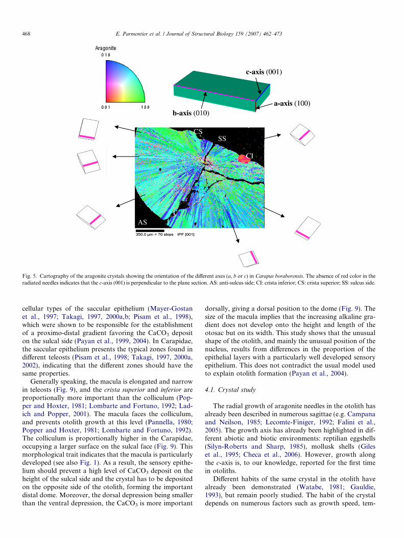
Fig. 5. Cartography of the aragonite crystals showing the orientation of the different axes (a, b or c) in Carapus boraborensis. The absence of red color in theradiated needles indicates that the c-axis (001) is perpendicular to the plane section. AS: anti-sulcus side; CI: crista inferior; CS: crista superior; SS: sulcus side.
468 E. Parmentier et al. / Journal of Structural Biology 159 (2007) 462–473
cellular types of the saccular epithelium (Mayer-Gostanet al., 1997; Takagi, 1997, 2000a,b; Pisam et al., 1998),which were shown to be responsible for the establishmentof a proximo-distal gradient favoring the CaCO3 depositon the sulcal side (Payan et al., 1999, 2004). In Carapidae,the saccular epithelium presents the typical zones found indifferent teleosts (Pisam et al., 1998; Takagi, 1997, 2000a,2002), indicating that the different zones should have thesame properties.
Generally speaking, the macula is elongated and narrowin teleosts (Fig. 9), and the crista superior and inferior areproportionally more important than the colliculum (Pop-per and Hoxter, 1981; Lombarte and Fortuno, 1992; Lad-ich and Popper, 2001). The macula faces the colliculum,and prevents otolith growth at this level (Pannella, 1980;Popper and Hoxter, 1981; Lombarte and Fortuno, 1992).The colliculum is proportionally higher in the Carapidae,occupying a larger surface on the sulcal face (Fig. 9). Thismorphological trait indicates that the macula is particularlydeveloped (see also Fig. 1). As a result, the sensory epithe-lium should prevent a high level of CaCO3 deposit on theheight of the sulcal side and the crystal has to be depositedon the opposite side of the otolith, forming the importantdistal dome. Moreover, the dorsal depression being smallerthan the ventral depression, the CaCO3 is more important
dorsally, giving a dorsal position to the dome (Fig. 9). Thesize of the macula implies that the increasing alkaline gra-dient does not develop onto the height and length of theotosac but on its width. This study shows that the unusualshape of the otolith, and mainly the unusual position of thenucleus, results from differences in the proportion of theepithelial layers with a particularly well developed sensoryepithelium. This does not contradict the usual model usedto explain otolith formation (Payan et al., 2004).
4.1. Crystal study
The radial growth of aragonite needles in the otolith hasalready been described in numerous sagittae (e.g. Campanaand Neilson, 1985; Lecomte-Finiger, 1992; Falini et al.,2005). The growth axis has already been highlighted in dif-ferent abiotic and biotic environments: reptilian eggshells(Silyn-Roberts and Sharp, 1985), mollusk shells (Gileset al., 1995; Checa et al., 2006). However, growth alongthe c-axis is, to our knowledge, reported for the first timein otoliths.
Different habits of the same crystal in the otolith havealready been demonstrated (Watabe, 1981; Gauldie,1993), but remain poorly studied. The habit of the crystaldepends on numerous factors such as growth speed, tem-

Fig. 6. Modification of the anti-sulcal surface of Carapus boraborensis during a period of 24 h.
E. Parmentier et al. / Journal of Structural Biology 159 (2007) 462–473 469
perature, viscosity of the medium and the presence ofimpurities that could prevent growth in some directions(Li et al., 1991). Growth control can be effective at two lev-els. (1) The thermodynamic level indicates that the presenceof aragonite in otoliths is due to the presence of proteic pre-
cursors (Borelli et al., 2003; Guibbolini et al., 2006).Calcium carbonate crystallizes into three different poly-morphs: calcite, aragonite and vaterite. Experimentalevidence from calcium carbonate overgrowth on thesurface of otoliths has suggested that the intracrystalline

Fig. 7. Spherules found on the proximal (A) and distal (B) sides in the sagitta of Carapus boraborensis.
Fig. 8. Spherules of different shapes and sizes on the otolith surface in Carapus boraborensis. Flattening and spreading of the spherules onto the otolithsurface in Carapus boraborensis, viewed by SEM, and corresponding modification of elemental composition determined by EDX. The progressivediminution of the spherules corresponds to a relative modification in the ration Ca:C.
470 E. Parmentier et al. / Journal of Structural Biology 159 (2007) 462–473

sulcus
dorsal depression
sulcus nucleus
ventral depression
nucleus
sulcus
dorsal depression
ventral depression
ventral depression
dorsal depression
A B
Fig. 9. Comparison of a ‘‘generalized’’ otolith model of Teleostean (A) with a carapid otolith (B). 1: sulcal side; 2: schematic view of a transverse section.
E. Parmentier et al. / Journal of Structural Biology 159 (2007) 462–473 471
macromolecules associated to the otolith favor aragonitepolymorphism (Falini et al., 2005). (2) The kinetic levelcould be responsible for the habit type. This depends onphysico-chemical factors such as the lack of uniformity inthe chemical composition of the saccular endolymph(Payan et al., 1999). Moreover, the adsorption of differentions can stimulate or inhibit the growth of different faces.
This study reports the presence of (at least) two differenthabits in the C. boraborensis sagitta. The first, the prismaticform, is found on the anti-sulcal side and the second, theacicular form, on the sulcal side. These two types of habitcould result from different environmental conditions. Onthe sulcal side, the ionic concentration is important (Payanet al., 1999) and could be at the origin of a rapid growthduring a short period of time. On the anti-sulcal side, theionic concentration is lower (Payan et al., 1999), leadingto lower growth speed but over longer periods. However,the idea cannot be excluded that habit type is partly deter-mined by matrix proteins.
4.2. Circadian cycle
The alternate deposit of organic and mineral layersseems dependent on the photoperiod (Tanaka et al.,1981; Radtke and Dean, 1982; Mugiya, 1984, 1987). Thereare few studies dealing with the circadian cycle of layerdeposition. In the Oncorhynchus mykiss trout, the bilayerrings of otolith growth during the day:night cycle corre-spond to anti-phasic mechanisms: organic matrix deposi-tion starts at the end of the day, whereas CaCO3
deposition starts at the beginning of the day (Borelliet al., 2003). On the other hand, Edeyer et al. (2000)found that, in the Psetta maxima turbot, the incremental
zone (L-zone) was formed at night, whereas the discontin-uous zone (D-zone) was formed during the daytime. Inboth cases, these circadian variations were deduced fromthe ionic composition and protein concentration in theplasma and endolymph.
For the first time, the deposition of the two layers (L-zone and D-zone) has been observed here directly on theotolith surface. In C. boraborensis, the organic-rich layerdevelops during the day, whereas the CaCO3 layer seemsto be deposited during the night, the small spherules givinga globular appearance to the otolith surface. It was alsoobserved that spherules on the proximal side are biggerthan on the distal side. These bigger spherules appear tobe made of smaller particles that were found only on thedistal side, suggesting the degradation of the large spher-ules and the migrations of the particles. In Oreochromismossambicus, discrete calcium carbonate precipitationswere also more numerous in the proximal endolymph thanin the distal endolymph, clearly indicating a decreasingproximo-distal gradient and that the calcium supply ofthe otoliths takes place via the proximal endolymph (Ibschet al., 2004).
Orchestration of extracellular calcification requiresbringing together ionic and proteinaceous components intime and space. Local alkaline microenvironments in theendolymph are required to promote the precipitation ofthe calcium carbonate (Mugiya et al., 1981; Payan et al.,1997). It has been accepted that the organic matrix, mainlycomposed of proteins such as glycoproteins, collagens, pro-teoglycans and Ca2+ binding proteins, was essential in thecalcification process (Murayama et al. 2005, Guibboliniet al., 2006) and in the control of the calcium carbonatemorphology and polymorphism (Falini et al., 2005).

472 E. Parmentier et al. / Journal of Structural Biology 159 (2007) 462–473
Flattening and spreading of the spherules onto the otolithsurface in Carapus boraborensis seems correspond to theCaCO3 precipitation, and the resolubilisation ofmacromolecules.
Acknowledgments
We thank C. Brie (Tropical Fish Tahiti, Rangiroa), Y.Chancerelle (CRIOBE, Moorea, French Polynesia) andtheir respective team for helping to obtain living carapids.N. Decloux kindly helped in the microscopic study. Thisstudy was supported by Grant C-05/77 from ‘‘Fonds speci-aux, Universite de Liege’’.
References
Borelli, G., Mayer-Gostan, N., De Pontual, H., Bœuf, G., Payan, P., 2001.Biochemical relationships between endolymph and otolith matrix inthe trout (Oncorhynchus mykiss) and turbot (Psetta maxima). Calcif.Tissue Int. 69, 356–364.
Borelli, G., Guibbolini, M.E., Mayer-Gostan, N., Priouzeau, F., DePontual, H., Allemand, D., Puverel, S., Tambutte, E., Payan, P., 2003.Daily variations of endolymph composition: relationship with theotolith calcification process in trout. J. Exp. Biol. 206, 2685–2692.
Carlstrom, D., 1963. A crystallographic study of vertebrate otoliths. Biol.Bull., Woods Hole 125, 441–463.
Campana, S.E., 1999. Chemistry and composition of fish otoliths:pathways, mechanisms and applications. Mar. Ecol. Prog. Ser. 188,263–297.
Campana, S.E., Neilson, J.D., 1985. Microstructure of fish otoliths. Can.J. Fish Aquatic Sci. 42, 1014–1031.
Checa, A.G., Okamoto, T., Ramırez, J., 2006. Organization pattern ofnacre in Pteriidae (Bivalvia: Mollusca) explained by crystal competi-tion. Proc. R. Soc. B 273, 1329–1337.
Davis, J.G., Burns, F.R., Navaratnam, D., Masaji Lee, A., Ichimiya, S.,Oberholtzer, J.C., Greene, M.I., 1997. Identification of a structuralconstituent and one possible site of postembryonic formation of ateleost otolithic membrane. Proc. Natl. Acad. Sci. 94, 707–712.
Degens, E.T., Deuser, W.G., Haedrich, R.L., 1969. Molecular structureand composition of fish otoliths. Mar. Biol. 2, 105–113.
Dunkelberger, D.G., Dean, J.M., Watabe, N., 1980. The ultrastructure ofthe otolithic membrane and otolith in the juvenile mummichog,Fundulus heteroclitus. J. Morph. 163, 367–377.
Edeyer, A., De Pontual, H., Payan, P., Troadec, H., Severe, A., Mayer-Gostan, N., 2000. Daily variations of saccular endolymph and plasmacompositions in the turbot Psetta maxima: relationship with the diurnalrhythm in otolith formation. Mar. Ecol. Prog. Ser. 198, 287–294.
Falini, G., Albeck, S., Weiner, S., Addadi, L., 1996. Control of aragoniteor calcite polymorphism by mollusc shell macromolecules. Science 271,67–69.
Falini, G., Fermani, S., Vanzo, S., Miletic, M., Zaffino, G., 2005. Influenceon the formation of aragonite or vaterite by otolith macromolecules.Eur. J. Inorg. Chem. 1, 162–167.
Gauldie, R.W., 1988. Function, form and time-keeping properties of fishotoliths. Comp. Biochem. Physiol. A 91, 395–402.
Gauldie, R.W., Nelson, D.G.A., 1988. Aragonite twinning and neuro-protein secretion are the cause of daily growth rings in fish otoliths.Comp. Biochem. Physiol. A 90, 501–509.
Gauldie, R.W., Nelson, D.G.A., 1990. Otolith Growth in fishes. Comp.Biochem. Physiol. A 97, 119–135.
Gauldie, R.W., 1993. Polymorphic crystalline structure of fish otoliths. J.Morphol. 218, 1–28.
Gauldie, R.W., 1999. Ultrastructure of lamellae, mineral and matrixcomponents of fish otolith twinned aragonite crystals: implications forestimating age in fish. Tissue Cell 31, 138–153.
Giles, R., Manne, S., Mann, S., Morse, S.E., Stucky, G.D., Hansma, P.K.,1995. Inorganic overgrowth of aragonite on molluscan nacre examinedby atomic force microscopy. Biol. Bull. 188, 8–15.
Guibbolini, M., Borelli, G., Mayer-Gostan, N., Priouzeau, F., DePontual, H., Allemand, D., Payan, P., 2006. Characterization andvariations of organic parameters in teleost fish endolymph during day-night cycle, starvation and stress conditions. Comp. Biochem. Physiol.A 145, 99–107.
Ibsch, M., Anken, R.H., Rahmann, H., 2004. Calcium gradients in the fishinner ear sensory epithelium and otolithic membrane visualized byenergy filtering transmission electron microscopy (EFTEM). Adv. Sp.Res. 33, 1395–1400.
Ladich, F., Popper, A.N., 2001. Comparison of the inner ear ultrastruc-ture between teleost fishes using different channels for communication.Hear. Res 154, 62–72.
Lecomte-Finiger, R., 1992. Growth history and age at recruitment ofEuropean glass eels (Anguilla anguilla) as revealed by otolith micro-structure. Mar. Biol. 144, 205–210.
Li, Y., Stein, M., Jungnickel, B.J., 1991. Competition betweencrystallisation and phase separation in polymers blends. Diffusioncontrolled supermolecular structure and phase morphologies in poly(e-caprolactone) (polystyrene blends). Colloid. Polym. Sci. 269,772–780.
Lombarte, A., Fortuno, J.M., 1992. Differences in morphological featuresof the sacculus of the inner ear of two hakes (Merluccius capensis andM. paradoxus, Gadiformes) inhabits from different depth of sea. J.Morph. 214, 97–107.
Lowenstein, O., 1971. The labyrinth. In: Hoar, W.S., Randall, D.J. (Eds.),Fish Physiology, vol. 5. Academic Press, New York, pp. 207–240.
Mayer-Gostan, N., Kossmann, H., Watrin, A., Payan, P., Bœuf, G., 1997.Distribution of ionocytes in the saccular epithelium of the inner ear oftwo teleosts (Oncorhynchus mykiss and Scophthalmus maximus). CellTissue Res. 289, 53–61.
Mugiya, Y., 1974. Calcium-45 behavior at the level of the otolith organs ofrainbow trout. Bull. Jpn. Soc. Sci. Fish. 40, 457–463.
Mugiya, Y., 1984. Diurnal rhythm in otolith formation in the rainbowtrout, Salmo gairdeni: seasonal reversal of the rhythm in relation toplasma calcium concentrations. Comp. Biochem. Physiol A 78,289–293.
Mugiya, Y., 1987. Phase difference between calcification and organicmatrix formation in the diurnal growth of otoliths in the rainbowtrout, Salmo gairdeneri. Comp. Biochem. Physiol. A 84, 57–60.
Mugiya, Y., Watabe, N., Yamada, J., Dean, J.M., Dunkelberger, D.G.,Sjimizu, M., 1981. Diurnal rhythm in otolith formation in the goldfish(Carassius auratus). Comp. Biochem. Physiol. 68A, 659–662.
Murayama, E., Takagi, Y., Ohira, T., Davis, J.G., Greene, M.I.,Nagasawa, H., 2002. Fish otolith contains a unique structural protein,otolin-1. Eur. J. Biochem. 269, 688–696.
Murayama, E., Herbomel, P., Kawakami, A., Takeda, H., Nagasawa, H.,2005. Otolith matrix proteins OMP-1 and Otolin-1 are necessary fornormal otolith growth and their correct anchoring onto the sensorymaculae. Mech. Dev. 122, 791–803.
Pannella, G., 1971. Fish otoliths: daily growth layers and periodicalpatterns. Science 173, 1124–1127.
Pannella, G., 1980. Growth pattern of fish sagittae. In: Rhoad, D.C., Lutz,R.A. (Eds.), Skeletal Growth of Aquatic Organisms: BiologicalRecords of Environmental Change, Topics in geobiology, vol. 1.Plenum Press, New York, pp. 519–560.
Parmentier, E., Vandewalle, P., Lagardere, F., 2001. Morpho-anatomy ofthe otic region in carapid fishes: eco-morphological study of theirotoliths. J. Fish Biol. 58, 1046–1061.
Parmentier, E., Lagardere, F., Vandewalle, P., 2002. Relationshipsbetween inner ear and sagitta growth during ontogenesis of threeCarapini species and consequences of life-history events on the otolithmicrostructure. Mar. Biol. 141, 491–501.
Parmentier, E., Das, K., 2004. Commensal vs parasitic relationshipbetween Carapini fish and their host: some further insight through (13Cand (15N measurements. J. Exp. Mar. Biol. Ecol. 310, 47–58.

E. Parmentier et al. / Journal of Structural Biology 159 (2007) 462–473 473
Payan, P., Kossman, H., Watrin, A., Mayer-Gostan, N., Boeuf, G., 1997.Ionic composition of endolymph in teleosts: origin and importance ofendolymph alkalinity. J. Exp. Biol. 200, 1905–1912.
Payan, P., Edeyer, A., de Pontual, H., Borelli, G., Bœuf, G., Mayer-Gostan, N., 1999. Chemical composition of saccular endolymph andotolith in fish inner ear: lack of spatial uniformity. Am. J. Physiol. 277,123–131.
Payan, P., Borelli, G., Priouzeau, F., De Pontual, H., Bœuf, G., Mayer-Gostan, N., 2002. Otolith growth in trout Onchorynchus mykiss:supply of Ca2+ and Sr2+ to the saccular endolymph. J. Exp. Biol. 205,2687–2695.
Payan, P., De Pontual, H., Bœuf, G., Mayer-Gostan, N., 2004.Endolymph chemistry and otolith growth in fish. C.R. Palevol. 3,535–547.
Pisam, M., Payan, P., LeMoal, C., Edeyer, A., Bœuf, G., Mayer-Gostan,N., 1998. Ultrastructural study of the saccular epithelium of the innerear of two teleosts, Oncorhynchus mykiss and Scophthalmus maximus.Cell Tissue Res. 294, 261–270.
Platt, C., Popper, A.N., 1981. Fine structure and function of the ear. In:Tavolga, W.N., Popper, A.N., Fay, R.R. (Eds.), Hearing and SoundCommunication in Fishes. Springer-Verlag, New York, pp. 2–37.
Popper, A.N., 1982. Organization of the inner ear and auditory process-ing. In: Northcutt, E.G., Davis, R.E. (Eds.), Fish Neurobiology, vol. 1.The University of Michigan Press, Ann Arbor, pp. 126–178.
Popper, A.N., Hoxter, B., 1981. The fine structure of the sacculus andlagena of a teleost fish. Hear. Res. 5, 245–263.
Radtke, R.L., Dean, J., 1982. Increment formation in the embyos, larvaeand juveniles of the mummichog, Fundulus heteroclictus. Fish. Bull.U.S. 80, 41–55.
Schuijf, A., 1981. Models of acoustic localization. In: Tavolga, W.N.,Popper, A.N., Fay, R.R. (Eds.), Hearing and Sound Communicationin Fishes. Springer-Verlag, New York, pp. 267–310.
Sollner, C., Burghammer, M., Busch-Nentwich, E., Berger, J., Schwarz,H., Riekel, C., Nicolson, T., 2003. Control of crystal size and latticeformation by starmaker in otolith biomineralization. Science 302,282–286.
Silyn-Roberts, H., Sharp, R.M., 1985. Preferred orientation of calcite andaragonite in the reptilian eggshells. Proc. R. Soc. London B 225, 445–455.
Takagi, Y., 1997. Meshwork arrangement of mitochondria-rich, Na+,K+ -ATPase-rich celles in the saccular epithelium of rainbouwtrout (Onchorryhnchus mykiss) inner ear. Anat. Rec. 248, 483–489.
Takagi, Y., 2000a. Cellular contributions to the otolith formation inteleosts: a review. Otsuchi Mar. Sci. 25, 1–6.
Takagi, Y., 2000b. Ultrastructural immunolocalization of the otolithwater-soluble-matrix in the inner ear of rainbow trout just-hatched fry.Fish. Sci. 66, 71–77.
Takagi, Y., 2002. Otolith formation and endolymph chemistry: a strongcorrelation between the aragonite saturation state and pH in theendolymph of the trout otolith organ. Mar. Ecol. Prog. Ser. 231, 237–245.
Takagi, Y., Takahashi, A., 1999. Characterization of otolithsoluble-matrix producing cells in the saccular epithelium ofrainbow trout (Oncorhynchus mykiss) inner ear. Anat. Rec. 254,322–329.
Tanaka, K., Mugiya, Y., Yamada, J., 1981. Effects of photoperiod andfeeding on daily growth patterns in otoliths of juvenile Tilapia nilotica.Fish. Bull. 79, 459–466.
Tavolga, W.N., 1971. Sound production and detection. In: Hoar, W.S.,Randall, D.J. (Eds.), Fish Physiology, vol. 5. Academic Press, NewYork, pp. 135–205.
Toshe, H., Mugiya, Y., 2001. Effects of enzyme and anion transportinhibitors on in vitro incorporation of inorganic carbon and calciuminto endolymph and otoliths in salmon Oncorhyncus masou. Comp.Biochem. Physiol. 128A, 177–184.
Watabe, N., 1981. Crystal growth of calcium carbonate in the inverte-brates. Prog. Cryst. Growth Charact. 4, 99–147.
Wright, P.J., Panfili, J., Morales-Nin, B., Geffen, A.J., 2002. Differentstypes de piece calcifiee, In: Panfili, J., Pontual, H.D.E., Troadec, H.,Wright, P.J. (Eds.), Manuel de sclerochronologie des poissons.Coedition Ifremer – IRD, pp. 31–88.