Embed Size (px)
Citation preview

Muntasir Dhaka National Medical College Pathology
OSPE Question-Pathology

OSPE Question Pathology
2

OSPE Question Pathology
3
Specimen#1 Acute appenidicitis
This is a jar containing Specimen of Vermiform Appendix with Mesoappendix.It’s outer
surface is swallowen,Oedematous & congested subserosal vessel(& lumen contain exudate
External surface is lusterless),Probable diagnosis is Acute appenidicitis Confirmed by
Histopathology
What is the Complication of Appendicitis? 1. Appendicular lump
2. Appendicular abscess
3. Burst of appendix
4. Peritonitis
Hallmark of Diagnosis:Presence of Neutrophil in Muscle coat
Common Tumour:Carcinoid Tumour(Neuroendocrine Tumour)
Specimen#2 Chronic cholesystitis
This is a jar containing specimen of galbladder.It is swallowen, wall is Fibrosed &
thickened, probable diagnosis is chronic cholesystitis confirmed by Hystopathology
What is the Common Carcinoma of this viscera?
Adenocarcinoma
What are the Types of gallbladder Stone Cholesterol stone
Pigment stone
Mixed Stone
What is it’s Complication? Empyema
(Acute cholecystitis in the presence of bacteria-containing bile may progress to
suppurative infection in which the gallbladder fills with purulent material, a
condition referred to as empyema of the gallbladder)
Mycocele of Gallbladder
(Mucocele, or hydrops, of the gallbladder describes an overdistended gallbladder
filled with mucoid or clear and watery content)
OSPE Question Pathology
4
Specimen#3 Leiomyoma of Uterus
This is a jar containing a cut section of totally Resected Uterus.The Outer surface shows
multiple nodular greyish white lesion.My probable diagnosis is Leiomyoma of uterus
Confirmed by Histopathology
What’s it’s Malignant Counter part?
Leiomyosarcoma
What are the types of Leiomyoma?
1. Subserosal(Beneath the endometrium)
2. Intramural(Within the myometrium)
3. Submucosal(Beneath the Endometrium)
Specimen#4
Ca Cervix This is a specimen of totally receted uterus with both sided adnexi/Both sided fallopian
tube & ovary.The cut section shows an irregular greyish-white exophytic lesion.My
probable diagnosis is Ca Cervix Confirmed by Histopathology
What are the common Histological pattern?
Squamous cell carcinoma
Adenocarcinoma
Adenosquamous Carcinoma
Specimen#5 Carcinoma of Breast
This is a Jar containing Mastectomy specimen;which is partaily covered with skin
containing areola & nipple.Cut surface shows an irregular greyish-White tumourous
lesion.My probable diagonosis is Carcinoma of Breast Confirmed by Histopathology
What do you mean by Scirrhous Carcinoma?
It is one form of Desmoplasia of breast in which there is Greater extend of fibrosis which
forms a stony mass of breast
What do you mean by Comedo Carcinoma?
Dutctule carcinoma in situ with Central necrosis in situ
What is the Common Histologic Type?
Duct cell Carcinoma What’s Sentinile Lymphnod?What’s it’s significance

OSPE Question Pathology
5
The nearest lymphnode of a Neoplastic growth is called Sentinile Lymphnode
Significance:If sentinile Lymphnode is involved it is sign of malignancy What are the 4 prognostic criteria ?
1. Size
2. Involvement of Lymphnode & In situ
3. Invasiveness
4. ERPR
Specimen#6 Carcinoma of Colon
This is a Jar containing part of intestine.The mucosa shows a cauliflower/Fungative/ Exophytic growth.My probable diagnosis is carcinoma in colon confirmed by
histopathology
What are the common malignant tumour of this organ?
Adenocarcinoma
Carcinoid tunour
GIST(Gastrointestinal stromal tumour)
Lymphnode
What are the common Benigntumour of this organ?
Adenoma of colon
Specimen#7 Lymphnode
It is a jar containing decested section of Lymphnode showing-
Macroscopically it is Cheesy white material & Microscopically it shows Amorphous,
Granular, Eosinophilic cellular Debris my impression is tubercular granuloma confirmed
by Histopathology What’s Granuloma? A granuloma is a focus of chronic inflammation consisting of a microscopic aggregation
of epithelioid cell surrounded by a collar of mononuclear leukocytes & occationally
plasma cells. Older granulomas develop an enclosing rim of fibroblast & connective tissue What are the Gian cell?
physiological
SyncytoTrophoblast
MegaKaryoCyte
Oesteoblast
Pathological
Langhan’s Type of Giant cell
Foreign body giant cell
Tutan giant cell
Tumour giant
Reed-SteinBerg Giant cell

OSPE Question Pathology
6
Specimen#8 Renal Cell Carcinoma
This is a Jar containing Specimen of Resected Kidney.The cut section shows the upper pole with
large greyish-white Tumourous lesion.My probable diagnosis is Renal cell carcinoma confirmed
by histopathology
What are the Types of Renal cell carnoma?
1. Clear cell carcinoma
2. Papillary carcinoma
3. Chromophobe carcinoma
What are the common childhood tumour of this organ?
Wilm’s Tumour/Nephroblastoma
Specimen#9 Squamous cell carcinoma
This is a specimen of a peice of lung tissue.The cut surface shows greyish-white tumourous lesion at
base.My probable diagnosis is lung cancer
What are the common histologic type?
Squamous cell carcinoma
Small cell carcinoma
Adenocarcinoma
Largecell caecinoma
Carcinoid tumour
What is the most common aetiologic factor?
Smoking
Name the most common industrial factor?
Asbetosis
Specimen:10 Nodular goitre
This is jar containing a cut a cut surface of thyroid gland.This sliced specimen of thyroid tissue give
multipleee meaty nodular appearance.My probable diagnosis is nodular goitre confirmed by
histopathology
What’s the Most common malignant Type?
Papillary carcinoma
What do you mean by cold nodule? A nonfunctioning thyroid nodule/lump that does not concentrate radioactive isotopes in a thyroid
scan and may be indicative of cancer.

OSPE Question Pathology
7
Microscopic Slides Slide#1
Nodular goitre Section shows thyroid goitre.It reveals thyroid follicle of various sizes containing
Colloid.Follicle of various sizes containg colloid.Follicles are fibrosed, hyalinization &
evidence of haemorrhage-haemosiderin laden macrophage
No maignancy seen
My diagnosis:Nodular goitre of thyroid gland
What are causes/Types of goitre?
1. Colloid goitre,Simple goitre or, diffuse non-toxic goitre
2. Multinodular goitre
3. Diffuse toxic goitre(Grave’s disease)
4. Thyroiditis
5. Neoplasm
What are the nodular lesions of thyroid?
1. Mulinodular goitre
2. Adenomas
3. Carcinomas
4. Other
What are the autoimmuno thyroid disease?
Grave’s disease
Hashimoto’s thyroiditis
Primary myxoedema

OSPE Question Pathology
8
Slide#2 Granulomatous lymphadenitis
Section shows lympnode.It reveals many granulomas composed of area of Caesiation
necrosis surrounded by epithilioid cells, rim of lymphocyte, small number of plasma cells
& occasionally Langhan’s type of giant cells & surrounded by fibroblast.
Diagnosis:Granulomatous Lymphadenitis
What are the Gian cell?
physiological
SyncytoTrophoblast
MegaKaryoCyte
Oesteoblast
Pathological
Langhan’s Type of Giant cell
Foreign body giant cell
Tutan giant cell
Tumour giant
Reed-SteinBerg Giant cell

OSPE Question Pathology
9
Slide#3 Chronic Cholecystitis
Section shows wall of gallbladder.The wall has been invaded by many chronic
inflammatory celll.Inflammatory fibrosis is present in the peerimuscular coat &
Rocky-Tansky Aschoff sinus present
Diagnosis:Chronic cholecystitis
What is the Common Carcinoma of this viscera?
Adenocarcinoma
What are the Types of gallbladder Stone
Cholesterol stone
Pigment stone
Mixed Stone What is it’s Complication? Empyema
(Acute cholecystitis in the presence of bacteria-containing bile may progress to suppurative
infection in which the gallbladder fills with purulent material, a condition referred to as
empyema of the gallbladder)
Mycocele of Gallbladder
(Mucocele, or hydrops, of the gallbladder describes an overdistended gallbladder filled with
mucoid or clear and watery content)
Rockitansky-aschoff sinus

OSPE Question Pathology
10
Slide#4 Invasive Squamous cell carcinoma
Section shows a maignant tumour composed of anaplastic epithelial cell aranged in sheets
& nests.These cells have hyperchromatic nuclei, coarse chromatin & eosinophillic
cytoplasm.The tumour has invaded underlying stroma.Keratin pearl is also seen
Diagnosis:Invasive Squamous cell carcinoma grade-I
What are the common histologic type in case of Lung specimen?
Squamous cell carcinoma
Small cell carcinoma
Adenocarcinoma
Largecell caecinoma
Carcinoid tumour
What is the most common aetiologic factor of SSC of Lung?
Smoking
Name the most common industrial factor of SSC of Lung?
Asbetosis
Keratin pearl

OSPE Question Pathology
11
Slide#5 Adenocarcinoma
The section shows a malignant tumour composed of Anaplastic epithelial cells arranged in
clusters & glandular pattern.These cells have large nuclei with prominent nucleoli,coarse
chromatin & mild to moderate amount of cytoplasm.The tumour has invaded into the
muscle coat Diagnosis:Adenocarcinoma

OSPE Question Pathology
12
Slide#6 Fibroadenoma of breast
The section shows breast tissue.It reveals proloferation of ductular & fibrous tissue
elements.The ductules are compressed formiong slit like pattern.The background shows
myxoid stroma
No evidence of malignancy
Diagnosis:Breast fibroadenoma What are the Histologic types of fibroadenoma?
1. Pericanalicular fibroadenoma
2. Intracnalicular fibroadenoma What is the malignant counter part of it?
Adenocarcinoma

OSPE Question Pathology
13
Slide#7 Leiomyoma
Section shows interlacing bundle of smooth muscle fibre arranged in whorled bundle
pattern
No malignancy is seen
Diagnosis:Leiomymyoma
What’s it’s Malignant Counter part?
Leiomyosarcoma
What are the types of Leiomyoma?
1. Subserosal(Beneath the endometrium)
2. Intramural(Within the myometrium)
3. Submucosal(Beneath the Endometrium)

OSPE Question Pathology
14
Slide#8 Acute Appendicitis
The section shows appendix.The mucosa is partly disrupted.The muscle coat contains
infiltration of acute inflammatory cell mostly meutrophills.The muscle fibres are are
sperated due to inflemmatory exudate
Diagnosis:Acute appendicitis
What are the complication of Acute appendicitis?
5. Appendicular lump
6. Appendicular abscess
7. Burst of appendix
8. Peritonitis What are the Tumour of appendix?
1. Carcinoid tumour(Most common)
2. Adenocarcinoma

OSPE Question Pathology
15
Slide#9 Lipoma
Section shows nodules of mature adipose tissue partially covered by a capsule
No malignancy is seen
Diagnosis:Lipoma
What is the malignant Counterpart of It?
Liposacrcoma

OSPE Question Pathology
16
Slide#10 Nodular Hyperplasia of Prostate
Section shows multiple irregular fragmented piece of prostatic tissue.This revealed
hyperplastic gland & fibromusclular stroma arranged in nodules.The glands are lined by
multilayered epithelium projecting into the lumen.Some of the glands contains corpora
amylacea
What are the common carcinoma of prostate?
Adenocarcinoma Which age group is commonly affected?
50-60 age group

OSPE Question Pathology
17
Histophathology Diagnosis of diseases by examinating the tissues obtained by Biopsy or, surgically
resected specimen
Methods of histopathology 1. Paraffin section method
2. Frozen section method
Method of Specimen Collection 1. Biopsy
2. Surgical resection
Types of Collection of biopsy marterial-
Biopsy Example
1. Simple incisional biopsy
(Part of organ removed) Breast Lump
2. Excisional biopsy
(Whole organ removed) Lymph node
3. Wedge BIopsy Liver
4. Neddle biopsy Liver
5. Instrumental Biopsy Endoscopy, Colonoscopy
6. Truecut Biopsy From Liver & Kidney
7. CT Guided Biopsy From Lung
Common preservatives or, fixatives: 1. 10% formaline
2. Carnoy’s fixative
3. Bonin’s Fixative
4. Zenker’s fixative
Methods of Preservation(Why 10% formaline is most commonly used as preservatives) Prevention of autolysis
Maintainan tissue structure in life like manner
Maintain the architecture
Cut by-
Microtomein 3-5 µm thickness
OSPE Question Pathology
18
Staining Method
Stain Example Haematoxylin Eosin stain Haematoxylin:Basic & stains the Nucleus blue
Eosine:Acidic & stains the cytoplasm red
PAS(Periodic acid shift test) Mucin & glycogem. basemenst memberane of Blood vessel
Congo Red Amyloid fribils
Sudan IV & Oil Red-O Fat tissue
Masson’s Trichoma Muscle element
Geimsa Stain LD body for Kala-a-zar
Prussian Blue Iron Stain
Fite stain AFB bacilli
Frozen Section Method
It is histopathological method which mainly helps the surgeon in Determining the surgical
procedure to be applied to the pt.; by quick diagnosis of a specimen within 20 min while
the pt. is at OT bed.
Procedure
The Specimen comes from the Operation theatre to pathology
The pathologist then quickly paerforms the Frozen section method who is previously
informed for this procedure with a short history
Then the Specimen is cut with an instrument microtome into 3-5µm thick peices
The section is then placed into a freezer Cryostate which quickly freezes the section
into -20 to -30°C whinin 20 minute
Then the section is stained with HE stain & observed under microscope
OSPE Question Pathology
19
Cytopathology Study of individual cells for thedetection & diagnosis of cancerous or, precancerous lesion
of tissue Method of specimen collection
Method Example
1. Exfoliative Urine, Sputum
2. Abrassive
a) Scrapping In between the junction of Ecto & endocervix with
a spacula
b) Baloon A baloon is introduced Stomach then inflated & cell
is collected when it touches the stomach wall c) Touch imprint During brain surgery the specimen is collected from
the surgical instrument which came in touch with
the brain tissue a the cells are imprinted onto the
tissue
3. FNAC(Fine needle
aspiration cytology)
(With a 5-10cc syringe by creating
negative pressure)
Visible organ
eg.Breast,Thyroid
Invisible organ
eg.Intraabdominal, Intrathoracic organ
Stains used
Papanicolaou stain
Other stains
Geimsa stain
HE satin
PAS
Diff quick stain
OSPE Question Pathology
20
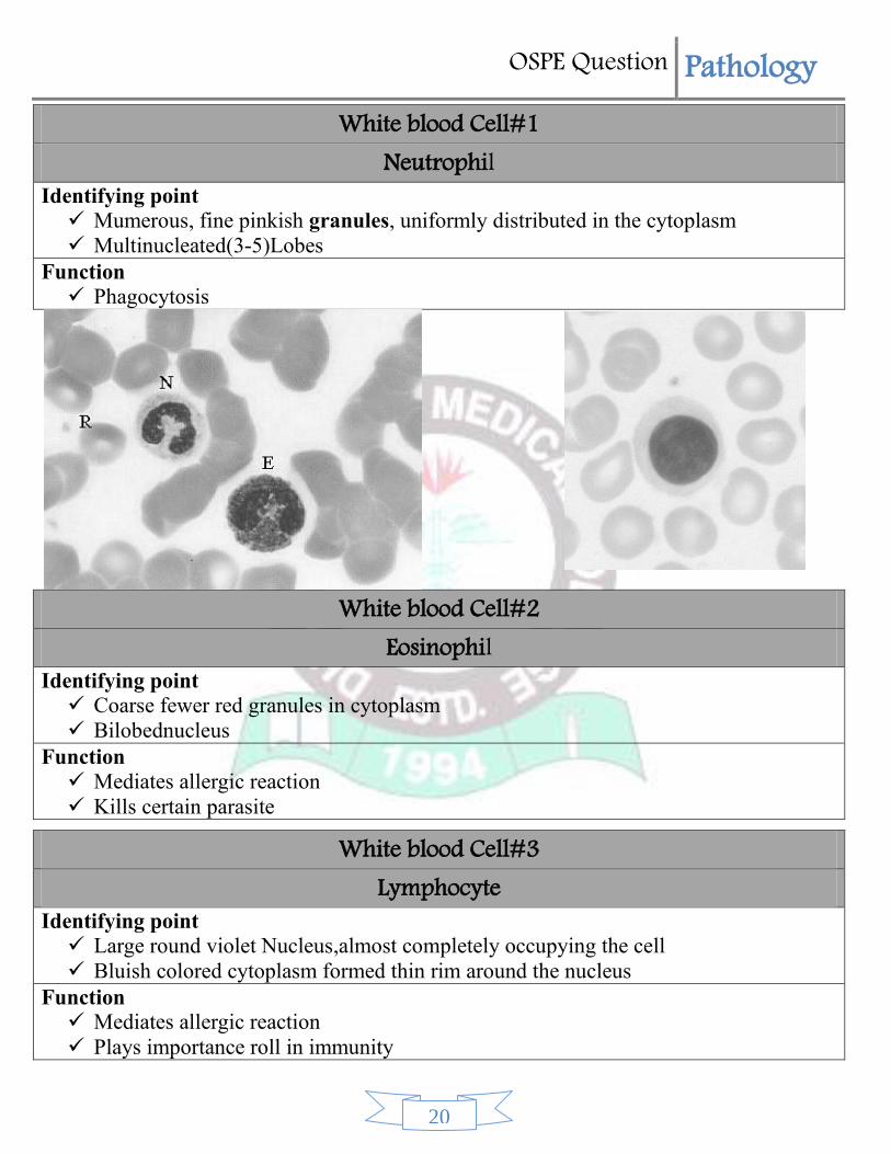
White blood Cell#1 Neutrophil
Identifying point
Mumerous, fine pinkish granules, uniformly distributed in the cytoplasm
Multinucleated(3-5)Lobes
Function
Phagocytosis
White blood Cell#2
Eosinophil Identifying point
Coarse fewer red granules in cytoplasm
Bilobednucleus
Function
Mediates allergic reaction
Kills certain parasite
White blood Cell#3 Lymphocyte
Identifying point
Large round violet Nucleus,almost completely occupying the cell
Bluish colored cytoplasm formed thin rim around the nucleus
Function
Mediates allergic reaction
Plays importance roll in immunity

OSPE Question Pathology
21
Instruments#1 Sahli’s Haemoglobinometer
Identifying point
It contains-
1. Colour matching
2. Haemoglobin pipette(with.02ml marking)
3. Graduated mixing tube
4. Dropper
5. Stirrer
6. Brush
Use-Estimation of haemoglobin by sahli’s Acid haematin Method
Name methods of Haemoglobin estimation.
1. Sahli’s Acid Haematin method
2. Alkaline haematin method
3. OxyHaemoglobin Method
4. Cyanmethhaemogllobin method
Which Method in is accurate?Why?
- Cyanmethhaemoglobin method.
Cause-
The result is accurate
No eye color variation(As in sahli’s Method)
Sulph-Hb, Carboxy-Hb etc. can be estimatd also
Calculation
Instruments#2 Wintrobe’s Haematocrit Tube
Identifying Point
Cylindrical Glass tube with one Open End
Marked from 0-10cm (Above downward-For ESR)& 10-0cm(Downward to above for
PCV)
Use-
Detrmenation of PCV by Wintrobe’s Method
Determination of ESR by Wintrobe’s Method
What is the PCV?its normal Value.
Male:.40-.54L/L(40%-50%)
Femlae:.37-.47L/L(37-47%)
OSPE Question Pathology
22
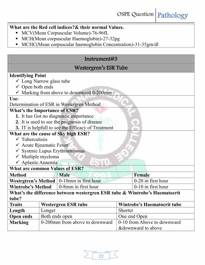
What are the Red cell indices?& their normal Values.
MCV(Mean Corpuscular Volume)-76-96fL
MCH(Mean corpuscular Haemoglobin)-27-32pg
MCHC(Mean corpuscular haemoglobin Concentration)-31-35gm/dl
Instrument#3
Westergren’s ESR Tube Identifying Point
Long Narrow glass tube
Open both ends
Marking from above to downward 0-200mm
Use-
Determination of ESR in Westergren Method
What’s the Importance of ESR?
1. It has Got no diagnostic importance
2. It is used to see the prognosis of disease
3. IT is helpfull to see the Efficacy of Treatment What are the cause of Sky high ESR?
Tuberculosis
Acute Rjeumatic Fever
Systmic Lupus Erythromatosus
Mutliple myeloma
Aplastic Anaemia
What are common Values of ESR?
Method Male Female
Westrgtren’s Method 0-10mm in first hour 0-20 in first hour
Wintrobe’s Method 0-8mm in first hour 0-10 in first hour
What’s the difference between westergren ESR tube & Wintrobe’s Haematocrit
tube?
Traits Westergren ESR tube Wintrobe’s Haematocrit tube
Length Longer Shorter
Open ends Both ends open One end Open
Marking 0-200mm from above to downward 0-10 from Above to downward
&downward to above

OSPE Question Pathology
23
Instrument#4 Haemocytometer
Identifying points-
RBC pipette
WBC pipette
Improved Neubauer’s counting chamber
Cover slip
Use-
TC of RBC
TC of WBC
Semen ananlysis
What’sTC of RBC?
4.5-5.5million/mm3
What’s the TC & DC of WBC?
TC of WBC 4000-7000/mm3
TC &DC of WBC- DC TC
Neutrophil 40-75% 2,000-7,500/mm3
Lymphocyte 20-50% 1,500-4,000/mm3
Eosinophil 2-10% 200-800/mm3
Monocyte 1-6% 40-400/mm3
Basophil 0-1% 0-100/mm3
Instrument#5 Spinocan needle
Identifying points
1. Trocar
2. Cannula
What are the Indication of lumber puncture?
1. Meningitis
2. Encephalitis
3. Subarachnoid haemorrhage
4. Brain Tumour
What are the contraindication of lumber puncture?
↑ICP

OSPE Question Pathology
24
Papillooedema
Head injury
What are the cause of increase Intracranial pressure?
Head injury
Hydrocephalus
Brain Tumour
Subdural haematoma
Instrument#6 Bone Marrow Aspiration Needle
Identifying Point
1. Trocar
2. Cannula
3. Gaurd
What are the indications of Bone Marrow Aspiration?
1. Aplastic Anaemia
2. Megaloblastic Anaemia
3. Leukaemia
4. Sublaeukaemic Acute Leukaemia
5. Multiple Myeloma
6. ITP
What are the Methods of Bone Marrow? 1. Wide bore method
2. Trephine Biopsy What are the site of Bone Marrow Aspiratoin?
1. Body of the Sternum opposite to 2nd to 3rd intrcostal space
2. Ilieac Crest
3. Medial Aspect of Upper pole of Tibia What are the contraindication of Bone Marrow aspiration?
1. Haemophillia
2. Other Coagulation Disorder What are the Common Complications of Bone Marrow Aspiration?
1. Suction Pain
2. Infection
3. Uncontrolled Bleeding
4. Injury to underlying Organ

OSPE Question Pathology
25
Instrument#7 Paraffin Block
Identifying Point
Whitish Cube made of Paraffin
How many Surface it has?
It has 6 surface
Which side is used for Which Purpose?
One side contain registration Number
The opposite site contain Tissue It is used For which Purpose?
For cutting the tissue evenly for Histopathologic Purpose
What are Stains Used for this Purpose?
1. HE stain
2. Congo Red
3. Sudan IV
4. Prussian Blue What are the methods of Histopathology?
1. Paraffin Block Method
2. Frozen Section Method Which instrument is used to cut the Paraffin Block?
Microtome
Instrument#8 Capillary Pipette
Identifying Point-
Micro Tube with capillary property which has 2 open or, patent ends
Why it is used?
To Measure the CT
Name the causes of increased CT.
Hemophillia
Other Coagulation dis order.eg.-Factor II, III, IV, V VII etc. defficiency
Name the causes of increased BT.
1. Thrombocytopenia
2. ITP
3. Dengue

OSPE Question Pathology
26
Procedure Station#1 Blood film Preparation
Two Slide is taken and made clean & greese-free with a cotton
¯ One slide is Fixed by left hand between thumb & index finger
¯ One Drop of Blood is added by right hand into the right side of the Slide
¯ Another Slide having smooth, even edge is taken as spreader
¯ The slide is Hold by right hand & place in the left side of Slide infront of the blood at
an angle 45°
¯ The spreader is slided towards right over the blood film & Wait untill the blood film is
spread Evenly over the edge of spreader
¯
Again Slide the spreader towards left & form an even blood smear
OSPE Question Pathology
27
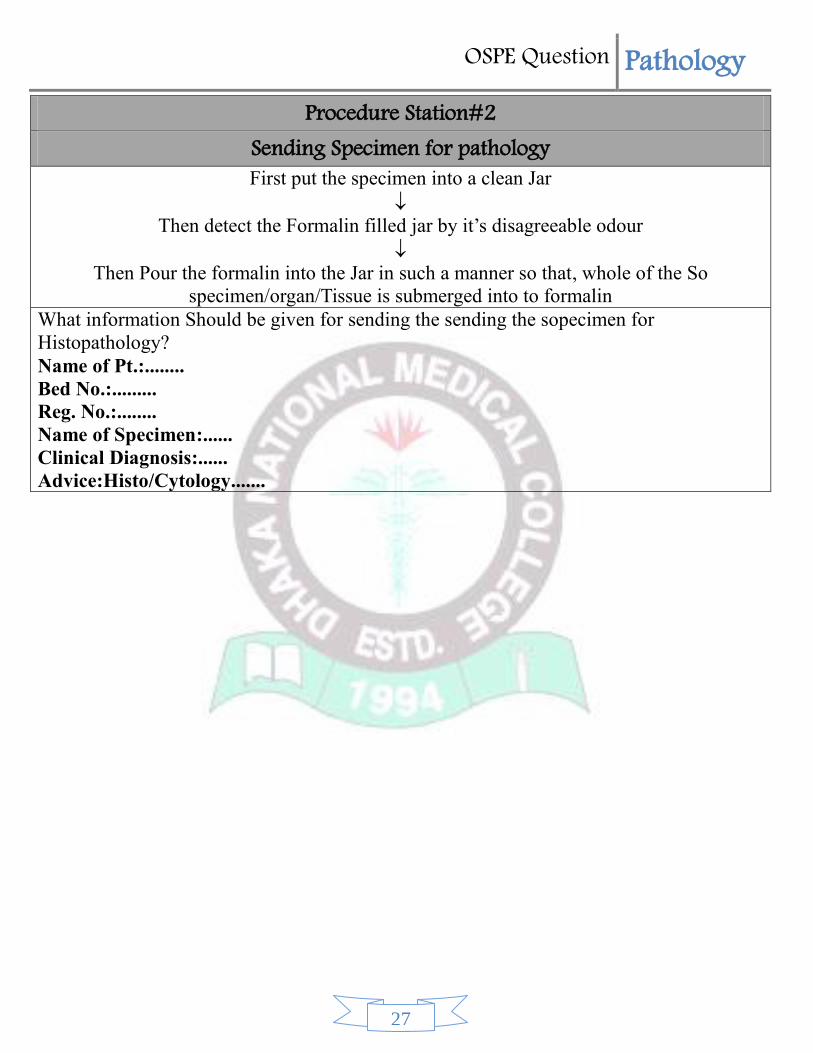
Procedure Station#2 Sending Specimen for pathology First put the specimen into a clean Jar
¯ Then detect the Formalin filled jar by it’s disagreeable odour
¯ Then Pour the formalin into the Jar in such a manner so that, whole of the So
specimen/organ/Tissue is submerged into to formalin What information Should be given for sending the sending the sopecimen for
Histopathology?
Name of Pt.:........
Bed No.:.........
Reg. No.:........
Name of Specimen:......
Clinical Diagnosis:......
Advice:Histo/Cytology.......


![[OSPE] Praktikum](https://img.dokumen.tips/doc/110x75/54e1ea4e4a7959f2578b4a5b/ospe-praktikum.jpg)