Embed Size (px)
Citation preview
Vol. 51, No. 3INFECTION AND IMMUNITY, Mar. 1986, p. 975-9780019-9567/86/030975-04$02.00/0Copyright (© 1986, American Society for Microbiology
Effect of Guinea Pig or Monkey Colonic Mucus on ShigellaAggregation and Invasion of HeLa Cells by Shigella flexneri
lb and 2aGABRIEL DINARI,' THOMAS L. HALE,2* OTHELLO WASHINGTON,2 AND SAMUEL B. FORMAL2
Pediatric Gastroenterology Unit, Beilinson Medical Center, Petah Tiqva 49100, Israel1 and Department ofEnteric Infections, Division of Communicable Diseases and Immunology, Walter Reed Army Institute of Research,
Washington, D.C. 203072
Received 19 July 1985/Accepted 23 November 1985
The effects of guinea pig and rhesus monkey colonic mucus preparations on Shigella aggregation andinvasion of HeLa cell monolayers by Shigellaflexneri serotype lb, 2a, and 5 strains were investigated. Guineapig mucus caused agglutination of S. flexneri serotype lb but not of S. flexneri serotype 2a or 5. Guinea pigmucus also inhibited HeLa cell invasion by S. flexneri serotypes lb and 2a. Monkey mucus neither agglutinatedany Shigella strain nor inhibited HeLa cell invasion.
Invasion of intestinal epithelium by virulent shigellae is acomplex process, which depends on both host factors andbacterial factors (8, 10) and occurs naturally only in humansand subhuman primates (16, 17). Adherence to the intestinalmucosa, which is an important early stage for colonizationand virulence expression of many microorganisms (15),occurs when various animal species, such as guinea pigs andmice, are exposed to Shigella strains under laboratoryconditions (1, 7, 11).
birth, but it is secreted at adult levels by about 2 weeks ofage (1). Preliminary results suggest that the molecular weightof the adhesin subunits is less than 60,000 (D. Mirelman, Y.Nuchamowitz, and M. Izhar, Abstr. J. Cell. Biochem.15(Suppl. 7A):9, 1983), and, since the adhesin can be par-tially washed off the mucosa, it appears to be one of thecomponents of the colonic mucus that coats the epithelialcells. In addition, Duguid and Gillies have found that guineapig mucus agglutinates S. flexneri (3), and more recently
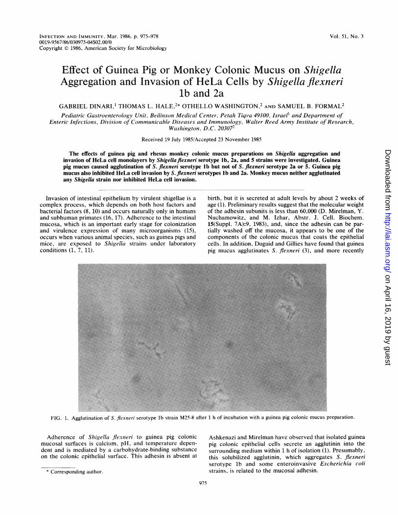
FIG. 1. Agglutination of S. flexneri serotype lb strain M25-8 after 1 h of incubation with a guinea pig colonic mucus preparation.
Adherence of Shigella flexneri to guinea pig colonicmucosal surfaces is calcium, pH, and temperature depen-dent and is mediated by a carbohydrate-binding substanceon the colonic epithelial surface. This adhesin is absent at
* Corresponding author.
Ashkenazi and Mirelman have observed that isolated guineapig colonic epithelial cells secrete an agglutinin into thesurrounding medium within 1 h of isolation (1). Presumably,this solubilized agglutinin, which aggregates S. flexneriserotype lb and some enteroinvasive Escherichia colistrains, is related to the mucosal adhesin.
975
on April 16, 2019 by guest
http://iai.asm.org/
Dow
nloaded from

976 NOTES
TABLE 1. Inhibition of Helta cell invasion by preincubation ofbacteria with different dilutions of guinea pig colonic mucus
preparations'% Cells invaded by S. flxnieri preinculbated
D)ilutionofwith guinea pig MLICULSmucus Serotype lb Scrotype 2a
strain M25-8 strain 2457T(/I = 9) (n = 7)
Undiluted 31.4 ± 6.9" 36.4 + 5.0"1:2 37.5 ± 5.8" 51.3 + 3.4"1:4 73.2 ± 4.2' 41.6 + 3.0"1:8 75.1 ± 6.6 40.1 + 3.1"1:16 79.1 ± 4.3 48.7 + 4.7"
Controls 100 83.6 ± 2.8
When we preincubated S. le.vneri serotype lb str-ain M25-8 or serotvpe 2astrain 2457T with different dilutions of monkey colonic mucus. 10)0.f of theHeLa cells were invaded. S. fle.v-neri serotype lb was agglutinated by themucus preparations, while serotype 2a was not. Results are expressed asmeans ± standard errors of the mean."P < 0.01 compared with controls.P < 0.05 compatred with controls.
"P < 0.001 compaired with controls.
Although the physiologic role of this adhesin is unclear, ithas been suggested that its presence in the colonic mucuscauses bacteria to agglutinate in the mucus layer and facili-tates the removal of the bacteria from the intestinal tract byperistalsis (1). This host defense mechanism might be sup-pressed by opiates and starvation, treatments which greatlyenhance the susceptibility of guinea pigs to Shigella infec-
tions (4, 17). Therefore, it is possible that normal gut motilityand mucus secretion protect the guinea pig mucosa and thatthe mucus agglutinin is involved in this protection. Sincerhesus monkeys are much more susceptible to Shigellainfections than guinea pigs, we compared the abilities ofmucus preparations from these two species to aggregatevarious S. flexnielri serotypes and to inhibit the invasion ofHeLa cells by these organisms.Crude mucus was isolated from the colons of 300- to 400-g
Hartley guinea pigs (Charles River Breeding Laboratories,Inc., Wilmington, Mass.) by using the method described byLaux et al. (12). Briefly, the colon distal to the hepaticflexure was removed, the mucosal layer with the overlyingmucus gel was scraped with a scalpel, placed in 4 ml ofHEPES (N-2-hydroxyethylpiperazine-N'-2-ethanesulfonicacid)-Hanks buffer, and centrifuged at 30,000 x g for 25 min,and the supernatant was collected. Protein levels weredetermined (13), and the protein concentration was adjustedto 2.5 mg/ml. Colons from 1- to 2-year-old rhesus monkeys(sacrificed by penthotal overdose as part of a polio vaccineefficacy evaluation protocol, in which the central nervoussystems of monkeys vaccinated 3 to 4 months earlier wereexamined for evidence of disease) were processed in anidentical manner, except that the protein concentration wasadjusted to 5 mg/ml. All mucus preparations were frozen at-20°C until they were used.Samples (100 ,ul) of twofold serial dilutions of crude mucus
prepared in basal medium Eagle (GIBCO Laboratories,Grand Island, N.Y.) were mixed in 96-well plates with 100-RlIportions of logarithmic-phase bacterial cultures grown inPenassay broth (Difco Laboratories, Detroit, Mich.). The
..1
M..
FIG. 2. Inhibition of HeLa cell invasion by S. flexneri serotype lb strain M25-8 after prior incubation with guinea pig colonic mucus. Onlya few cells were invaded. Magnification, x 1200.
INFECT. IMMUN.
.0
:: -.4
Awk.
1, p
..f.
on April 16, 2019 by guest
http://iai.asm.org/
Dow
nloaded from
NOTES 977
t
.. 4.
i k.
#;...p
/1iv,~~ I4. 0
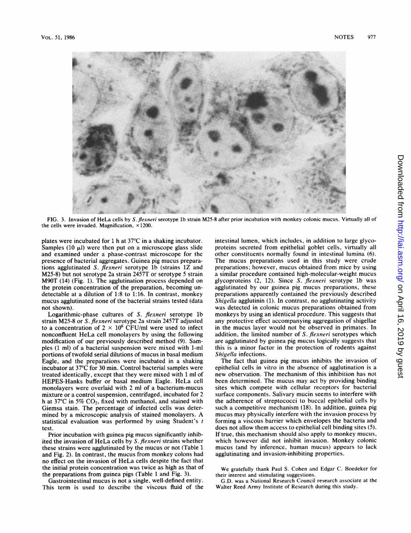
FIG. 3. Invasion of HeLa cells by S. flexneri serotype lb strain M25-8 after prior incubation with monkey colonic mucus. Virtually all ofthe cells were invaded. Magnification, x 1200.
plates were incubated for 1 h at 37°C in a shaking incubator.Samples (10 ,ul) were then put on a microscope glass slideand examined under a phase-contrast microscope for thepresence of bacterial aggregates. Guinea pig mucus prepara-tions agglutinated S. flexneri serotype lb (strains 1Z andM25-8) but not serotype 2a strain 2457T or serotype 5 strainM9OT (14) (Fig. 1). The agglutination process depended onthe protein concentration of the preparation, becoming un-detectable at a dilution of 1:8 to 1:16. In contrast, monkeymucus agglutinated none of the bacterial strains tested (datanot shown).
Logarithmic-phase cultures of S. flexneri serotype lbstrain M25-8 or S. flexneri serotype 2a strain 2457T adjustedto a concentration of 2 x 10⁸ CFU/ml were used to infectnonconfluent HeLa cell monolayers by using the followingmodification of our previously described method (9). Sam-ples (1 ml) of a bacterial suspension were mixed with 1-mlportions of twofold serial dilutions of mucus in basal mediumEagle, and the preparations were incubated in a shakingincubator at 37°C for 30 min. Control bacterial samples weretreated identically, except that they were mixed with 1 ml ofHEPES-Hanks buffer or basal medium Eagle. HeLa cellmonolayers were overlaid with 2 ml of a bacterium-mucusmixture or a control suspension, centrifuged, incubated for 2h at 37°C in 5% CO2, fixed with methanol, and stained withGiemsa stain. The percentage of infected cells was deter-mined by a microscopic analysis of stained monolayers. Astatistical evaluation was performed by using Student's ttest.
Prior incubation with guinea pig mucus significantly inhib-ited the invasion of HeLa cells by S. flexneri strains whetherthese strains were agglutinated by the mucus or not (Table 1and Fig. 2). In contrast, the mucus from monkey colons hadno effect on the invasion of HeLa cells despite the fact thatthe initial protein concentration was twice as high as that ofthe preparations from guinea pigs (Table 1 and Fig. 3).
Gastrointestinal mucus is not a single, well-defined entity.This term is used to describe the viscous fluid of the
intestinal lumen, which includes, in addition to large glyco-proteins secreted from epithelial goblet cells, virtually allother constituents normally found in intestinal lumina (6).The mucus preparations used in this study were crudepreparations; however, mucus obtained from mice by usinga similar procedure contained high-molecular-weight mucusglycoproteins (2, 12). Since S. flexneri serotype lb wasagglutinated by our guinea pig mucus preparations, thesepreparations apparently contained the previously describedShigella agglutinin (1). In contrast, no agglutinating activitywas detected in colonic mucus preparations obtained frommonkeys by using an identical procedure. This suggests thatany protective effect accompanying aggregation of shigellaein the mucus layer would not be observed in primates. Inaddition, the limited number of S. flexneri serotypes whichare agglutinated by guinea pig mucus logically suggests thatthis is a minor factor in the protection of rodents againstShigella infections.The fact that guinea pig mucus inhibits the invasion of
epithelial cells in vitro in the absence of agglutination is anew observation. The mechanism of this inhibition has notbeen determined. The mucus may act by providing bindingsites which compete with cellular receptors for bacterialsurface components. Salivary mucin seems to interfere withthe adherence of streptococci to buccal epithelial cells bysuch a competitive mechanism (18). In addition, guinea pigmucus may physically interfere with the invasion process byforming a viscous barrier which envelopes the bacteria anddoes not allow them access to epithelial cell binding sites (5).If true, this mechanism should also apply to monkey mucus,which however did not inhibit invasion. Monkey colonicmucus (and by inference, human mucus) appears to lackagglutinating and invasion-inhibiting properties.
We gratefully thank Paul S. Cohen and Edgar C. Boedeker fortheir interest and stimulating suggestions.G.D. was a National Research Council research associate at the
Walter Reed Army institute of Research during this study.
VOL. 51, 1986
A.F.
.OJI
*:
Womol..:
t.
11 'I- ..-i"or-41 k
on April 16, 2019 by guest
http://iai.asm.org/
Dow
nloaded from

INFECT. IMMUN.
LITERATURE CITED1. Ashkenazi, S., and D. Mirelman. 1984. The effect of postnatal
age on the adherence of Shigellaflexneri, Escherichia coli 0124,and E. coli 0128 to guinea pig intestinal cells. Pediatr. Res.18:1366-1371.
2. Cohen, P. S., J. C. Arruda, T. J. Williams, and D. C. Laux.1985. Adhesion of a human fecal Escherichia coli strain tomouse colonic mucus. Infect. Immun. 48:139-145.
3. Duguid, J. P., and R. R. Gillies. 1957. Fimbriae and adhesiveproperties in dysentery bacilli. J. Pathol. Bacteriol. 74:397-411.
4. Formal, S. B., G. J. Dammin, E. H. LaBrec, and H. Schneider.1958. Experimental shigella infections: characteristics of fatalinfection in guinea pigs. J. Bacteriol. 46:1-13.
5. Forstner, G., A. Wesley, and J. F. Forstner. 1982. Clinicalaspects of gastrointestinal mucus. Adv. Exp. Med. Biol.144:199-224.
6. Forstner, J. F. 1978. Intestinal mucins in health and disease.Digestion 1:234-263.
7. Golderman, L., and E. Rubinstein. 1982. Salmonella and Shi-gella adherence to the intestine of mice. Isr. J. Med. Sci.18:1032-1036.
8. Hale, T. L., and P. F. Bonventre. 1979. Shigella infection ofHenle intestinal epithelial cells. Role of the bacterium. Infect.Immun. 24:879-886.
9. Hale, T. L., and S. B. Formal. 1981. Protein synthesis in HeLaor Henle 407 cells infected with Shigella dysenteriae 1, Shigellaflexneri 2a, or Salmonella typhimurium W118. Infect. Immun.32:137-144.
10. Hale, T. L., R. E. Morris, and P. F. Bonventre. 1979. Shigellainfection of Henle epithelial intestinal cells. Role of the hostcell. Infect. Immun. 24:887-894.
11. Izhar, M., Y. Nuchamowitz, and D. Mirelman. 1982. Adherenceof Shigellaflexneri to guinea pig intestinal cells is mediated by amucosal adhesin. Infect. Immun. 35:1110-1118.
12. Laux, D. C., E. F. McSweegan, and P. S. Cohen. 1984. Adhesionof enterotoxigenic Escherichia coli to immobilized intestinalmucosal preparation: a model for adhesion to mucosal surfacecomponents. J. Microbiol. Methods 2:27-39.
13. Lowry, 0. H., N. J. Rosebrough, A. L. Farr, and R. J. Randall.1951. Protein measurement with the Folin phenol reagent. J.Biol. Chem. 193:265-275.
14. Oaks, E. V., M. E. Wingfield, and S. B. Formal. 1985. Plaqueformation by virulent Shigella flexneri. Infect. Immun.48:124-129.
15. Ofek, I., and E. H. Beachey. 1980. General concepts andprinciples of bacterial adherence in animal and man, p. 2-29. InE. H. Beachey (ed.), Bacterial adherence, series B, vol. 6.Receptors and recognition. Chapman & Hall, London.
16. Takeuchi, A., H. R. Jervis, and S. B. Formal. 1975. Monkeyshigellosis or dysentery. Am. J. Pathol. 81:251-254.
17. Takeuchi, A., H. Sprinz, E. H. LaBrec, and S. B. Formal. 1965.Experimental bacillary dysentery. Am. J. Pathol. 47:1011-1044.
18. Williams, R. C., and R. J. Gibbons. 1975. Inhibition of strepto-coccal attachment to receptors on human buccal epithelial cellsby antigenically similar salivary glycoproteins. Infect. Immun.11:711-718.
978 NOTES
on April 16, 2019 by guest
http://iai.asm.org/
Dow
nloaded from