Embed Size (px)
Citation preview
Sensors 2015, 15, 465-484; doi:10.3390/s150100465
sensors ISSN 1424-8220
www.mdpi.com/journal/sensors
Review
Optofluidic Approaches for Enhanced Microsensor Performances
Genni Testa *, Gianluca Persichetti and Romeo Bernini
Institute for Electromagnetic Monitoring of the Environment (IREA), National Research Council (CNR),
Via Diocleziano, 328, Naples 80124, Italy; E-Mails: [email protected] (G.P.);
[email protected] (R.B.)
* Author to whom correspondence should be addressed; E-Mail: [email protected];
Tel.: +39-081-7620-643; Fax: +39-081-5705-734.
Academic Editor: Stefano Mariani
Received: 11 November 2014 / Accepted: 15 December 2014 / Published: 30 December 2014
Abstract: Optofluidics is a relatively young research field able to create a tight synergy
between optics and micro/nano-fluidics. The high level of integration between fluidic and
optical elements achievable by means of optofluidic approaches makes it possible to
realize an innovative class of sensors, which have been demonstrated to have an improved
sensitivity, adaptability and compactness. Many developments in this field have been made
in the last years thanks to the availability of a new class of low cost materials and new
technologies. This review describes the Italian state of art on optofluidic devices for
sensing applications and offers a perspective for further future advances. We introduce the
optofluidic concept and describe the advantages of merging photonic and fluidic elements,
focusing on sensor developments for both environmental and biomedical monitoring.
Keywords: optofluidics; sensors; microfluidic; ring resonator; Mach-Zehnder; liquid core
waveguides; lab-on-a-chip (LOC); micro total analysis systems (µTAS)
1. Introduction
Optofluidics is a rapidly growing interdisciplinary research field combining micro/nano-fluidics and
optics in a very fascinating and valuable way [1]. The concept behind optofluidics is to build up new
forms of reconfigurable photonics in which optics and fluidics enable each other. This approach has
had a strong impact in the field of optical sensing, leading to the development of a new class of
OPEN ACCESS
Sensors 2015, 15 466
innovative and reconfigurable optofluidic sensors where the fluidic is exploited not only to efficiently
handle the sample to be tested, but also as an optical material in order to reconfigure the photonic part
of microdevices. Not surprisingly, recent advances in optofluidics have been supported by recent
developments in microfluidics, which have led to simpler and precise control of fluids on the micron
scale [2], opening new ways to tune microphotonic devices. Additional advantages can be gained by
the distinctive properties of fluids interfaces of being naturally smooth and flexible. Some examples of
such adaptable devices include fluidic lenses [3] and optofluidic light source [4,5].
In the context of sensing applications, optofluidics arises from the need to hold up very high sensor
sensitivity with smaller sample consumption and miniaturized devices. The growing requests of
compact devices for complete biochemical and chemical analysis on micron-sized scale have
recently seen the development of the so called lab on a chips (LOCs) or micro total analysis systems
(µTASs) [6,7]. The optofluidic approaches allow adding further advances to confer optical functions
on fluidic elements to such systems. In an optofluidic device microfluidic and optical parts are not
separate entities, merely integrated on the same chip, but rather they are strongly tied together,
resulting in high interaction efficiency between light and fluids, which is very desirable for sensing
applications. The simplest, but not less remarkable example of this mutual correlation is offered by the
liquid core optical waveguides, where the fluid is a constituent part of the photonic structure itself [8].
In order to increase device sensitivity, optofluidic sensors are often realized by employing liquid core
waveguides or by suitably integrating microfluidic channels along the optical path. In the former case
the fluid provides the means to guide the light, enabling a maximized optical coupling across the entire
photonic structure. Different approaches for optofluidic sensors consist in exploiting inherent fluidic
capability of photonic structure, while preserving evanescent coupling between light and fluids.
Fluids can be efficiently used to transport cells or other biological molecules that are typically
suspended in aqueous solutions. In particular, since the detection volumes can be very small (typically
of the order of nL), optofluidic microsensors are ideally suited for carrying out single molecule
detection [9]. Moreover, the possibility to drive fluids into and out of the fluidic handling system with
a continuous piping network makes these devices very attractive for ambient bio-threat detection,
where the local environment is continuously tested online [10].
In the following, we describe the recent progress in optofluidic approaches for sensing applications
focusing on Italian developments. We firstly offer an overview on optofluidic waveguides, as they
often represent the building block of an optofluidic sensor. We present optofluidic devices for
spectroscopy and discuss the advantages of the proposed approaches. Optofluidic sensors are also
presented and classified on the basis of the basic photonic structure; in particular, interferometric and
resonant sensors and their applications are described.
2. Optofluidic Waveguides
An optofluidic waveguide can be defined as a structure able to perform optical confinement and
transmission of light through a fluid. Optofluidic waveguide represents one of the more significant
elements of an optofluidic sensor. As in the approach for solid state optical waveguides, the more
obvious solution to confine the light in optofluidic waveguides is based on the total internal reflection
(TIR) effect. TIR arises when light propagates, at angles greater than the critical angle, into a material

Sensors 2015, 15 467
(core) with refractive index nc surrounded by another material with lower refractive index ncl
(cladding). The guiding of the light into a liquid core has been proposed in the 1970s, long before the
concept of optofluidics was defined, by introducing liquid-waveguide-capillary cells (LWCCs) [11]. In
their seminal work, Walrafen and Stone reported intensity enhancement in Raman spectroscopy by a
factor of 3000 with respect conventional approaches, using fused quartz capillaries up to 25 m in
length, filled with benzene and tetrachloroethylene.
However, as fused silica and other solid materials commonly employed for microelectronic and
microfluidic fabrication exhibit higher refractive indexes (1.4–3.5), the condition to fulfill for TIR
propagation (nc > ncl) poses a severe limitation in the possible liquid core refractive index for
optofluidic waveguides. In particular, the use of water solutions (nH2O = 1.33), of great interest in
biological applications, is precluded. Most of the optofluidic waveguides described in the following
represent the technological and scientific effort to overcome this problem. The first solution was
achieved with liquid core waveguides made entirely of Teflon AF2400 (n = 1.29 < 1.33). However,
despite the fact this material has a suitable refractive index, it also exhibits autofluorescence that
contributes to increasing the background noise measurement.
2.1. ARROWs Waveguides
Very interesting approaches to confine the light in low index media are based on the use of
interference effect in cladding layers able to reflect light back into the core. Liquid core antiresonant
reflecting optical waveguides (ARROWs) use such an effect for light propagation [12,13]. A cross
section of an ARROW with two layers is represented in Figure 1a. The confinement mechanism is
based on the interferences arising in the multilayered structure between reflected and refracted rays in
the same way as in Fabry-Pérot mirrors, if specific conditions are fulfilled (antiresonant conditions).
The multilayer is designed to operate like a high reflectivity mirror (reflectivity up to 99% can be
obtained with four cladding layers) at a specific wavelength. The transmitted spectrum is characterized
by a broad spectral range around the design wavelength. ARROWs are fabricated in silicon substrate
by employing typical dielectric materials such as SiO2 and Si3N4 for claddings deposition, which are
widely compatible with standard silicon process. Recently in the work of Testa et al. [14], TiO2
deposition by atomic layer deposition (ALD) technique was used to fabricate the high index cladding
layer in order to improve the overall optical performance of these waveguides by taking advantage of
improved conformality, uniformity and reduced surface roughness which are typical of ALD.
Since 2004, Bernini et al. [15] have demonstrated multimode propagation in ARROW proposing a
structure able to guide light in low index media (air, gases, and liquids). A very important inherent
property of ARROWs is that propagating modes experience attenuation losses depending upon their
state of polarization. In particular, the core geometry can be properly chosen in order to select only one
polarization state (TE or TM) [12]. Even more significantly, ARROWs can be designed to operate on
single polarized optical mode, which allow these waveguides to be applied for designing
interferometric optofluidic devices.
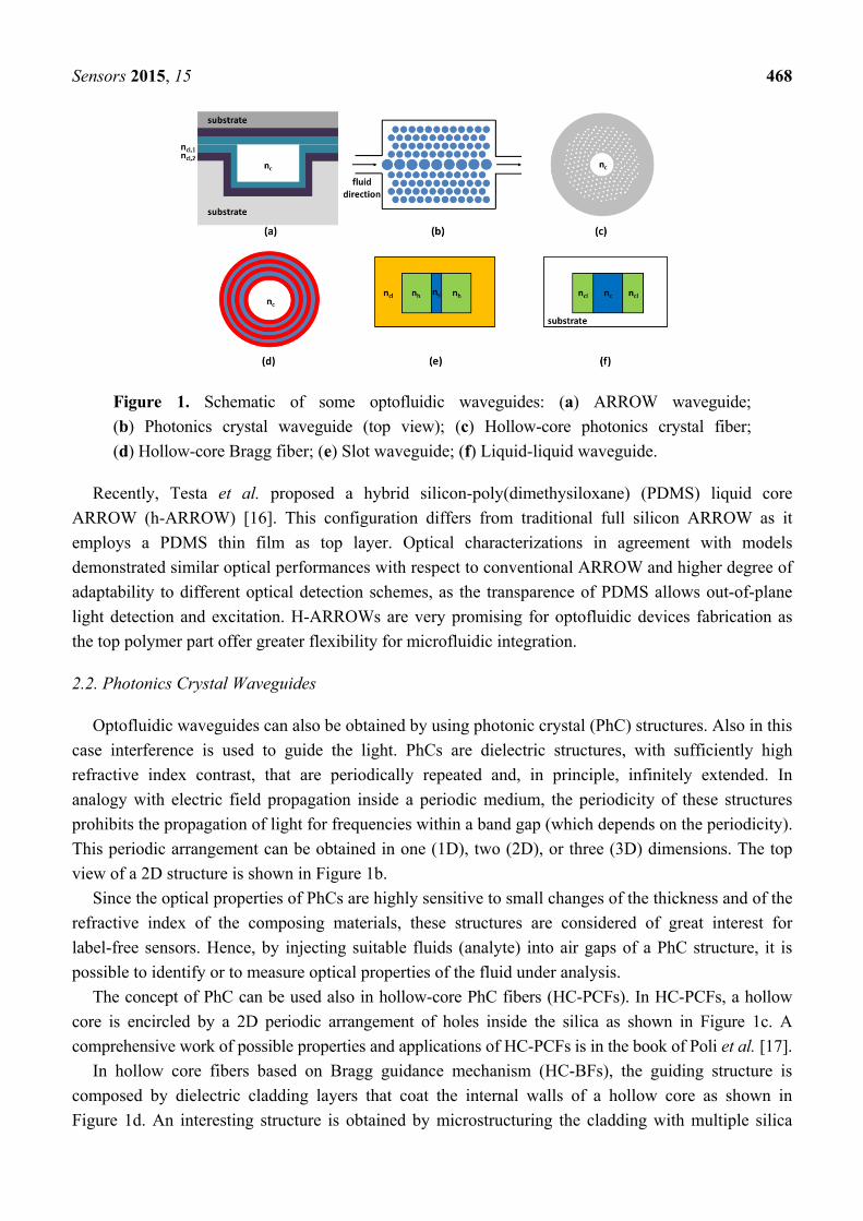
Sensors 2015, 15 468
Figure 1. Schematic of some optofluidic waveguides: (a) ARROW waveguide;
(b) Photonics crystal waveguide (top view); (c) Hollow-core photonics crystal fiber;
(d) Hollow-core Bragg fiber; (e) Slot waveguide; (f) Liquid-liquid waveguide.
Recently, Testa et al. proposed a hybrid silicon-poly(dimethysiloxane) (PDMS) liquid core
ARROW (h-ARROW) [16]. This configuration differs from traditional full silicon ARROW as it
employs a PDMS thin film as top layer. Optical characterizations in agreement with models
demonstrated similar optical performances with respect to conventional ARROW and higher degree of
adaptability to different optical detection schemes, as the transparence of PDMS allows out-of-plane
light detection and excitation. H-ARROWs are very promising for optofluidic devices fabrication as
the top polymer part offer greater flexibility for microfluidic integration.
2.2. Photonics Crystal Waveguides
Optofluidic waveguides can also be obtained by using photonic crystal (PhC) structures. Also in this
case interference is used to guide the light. PhCs are dielectric structures, with sufficiently high
refractive index contrast, that are periodically repeated and, in principle, infinitely extended. In
analogy with electric field propagation inside a periodic medium, the periodicity of these structures
prohibits the propagation of light for frequencies within a band gap (which depends on the periodicity).
This periodic arrangement can be obtained in one (1D), two (2D), or three (3D) dimensions. The top
view of a 2D structure is shown in Figure 1b.
Since the optical properties of PhCs are highly sensitive to small changes of the thickness and of the
refractive index of the composing materials, these structures are considered of great interest for
label-free sensors. Hence, by injecting suitable fluids (analyte) into air gaps of a PhC structure, it is
possible to identify or to measure optical properties of the fluid under analysis.
The concept of PhC can be used also in hollow-core PhC fibers (HC-PCFs). In HC-PCFs, a hollow
core is encircled by a 2D periodic arrangement of holes inside the silica as shown in Figure 1c. A
comprehensive work of possible properties and applications of HC-PCFs is in the book of Poli et al. [17].
In hollow core fibers based on Bragg guidance mechanism (HC-BFs), the guiding structure is
composed by dielectric cladding layers that coat the internal walls of a hollow core as shown in
Figure 1d. An interesting structure is obtained by microstructuring the cladding with multiple silica

Sensors 2015, 15 469
rings joint by nanoscale silica supports. These waveguide, called all-silica hollow-core microstructured
Bragg fibers, have been investigated and proposed for biosensing application by Passaro et al. [18].
PhC fibers are not limited at hollow core structures as also solid core with microstructured cladding
are possible. Among possible solid-core PhC fibers, suspended-core PhC fibers (SC-PhCFs) seem to
be very promising for developing efficient biological sensors [19]. The cross-section of a SC-PhCF is
composed of a solid rod suspended by three thin supports and it is surrounded by noncircular air-holes.
In particular, those waveguides have been investigated for a highly specific DNA biosensor by
Coscelli et al. [20].
2.3. Slot Waveguides
The typical core sizes of a liquid waveguide range from few microns to some hundreds of microns.
However, waveguiding properties can be realized on a nanoscale cross section, considering slot
waveguides (SWs). This approach has been proposed in 2004 by Almeida et al. [21] and subsequently
demonstrated experimentally by the same group [22]. The structure consists in a nanometer sized low
refractive index (ns) slot between two region with high refractive index (nh). The whole structure is
surrounded by a cladding with low refractive index ncl. A typical cross section of a SW waveguide is
shown in Figure 1e.
In a SW, an enhanced and confined optical field is observed in the low refractive index (ns) material
slot even if light propagates exploiting TIR. As the normal component of the electric displacement
field D has to be continuous at the interface of two dielectric materials, the normal component of the
electric field E experiences a discontinuity at this interface (D = n2ε0E where n and ε0 are the refractive
index and the vacuum permittivity, respectively). In particular, the normal component of E has to be
higher at the low-index side and lower at the high-index side. In structures with high index contrast as
SW, this discontinuity is particularly significant and up to 30% of the total modal power is confined in
the slot region in a 100 nm narrow core considering a high index contrast system such as Si/air [21].
The small size where the electric field is confined (which is comparable to the decay length of E)
and the high intensities available in a SW, make those structures very attractive for optofluidic
applications. For this purpose, extensive investigation has been carried out in order to optimize
the sensing properties of SW. In particular, in the works of Passaro et al., the optimization of
silicon-on-insulator SW has been carried out considering also the influence of fabrication tolerances in
order to enhance optical sensing [23–25].
Silicon nanocrystals-based sandwiched SWs has been theoretically investigated by Passaro et al. [26].
By changing the upper layer thickness only of few nanometers, the authors demonstrated, for the first
time, the possibility to create or to annihilate an optical solitons inside those waveguides.
Moreover, Bettotti et al. [27] explored the possibility of using SWs realized by considering very
low refractive index materials such as for instance, polymers. Since polymers are particularly flexible
materials (and transparent at visible wavelengths) also allowing surface functionalization, the proposed
SWs are very promising optical waveguides in sensors designing.
Recently, a novel configuration called Layer-slot (L-slot) has been proposed by Testa et al. [28],
which is able to operate in the visible wavelength range. In this range the optical absorption of water is
about three orders of magnitude lower than in IR region, reducing optical losses and thermal heating.
Sensors 2015, 15 470
In addition, the proposed configuration contributes to simplify the SW fabrication process as it is based
on a deposition step instead of the conventional etching processes, hence high-resolution techniques
are not required.
2.4. Liquid-Liquid Waveguides
Another approach to realize an optical waveguide consists in using liquids cladding inside a larger
fluidic channel. In this approach, by adopting a suitable flow rate, the two liquids will flow in laminar
condition and the only diffusion process will be responsible of the mixing of the fluids. This means
that the waveguiding properties are ensured by TIR, until the cladding refractive index ncl remains
smaller than the core refractive index nc and the cladding thickness is reduced to a few tens of microns.
A cross section of such a liquid-liquid waveguide (L2) is shown in Figure 1f. The L2 approach has
been successfully demonstrated by Wolfe et al. [29] employing CaCl2 water solution (n = 1.445) as
core and water as cladding, both embedded in PDMS (n = 1.4).
Manipulating the flow rate and the liquids composition, it is possible to tune many optical
properties, such as for instance refractive index contrast, size and loss/gain properties. The attainable
optical tunability is one of the more interesting properties offered by L2 waveguides. The L2 concept
has been extensively proposed in a 2D geometry, i.e., the liquid core is bordering on the liquid
claddings only in one transverse direction, whereas in the other transverse direction, the liquids are
surrounded by solid materials. This circumstance leads to detrimental effects on the waveguiding
properties, preventing the possibility to use water and very dilute water solution as core materials. A
possible solution has been proposed by Bernini et al. [30] by adopting a hybrid approach in which L2
waveguide is accomplished in the microfluidic channel of an ARROW waveguide, providing light
confinement in both of the transverse directions. This same difficulty has been differently solved by
exploiting 3D hydrodynamic focusing effect [31]. In this 3D approach, the liquid core is completely
surrounded by the liquid cladding using fluid flows from concentric glass capillaries. Since the first
implementation of a 3D hydrodynamic focusing effect, proposed by Takiguchi et al., several
improvements have been proposed [32–34]. In 2012, Testa et al. [35] numerically and experimentally
demonstrated an innovative scheme, with reduced input channels, for 3D hydrodynamic focusing
which is able to produce a tunable and circular liquid core placed in the center of the focusing channel.
2.5. Jet Waveguides
The last optofluidic waveguide we describe is based on a simple and clever idea coming from the
past. The first demonstration of an optofluidic waveguide was given almost two centuries ago, when
the waveguiding nature of a water jet was discovered and explained by Colladon [36] as due to TIR at
the water-air interface. Microfluidic liquid jets are regular cylinder of fluids in air that can be obtained
by injecting the liquid into a channel or a capillary at a specific range of flow rate, depending on the
channel size diameter. This regular shape in the jet is observed up to a specific length (breakup
length).Beyond this length, the jet breaks up into drops.
As other liquid core waveguides, jet waveguides exploit TIR for light propagation. However, a very
high refractive index contrast between the core (liquid) and cladding (air) leads to a very high
numerical aperture (NA = 0.88 for water core). Hence, in principle, liquid jets exhibit significant

Sensors 2015, 15 471
collection efficiency. The advantages of these simple but effective optical waveguides have been
neglected until recent times [37] when the group led by Bernini proposed an experimental
configuration able to overcome the typical complications arising in conventional approaches. Liquid
core waveguide with solid or liquid cladding are usually surrounded by solid structures that are
potential sources of scattering (by surface roughness) or autofluorescence. Beside the advantage
related to its collection efficiency, in a jet waveguide, the absence of solid walls to contain the liquid
waveguide and the smoothness of the liquid/air surface enable very low background signal arising
from scattering.
3. Optofluidic Devices for Spectroscopy
Spectroscopy methods like absorption, fluorescence, and Raman are mostly used as optical
detection methods, as they enable highly selective and sensitive detection of analytes at a very low
concentration level. However, device miniaturization has the unavoidable effect of reducing the
interaction length of light with fluid due to the overall device size reduction, thus limiting the sensor
sensitivity [38]. The use of optofluidic waveguides or microfluidic channels designed to catch directly
the optical probe enables an enhanced optical interaction with sample under analysis and hence an
increased sensitivity. Optofluidic chips, besides retaining the advantages of high optical sensitivity, have
the potential to address the growing needs of reduced device size, low sample consumption and field
deployability. In the following we present the recent progress in development of optofluidic devices for
spectroscopic analysis of fluids. Among others spectroscopic methods, fluorescence and Raman
spectroscopy have been demonstrated as very sensitive techniques; analysis and detection of molecules
on the single particle level has also been demonstrated by implementing these techniques on
optofluidic chip [39,40].
In this scenario liquid core ARROWs play a very remarkable role. Liquid core waveguides have the
inherent advantage of exposing the liquid analyte to the entire optical power as both light and liquid are
being guided through the same channels. As a consequence, maximized optical sensitivity in bulk
sensing distinguishes these waveguides among others. Moreover, like optofluidic slot- and
PC-waveguides, ARROWs are photonic structures that could be realized using silicon technology and
hence they are highly attractive for planar optofluidic integration [13]. The potentialities of these
waveguides in the field of optical sensing have been explored since the first demonstration of their
guiding capability [39,41]. In the work of Campopiano et al. [42], an integrated optical bulk
refractometer based on multimodal liquid ARROW has been demonstrated. The waveguide itself
constituted the optical sensor, with input and output optical fibers directly inserted in the waveguide
hollow core for the connection with off-chip optical source and detection system. The operating
principle was based on the shift of the minimum of the transmitted spectrum upon variation of the
refractive index (RI) of the fluid filling the core. A linear response and a sensitivity of about
555 nm/RIU were measured, with an LOD of 9 × 10⁻⁴ refractive index unit (RIU). Fluid
injection/ejection in the microfluidic core was achieved via lateral channels, orthogonally crossing the
core (Figure 2a). The potential for use in spectroscopy of the ARROW microfluidic channel has been
demonstrated by using the same configuration to realize a long path absorbance cell, for colorimetric
determination of concentration of specific protein in water solution [43]. In particular, by using

Sensors 2015, 15 472
Bradford assay, authors were able to detect bovine serum albumin with an LOD of about
1.5 × 10⁻³ mg/mL. Certainly these sensors, together with other existing examples in literature [39,41],
can be considered as starting points for ARROW based optofluidic chip development for spectroscopic
measurements. However, further advances can be gained by optofluidic approaches, obtained through
the integration of microfluidic components that allow a more precise control and handling of fluid
sample [44].
Figure 2. (a) Schematic of the microfluidic sensor for bulk refractometer RI measurements.
Reprinted with permission from [42] (Copyright (2004) OSA) (b) Schematic drawing of the
hybrid optofluidic platform. Reprinted with permission from [45] (Copyright (2014) OSA.)
Recently, in the work of Testa et al., an optofluidic platform based on hybrid liquid core ARROW
waveguides for fluorescence spectroscopy of liquids has been presented [45]. The chip was assembled
in a modular structure, with the upper polymeric part including solely microfluidic functionalities
(Figure 2b). The optical part has been realized developing a polymer-silicon hybrid solution in order to
form a fully integrated platform connected with the upwards microfluidic system. Solid core hybrid
ARROWs (solid h-ARROWs) have been suitably integrated in the polymeric part in a self-aligned
configuration with the liquid core waveguides containing the sample to be tested. This hybrid solution
allows us to seal the liquid h-ARROW and to couple light from/toward the exciting/collecting off-chip
optical fibers. The presented approach is unique among other as hybrid integration was implemented
for the fabrication of the both photonic and fluidic elements on the same chip. A first prototype has
been fabricated with integrated passive micromixer as microfluidic element. The sensing performance
of the device was tested by performing fluorescence measurements at different concentration,
controlled by means of the integrated micromixer; an LOD of 2.5 nM was demonstrated. The proposed
modular approach represents the major step toward a truly hybrid optofluidic chip and offers prospects
of high functional flexibility inherent to optofluidics, as the microfluidic part can be easily replaced
and adapted to different detection schemes.
Planar optofluidic chip has also been developed in bulk fused silica by using femtosecond laser
technology (FLT). FLT is a well-established fabrication technique that exploits refractive index
modifications induced by focused femtosecond pulses to inscribe photonic circuits in glass substrates;
it has also been demonstrated as a powerful tool for microfluidic chip fabrication [46]. In the work of

Sensors 2015, 15 473
Osellame et al., it has been demonstrated that FLT can be exploited to fabricate both microfluidic
channels and optical waveguides on the same fused silica substrate [47], paving the way towards the
realization of monolithic optofluidic devices. Even if polymer materials are going to became more and
more preferred for LOC fabrication, glass is still used in some applications. Based on FLT, an
optofluidic chip for dual-wavelength fluorescent DNA analysis is presented in the work of
Dongre et al. [48]. A microfluidic channel network has been suitably integrated with crossing
exciting/collecting optical waveguides by means of FLT on a fused silica glass slide, towards an on
chip fluorescence excitation of DNA molecules. Capillary electrophoresis has been implemented on
the chip for separation of fluorescently labeled-DNA molecules. Dual point, dual-wavelength laser
induced fluorescence has been used to optically resolve separation of two equally sized DNA
molecules, which could not be electrophoretically distinguished. Similar device design has also been
used to demonstrate a proof of principle color-end-labeled DNA identification by means of
modulation-frequency-encoded multi-wavelength excitation [49]. The potential of the proposed chips
makes them very interesting, in particular for prospective use in point-of-care diagnostics.
Spectroscopic optofluidic devices are also finding significant applications in the field of flow
cytometry for cell counting, analysis and sorting. In the last years, many research efforts have been
dedicated to find new solutions to the question of handling particles at a single level on planar
miniaturized structures [50]. Like in a bulk cytometer, cells must be organized in a single line to pass
through the detection region, where a collimated optical beam can provide single cell interrogation at a
time. Microfluidic strategies for arranging particles in a single line are mainly based on hydrodynamic
focusing effect. In this case, a sample flow with suspended cells is wrapped and squeezed by suitable
sheathing fluids. Miniaturization of flow cytometers with planar approach is a challenging objective
that has seen a growing interest by the research community in the last years [34,51]. An integrated
micro flow cytometer has been proposed in the work of Bernini et al. [52]. A hydrodynamically
focused stream of fluorescently-labeled human T leukemia cells (Jurkat) has been accomplished in the
optofluidic channel of an ARROW waveguide. The authors exploit the dual ability of an ARROW
waveguide to both serve as a microfluidic channel for flow focusing and efficiently confine the
excitation light for cell interrogation. Optical fibers for fluorescent detection have been integrated on
the same platform, orthogonally crossing the focusing channel to reduce the pump contribution to
detected signals. The same samples were also tested by using a bench-top flow cytometer. Qualitative
accordance has been achieved between results, demonstrating the validity of the proposed approach.
Sorting of cells is another very useful functionality in many biological applications. It can be
applied to isolate cells with specific properties like particular size and weight. By using FLT, an
optofluidic device implementing an integrated fluorescence-activated cell sorter has been
demonstrated [53]. The device comprises a microfluidic channel for cells flowing and two integrated
optical waveguides for cells fluorescent excitation and sorting via optical force, respectively. Optical
waveguides were arranged in an orthogonal scheme with the flow channel. The optical force is
activated when fluorescent cells pass through the interrogation region produced by the excitation
waveguide, where a CCD camera for fluorescent cells recognition is located. Isolation of subpopulations
with high selectivity from heterogeneous samples has been demonstrated by authors. A similar device
employing two facing optical waveguides, crossing the microfluidic channels, has been demonstrated
for optical trapping of single red blood cells by means of counterpropagating dual beams traps [54].
Sensors 2015, 15 474
These results are very promising in the view of realizing micro-sized devices for manipulation of cells
at the single particle level.
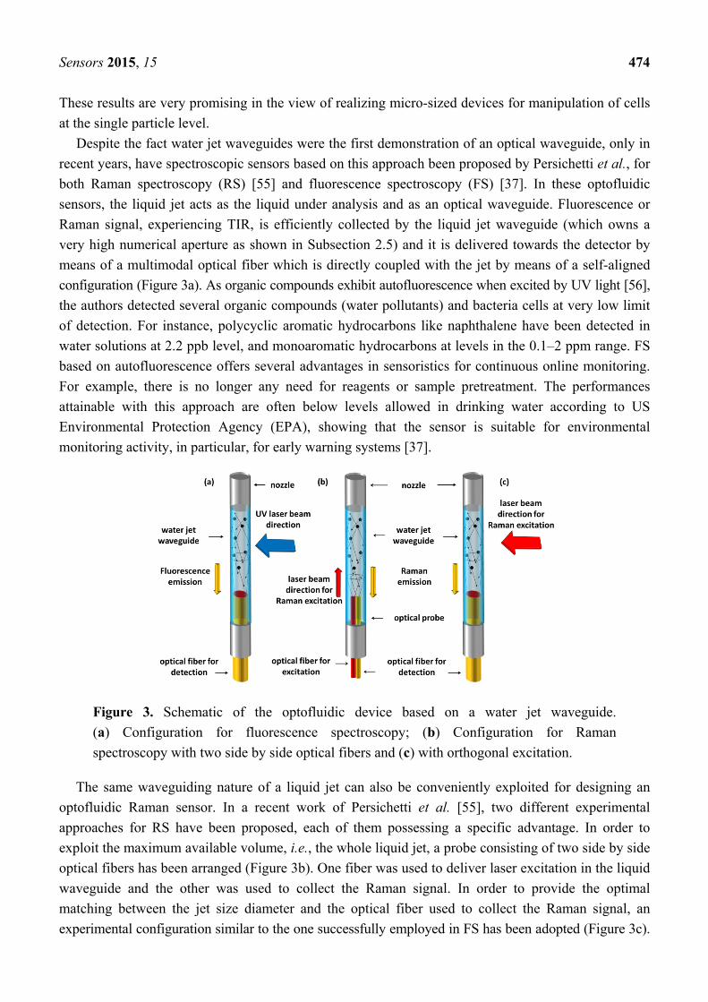
Despite the fact water jet waveguides were the first demonstration of an optical waveguide, only in
recent years, have spectroscopic sensors based on this approach been proposed by Persichetti et al., for
both Raman spectroscopy (RS) [55] and fluorescence spectroscopy (FS) [37]. In these optofluidic
sensors, the liquid jet acts as the liquid under analysis and as an optical waveguide. Fluorescence or
Raman signal, experiencing TIR, is efficiently collected by the liquid jet waveguide (which owns a
very high numerical aperture as shown in Subsection 2.5) and it is delivered towards the detector by
means of a multimodal optical fiber which is directly coupled with the jet by means of a self-aligned
configuration (Figure 3a). As organic compounds exhibit autofluorescence when excited by UV light [56],
the authors detected several organic compounds (water pollutants) and bacteria cells at very low limit
of detection. For instance, polycyclic aromatic hydrocarbons like naphthalene have been detected in
water solutions at 2.2 ppb level, and monoaromatic hydrocarbons at levels in the 0.1–2 ppm range. FS
based on autofluorescence offers several advantages in sensoristics for continuous online monitoring.
For example, there is no longer any need for reagents or sample pretreatment. The performances
attainable with this approach are often below levels allowed in drinking water according to US
Environmental Protection Agency (EPA), showing that the sensor is suitable for environmental
monitoring activity, in particular, for early warning systems [37].
Figure 3. Schematic of the optofluidic device based on a water jet waveguide.
(a) Configuration for fluorescence spectroscopy; (b) Configuration for Raman
spectroscopy with two side by side optical fibers and (c) with orthogonal excitation.
The same waveguiding nature of a liquid jet can also be conveniently exploited for designing an
optofluidic Raman sensor. In a recent work of Persichetti et al. [55], two different experimental
approaches for RS have been proposed, each of them possessing a specific advantage. In order to
exploit the maximum available volume, i.e., the whole liquid jet, a probe consisting of two side by side
optical fibers has been arranged (Figure 3b). One fiber was used to deliver laser excitation in the liquid
waveguide and the other was used to collect the Raman signal. In order to provide the optimal
matching between the jet size diameter and the optical fiber used to collect the Raman signal, an
experimental configuration similar to the one successfully employed in FS has been adopted (Figure 3c).

Sensors 2015, 15 475
By exciting the liquid waveguide in the orthogonal direction with respect to the flow direction, it is
possible to use a collecting optical fiber with a diameter size close to the jet diameter, ensuring
minimal diameter mismatching losses. Both of the configurations have shown high performance
detection in measurements performed using water-ethanol solutions, leading to competitive results
with respect more complex optofluidic approaches. For instance, in the measurement configuration
with optical fiber excitation, the authors observed an LOD = 0.022% of ethanol concentration which is
a competitive value with respect an LOD of 0.2% reported using FT-Raman spectroscopy [57] or the
1% level reported for hollow core based sensor [58].
4. Optofluidic Interferometer
Integrated interferometric devices are widely applied in sensing applications and have been
demonstrated mainly in two optical configurations: Mach-Zenhder (MZ) and Young interferometers [59].
Integrated interferometers have proved very high sensitivity for biochemical and biomedical detection,
which is the reason why innovative optofluidic architectures have been explored, to provide these
devices with the further improvement arising from microfluidic integration.
Unlike Michelson interferometer which requires optical mirrors to build the optical path for beams
recombination, MZ design lend itself to chip integration more easily, as it is demonstrated by the large
amount of publications appeared in the last decades. However, using the flexibility given by
optofluidics, a Michelson interferometer has been demonstrated using a droplets grating created by
using two immiscible liquids [60]. Optofluidic MZs have mainly been realized either by employing
liquid core waveguides or by inserting microfluidic channels along the optical path of a solid core
interferometer [61]. Nevertheless, novel solutions arise from optofluidic that have led to innovative
schemes which do not necessarily require splitting a beam into two separated arms to achieve phase
difference. Optofluidic MZ schemes for sensing applications have been demonstrated where a phase
delay is induced on a portion of a beam propagating across a liquid/air interface [62] or a liquid/solid
interface [63], with a single collimated light beam.
A liquid core MZ (LC-MZ) has been proposed by Bernini et al., based on liquid core ARROW
waveguides (LCAs) [64]. Basically, the design consists of an input LCA waveguide that is split into
two separate interferometer arms before recombining to form the output waveguide. Since the core
refractive index is set by the fluid flowing in the entire device, phase difference between the interfering
beams is achieved by employing an asymmetric configuration, with two arms having different physical
length. In a first generation LC-MZI a Y-junction and cosine-shaped arms have been employed in
order to reduce the bending losses. However, the asymmetric geometry explored in the first prototype
causes a degradation of the device’s performances due to the poor visibility of the interferometer. The
visibility of a MZI is strictly related to the polarization and the intensity balancing between the beams
emerging from the two arms of the interferometer. In the first LC-MZI prototype, the power
unbalancing comes from the difference in curvature between the two arms, which causes different
bending losses. A great improvement of interferometer’s visibility has been demonstrated by
Testa et al., with the second generation LC-MZI [65]. In order to achieve a better visibility factor, the
shape of the arms has been adjusted in order to balance, as well as possible, the intensities of the
interfering beams. The device is constituted by an input straight LCA ending in a tapered T junction

Sensors 2015, 15 476
that splits equally the input power into the two arms of the interferometer (Figure 4a). With the
proposed design the path-length difference between the two arms is attained by modifying the length
of the straight section, instead of altering the curvature between the two arms. As a consequence, the
power unbalancing is minimized and a best visibility of 0.99 was demonstrated. About sensing
capability, a best value of bulk sensitivity of Δn = 1.6 × 10⁻⁵ RIU has been estimated. The device
exploited a large refractive index tunability, distinctive of optofluidic microsystem; in fact, the guiding
condition in the LCA is satisfied for a quite large index range nc = 1.32/1.45, that corresponds to the
detection range of the sensing device. To the best of our knowledge, the proposed LC-MZIs were the
first examples of integrated MZ interferometers composed entirely of liquid core waveguides.
Compactness and fluidic-photonic cross correlation are remarkable in the proposed LC-MZI; however,
further improvements can be envisioned, achievable by means of hybrid approaches which benefit
from simpler and faster fabrication procedures for incorporating advanced microfluidic functionalities
into the chip.
Figure 4. (a) SEM image of the second generation LC-MZI. Reprinted with permission
from [65] (Copyright (2010) OSA) (b) Schematic of the femtosecond-laser-fabricated
microfluidic channel and integrated MZI. Reprinted with permission from [66] (Copyright
(2010) RSC Publishing).
Other interesting optofluidic approaches have been developed integrating solid-core waveguides
with liquid-core waveguides or microchannels [61]. Femtosecond laser technology (FLT) has recently
offered the opportunity for 3D architectures of microchannels and optical waveguides, without the
requirement for top sealing. In an earlier work [66], Crespi et al., exploited FLT to fabricate a MZI
with the sensing arm orthogonally crossing a microfluidic channel for sample flowing and the
reference arm passes over it (Figure 4b). The authors claimed an increased sensitivity due to direct
optical coupling between the propagating waveguide mode and the fluid sample in the crossing region.
In this configuration the interaction length is limited by the width of the microfluidic channel and
calibration of the MZI device, carried out by using glucose solutions at different concentrations,
revealed a bulk sensitivity of about 10−4 RIU. Despite the moderate sensitivity, the proposed approach
retain the merit of high level of optofluidic integration and of very promising future prospects for
direct on-chip integration of photonic and fluidic devices. After demonstrating the proof of concept of
label-free sensing in an optofluidic MZI entirely fabricated by FLT, a further step toward application
of their approach to LOC devices is performed by integrating the MZI in a commercial LOC for
Sensors 2015, 15 477
capillary electrophoresis. In this case the authors demonstrated an LOD for monopeptide of 10 mM,
which makes the device very promising for assaying the products of chemical reactions.
5. Optofluidic Resonators
Optical microresonators are very interesting and promising photonic structures for high sensitivity
miniaturized biological or biochemical sensing [67]. The main advance of these structures is the
recursive interaction between light and sample, achieved thanks to the resonant recirculation of light in
the cavity. This phenomenon has the effect to enhance the coupling strength of light with the sample
and, in the sensing applications, to improve sensitivity. Optical resonator architecture allows
overcoming the typical limitation occurring in a MZI, i.e., a reduced interaction length limited by the
physical length of the sensing path. The effective light-sample interaction length Leff is governed by
Leff = Qλ/2 πn where Q is the ring resonator quality factor, λ is the resonant wavelength, and n is the
optical mode effective refractive index. Optofluidic resonators for sensing applications have been
recently demonstrated by using the same fluids as the only element to realize the resonant cavity, such
as microdroplet resonators [68], or by efficiently including fluidic capability in the photonic structure.
A very interesting approach to realize optofluidic resonators is based on the use of liquid core
waveguides. A high level of integration can be achieved with this approach as the optical and
microfluidic functionalities are closely combined, as the waveguide core serves also as microfluidic
channel. An optofluidic ring resonator (ORR) for sensing application based on liquid core ARROW
has been demonstrated by Testa et al. [69]. The ARROW’s liquid core itself is used to give shape to
the resonant cavity loop with great advantage in terms of compactness and integration. 90°-bent
ARROW waveguides have been used to connect straight optofluidic channels in order to form a
rectangular shaped ring resonator (Figure 5a). The coupling of light from input waveguide to the
resonant cavity is achieved by means of a multi-mode interference (MMI) liquid core ARROW with a
50:50 splitting ratio. Moderate Q factor of the order of 103 were experimentally demonstrated, opening
the way for further optimization and better performances. ARROWs are leaky waveguides and require
an accurate design in order to minimize the propagation losses, especially in the curved sections, where
higher order modes are excited. Recently, the possibility to use a hybrid version of ARROWs offered
the opportunity to improve the microfluidic functionalities of the ORR by integrating microfluidic
elements in the polymer part, for manipulating and delivering fluids into the resonant cavity. A hybrid
version of the ARROW-based optofluidic ring resonator could take advantage of low cost and fast
fabrication technique nowadays available to handle polymer materials. In order to compensate for
slightly higher propagation losses arising in the hybrid version, different ARROW configurations have
been explored by varying the number of cladding bilayer with the aim of improving the confinement
effect in the liquid core. A detailed design procedure, based on the optimization of each optical
element composing the hybrid ring has been reported [70]. Overall the optimization criterion was the
requirement of low propagation losses for a high sensitivity and high quality factor ring resonator.
Quality factor up to 4 × 10⁴ has been estimated by simulations and bulk RI detection limit of
Δnmin = 3.7 × 10⁻⁶.
Optofluidic resonators have been also fabricated using photonic crystals. These structures are a
powerful building block for optofluidics as the cavity holes, designed to provide strong light
Sensors 2015, 15 478
confinement, can also be exploited for fluid flowing [71]. The group of Barillaro et al., have recently
demonstrated that a high-aspect ratio vertical silicon/air walls 1D PhCs is a very powerful tool for
optofluidic applications [72]. Basically, by exploiting the air gaps as microfluidic cavity, they
developed a first generation prototype of an integrated refractive index optofluidic sensor by using a
1D PhC sealed with a borosilicate glass cover with integrated a fluid inlet for liquid injection into the
air-gaps. A bulk sensitivity of 1049 nm/RIU at λ = 1.55 μm has been demonstrated, which is well
compared to sensitivities of state-of-the-art integrated refractive index sensors. As it has been
envisioned by the group, high aspect ratio 1D PhCs are very promising key structures for the
realization of more sophisticated optofluidic microsystem [73]. A second generation PhCs optofluidic
microsystem (PhC-OFM) has been realized by the group, with integrated fluidic and optical sections
on the same chip [74] (Figure 5c). By exploiting electrochemical machining (ECM) technology,
vertical silicon walls with height comparable with standard optical fiber diameter were fabricated.
Moreover grooves etched in silicon were suitable integrated, thus allowing a simple alignment
procedure between the PhC structure and the readout optical fibres. Fluidic section for handling
injection of fluids in the 1D PhC air gaps comprise reservoirs/microfluidic channels integrated in
silicon. A best bulk sensitivity value of 670 nm/RIU and a best limit of detection of the order of
10−3 RIU were demonstrated. A very promising proof of concept about the biosensing potential of
PhC-OFM has been also demonstrated by carrying out an immunoassay for C-reactive protein, paving
the way to the opportunity of realizing a fully integrated PhC optofluidic biosensors.
The capillary-based optofluidic resonator (CORR) represents another interesting architecture where
the fluidic and photonic elements are elegantly merged [75]. The thin wall of a silica microcapillary
forms a ring resonator able to support whispering gallery modes (WGMs) and, at the same time, has
the function of a microfluidic core for fluid flowing. The WGM is a surface mode which evanescently
couples with liquid sample flowing through the capillary or with the analyte on the interior surface
wall. Capillaries that have very smooth sidewalls can be used, thus allowing high Q factors of the order
of 106. The sensing capability to bulk RI changes in the core has been successfully demonstrated and a
bulk detection limit of approximately 10−7 RIU has been proved. Detection of biomolecules adhered on
the interior wall surface has been also carried out with a detection-limit of approximately
1.6 pg/mm2 [76]. CORRs are very promising devices, however they suffer from some limitations that
have slowed their integration onto lab on chip platform. While the microfluidic integration is
straightforward, being inherent to capillaries, main photonic elements, such as the waveguide to excite
WGMs, need to be fabricated separately and then included on the same platform. This step is basically
hindered by low tolerance to angular misalignments of the optical excitation system, due to the poor
2D light confinement of capillary WGMs. In this contest, recent advances have been made to simplify
the WGMs optical excitation. In the work of Berneschi et al. [77], a novel fabrication technique based
on arc discharge has been successfully used to produce highly controllable microbubbles in a glass
capillary with Q factors larger than 107 (Figure 5c). In this way, besides the intrinsic microfluidic
capability of a glass capillary, relaxed tolerance in optical excitation can be achieved due to 3D
confinement of WGMs supported by the microbubble. Bulk RI sensing performance of 0.5 nm/RIU
and a detection limit of 10−6 have been experimentally measured by the group.

Sensors 2015, 15 479
Figure 5. SEM image of (a) the integrated optofluidic ring resonator based on liquid
ARROWs. Reprinted with permission from [69] (Copyright (2010) AIP); (b) PhC-OFM
with PhC structure height and width of 90 μm and 130 µm, respectively. In the image are
also shown the fiber grooves. Reprinted with permission from [74] (Copyright (2012) RSC
Publishing); (c) Optical image of microbubble with outer diameter of ~340 μm. (Reprinted
with permission from [77] Copyright (2011) OSA.)
6. Conclusions
Optofluidics is still a relatively young research field that has seen rapid progress in the last few
years as demonstrated by the large number of papers dedicated to this concept. As illustrated by the
discussed examples, optofluidic approaches offer bright prospects for sensor development. Italian
researchers have played an important role in this context, establishing very interesting strategies to
design optofluidic devices for enhanced sensing performances. Major efforts in this direction have
been made by developing innovative design and fabrication solutions able to improve, and potentially
maximize, the coupling strength between light and fluids. These strategies include the use of liquid
core waveguides or fluid filled photonic bandgap structures to realize the photonic architecture, and the
monolithic integration of microfluidic channels crossing optical waveguides in glass substrate. Many
valuable applications have been demonstrated using these devices, ranging from environmental
sensing to biosensing, like detection of water pollutants for drinking water monitoring or DNA
investigation as relevant in genetic diagnostics. The obtained results are very competitive in the
international scenario and further developments could be envisioned in the future. However, most of
the research done so far has addressed only some of the requirements for a complete LOC optofluidic
device, i.e., the fluidic-photonic mutual integration for enhanced sensitivity and functionality. Optical
components for interrogation and signal collection continue to consist mainly of off-chip bulk
components. Great efforts need to be devoted in the near future to solve this aspect, otherwise it will

Sensors 2015, 15 480
continue to hinder the use of these devices for point-of-care-diagnosis or in situ analysis in
environmental monitoring. One possible solution to this concern, where Italian researchers are also
involved, is to use hybrid approaches in the manufacturing process in which silicon is combined with
polymer or glass materials. Nowadays, the use of silicon substrates makes the integration of photonic
source and detection system straightforward. Also glass-based substrates manufactured by FTL have
demonstrated a great potential for optofluidic integration. The possibility to use hybrid approaches to
fabricate the microfluidic system and/or parts of the photonic structure could offer great advantages in
terms of costs of the involved materials and technologies, without compromising the optical
performance of the resulting devices. This approach could significantly increase the portability of such
microsystem on the way to the realization of reliable LOCs, enabling their large-scale diffusion
and commercialization.
Acknowledgments
The research leading to these results has received the financial support of the Italian Minister
of University and Research (MIUR), Futuro in Ricerca (FIR) programme under the Grant
N. RBFR122KL1 (SENS4BIO).
Conflicts of Interest
The authors declare no conflict of interest.
References
1. Monat, C.; Domachuk, P.; Eggleton, P.G. Integrated optofluidics: A new river of light.
Nat. Photon. 2007, 1, 106–114.
2. Mark, D.; Haeberle, S.; Roth, G.; von Stetten, F.; Zengerle, R. Microfluidic lab-on-a-chip
platforms: Requirements, characteristics and applications. Chem. Soc. Rev. 2010, 39, 1153–1182.
3. Dong, L.; Agarwal, A.K.; Beebe, D.J.; Jiang, H. Adaptive liquid microlenses activated by
stimuliresponsive hydrogels. Nature 2006, 442, 551–554.
4. Galas, J.C.; Torres, J.; Belotti, M.; Kou, Q.; Chen, Y. Microfluidic tunable dye laser with
integrated mixer and ring resonator. Appl. Phys. Lett. 2005, 86, 264101.
5. Fan, X.; Yun, S.H. The potential of optofluidic biolasers. Nat. Methods 2014, 11, 141–147.
6. Abgrall, P.; Gue, A.M. Lab-on-chip technologies: Making a microfluidic network and coupling it
into a complete microsystem-a review, J. Micromech. Microeng. 2007, 17, R15–R49.
7. Lee, S.J.; Lee S.Y. Micro total analysis system (micro-TAS) in biotechnology. Appl. Microbiol.
Biotechnol. 2004, 64, 289–299.
8. Schmidt, H.; Hawkins, A.R. Optofluidic waveguides: I. Concepts and implementations.
Microfluid Nanofluid 2008, 4, 3–16.
9. Zheng, Y.; Nguyen, J.; Weia, Y.; Sun, Y. Recent advances in microfluidic techniques for single
cell biophysical characterization. Lab Chip 2013, 13, 2464–248.
10. Persichetti, G.; Testa, G.; Bernini, R. High sensitivity UV fluorescence spectroscopy based on an
optofluidic jet waveguide. Opt. Express 2013, 21, 24219–24230.

Sensors 2015, 15 481
11. Walrafen, G.E.; Stone, J. Intensification of Spontaneous Raman Spectra by Use of Liquid Core
Optical Fibers. Appl. Spectrosc. 1972, 26, 585–589.
12. Yin, D.; Deamer, D.W.; Schmidt, H.; Barber, J.P.; Hawkins, A.R. Integrated optical waveguides
with liquid cores. Appl. Phys. Lett. 2004, 85, 3477–3479.
13. Bernini, R.; Campopiano, S.; Zeni, L. Silicon micromachined hollow optical waveguides for
sensing applications. IEEE J. Sel. Top. Quant. Electron. 2002, 8, 106–110.
14. Testa, G.; Huang, Y.; Zeni, L.; Sarro, P.M.; Bernini, R. Liquid core ARROW waveguides by
atomic layer deposition. IEEE Photon. Technol. Lett. 2010, 22, 616–618.
15. Bernini, R.; Campopiano, S.; Zeni, L.; Sarro, P.M. ARROW optical waveguides based sensors.
Sens. Actuators B Chem. 2004, 100, 143–146.
16. Testa, G.; Huang, Y.; Zeni, L.; Sarro, P.M.; Bernini, R. Hybrid Silicon-PDMS Optofluidic
ARROW Waveguide. IEEE Photon. Technol. Lett. 2012, 24, 1307–1309.
17. Poli, F.; Cucinotta, A.; Selleri, S. Photonic Crystal Fibers: Properties and Application;
Springer: Dordrecht, The Netherlands, 2007.
18. Passaro, D.; Foroni, M.; Poli, F.; Cucinotta, A.; Selleri, S.; Lægsgaard, J.; Bjarklev, A.O.
All-silica hollow-core microstructured bragg fibers for biosensor application. IEEE Sens. J. 2008,
8, 1280–1286.
19. Webb, A.S.; Poletti, F.; Richardson, D.J.; Sahu, J.K. Suspended core holey fiber for
evanescent-field sensing. Opt. Eng. Lett. 2007, 46, 010503.
20. Coscelli, E.; Sozzi, M.; Poli, F.; Passaro, D.; Cucinotta, A.; Selleri, S.; Corradini, R.; Marchelli, R.
Toward A Highly Specific DNA Biosensor: PNA-Modified Suspended-Core Photonic Crystal
Fibers. IEEE J. Sel. Top. Quantum Electron. 2010, 16, 967–972.
21. Almeida, V.R.; Xu, Q.; Barrios, C.A.; Lipson, M. Guiding and confining light in void
nanostructure. Opt. Lett. 2004, 29, 1209–1211.
22. Xu, Q.; Almeida, V.R.; Panepucci, R.R.; Lipson, M. Experimental demonstration of guiding and
confining light in nanometer-size low-refractive-index material. Opt. Lett. 2004, 29, 1626–1628.
23. Dell’Olio, F.; Passaro, V.M.N. Optical sensing by optimized silicon slot waveguides.
Opt. Express 2007, 15, 4977–4993.
24. Passaro, V.M.N.; la Notte, M. Optimizing SOI slot waveguide fabrication tolerances and strip-slot
coupling for very efficient optical sensing. Sensors 2012, 12, 2436–2455.
25. Dell’Olio, F.; Passaro, V.M.N.; Ciminelli, C.; Armenise, M.N. Efficient chemical sensing by
coupled slot SOI waveguides. Sensors 2009, 9, 1012–1032.
26. Passaro, V.M.N.; de Leonardis, F.; Perri, A.G. Investigation of dispersion and nonlinear effects in
silicon nanocrystal slot waveguides for surface optical sensing. IEEE Sens. J. 2012, 9, 2776–2783.
27. Bettotti, P.; Pitanti, A.; Rigo, E.; de Leonardis, F.; Passaro, V.M.N.; Pavesi, L. Modeling of slot
waveguide sensors based on polymeric materials. Sensors 2011, 11, 7327–7340.
28. Testa, G.; Bernini, R. Slot and Layer-Slot Waveguide in the Visible Spectrum. J. Lightwave
Technol. 2011, 29, 2979–2984.
29. Wolfe, D.B.; Conroy, R.S.; Garstecki, P.; Mayers, B.T.; Fischbach, M.A.; Paul, K.E.; Prentiss, M.;
Whitesides, G.M. Dynamic control of liquid-core/liquid-cladding optical waveguides. Proc. Natl.
Acad. Sci. USA 2004, 101, 12434–12438.

Sensors 2015, 15 482
30. Bernini, R.; de Nuccio, E.; Minardo, A.; Zeni, L.; Sarro, P.M. Liquid-core/liquid-cladding
integrated silicon arrow waveguides. Opt. Commun. 2008, 281, 2062–2066.
31. Takiguchi, H.; Odake, T.; Ozaki, M.; Umemura, T.; Tsunoda, K.I. Liquid/liquid optical
waveguides using sheath flow as a new tool for liquid/liquid interfacial measurements.
Appl. Spectrosc. 2003, 57, 1039–1041.
32. Hairer, G.; Vellekoop, M. An integrated flow-cell for full sample stream control. Microfluid Nanofluid
2009, 7, 647–658.
33. Lee, K.S.; Kim, S.B.; Lee, K.H.; Sung, H.J.; Kimb, S.S. Three-dimensional microfluidic
liquid-core/liquid-cladding waveguide. Appl. Phys. Lett. 2010, 97, 021109.
34. Ateya, D.A.; Erickson, J.S.; Howell, P.B.; Hilliard, L.R.; Golden, J.P.F.S.; Ligler, J.P. The good,
the bad, and the tiny: A review of microflow cytometry. Anal. Bioanal. Chem. 2008, 391,
1485–1498.
35. Testa, G.; Bernini, R. Integrated tunable liquid optical fiber. Lab Chip 2012, 12, 3670–3672.
36. Colladon, D. On the reflections of a ray of light inside a parabolic liquid stream. Compt. Rend.
1842, 15, 800–802.
37. Persichetti, G.; Testa, G.; Bernini, R. Optofluidic jet waveguide for laser-induced fluorescence
spectroscopy. Opt. Lett. 2012 37, 5115–5117.
38. Fan, X.; White, I.M. Optofluidic microsystems for chemical and biological analysis. Nat. Photon.
2011, 5, 591–597.
39. Yin, D.; Deamer, D.W.; Schmidt, H.; Barber, J.P.; Hawkins, A.R. Single-molecule detection
sensitivity using planar integrated optics on a chip. Opt. Lett. 2006, 3, 2136–2138.
40. Measor, P.; Seballos, L.; Yin, D.; Zhang, J.Z.; Lunt, E.J.; Hawkins, A.R.; Schmidt, H.
On-chip surface-enhanced Raman scattering detection using integrated liquid-core waveguides.
Appl. Phys. Lett. 2007, 90, 211107.
41. Yin, D.; Barber, J.P.; Hawkins, A.R.; Schmidt, H. Highly efficient fluorescence detection in
picoliter volume liquid-core waveguides. Appl. Phys. Lett. 2005, 87, 211111.
42. Campopiano, S.; Bernini, R.; Zeni Sarro, P.M. Microfluidic sensor based on integrated optical
hollow waveguides. Opt. Lett. 2004, 29, 1894–1896.
43. Bernini, R.; de Nuccio, E.; Minardo, A.; Zeni, L.; Sarro, P.M. Integrated silicon optical sensors
based on hollow core waveguide. Proc. SPIE 2007, 6477, doi:10.1117/12.700410.
44. Parks, J.W.; Cai, H.; Zempoaltecat, L.; Yuzvinsky, T.D.; Leake, K.; Hawkins, A.R.; Schmidt, H.
Hybrid optofluidic integration. Lab Chip 2013, 13, 4118–4123.
45. Testa, G.; Persichetti, G.; Sarro, P.M.; Bernini, R. A hybrid silicon-PDMS optofluidic platform
for sensing applications. Biomed. Opt. Express 2014, 15, 417–426.
46. Liao, Y.; Song, J.; Li, E.; Luo, Y.; Shen, Y.; Chen, D.; Cheng, Y.; Xu, Z.; Sugiokad, K.;
Midorikawa, K. Rapid prototyping of three-dimensional microfluidic mixers in glass by
femtosecond laser direct writing. Lab Chip 2012, 12, 746–749.
47. Osellame, R.; Maselli, V.; Vazquez, R.M.; Ramponi, R.; Cerullo, G. Integration of optical
waveguides and microfluidic channels both fabricated by femtosecond laser irradiation. Appl. Phys.
Lett. 2007, 90, 231118.
Sensors 2015, 15 483
48. Dongre, C.; van Weerd, J.; Bellini, N.; Osellame, R.; Cerullo, G.; van Weeghel, R.;
Hugo, H.J.W.M.; Pollnau, M. Dual-point dual-wavelength fluorescence monitoring of DNA
separation in a lab on a chip. Biomed. Opt. Express 2010, 1, 729–735.
49. Dongre, C.; van Weerd, J.; Besselink, G.A.J.; Martinez Vazquez, R.; Osellame, R.; Cerullo, G.;
van Weeghel, R.; van den Vlekkert, H.H.; Hoekstra, H.J.W.M.; Pollnau, M. Modulation-frequency
encoded multi-color fluorescent DNA analysis in an optofluidic chip. Lab Chip 2011, 11, 679–683.
50. Xuan, X.; Zhu, J.; Church, C. Particle focusing in microfluidic devices. Microfluid Nanofluidics
2010, 9, 1–16.
51. Rosenauer, M.; Buchegger, W.; Finoulst, I.; Verhaert, P.; Vellekoop, M. Miniaturized flow
cytometer with 3D hydrodynamic particle focusing and integrated optical elements applying
silicon photodiodes. Microfluid Nanofluid 2011, 10, 761–771.
52. Bernini, R.; de Nuccio, E.; Brescia, F.; Minardo, A.; Zeni, L.; Sarro, P.M., Palumbo, R.;
Scarfi, M.R. Development and characterization of an integrated silicon micro flow cytometer.
Anal. Bioanal. Chem. 2006, 386, 1267–1272.
53. Bragheri, F.; Minzioni, P.; Martinez Vazquez, R.; Bellini, N.; Paiè, P.; Mondello, C.; Ramponi, R.;
Cristiani, I.; Osellame R. Optofluidic integrated cell sorter fabricated by femtosecond lasers.
Lab Chip 2012, 12, 3779–3784.
54. Bragheri, F.; Ferrara, L.; Bellini, N.; Vishnubhatla, K.C.; Minzioni, P.; Ramponi, R.; Osellame, R.;
Cristiani, I. Optofluidic chip for single cell trapping and stretching fabricated by a femtosecond
laser. J. Biophoton. 2010, 3, 234–243.
55. Persichetti, G.; Testa, G.; Bernini, R. Optofluidic jet waveguide enhanced Raman spectroscopy.
Sens. Actuators B Chem. 2015, 207, 732–739.
56. Henderson, R.K.; Baker, A.; Murphy, K.R.; Hambly, A.; Stuetz, R.M.; Khan, S.J. Fluorescence as
a potential monitoring tool for recycled water systems: A review. Water Res. 2009, 43, 863–881.
57. Mendes, L.S.; Oliveira, F.C.C.; Suarez, P.A.Z.; Rubim, J.C. Determination of ethanol in fuel
ethanol and beverages by Fourier-transform (FT)-near-infra-red and FT Raman spectrometries.
Anal. Chim. Acta 2003, 493, 219–231.
58. Meneghini, C.; Caron, S.; Jacob Poulin, A.C.; Proulx, A.; Émond, F.; Paradis, P.; Paré, C.;
Fougères, A. Determination of Ethanol Concentration by Raman Spectroscopy in Liquid-Core
Microstructured Optical Fiber. IEEE Sens. J. 2008, 8, 1250–1255.
59. Fan, X.; White, I.M.; Shopova, S.I.; Zhu, H.; Suter, J.D.; Sun, Y. Sensitive optical biosensors for
unlabeled targets: A review. Anal. Chim. Acta 2008, 620, 8–26.
60. Chin, L.K.; Liu, A.Q.; Soh, Y.C.; Limb, C.S.; Lin, C.L. A reconfigurable optofluidic Michelson
interferometer using tunable droplet grating. Lab Chip 2010, 10, 1072–1078.
61. Dumais, P.; Callender, C.L.; Noad, J.P.; Ledderhof, C.J. Integrated optical sensor using a
liquid-core waveguide in a Mach-Zehnder interferometer. Opt. Express 2008, 16, 18164–1972.
62. Grillet, C.; Domachuk, P.; Ta’eed, V.; Mägi, E.; Bolger, J.A.; Eggleton, B.J. Compact tunable
microfluidic interferometer. Opt. Express 2004, 12, 5440–5447.
63. Bedoya, A.C.; Monat, C.; Domachuk, P.; Grillet, C.; Eggleton, B.J. Measuring the dispersive
properties of liquids using a microinterferometer. Appl. Opt. 2011, 50, 2408–2412.
64. Bernini, R.; Testa, G.; Zeni, L.; Sarro, P.M. Integrated optofluidic Mach-Zehnder interferometer
based on liquid core waveguides. Appl. Phys. Lett. 2008, 93, 011106.
Sensors 2015, 15 484
65. Testa, G.; Huang, Y.; Sarro, P.M.; Zeni, L.; Bernini, R. High-visibility optofluidic Mach-Zehnder
interferometer. Opt. Lett. 2010, 35, 1584–1586.
66. Crespi, A.; Gu, Y.; Ngamsom, B.; Hoekstra, H.J.W.M.; Dongre, C.; Pollnau, M.; Ramponi, R.;
van den Vlekkert, H.H.; Watts, P.; Cerullo, G.; et al. Three-dimensional Mach-Zehnder
interferometer in a microfluidic chip for spatially-resolved label-free detection. Lab Chip 2010,
10, 1167–1117.
67. Sun, Y.; Fan, X. Optical ring resonators for biochemical and chemical sensing. Anal. Bioanal.
Chem. 2011, 399, 205–211.
68. Tanyeri, M.; Kennedy, I.M. Detecting single bacterial cells through optical resonances in
microdroplets. Sens. Lett. 2008, 6, 326–329.
69. Testa, G.; Huang, Y.; Sarro, P.M.; Zeni, L.; Bernini, R. Integrated optofluidic ring resonator.
Appl. Phys. Lett. 2010, 97, 131110.
70. Testa, G.; Persichetti, G.; Bernini, R. Design and optimization of an optofluidic ring resonator
based on liquid-core hybrid ARROWs. IEEE Photon. J. 2014, 6, 2201614.
71. Erickson, D.; Rockwood, T.; Emery, T.; Scherer, A.; Psaltis, D. Nanofluidic tuning of photonic
crystal circuits. Opt. Lett. 2006, 31, 59–61.
72. Barillaro, G.; Merlo, S.; Surdo, S.; Strambini, L.M.; Carpignano, F. Integrated optofluidic
microsystem based on vertical high-order one-dimensional silicon photonic crystals. Microfluid
Nanofluid 2012, 12, 545–552.
73. Surdo, S.; Carpignano, F.; Silva, G.; Merlo, S.; Barillaro, G. An all-silicon optical platform based
on linear array of vertical high-aspect-ratio silicon/air photonic crystals. Appl. Phys. Lett. 2013,
103, 171103.
74. Surdo, S.; Merlo, S.; Carpignano, F.; Strambini, L.M.; Trono, C.; Giannetti, A.; Baldini, F.;
Barillaro, G. Optofluidic microsystems with integrated vertical one-dimensional photonic crystals
for chemical analysis. Lab Chip 2012, 12, 4403–4415.
75. White, I.M.; Oveys, H.; Fan, X. Liquid-core optical ring-resonator sensors. Opt. Lett. 2006, 31,
1319–1321.
76. Li, H.; Fan, X. Characterization of sensing capability of optofluidic ring resonator biosensors.
Appl. Phys. Lett. 2010, 97, 011105.
77. Berneschi, S.; Farnesi, D.; Cosi, F.; Nunzi Conti, G.; Pelli, S.; Righini, G.C.; Soria, S. High Q
silica microbubble resonators fabricated by arc discharge. Opt. Lett. 2011, 36, 3521–3523.
© 2014 by the authors; licensee MDPI, Basel, Switzerland. This article is an open access article
distributed under the terms and conditions of the Creative Commons Attribution license
(http://creativecommons.org/licenses/by/4.0/).