Embed Size (px)
Citation preview

Operator
of Portable X-ray
Fluorescence Analyzers
Certification Information and
Examination Preparation Booklet
Copyright
Natural Resources Canada (NRCan)
Government of Canada
Version 3 December 2010

NRCan Certification Info & Examination Preparation Booklet – Operator of Portable XRF Analyzers Version 3 - Revised December 17, 2010
© 2010 Natural Resources Canada (NRCan) - i -
Forward Important Note to Candidates for NRCan Certification as XRF Operators Authors: Original Author - Dr. Richard V. Murphy Contributing Authors – Dr. Harri Maharaj, Julie Lachapelle, Pui Kei Yuen * Version 3 (December and October 2010) of this Booklet contains updates from Version 2 (September 2006) that captures the improvements and changes in the administrative processes of the certification program. * Version 2 (September 2006) of the NRCan Examination Preparation Booklet contains significant changes from Version 1 (June 2004) in respect of Health Canada and NRCan requirements for XRF operations and certification. The changed requirements are based upon: - Evolution in XRF analyzer designs that have improved safety. E.g. addition of sample-proximity sensors, improved internal shielding, etc. - Dose records of persons who have worn dosimeters since July 2004 showed no radiation exposure approaching the ICRP annual occupational shallow dosage limit of 50 rem/500 mSv. Summary of Health Canada/NRCan Changes 1 - Requirement for Barriers and Warning Signs at 1.5 meters – ELIMINATED 2 - Requirement for Finger Ring Dosimeter – ELIMINATED* 3 - Reduction in classroom training requirements from 12 hours to 7 hours. 4 - Introduction of Level 2 XRF Operator – able to train company staff in XRF. 5 - Period of XRF Certification – changed from sliding 3 years to fixed 3 years. * Health Canada no longer requires an XRF operator to wear a finger ring dosimeter; however operational rules differ amongst provinces and territories; thus owners and users of XRF devices shall contact their appropriate provincial or territorial jurisdiction to determine specific rules regarding finger-ringer dosimeters. The certification program, for Operators of Portable X-ray Fluorescence Analyzers, was developed jointly by Natural Resources Canada and Health Canada in accordance with the requirements of International Standard ISO 20807:2004, “Non-destructive testing — Qualification of personnel for limited applications of non-destructive testing”. Within the context of ISO 20807:2004, NRCan is the “Certification body”. This NRCan Examination Preparation Booklet is an aid to help candidates prepare for the qualification examination associated with becoming certified by NRCan as an Operator of Portable X-ray Fluorescence Analyzers. The operator must possess a basic knowledge of XRF theory and applications as well as the principles and practices of radiation safety that apply to x-ray fluorescence analyzers

NRCan Certification Info & Examination Preparation Booklet – Operator of Portable XRF Analyzers Version 3 - Revised December 17, 2010
© 2010 Natural Resources Canada (NRCan) - ii -
The information provided in this booklet is specific to the theory, use, maintenance and storage of portable XRF equipment using an x-ray tube. As well as general radiation protection information, this booklet presents selected and condensed information from three Canadian Government sources: Radiation Emitting Devices Act 1985 (RED Act) Radiation Emitting Devices Regulations (amended 1997), Part XIV, Analytical X-ray
Equipment Health Canada Safety Code 34: Radiation Protection and Safety for Industrial X-ray
Equipment (2003) While care has been taken to condense the available information into a succinct booklet, the reader is warned that there may be errors and omissions. NRCan will not be held responsible for the accuracy of the information presented; the reader is encouraged to consult other reference materials and the applicable Acts, Regulations and Safety Codes for the full exact text and subordinate references. This material is protected by Copyright. Unauthorized use, reproduction or distribution of this material is a violation of Copyright and subject to legal penalties. Be certain to use the latest version of this booklet to prepare for your examination. NRCan wishes to acknowledge the assistance of the following XRF manufacturers in the preparation of this booklet: Innov-X Systems, Metorex Inc., and Niton LLC. NRCan wishes to acknowledge the following individuals for their suggestions leading to Version 2 of this booklet: Mr. Barry DeLong (GE Industrial Inspection Technologies) and Mr. Brian Paradis (Acuren). For user certification, please contact: Natural Resources Canada, Nondestructive Testing Certification, CANMET ndt.nrcan.gc.ca Telephone: 1-866-858-0473 Users are advised to contact their appropriate Federal/Provincial/Territorial radiation protection agency for applicable rules of operation.

NRCan Certification Info & Examination Preparation Booklet – Operator of Portable XRF Analyzers Version 3 - Revised December 17, 2010
© 2010 Natural Resources Canada (NRCan) - iii -
Introduction X-ray fluorescence (XRF) is diagnostic NDT technique that can be used to detect and measure the concentration of elements in substances. Fluorescence is the phenomena of absorbing incoming radiation and re-radiating it as lower-energy radiation. An example of fluorescence is the 'T' shirt that visibly glows when exposed to invisible ultraviolet light in a disco-club. Fluorescence occurs with certain minerals; when exposed to ultraviolet light, they fluoresce - giving off visible light. On an atomic scale, visible-light fluorescence is caused by incoming ultraviolet light that ejects low energy electrons from the outer-electron shells of atoms. The vacancies, left by the ejected electrons, are filled by electrons 'dropping in' from outermost shells. This 'dropping in' releases a specific amount of energy in the form of visible light of a certain colour (energy). In x-ray fluorescence, the same principle applies but the energy of the incoming radiation is higher. Instead of exposing a substance to ultraviolet radiation and observing visible-light fluorescence, a substance is exposed to x-rays and fluoresces giving off lower energy x-rays. On an atomic scale, x-ray fluorescence is caused by incoming x-rays that eject high-energy electrons from the innermost-electron shells of atoms. The vacancies, left by the ejected electrons, are filled by electrons 'dropping in' from outer shells. This 'dropping in' releases a specific amount of energy in the form of a characteristic x-ray. The discreet energies of fluorescent x-rays are 'characteristic' of the energy levels of the electron shells in the element. These energy levels are different for each element. Thus, by analysing the energies of the spectrum of fluorescent x-rays emitted by a substance, one can determine what elements are present, and their concentration in the substance. This information may be sufficient to identify the substance. To competently use portable XRF analyzers, candidates must understand how an XRF analyzer works, how to operate it, potential sources of measurement error and general XRF applications. To safely use portable XRF analyzers, candidates must understand the dangers of x-rays, know the fundamentals of radiation protection and apply safe XRF work practices in accordance with the applicable regulatory requirements.

NRCan Certification Info & Examination Preparation Booklet – Operator of Portable XRF Analyzers Version 3 - Revised December 17, 2010
© 2010 Natural Resources Canada (NRCan) - iv -
Levels of XRF Certification and Examinations All questions in the examinations are based upon material and references found in this NRCan Examination Preparation Booklet for X-ray Fluorescence Operators. There are two levels of XRF operator certification: Level 1 Level 1 is sufficient to perform most all XRF operations. A Level 1 XRF operator can be the XRF radiation safety officer (RSO) for a company (the equipment owner). However, the Level 1 XRF operator, acting in the role of company XRF RSO, cannot train company personnel to become XRF operators; the manufacturer’s representative (an employee of the manufacturer) must provide the XRF training. The Level 1 examination is divided into two parts. To pass the examination, the candidate must achieve a minimum grade of 70% in each part. Part 1: 30 multiple choice questions on basic knowledge of XRF theory and application. The maximum time allowed is 60 minutes. Passing grade is 70%. Part 2: 30 multiple choice questions on principles and practices of XRF radiation safety. The maximum time allowed is 60 minutes. Passing grade is 70%. Level 2 Manufacturer’s representatives (includes Canadian suppliers) must hold Level 2 in order to be able to train persons working for a company that purchased an XRF device. A company that purchased an XRF device can use an employee certified to Level 2 to train other company employees in the use of that specific XRF device At renewal, the Level 2 personnel must receive update training from the manufacturer’s representative or by a Level 2 certified XRF operator who is a Canadian supplier of the XRF analyzer or who is employed by the same company as the candidate else or revert to Level 1. Compared to the Level 1, the Level 2 is required to successfully complete an additional radiation-safety examination. Thus, the Level 2 examination consists of the Level 1 examination (part 1 and part 2) plus an additional part 3. Part 3: In respect of XRF radiation safety and Safety Code 34, 30 multiple choice questions, mostly on the responsibilities of the various parties such as the: equipment manufacturer, equipment owner, employer, manager, safety personnel, workers, Radiation Safety Officer (RSO), XRF operator, Health Canada, etc. The maximum time allowed is 60 minutes. Passing grade is 70%.

NRCan Certification Info & Examination Preparation Booklet – Operator of Portable XRF Analyzers Version 3 - Revised December 17, 2010
© 2010 Natural Resources Canada (NRCan) - v -
Requirements for Certification, Renewal and Recertification NOTE: Appendix 2 contains all the necessary forms in respect to XRF certification, renewal and recertification. - Application - Identity and photo verification - Training - Vision - NRCan Code of Conduct Initial XRF Certification To be eligible for initial XRF certification:
The candidate shall complete an NRCan application form for XRF certification. The candidate shall submit a completed photograph verification form along with
two photos. The candidate shall provide documentary evidence of 7 hours of formal training
provided by and signed by the manufacturer’s representative, or by a Level 2 certified XRF operator who is a Canadian supplier of the XRF analyzer or who is employed by the same company as the candidate.
o These trainers and signees must be approved by NRCan, and verified against an active listing of acceptable personnel that is maintained by NRCan.
The candidate shall submit a completed NRCan form of satisfactory vision. The candidate shall submit a signed NRCan Code of Conduct. The candidate shall pay the application, examination and test centre fees. Please allow two (2) weeks for the NRCan NDT Certifying Agency to properly
process an application and provide its notification to the candidate. The candidate shall pass the initial certification examinations. After each examination attempt, please allow three (3) weeks for the NRCan
NDT Certifying Agency to process the examination results and provide its notification to the candidate.
Fees for Initial Certification Payable to: NRCan Examination Centre Application Fee $250 Method Exam Level 1 $150 $ 50 Safety Exam Level 1 $150 $ 50 Total Level 1 $550 $100 Code 34 Exam Level 2 $150 $ 50 Total Level 2 $700 $150
Initial XRF certification is valid for three (3) years from date of issue of certification.

NRCan Certification Info & Examination Preparation Booklet – Operator of Portable XRF Analyzers Version 3 - Revised December 17, 2010
© 2010 Natural Resources Canada (NRCan) - vi -
Renewal of XRF Certification As the date of expiration* of the initial certification approaches (e.g. ~three years after initial certification), the candidate may apply for renewal.
The candidate shall complete an NRCan application form for XRF renewal. The candidate shall submit a completed photograph verification form along with
two photos. Level 2 operators shall provide documentary evidence of formal training signed
by the manufacturer’s representative, or by a Level 2 certified XRF operator who is a Canadian supplier of the XRF analyzer or who is employed by the same company as the candidate.
Level 1 operators are exempt from additional training at renewal. The candidate shall submit a copy of the log of safety checks as required by
Safety Code 34 Section 2.3, Article 9.** The candidate shall submit a completed NRCan form of satisfactory vision. The candidate shall submit a signed NRCan Code of Conduct. The candidate shall pay the renewal fees.
Renewal Fees: Payable to NRCan Application Fee $100 Total $100
Please allow two (2) weeks for the NRCan NDT Certifying Agency to properly process an application for renewal and provide its notification to the candidate.
If the candidate is late for renewal, up to six months past the date of expiration of his certification, he may renew upon an additional payment of $50. If more than six months have past since the date of expiration of his certification, the candidate cannot renew but must apply for recertification. Renewal extends the period of certification by three (3) years from date of issue of last certification. Recertification of XRF Operator As the date of expiration* of the renewal approaches (e.g. ~six years after initial certification), the candidate may apply for recertification.
The candidate shall complete an NRCan application form for recertification. The candidate shall submit a completed photograph verification form along with
two photos. The Level 1 operator shall provide documentary evidence of formal training
signed by a Level 2 certified XRF operator who is a manufacturer’s representative or who is employed by the same company as the candidate.
o These trainers and signees must be approved by NRCan, and verified against an active listing of acceptable personnel that is maintained by NRCan.

NRCan Certification Info & Examination Preparation Booklet – Operator of Portable XRF Analyzers Version 3 - Revised December 17, 2010
© 2010 Natural Resources Canada (NRCan) - vii -
The Level 2 operator shall provide documentary evidence of formal training signed by a Level 2 manufacturer’s representative or who is employed by the same company as the candidate.
o These trainers and signees must be approved by NRCan, and verified against an active listing of acceptable personnel that is maintained by NRCan.
The candidate shall submit a copy of the log of safety checks as required by Safety Code 34 Section 2.3, Article 9. **
The candidate shall submit a completed NRCan form of satisfactory vision. The candidate shall submit a signed NRCan Code of Conduct. The candidate shall pay the application, examination and test centre fees. Please allow two (2) weeks for the NRCan NDT Certifying Agency to properly
process an application and provide its notification to the candidate. The candidate shall pass the recertification examinations. After each examination attempt, please allow three (3) weeks for the NRCan
NDT Certifying Agency to process the examination results and provide its notification to the candidate. Fees for Recertification Payable to: NRCan Examination Centre Application Fee $100 Safety Exam Level 1 $150 $ 50 Total Level 1 $250 $ 50 Code 34 Exam Level 2 $150 $ 50 Total Level 2 $400 $100
Recertification is valid for three (3) years from date of recertification. One renewal is permitted after which the operator shall recertify. Important Information Concerning Renewal and Recertification *It is the responsibility of the XRF operator (not NRCan) to be aware of the date of expiration of his XRF certification. As the date of expiration approaches, it is the responsibility the candidate to apply to NRCan for renewal or recertification. Note: NRCan will not notify the XRF operator that it is time for renewal or recertification.
Termination of XRF Certification ** In the event that the candidate cannot provide a copy of the log of safety checks as required by Safety Code 34 Section 2.3, Article 9, the candidate’s XRF certification will be permanently terminated because this is a violation of Article 2 of the NRCan Code of Conduct, a noncompliance with a code under which he is working. In addition, the XRF certification of the Company RSO for XRF will be permanently terminated because under Safety Code 34 (Section 2.2.1 Article 15), the RSO is responsible to conduct quarterly reviews of the XRF operator's signed and dated log. This is a violation of Article 2 of the NRCan Code of Conduct, a noncompliance with a code under which he is working.

NRCan Certification Info & Examination Preparation Booklet – Operator of Portable XRF Analyzers Version 3 - Revised December 17, 2010
© 2010 Natural Resources Canada (NRCan) - viii -
Any person who is not certified in XRF or whose XRF certification has lapsed may not operate x-ray tube based XRF analyzers. Failure to comply with this requirement will invoke penalties upon the person and his employer.

NRCan Certification Info & Examination Preparation Booklet – Operator of Portable XRF Analyzers Version 3 - Revised December 17, 2010
© 2010 Natural Resources Canada (NRCan) - ix -
Training Curriculum for XRF Operators The formal training is to be provided by a Level 2 certified XRF operator who is either: a) an XRF manufacturer’s representative (includes Canadian suppliers) - who may train anyone in the use of that manufacturer's XRF devices, or b) an employee of a company who purchased an XRF device - who may train other employees of that company in the use of that specific XRF device. All training must be attested to in writing by the training instructor.
2 hour minimum - demonstration and practice in using the analyzer to make accurate measurements
2 hour minimum - demonstration and practice in the safe set up, handling,
operation, maintenance and storage of the analyzer
3 hour minimum overview of the material that is contained in this XRF booklet Note: All examination questions are based on this XRF booklet. In order to pass the XRF examinations, the 3-hour overview training must be supplemented by candidate self-study of all material in the booklet.
The recommended training hours for subject material in the XRF booklet is as shown. SUBJECT RECOMMENDED TRAINING HOURS 1. Fundamental properties of matter 1.0 2. Types of radiation 1.5 3. Process of XRF 0.5 4. XRF analyzers 1.5 5. Sources of Error 1.0 6. XRF Analyzer Operation 1.0 7. XRF Applications 1.0 8. Interaction of radiation with matter 0.5 9. Biological effects of radiation 1.5 10. Radiation Detection 0.5 11. Safe XRF work practices 0.5 12. Applicable regulatory requirements 1.5 Total Time (hours) 12.0
Note: In this booklet, certain sections are designated “Reference”. The qualification examination contains no questions on such reference material. The reference material is provided to help the XRF operator/candidate to more fully understand the subject.

NRCan Certification Info & Examination Preparation Booklet – Operator of Portable XRF Analyzers Version 3 - Revised December 17, 2010
© 2010 Natural Resources Canada (NRCan) - x -
Table of Contents Forward ........................................................................................................................... i Introduction .................................................................................................................. iii Levels of XRF Certification and Examinations .......................................................... iv
Level 1 ............................................................................................................................................ iv Level 2 ............................................................................................................................................ iv
Requirements for Certification, Renewal and Recertification ................................... v Initial XRF Certification ..................................................................................................................... v Renewal of XRF Certification........................................................................................................... vi Recertification of XRF Operator ....................................................................................................... vi Important Information Concerning Renewal and Recertification........................................................vii
Training Curriculum for XRF Operators ..................................................................... ix Table of Contents.......................................................................................................... x 1. Fundamental Properties of Matter ........................................................................... 1
1.1 Atoms, elements, molecules and compounds ......................................................... 1 1.2 Atomic particles - properties of protons, electrons, and neutrons............................ 2 1.3 Atomic structure ...................................................................................................... 2 1.4 Atomic number and mass number........................................................................... 3
2. Types of Radiation .................................................................................................... 4 2.1 Electromagnetic spectrum....................................................................................... 6 2.2 Penetrating radiation: x-rays and gamma rays........................................................ 6 2.3 Gamma-ray sources (Reference) ............................................................................ 7 2.4 Gamma-ray production (Reference)........................................................................ 7
Reference......................................................................................................................................... 8 2.5 X-ray Sources.......................................................................................................... 9 2.6 Bremsstrahlung x-rays ............................................................................................ 9 2.7 Characteristic x-rays.............................................................................................. 10
3 The Process of X-ray Fluorescence........................................................................ 12 3.1 Characteristic X-ray Energies of the Elements...................................................... 13
4. XRF analyzers.......................................................................................................... 14 4.1 Basic Components ................................................................................................ 14 4.2 X-ray tube.............................................................................................................. 14 4.3 X-ray Detection...................................................................................................... 15 4.4 Multi-Channel Analyser ......................................................................................... 17
Challenges - Energy Spectrum ....................................................................................................... 18 4.5 Computer .............................................................................................................. 18
Eliminating spectral interference due to background and overlap .................................................... 18 Calculating Concentrations ............................................................................................................. 19 Calibration...................................................................................................................................... 19
5. Sources of Error ...................................................................................................... 20 5.1 Systematic and Random Errors............................................................................. 20
Systematic errors (Bias).................................................................................................................. 20 Random errors (Imprecision) .......................................................................................................... 20
5.2 Accuracy, Precision, and Bias............................................................................... 20 Bias................................................................................................................................................ 20 Precision ........................................................................................................................................ 21 Accuracy ........................................................................................................................................ 21

NRCan Certification Info & Examination Preparation Booklet – Operator of Portable XRF Analyzers Version 3 - Revised December 17, 2010
© 2010 Natural Resources Canada (NRCan) - xi -
Standard Deviation (SD or ).......................................................................................................... 22 Reference....................................................................................................................................... 22
6. XRF Analyzer Operation ......................................................................................... 23 6.1 Advantages of Portable XRF................................................................................. 23 6.2 Limitations of Portable XRF................................................................................... 23 6.3 Typical Analyzer Features..................................................................................... 24
1 – Libraries (for alloy analysis)....................................................................................................... 24 2 – Instrument parameters and calibrations..................................................................................... 24 3 - Displays .................................................................................................................................... 24 4 - External Connections................................................................................................................. 24
6.4 Testing Modes....................................................................................................... 25 Fundamental Parameters ............................................................................................................... 25 Compton Normalization .................................................................................................................. 25 Spectral Matching........................................................................................................................... 26 Empirical Calibration....................................................................................................................... 26
6.5 Using the XRF analyzer - General......................................................................... 27 7. XRF Applications..................................................................................................... 28
7.1 Alloy Determination / Chemical Analysis ............................................................... 28 Alloy Sample Considerations .......................................................................................................... 28 Alloy – Error Reduction................................................................................................................... 28
7.2 Soil Analysis.......................................................................................................... 29 Soil Sample Considerations............................................................................................................ 29 Soil - Error Reduction ..................................................................................................................... 29
7.3 Mineral Analysis .................................................................................................... 30 Prospecting and Exploration ........................................................................................................... 30 Mining Applications......................................................................................................................... 30 Mineral Sample Considerations ...................................................................................................... 30 Mineral – Error Reduction ............................................................................................................... 30
7.4 Obtaining Quality Data .......................................................................................... 32 Performance Based Measurement Systems ................................................................................... 32 Sampling Analysis Plan .................................................................................................................. 32 Data Quality Objective - Process .................................................................................................... 32
8. Interaction of Radiation with Matter ...................................................................... 33 8.1 Ionization............................................................................................................... 33 8.2 Radiation interaction with matter ........................................................................... 33 8.3 Unit of radiation exposure – Coulomb/Kg and the Roentgen ................................ 34 8.4 Attenuation of electromagnetic radiation ............................................................... 34
9. Biological Effects of Radiation............................................................................... 35 9.1 Absorbed dose units: Grey and Rad (any material)............................................... 35 9.2 Dose equivalent units: Sievert and Rem (Biological Effect)................................... 35 9.3 ICRP 1990 radiation dose limits ............................................................................ 36 9.4 Natural background radiation ................................................................................ 37 9.5 Organ radio-sensitivity - Radiation damage/repair ................................................ 37 9.6 Symptoms of radiation injury ................................................................................. 37 9.7 Acute radiation exposure and somatic injury......................................................... 38 9.8 Personnel monitoring for tracking exposure .......................................................... 39 9.9 ALARA (As Low As Reasonably Achievable) concept .......................................... 39
10. Radiation Detection............................................................................................... 41 10.1 Instruments for radiation detection and measurement ........................................ 41
Ionization Chamber......................................................................................................................... 41

NRCan Certification Info & Examination Preparation Booklet – Operator of Portable XRF Analyzers Version 3 - Revised December 17, 2010
© 2010 Natural Resources Canada (NRCan) - xii -
Geiger-Mueller Tube....................................................................................................................... 41 Pocket Dosimeter ........................................................................................................................... 41 Film Badge [Rarely used today] ...................................................................................................... 42
10.2 Personnel monitoring .......................................................................................... 42 Whole body monitoring ................................................................................................................... 42 Extremity monitoring (Finger ring) ................................................................................................... 42 Personnel dosimeters: pocket and electronic .................................................................................. 42
10.3 Survey instruments.............................................................................................. 43 10.4 Radiation survey reports ..................................................................................... 43
11. XRF Safe Work Practices...................................................................................... 44 11.1 Knowledge of the XRF analyzer .......................................................................... 44 11.2 XRF labels........................................................................................................... 44 11.3 Establishment, posting and surveillance of restricted areas................................ 45 11.4 Use of time, distance, and shielding to reduce radiation exposure...................... 45 11.5 Control of XRF analyzers when not in use .......................................................... 47
12. Applicable Canadian Regulatory Requirements................................................. 48 12.1 RED Act: Radiation Emitting Devices Act 1985 (RED Act).................................. 48 12.2 Part XIV of Schedule II RED Regulations for equipment design ......................... 50 12.3 Safety Code 34: Radiation Protection and Safety for Industrial X-ray Equipment 52 12.4 Summary of XRF safety responsibilities.............................................................. 58
Appendix 1: Energies (keV) of K and L lines (Reference)........................................ 62 Appendix 2: XRF Forms.............................................................................................. 64

NRCan Certification Info & Examination Preparation Booklet – Operator of Portable XRF Analyzers Version 3 - Revised December 17, 2010
© 2010 Natural Resources Canada (NRCan) - Page 1 -
1. Fundamental Properties of Matter
1.1 Atoms, elements, molecules and compounds The physical world is composed of key materials called elements. Element: A basic chemical substance that cannot be divided into a simpler substance by chemical means. (E.g. hydrogen, oxygen, sodium, chlorine, etc.)
The basic unit of every element is the atom. Although microscopic, each atom has all of the chemical characteristics of its element. Atom: The smallest portion of an element that exhibits all the chemical properties of that element. It consists of a central nucleus, composed of positively charged protons and no-charge neutrons, with negatively charged electrons orbiting around the nucleus.
All substances are made from combinations of atoms. Atoms from the same element may combine to form molecules of that same element. In other cases, atoms from different elements may combine to form molecules of a new substance. Molecule: The smallest particle of a substance that retains the physical and chemical properties of that substance. A molecule consists of one or more atoms of one or more elements. Example: The element hydrogen (H) does not like to exist as one atom but combines with a second hydrogen atom to from a molecule of hydrogen gas H2. Example: The smallest particle of water (H2O) that can exist is a molecule composed of two atoms of the element hydrogen and one atom of the element oxygen. Compound: A pure substance composed of two or more elements that are chemically united in a fixed and definite proportion by weight. Example: Water (H2O) is a compound formed by the chemical union of two atoms of the element hydrogen (H) and one atom of the element oxygen (O). Thus, 2H + O yields H2O.

NRCan Certification Info & Examination Preparation Booklet – Operator of Portable XRF Analyzers Version 3 - Revised December 17, 2010
© 2010 Natural Resources Canada (NRCan) - Page 2 -
1.2 Atomic particles - properties of protons, electrons, and neutrons Parts of the Atom
Just as all things are composed of atoms, atoms are made up of three basic particles called protons, neutrons, and electrons. Together, these particles determine the properties, electrical charge, and stability of an atom.
Protons: Are found in the nucleus (centre) of the atom. Have a positive electrical charge. The number of protons in the nucleus determines the element.
Neutrons:
Are found in the nucleus of every atom except Hydrogen (H). Have no electrical charge. Mass is slightly greater than a proton
Electrons:
Like planets around the sun, electrons rotate around the nucleus in precisely spaced orbits termed shells.
Have a negative electrical charge. Determine chemical properties of an atom. Mass is small, only 1/1840 that of a proton.
1.3 Atomic structure The structure of the atom has two main parts: The nucleus and the electron shells that surround the nucleus.
Nucleus: Is the centre of an atom. Is composed of protons and neutrons. Produces a positive electrical field. Makes up nearly the entire mass of the atom.
Protons and neutrons in the nucleus are bound tightly together by nuclear forces.

NRCan Certification Info & Examination Preparation Booklet – Operator of Portable XRF Analyzers Version 3 - Revised December 17, 2010
© 2010 Natural Resources Canada (NRCan) - Page 3 -
Electron Shells: Encircle the nucleus of an atom at fixed distances of different
discreet energy levels, the inner most shells having more energy.
Have a specific number of electrons. Produce a negative electrical field. Are the principle controls in chemical reactions.
Electrons (-) are held in orbit by their electromagnetic attraction to protons (+) in the nucleus. Like planets around the sun, electrons circle around the nucleus in precisely spaced orbits termed shells. The distance between shells is different for each element. The energy of an electron varies inversely with distance from the nucleus. Electrons in the inner shell (K) are more tightly bound by the nucleus and have greater energy than electrons in outer shells (L, M, N, O and P). Each electron shell has a maximum capacity of electrons that it can support: Reference: e.g. K = 2 and L = 8, M = 8 or 18, N = 8, 18 or 32, O = 8 or 18, and P=8
Each element has a unique set of electrons orbiting in shells of different energy. The unit of energy used at the atomic scale is the electron-volt (eV). 1 eV = the energy that an electron acquires in passing through a potential of 1 volt. Since this is a very small unit of energy, one often observes the use of larger units: keV = thousand electron-volts MeV = million electron-volts
1.4 Atomic number and mass number Each element has a different number of protons in its nucleus. This Atomic Number can be used to uniquely identify the element. Atomic Number (Z): The number of protons in the nucleus of an atom. Mass Number (A): The sum of the number of protons and neutrons in the nucleus. Atomic Weight: The average weight of the mass numbers of the isotopes in an
element. Isotopes: Atoms of an element having the same atomic number (similar chemical
behaviour) but different mass numbers (different number of neutrons).
K L M N O

NRCan Certification Info & Examination Preparation Booklet – Operator of Portable XRF Analyzers Version 3 - Revised December 17, 2010
© 2010 Natural Resources Canada (NRCan) - Page 4 -
Gamma Ray
Alpha Particle
Beta Particle
Neutron Particle
X-ray
2. Types of Radiation Radiation consists of invisible waves or particles of energy that can have a health effect on humans if received in too large a quantity. There are two distinct types of radiation: non-ionizing and ionizing. Ionization: The dissociation of a molecule into constituent electrically charged atoms (ions). The process of removing electrons from a neutral atom. Ions have a positive (lack of electrons) or negative (excess of electrons) charge. Ions are highly reactive in a chemical sense and seek to achieve a state of neutral charge by combining with oppositely charged ions. When ions from two different elements combine, a chemical reaction occurs and a new compound is created. Non-ionizing Radiation: Non-ionizing radiation does not have the energy needed to ionize an atom (e.g. ~4 to 25 eV to remove electrons from neutral atoms). Non-ionizing radiation includes radio waves, microwaves, light, etc. Although this radiation can cause biological damage, like burns, it is less hazardous than ionizing radiation. Ionizing Radiation: Ionizing radiation has enough energy to ionize an atom (>4 eV). Ionizing radiation is a health concern as it can alter the chemical structure of living cells. Chemical changes can impair the normal functions of cells. Sufficient amounts of ionizing radiation can cause hair loss, blood changes, degrees of illness and death. Four types of ionizing radiation are: Alpha particles Beta Particles Neutron Particles Gamma rays or x-rays (XRF analyzers)

NRCan Certification Info & Examination Preparation Booklet – Operator of Portable XRF Analyzers Version 3 - Revised December 17, 2010
© 2010 Natural Resources Canada (NRCan) - Page 5 -
The penetrating power of the four radiation types varies significantly.
Alpha particles: (Reference)
Have a large mass, consisting of two protons and two neutrons. Have a +2 positive charge and are emitted from the nucleus. Ionize by stripping away electrons (-) from other atoms.
Range: Alpha particles travel about one to two inches in air. Shielding: Stopped by a piece of paper or the outer skin layer (i.e. dead layer). Hazard: Not an external radiation hazard but a potent internal hazard.
Beta Particles: Have a small mass and a negative charge (-), same as electrons. Are emitted from the nucleus of an atom. Ionize other atoms by ejecting electrons (-) from their orbits.
Range: Beta particles travel about 10 feet in air. Shielding: Stopped by a few millimetres of plastic, glass, or metal foil. Hazard: A short range external radiation hazard to the skin and eyes.
If ingested/inhaled, beta radiation may pose a hazard to internal tissues.
Neutron Particles: (Reference) Are produced by the natural decay process of some radioactive
elements as well as in a nuclear reactor or particle accelerator. Can split atoms by fission, forming two or more unstable atoms that
decay creating ionizing radiation of the alpha, beta and gamma type. Neutrons can also be absorbed by some atoms (fusion) resulting in
creation of a possible radioactive atom dependent on the absorber. Range: Neutrons travel several hundred feet in air. Shielding: Highly penetrating, require thick shielding material to stop. Best shielding
materials are rich in hydrogen (water, concrete or plastic). Hazard: Primarily an external hazard due to its range and penetrating ability.

NRCan Certification Info & Examination Preparation Booklet – Operator of Portable XRF Analyzers Version 3 - Revised December 17, 2010
© 2010 Natural Resources Canada (NRCan) - Page 6 -
2.1 Electromagnetic spectrum The electromagnetic spectrum is a term used to describe the entire frequency range of all electromagnetic waves and their energies - from 60 Hz electric power through high-energy cosmic rays. Recall that ionization requires about 4 electron volts of energy. That energy level is reached as one moves into the ultraviolet and beyond into x-rays and gamma rays.
2.2 Penetrating radiation: x-rays and gamma rays X-rays and gamma rays are electromagnetic waves or photons of high energy that have no mass or electrical charge.
Range: Because x-rays and gamma rays have high energy and no charge or mass, they are highly penetrating and can travel quite far. For low energy (~40 keV) XRF radiation,
range in air is limited to about 3 metres.
Shielding: X-rays and gamma rays are best shielded by use of dense materials, such as lead, steel or concrete.
Gamma or x-ray
10-12 10-10 10-8 10-6 10-4 10-2 100 102 104 106 108 1010 Energy (keV)
Frequency (Hz) 102 104 106 108 1010 1012 1014 1016 1018 1020 1022 1022
Elec
tric
Pow
er
A
M B
road
cast
ing
FM B
road
cast
ing
M
icro
wav
es
Infr
ared
U
ltrav
iole
t
G
amm
a R
ays
C
osm
ic R
ays
Visible Light
X-R
ays
There is no observable difference between x-rays and gamma rays. The fundamental difference between x-rays and gamma rays is their origin. X-rays originate from the acceleration of electrons while gamma rays originate from the radioactive decay of the atomic nuclei.

NRCan Certification Info & Examination Preparation Booklet – Operator of Portable XRF Analyzers Version 3 - Revised December 17, 2010
© 2010 Natural Resources Canada (NRCan) - Page 7 -
2.3 Gamma-ray sources (Reference) Stable atoms have nuclei with a specific combination of neutrons and protons. Any other combination results in unstable nuclei that will eventually disintegrate, releasing energy in the form of charged particles and gamma rays. Gamma rays originate from the disintegration of the nuclei of atoms due to radioactivity. Radioactivity: A property exhibited by certain elements, the atomic nuclei of which spontaneously disintegrate and gradually transmute the original element into stable isotopes of that element or into another element. The process causes the emission of energetic particles and gamma rays.
Gamma-ray sources used in XRF analyzers have energies from 5 keV to 90 keV.
2.4 Gamma-ray production (Reference) Gamma rays are emitted when the nucleus of an atom spontaneously disintegrates. Unstable isotopes of certain elements are more prone to radioactive disintegration. For XRF applications, the commonly used radioactive isotopes are Cadmium-109 and Americium-241. These radioactive isotopes are purposely produced in a nuclear reactor by exposure of the non-radioactive isotopes of Cadmium and Americium to an intense neutron flux.
Unit of Radioactivity - Curie (Ci) The radioactivity of a material is quantified by the number of atoms undergoing radioactive decay in a given period of time. The unit for quantifying this rate of decay is the Curie. 1 Curie = 3.7 x 10¹⁰ disintegrations per second. The international unit of decay is the Becquerel (Bq). 1 Bq = 1 disintegrations per second. Half-Life The rate of decay of an unstable atom is predictable and is specific to the given radioisotope. Starting with 100 atoms, after a given period of time, 50 atoms (half the original number) will have undergone radioactive decay. This time period is called a half-life; it is the time necessary for a radioisotope to decay to half its original activity.
nucleus
ejected particle
gamma ray
Note: Emitted gamma rays do not have a continuous spectrum but instead have discreet energies determined by the isotope undergoing disintegration.

NRCan Certification Info & Examination Preparation Booklet – Operator of Portable XRF Analyzers Version 3 - Revised December 17, 2010
© 2010 Natural Resources Canada (NRCan) - Page 8 -
Reference The table below shows the energies, intensities and half-lives of common radioisotopes. Gamma-ray energy, intensity and half-life for the more common radioisotopes Isotope Energy (keV) Intensity (R/h/Ci at 1 metre) Half-Life Cobalt 60 1,170 and 1,330 1.3 5.3 years Iridium 192 310 and 470 0.48 74 days Caesium 137 660 0.32 30 years Ytterbium 169 60, 200 0.125 32 days
Below are radioisotopes used in some XRF analyzers – source size ~ 10-50 mCi Thulium 170 52, 84 0.025 129 days Americium 241 Lines up to 59.5 0.31 432.7 years Cadmium 109 214 462.6 days Iron 55 231 2.73 years Cobalt 57 836 271.8 days

NRCan Certification Info & Examination Preparation Booklet – Operator of Portable XRF Analyzers Version 3 - Revised December 17, 2010
© 2010 Natural Resources Canada (NRCan) - Page 9 -
2.5 X-ray Sources There are two types of x-rays, created in two different processes: Bremsstrahlung: continuous energy spectrum, process = acceleration of electron Characteristic: single energy line, process = electron shell transition
2.6 Bremsstrahlung x-rays Bremsstrahlung is the German term for “braking” and was originally used to describe the unknown penetrating radiation (x-rays) released when high-speed electrons were stopped by sudden impact with a metal target. Bremsstrahlung or "braking" radiation can occur in a purposely-designed x-ray tube or in a material when high-speed electrons are suddenly slowed down or change direction. X-ray Production A modern industrial x-ray tube consists of a ceramic
container that is under vacuum. Electrons are emitted by a negative cathode (-). Accelerated by a voltage of several thousand volts, the fast moving electrons stop abruptly when they impact a positively charged anode (+). This rapid deceleration of the electrons generates Bremsstrahlung - a spectrum of x-rays ranging in energy from a few thousand volts up to the maximum voltage applied across the tube.
Typical industrial x-ray tubes generate x-rays of energies from 10 to 400 kilovolts (keV) while linear accelerators produce x-rays of energy up to 10 Mev.
A typical 40 keV XRF x-ray tube will emit x-rays from 8 keV to 40 keV with the maximum intensity occurring at about one-half the maximum keV or ~20 keV.
Voltage across an x-ray tube determines the maximum energy of the x-rays produced.
Typical x-ray tubes in XRF analyzers use a voltage of ~40 keV that produces a spectrum of x-rays to a maximum energy of 40 keV (40,000 electron volts).
Current passing through an x-ray tube determines the intensity of the x-ray beam.
Typical XRF analyzers operate with x-ray tube currents of ~2 to 25 microamperes.
For efficient x-ray production, the target anode must have high stopping power (i.e. high density) and not melt under the heat generated by electron impact. The target anode is often tungsten but silver (Ag) is used in XRF applications. The target anode is often tilted at 45 degrees in order to direct the shower of x-rays in a preferred direction.
When a charged particle accelerates it emits electromagnetic radiation. Acceleration = any change in speed [faster or slower] or direction.

NRCan Certification Info & Examination Preparation Booklet – Operator of Portable XRF Analyzers Version 3 - Revised December 17, 2010
© 2010 Natural Resources Canada (NRCan) - Page 10 -
Rel
ativ
e In
tens
ity
Bremsstrahlung
Energy (keV)
Characteristic X-rays
X-rays from a molybdenum target bombarded at 30 keV electrons.
0 10 20 30
K() = 17.4 keV
K() = 19.8 keV
In XRF analyzers there is a special 'window', constructed of a thin, hard, low-density metal such as beryllium, to allow lower energy x-rays to escape the tube.
The majority of x-rays generated by an x-ray tube form a continuous x-ray spectrum or Bremsstrahlung radiation - generated by deceleration of electrons. The x-ray spectrum has energies up to the maximum applied voltage across the x-ray tube. E.g. a 50 keV tube will create x-rays with energies up to a maximum of 50 keV. However, the majority of x-rays will have lower energy. Peak output = ~1/2 Max = ~25 keV.
2.7 Characteristic x-rays When certain elements are bombarded with electrons, superimposed on the Bremsstrahlung x-ray radiation are x-ray lines of energy that are unique to or “characteristic” of those elements. Characteristic x-rays: X-rays emitted from electrons during electron shell transfers.
High-speed electrons and Bremsstrahlung x-rays can eject electrons from the inner shells of atoms. These vacancies are quickly filled by electrons dropping down from higher-level, outer shells. When this happens, 'characteristic x-rays' are emitted, having precise energies associated with the difference between the energy level of the outer and inner electron shells of the atom.
The emission of characteristic x-rays is the foundation for x-ray fluorescence analysis.
0 0.2 0.4 0.6 0.8 1.0
9 8 7 6 5 4 3 2 1 0
Wavelength x 10-10 m
50 keV
40 keV
30 keV
20 keV
X-rays from a tungsten target bombarded at varying voltage.
Rel
ativ
e In
tens
ity
M L K
K-alpha
K-beta

NRCan Certification Info & Examination Preparation Booklet – Operator of Portable XRF Analyzers Version 3 - Revised December 17, 2010
© 2010 Natural Resources Canada (NRCan) - Page 11 -
In the XRF method, important sources of x-rays are electron movements within the atoms of elements. When an electron moves from an outer electron shell to an inner electron shell, an x-ray of precise energy is emitted. The energy of the x-ray is characteristic of the difference in energy levels between the two electron shells. The distance between electron shells is different for each element. Thus the energy level of each electron shell, and the difference in energy between the shells is different for each element. That is why these x-rays are termed characteristic - characteristic of the element that emitted them. Special terminology describes x-rays emitted in electron shell transitions. (E.g. K-alpha) The 1st letter (K, L, M, N or O) is the shell into which the electron moves. The 2nd letter (alpha [] or beta []) describes the shell of origin of the electron.
= next outer shell. = next-next outer shell. K-alpha = electron dropped into the K shell from the L shell
K-beta = electron dropped into the K shell from the M shell L-alpha = electron dropped into the L shell from the M shell L-beta = electron dropped into the L shell from the N shell
Here are energies of characteristic x-rays emitted by common alloy elements in steel.
Reference: Characteristic X-ray Energies in keV Element Symbol Atomic number K-alpha K-beta L-alpha L-beta Vanadium V 23 4.95 5.43 0.51 0.52 Chromium Cr 24 5.41 5.95 0.57 0.58 Manganese Mn 25 5.9 6.49 0.64 0.65 Iron Fe 26 6.4 7.06 0.70 0.72 Cobalt Co 27 6.93 7.65 0.78 0.79 Nickel Ni 28 7.47 8.26 0.85 0.87
Each element can be distinguished by different energies of the two characteristic x-rays coming from the K-shell. For a given element, there is about a 500 eV difference between the K-alpha and K-beta lines. However, when many of these elements are present in an alloy, it can be a challenge to separate and identify certain closely spaced energy lines. E.g. Chromium (Cr) K-beta (5.95 keV) and manganese (Mn) K-alpha (5.9 keV)

NRCan Certification Info & Examination Preparation Booklet – Operator of Portable XRF Analyzers Version 3 - Revised December 17, 2010
© 2010 Natural Resources Canada (NRCan) - Page 12 -
3 The Process of X-ray Fluorescence An XRF analyzer bombards the atoms of the sample with x-rays. This creates a shower of electrons, Bremsstrahlung x-rays and characteristic x-rays. Some of the x-rays collide with K and L shell electrons of the sample, ejecting electrons from their atomic orbits. This leaves vacancies in the K (or L shell) that are immediately filled by electrons transiting from outer L, M, or N shells. Each electron transition emits a characteristic x-ray (fluorescence photon) with an energy equal to the energy differences between the two shells for the specific element.
Since the electron shells have the same fixed energy levels in all atoms of the same element, each similar electron transition emits an x-ray of the same discreet energy. Thus when electrons are ejected from atoms of the same element, the emitted x-rays are identical. These x-rays can be detected and the quantity of K shell and/or L shell x-rays measured will be proportional to the number of atoms of the particular element or elements present in the sample.
The figure above is a typical XRF Energy Spectrum (Intensity versus Energy) showing the concentration (Intensity) of various elements detected in a soil sample. The characteristic x-rays from several elements are clearly visible. The greater the peak height, the greater is the concentration of that element.

NRCan Certification Info & Examination Preparation Booklet – Operator of Portable XRF Analyzers Version 3 - Revised December 17, 2010
© 2010 Natural Resources Canada (NRCan) - Page 13 -
3.1 Characteristic X-ray Energies of the Elements For reference, Appendix 1 provides a listing of characteristic x-rays of several elements. For reference, the table below shows characteristic x-rays of some selected elements.
Reference: Characteristic X-ray Energies in keV
Element Symbol Atomic number
K-alpha line
K-beta line
L-alpha line
L-beta line
Hydrogen H 1 0 0 0 0 Carbon C 6 0.282 0 0 0 Neon Ne 10 0.851 0.86 0 0 Sodium Na 11 1.04 1.07 0 0 Magnesium Mg 12 1.25 1.30 0 0 Silicon Si 14 1.74 1.83 0 0 Calcium Ca 20 3.69 4.01 0.34 0 Copper Cu 29 8.04 8.9 0.93 0.95 Zinc Zn 30 8.63 9.57 1.01 1.03 Molybdenum Mo 42 17.48 19.63 2.29 2.4 Tin Sb 50 25.27 28.5 3.44 3.66 Gadolinium Gd 64 42.6 49.3 6.06 6.71 Tungsten W 74 59.31 67.23 8.39 9.67 Bismuth Bi 83 77.1 87.34 10.84 13.02 Uranium U 92 98.43 111.29 13.61 17.22
This table shows several important facts: K-lines are much more (~7X) energetic than L-lines from the same element. As the atomic number Z rises, the characteristic x-rays have higher energy. Measurement of light elements (Z<14 Si) is difficult due to attenuation of the low
energy x-rays (<2 keV) and detector/electronic issues. E.g. portable XRF can’t do carbon.
To measure high Z elements (U 92), use the L-lines an x-ray a tube of ~ 20-40 keV. Overall, an x-ray tube of 20 to 40 keV output should give good results.
XRF Summary: The energy level of each fluorescent x-ray is characteristic of the element excited. Thus, by analysing the energies of the x-rays emitted, one can determine what elements are present in a sample. Further, by analysing the intensity of the x-rays emitted, one can determine the relative amount of each element present in a sample. In ‘alloy analysis’, one can compare the analysis to the known composition of several alloys and make a positive identification of the alloy.
An x-ray source can excite characteristic x-rays only if the source x-ray energy is greater the energy of the characteristic x-ray emitted. Energy in > Energy out

NRCan Certification Info & Examination Preparation Booklet – Operator of Portable XRF Analyzers Version 3 - Revised December 17, 2010
© 2010 Natural Resources Canada (NRCan) - Page 14 -
4. XRF analyzers
4.1 Basic Components
An XRF analyzer consists of four basic components: Miniature x-ray tube X-ray detector Multi-channel analyser Computer
X-rays from the x-ray tube interact with sample producing fluorescent x-rays that are ‘characteristic’ of elements in sample. The fluorescent x-rays are detected by a detector and converted to voltage pulses. A multi-channel analyzer categorizes the voltage pulses into a fixed number of quantized (digital) energy values and counts the number of times each energy value occurs. The output is an energy spectrum: counts per second versus photon energy in keV. A computer takes the data from the multi-channel analyzer, adjusts the data for several factors, and calculates the sample chemistry from the ‘adjusted’ energy spectrum.
4.2 X-ray tube The x-ray tube used in portable x-ray fluorescence equipment is miniaturized, about 20 mm in diameter (1¢ coin), operates at about 15 to 40 keV maximum with a current of 2 to 25 micro-amperes. Small as it is, the x-ray tube can still produce an output of approximately 30 R/h at the face of the instrument window.
Various filters may be placed in front of the tube to alter the output energy spectrum.
X-Ray Tube
Sample
Detector
Computer
MC Analyser
X-ray tube energy spectrum, 35keV, filters
Filter 3 Filter 2 Filter 1
0 5 10 15 20 25 30 35 Energy (keV)
140 100 60 20
Inte
nsity
(cou
nts
per s
econ
d)

NRCan Certification Info & Examination Preparation Booklet – Operator of Portable XRF Analyzers Version 3 - Revised December 17, 2010
© 2010 Natural Resources Canada (NRCan) - Page 15 -
4.3 X-ray Detection The x-ray detector used in portable x-ray fluorescence
analyzers is miniaturized, ~8 millimetres in diameter. The detector features a beryllium (Z=4) window to allow transmission of low energy x-rays - without the creation of additional characteristic x-rays. The
solid-state detector reduces background electronic noise by operating at low temperature via a peltier cooler. The x-ray detection process involves the following steps: 1. Conversion of x-ray photon into electrical charges 2. Accumulation of total charge and conversion to voltage pulse 3. Amplification so that pulse height in volts is proportional to x-ray energy
An incoming x-ray photon enters the detector and begins to release a number of electrical charges. A high voltage applied across the detector causes these charges to move to sides of the detector where they create a small voltage change that is amplified and output from the detector. With time, the x-ray photon passes deeper into the detector, releasing more charges while losing energy until the photon is finally absorbed. In this process, the detector output voltage goes from near zero background to some maximum peak voltage and back to background in a very short time - creating a voltage pulse. A high-energy photon will release more charges than a low-energy photon, creating a larger voltage pulse from the detector.

NRCan Certification Info & Examination Preparation Booklet – Operator of Portable XRF Analyzers Version 3 - Revised December 17, 2010
© 2010 Natural Resources Canada (NRCan) - Page 16 -
The peak height of the voltage pulse is proportional to the energy of the x-ray photon. The detector outputs a series of voltage pulses of height proportional to the energy of the x-ray photon that struck the detector. Knowing the energies of the characteristic x-rays from each element, the pulse heights can be used to identify the elements that are present in the substance under investigation. Detection is a statistical process. Thus, there is some variation in the height of pulses observed from photons of identical energy.
Example: One observes pulse heights of 9.7 and 19.5 volts. This corresponds to K-alpha for zinc at 9.6 keV and K-alpha for molybdenum at 19.6 keV.
Consider a zinc sample with no molybdenum present. It is possible that two photons of energy 9.7 keV from zinc could strike the detector at almost the same time. This would produce a single pulse of height = 9.7 + 9.7 = 19.4 volts. This would make it appear that the sample contained molybdenum.
Note too that a detector processes thousands of x-rays per second.
The detector must have a fast response time so that each X-ray photon is detected individually.

NRCan Certification Info & Examination Preparation Booklet – Operator of Portable XRF Analyzers Version 3 - Revised December 17, 2010
© 2010 Natural Resources Canada (NRCan) - Page 17 -
4.4 Multi-Channel Analyser The nature of electron-shell transfer fixes the energy of a characteristic x-ray. To identify the elements present in a substance, the pulse height from each x-ray photon striking the detector must be recorded for analysis. The intensity of a characteristic x-ray depends on number of identical electron-shell transfers occurring. The greater the concentration of an element, the greater is the occurrence of the x-rays with the same energy or pulse height. To determine the concentration of the elements present in a substance, the number of times the same pulse height occurs (counts) must be recorded for analysis.
The multi-channel analyzer sorts and counts voltage pulses. As shown in the figure, the process is similar to counting pocket change. A person takes each coin in turn, determines its value (sorting), then drops the coin into the appropriate value box (1¢, 5¢, 10¢ or 25¢) and increases the count of that value box by one. The multi-channel analyzer takes each voltage pulse in turn as it arrives from the detector, determines its pulse height in volts (sorting), then increments the count of the appropriate voltage box by one. This creates a chart of counts (y-axis) versus pulse height in volts (x-axis). The x-ray energy spectrum: Pulse height denotes x-ray photon energy (keV). Counts denotes intensity of photon radiation. To compare charts, the counts are normalized to an interval of one second, and the y-axis becomes counts per second (cps).

NRCan Certification Info & Examination Preparation Booklet – Operator of Portable XRF Analyzers Version 3 - Revised December 17, 2010
© 2010 Natural Resources Canada (NRCan) - Page 18 -
Challenges - Energy Spectrum In practice, the production of an x-ray energy spectrum is not so simple because of: Bremsstrahlung radiation from the x-ray tube and scattered electrons low-energy background from light elements widening of the pulse caused by the detector variations in pulse height and count rate caused by statistical processes overlapping of pulses from different elements matrix absorption effects
Spectral Resolution The statistics of the detection process cause pulse height to vary randomly about a mean value. The detector converts each narrow x-ray energy line into a wider bell-shaped voltage pulse a couple of hundred eV wide. Spectral overlap occurs when two peaks are not completely resolved – a problem with elements of adjacent atomic numbers such as Cu and Zn.
The two energy pulses overlap, forming two humps instead of two clearly separated lines. The analyser, having divided the energy spectrum into a fixed number of slots (1024, 2048, etc.), must assign the two humps to a number of different energy slots - instead of just two. This limits the resolution of the XRF system. Resolution of the XRF system is also dependent on the energy level measured.
4.5 Computer The computer is an integral part of the XRF instrument. The computer has several functions: to control the x-ray tube, detector and multi-channel analyser to calculate and apply corrections to the energy spectrum to display details of the chemistry of the sample to identify the alloy by comparing the sample's energy spectrum to a library
Eliminating spectral interference due to background and overlap After accumulation of an energy spectrum by the multi-channel analyser, the first step is the mathematical elimination of background and spectral peak overlap. Background interference arises from gamma ray and x-ray backscatter and the low-energy tail associated with each energy pulse. This background interference is a result of an imperfect detection process and is proportional to the peak causing it. Calculations on spectral peak overlap are made during instrument calibration using spectra taken from reference standards (standard reference materials or site-specific standards).

NRCan Certification Info & Examination Preparation Booklet – Operator of Portable XRF Analyzers Version 3 - Revised December 17, 2010
© 2010 Natural Resources Canada (NRCan) - Page 19 -
Calculating Concentrations After obtaining net x-ray intensities, the second step is to convert the net intensities to element concentrations. This is done in a mathematical process (algorithm), using empirical coefficients and linear and/or polynomial multi-parameter regressions. Calibration is achieved by measuring many reference standards of accurately and precisely known element concentrations. The microprocessor inside the analyzer calculates the correction factors for each element.
Intensity Concentration Relation The concentration (C) of the elements in the sample is directly proportional to the x-ray intensity [I] (in counts per second) in the energy spectrum. I = N/t = k x Io x C Where: I = X-ray Intensity (counts per second) N = Net count (after background and overlap subtractions) t = Measurement time (seconds) k = Constant (detector/sample geometry, cross-section, matrix) Io = Original x-ray intensity C = Weight fraction of the element
Matrix Absorption Coefficient The presence of an element with a much higher (or lower) x-ray absorption coefficient than the rest of the sample can alter the apparent intensity from a target element - even though the concentration (C) of the target element has not changed. This can cause an error in the estimated concentration of the target element.
Calibration The purpose of calibration is to calibrate for the elements’ energy scale XRF analyzers come with factory calibration User may also perform calibration if they maintain the factory calibration conditions Some XRF analyzers use an internal reference to maintain/verify factory calibration
- Entire recalibration of energy scale is automated - Stable electronics and detector allow days or weeks between recalibration
Use a 'check sample' provided by factory to verify accuracy of calibration/results

NRCan Certification Info & Examination Preparation Booklet – Operator of Portable XRF Analyzers Version 3 - Revised December 17, 2010
© 2010 Natural Resources Canada (NRCan) - Page 20 -
5. Sources of Error
5.1 Systematic and Random Errors All errors may be classified as either systematic errors or random errors.
Systematic errors (Bias) Systematic errors are due to bias in the measurement system - consistently producing either too low a value or too high a value. Systematic errors can be reduced by calibration and careful procedures.
Random errors (Imprecision) Random errors are due to uncertainty (imprecision) in the measurement system - randomly producing a statistical variation, either too low or too high, about the true value. Random errors can be reduced by averaging results of repeated measurements.
5.2 Accuracy, Precision, and Bias For the measurement to be accurate, it must be both unbiased and precise. There is confusion about the terms precision, accuracy, and bias in measurements. The figure below illustrates the relationship between bias, precision and accuracy.
Bias Bias is due to systematic errors such as a change in voltage since calibration or wrong calibration constants that introduce a constant error into each measurement. Bias can be reduced by calibration and carefully following established measurement procedures.
Example: In the figure, 2 and 3 show a bias toward the upper-right. This can be fixed by applying a correction factor toward the lower-left.
1 Unbiased Imprecise Inaccurate
2 Biased Imprecise Inaccurate
3 Biased Precise Inaccurate
4 Unbiased Precise Accurate

NRCan Certification Info & Examination Preparation Booklet – Operator of Portable XRF Analyzers Version 3 - Revised December 17, 2010
© 2010 Natural Resources Canada (NRCan) - Page 21 -
Precision Precision is a measure of the agreement among a group of individual measurements. (How close repeat measurements are to one another.)
Example: In the target figure, 3 shows values that are precise ('agree' with one another) but are not accurate (close to the true value).
XRF Precision is influenced by random factors such as: 1. statistical nature of the x-ray tube emission process 2. statistical nature of the sample's x-ray absorption/emission process 3. statistical nature of detection process 4. unpredictable variations in substrate/matrix effects One through three above occur because the x-ray fluorescence process is random; atoms in samples are excited randomly. The detector processes thousands of counts per second. Typical readings are several seconds long. Since the data set is large, statistics can be applied. For such a process, precision increases with the square root
N of the number (N) of measurements. 4 X the number of measurements yields 4 = 2X the precision 100 X the number of measurements yields 100 = 10X the precision 10000 X the number of measurements yields 10000 = 100X the precision For XRF, as the number of counts increases, the uncertainty in the result decreases. Thus, the operator can increase precision by increasing the measurement time. Practical Application
Low levels of Manganese (Mn) in a stainless steel can be difficult to detect and can sometimes be missed in a short test. In contrast, Molybdenum (Mo) gives good readings even with short test.
Accuracy Accuracy is a measure of how close the measured value is to the true value. Error (E) = Measured value (M) - True value (T) The True value (T) can never be determined with absolute certainty. In the real world, the best approximation of the True value is the arithmetic mean (precision N ) of a number of measurements, but only if systematic type errors (bias) can be corrected or reduced to a negligible value. XRF accuracy is influenced by factors such as: the quality of reference standards used in the calibration the calibration procedure the duration of the measurement

NRCan Certification Info & Examination Preparation Booklet – Operator of Portable XRF Analyzers Version 3 - Revised December 17, 2010
© 2010 Natural Resources Canada (NRCan) - Page 22 -
Standard Deviation (SD or ) The random error in repeated measurements is often expressed in terms of Standard Deviation SD, where 1 standard deviation is referred to as Sigma (). Therefore, it is important that the XRF operator have some basic knowledge of this concept.
In any measurement process, if we take repeated readings of a quantity 'x', there will be a spread in the measured values. Plot the number of times each measured value occurs (Y-axis) against the measured values (X-axis). We will obtain a bell-shaped (Gaussian) curve centred about the mean value x (average). To specify error, we are interested in the magnitude of the deviation (spread) of measured values about the mean value. This is termed standard deviation (SD).
Reference Standard Deviation = )1/()( 2 Nxxi
= math symbol for square root = math symbol for summation x = mean over 'i' measurements = xi /N xi - x = deviation of the ith value
of x from the mean x N = number of readings Relative Standard Deviation RSD % RSD % = (/ x ) x100
Limit Of Detection (LOD) The limit of detection is the level of concentration at which the presence of an element in a sample can be detected above background. LOD = 3 as measured on a test blank.
Limit of Quantitation (LOQ) The limit of quantitation is the level at which the concentration of an element in a sample can be quantified. LOQ = 10 as measured on a test blank
Example of RSD, LOD and LOQ Measured concentration of lead in a sample is specified as 104 ppm with a standard deviation of 10 ppm. This means: Mean x = 104 ppm, = 10 ppm, Relative SD % = / x x 100 = 9.6% LOD = 3 = 3 x 10 ppm = 30 ppm (Lead content of sample must be at least 30 ppm or 30/104 x 100 = 29% to be detected.) LOQ = 10 = 10 x 10 ppm = 100 ppm (Lead content of sample must be at least 100 ppm or 100/104 x 100 = 96% to be quantified.)
The “68.3 – 95.5– 99.7” Rule 68.3% will be within ±1 of mean 95.5% will be within ±2 of mean 99.7% will be within ±3 of mean Example: Suppose the concentration of nickel in a sample is specified as 2.5±0.2. This means that = 0.2. Thus: 68.3% of values are from 2.3 to 2.7. 95.5% of values are from 2.1 to 2.9. 99.7% of values are from 1.9 to 3.1.

NRCan Certification Info & Examination Preparation Booklet – Operator of Portable XRF Analyzers Version 3 - Revised December 17, 2010
© 2010 Natural Resources Canada (NRCan) - Page 23 -
6. XRF Analyzer Operation
6.1 Advantages of Portable XRF The Advantages of the portable XRF method are: Completely nondestructive Fast results Portable and easily transported to job sites Minimum set up and calibration required Delivers qualitative and/or quantitative multi-element analysis Wide range of application Alloy determination / chemical analysis Soil analysis Mineral analysis
Can be used on many different samples - solids, turnings, powders, liquids, etc. Little or no sample preparation required
6.2 Limitations of Portable XRF Very difficult to detect elements lighter than Atomic Number Z = 14, Silicon (Si)
e.g. lithium (Li), beryllium (Be) boron (B), carbon (C) Difficult to detect elements lighter than Atomic Number Z = 22, Titanium (Ti):
e.g. calcium, sulphur, phosphorus, silicon, aluminum Many carbon steels, aluminum alloys and magnesium alloys cannot be measured.
- Can only sort a few wrought aluminum alloys by series Minimum detection limits is in the parts per million (ppm) range XRF is a surface/near surface technique Analyzer only measures the portion of the sample directly in front of window
Analyzer measures ALL material in front of analyzing window

NRCan Certification Info & Examination Preparation Booklet – Operator of Portable XRF Analyzers Version 3 - Revised December 17, 2010
© 2010 Natural Resources Canada (NRCan) - Page 24 -
6.3 Typical Analyzer Features
1 – Libraries (for alloy analysis) Stored in memory are one or more libraries or look-up tables such as: Manufacturer's library: all common Fe, Ni, Co, Ti, and Cu based alloys/elements e.g. many hundreds of alloy grades may be found in a typical library user may add, delete, rename, or modify the manufacture's alloy library
Users library: users may generate their own custom library (multiple libraries) e.g. load only the alloys relevant to the current project
2 – Instrument parameters and calibrations Hidden from the user, but stored in memory are instrument dependent factors, calibration references and correction algorithms for: Excitation cross-section of the elements Geometry of the tube/detector Matrix effects Variable tube voltage and/or x-ray filters
3 - Displays Portable analyzers may incorporate a visual screen that may be able to display: Chemical results (concentration of each element) and grade identification A graph of the energy spectrum - cps of each energy line in the sample Details of library in use, mode in use, etc.
4 - External Connections Portable analyzers may incorporate software and hardware to permit: Data logging - storing thousands of measurements in removable memory Sending of XRF analyzer information to an external computer. e.g. spread sheet Sending of external computer information to the XRF analyzer. e.g. library, factors

NRCan Certification Info & Examination Preparation Booklet – Operator of Portable XRF Analyzers Version 3 - Revised December 17, 2010
© 2010 Natural Resources Canada (NRCan) - Page 25 -
6.4 Testing Modes Most portable XRF manufactures offer a variety of analytical modes. The names will vary from one manufacturer to the next. These names may relate to the function of the mode (e.g. Soil or Alloy Analysis) or refer to the calibration method used. Consult your operator manual for further information on available modes. Common analysis methods include:
1. Fundamental Parameters - mining samples and alloys 2. Compton Normalization - soil samples 3. Spectral Matching - alloy analysis 4. Empirical - precision matrix matching
Fundamental Parameters The fundamental-parameters calibration is a "standardless" calibration. Fundamental-parameters calibration relies on the known physics of the spectrometer's response to pure elements to establish the calibration theoretically. Built-in mathematical algorithms are used to correct for matrix effects. A fundamental-parameters calibration is performed by the manufacturer, but the analyst may be able to adjust the calibration curves (slope and y-intercept) on the bases of results of analyses of check samples. ‘Fundamental parameters’ is typically used for analysis of mining samples and alloys. Alloy software packages often include an alloy grade library containing a series of alloy min/max specifications. After the chemistry calculations are preformed, the software searches the library to produce a grade match to the calculated chemistry. The fundamental-parameters method is well suited for applications that require analysis of proprietary or uncommon alloys, for monitoring chemistry of tramp elements, or for monitoring chemistry during processing. The fundamental-parameters method is also ideal for obtaining an average chemistry of turnings, especially mixed turnings. (Turnings = usually metal shavings/cuttings from a lath.)
Compton Normalization The sample matrix affects the way in which source radiation is scattered by the sample. Compton backscatter (Section 8.2) is proportional to total Light Element Content (LEC) in the sample and is directly related to the intensity of the Compton peak. The Compton normalization method of calibration consists of the analysis of a single, well-characterized standard in order to store the intensity of the Compton peak. In use, Compton peaks in the spectra of samples are normalized to the ‘standard’ Compton peak. This approach reduces problems with matrix effects that vary among samples. Compton normalization is similar to the use of internal standards in analysis for organic analytes. Compton normalization is used primarily for soil samples.

NRCan Certification Info & Examination Preparation Booklet – Operator of Portable XRF Analyzers Version 3 - Revised December 17, 2010
© 2010 Natural Resources Canada (NRCan) - Page 26 -
Spectral Matching The spectral-matching method compares the x-ray spectrum from the sample to many x-ray spectra previously stored in the analyzer memory. The closest match is displayed along with a number indicating the quality of the match. e.g. Stainless Steel, SS 316, 98% Spectral-matching is primarily used for alloy analysis. This method is used to quickly (3 seconds) match a test sample against known alloy spectra stored in analyzer memory. Analyzers may be shipped with factory-installed libraries, or the operator may create custom library entries by testing a known alloy and storing its signature in the library. Spectral matching may also be used to produce precise chemistry information if assay information is stored. Chemistry may be extrapolated from a single point value stored for a matching alloy, or by extrapolating between minor differences in reference samples.
Empirical Calibration The empirical calibration method uses a series of calibration standards to establish calibration parameters. The instrument response to known analytes is measured and used to create calibration curves. Empirical calibration is effective because the samples used closely match the sample matrix. The standards should contain all the analytes of interest and interfering analytes. Empirical calibrations can be installed by the manufacturer and some analyzers permit the user to create their own calibration curves. Empirical calibration may be used for any type of samples provided sufficient standards are available. Note: Manufacturers of portable XRF analyzers offer a variety of analytical methods that may combine some of the features described above. Consult your operator manual for further information on available modes.
NRCan Certification Info & Examination Preparation Booklet – Operator of Portable XRF Analyzers Version 3 - Revised December 17, 2010
© 2010 Natural Resources Canada (NRCan) - Page 27 -
6.5 Using the XRF analyzer - General An XRF analyzer is usually delivered to the user fully calibrated. Therefore it can be used for daily work without any major preparation. However, calibration accuracy is vitally important, so it is wise to verify calibration on test standards before starting analysis. Errors from counting statistics are a main source of error in most analyses. A longer reading may provide the precision needed to identify more elements in the sample. An XRF analyzer is operated through a control program. Using this program the user can: 1. Select the mode of operation 2. Make the measurements 3. View spectra 4. Save results and/or spectra The XRF analyzer should be turned on and allowed to 'warmed-up' for 1-2 minutes in order to allow the peltier cooler of the detector time to stabilize. To make a measurement, place the analyzer nose on the sample and press the trigger. Be sure that the proximity sensor on the nose of the instrument is activated or the measurement will not start. A warning light indicates that x-ray tube is generating x-rays. Make sure that you keep the probe on the sample during the entire measurement. If the sample doesn’t activate the proximity sensor, no data will be acquired. In that case release the trigger immediately and re-centre the sample to activate the sensor. The first result is displayed after few seconds. The result is updated continuously as the trigger is pressed. To stop the measurement, simply release the trigger. During the measurement, the elapsed measurement time may be updated continuously on the analyzer screen. Increasing measurement time will improve the precision of the measurement. A simple rule is: Increasing measurement time by four doubles the precision. When the measurement is completed and results are shown you can start new measurement simply by pressing the trigger. Some analyzers may be able to display standard deviations along with the assay concentrations.
NRCan Certification Info & Examination Preparation Booklet – Operator of Portable XRF Analyzers Version 3 - Revised December 17, 2010
© 2010 Natural Resources Canada (NRCan) - Page 28 -
7. XRF Applications The three main applications of portable XRF analyzers are: Alloy determination / chemical analysis Soil analysis Mineral analysis
7.1 Alloy Determination / Chemical Analysis By far the greatest usage of portable XRF analyzers is for determining the alloy under examination and in obtaining a chemical analysis of the constituent elements of samples.
Alloy Sample Considerations
Sample Preparation - General The sample surface should be clean of dust, corrosion, oil etc. The analysis is done on the surface of the sample, so the surface must be representative of the whole material. If the sample is flat and clean (no rust, oil, dirt etc.), no sample preparation is necessary. If the sample surface is dirty it should be cleaned. Contamination on the sample surface will have the greatest effect on light element analysis (Ti, V, Cr). Dirt and oil can be simply cleaned from the surface with a cloth. Rust, paint and coatings should be removed by grinding the sample surface.
Alloy – Error Reduction Measurement errors set the limit of detection for the elements being measured. Analyzer only detects surface of sample directly in front of analyzer window. If
concentration is higher/lower on surface particles, analyzer will read accordingly - Clean or remove any surface metallic dust, corrosion, oxide layers (rust,
aluminum oxide, anodizing - grinding), coatings (paint) or films (plastic, oil, etc.) - Shot-blasting to clean may itself leave residue from pellets on/in the surface. - Metallic plating on the surface, will dominate the reading from the substrate.
Readings can be affected by the presence of other elements in sample (matrix). Readings on non-homogenous samples will represent that portion of the sample. Large ‘chunky’ sample pieces cause voids between pieces. If sample is undersize (does not cover aperture) try to obtain larger specimen.
e.g., instead of single wire strand, measure on the wire spool. Follow manufacturers' recommended procedure for analysis of small parts.
Read-through: x-ray will penetrate thin samples and read from what is below.
- Be careful how you support lath/drill turnings and powders. - Don't place a thin aluminum sample on a steel desk - lots of Fe!

NRCan Certification Info & Examination Preparation Booklet – Operator of Portable XRF Analyzers Version 3 - Revised December 17, 2010
© 2010 Natural Resources Canada (NRCan) - Page 29 -
Problem Samples - Solutions Painted, coated surfaces - analyzer measures the paint/coating plus the sample. Surface treatment - hardening processes, shot peening - change surface chemistry.
- Painted, coated, chromed, treated surfaces, can be ground to remove top layer. Non-homogeneous samples - how to measure? Welds - weld metal, heat-affected zone - have different chemistry than parent metal. Small samples
- If many small pieces exist of same material (turnings, powders, etc) analyse multiple pieces at the same time.
Hot surfaces - Work on hot surfaces may require a hot surface adapter from the manufacturer.
7.2 Soil Analysis XRF has application to analysis of soil for determining metal contamination. High levels (several hundred ppm); quick screen <30 seconds Near detection limits; 60 second or longer test
Soil Sample Considerations Soil type, heterogeneous sample, particle size, element concentration distribution, moisture, depth and area profiles. Don’t test in-situ soil with standing water on top of it. Lab Work: A few samples sent to a laboratory may yield almost perfect analytical data that is non-representative of site. Field Work: Many samples are read in the field and the data is more representative of site. However, there must be a proper site characterization strategy to yield good site characterization results (e.g. EPA Method 6200).
Types of Sampling Direct (in-situ): field-screening technique, no sample preparation performed. Indirect: bagged samples - field-screening technique, no sample preparation. Indirect (ex-situ): analytical grade accuracy using prepared samples that are dried,
ground, sieved and mixed.
Soil - Error Reduction Calibration instability or long-term drift: minimize by regular calibration. Chemical matrix effects: minimize by Compton normalization calibration. Physical matrix effects such as particle size, moisture: minimized by drying/grinding. Sample presentation errors such as non-representative material in measurement
area, re-entrant surfaces: minimize by clearing & levelling soil measurement surface, or intrusive sampling followed by sample preparation procedure.

NRCan Certification Info & Examination Preparation Booklet – Operator of Portable XRF Analyzers Version 3 - Revised December 17, 2010
© 2010 Natural Resources Canada (NRCan) - Page 30 -
7.3 Mineral Analysis XRF has application to analysis of minerals to determine concentration. Prospecting and exploration Mining: concentration of metals in drill core, ore samples, directly on rock face Plant process: Check concentration of metals through the refining process
(Alloy check: ensure correct alloy grades in process piping and machinery) Determining metal content in slag Prospecting and Exploration For screening/prospecting, it is sufficient to show regions of increased concentration
- in-situ testing provides screening level data - calibration factors not necessary but may improve accuracy
For accurate assays, preparation in field can improve data quality XRF cannot be used to prospect for gold, silver or low levels of any precious
metals; limits of detection are in the range of ~75-150 ppm in soil/rock matrices. Mining Applications Drill Core Ore Grade control - open cut Blast hole cuttings
Mineral Sample Considerations Samples must be representative of material being analyzed
- samples measured “in-situ” require multiple location, short-shots (10-20 seconds) - intrusive samples in plastic bags require several locations, short-shots - finely ground (~20 um is optimum) representative, homogenous samples in x-ray cups can be measured for long periods (few hundred seconds)
Precision/accuracy depend on sample preparation and presentation to XRF window Lab quality data is achievable but sample must often be dried and ground <150 µm
Mineral – Error Reduction Non-representative samples Spatial extension errors (geo-statistical errors) Lack of homogeneity Variable particle size Bulk density changes Porosity (especially rocks, etc) Significant moisture changes Referee analysis errors Lack of sufficient specimen quantity
- for cups, fill to max possible - in plastic baggies, measure with about 40 mm depth

NRCan Certification Info & Examination Preparation Booklet – Operator of Portable XRF Analyzers Version 3 - Revised December 17, 2010
© 2010 Natural Resources Canada (NRCan) - Page 31 -
In mineral analysis, empirical correction factors are sometimes necessary. The matrix of geological samples can vary greatly. Samples can exist as oxides,
carbonates, sulfides, etc Significant amounts of water, light elements, may exist in industrial processes Slag & waste products contain C, P, S, etc. in addition to metallic elements Light element variations may affect x-rays from target elements (light matrix effects)
Example: Light Matrix Effects - effect of matrix on Copper (Cu) readings
Actual Copper Conc. Matrix 24.96 Ratio
Cellulose 26.38 1.06 SiO2 23.36 0.94
XRF Reading
CaCO3 20.32 0.81
Type of matrix, not concentration, affects calibration factors Calibration factors are valid for wide concentration range
Example: In Zinc/Oxygen samples, Zinc varies from 2-60 % and oxygen varies from 40-98 %, yet the same calibration factor applies. However, if samples consisted of Zinc/Aluminum, a different calibration factor would likely apply
Calibration Factors Refer to your manufacturer's literature Calibration factors can be set for a single element, or a suite of elements. Changing calibration factors for one element will not affect other elements being
measured. Multiple calibration factors may be stored simultaneously. The entire procedure can take as little as 10-15 minutes (depending on number of
calibration samples used). Process needs only be done once if sample type remains constant (empirical
calibrations must be redone periodically).

NRCan Certification Info & Examination Preparation Booklet – Operator of Portable XRF Analyzers Version 3 - Revised December 17, 2010
© 2010 Natural Resources Canada (NRCan) - Page 32 -
7.4 Obtaining Quality Data In order to obtain quality data using portable XRF analyzers, the operator should apply one or more of the following strategies.
Performance Based Measurement Systems This is a systematic planning approach that requires operator to establish goals and data quality needs that answer the following questions: 1. What is the purpose of this analysis? (Why are we doing this?) 2. How will the data be used? (What decisions will it support?) 3. How good does the data quality have to be to support the decision?
Sampling Analysis Plan The plan for analyzing sample should answer the basic questions: 1. Why are we doing this project? 2. What is the regulatory driver? 3. How will the data ultimately be used? 4. Will the ultimate decision be risk-based or technology based?
Data Quality Objective - Process 1. State the problem 2. Identify the decision 3. Identify inputs to the decision 4. Define the study boundaries 5. Develop a decision rule 6. Specify limits on decision errors 7. Optimize the design for obtaining data

NRCan Certification Info & Examination Preparation Booklet – Operator of Portable XRF Analyzers Version 3 - Revised December 17, 2010
© 2010 Natural Resources Canada (NRCan) - Page 33 -
8. Interaction of Radiation with Matter
8.1 Ionization Ionization is the dissociation of an atom or molecule into positively or negatively charged ions (ion pairs) or electrons. It can also be thought of as the process of removing electrons from a neutral atom. Since x-rays and gamma rays can ionize matter, they are often called ionizing radiation. Ionizing radiation is a safety concern due to its potential to alter the chemical structure of living cells. These changes can alter or impair the normal functions of a cell.
8.2 Radiation interaction with matter Process of x-ray fluorescence An XRF analyzer bombards the atoms of a metal sample with x-rays. This creates a shower of free electrons, Bremsstrahlung x-rays and characteristic x-rays. X-rays and gamma rays lose energy to atoms by three main processes: Photoelectric absorption (low energy)
50 eV to 0.1 Mev Compton scattering (medium energy)
1 keV to 1 Mev Pair production (high energy)
>1.2 Mev) [not applicable to XRF] Photoelectric absorption An incoming low energy x-ray photon is totally absorbed. Some of its energy goes to eject an orbital electron (30 to 50 eV) and the remainder of its energy imparts speed to the electron. The moving electron then loses energy by causing further ionization. Compton scattering At higher energy, an incoming x-ray photon transfers only part of its energy to ejecting an orbital electron and imparting speed to the ejected electron. The now lower energy x-ray is scattered at an angle to its original path. One high-energy x-ray may trigger off an entire shower of free electrons and x-rays. E.g. the ejected electrons may change direction/speed in passing close to an atom - creating Bremsstrahlung x-rays. The ejected electrons, Compton scattered x-rays and Bremsstrahlung x-rays may eject other electrons from inner atomic shells, causing emission of characteristic x-rays as outer-shell electrons drop into the vacancies. The many processes repeat with decreasing energy until the energy of the emitted electrons or radiation is too low to cause further ionization.

NRCan Certification Info & Examination Preparation Booklet – Operator of Portable XRF Analyzers Version 3 - Revised December 17, 2010
© 2010 Natural Resources Canada (NRCan) - Page 34 -
8.3 Unit of radiation exposure – Coulomb/Kg and the Roentgen The unit of measure for intensity of x-ray radiation is based upon the ability to produce ionization in air. The electrical charge (coulombs) deposited per kilogram of air. Roentgen: Unit of exposure to x-rays or gamma rays having energies < 3 MeV. One roentgen = 2.58 x 10⁻⁴ coulomb per kilogram of air.
8.4 Attenuation of electromagnetic radiation Radiation loses intensity in passing through material. The greater the energy (keV) of the x-ray or gamma ray, the greater is the penetration into material. The greater the density of the material, the less is the penetration.
For XRF applications, the operator should be aware that low energy (<40keV) x-ray radiation can penetrate and pass through thin samples.
Inverse-square law for distance Radiation loses intensity with distance from the radiation source. For a point source of radiation, the decrease in intensity is proportional the square of the distance.
Doubling the distance reduces the intensity to 1/4 of the original. Tripling the distance reduces the intensity to 1/9 of its original value.
Sample Calculation:
An XRF analyzer has an intensity of 24 mR/h at a distance of 1 metre, what will be the intensity at two metres? Ans: 6 mR/h
The inverse square law does not work well with a large source or multiple sources at close distances or with low energy sources in an enclosed room where the low energy radiation can scatter off the walls. In these cases, the intensity does not decrease as rapidly with distance from the source as the inverse-square law would suggest.

NRCan Certification Info & Examination Preparation Booklet – Operator of Portable XRF Analyzers Version 3 - Revised December 17, 2010
© 2010 Natural Resources Canada (NRCan) - Page 35 -
9. Biological Effects of Radiation
9.1 Absorbed dose units: Grey and Rad (any material) Dose: the energy absorbed per unit mass caused by exposure to ionizing radiation. Rad: Unit of Radiation Absorbed Dose (for any material) One rad = 100 erg of energy absorbed per gram of material. Gray: International Standard (SI) unit for measurement of radiation absorbed dose. One gray = 10,000 erg of energy absorbed per gram of material. 1 gray (Gy) = 100 rad or 1 rad = 10 milligray Dose rate: the rate at which energy is absorbed per unit mass (e.g. gray/h or rad/h) caused by exposure to ionizing radiation.
9.2 Dose equivalent units: Sievert and Rem (Biological Effect) Dose equivalency relates radiation dose (in rads) to biological damage in humans. The damage sustained depends upon the type (quality) of radiation. Dose equivalency: The product of dose and a radiation quality factor. The quality factor is based on the radiation's ability to cause biological damage. Radiation Quality Factor X-ray, Gamma ray, beta 1 Neutron (Fast) 10 Alpha 20 Dose equivalence is expressed in units of rem (Roentgen Equivalent Man) The rem is used to specify dose equivalence and is equal to the dose in rads times the quality factor: rem = rad x quality factor. For x-rays, 1 rem ~ 1 rad for soft tissue In 1990, the ICRP proposed using the term ‘equivalent dose’ instead of dose equivalent. Equivalent dose: The product of dose and a radiation quality factor expressed in Seiverts: 1 Seivert (Sv) = 100 rem Overview: The term “dose” applies to absorption in any material while dose equivalency or equivalent dose applies strictly to human tissue. However, when dealing with x-rays and gamma rays where the Quality Factor = 1, it is common in the work place to use the expression ‘dose’ to describe how much absorbed radiation a person has received in milliseiverts or mrem. e.g. “How much dose did you get?” “I received three mrem.”

NRCan Certification Info & Examination Preparation Booklet – Operator of Portable XRF Analyzers Version 3 - Revised December 17, 2010
© 2010 Natural Resources Canada (NRCan) - Page 36 -
Biological Damage Factors Biological damage factors determine how much damage living tissue receives from radiation exposure, and include: Total dose: the larger the dose, the greater the biological effects. Dose rate: the faster the dose is received, the less time for the cell to repair. Type of radiation: the more energy deposited the greater the effect. Area exposed: the more body area exposed, the greater the biological effects. Cell sensitivity: rapidly dividing cells are the most vulnerable. Individual sensitivity to ionizing radiation:
a) developing embryo/fetus is the most sensitive. b) children are the second most vulnerable. c) the elderly are more sensitive than middle-aged adults. d) young to middle-aged adults are the least sensitive.
9.3 ICRP 1990 radiation dose limits
ICRP: International Commission on Radiological Protection Maximum Allowable Canadian Radiation Dose Limits (as per ICRP, 1990)
Annual Occupational Dosage Limits for General Public: Whole body: ....................................................................................100 mrem, 1 mSv Shallow dose equivalent to the skin or any extremity or organ......... 5 rem, 50 mSv Eye dose equivalent......................................................................... 1.5 rem, 15 mSv Annual Occupational Dosage Limits for Adults and Radiation Workers: Whole body: ..................................................................................... 2 rem, 20 mSv Shallow dose equivalent to the skin or any extremity or organ.....… 50 rem, 500 mSv Eye dose equivalent......................................................................... 15 rem, 150 mSv For a pregnant worker (during gestation period).............................200 mrem, 2 mSv These dose limits do not include medical and natural background ionizing radiation exposures. In general, the larger the area of the body that is exposed, the greater the biological effects for a given dose. Extremities are less sensitive than internal organs. That is why the annual dose limit for extremities (50 rem, 500 mSv) is 25 times higher than for a whole body exposure (2 rem, 20 mSv).
Conversions: SI units are typically 100x larger than the American units. Dose: 1 gray (Gy) = 100 rads or 1 rad = 10 milligray Dose equivalent: 1 Seivert (Sv) = 100 rem or 1 rem = 10 milliseiverts
NRCan Certification Info & Examination Preparation Booklet – Operator of Portable XRF Analyzers Version 3 - Revised December 17, 2010
© 2010 Natural Resources Canada (NRCan) - Page 37 -
9.4 Natural background radiation We live in a radioactive world and always have. We are continually exposed to sources of radiation from our environment, both natural and man-made. The average person in North America receives about 365 millirem (mrem) of radiation per year. Most radiation exposure comes from natural sources (about 300 mrem per year). The four major sources of natural background radiation are:
Cosmic radiation (sun and outer space) 30 mrem/yr Terrestrial radiation (soil, rock, uranium, thorium) 30 mrem/yr Internal sources (drinking water, food e.g. K-40 from bananas) 40 mrem/yr Radon gas in homes 200 mrem/yr
The rest of radiation exposure comes from man-made sources (65 mrem per year). Man-made sources tend to be x-ray and gamma ray radiation. The four major sources of man-made radiation exposures are:
Medical radiation (chest/dental x-rays, CAT scans, radiotherapy) 53 mrem/yr Atmospheric testing of nuclear weapons (radioactive fall out) 1 mrem/yr Consumer products (TVs, buildings, burning fuel, smoke detector) 10 mrem/yr Industrial uses (x-rays) 1 mrem/yr
9.5 Organ radio-sensitivity - Radiation damage/repair Cell Sensitivity The human body is composed of over 50,000 billion living cells. Groups of these cells make up tissues, which in turn make up the body’s organs. Some cells are more resistant to radiation damage than others. The most sensitive cells are those that are rapidly dividing, that is why exposure to a fetus is so carefully controlled. Radiation damage and repair depends on cell resistance and level of exposure.
9.6 Symptoms of radiation injury reduced white blood cell count hair loss nausea increased temperature fatigue vomiting loss of the exposed body part death

NRCan Certification Info & Examination Preparation Booklet – Operator of Portable XRF Analyzers Version 3 - Revised December 17, 2010
© 2010 Natural Resources Canada (NRCan) - Page 38 -
9.7 Acute radiation exposure and somatic injury Acute and Chronic Doses of Radiation Acute Dose An acute dose is a large dose of radiation received in a short period of time that results in physical reactions due to massive cell damage (acute effects). The body can't replace or repair cells fast enough to undo the damage right away, so the individual may remain ill for a long period of time. Acute doses of radiation can result in reduced blood count and hair loss. Recorded whole body doses of 10 rem to 25rem have resulted only in slight blood changes with no other apparent effects. Radiation Sickness Radiation sickness occurs at acute doses greater than 100 rem. Radiation therapy patients often experience it as a side effect of high-level exposures to singular areas. Radiation sickness may cause nausea (from cell damage to the intestinal lining), and additional symptoms such as fatigue, vomiting, increased temperature, and reduced white blood cell count. Acute Dose to the Whole Body Recovery from an acute dose to the whole body may require months. Whole body doses of 500 rem or more may result in damage too great for the body to recover from. Mean Lethal Dose (MLD) = 500 rem Acute Dose to Part of the Body Acute dose to a part of the body most commonly occur in industry (use of x-ray machines), and often involve exposure of extremities (hand, fingers, etc.). Sufficient radiation doses may result in loss of the exposed body part. The prevention of acute doses to part of the body is one of the most important reasons for proper training of personnel. Chronic Dose A chronic dose is a small amount of radiation received continually over a long period of time, such as the dose of radiation we receive from natural background sources every day. Chronic Dose versus Acute Dose Chronic = small radiation dose over a long time Acute = large radiation dose in a short time The body tolerates chronic doses better than acute doses because: Only a small number of cells need repair at any one time. The body has more time to replace dead or non-working cells with new ones. Radical physical changes do not occur as with acute doses.

NRCan Certification Info & Examination Preparation Booklet – Operator of Portable XRF Analyzers Version 3 - Revised December 17, 2010
© 2010 Natural Resources Canada (NRCan) - Page 39 -
Genetic Effects Genetic effects involve changes in chromosomes or direct irradiation of the fetus. Effects can be somatic (cancer, tumours, etc.) and may be heritable (passed on to offspring). Somatic Effects
Somatic effects apply directly to the person exposed, where damage has occurred to the genetic material of a cell that could eventually change it to a cancer cell. It should be noted that the chance of this occurring at occupational doses is very low.
Heritable Effects
This effect applies to the offspring of the individual exposed, where damage has occurred to genetic material that doesn't affect the person exposed, but will be passed on to offspring. To date, only plants and animals have exhibited signs of heritable effects from radiation.
9.8 Personnel monitoring for tracking exposure We cannot detect radiation through our senses. Special devices are needed to detect and record radiation exposure. These devices are required by law to be wore by radiation workers and personnel operating x-ray equipment (XRF operator) to monitor and record the operator’s exposure. These devices are commonly referred to as dosimeters, and the use of them for monitoring is called dosimetry.
9.9 ALARA (As Low As Reasonably Achievable) concept While dose limits and administrative control levels try to ensure very low radiation doses, it is the goal of the ALARA program is to reduce ionizing radiation doses to a level that is As Low As Reasonably Achievable (ALARA). ALARA is designed to prevent unnecessary exposures to employees, the public, and to protect the environment. It is the responsibility of all workers, managers, and safety personnel alike to ensure that radiation doses are maintained ALARA.
Three basic practices to maintain external radiation ALARA: Time Distance Shielding

NRCan Certification Info & Examination Preparation Booklet – Operator of Portable XRF Analyzers Version 3 - Revised December 17, 2010
© 2010 Natural Resources Canada (NRCan) - Page 40 -
Time The less time you are exposed to ionizing radiation, the smaller the dose you will receive. Reduce dose by limiting the time spent in a radioactive area. The effect of time on radiation can be stated as: Dose = Dose Rate x Time
Example: If 1 hour of exposure time results in 20 mrem of dose, then 1/2 an hour would result in 10 mrem of dose.
Distance The further you are from the source of ionizing radiation, the smaller the dose you will receive. Reduce dose by maintaining the maximum possible distance from the radiation source. The effect of distance can be determined by the inverse square law. In simple cases, one can perform quick approximate calculations by remembering that: a) doubling the distance from the source reduces the dose or dose rate to 1/4 b) tripling the distance reduces the dose or dose rate to 1/9
Sample Calculation: If the dose rate from a source is 2 mrem/h at 3 metres, what would be the dose rate at 1 meter? Ans: 9 x 2 mrem = 18 mrem/h
Shielding The more material between you and ionizing radiation, the smaller the dose you will receive. Reduce dose by placing dense shielding material about the radiation source. Shielding is considered to be the most effective method of reducing radiation exposure. The shielding ability of a material is proportional to its density (mass per unit volume) and depends upon the energy of the ionizing radiation. Which is the best shielding material, lead, steel or aluminum? Ans: Lead because it has the highest density.
NRCan Certification Info & Examination Preparation Booklet – Operator of Portable XRF Analyzers Version 3 - Revised December 17, 2010
© 2010 Natural Resources Canada (NRCan) - Page 41 -
10. Radiation Detection
10.1 Instruments for radiation detection and measurement Several instruments are used for detection and measurement of radiation dose and dose rate. Recall that dose is a measure of energy per unit mass caused by ionizing radiation. The underlying principle of operation for all these instruments is ionization. These instruments include:
Ionization chambers (survey meters) Geiger-Mueller tubes Pocket dosimeters Film badges (photographic film) Thermoluminescence detectors (TLD’s) Optically stimulated luminescence dosimeters (OSL)
Ionization Chamber The Ionization chamber is the simplest type of detector for measuring radiation. It consists of a sealed cylindrical chamber containing air with an insulated wire running through its centre length with a voltage applied between the wire and outside cylinder. When radiation passes through the chamber, ion pairs are created that alter the charge on the wire. This charge is used as a measure of the exposure received. The measurement is only 30-40% efficient, as radiation can pass through the chamber without creating enough ion pairs for proper measurement. Most survey meters use ionization chambers. Geiger-Mueller Tube The Geiger-Mueller (GM) tube is very similar to the ion chamber, but is much more sensitive. The voltage of its electrostatic charge is so high that even a very small number of ion pairs will cause it to discharge. Thus, a GM tube can detect and measure very small amounts of beta or gamma radiation. However, a GM tube it is prone to overload in high radiation fields. Pocket Dosimeter The pocket dosimeter is a specialized miniature version of the ionization chamber. It is basically a small quartz-fibre electroscope. The chamber is given a charge of static electricity, which it stores like a condenser. As radiation passes through the chamber, ion pairs reduce the charge in proportion to the amount of radiation received, and the quartz-fibre indicator moves across a measurement scale. A pocket dosimeter is charged and ‘zeroed’ by the operator using a charging station.

NRCan Certification Info & Examination Preparation Booklet – Operator of Portable XRF Analyzers Version 3 - Revised December 17, 2010
© 2010 Natural Resources Canada (NRCan) - Page 42 -
Film Badge [Rarely used today] The film badge is a light tight case that contains photographic film. The film will darkens in proportion to the amount of radiation received (dose). The degree of film darkening is determined by dosimetry service. Thermoluminescence Detector (TLD) & Optically Stimulated Luminescence Dosimeter (OSL) TLDs and OSLs are small badge-like devices that use materials in the form of crystals, which can store free electrons when exposed to ionizing radiation. These electrons remain trapped until the crystals are read by a special reader or processor, using heat (TLD) or light (OSL). Upon reading, the electrons are released and the crystals produce light. The intensity of the light can be measured and related directly to the amount of radiation received (dose). It is the responsibility of your Radiation Safety Officer (RSO) to specify and acquire for each individual the dosimetry devices specified by your regulatory bodies.
10.2 Personnel monitoring Whole body monitoring A TLD or OSL whole body dosimeter is used to measure both shallow and deep penetrating radiation doses. It is normally worn between the neck and waist. The measured dose this device records is used as an individual's legal occupational exposure. In Canada, an XRF operator is not required to wear a whole body dosimeter. Extremity monitoring (Finger ring) A finger ring is a TLD in the shape of a ring, which is worn by workers to measure the radiation exposure to the extremities. The measured dose this device records is used as the worker's legal occupational extremity exposure. In XRF applications, the operator wears the finger ring on the hand he/she uses to operate the XRF analyzer. In Canada, based upon historical data of persons who have worn dosimeters since July 2004, Health Canada does not require an XRF operator to wear a finger ring dosimeter. However, operational rules differ from amongst provinces and territories; thus owners and users of XRF devices shall contact their appropriate provincial or territorial jurisdiction to determine specific rules regarding the need for wearing such finger-ringer dosimeters. Personnel dosimeters: pocket and electronic A pocket ionization chamber is often used to record dose on a daily basis if working in high radiation areas or for a specific work activity when a person knows that they receive a dose. Electronic alarming dosimeters sound an audible alarm if the dose rate or accumulated dose exceeds values set by the operator. In that way, the operator knows its time to leave the area.

NRCan Certification Info & Examination Preparation Booklet – Operator of Portable XRF Analyzers Version 3 - Revised December 17, 2010
© 2010 Natural Resources Canada (NRCan) - Page 43 -
In Canada, an XRF operator is not required to wear a personnel dosimeter.
10.3 Survey instruments Survey meters are typically used for monitoring an area when ionizing radiation is present. For XRF operation, the survey meter must be responsive to very low energy - approximately 5 keV. For operators of XRF analyzers, a survey meter has the following applications: To detect the presence of scatter radiation in the vicinity of the operators hands. To verify the presence of x-rays passing around or through small or thin samples. To determine if radiation is passing through a table top (and striking the legs of the
XRF operator) or passing through partitions or walls into occupied areas. To assure the public adjacent to an XRF-test area that no undue radiation is present. To determine if radiation is present in the event of damage to the XRF analyzer. In Canada, an XRF operator must have a survey instrument at the job site. In Canada, calibration of the survey instrument must be performed once a year.
10.4 Radiation survey reports In Canada, the Radiation Safety Officer (RSO) is responsible for keeping records of the radiation scatter profiles for any XRF analyzers under his authority.
Typical XRF Analyzer Radiation Survey Results (mR/h) Analyzer
With Sample
Sides Contact
Top Contact
Trigger Contact
In Beam Contact
In Beam 1 metre
1 Yes 10 10 - 5 No 5 5 - 450 20 2 Yes 0.1 0.1 0.1 No 0.1 0.1 0.1 28,000 24 3 No 0.1 0.3 0.12 2,000 3

NRCan Certification Info & Examination Preparation Booklet – Operator of Portable XRF Analyzers Version 3 - Revised December 17, 2010
© 2010 Natural Resources Canada (NRCan) - Page 44 -
11. XRF Safe Work Practices
11.1 Knowledge of the XRF analyzer The highest dose rates are at the beam end of the XRF analyzer. Avoid placing hands or portions of the body in the beam path. The XRF operator must be thoroughly familiar with the safety features and manufacturer’s recommended operating procedures for the specific XRF analyzer that he will be using. Designed to prevent operator exposure, XRF analyzer safety features may include: - internal shielding - beam collimator - shutter over aperture - key lock switch for primary power - power interrupter switch - proximity sensor - warning lights: power on, x-ray on, specimen present - safety switch – activates upon pressing analyzer against specimen - operator trigger lock – deadman switch that must be pulled to generate x-rays
11.2 XRF labels Warning labels must be in place on the XRF analyzer.
a) a label bearing the X-radiation warning symbol. b) labels stating:
CAUTION – X-RADIATION This equipment produces High Intensity X-radiation when Energized. To be operated only by Qualified Personnel ATTENTION – RAYONNEMENTS X Cet appareil produit des rayonnements X haute intensité lorsqu’il est sous tension. Son utilisation est réservée au personnel compétent
c) a label setting out details of the model and manufacturer (i) the name and address of the manufacturer, (ii) the model designation,

NRCan Certification Info & Examination Preparation Booklet – Operator of Portable XRF Analyzers Version 3 - Revised December 17, 2010
© 2010 Natural Resources Canada (NRCan) - Page 45 -
(iii) the serial number, (iv) the date of manufacture, and (v) the country of manufacture.
11.3 Establishment, posting and surveillance of restricted areas Scatter radiation is produced when an absorbing material is irradiated. Scatter radiation is more of a problem at low energies (<100 keV) such as employed by XRF analyzers. XRF analyzers function by creating backscatter radiation from the specimen. Although designed to limit backscatter to the detector of the XRF analyzer, some x-rays will scatter beyond the detector. To ensure safe operation of the system, it is vital that the XRF operator understands the radiation profile of the specific XRF analyzer he is operating. In Canada, the XRF operator shall:
carry his certification wallet card with him to all job sites and be prepared to show it upon request.
disallow any unauthorized personnel from being in the immediate area (approximately 1 meter) around him as he performs XRF measurements.
use an appropriately calibrated radiation survey meter at the job site and be prepared to demonstrate to the client what the radiation levels are in the immediate workplace while performing XRF measurements.
Note: The above relaxed requirements replace those previously (June 2004) required. Formerly, an XRF Operator was required to protect persons in the immediate area of XRF operations by establishing a restricted area 1.5 meters (5 feet) in all directions from the XRF analyzer. This restricted area had to be posted with radiation warning signs.
11.4 Use of time, distance, and shielding to reduce radiation exposure Whenever practical, the useful beam of radiation shall be directed away from occupied spaces. The XRF operator should perform his work in such a way as to promote ALARA concepts (time, distance and shielding):
- Minimize the exposure time, - Maximize the distance from persons to the source radiation, and - Shield persons from the radiation.
Radiation safety tips for using XRF analyzers
Do not allow anyone other than a certified XRF operator to operate the analyzer. Be aware of the direction that the x-rays travel.

NRCan Certification Info & Examination Preparation Booklet – Operator of Portable XRF Analyzers Version 3 - Revised December 17, 2010
© 2010 Natural Resources Canada (NRCan) - Page 46 -
Avoid placing any part of your body (especially the eyes or hands) near the x-ray port to adjust the instrument during operation.
Allow no one closer than 1 meter during operation of the analyzer. Never hold sample up to the analyzer by hand; hold the instrument up to sample Never defeat safety devices on analyzers such as sensors, switches, etc. Wear appropriate dosimetry as required by the regulatory authorities During transport to and from the field, store the instrument in a cool, dry location. The XRF operator is responsible for the security of the analyzer.
o Never leave the analyzer unattended when in use. o When in use, the analyzer should be in the operator's possession at all
times (i.e. either in direct sight or a secure area). o The key should not be left in an unattended analyzer. o Always store the instrument in a secure location when not in use; storage
of the key in a separate location is recommended in order to avoid unauthorized usage.
Small Samples Small samples that do not cover the measurement window entirely are potentially risky because part of the primary radiation may not be attenuated by the sample. To eliminate this risk, some XRF analyzers have a protective safety cap or test stand designed to completely enclose a small sample. Thin Samples Radiation from the x-ray tube can penetrate thin samples if they are of low atomic number material. The following table gives an idea of the relative intensities after 40 keV radiation has gone through aluminum and iron sheets of various thickness.
Aluminum Sheet Iron Sheet Thickness
(mm) Relative Intensity
(%) Thickness
(mm) Relative Intensity (%)
0 100 0.0 100 1 46 0.1 23 2 26 0.2 9 4 11 0.4 2.1
10 1.5 1.0 0.08 To absorb 90% of the radiation (1 tenth-value layer), aluminum samples must be 4 mm thick while iron samples need only be 0.2 mm thick. In practice, this means that it is not safe to measure samples: while holding them in your hand. while sitting at a desk - radiation may go through the desktop and strike your legs.

NRCan Certification Info & Examination Preparation Booklet – Operator of Portable XRF Analyzers Version 3 - Revised December 17, 2010
© 2010 Natural Resources Canada (NRCan) - Page 47 -
11.5 Control of XRF analyzers when not in use The Radiation Safety Officer is responsible for the safe keeping (under lock and key) of XRF analyzers under his authority. When not in use, always keep the analyzer in its shipping container and store it in a
locked vehicle or in a secured area. Keep the key separate from the analyzer. Maintain records to keep track of all XRF analyzers owned and the operators
assigned to use them and where they were used. In Case of Emergencies
Lost or Stolen: Notify the regulatory authorities (Health Canada and the appropriate provincial or territorial jurisdiction) as soon as possible. In addition, contact the police and the manufacturer.
Accident
The first step is to turn off the analyzer and: remove the battery pack and other power sources (e.g. disconnect line power).
Minor Damage: If any hardware item appears to be damaged, even if the analyzer remains operable, contact the RSO for advice immediately. The RSO is to contact the manufacturer for advice as soon as possible.
Major Damage: If the analyzer is severely damaged, immediately contact both the manufacturer and the regulatory authorities (Health Canada and the appropriate provincial or territorial jurisdiction).

NRCan Certification Info & Examination Preparation Booklet – Operator of Portable XRF Analyzers Version 3 - Revised December 17, 2010
© 2010 Natural Resources Canada (NRCan) - Page 48 -
12. Applicable Canadian Regulatory Requirements
12.1 RED Act: Radiation Emitting Devices Act 1985 (RED Act) The RED Act is an Act respecting the sale and importation of certain radiation emitting devices. Section 2. Important definitions “Minister” means Minister of Health. Health Canada administers (the RED Act). "Radiation emitting device" means: (a) any device that is capable of producing and emitting radiation, and (b) any component of or accessory to a device described in (a) Section 4. Prohibitions No person shall sell, lease or import into Canada a radiation emitting device if the device: (a) does not comply with the standards regulating the design, construction and functioning of any prescribed class of radiation emitting devices for the purpose of protecting persons against genetic or personal injury, impairment of health or death from radiation (b) creates a risk to any person of genetic or personal injury, impairment of health or death from radiation by reason of the fact that it”
(i) does not perform according to the performance characteristics claimed for it, (ii) does not accomplish its claimed purpose, or (iii) emits radiation that is not necessary in order for it to accomplish its claimed purpose.
Section 5. Deception In respect to the emission of radiation: No person shall label, package or advertise a radiation emitting device in a manner that is false, misleading or deceptive or is likely to create an erroneous impression regarding its design, construction, performance, intended use, character, value, composition, merit or safety. No person shall fail to comply with the regulations respecting the labelling, packaging or advertising of radiation emitting devices.

NRCan Certification Info & Examination Preparation Booklet – Operator of Portable XRF Analyzers Version 3 - Revised December 17, 2010
© 2010 Natural Resources Canada (NRCan) - Page 49 -
Section 7. Enforcement For the purposes of this Act, the Minister may designate inspectors who, upon producing certificate of his designation as an inspector, may enter certain premises as follows: An inspector may at any reasonable time enter the premises (not a dwelling-house) of any manufacturer, distributor or importer of a radiation emitting device in which he believes on reasonable grounds there is a radiation emitting device, or any place in which he believes on reasonable grounds there is a radiation emitting device that is owned by a manufacturer, distributor or importer, and may: (a) examine any radiation emitting device found therein and take it away for further examination; (b) open and examine any package that the inspector believes on reasonable grounds contains a radiation emitting device and take it away for further examination; and (c) examine any books, reports, test data, records, shipping bills and bills of lading or other documents or papers found in any premises or place referred to in this subsection that the inspector believes on reasonable grounds contain any information relevant to the enforcement of this Act and make copies thereof or extracts there from. A radiation emitting device taken away in accordance with (a) or (b) shall not be kept longer than ninety (90) days from the day it was taken away unless, before that time, proceedings have been instituted in respect of a contravention of this Act, in which case the device may be detained until the proceedings are finally concluded. Section 9. Obstruction No person shall obstruct or hinder, or knowingly make any false or misleading statement either orally or in writing to, an inspector while the inspector is engaged in carrying out his duties or functions under this Act. Except with the authority of an inspector, no person shall remove or interfere in any way with a radiation-emitting device detained by an inspector. Section 10. Seizure Where an inspector believes on reasonable grounds that this Act has been contravened, the inspector may seize any radiation-emitting device in relation to which he believes on reasonable grounds the contravention was committed. Section 14. Offence and Punishment Every person who, or whose employee or agent, contravenes section 4, 5 or 6 is guilty of an offence and liable:
(a) on summary conviction, to a fine not exceeding five thousand dollars; or (b) on conviction on indictment, to a fine not exceeding ten thousand dollars.

NRCan Certification Info & Examination Preparation Booklet – Operator of Portable XRF Analyzers Version 3 - Revised December 17, 2010
© 2010 Natural Resources Canada (NRCan) - Page 50 -
Every person who, or whose employee or agent, contravenes any provision of this Act other than section 4, 5 or 6, or any regulation, is guilty of an offence and liable: (a) on summary conviction, to a fine not exceeding one thousand dollars or to imprisonment for a term not exceeding six months or to both; or (b) on conviction on indictment, to a fine not exceeding three thousand dollars or to imprisonment for a term not exceeding one year or to both. Section 16. Forfeiture on Conviction Where a person has been convicted of an offence under this Act, any radiation emitting device in relation to which the offence was committed is, on the conviction, in addition to any punishment imposed for the offence, forfeited to Her Majesty if the forfeiture is directed by the court.
12.2 Part XIV of Schedule II RED Regulations for equipment design Radiation Emitting Devices Regulations (amended 1997) Part XIV Analytical X-ray Equipment Interpretation 1. Definitions "beam-limiting device" means a device that limits the dimensions of a radiation beam. "control panel" means that part of analytical X-ray equipment on which are mounted: (a) one or more manually-operated controls that regulate all, or some, of the functions of the equipment; and (b) meters, lights or other indicators that disclose operating factors and conditions. "interlock" means a system that prevents the start or the continued operation of equipment unless certain predetermined conditions prevail. "protective shielding" means material that limits the extent of a radiation beam or attenuates stray radiation. "stray radiation" means all ionizing radiation except that of the specified radiation beam under consideration, but includes that part of the radiation beam which emerges from the material being irradiated. "timing device" means a device that
(a) integrates or presents the time elapsed during an equipment function; and (b) changes the state of operation at the end of a predetermined time interval.

NRCan Certification Info & Examination Preparation Booklet – Operator of Portable XRF Analyzers Version 3 - Revised December 17, 2010
© 2010 Natural Resources Canada (NRCan) - Page 51 -
Standards of Design and Construction 2. Analytical X-ray equipment shall be designed and constructed in such a manner that, when installed, operated and maintained in accordance with the instructions referred to in paragraph 3(a), it functions in accordance with section 10. 3. Analytical X-ray equipment shall be accompanied by (a) instructions from the manufacturer as to the installation, interconnection, testing, operation and maintenance of the equipment and its accessories and replacement components; and (b) information from the manufacturer on the recommended accessories and replacement components. 4. Analytical X-ray equipment shall bear on the control panel, next to any one switch that turns on an X-ray tube, each of the following labels: (a) a label bearing an X-radiation warning symbol (b) a label bearing the words “CAUTION--X-RADIATION. This Equipment Produces High Intensity X-radiation When Energized. To be Operated Only by Qualified Personnel.” (c) a label setting out details of the model and manufacturer
(i) the name and address of the manufacturer, (ii) the model designation, (iii) the serial number, (iv) the date of manufacture, and (v) the country of manufacture.
6. Analytical X-ray equipment shall be designed and constructed to include (a) a control panel that regulates one or more X-ray tubes and that includes:
(i) an "ON/OFF" switch equipped with warning lights that indicate when the control panel is energized, (iii) a warning light that indicates when X-radiation is being produced, (vi) a timing device for each X-ray tube, unless the device is a component of an accessory;
8. Every accessory associated with the production, collimation, transmission or detection of X-radiation shall bear on its external surface a label that sets out, in respect of that accessory, (a) the name and address of the manufacturer; (b) the model designation;

NRCan Certification Info & Examination Preparation Booklet – Operator of Portable XRF Analyzers Version 3 - Revised December 17, 2010
© 2010 Natural Resources Canada (NRCan) - Page 52 -
(c) the serial number; (d) the date of manufacture; and (e) the country of manufacture. Standards of Functioning 10. Assembled analytical X-ray equipment, when operated at any of its designed X-ray tube voltage and X-ray tube current specifications, shall function in such a way that the X-radiation emitted does not exceed 0.5 milliroentgen (~4.38 microgray air kerma) in one hour at a distance of 5 cm from: (a) any accessible external surfaces of any X-ray tube housing, beam-limiting device, protective shielding or accessory;
12.3 Safety Code 34: Radiation Protection and Safety for Industrial X-ray Equipment Note:
General references to industrial x-ray equipment are to be considered as including portable x-ray fluorescence analyser equipment.
General references to industrial radiography are to be considered as including the use of portable x-ray fluorescence analysers.
References to a certified industrial radiographer are not to be considered as including certified operators of portable x-ray fluorescence analysers.
Foreword Safety Code 34 is published by Health Canada, in accordance with Treasury Board Hazardous Substances Directive, to promote radiation protection and safety in the federal workplace. Facilities subject to the Canada Labour Code Part II, Occupational Health and Safety Legislation must comply with the requirements of this Safety Code. It draws on the objectives, concepts and recommendations of the: a) International Commission on Radiological Protection (ICRP), and b) International Atomic Energy Agency (IAEA) Section 1.2 Objective The objective of Safety Code 34 is to present information for the radiation protection and safety of individuals operating, using and servicing industrial radiography x-ray equipment at permanent installations or at temporary job sites, and of persons proximal to such work areas.

NRCan Certification Info & Examination Preparation Booklet – Operator of Portable XRF Analyzers Version 3 - Revised December 17, 2010
© 2010 Natural Resources Canada (NRCan) - Page 53 -
Section 2. Responsibilities of Relevant Parties Mitigation of radiation risks associated with the use of industrial x-ray equipment requires the collective action of various stakeholders:
the regulatory authority, the equipment manufacturers, equipment owners, equipment operators, equipment servicing personnel, organizations carrying out industrial radiography, clients responsible for hiring such organizations, and the public.
Section 2.1 Regulatory Authority Health Canada’s role is to develop standards, to verify and enforce compliance. Section 2.2 Equipment Owner Only products that are compliant with the RED Act shall be procured. The owner of industrial x-ray equipment is ultimately responsible for the radiation safety of that equipment.
The owner is responsible for ensuring that it is used in accordance with all legislation and regulations, including conformance with other safety standards that might be applicable for the safe use of the equipment in the intended workplace.
The owner must establish and use a radiation safety program.
The owner must appoint a Radiation Safety Officer (RSO) to oversee and
implement the radiation safety program at each facility in which industrial radiography is carried out.
The RSO is empowered to undertake necessary supervision of radiation protection activities, stop unsafe practices, implement safety directives and enforce prohibitions issued by the regulatory authority pursuant to this Safety Code.
XRF MOD - Radiation Safety Officer (RSO) In respect of XRF applications, the RSO must be certified by Natural Resources Canada as an operator of portable x-ray fluorescence analyzers.

NRCan Certification Info & Examination Preparation Booklet – Operator of Portable XRF Analyzers Version 3 - Revised December 17, 2010
© 2010 Natural Resources Canada (NRCan) - Page 54 -
Section 2.2.1. Radiation Safety Officer Prior to procurement of the XRF analyzer, the RSO must receive written confirmation from the equipment manufacturer or its authorized marketing agent that the product complies with the RED Act. The onus is on the equipment manufacturer to show proof of or demonstrate regulatory compliance with the RED Act. The RSO must ensure that the industrial x-ray equipment and the facility(ies) in which it will be in use meet all applicable radiation safety and regulatory requirements in this Safety Code, including conformance with other applicable standards (e.g., electrical, explosive). The RSO shall be aware of and adhere to, as may be appropriate for XRF work, the actions outlined in section 2.2.1 of Safety Code 34 with emphasis on the following: 10. To acquire a calibrated and operable survey meter at each facility where
industrial x-ray equipment is in use. Such meters shall be calibrated by a national calibration ionizing radiation laboratory (e.g., National Research Council, Ottawa, Canada; NIST, USA) or, a laboratory that has been accredited by a national laboratory to perform such calibrations.
XRF MOD Calibrations of survey meters shall be done at the photon energy generated by the x-ray equipment that will be in use. Calibration frequency is once a year or after the meter has been serviced or repaired. Records of the results of survey meter calibrations shall be kept at the facility.
11. To undertake reviews and modifications, as necessary, of operational procedures relevant to protection and safety to ensure radiation exposure to XRF operators is ALARA and dose limits (in Appendix II, Tables 1 and 2 of Safety Code 34) are not exceeded.
12. XRF MOD
To ensure that any person operating portable x-ray fluorescence equipment is certified by Natural Resources Canada in accordance with the requirements of International Standard ISO 20807, “Non-destructive Testing – Qualification of personnel for limited application of non-destructive testing”.
15. XRF MOD
To conduct quarterly reviews of the XRF operator’s signed and dated log reflecting periodic safety checks done on the industrial x-ray equipment and its safety-related components. Not to comply will result in permanent suspension of XRF certification.

NRCan Certification Info & Examination Preparation Booklet – Operator of Portable XRF Analyzers Version 3 - Revised December 17, 2010
© 2010 Natural Resources Canada (NRCan) - Page 55 -
17. To arrange for training, where applicable, and briefing of industrial radiographers, other users and allied personnel on radiation safety issues or procedures, and maintain appropriate record keeping of training material and a roster of trained personnel.
20. To develop and maintain a system of personnel monitoring that includes periodic
reviews of personnel monitoring data, investigation of inconsistencies, implementation of remedial actions in a timely manner, and retention of all such documents and records.
Section 2.3. MOD Certified XRF-Operator A certified XRF-operator is an individual meeting the requirements as described in section 2.2.1.12. of this Safety Code. The individual has a responsibility to carry out the XRF work in accordance with instructions and defined operating and safety procedures to ensure his or her protection and that of others. It is therefore imperative that there be strict adherence of the safety instructions, procedures and precautions provided in the operating manual of the specific industrial x-ray equipment to be used. The individual shall adopt an overall safety philosophy when doing XRF analysis and, in this context, exercise vigilance with respect to the interlocks, alarms, warning indicators and signs. A certified XRF-operator shall: 2. provide the facility RSO with appropriate evidence affirming identification,
certification and training; 3. provide the facility RSO with a record of any radiation exposure history,
especially if the XRF-operator has been or is engaged in service contractual agreements with multiple employers at any time;
4. ensure that his or her annual permissible occupational equivalent dose limits do
not exceed the values for a radiation worker (refer to Appendix II, Table 1 of this Safety Code), prior to undertaking industrial radiography at a facility;
5. receive safety instructions and training specific to the industrial x-ray equipment
and survey meter(s) to be used at the permanent installation or temporary job site, and confirm with the facility RSO that this action has been taken;
8. not wear another individual’s passive radiation dosimeter; 9. perform preliminary checks on all safety devices (interlocks, warning lights,
timers, shields, survey meters, personnel alarm dosimeters, etc.) for proper functioning and resolve any inadequacies identified, before undertaking industrial radiography, and maintain an appropriately signed and dated log of such checks;
*** Copy of log required for renewal and recertification *** Not to comply will result in permanent suspension of XRF certification.
NRCan Certification Info & Examination Preparation Booklet – Operator of Portable XRF Analyzers Version 3 - Revised December 17, 2010
© 2010 Natural Resources Canada (NRCan) - Page 56 -
11. follow all protection and safety rules, including procedures established for the
pending radiography work at the facility; 12. notify the facility RSO of any known or suspected abnormal radiation exposure to
any person, and maintain a record of such notification; 13. supervise, as delegated by the owner of the industrial x-ray equipment in
collaboration with the facility RSO, any individual in training for XRF-operator certification;
14. exercise vigilance and check, at least once a month, the warning labels and
signs on the portable XRF-analyzer control console and housing for legibility; 15. not use any defective industrial x-ray equipment or survey meters or radiation
dosimeters or shielding; and 16. where applicable, when XRF analysis work is to be carried out at a client’s facility
which is off site from the facility in which the industrial radiographer is registered, brief the client as described in section 2.5 of this Safety Code.
Due consideration shall be given to female industrial radiographers of reproductive capacity. Section 2.4 Manufacturer of Industrial X-ray Equipment It is the manufacturers’ responsibility to ensure that their products comply with the RED Act [9] before they are imported, leased or sold in Canada. The importation, lease or sale of XRF analytical x-ray equipment that does not conform to the RED Act is unlawful. Any violation of the RED Act is a criminal offence. The manufacturer is solely responsible for providing training on the servicing, safety and radiation protection specific to the XRF analytical x-ray equipment, and for issuing appropriately written authorizations to individuals so trained to carry out servicing. 2.4.1. Service Personnel Personnel responsible for servicing industrial x-ray equipment in a facility or other workplace shall provide written evidence to the facility RSO or workplace manager that they are authorized by the industrial x-ray equipment manufacturer to service that specific industrial x-ray equipment. They are to specify any unsafe conditions that could arise during operation of the industrial x-ray equipment in a written, signed and dated report which shall be submitted to the facility RSO or workplace manager.

NRCan Certification Info & Examination Preparation Booklet – Operator of Portable XRF Analyzers Version 3 - Revised December 17, 2010
© 2010 Natural Resources Canada (NRCan) - Page 57 -
Section 2.5 XRF MOD Clients Hiring Organizations or Individuals to Perform XRF-Analysis Before any XRF-analytical services are undertaken at a client’s facility, the client shall be briefed on the potential risks and hazards that are intrinsic to the work, the safety procedures that will be followed for the client’s specific work required, applicable regulations, and proof of XRF-operator certification. This briefing shall be done by the certified XRF-operator hired or authorized to perform the XRF-analytical services at the client’s premises, before any such services are carried out. The client shall acknowledge that contractual conditions must not hinder the operating organization or individuals from performing the XRF work safely, that is, regulatory and safety requirements take precedence in XRF-analytical service. The client must ensure that XRF work is conducted in a safe manner to minimize the risk of ionizing radiation exposure to the operators and others on site. The client shall, therefore, designate a safety officer to ensure that all safety procedures are being adhered to at the facility. The client in conjunction with the XRF service provider shall prepare and retain a document (e.g., a checklist) that incorporates appropriate signatures and dates in order to demonstrate that the client was briefed on the safety issues relevant to the job, and is satisfied that appropriate safety actions were taken at the work site. A copy of all documents so prepared shall be kept on file at the XRF operator’s facility and at the client’s premises where the XRF service was carried out. Section 3.2. XRF MOD Legal Registration of XRF X-ray Equipment in Federal Facilities Pursuant to Canada Labour Code Part II, Occupational Safety and Health Regulations, Part X, Section 10.26, all federal departments and agencies under federal jurisdiction must register their XRF analytical x-ray equipment with the regulatory authority, Health Canada. Section 3.3 Job Site Requirements for XRF-analysis 3.3.2. Temporary job site The use of engineering controls at temporary job sites is limited. Consequently, strict adherence to administrative measures is necessary to reduce the likelihood of radiation accidents and unintentional exposures. Individuals must therefore receive training and instructions commensurate with the work procedures, and they must be committed to safety. The minimum requirements for XRF analysis at a temporary job site are:

NRCan Certification Info & Examination Preparation Booklet – Operator of Portable XRF Analyzers Version 3 - Revised December 17, 2010
© 2010 Natural Resources Canada (NRCan) - Page 58 -
1. A controlled area must be established to prevent any unauthorized personnel from being in the immediate area (approximately 1 meter) around the operator as he performs XRF measurements. 3.4. Facility On-Site Evaluation On-site evaluation is intended to ensure compliance with federal requirements. In respect of XRF, an on-site evaluation may be conducted at any time by the regulatory authority; equivalents may be permitted provided that prior consent has been obtained from the regulatory authority. Such an evaluation should confirm that (ii) safety systems and components function as intended; (iii) warning signs are appropriate and are correctly deployed;
(v) stray radiation levels are within limits to ensure that no undue risks are presented;
(vii) XRF users are trained to conduct checks that establish the proper functioning of safety components associated with the ionizing radiation producing equipment or the permanent facility or the temporary job site; and
(viii) personnel monitoring and control are in accordance with the requirements of sections 2.2.1.16, 2.2.1.20 to 2.2.1.23, and 2.3.6- to 2.3.8 of this Safety Code.
APPENDIX II Recommended dose limits for ionizing radiation are indicated in Tables 1, 2 and 3 of Safety Code 34; they should be consulted whenever necessary.
12.4 Summary of XRF safety responsibilities Sections 9 through 12 discuss various safety responsibilities of those involved with XRF analyzers. The intent of this section is present those responsibilities in one place. Responsibilities of the regulatory authority – Health Canada To develop standards, to verify and enforce compliance To designate inspectors who may enter certain premises in search of x-ray devices To conduct on-site evaluation intended to ensure compliance with the requirements
of Safety Code 34 Responsibilities of the equipment manufacturer To ensure that their products comply with the RED Act before they are imported,
leased or sold in Canada. To show proof of or demonstrate regulatory compliance with the RED Act To provide training on the servicing, safety and radiation protection specific to the
XRF analytical x-ray equipment, and for issuing appropriately written authorizations to individuals so trained to carry out servicing

NRCan Certification Info & Examination Preparation Booklet – Operator of Portable XRF Analyzers Version 3 - Revised December 17, 2010
© 2010 Natural Resources Canada (NRCan) - Page 59 -
Responsibilities of service personnel To provide written evidence to the facility RSO or workplace manager that they are
authorized by the industrial x-ray equipment manufacturer to service that specific industrial x-ray equipment and to specify any unsafe conditions that could arise during operation of the industrial x-ray equipment in a written, signed and dated report which shall be submitted to the facility RSO or workplace manager
Responsibilities of all workers, managers, and safety personnel To ensure that radiation doses are maintained ALARA Responsibilities of the equipment owner To procure only products that are compliant with the RED Act To register their XRF x-ray equipment with the appropriate regulatory authority To ensure that x-ray equipment is used in accordance with all legislation and
regulations, including conformance with other safety standards that might be applicable for the safe use of the equipment in the intended workplace
To establish and use a radiation safety program To appoint a Radiation Safety Officer (RSO) Responsibilities of the Radiation Safety Officer (RSO) To receive written confirmation from the equipment manufacturer or its authorized
marketing agent that the product complies with the RED Act For the safe keeping (under lock and key) of XRF analyzers under his authority For keeping records of the radiation scatter profiles for any XRF analyzers under his
authority To ensure that the analyzer and the facility(ies) in which the analyzer will be in use
meet all applicable radiation safety and regulatory requirements To maintain records to keep track of all XRF analyzers owned and the operators
assigned to use them and where they were used To undertake necessary supervision of radiation protection activities, stop unsafe
practices, implement safety directives and enforce prohibitions issued by the regulatory authority pursuant to Safety Code 34
To undertake reviews and modifications of operational procedures relevant to protection and safety to ensure radiation exposure to XRF operators is ALARA and dose limits are not exceeded
To specify and acquire for each XRF operator the dosimetry devices specified by the regulatory bodies
To acquire a calibrated and operable survey meter at each facility where industrial x-ray equipment is in use
To maintain records at the facility of the results of survey meter calibrations To develop and maintain a system of personnel monitoring that includes periodic
reviews of personnel monitoring data, investigation of inconsistencies, implementation of remedial actions in a timely manner, and retention of all such documents and records

NRCan Certification Info & Examination Preparation Booklet – Operator of Portable XRF Analyzers Version 3 - Revised December 17, 2010
© 2010 Natural Resources Canada (NRCan) - Page 60 -
To arrange for training of personnel on radiation safety issues or procedures, and maintain appropriate record keeping of training material and a roster of trained personnel
To ensure that any person operating portable x-ray fluorescence equipment is certified in XRF by Natural Resources Canada
To conduct quarterly reviews of the XRF operators’ signed and dated log reflecting periodic safety checks done on the XRF equipment and its safety-related components.
Not to comply will result in permanent suspension of XRF certification. Responsibilities of the XRF Operator To be thoroughly familiar with the safety features and manufacturer’s recommended
operating procedures for the specific XRF analyzer that he will be using To understand the radiation profile of the specific XRF analyzer he is operating To receive safety instructions and training specific to the XRF equipment and survey
meter(s) to be used at the permanent installation or temporary job site, and confirm with the facility RSO that this action has been taken
To ensure there is a survey instrument at the job site For the security of the analyzer in transit to/from and at temporary job site In case of lost or stolen analyzer, to notify the regulatory authorities (Health Canada
and the appropriate provincial or territorial jurisdiction) as soon as possible and to contact the police and the manufacturer
To provide the facility RSO with appropriate evidence affirming identification, certification and training
To provide the facility RSO with records of any radiation exposure history To ensure that his/her annual permissible occupational equivalent dose limits do not
exceed the values for a radiation worker To perform preliminary checks on all safety devices (interlocks, warning lights,
timers, shields, survey meters, personnel alarm dosimeters, etc.) for proper functioning and resolve any inadequacies identified, before undertaking XRF analysis, and to maintain an appropriately signed and dated log of such checks
*** Copy of log required for renewal and recertification *** Not to comply will result in permanent suspension of XRF certification.
To follow all protection and safety rules, including procedures established for the pending radiography work at the facility
To notify the facility RSO of any known or suspected abnormal radiation exposure to any person, and maintain a record of such notification
To supervise, as delegated by the owner of the XRF analyzer in collaboration with the facility RSO, any individual in training for XRF-operator certification
To exercise vigilance and check, at least once a month, the warning labels and signs on the portable XRF-analyzer control console and housing for legibility
Not to use any defective industrial x-ray equipment or survey meters or radiation dosimeters or shielding
Before any XRF-analytical services are undertaken at a client’s facility, to brief the client on the potential risks and hazards that are intrinsic to the work, the safety

NRCan Certification Info & Examination Preparation Booklet – Operator of Portable XRF Analyzers Version 3 - Revised December 17, 2010
© 2010 Natural Resources Canada (NRCan) - Page 61 -
procedures that will be followed for the client’s specific work required, applicable regulations, and proof of XRF-operator certification
In conjunction with the client, to prepare and retain a document (e.g., a checklist) that incorporates appropriate signatures and dates in order to demonstrate that the client was briefed on the safety issues relevant to the job, and is satisfied that appropriate safety actions were taken at the work site
Responsibilities of the client To ensure that XRF work is conducted in a safe manner, to designate a safety
officer to ensure that all safety procedures are being adhered to at the facility Accidents In the case of a minor damage accident:
o XRF Operator – to contact the RSO for advice immediately o RSO - to contact the manufacturer for advice as soon as possible
In the case of a major damage accident:
o RSO to immediately contact both the manufacturer and the regulatory authorities (Health Canada and the appropriate provincial or territorial jurisdiction)

NRCan Certification Info & Examination Preparation Booklet – Operator of Portable XRF Analyzers Version 3 - Revised December 17, 2010
© 2010 Natural Resources Canada (NRCan) - Page 62 -
Appendix 1: Energies (keV) of K and L lines (Reference) Atomic number
Symbol Element Intensity Ratios K K L L Ly
1 H Hydrogen - - - - - 2 He Helium - - - - - 3 Li Lithium 0.052 - - - - 4 Be Beryllium 0.110 - - - - 5 B Boron 0.185 - - - - 6 C Carbon 0.282 - - - - 7 N Nitrogen 0.392 - - - - 8 O Oxygen 0.526 - - - - 9 F Fluorine 0.677 - - - - 10 Ne Neon 0.851 0.86 - - - 11 Na Sodium 1.04 1.07 - - - 12 Mg Magnesium 1.25 1.30 - - - 13 Al Aluminum 1.49 1.55 - - - 14 Si Silicon 1.74 1.83 - - - 15 P Phosphorus 2.02 2.14 - - - 16 S Sulfur 2.31 2.46 - - - 17 Cl Chlorine 2.62 2.82 - - - 18 Ar Argon 2.96 3.19 - - - 19 K Potassium 3.31 3.59 - - - 20 Ca Calcium 3.69 4.01 0.34 - - 21 Sc Scandium 4.09 4.46 0.40 - - 22 Ti Titanium 4.51 4.93 0.45 0.46 23 V Vanadium 4.95 5.43 0.51 0.52 24 Cr Chromium 5.41 5.95 0.57 0.58 25 Mn Manganese 5.90 6.49 0.64 0.65 26 Fe Iron 6.40 7.06 0.70 0.72 27 Co Cobalt 6.93 7.65 0.78 0.79 28 Ni Nickel 7.47 8.26 0.85 0.87 29 Cu Copper 8.04 8.90 0.93 0.95 30 Zn Zinc 8.63 9.57 1.01 1.03 31 Ga Gallium 9.24 10.26 1.10 1.12 32 Ge Germanium 9.88 10.98 1.19 1.21 33 As Arsenic 10.53 11.73 1.28 1.32 34 Se Selenium 11.21 12.50 1.38 1.42 35 Br Bromine 11.91 13.30 1.48 1.53 36 Kr Krypton 12.64 14.11 1.59 1.64 37 Rb Rubidium 13.38 14.97 1.69 1.75 38 Sr Strontium 14.15 15.85 1.81 1.87 39 Y Yttrium 14.95 16.74 1.92 2.00 40 Zr Zirconium 15.77 17.68 2.04 2.12 2.30 41 Nb Niobium 16.61 18.64 2.17 2.26 2.46 42 Mo Molybdenum 17.48 19.63 2.29 2.40 2.62 43 Tc Technetium 18.41 20.66 2.42 2.54 2.79 44 Ru Ruthenium 19.28 21.66 2.56 2.68 2.96 45 Rh Rhodium 20.21 22.75 2.70 2.83 3.14 46 Pd Palladium 21.18 23.85 2.84 2.99 3.33 47 Ag Silver 22.16 24.96 2.98 3.16 3.52 48 Cd Cadmium 23.17 26.12 3.13 3.32 3.72 49 In Indium 24.21 27.30 3.29 3.49 3.92 50 Sn Tin 25.27 28.5 3.44 3.66 4.13
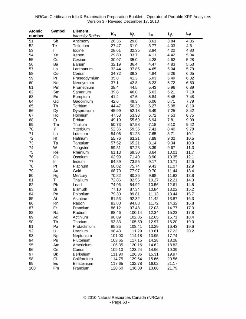
NRCan Certification Info & Examination Preparation Booklet – Operator of Portable XRF Analyzers Version 3 - Revised December 17, 2010
© 2010 Natural Resources Canada (NRCan) - Page 63 -
Atomic number
Symbol Element Intensity Ratios K K L L Ly
51 Sb Antimony 26.36 29.8 3.61 3.84 4.35 52 Te Tellurium 27.47 31.0 3.77 4.03 4.5 53 I Iodine 28.61 32.35 3.94 4.22 4.80 54 Xe Xenon 29.80 33.7 4.11 4.42 5.04 55 Cs Cesium 30.97 35.0 4.28 4.62 5.28 56 Ba Barium 32.19 36.4 4.47 4.83 5.53 57 La Lanthanum 33.44 37.85 4.65 5.04 5.79 58 Ce Cerium 34.72 39.3 4.84 5.26 6.05 59 Pr Praseodymium 35.8 41.3 5.03 5.49 6.32 60 Nd Neodymium 37.1 42.8 5.23 5.72 6.60 61 Pm Promethium 38.4 44.5 5.43 5.96 6.89 62 Sm Samarium 39.8 46.0 5.63 6.21 7.18 63 Eu Europium 41.2 47.6 5.84 6.46 7.48 64 Gd Gadolinium 42.6 49.3 6.06 6.71 7.79 65 Tb Terbium 44.47 50.39 6.27 6.98 8.10 66 Dy Dysprosium 45.99 52.18 6.49 7.25 8.42 67 Ho Holmium 47.53 53.93 6.72 7.53 8.75 68 Er Erbium 49.10 55.69 6.94 7.81 9.09 69 Tm Thulium 50.73 57.58 7.18 8.10 9.42 70 Y Ytterbium 52.36 59.35 7.41 8.40 9.78 71 Lu Lutetium 54.06 61.28 7.65 8.71 10.1 72 Hf Hafnium 55.76 63.21 7.89 9.02 10.5 73 Ta Tantalum 57.52 65.21 8.14 9.34 10.9 74 W Tungsten 59.31 67.23 8.39 9.67 11.3 75 Re Rhenium 61.13 69.30 8.64 10.01 11.7 76 Os Osmium 62.99 71.40 8.90 10.35 12.1 77 Ir Iridium 64.89 73.55 9.17 10.71 12.5 78 Pt Platinum 66.82 75.74 9.43 11.07 12.9 79 Au Gold 68.79 77.97 9.70 11.44 13.4 80 Hg Mercury 70.82 80.26 9.98 11.82 13.8 81 Tl Thallium 72.86 82.56 10.27 12.21 14.3 82 Pb Lead 74.96 84.92 10.56 12.61 14.8 83 Bi Bismuth 77.10 87.34 10.84 13.02 15.2 84 Po Polonium 79.30 89.81 11.13 13.44 15.7 85 At Astatine 81.53 92.32 11.42 13.87 16.3 86 Rn Radon 83.80 94.88 11.72 14.32 16.8 87 Fr Francium 86.12 97.48 12.03 14.77 17.3 88 Ra Radium 88.46 100.14 12.34 15.23 17.8 89 Ac Actinium 90.89 102.85 12.65 15.71 18.4 90 Th Thorium 93.33 105.59 12.97 16.20 19.0 91 Pa Protactinium 95.85 108.41 13.29 16.43 19.6 92 U Uranium 98.43 111.29 13.61 17.22 20.2 93 Np Neptunium 101.00 114.18 13.95 17.74 94 Pu Plutonium 103.65 117.15 14.28 18.28 95 Am Americium 106.35 120.16 14.62 18.83 96 Cm Curium 109.10 123.24 14.96 19.39 97 Bk Berkelium 111.90 126.36 15.31 19.97 98 Cf Californium 114.75 129.54 15.66 20.56 99 Es Einsteinium 117.65 132.78 16.02 21.17 100 Fm Francium 120.60 136.08 13.68 21.79

NRCan Certification Info & Examination Preparation Booklet – Operator of Portable XRF Analyzers Version 3 - Revised December 17, 2010
© 2010 Natural Resources Canada (NRCan) - Page 64 -
Appendix 2: XRF Forms Appendix 2 contains the forms necessary to apply for certification as an XRF operator:
1. Application 2. Identity verification 3. Training 4. Vision 5. Code of conduct

Application for X-Ray Fluorescence Operator (XRF) Certification, Renewal and Recertification
Mr. Mrs. Ms Language: English Français NAME: ________________________________________________________________________________ SURNAME GIVEN NAMES ADDRESS: _____________________________________________________________________________ _____________________________________________________________________________ CITY PROVINCE POSTAL CODE TELEPHONE: ___________________________ DATE OF BIRTH: yyyy________mm_________dd______
By signing this application, the candidate authorizes NRCan to disclose the candidates name, certification status, expiry date, city and province. Example: Web Listing of NDT Certified Personnel CANDIDATE’S SIGNATURE The signature MUST NOT exceed the inner limits of the signature box _________/____________/______ DATE (yyyy/mm/dd)
For NRCan Use Only __________________________ Name: __________________________ Registration Number: __________________________ Received Date: __________________________ Approved By: __________________________ Approval Date: Attached: Verification and two photos Training declaration Eye exam report Code of conduct Copy of log (Renewal) Application fee Examination fees
Employer: ______________________________________________________________________________ (Please print) Address: ______________________________________________________________________________ _______________________ / __________ / _________________ / ______________________ City Province Postal Code Telephone
Application for: Initial certification Level 1 Level 2 Rewrite Renewal Recertification
Fees Initial certification Renewal Recertification payable to NRCan Test centre NRCan NRCan Test centre
Application $250 $100 $100 Method Exam L1 $150 $ 50 Safety Exam L1 $150 $ 50 $150 $ 50 Total Level 1 $550 $100 $250 $ 50 Safety Exam L2 $150
Submit this application to: NDT Certification Body Telephone: 1-866-858-0473 (Please allow (2) weeks for processing. Processing times CanmetMATERIALS, Natural Resources Canada Fax: 905-645-0836
only start once we have a complete and accurate application 183 Longwood Road South Email: [email protected] and do not include postal delivery time.) Hamilton, Ontario L8P 0A5 Website: http://ndt.nrcan.gc.ca
$ 50 $150 $ 50 Total Level 2 $700 $150 $400 $100 If applicable: Renewal late fee $50 Exam rewrites $150 / exam $50 / exam Method L1 Safety L1 Safety L2
TYPE OF PAYMENT: Cheque Money Order (Cheques & money orders are payable to the Receiver General for Canada) Credit Card: Personal Company MasterCard Visa AMEX Cardholder's Name___________________________, Card Number______________________________, Expiry Date_______, Amount: $_______
With your application you are required to submit the following forms: ☞ Signed photo verification form + 2 passport photos ( 2 in x 2 ¾ in) ☞ Signed training declaration (for initial certification and recertification) ☞ Signed eye exam report ☞ Signed code of conduct
For Renewal and Recertification ☞ A copy of the log of safety checks as required by Safety Code 34 Section 2.3, Article 9.
Revised December 2012

PHOTOGRAPH VERIFICATION FORM Please use passport photograph only-size 2 in x 2 3/4 in
Candidate's name: _____________________________________________Reg. #: _________________ Employer: _______________________________________________________________________ Address: _______________________________________________________________________
_______________________________________________________________________ city province postal code Phone: ____________________________________ Fax: ___________________________ EMPLOYER/SUPERVISOR/SPONSOR VERIFICATION: I verify that the attached photographs are those of the employee whose name appears on this form. Name: ________________________ Signature: ______________________ (Please print) Date: ______/____/____ YYYY/MM/DD If you are not currently employed, you can ask one of the following people to attest to your photograph: Accountant; Chief of First Nation Band; Employee of Natural Resources Canada acting in an official capacity; Funeral Director; Justice of the Peace; Lawyer, Notary, Magistrate; Manager of Financial Institution; Medical and Health Practitioners: Chiropractor, Dentist, Doctor, Ophthalmologist, Optometrist, Pharmacist, Psychologist, Nurse Practitioners, Registered Nurse; Member of Parliament or their staff; Member of Provincial Legislature or their staff; Minister of Religion; Municipal Clerk; Official of a federal or provincial government department, or one of its agencies; Official of an Embassy; Consulate or high Commission, Official of a country with which Canada has a reciprocal social security agreement; Police Officer; Postmaster; Professional Engineer; Social Worker; Teacher Please submit photographs with this form to: NDT Certifying Agency CANMET Materials Technology Laboratory Natural Resources Canada 183 Longwood Road South Telephone: 866-858-0473 Hamilton, ON L8P 0A5 Fax: (905) 645-0836 IDVerifi-EF revised 08/11

NRCan Certification Info & Examination Preparation Booklet – Operator of Portable XRF Analyzers Version 3 - Revised December 17, 2010
XRF Training declaration I attest that _____________________________________ has successfully completed on this ______ day of _____________, __________ the following training sessions: ______ hours (minimum 3 hours) of classroom training in the theory of the XRF method and radiation safety in accordance with the NRCan Training Curriculum for XRF Operators as published in the Examination Preparation Booklet. ______ hours (minimum 2 hours) demonstration and practice in using XRF analyzer(s) to make accurate measurements ______ hours (minimum 2 hours) demonstration and practice in radiation safety including the safe set up, handling, operating, maintenance and storage of XRF analyzers. Instructor Name: _____________________________________ (Please print)
The Instructor/Trainor/Signee must be approved by NRCan, and verified against an active listing of acceptable personnel that is maintained by NRCan.
Signature: _____________________________________ XRF Manufacture: _____________________________________ NRCan Registration number: _______________ Telephone: _____________________________________ Date: ___________/________________/_________ (yyyy/mm/dd)

NRCan Certification Info & Examination Preparation Booklet – Operator of Portable XRF Analyzers Version 3 - Revised December 17, 2010
NRCAN EYE EXAMINATION REPORT - NDT PERSONNEL
Three vision assessments may be required: Near Vision, Distance Vision (visual testing method only) and Colour Vision (initial certification only). This form must be completed and returned to the NDT Certifying Agency when applying for examination in any NDT method, renewal of certification or recertification. CANDIDATE'S NAME: _______________________________ REGISTRATION NUMBER: __________ Near Vision and Distance Vision – to be completed by medically recognized personnel (ophthalmologist, optometrist, physician, nurse, etc.)
____________________________________ __________________________________________ Examiner's Name (Please Print/Type) Examiner's Signature ____________________________________ ____________/_________________/____________ Appointment/Title Date of Eye Examination (yyyy/mm/dd) Colour Vision (required only for initial certification, not for renewal or recertification) - to be completed by medically recognized personnel or the employer or certified level 3 NDT personnel. NOTE: A candidate who passes an Ishihara test (short or long) is acceptable. As an alternative or in case of a failure of an Ishihara test, the employer or Level 3 NDT personnel may administer a performance test to confirm if the candidate can see flaw indications that are typical of the method. Example: In liquid penetrant, confirm that the candidate can see red indications on a white background and fluorescent-green indications on a variety of backgrounds. I CONFIRM THAT THE CANDIDATE CAN DISTINGUISH CONTRAST BETWEEN THE COLOURS USED IN THE NDT METHOD(S) CONCERNED AS SPECIFIED BY THE EMPLOYER (OR PASSED AN ISHIHARA TEST). ____________________________________ __________________________________________ Examiner's Name (Please Print/Type) Examiner's Signature ____________________________________ _____________/_________________/__________ Appointment/Title Date of Eye Examination (yyyy/mm/dd)
NOTE: PROVINCIAL HEALTH CARE PROGRAMS MAY NOT COVER THE COST FOR AN EYE EXAMINATION
Near vision acuity: shall permit reading Times Roman N4.5 (Jaeger number 2 ) or equivalent letters at not less than 30 cm with one or both eyes, either corrected or uncorrected. I CONFIRM THAT THE CANDIDATE: (Please check one) Meets the requirement without correction Meets the requirement with correction Does not meet the requirement
Distance vision acuity: (required only for the visual testing method) shall equal Snellen Fraction 20/30 or better in at least one eye, either corrected or uncorrected. I CONFIRM THAT THE CANDIDATE: (Please check one) Meets the requirement without correction Meets the requirement with correction Does not meet the requirement

NRCan Certification Info & Examination Preparation Booklet – Operator of Portable XRF Analyzers Version 3 - Revised December 17, 2010
CODE OF CONDUCT FOR TESTING PERSONNEL
Individuals who are certified or individuals who are in process of being certified by NRCan must recognize that personal integrity and professional competence are the fundamental principles on which their testing activities are founded. Accordingly, it is a condition of NRCan certification that these individuals shall: 1. Comply with the relevant provisions of the applicable NRCan certification scheme(s); 2. Comply with the regulations, codes and standards under which they are working; 3. Immediately report to NRCan any perceived violation(s) of this Code-of-Conduct or any attempt to pressure or force a
certified individual to violate this Code-of-Conduct; 4. Verify the information on their certificates and/or wallet card. If the information is incorrect, it is their responsibility to
inform NRCan as soon as possible so that a new, corrected certificate and/or wallet card can be issued; 5. Not attempt to cheat on NRCan examinations, attempt to bribe or threaten NRCan invigilators or examiners, falsify
documents, falsely claim, misrepresent or permit misrepresentation or misuse of their own or their associate’s academic or professional qualifications, knowledge, training, experience, work responsibilities or certifications;
6. Discontinue all claims to certification upon expiry, suspension or withdrawal of certification, and upon request return to NRCan any certificates and/or wallet cards issued by NRCan;
7. Inform their employer in the event that their certification has expired, was suspended or withdrawn; NOTE: To protect certified individuals, employers, regulators and the public, NRCan maintains a publicly available listing of all currently certified testing personnel.
8. Only sign documents for which they have personal professional knowledge and/or direct supervisory control; 9. Undertake only those testing activities for which they are competent by virtue of their training, experience, qualification and
certification; 10. When required, engage or advise the engagement of specialists to enable testing activities to be properly completed; 11. Indicate to the employer or client any adverse consequences which may result from an overruling of their technical
judgment by a non-technical authority; 12. Perform their testing activities with proper regard for the environment and the safety, health and well-being of the public; 13. Consistent with the provisions of this Code-of-Conduct and the well-being of the public, respect the confidentiality of
information provided by an employer, colleague or member of the public; 14. Conduct themselves in a responsible manner and utilize fair and equitable business practices in dealing with employers,
colleagues, clients and associates; this includes disclosing any potential conflicts of interests; 15. Maintain their proficiency by updating their technical knowledge in their certified methods, levels and sectors; 16. Refrain from performing unethical acts that would discredit the NRCan certification schemes or bring NRCan into disrepute,
and refrain from making statements that NRCan could consider as misleading or unauthorized; Failure to comply with this Code-of-Conduct shall be dealt with under arrangements for handling complaints/appeals and may necessitate corrective measures such as the termination of the certification process, the suspension or withdrawal of certification, publication of the violation, notification of the employer(s), union(s) and appropriate regulatory authorities and, if appropriate, additional legal action. I agree to abide by this Code-of-Conduct. If I violate this Code-of-Conduct:
I accept the right of NRCan to suspend or withdraw any and all certifications granted to me by NRCan and/or terminate the certification process.
I accept the right of NRCan to notify my employer(s), union(s) and appropriate regulatory authorities and to
publish the details of the violation.
_____________________________________________ Name (Please Print)
_____________________________________________ ______________/______________/_________ Signature Date (yyyy/mm/dd)