Embed Size (px)
Citation preview
On the Blood-Vase alar System of the EarthwormPheret ima, and the Course of the Circu-lation in Earthworms.
By
Kami Narayan Bald, D.Sc,
Of the Muir Central College, Allahabad, India.(From the Department of Comparative Anatomy. Oxford.)
With 11 Text-figures.
TABLE OF CONTENTS.
1»AGK
1. I N T R O D U C T O R Y . . . . . . . . . 3 5 0
2 . T H E T Y P I C A L A R R A N G E M E N T O F T H E B L O O D - S Y S T E M I N T H E
I N T E S T I N A L R E G I O N O F T H E B O D Y B E H I N D T H E F O U R -
T E E N T H S E G M E N T . . . . . . . . 3 5 2
(a) T h e L o n g i t u d i n a l T r u n k s . . . . . . 3 5 2
(6) T h e I n t e s t i n a l B l o o d - p l e x u s 357
(c) T h e C o m m i s s u r a l , I n t e g u m e n t a r y , a n d N e p h r i d i a l Vesse ls . 3 6 3
(«!) T h e D o r s o - a n d V e n t r o - i n t e s t i n a l s . . . . . 368
3. T H E B L O O D - S Y S T E M I N T H E F I R S T F O U R T E E N C E P H A L I Z E D
S E G M E N T S . . . . . . . . . 3 6 9
(a) T h e L o n g i t u d i n a l T r u n k s 3 7 0
(6) T h e ' H e a r t s ' a n d t h e A n t e r i o r L o o p s . . . . 3 7 3
(c) T h e B l o o d - v e s s e l s of t h e O e s o p h a g u s a n d P h a r y n x . . 378
i. C O M P A R I S O N O F T H E B L O O D - S Y S T E M O F P H E R E T I M A W I T H
T H A T O F L U M B K I C U S A N D A L L O L O B O P H O R A . . 3 7 8
5 . T H E V A L V E S I N T H E B L O O D - V E S S E L S A N D T H E C O U R S E O F T H E
C I R C U L A T I O N O F T H E B L O O D . . * . . . 3 8 0
6. S U M M A R Y . . . . . . . . . . 391
7. L I S T O F R E F E R E N C E S . . . . . . . . 3 9 2

350 KABM NAHAYAN BAHL
1. INTRODUCTORY.
THE blood-vascular system of earthworms has engaged theattention of many distinguished observers. Lankester (12)described the blood-vessels of L u m b r i c u s in one of hismemoirs on the ' Anatomy of the Earthworm ', which formsabout the earliest contribution to this subject. Jaquet (9)gives a comparative account of the vascular system in Annelids,describing the system in typical genera of the various classesof the group. Of the Oligochaeta, he selects L u m b r i c u sas a type. Perrier (13) and Benham (5), also working onL u m b r i c u s , describe the course of flow in all the blood-vessels from a study of the disposition of the valves ; toBonham we also owe our knowledge of the blood-supply ofthe nephridium in L u m b r i c u s (6). Harrington (8) givesa detailed account of the anatomy of the blood-system inL u m b r i c u s with elaborate diagrams, and was the firstto describe the arrangement of blood-vessels in the integument.Recently, Johnstone and his student, Miss Johnson (10 and 11),have published two papers on the course of blood-flow inL u m b r i c u s demonstrating the course in various vessels bya series of interesting experiments and observations. Theblood-system has thus been thoroughly studied in L u m b r i c u ssince that is the form studied as a type in Europe and America.Amongst the Oriental forms of Oligochaeta, Bourne (1) hasdescribed the blood-system in some detail in the Perichaetoworm M e g a s c o l e x and also in M o n i l i g a s t e r g r a n d is(2, 1894), a huge worm about two feet long placed by Beddardin the group Mierodrili. Besides Bourne's work on Mega-s c o l e x , very little attention has been paid to the blood-system of the Perichaetidae, the largest family of earthworms.
Tho earthworm P h e r e t i m a (the genus Perichaeta s ensus t r i c t o) is now studied as a type of the Oligochaeta inNorthern India and also at the Universities of Bombay andCalcutta, and it has become necessary, therefore, to have ascomplete a knowledge as possible of the anatomy of this form.An attempt has been made in this paper to present an account

VASCULAR RYSTKM OF PHERRTIMA 851
of the blood-system of P h e r e t i m a and the course of blood-flow about which, even in L u m b r i c u s , there has beena great divergence of opinion amongst the various observers.Some of the observations were made in India, but in thiscountry, besides having an opportunity of examining thetwo Englisl i genera L u m b r i c u s and A11 o 1 o b o p h o r a,I was able to complete my work on P h e r e t i m a, havingbeen lucky to obtain specimens of this Oriental form in theLily-house of Kew Gardens.
The work was carried out in the Department of ComparativeAnatomy at Oxford. I am indebted to Professor E. S. Goodrichfor his keen interest in my work ; he has made valuablesuggestions, and has also found time to read through and correctthe manuscript of the paper.
Although essentially the blood-systems of both L u m -b r i c u s and P h e r e t i m a can be reduced to a, commontype, there are important differences in the system in the twogenera, which I have indicated in the text. P h e r e t i m aresembles A l l o l o b o p h o r a rather than L u m b r i c n s sofar as the blood-system in the general body-region is concerned,while the system differs in important respects from that ofMegascolex. As regards the course of the blood-flow studiedby holding the vessels with fine forceps, by cutting the vesselsand observing the direction of blood-flow, and by a study ofthe valves, I am led to confirm the observations and con-clusions of Johnstone (10 and 11) and to reject part of Bourne'stheory of the course of the circulation (1).
The typical arrangement of the blood-system in P h e r e-t i m a is found behind the fourteenth segment, being meta-merically repeated behind that segment. In the first fourteensegments, on the other hand, this typical arrangement is con-siderably modified, this modification, together with that shownin the digestive, reproductive, and nervous systems, being spokenof as c e p h a l i z a t i o n . It will be convenient, therefore, todescribe, as Harrington (8) does in the case of L u m b r i c u s ,first, the typical arrangement as it occurs in the region of thebody of the worm behind the fourteenth segment, and then the

8 5 2 KATCM NARAYAN BAHL
blood-vessels in the first fourteen cephalized segments, andfinally to discuss the course of the circulation in the system.
2. THE TYPICAL ARRANGEMENT OF THE BLOOD-SYSTEM IN
THE INTESTINAL EEOION OF THE BODY BEHIND THE
.FOURTEENTH SEGMENT.
The Mood-system in this system in this region of the bodyconsists of (a) three longitudinal trunks running parallel toone another, namely, the dorsal, the ventral, and the sub-neural vessels; (b) the intestinal blood-plexus, situated in thewall of the gut, is directly connected with the dorsal andventral vessels, and indirectly with the subnenral; and(e) the commissural, integumentary, and nephridial vessels.
(a) The L o n g i t u d i n a l T r n n k s .
1. The do r sa l vessel.—The dorsal vessel is the mostprominent of all the blood-vessels in the worm and is rhythmi-cally contractile. It runs along the mid-dorsal line immediatelybeneath the body-wall, between the latter and the intestine,and is at once seen lying on the gut, when the worm is openedby a mid-dorsal incision. In L u m b r i c u s the dorsal vesselis heavily covered over with ' yellow cells ', which must beremoved before the vessel is seen; but in P h e r e t i m athe ' yellow cells ' do not cover the dorsal vessel, so that thelatter is at once prominent on dissection. Although lying closeupon the gut, the dorsal vessel is not actually attached to thewall of the former in any portion of its course. It is singlethroughout its length and has thick muscular walls which areresponsible for its contractility. The average diameter of thisvessel is about 220 fi; it is narrowest at places where it piercesthe intersegmental septa. On opening a narcotized worm,we can easily see the wave of contraction in this vessel travellingfrom behind forwards and consequently driving the bloodin that direction. During its course through the body, thedorsal vessel, on piercing each septum, has a pair of forwardly-directed valves (figs. 7 and 10) in its lumen. These valves,

VASCULAR SYSTEM OF PH.BRETIMA 858
as I shall show later, prevent the flow of blood backwardswhen: the vessel contracts. There are also valves (videinfra) at the orifices of the dorso-int.estin.al and commissuralvessels.
It will be seen from fig. 1 that the dorsal vessel is connectedwith the intestine by two pairs of dorso-intestinal vessels(rti.v.) in each segment; these vessels serve to establish a com-munication between the internal intestinal plexus and thedorsal blood-vessel (fig. 2). The anterior pair of dorso-intestinalscome off from the dorsal in the anterior third of the segment,while the posterior pair lie in the posterior third, nearing thehinder septum of the segment, in close association with theso-called ' lymph-glands ' which lie on each side of the dorsalvessel in every segment here. These dorso-intestinals arevery short vessels, being only about 450 ft in length, on anaverage. They soon enter the intestinal wall, in which theyare continued as ' transverse vessels' (vide infra) .
Again, just before piercing each septum from behind, thedorsal vessel receives a commissural vessel (the dorso-lateralor the parietal vessel), which is connected ventrally with thesuhneural (covun.v., figs. 1 and 2). This commissural vesselruns along the posterior face of each septum very near andparallel to its outer edge, i. e. the edge joining the body-wall;and is connected with capillaries of the nephridia and thebody-wall.
As I shall show later on, both the dorso-intestinal and thecommissural vessels bring blood into the dorsal vessel andreplenish its supply. No blood leaves the dorsal vessel in thisregion of the body.
2. The v e n t r a l vessel.—The ventral vessel, like thedorsal, is single throughout its length and extends from theanterior to the posterior end of the body. In the region ofthe intestine it has an average diameter of 115 fi and givesoff a pair of v e n t r o - t e g u m e n t a r y branches in eachsegment. Each of these branches leaves the ventral vesseljust anterior to the septal wall in each segment and, afterrunning alongside the anterior face of each septum for a little
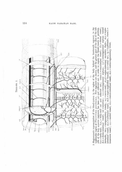
A diagrammatic representation of the blood-system in the region of the body behind the fourteenth segment, in thetyphlosolar region of Pheretima posthuma. Five segments are shown, and the greater part of the skin of the leftside of the four anterior segments has,been cut and reflected out, in order to expose the blood-vessels in position.b.w. = body-wall; b.w.i= body-wall cut and reflected out; co»m.i?. = commissural vessel; d.v. = dorsal vessel;cK.D. = dorso-intestinal vessel; vnl. = intestine; i.s. = intersegmental septum; i.*.
1=intersegmental septum turned
forwards; int.v. = integumentary vessels; n.c — nerve-cord ; si.e.<2. = supra-intestinal excretory ducts; sz'.«. = septo-intestinal vessel; sn.v. = sabneura,] vessel; sre.6. = septo-nephridial branch; «.s. = setal sac; <f/. = typhlosole; trans.v.^transverse vessel ; v.v. = ventral vessel; w.w.= ventro-intestinal vessel; i><.p.=ventro-tegumentary vessel.

VASCULAR SYSTEM OF PHERETIMA 355
distance, it pierces the septum and gets into the succeedingsegment (vt.v., fig. 1). Here it lies on the inner surface of thebody-wall near the middle line of the segment just in front ofthe row of setal sacs, going right up near the mid-dorsal line(figs. 1 and 2). As it ascends along the body-wall transversely,the ventro-tegumentary vessel (vt.v.) gives off backwards andforwards capillaries that supply blood to the body-wall(epidermis and the muscles) and the integumentary nephridia.Besides, the septal nephridia and the prostates also receivetheir blood-supply from the ventro-tegumentaries. The septalnephridia are supplied by a septo-nephridial branch (sn.b.,fig. 1) of the ventro-tegumentary given off in each segmentat the place where it pierces the septum; while the prostateglands in the segments sixteen to twenty-one receive smallbranches from the ventro-tegumentary in each of thesesegments.
Besides the paired ventro-tegumentary branches the ventralvessel gives off dorsally a single unpaired v e n t r o - i n t e s t i n a lv e s s e l in each segment (vi.v., fig. 1). This vessel originatesfrom the ventral a little behind the middle of each segment,and runs forward to enter the ventral wall of the intestine,by three or four branches, close to the anterior intersegmentalseptum. The ventro-intestinal, though generally overlookedin this worm, is, however, an important vessel, and measuresas much as 1 -5 mm. in length in some worms from its place oforigin on the ventral vessel to its place of entrance into theintestinal wall. It puts the ventral vessel into communicationwith the intestinal plexus. There are no valves anywhere alongthe course of the ventral vessel.
The ventral vessel is the main and, in fact, the only distribut-ing channel in the intestinal region of the body. All partsin this region get their supply of blood from the ventralvessel.
3. The s u b n e u r a l vessel.—The subneural vessel runsalong the mid-ventral line of the body-wall, being intimatelyattached to it, and lies, as its name indicates, beneath thenerve-cord. It is a very slender vessel and extends from the
NO. 259 B b

356 KAEM NARAYAN BAHL
posterior end of the •worm to the fourteenth segment anteriorly,being absent from the first fourteen segments. The com-missural vessel, connecting the subneural with the dorsal inthe septal regions, has already been referred to above. Atabout the middle of each segment just in front of the line
d.v.
trans.vii
c.i.p.
vt.v/
sn.v.A diagrammatic transverse section through the region of the intes-
tine, the right half showing a section through the intersegmentalregion and the left half through a segment proper passing throughone of the dorso-intestinals. ?>.?<;. = body-wall; c.e.j). = capil-laries of the external plexus ; c.»'.}). = capillaries of the internalplexus ; co?ra?)i.'w. = commissural vessel; d.v. = dorsal vessel;di.i). — dorso-intestinal vessel; si.v.—septo-intestinal vessel;s.v. = subneural vessel; trans.v. = transverse vessel; ty.v.=typhlosolar vessel; v.v. = ventral vessel; tM.i>. = ventro-tegu-mentary vessel.
of setal sacs, the subneural receives a pair of very small branchesfrom the ventral part of the body-wall. One also finds insections the subneural receiving a branch on its ventral sidefrom the body-wall in the mid-ventral line (fig. 2).
The subneural is connected with the intestinal plexus

VASCULAR SYSTEM OF PHKRETIMA 857
through the s e p t o - i n t e s t i n a l (si.v., figs. 1 and 2), avessel which I describe below along with the commissuralvessel.
This vessel collects blood from the small ventral part of thebody-wall and the nerve-cord ; and as the area over whichits branches ramify is very small and the quantity of bloodreceived is also small, the vessel itself is very slender as com-pared with the other longitudinal trunks.
There are no s u p r a - i n t e s t i n a l vesse ls in this regionin this worm : a pair of longitudinal ducts attached to the mid-dorsal line of the gut and described as supra-intestinal blood-vessels by Stephenson (14) have already been shown by me tobe excretory ducts (7).
There are also no lateral neural vessels as found in Lurn-b r i c u s .
(b) The I n t e s t i n a l B l o o d - p l e x u s .
The intestinal blood-plexus (fig. 8) consists of a close networkof capillaries and blood-vessels in the walls of the intestine.In P h e r e t i m a as in Megascolex (1) there are twocapillary networks in the alimentary canal, i.e. (1) an internaldeep-lying network, and (2) an external more superficial one.The internal network lies deep in the wall of the gut inside thelayer of circular muscle-fibres, between it and the internalepithelial lining ; while the capillaries belonging to the externalnetwork lie on the surface of the gut-wall amongst or evenoutside the yellow cells (chloragogen cells) which form thesplanchnic layer of the peritoneal lining of the coelom. Whena freshly-killed worm is opened in saline solution it is at onceseen that the blood-plexus on the gut is marked out into threedistinct regions—the first region is from the fourteenth to thetwenty-sixth segment, where the intestinal capillaries are verythickly set and lie at right angles to the longitudinal axis ofthe body (transverse capillaries) ; the second is the longestportion and extends from the twenty-sixth segment to twenty-three to twenty-eight segments in front of the anus, the main
B b 2

858 KARM NARAYAN BAHIJ
portion of the plexus in this region consisting of longitudinalcapillaries lying parallel with one another along the intestineall round the circumference ; and the third region comprisesthe last twenty-three to twenty-eight segments of the animal,where the blood-plexus differs markedly from what we havein the first two regions. The difference in appearance of theblood-plexus in the three regions is illustrated in fig. 3, whereat the point marked x there is a sudden change in the arrange-ment of capillaries from the second to the third region. Whilethere is a regular, almost rectangular arrangement of thecapillaries in the anterior two regions of the gut, the capillariesin the posterior region (last twenty-three to twenty-eightsegments) branch off in a tree-like fashion from the dorso-intestinal vessels. That the three regions mentioned above aredistinct from one another will be evident from the fact, ascer-tained by a study of sections passing through the three regions,that in the first region (fourteenth to twenty-sixth segment)the intestinal capillaries form only the internal plexus, theexternal plexus being absent, that in the second region(twenty-sixth segment onwards) there are both the internaland external plexuses well developed, while in the third region(last twenty-three to twenty-eight segments) we have nointernal plexus at all, all the capillaries belonging to an externalplexus.
Besides the difference in the arrangement and position ofcapillaries in the three regions there is another feature whichalso distinguishes these three regions from one another, andthat is the presence and absence of a • typhlosole and thetyphlosolar vessel. Taking the last region first, we have tonote the entire absence of a typhlosole in this region. Bed-dard (3) describes the absence of typhlosole in the last fewsegments of A c a n t h o d r i l u s , and calls this last part ofthe gut without a typhlosole the 'rectum'. Similarly, thetyphlosole is absent in the gut in the last thirty-six segmentsof L u m b r i c u s , and we can apply the term ' rectum' tothese last thirty-six segments of L u m b r i c u s and the lasttwenty-three to twenty-eight segments of P h e r e t i m a .

VASCULAR SYSTEM OF PHERETIMA 359
It seems reasonable to suppose that by the time the earthreaches the last rectal portion of the gut there is hardly anynutriment left in it for absorption, and hence we have theabsence of the typhlosole as well as of internal blood-plexusin this region, both of these structures being the likely mediafor absorption of nutriment from the earth. A well-developedexternal network of capillaries is, however, present in the
TEXT-FIG. X.
1st region 2nd region x d.v. 3rd reg-ionSemi-diagrammatic representation of the intestinal blood-plexus
in the three regions of the intestine. The 1st region extends fromthe fourteenth to tho twenty-sixth segment; the 2nd regionfrom the twenty-sixth to twenty-three to twenty-six segments infront of the anus and the region includes the last twenty-threeto twenty-six .segments (rectal region). d.v. = dorsal vessel;.c=the place where there is a change from the regular geometricalplexus to the branching tree-like plexus of the rectum.
rectal region and serves to supply blood to the wall of the gut,and also, being distributed amongst the chloragogen cells,allows the latter to take up the excretory products from theblood capillaries.
In the second region, which is the most extensive (twenty-sixth segment to twenty-three to twenty-eight segments infront of the anus) of the three regions, we have a typhlosoleas well as both the internal and the external plexus equally

360 KARM NARAYAN BAHL
well developed. The internal plexus is a dense network ofcapillaries appearing as a sort of blood-sinus interrupted atplaces by the foldings of the gut epithelium (fig. 2). Thetyphlosolar vessel, which should be regarded as part of theinternal plexus, communicates with it at two places in eachsegment. The external blood-plexus, which is not continuousfrom segment to segment, has capillaries of varying diameters.The blood apparently passes from the external to the internalplexus, as, like the case in M e g a s c o l e x (1), we can see thecapillaries of the external network communicating with thecapillaries of the internal network at numerous places insections.
In the first region we have only a well-developed internalplexus but no external one. Neither is there a typhlosole,although, of the specially large mid-dorsal and mid-ventralcapillaries, the mid-dorsal one simulates the typhlosolarvessel.
(1) Alimentary plexus in the first region (fourteenth totwenty-sixth segment).
In this region of the gut the internal blood-plexus is bestdeveloped. The network is very dense, almost a blood-sinusinterrupted at certain places ; the interspaces in the dorsalhalf of the" plexus are very small indeed, even less than one-fourth the size of the vessels which surround them. Thecapillaries run parallel to one another transversely to thelength of the gut, and towards the ventral half break up intocapillaries of smaller calibre, so that in the ventral half of thegut a continuous blood-sinus gives place to a coarse networkof capillaries. In a freshly-opened worm this region of thegut presents a very bloody appearance.
Besides the richness in capillaries of this region we havea pair of well-marked vessels lying on the dorso-lateral aspect.These begin ventrally in the intestinal plexus about the four-teenth segment, and incline gradually dorsalwards up to thetwenty-sixth segment, where they join the posterior pair ofdorso-intestinal vessels of that segment at the inner angles ofthe roots of the intestinal coeca, and also communicate at that

VASCULAR SYSTEM OF l'HEHETIMA 361
place with the other blood-vessels on the walls of the coecathemselves.
An equally well-developed vessel runs along the mid-dorsalline of the gut, being only a specialized capillary of the internalplexus and being also continuous with the typhlosolar vesselbehind.
The external blood-plexus is almost completely absent inthis region. There are, however, a few capillaries present,which can be seen attached to the outside of the gut; forexample, at places where the ventro-intestinal and septo-intestinal vessels join the wall of the intestine. But they soononter the intestinal wall and pour their blood into the internalblood-plexus ; so that a regular external plexus such as we findin the second and third regions (vide infra) is absent in thispart of the gut, the internal plexus being very strongly developed.
(2) The alimentary plexus in the second region (twenty-sixthsegment to twenty-three to twenty-eight segments in frontof the anus).
In this region we have both the external and internal plexuseswell developed. The external plexus consists of capillaries ofvarious sizes which are continuous on the ventral Avail of thegut but not on the dorsal. They are connected with the septo-intestinals and the ventro-intestinals which apparently formtheir source of blood-supply. They open into the capillariesof the internal plexus as shown in fig. 2.
The internal plexus in this region of the gut presents a veryregular geometrical arrangement, as shown in fig. 3. Thisnetwork consists of (a) L o n g i t u d i n a l c a p i l l a r i e s , whichare very closely set around the wall of the gut, extending allalong its length. They are continuous from segment to seg-ment and number about forty all round. These capillariesform the main portion of the plexus and in transverse sectionsare seen to lie in the folds of internal gut-epithelium.
(b) T r a n s v e r s e Channels .—We have already men-tioned that in each segment the dorsal vessel is connected withthe gut by means of two pairs of dorso-intestinal vessels.These dorso-intestinals on leaving the dorsal vessel enter the

362 KARM NARAYAN BAHL
intestinal wall about \ mm. from their origin and go roundthe wall of the gut to its ventral side. I propose to apply theterm dor s o - i n t e s t i n a l to the vessel from its point oforigin from the dorsal to the point of its entrance into theintestinal wall. The continuation of the dorso-intestinalon the wall of the gut I propose to call a t r a n s v e r s ec h a n n e l . 1 Corresponding to the two pairs of dorso-intes-tinals there are two pairs of transverse channels in eachsegment; each of these transverse channels is joined at itspoint of junction with the dorso-intestinal by a branch fromthe typhlosolar vessel (vide infra) (fig. 2, left half) : so thatthese transverse channels serve to connect not only the longitu-dinal capillaries with each other but also the whole plexuswith the typhlosolar vessel.
(c) O b l i q u e Channels.—These begin at the mid-ventralline of the intestine at the intersegmental plane and jrunforwards and dorsalwards, passing through three segmentsbefore reaching the mid-dorsal line, where they join thetyphlosolar just in front of the septa (fig. 3).
(d) T y p h l o s o l a r Vessel.—The typhlosolar vessel runsalong the free edge of the typhlosole all down the second regionof the gut (fig. 2). The typhlosole itself cannot be comparedto the structure of the same name in L u m b r i c u s , for inP h e r e t i m a it is really a bigger fold of the gut-epitheliumcontaining not yellow cells, like those which fill up the typhlo-sole of L u m b r i o u s , but only connective tissue which hasthe same staining qualities as the connective-tissue matrixin the layer of circular muscle-fibres of the body-wall. Thetyphlosolar vessel does not seem to possess a definite wall likethe capillaries of the external plexus in P h e r e t i m a orthe typhlosolar vessel of L u m b r i c u s , but is only a partof the blood-sinus like the longitudinal capillaries, being, likethem, in communication with the two pairs of transversechannels in each segment. We can therefore think of thesetransverse channels as circular ring-vessels which collect blood
1 I have called these channels as they are thicker than the longitudinalcapillaries.

VASCULAR SYSTEM OF PHEEBTIMA 363
from the longitudinal capillaries and the typhlosolar vessel(which we may regard as a specialized longitudinal capillarylying in the mid-dorsal line), and convey it to the dorsal vesselby means of the two pairs of dorso-intestinals in the sameway as the ring-vessels of the oesophagus convey its blood tothe supra-oesophageal vessel there (vide infra). It wouldbe interesting to note here that, although the typhlosole isabsent in the segments fourteen to twenty-six, there isa prominent blood-vessel in the mid-dorsal line of the gut-epithelium, the vessel corresponding to the typhlosolar behind,with which it is directly continuous.
(8) The blood-plexus in the third region (last twenty-threeto twenty-eight segments).
In the last twenty-three to twenty-eight segments of theworm where the typhlosole in the gut is absent, and whichregion Beddard (3, p. 18) has referred to as the ' rectum ',the intestinal plexus is different from what we have seen in thefirst two regions. The whole of the plexus is external, i. e. liesoutside the muscular coats, .there being no internal plexus.The regular and rectangular arrangement of capillaries in thetyphlosolar (second) region at once changes into a branchingtree-like plexus as shown in fig. 8. There is only one pair ofdorso-intestinals in this rectal region in place of two pairsin the first two regions. Since there is no internal plexusthe dorso-intestinals change their connexions and communicatein this region with the external blood-plexus.
The blood coming to the rectum from the ventro-intestinalsand septo-intestinals goes to the external plexus, from whereit passes to the dorsal through the dorso-intestinals, the partof the course involving the internal plexus having been cutout (vide infra).
(c) The Commissura l , I n t e g u m e n t a r y , andN e p h r i d i a l Vesse l s .
1. The Commissura l Vessel.—As already mentioned,there is a pair of commissural vessels (parietal vessels) in eachsegment connecting the dorsal with the subneural vessel

364 KAKM NARAYAN BAHL
(tigs. 1 and 2). The commissural lies in the most anteriorposition in each segment, since the posterior faco of a septum,on which this vessel lies, forms the anterior boundary of a seg-ment. In its ventro-lateral part each commissural vessel isjoined by a ' septo-intestinal' branch (figs. 1 and 2) whichputs the commissural vessel in communication with theintestinal plexus, so that the commissural joins the dorsaland subneural vessels at its two ends, while in its ventral thirdit gives the septo-intestinal branch to the intestinal blood-plexus. It is interesting to note the Y-shaped places of junc-tion (fig. 2) one comes across in sections, where the three limbsof the Y represent the branches of the commissural going tothe dorsal and subneural vessels and the intestinal plexusrespectively. All along its length the commissural vessel isjoined by branches coming from the septal nephridia and thebody-wall. In segments sixteen to twenty-one the com-missural vessel also receives the efferent capillaries from theprostates which get their blood-supply from the branchesof the ventro-tegumentaries. As shown in fig. 1.1 could countin one preparation as many as eight branches entering thecommissural, each of these branches being formed by theunion of several branchlets.
The commissural vessel of P h e r e t i m a is a very interestingstructure when we compare it with similar structures in otherearthworms. Bourne (1) describes in M e g a s c o l e x twovessels, which he calls ' intestino-tegurnentary ' and ' dorso-tegumentary ', as follows : ' The main portion of the intestino-tegumentary vessel lies closely adherent to the body-wall justbehind a septum, i. e. in the anterior portion of a segment ',and ' the dorso-tegumentary arises in all segments regularlyfrom the dorsal vessel immediately posterior to the septumwhich forms the anterior boundary of the segment in whichit lies'. It is clear from this description and also from hisdiagram (PI. IX, fig. 7, in his paper) that these two vesselsof M e g a s c o l e x run in the same transverse plane, and wouldthus correspond exactly to the commissural vessel of P h e r e -t i m a minus its small ventral portion, since the commissural

VASCULAR SYSTEM OF PHEHE'TOIA " 865
also lies in exactly the same position. Its dorsal part with itsconnexions with both the dorsal vessel and the body-wallwould correspond to the ' dorso-tegumentary ', and its lateralpart together with the septo-intestinal having connexionswith the body-wall on the one hand and the intestinal plexuson the other would correspond to the ' intestino-tegumentary 'of Megascolex. There being no subneural vessel in thelatter genus, there is nothing in its blood-system correspondingto the ventral part of the commissural of Phere t ima.
Again, the 'dorso-tegumentary' of Moniligaster (2)and Lumbricus (8) corresponds to the commissural vesselof Pheret ima minus the septo-intestinal. Unlike Mega-scolex, these two genera (Moniligaster and Lumbricu s)possess a subneural vessel like Phere t ima, and we havea loop or commissural vessel connecting the dorsal with thesubneural, which has been described by Jaquet (9) in Lum-bricus as the ' branche dorso-sous-nervienne ', a term adoptedby Bourne for the same structure in Moniligaster. Jaquetalso describes a ' branche tegumentaire ' from the dorso-tegumentary ; but I have examined the tegumentary (com-missural or parietal) of Lumbricus and do not finda special ' branche tegumentaire ' as Jaquet makes out. Ofcourse, there are several branches from the body-wall (tegu-mentary branches) joining the commissural all along itscourse as in Pheret ima, to which the term ' branchutegumentaire ' can be applied ; but the real point in whichthe commissural of Lumbricus and Moniligasterdiffers from that of Pheret ima is that in the former twogenera it has no connexion with the intestinal plexus, therebeing nothing corresponding to the ' septo-intestinal' ofPhere t ima.
From the comparisons made above it seems reasonable todeduce that the commissural vessel of Pheret ima is a com-pound vessel which combines in itself the ' dorso-tegumentary '(commissural or parietal) of Lumbricus and Moniligaster(the dorso-tegumentary of Megascolex corresponding onlyto one of the tegumentary branches joining the commissural

366 KAEM NARAYAN BAHL
in the other earthworms) and the ' intestino-tegumentary ' ofM e g a s c o l e x . The probable homologies are set out in thefollowing table :
Branohe dorso-sous-nervienne
Only partially re-presented by thetegumentary partof the ' intestino-tcgiuiientary '
CommisKiiral ves-sel
Absent
Intestinalpart ofintestino-tegumen-tary '
iSepto-intea-tinal.
1. L U m b t i c u s Branche tegumen-taire
2. Moniligaster „ „
3. Megasoolex Dorso - tegumen -tary
4. P h e r e t i m a Uric of tlic capil-laries from thebody-wall joiningthe dorsal por-tion of the com-missural
In describing the ' ventro-intestinals', of which there isa pair in each segment in M o n i l i g a s t e r (2, 1894, p. 380),Bourne remarks : ' They are the sole afferent vessels of theintestinal walls. There are no such vessels in M e g a s c o l e xc o e r u l e u s , their function being performed by the " intes-Iino-teguinentary " vessels.' In P h e r e t i m a we have boththe ' intestino-tegumentary' (represented by the septo-intestinal) as well as the Arentro-intestinal vessel in eachsegment ; and if both are afferent A'essels of the gut-wall, asI believe they are, there is a double source of supply of bloodto the gut in P h e r e t i m a .
As I shall discuss later on, I believe that the course of bloodin the commissural is towards the dorsal vessel. The bloodfrom the subneural goes to the intestinal plexus through thesepto-intestinal, and the branches joining the commissural allalong its course bring blood into it from the body-wall and theseptal and integumentary nephridia.
2. The I n t e g u m e n t a r y Vessels.—The body-wall,consisting of its muscular layers, and the epidermis receives itssupply of blood from the ventro-tegumentary branches, a pairof which comes off from the ventral vessel in each segment.I have already stated that these ventro-tegumentary branches

V A S C U I J A R SYSTEM OF PHERETIMA 867
supply the body-wall of the segment succeeding the one inwhich they arise from the ventral vessel (e. g. the ventro-tegumentary arising from the ventral in the fortieth segmentruns along and supplies the body-wall of the forty-first segmentand so on). The ventro-tegumentaries give off numerousbranches backwards and forwards (fig. 1), which are distributedover the body-wall and also supply blood to the integumentarynephridia (vide infra) . The ventro-tegumentaries growthinner and thinner along their course towards the mid-dorsalline near which they end in the body-wall.
TfiXT-FIG. 4.
long. ra.A diagrammatic reconstruction of three serial sections showing
the close parallelism of ' arterial' and ' venous ' capillaries in thebody-wall. ep. = epidermis ; cir.m. = layer of circular muscle-fibres; long.m. = layer of longitudinal muscle-fibres; vl.b.=a branch of the ventro-tegumentary vessel; comm.v.h. = a, branchof the commissural vessel.
The efferent vessels of the body-wall are the paired branchesof the subneural in each segment and the numerous branchesjoining the commissural vessel in each segment.
The afferent and efferent capillaries run side by side in thesubstance of the body-wall, and can always be followed fromthe coelomic epithelium through the muscular layers to theepidermis. I can confirm for P h e r e t i i n a Bourne's state-ment (2) with regard to the peripheral capillaries in M o n i 1 i -g a s t e r , that ' the most striking feature of these networks(he is s p e a k i n g of c a p i l l a r i e s in t h e b o d y - w a 11)

368 KARM NARAYAN BAHL
is the strict parallelism which obtains throughout between" artery " and " vein " '. In serial sections it is very interestingto follow pairs of parallel capillaries in the body-wall, and onecan invariably trace them to their afferent and efferent vessels.Kg. 4, reconstructed from three sections of G (i thickness,serves to illustrate the parallelism obtained in sections, whilefig. 4 A gives an accurate camera luc ida drawing of partof the body-wall mounted flat after the removal of longitudinalmuscles. The strict parallelism between an ' artery' anda vein together with the capillary loops connecting them arevery clearly displayed.
3. The N e p h r i d i a l Blood-system.—The blood-supplyof the three kinds of nephridia in P h e r e t i m a has alreadybeen described by me elsewhere (3), and I have nothing furtherto add here.
(d) The .Dorso - in t e s t ina l s and the V e n t r o -i n t e s t i n a l s .
The Dor so - in t e s t i na l s .—I have referred to thesevessels already in describing the dorsal vessel. The dorso-intestinals form, so to speak, the efferert vessels (veins) ofthe intestinal blood-plexus, as all the blood in the intestineis returned to the dorsal vessel through these dorso-intestinals.There is a single pair of them in the fourteenth segment and inall the segments of the rectal (post-typhlosolar) region, whilein the remaining large part of the intestine we have two pairsto each segment. We have already noted that the dorso-intestinals communicate with the external plexus in the rectalregion but with the internal plexus in the first and secondregions. At the place where the dorso-intestinal leaves thegut, it also receives a branch from the typhlosolar vessel
(%• 2).The Ventro- intes t inals .—These single unpaired
vessels in each segment have also been referred to above.They form the afferent vessels (arteries) of the gut, and arepresent in all the three regions.

VASCULAR SYSTEM OK PHERET1MA 869
3. THE BLOOD-SYSTEM IN THE FIRST FOURTEEN SEGMENTS.
In the first fourteen segments the blood-system is highlymodified on account of the cephalization of this region, anddiffers a good deal from the system in the general body-region.
Amongst the longitudinal trunks the subneural as such is
TEXT-FIG. 4 A.
eff.v.c aff.v.c.
intl.c.
Disposition of blood-capillaries in the body-wall from a wholemount of a portion of the body-wall treated with caustic potash,showing how a ' venous ' capillary passes into an ' arterial' one.aff.v.c. = capillary of the afferent vessel ; eff.v.c. = capillary of theefferent vessel; inl.l.c. = capillary loop connecting the afferentand efferent vessels.

370 KARM NARAYAN BAHL
absent; it bifurcates in the fourteenth segment, and the twobranches curve round (fig. 5) the nerve-cord to be continuedinto the two lateral oesophageal vessels. A new large vesselin this region limited in extent is the supra-intestinal vessel,which is closely attached to the oesophagus in the mid-dorsalline and communicates freely with the blood-plexus of theoesophagus. Besides these there are the big pulsating ' hearts 'in many of the segments of this region, by means of which thedorsal vessel pumps out all the blood it receives either into theventral vessel to be distributed by it or directly to the variousorgans in this part of the body.
(a) The L o n g i t u d i n a l T r u n k s .
1. The Dorsa l Vessel.—The dorsal vessel continuesin front up to the third segment, where it divides into threebranches near the cerebral ganglion, these branches beingdistributed over the pharyngeal mass and the wall of thebuccal cavity. While in the region of the intestine the dorsalvessel lies close upon the gut, being connected with it by twopairs of dorso-intestinals ; in this anterior region it is removedconsiderably away from the oesophagus. Except in the four-teenth segment, where the dorsal vessel is connected by a single(not two) pair of dorso-intestinals, there are no such venousbranches at all in the anterior cephalized region. Since thereis no subneural vessel in this region the commissural vesselsconnecting the dorsal with the subneural in the intestinalregion are absent in this anterior region. However, the dorsalvessel here gives off, in many segments, pulsatile vessels calledthe 'hearts'. These structures I shall describe separately below.
The intersegmental valves present in the posterior part ofthe dorsal vessel are present here also, and have the samestructure and disposition, making the blood flow in the anteriordirection. But the valves at the orifices of the dorso-intestinalsand commissurals into the dorsal (vide i n f r a) in the posterior-region have no counterpart here ; in their place there are othervalves away from these orifices, leading the blood outwardsfrom the dorsal vessel.

TEXT-FIG. 5.
A semi-diagrammatic representation of blood-vessels in the first sixteen segments of P h e r e t i m a . d.i\ = dorsalvessel; giz. = gizzard; int. = intestine; lot.oes.v. — lateraloesophagealvessel; oes. — oesophagus; ph. = pharynx;sn.v. = subneural vessel; v.v. = ventral vessel; t>t.'U. = ventro-tegumentary vessel. The ' latero-intestinal'hearts are in the twelfth and thirteenth segments, the ' lateral' hearts in the seventh and ninth segments,while the tenth and eleventh segments contain the ' anterior loops '.

872 TvARM NARAYAN BAHIJ
2. The S u p r a - i n t e s t i n a l Vessel.—The supra-intes-tinal vessel, which is confined to the oesophageal regionbehind the gizzard, occupies the same relative position withregard to the gut as the dorsal vessel does in the region of theintestine. It lies beneath the dorsal vessel rather closelyattached to the dorsal wall of the oesophagus, while the dorsalvessel itself is removed considerably away from the gut. It isusually double along its whole extent, but the two halves cometogether and communicate with each other at several places.The supra-intestinal vessel extends from the tenth to thethirteenth segment. In the tenth and eleventh segments itcommunicates with the lateral oesophageal vessels by largecommissural vessels or ' loops ' that go round free from thewall of the oesophagus ; while in the twelfth and thirteenthsegments it communicates with the ventral vessel through the' hearts '. The vessel ends anteriorly by breaking up intocapillaries in front of the tenth segment, and these capillariesare distributed over the walls of the oesophagus and thegizzard. Posteriorly the vessel ends by joining the posteriorpair of ' hearts ' in the thirteenth segment, although a slenderbranch very often continues backwards on the mid-dorsal lineof the gut for a segment or two.
The supra-intestinal is the efferent vessel for the gizzardand the oesophagus, and all the blood brought in it fromthese structures is no doubt carried into the ' hearts ' of thetwelfth and thirteenth segments.
3. The V e n t r a l Vessel.—The ventral vessel extendsanteriorly up to the second segment, and in each segmentgives off a pair of ventro-tegumentary branches as in theposterior region, with the difference that the branches froma particular segment are spread over and distribute blood tothe body-wall, the septa, and the nephridia in the s amesegment and n o t the succeeding one, as they do behind.All the special organs in this part of the body, e.g. the sperma-thecae, the seminal vesicles, the ovaries, and the oviductsare supplied with blood by little branches from the ventro-tegumentaries. The vessel ends anteriorly in a pair of branches

VASCULAR SYSTEM OP PHERET.IMA 878
in the second segment. There are no ventro-intestinals in thisregion of the body.
4. L a t e r a l - o e s o p h a g e a l Vessels.—These are a pairof fairly large vessels in the first fourteen segments of the animalsituated on the ventiro-lateral aspect of the oesophagus. Thevare always found full of blood and can be easily seen. Behindthe gizzard, i.e. in segments ten to thirteen, they are veryintimately attached to the wall of the oesophagus and, as canbe seen in sections, communicate with the oesophageal ring-vessels throughout these four segments by as many branchesas the number of ring-vessels. In the region of the gizzardand in front, however, they are free from the wall of the gut,but receive a branch in each segment from the wall of the gut.
The lateral oesophageals receive in each segment a pair ofbranches that bring back blood not only from the bod}r-walland septa of this region but also from the seminal vesicles andthe spermathecae. They thus function here like the branchesof the subneural and commissural vessels behind, which collectblood from the body-wall, the nephridia and other organs incoelom like the prostates.
It only remains to be added that the lateral oesophageals area continuation forward of the subneural vessel. In the four-teenth segment the subneural vessel forks into two, and eachof the two branches loops round the nerve-cord and comes tolie dorsal to it and is continued forward along the ventro-lateral aspect of the oesophagus as the lateral-oesophageal
(b) The ' H e a r t s ' a n d t h e A n t e r i o r L o o p s .
It will be seen from what we have described above that thereis no direct communication between the dorsal and ventralvessels in the region of the body behind the thirteenth segment,but in the anterior thirteen segments the dorsal vessel com-municates directly with the ventral through the ' hearts ' inthe seventh and ninth, and twelfth and thirteenth segments.It is only these four pairs of ' hearts ' that are connected withthe ventral vessel; but, besides these, there are other ' hearts '

374 KABM NAEAYAN BAHI,
which are also pulsatile but supply blood to some of the organsdirectly, e.g. the gizzard and the pharyngeal nephridia. I haveadopted Bourne's suggestion (1, p. 64 n.) of naming all rhythmi-cally contractile, circularly disposed vessels as 'hearts ', whichterm thus includes even the anterior branches of the dorsalvessel which do not join the ventral vessel.
TEXT-FIG. 6.
d.v.
latoesiP
l.SA diagrammatic transverse section of the earthworm through the
region of the ' latero-intestinal' hearts. In the right half is shownthe intersegmental septum just behind the 'heart ' . rf.«.=dorsal vessel; ht. = latero-intestinal heart; ».«. = intersegmentalseptum ; int.v.=integumentary vessels taking blood (venous) tothe lateral oesophageals and the supra-intestinals ; lat.oes.v.=latero-oesophageal vessels ; j'.u. = a ring-vessel in the oesophagus ;sv.i.v. = supra-intestinal vessel; v.v. = ventral vessel.
Again, Bourne (1, p. 64 n.), following Perrier, distinguishes' lateral hearts ' from the ' intestinal hearts ' according as theyare connected dorsally with the dorsal or supra-intestinalvessels. The ' hearts ' in the twelfth and thirteenth segmentsin P h e r e t i m a communicate dorsally with both the dorsaland supra-intestinal A'essels and are therefore ' latero-intestinal'hearts, while the ' hearts ' in the seventh and ninth segmentsbelong to the category of ' lateral hearts '. Coming to the' loops ' of the tenth and eleventh segments, we find that they

VASCULAR SYSTEM OF PHERETIMA 375
communicate dorsally with the supra-intestinal vessel, whileventrally they are connected with the lateral-oesophagealvessels. They might have been called ' intestinal hearts ' butfor the fact that those ' loops ' do not pulsate, have non-muscular walls unlike those of the ' hearts ', and I believe thatthe flow of blood in them is from the lateral oesophageals tothe supra-intestinal, a fact which I refer to again below. Onthese considerations I exclude these vessels from the categoryof ' hearts ' and call them ' anterior loops ', since they havenothing in common with the so-called ' hearts ' and ' anteriorloops ' in greater detail below ; they are shown in fig. 5.
T h i r t e e n t h and Twelf th Segments.—In each ofthose two segments there is a pair of ' latero-intestinal' hearts.In systematic accounts of the genus P h e r e t i m a it is onlythese two pairs that are described, and no mention is madeof the anterior pairs of ' hearts '. Even if the term ' hearts 'be restricted to those commissures which communicate withthe ventral vessel below it should include the ' hearts ' of theseventh and ninth segments. This diagnostic character forthe genus P h e r e t i m a is thus generally erroneously described,and the genus should be recognized to possess at least fourpairs of ' hearts ', two ' lateral' and two ' latero-intestinals '.
The ' hearts ' of the twelfth and thirteenth segments (fig. 5)are situated in the posterior parts of these two segments, andtheir walls are intimately attached to the septa behind them.They have thick muscular walls and a spacious cavity, and attheir dorsal ends communicate anteriorly with the supra-intestinal and posteriorly with the dorsal vessel. At theplaces where the branches from the dorsal and supra-intestinalmeet to enter the ' heart', each has a pair of valves leadingto the ' heart', and similarly there is a pair of valves at theventral end of each ' heart ' just above the place where itjoins the ventral vessel (fig. 11). The dorsal valves preventthe blood from going back to the dorsal or supra-intestinalvessels during systole, while the ventral valves prevent theblood from entering the ' heart' from the ventral vesselduring diastole.

376 KARM NAEAYAN BAHL
E l e v e n t h and T e n t h Segments.—These two seg-ments contain no ' hearts ', but each of them has a pair ofcommissural vessels connecting the supra-intestinal with thelateral oesophageal of each side. These vessels lie in theposterior parts of these segments near their posterior septa,and are partially covered by the latter. Unlike the ' hearts 'these ' loops ' of the tenth and eleventh segments are thin-walled, their walls being non-muscular, and they have no valvesanywhere along their length.
The blood, by means of these ' loops ', flows from the lateral-oesophageals into the supra-intestinals. The latter collectblood from the gizzard and oesophagus and also receive bloodin these two segments directly from the lateral oesophageals.All this blood they carry into the ventral vessel through the' hearts ' in the twelfth and thirteenth segments.
We may note here that the lateral oesophageals in Lum-b r i c u s pour their blood into the dorsal vessel in the tenthsegment and into the large parietal in the twelfth.
N i n t h Segment .—In the ninth segment there is a pairof ' lateral hearts ' connecting the dorsal with the ventralvossel. This pair of ' hearts ' is generally asymmetrical, theleft ' heart ' being large and well developed as compared withthe small thin-walled and ill-developed one of the right side,which, however, sends a branch to the oesophagus in thissegment. The ' heart ' on the left side has valves pointingdownwards along the greater part of its length, and there isalso a pair near the point of opening of the ' heart ' into theventral vessel. There are altogether four pairs of valves andtheir position and arrangement is illustrated in fig. 11A.
E i g h t h Segment .—In the eighth segment the dorsalvessel gives off a pair of large thick-walled branches whichdo not join the ventral vessel but on account of their contrac-tility are still called ' hearts ' ; each of them presents a bulb-like dilatation at some distance from its origin and immediatelyforks into two (fig. 5), the posterior branch going to the septumand body-wall, and the anterior dividing and distributingblood over the wall of the gizzard in a large number of capil-

VASCULAR SYSTEM 01' PHEKBTIMA 377
laries which ran longitudinally parallel to one another. Thesebranches of the dorsal vessel have a series of paired valvesalong their length between the point of their origin and theplace where there is the bulb-like dilatation. The bulb-likedilatation which occurs at the distal end of all the ' hearts 'contains a pair of thick valves pointing away from the dorsaland towards the ventral vessel, as shown in fig. 11.
The blood to the gizzard, therefore, is supplied from the dorsalvessel by the pair of branches in this segment ; while the capil-laries of the supra-intestinal vessel, which has its beginningshere, collect blood from the gizzard and take it into thatvessel.
Seven th Segment.—In the seventh segment there isa pair of ' lateral' hearts, each of which is joined below bothwith the ventral and the lateral oesophageal vessels, whichlatter are themselves joined together by a cross channel.In its upper part each of this pair of ' hearts ' is thick-walledand has A alves leading blood outwards, but in its ventral parteach ' heart' is thin-walled and has also no valves in it.There is no doubt that the blood flows from the dorsal to theventral vessel; but it seems probable that the supply of bloodin the ventral vessel, which is very thin in this region andcontains little blood, is also replenished from the lateraloesophageals, which are always large and full.
S i x t h , F i f t h , and F o u r t h Segments .—In thesixth segment, and also in the fifth and fourth, there is a pairof branches given off from the dorsal vessel each of whichhas a pair of valves leading outwards near its origin, andsupplies blood to the masses of pharyngeal nepliridia in each ofthese three segments. These branches are also pulsatile andcan therefore be named ' hearts '.
Th i rd Segment.—In the third segment before the dorsalvessel breaks up anteriorly, it gives off a pair of branchesto the pharyngeal mass behind the cerebral ganglion. Thesebranches also possess valves near their origin which directthe flow of blood outwards.

378 KAHM NAKAYAN BAHL
(c) The B l o o d - v e s s e l s of t he Gut in t h ef i r s t F o u r t e e n S e g m e n t s .
In segments ten to fourteen there are in the oesophagealwall a series of very definite and striking transverse vessels,about twelve pairs per segment, joining the supra-intestinalabove and the lateral oesophageals below ; the breadth ofthese vessels is at least equal to the intervals between them.They are not united by longitudinal connexions and are con-tinuous across the mid-ventral line. These ring-vessels (fig. 6)are very characteristic of the oesophagus behind the gizzard,and are situated inside the muscular coats of the oesophagus.In this region both the lateral oesophageals and the supra-intestinals are intimately attached to the oesophagus, and theblood flows from the former into the latter through thesetransverse ring-vessels, the latter receiving no supply at allfrom the ventral vessel.
Tn the eighth and ninth segments the gizzard receives itssupply of blood from the ' hearts ' of the eighth segment,the branches of which divide and run along the outer wall ofthe gizzard in about fourteen parallel longitudinal capillaries.There is a second set of parallel capillaries which collect bloodfrom the gizzard and join the supra-intestinal vessel.
In front of the gizzard, i.e. in the first seven segments,the pharynx and the oesophagus get their supply of blood fromthe ' hearts ' of the dorsal vessel, and branches of the lateraloesophageals collect blood and take it to the latter from thispart of the gut.
4. COMPARISON WITH THE BLOOD-SYSTEM OF THE
LUMBRICIDAE.
In main outline the arrangement of blood-vessels in P h e r e -t i m a resembles that of L u m b r i c u s and A11 o 1 o b o p h o r a ,the latter more than the former. The main longitudinaltrunks—the dorsal, the ventral, and the subneural—are thesame in the three genera, but in L u m b r i c u s there are alsoin addition the two lateral neurals which are absent in the

VASCULAR SYSTEM OF PHBRETIMA 379
other two genera. Moreover, while in Lumbricus andAllolobophora the subneural goes right up to the anteriorend of the body, in Pheret ima it passes into the lateraloesophageals in the fourteenth, as it also does in Monili-gaster (2), being absent in the first thirteen segments. Thevenous branches of the dorsal vessel bringing blood into itbehind the ' hearts ' are the ' dorso-intestinals ' and the' commissurals '. The latter, while they lie completely in onesegment in Pheret ima, occupy two segments in Lum-bricus and Allolobophora. In these the ventralportions of the commissurals lie on the posterior face ofa septum in one segment, while the dorsal portions lie on theanterior face of the same septum in the segment in front.In this way, while the commissural vessel enters the dorsalvessel in front of a septum, it enters the subneural imme-diately behind that septum ; but in Pheret ima, both theends of the commissural and, in fact, the whole of the com-missural, lies on the posterior face of a septum.
The ventro-tegumentaries in all the three genera arise in thesegment anterior to the one they supply ; but while in Phere-tima and Allolobophora the ventro-tegumentary runsalong the middle line of a segment (fig. 3), it runs very nearthe anterior septum alongside the commissural in Luni-bricus. The parallelism between an artery and vein shownin fig. 4 in Pheret ima in the body-wall is not found inLumbricus, in which the arterial branch lying inside themuscular layers of the body-wall takes a dip towards theepidermis, runs beneath this layer for a short distance, andruns back to the muscular layers to be continued as a venousbranch to the commissural into which it enters (6).
As regards the blood-vessels in connexion with the gut wemay notice the absence of septo-intestinal vessels in theLumbricidae, whereas in Pheret ima the gut hasa double source of blood-supply (the ventro-intestinals and thesepto-intestinals) ; in the other two genera it gets all its bloodfrom the ventral vessel only. The typhlosopar vessel ofPheret ima, unlike that of the Lumbr ic idae , is only

380 . KAEM NARAYAN BAHL
a specially developed mid-dorsal portion of the gut-plexus,and has no definite walls of its own, nor does it communicatedirectly with the dorsal vessel as it does in L u m b r i c u s .
In the anterior cephalized region of the body besides thedifferences in the number and position of the ' hearts ', thereis the presence in P h e r e t i m a of an additional ' supra-intestinal vessel' which receives all the blood from the lateraloesophageals and pours it into the ' hearts ' ; while in theother two genera, the blood from the lateral oesophagealsgoes directly to ' hearts ', and there is no ' supra-intestinal'vessel.
5. THE COURSE OF THE CIRCULATION OF THE BLOOD.
All observers are agreed upon the fact that the blood-currentin the dorsal vessel has a forward direction. I have alreadystated that just in front of each septal plane, where the dorsalvessel is very much constricted and has the narrowest lumen,there are forwardly-directed valves which, when the vesselcontracts, prevent the flow of blood backwards. These i n t e r -s e g m e n t a 1 v a l v e s , as we may call them, form an incom-plete circular ridge on the internal wall of the vessel at theirpoint of origin ; but it can easily be seen that the valvesconsist of two large dor s o - l a t e r a l valves, while there aresmall dorsal aDd ventral ones (figs. 7 and 10). These valvesare more or less continuous with one another, so that we canregard them as constituting one valve with small dorsal andventral lobes and large lateral lobes. The large dorso-laterallobes project forwards into the lumen of the vessel for somedistance, and are seen as two masses lying free in the dorsalvessel in transverse sections. Mg. 10 (a, b, and c) shows thedisposition of this intersegmental valve in serial sections.In L u m b r i c u s , on the other hand, there arc two largelateral valves, as shoAvn by Johnstone (9), in the same positionand having the same function.
The dorsal vessel receives two pairs of dorso-intestinalsand one pair of commissurals (' parietals ' or ' dorso-sous-nerviens') in each segment behind the fourteenth. The

VASCULAR SYSTEM OF PHERETIMA 381
question is, what is the course of blood in these two kinds ofvessels ? Does the blood come into the dorsal from both orfrom only one ? According to Bourne (1, p. 74) and Vejdovsky(11, p. 115), the blood flows from the intestinal capillaries intothe dorsal vessel through the dorso-intestinals, and in thisI agree with them. In recently-killed worms I have cut thesedorso-intestinals to see from which of the cut ends the bloodflows, and 1 have invariably found blood oozing out from theside of the intestinal capillaries. Moreover, the arrangementof valves which I refer to later confirms this view. Withregard to the course of blood in the commissural vessel (' dorso-tegumentary ' of Bourne in M o n i l i g a s t e r ) , I believe withPerrier (as quoted by Bourne in 1) and Benharn (1, p. 255)that blood enters the dorsal vessel from these commissurals.Bourne (1, p. 75), however, believes that blood leaves the dorsalvessel by the dorso-tegumentaries. But later on in his paper onM o n i l i g a s t e r , after discussing the point in an elaboratemanner (2, p. 335) and concluding that Benham's view isincorrect and that blood flows outwards from the dorsal bythe dorso-tegumentaries, he adds (2, p. 336), ' the peripheralcapillaries in the region of the body behind the hearts arealso supplied, to an extent-which probably varies from timeto time and is, I expect, n e v e r v e r y g r e a t , from the dorsalvessel by means of the dorso-tegumentary vessels.' Furtheron in the same paper (p. 350), while generalizing on the vascularsystem of earthworms, Bourne refers again to the course ofblood in the dorso-tegumentaries (commissurals) and says,' I have again and again returned to the course taken by theblood in these vessels (dorso-tegumentaries). I cannot helpthinking that primitively they are efferent vessels, and thatb o t h t h e y a n d t h e d o r s o - i n t e s t i n a l v e s s e l sb r i n g b lood to t h e d o r s a l v e s s e l . In this casethey can only have, in worms otherwise well provided with
. a venous system, the function suggested above for Moni l i -g a s t e r g r a n d i s of regulating the pressure in the peripheralcapillaries, and have practically no flow in them in one direc-tion or the other.' Bourne here seems to give away his case

382 KAItM NARAYAN BAHL
for the course of blood in the dorso-tegumentaries, and I amconvinced that his statement with regard to the primitivecondition that I have quoted above holds for adult P h e r e -t i m a , and in fact all earthworms. Both by a study ofthe disposition of valves, and by cutting the cormnissuralsand observing from which of the cut ends the blood flows,I am convinced that blood flows into the dorsal vessel from thecommissural vessels as it does in the case of the dorso-intestinals.In fact I believe that the dorsal vessel all along the body of theworm behind the first thirteen cephalized segments is a channelonly for collecting blood and propelling it forwards. It givesout no blood at all behind the thirteenth segment as it receivesnone in the first thirteen segments ; so that we havo twoclearly marked divisions of the dorsal vessel—the large pos-terior division of it behind the ' hearts ' being the collectingchannel, and the anterior short division of the first thirteensegments being the channel for distribution of all the bloodcollected behind.
As regards the disposition of the valves situated at theentrance of the dorso-intestinals and the commissurals intothe dorsal, they are easily seen in transverse sections projectinginto the lumen of the dorsal A'essel. In two lucky preparationsof the dorsal vessel, in which the latter was torn open and fixedwith the valves projecting out into the open lumen, I havebeen able to see the valves displayed in an admirable manner.They are shown in fig. 7. The valves are seen in two condi-tions, i.e. either protruding inwards into the lumen of the dorsalvessel or flush with the wall of the vessel. In the formercondition they are more or less conical in shape, the bluntapex of the cone forming the projecting end into the dorsalvessel, and the base being continuous with the wall of thevessel; in the latter condition there is nothing projectinginto the lumen of the dorsal vessel, and the valves look likeclosed sphincter muscles in the wall of the vessel, the actualvalves being contained in the upper ends of the dorso-intestinalsor commissurals. There can be no doubt that these twoconditions of the valves represent them as they are during the

VASCULAR SYSTEM OF PHERETIMA 988
diastole and systole of the dorsal vessel projecting inwardswhen the dorsal vessel is filling and the blood is coming inthrough both the dorso-intestinals and the commissurals, andlying flush with the wall with the apertures closed when thedorsal vessel contracts.
Bourne (2, p. 384) says, ' I n M o n i l i g a s t e r as in Mega-s c o l e x , while there are valves which would mechanically
Fio. 7. PIG. 8. Fia. 10.
FIG. 9.
Text-Eig. 7.—Portion of the dorsal vessel cut open along its mediandorsal line showing the valves in its lumen. i;. = the valves at theintersegmental septa ; v'. = valves at the entrance of the dorso-intestinals into the dorsal; v". — valves at the entrance of thecommissural vessels into the dorsal.
Text-Fig. 8.—Section of the dorsal vessel passing through the regionwhere the dorso-intestinals enter the dorsal vessel showing thevalves at the entrance, d.v. = dorsal vessel; di.v. = dorso-intestinalvessel.
Text-Fig. 9.—Section of the dorsal vessel showing the valves at theentrance of the commissural vessels into the dorsal. d.v. = dorsalvessel; comm.v. = commissural vessel.
Text-Fig. 10.—Three sections of the dorsal vessel showing the inter-sogmental valves. a. = about the place of origin of the valve ;6. = a little in front; c. = still further forward.
prevent blood flowing into the dorso-intestinal vessel from thedorsal vessel, there are no such valves where the dorso-tegumentary vessels join the dorsal vessel. I have, however,observed in M o n i l i g a s t e r and some other worms a sphinctermuscle in the wall of the dorso-tegumentary vessel close to its

384 KARM NARAYAN BAHL
origin.' As a matter of fact the valves at the point of entranceof both the dorso-intestinals and the commiRSural vessels(dorso-tegumentaries) look like sphincter muscles when theyare not in the protruding position and are flush with the wallof the dorsal vessel. It is not unlikely that the sphinctermuscles seen in M o n i l i g a s t e r by Bourne are really thevalves in the closed condition, which, like those of the dorsalvessel, have the form of circular ridges. In transverse sectionsof P h e r e t i m a they are seen as small club-shaped structures,attached to the inner wall of the commissural vessel justwhere the latter narrows to join the dorsal vessel, and havingtheir broad ends projecting freely into the cavity of the dorsalvessel (fig. 9). Johnstone (8 and 9) describes a similar disposi-tion of valves in L u m b r i c u s both in the dorso-intestinalsand the commissnrals, and I have verified it from my sectionsof L u m b r i c u s . The disposition of valves and the courseof blood-flow in these two vessels are therefore similar in boththe worms ( L u m b r i c u s and P h e r e t i m a ) and probabl}"-in all earthworms.
Another fact, which confirms my view with regard to theflow of blood into the dorsal vessel from the commissural(dorso-tegumentary) and not v ice v e r s a , is that in dissec-tions of the fresh worm when the flaps of body-wall are pinneddown after a mid-dorsal incision, the commissural vessels arealmost always torn off from the dorsal vessel near their pointof entrance into the latter, and the blood oozes out not fromthe dorsal vessel or the portion of the commissural left attachedto it, but a l w a y s from the cut end of the commissural nearthn outer edge of the flaps. This shows that the direction ofblood is towards the dorsal and not away from it. If the flowof blood were from the dorsal to the comrnissurals, we shouldsee the dorsal emptying itself through the upper cut piecesof the commissurals, especially since the dorsal vessel keepspulsating for some time after the worm is opened in the saltsolution. As a matter of fact no blood oozes out of the dorsal,which remains full.
Moreover, leaving aside the question of valves and the

VASCULAR SYSTEM OF PHERETIMA 885
flow of blood from cut ends, I think Bourne's view that, bloodin the commissural vessel comes out of the dorsal andflows towards the subneural is untenable oven on theoreticalgrounds. He is agreed on tho fact that branches joining thecommissural vessel are veins bringing blood to it from thebody-wall and the nephridia, and shows them as such in hisdiagrams (PI. 26, fig. 34, 2) ; but he believes that all theblood is collected in the subneural and passes forwards alongthe lateral longitudinals (lateral opsophageals) to enter theposterior pair of ' hearts '. Assuming for a moment thatBourne's view is correct (although I do not agree with it)and that the blood from the subneural goes all the way to thehearts, why should any part of this blood come from thedorsal in each segment via the commissurals ? If the commis-sural is a collecting channel for all the blood from the body-wall and the nephridia, why should it get any blood at allfrom the dorsal vessel ? There is no meaning in the bloodcoming from the dorsal into the subneural in each segmentand then entering the ' hearts ', while it could do so by goinginto the ' hearts ' straight along the dorsal vessel. It is toobviate this difficulty that Bourne takes the view that thecommissurals have practically no flow in them in one directionor the other and that they regulate the pressure in the peripheralcapillaries—a supposition which is easily disproved by cuttingthe commissurals and seeing that blood does flow in themtowards the dorsal vessel.
As a matter of fact, so much blood leaves the dorsal vesselanteriorly through the 'hearts ' , of •which there are four inP h e r e t i m a connected with the ventral vessel and otherssupplying the organs directly, that it is difficult to conceiveon a p r i o r i grounds that any blood leaves the dorsal vesselat all behind the thirteenth segment.
Having decided that the dorsal vessel all along the bodybehind the thirteenth segment is only a channel for collectionand propulsion forwards of the blood which enters it from theintestinal network and the commissural vessels, the rest of thecirculation in the worm becomes easy to follow.

380 KARM NARAYAN BAHL
The ventral vessel is the chief distributing channel and, soto speak, the arterial trunk of the body. All observers areagreed that blood flows backwards in this vessel in the regionof the body behind the ' hearts ', and that the blood is distri-buted to the body-wall and the other organs lying in the body-cavity (nephridia (septal and integumentary), nerve-cord,prostates, &c.) by means of the pair of ventro-tegumentariesin each segment, and to the gut by means of a single unpairedventro-intestinal. Every structure in the body region in factgets its supply from the ventral vessel.
The subneural vessel collects blood from the ventral partof the body-wall and the nerve-cord by means of a pair of smallbranches it receives in each segment. All this blood goes intothe commissural vessels, from which part of it goes to theintestine through the septo-intestinal and the rest to the dorsalall along the commissural, the latter receiving the greaterpart of its blood-supply from the capillaries that enter into itfrom the body-wall and the nephridia all along its length.The flow in the subneural is therefore from in front backwards.This can be easily seen by pinching or cutting the vessel ina narcotized worm and watching the direction of blood-flow.
It should be noted that the intestine has a double supply—one from the ventral through the single ventro-intestinal,and the other from the subneural through a pair of septo-intestinals in each segment; this is what we should expectconsidering the large amount of blood in the extensive networkof capillaries on the gut-wall. In L u m b r i c u s the onlysource of blood for the gut is the ventral vessel; but therethe gut receives two or more ventro-intestinal branches ineach segment, while in P h e r e t i m a , there being onlyone unpaired ventro-intestinal vessel in each segment, theamount of blood supplied to the gut from the ventral vesselis comparatively small, and I suppose it is to supplement thisthat we have blood brought to the gut by the septo-intestinals.Both the ventro-intestinals and septo-intestinals bring bloodto the external intestinal plexus from which the blood passesinto the internal intestinal plexus. Prom the internal plexus

VASCULAR SYSTEM OF PHERETIMA 887
he blood finally passes into the dorsal vessel through the twopairs of dorso-intestinals in each segment. In the posteriorregion of the gut—the post-typhlosolar or the rectal region,however, the blood brought to the external plexus passesdirectly into the dorsal vessel through a single pair of dorso-intestinals in each segment, which, as already mentioned,communicate with the external plexus, the internal plexusbeing absent in this region. The course of blood in the intestinalregion can be shown diagrammatically as follows :—
Dorsal vessel
\Hearts
\Ventral vessel
Ventro-tequmenlaries Ventro-intestinohsupplying _ supplying
Body-wall 4- Ntphridia, Intestine.
Branches from the Branches from theventral body ->vall body-wall £ nephridia toto the subneural. the commissural vessel-
ISubneural
I.Commissural Externalintestinal
~ \ •* Plexus,n i Septo-Lommissursl intestinal
vessel yInternal intestinal
pteA us
yDorso-intesti'nals
Dorsal vessel
It will be seen that the ventral vessel and its branches, theventro-tegumentaries and ventro-intestinals, form the arterialvessels, while the subneural, the commissurals, the dorso-intestinals, and the dorsal vessel itself are the chief veins(using the word in an anatomical sense) in the worm. Theblood in the dorsal vessel in a certain segment must go tothe ' hearts ', and return by the ventral vessel into that segment
NO. 269 D d
888 KARM NARAYAN BAHL
again—so that the blood-flow is not self-sufficient in onesegment; the blood must circulate in the whole body.
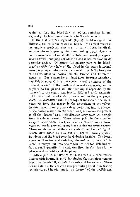
In the first thirteen segments (fig. 5) the blood-system isdifferent, and so is the course of blood. The dorsal vessel isno longer a receiving channel; it has no dorso-intestinalsand cornmissurals opening into it and feeding it with blood—infact it receives no blood at all, but behaves instead as a greatarterial trunk, pumping out all the blood it has received in itsposterior region. Of course the greater part of its blood,together with the whole of the blood in the supra-intestinalvessel, is pumped into the ventral vessel through the two pairsof ' latero-intestinal hearts ' in the twelfth and thirteenthsegments. But a quantity of blood flows forwards anteriorlyand this is pumped into the ventral vessel by means of the' lateral hearts ' of the ninth and seventh segments, and issupplied to the gizzard and the pharyngeal nephridia by the' hearts ' in the eighth and fourth, fifth and sixth segments,until the dorsal vessel ends by branching on the pharyngealmass. In accordance with the change of function of the dorsalvessel we have the change in the disposition of the valves.In this region there are no valves projecting into the lumenof the dorsal vessel; on the other hand, the valves are presentin all the ' hearts ' at a little distance away from their originfrom the dorsal vessel. These valves point in the directionaway from the dorsal vessel, and lead the blood from the dorsalvessel outwards, preventing any blood taking the reverse course.There are also valves at the distal ends of the ' hearts ' (fig. 11)which allow blood to flow out of ' hearts ' during systole,but do not let the blood come back during diastole. The dorsalvessel is therefore a distributing channel here ; most of itsblood it pumps out into the ventral vessel for distribution,but a small quantity it distributes itself to the gizzard, thepharyngeal nephridia and the pharynx.
With regard to the flow of the blood in the ventral vessel,I agree with Bourne (1, p. 77) in thinking that the blood comingfrom the ' hearts ' flows both forwards and backwards. Thereare no valves in the ventral vessel preventing blood from flowinganteriorly, and in addition to the ' hearts ' of the twelfth and

VASCULAR SYSTEM OF PHERET1MA S89
thirteenth segments there are ' hearts ' in the ninth andseventh segments also to take blood into the ventral vessel.I also agree with Bourne (1) when he says, ' All the blood whichenters the ventral vessel comes from the " hearts ", and thatall the ventro-integumentary branches—those anterior to the" hearts ", as well as those posterior to them—are efferentvessels. So far as the ventral vessel is concerned, they carryblood away from it.' The ventral vessel, therefore, here as in
T E X T - F I G . J l .
A BSemi-diagrammatic representation of ' hearts' in longitudinal
sections, A is one of the ' lateral' hearts of the ninth segmentwith the valves in its lumen and a bulb-like dilatation at itsventral end before it joins the ventral vessel, d.v. = dorsalvessel; ht. = heart; v. = valves ; si.v. = supra-intestinal vessel.
the region of the body behind the thirteenth segment, is thed i s t r i b u t i n g vessel and supplies blood through the ventro-tegumentaries to the body-wall, the integumentary nephridiaas well as the spermathecae and seminal vesicles, the ovaries,and the oviducts. . But it does not supply blood to the gutas it does in the hinder region ; there are no ventro-intestinalshere, and the function of supplying blood to the gut here istaken over partly by the dorsal vessel which supplies blood to thegizzard in the eighth segment and the pharynx and oesophagusin front, and partly by the lateral oesophageals. These vessels
D d 2

390 KAEM NARAYAN BAHL
in this region of the body are the counterpart of the subneural,the commissural, and the septo-intestinals of the hinderregion, and bring blood from the periphery to the main streamand to the gut. They receive a pair of branches bringing bloodfrom the body-wall, the septa, and other organs of the body,e.g. the nephridia (pharyngeal and integumentary) and thereproduction organs. The part of the oesophagus behind thegizzard is supplied with blood by the lateral oesophagealswhich lie intimately attached along the ventro-lateral aspectof the oesophagus. The blood from the oesophagus (ten tothirteen) (' ring-vessels ') and the gizzard is collected by thesupi'a-intestinal vessel, which also receives blood directlyfrom the lateral oesophageals through the ' anterior loops ' ofthe tenth and eleventh segments, and is conveyed to the heartsin the twelfth and thirteenth segments. The course of bloodcan be represented as follows :—
Pharynx £ Hearts PbaryngealNephridia Gizzardant. oesophagus. (I2th<£13th. (^tb 5th d 6th). (8th)
7th 49th).
Ventral vessel.
\Ventro-tegumentaries
throughout this region.\ (Body-wall $ septa \\\c£ Reproductive organs) i
Lateral- Oesophagsals
Oesophagus "Anterior loops"!(10-13 seqmts.) of loth 4 nth.)
ves
I^ Subneural vessel. Supra-intestinal vessels.
4Hearts
02 4- 13 th)

VASCULAR SYSTEM OF PHERETIMA 391
6. SUMMARY.
1. The typical arrangement of the blood-system in P h e r e -fcima occurs in the region of the body behind the fourteenthsegment, the first fourteen segments forming the cephalizedregion. The main longitudinal trunks are the same as inL u m b r i c u s , except that the lateral neurals are absent asin A l l o l o b o p h o r a . The dorsal vessel receives two pairsof dorso-intestinals and one pair of commissurals in eachsegment behind the cephalized region.
2. The intestinal blood-plexus is both an external and aninternal one, and three regions can easily be distinguished.The first is internal, and extends from the fourteenth to thetwenty-sixth segment; the second is both external andinternal, is co existent with the typhlosole, and extends overthe larger part of the gut; and the third is only external, andis confined to the rectal or post-typhlosolar part of the gut(last twenty-three to twenty-six segments).
3. The commissural vessel of P h e r e t i m a is a compoundvessel, and represents both the ' dorso-sous-nervien ' ofL u m b r i c u s and the intestino-tegumentary of Megasco lex .The capillaries of the integument are not like those of L u m -b r i c u s but like those of M o n i l i g a s t e r , and there isa close ' parallelism ' between an ' artery ' and a ' vein ' in thebody-wall, in which the two pass into each other througha number of capillary loops.
4. There are four pairs of ' hearts ' which connect the dorsalwith the ventral vessel, and five pairs which supply blooddirectly to the various organs in the cephalized region. Thereare two pairs of non-contractile ' anterior loops ' connectingthe lateral oesophageals with the supra-intestinals, these loopsbeing the counterpart of the connexions of the lateral oesopha-geals with the dorsal and the parietal in the tenth and twelfthsegments respectively of L u m b r i c u s . The subneuralTessel is absent in the first fourteen segments, and is con-tinuous with the lateral oesophageals of the anterior region.
5. As regards the course of circulation of the blood, the chief

392 HARM NAEAYAN BAHL
fact is that the dorsal vessel is wholly ' venous ' behind the' hearts ' and wholly ' arterial' in the region of the ' hearts 'and in front (the whole of the cephalized region). The examina-tion of valves and experiments by cutting and pinching theblood-vessels in P h e r e t i m a confirm the results of Johnstonefor L u m b r i c u s as regards the course of blood in dorso-intestinals and conimissurals and make Bourne's theory unten-able. The ventral vessel is the arterial trunk throughout,while the venous function of the dorsal and subneural behindis taken up by the lateral oesophageals in the cephalized region.The thin-walled and non-contractile ' loops ' of the tenth andeleventh segments must be distinguished from the thickwalled and contractile ' hearts ' of the other cephalized seg-ments, the ' loops ' being the channels for conveying bloodfrom the lateral oesophageals to the supra-intestinals.
LIST OF REFERENCES.
1. Bourne, A. G.—"On Megascolex coe ru leus and a Theory ofthe Course of the Blood in Earthworms ", ' Quart. Journ. Micro.Sci.', vol. 32, 1891.
2. "On Moni l i ga s t e r g r and i s ", ibid., vol. 36, 1894.3. Beddard, F. E.—" On the Structure of a New Genus of Oligochaeta,
and on the Presence of Anal Nephridia in Acanthodrilus ", ibid.,vol. 31, 1890.
4. ' A Monograph of the Oligochaeta'. Oxford, 1895.5. Benham, W.—" Studies on Earthworms ", ' Quart. Journ. Micro. Sci.',
vol. 26, 1886.6. " The Nephridiuni of L u m b r i c u s and itB Blood-supply",
ibid., vol. 36, 1891.7. Bahl, K. N.—" On a New TyPc °f Nephridia in Indian Earthworms
of the genus P h e r e t i m a " , ' Quart. Journ. Micro. Sci.', vol. 64,1919.
8. Harrington, N. R.—" The Calciferous Glands of Earthworms, with anAppendix on Circulation", 'Journ. of Morphology ', vol. 5, Supple-ment, 1899.
9. iTaquet.—" Recherches sur le syst£me vasculaire des Annelides ",1 Mitth. Zool. Stat. Neap.', Bd. VI, 1885-6.
10. Johnstone, J. B., and Johnson, Sarah W.—" The Course of Blood-flowin L u m b r i c u s " , 'American Naturalist', vol. 36, 1902.
11. Johnstone, J. B.—" On the Blood-vessels, their Valves, and the Courseof the Blood in L u m b r i c u s ", ' Biological Bulletin ', vol. 5, 1903.

VASCULAR SYSTEM OF PHERBTIMA 393
12. Lankester, E. Ray.—" The Anatomy of the Earthworm ", ' Quart.Journ. Micro. Sei.', 1865.
13. Perrier.—" Etudes sur l'organisation des lombriciens terrestres ",' Arch. Zool. Exp.\ t. ix, 1881.
14. Stephenson, J.—" On Intestinal Respiration in Annelids : with Con-siderations on the Oiigin and Evolution of the Vascular System inthat Group ". ' Trans. Roy. Soc. Edin.', vol. 49, 1914.
15. Vejdovaky, F.—' System und Morphologie der Oligochaeten ', Prag,