Embed Size (px)
Citation preview

On the Anatomy of some Fish Cestodes describedby Diesing from the Amazon.
By
W. N. F. Woodland,
Wellcome Bureau of Scientific Research, Euston Road, London, N.W.I.
With Platea 11 to 16 and 1 Text-figure.
I N 1850 and 1855 Diesing briefly described the superficialfeatures of about half a dozen Cestodes obtained by Nattererfrom fishes in the Amazon and on this basis he founded fivegenera. Zoologists have been familiar for many years withDiesing's figures of the remarkable scoleces of some of theseCestodes (as reproduced in Braun's account of the Cestodes inBronn's 'Klassen und Ordnungen des Thier-Eeichs', e.g.) butapparently, with the sole exceptions of E p h e d r o c e p h a l u sm i c r o c e p h a l u s redescribed in 1906 by Mola, and of theCestodarian Amph i l i na l i gu lo idea (the Monos tomuml igu lo ideum of Diesing and the Sch i zochoe rus l igu-lo ideus of Poche), no one has, up to the present, had theopportunity of ascertaining the strobilar anatomy correspondingto these scoleces. In fact fish Cestodes from the Amazon, apartfrom the descriptions mentioned and the papers by Fuhrmann(1916) on Goezeel la s i lu r i from Cetops is c a e c u t i e n s ,and by Janicki (1908) on Amph i l i na (Nesoleci thus)j a n i c k i i Poche (described under the name A m p h i l i n al iguloidea—a Cestodarian; see Poche 1925, p. 257), are, sofar as I am aware, quite unknown, though three or four specieshave been described from other parts of South America. Thisbeing the case, the outline description of the strobilar structureof six of Diesing's species of Cestodes contained in this papermay be of more than usual interest.
The material on which the following descriptions are basedwas obtained by me during the latter half of 1931. In that year
NO. 302 N

176 W. N. F. WOODLAND
I was able, thanks to the generosity of Sir Henry Wellcome,F.E.S., and the kind offices of Dr. C. M. Wenyon, F.B.S., totraverse the main channels and backwaters of more than1,100 miles of the Eio Amazonas and Bio Solimoes (AltoAmazonas) for the purpose of collecting Helminth parasites.Of fishes I examined altogether 312 individuals belonging tosixty-five distinct species, and the majority of these were caughton hook and line or in circular hand-nets by the crew of the twoboats which I purchased for the voyage back to Para fromManaos. I had at first hoped to be able to utilize the riversteamers as a means of transport and to engage local fishermento obtain fishes and other animals for my purposes, but a pre-liminary visit to a typical village (Codajaz, about 150 milesabove Manaos) soon convinced me that native indolence wasproof against all practicable forms of inducement. The fishmarket at Manaos was the only other source of supply availableto me, but this of course only represented one locality, and non-edible fish were rarely to be seen. I had also relied greatlyupon the fish markets at Obydos, Santarem, and other smalltowns or villages and especially at Para, but I found that allmarkets other than the large Portuguese-managed concerns atManaos and Para were very poorly supplied and quite uselessto me, and I was prevented from profiting by the excellentmarket at Para by becoming incapacitated with acute kidneytrouble towards the end of the voyage, and the last fish Iexamined was up a creek at Gurupa some 200 miles from Para.
The parasites I sought for were mainly Cestodes, and of theseI obtained a large number from fishes and birds, those fromfishes mostly new to science. Unfortunately, in the cases offive out of the six species described by Diesing, the material Iobtained was very scanty, though sufficient to describe the mainfeatures of the anatomy. Only in the case of A m p h o t e r o -m o r p h u s p e n i c u l u s was my material plentiful, though mydescription of this species is not correspondingly amplified.
Finally, I wish to acknowledge my obligations to the followinggentlemen for kind assistance in connexion with my visit toBrazil: Dr. L. W. 6. Malcolm of the Wellcome Historical MedicalMuseum, Mr. W. H. Parker, Professor E. Euggles Gates, His

CESTODES FROM THE AMAZON 177
Excellency Senhor Raul Regis de Oliveira of the BrazilianEmbassy, His Excellency Senhor Emmanuel Moraes, Acting-Governor of the State of Amazonas at the time of my visit, andMr. F. G. Coultas, British Consul at Para. I also wish to thankMessrs. J. R. Norman and N. B. Kinnear of the British Museumfor identification of the fishes and birds which I examined.
Z y g o b o t h r i u m m e g a c e p h a l u m Diesing, 1850.(Figs. 1-17, Pis. 11 and 12).
This tapeworm is found in the middle intestine of the Pirarara,the P i r a r a r a b icolor of Spixand Agassiz, and the P h r a c -t o c e p h a l u s h e m i l i o p t e r u s of Cuvier and Valenciennes,a common and ferocious fish and very conspicuous with itsdark grey dorsum, yellow lower surface, and sides with blackpatches, and brilliant scarlet fins. My largest examples wereabout 1 metre in length—not far short of the maximum lengthrecorded, viz. 1-2 metres. Out of 27 of these fishes examined,6, captured between Manaos and Santarem, contained Zygo-b o t h r i u m m e g a c e p h a l u m , and of this species I onlypossess 5 entire mature specimens, 2 large scoleces (with por-tions of strobila), and 2 small immature specimens.
E x t e r n a l F e a t u r e s .Diesing gives the maximum length of the entire worm (fig. 1,
PI. 11) as 3 in., which I assume to be 76 mm., and the maximumbreadth as 2£"' or about 5 mm. My largest specimen measured70 mm. in length and 3 mm. in maximum breadth (preservedin formalin). The scolex is, as the specific name implies, large,being, in my largest specimen, 3 mm. broad and 2 mm. high(these measurements were doubled in Diesing's largest specimen),and essentially consists of four large hollow globular partsunited centrally to a central column and also fused to eachother laterally (fig. 2, PI. 11). Each hollow globular part is inreality a large sucker which, however, differs from most othersuckers in having its wide opening to the exterior divided intotwo by a relatively narrow though conspicuous bridge of tissuedisposed tangentially to the scolex circumference (figs. 2-5,PI. 11)—a feature characteristic of this species. The figures of
N 2

178 W. N. F. WOODLAND
Diesing show the scolex in an unattached and somewhat con-tracted condition, the two openings of each sucker1 being verysmall and the surface very wrinkled. Pig. 2, PI. 11, shows thescolex less contracted, and fig. 5, PI. 11, is a vertical sectionthrough another scolex in the same condition. Fig. 4, PI. 11,shows an end-on view of the scolex in a contracted conditionsimilar to that figured by Diesing. The central area inside thesucker openings is sometimes slightly elevated. There is no'os terminale' (Diesing queried its existence) nor apical organof any kind. The axis of the central column of the scolex con-tains numerous muscle-fibres, and each globular wall of thesucker is lined with a layer of muscle specially thickened roundthe edge of the opening. Fig. 3, PI. 11, shows the end-onappearance of a scolex which was attached to the mucosa byfour large digitiform masses of mucosa which projected inthrough the inner openings and emerged as lumps through theouter openings of each sucker. In fig. 7, PI. 11, in which alleight openings of the suckers are in contact with the mucosa, itis possible that the inner and outer openings of the suckersacted each as a separate sucker. As this figure shows, the scolexcan become greatly deformed during attachment.
An unsegmented neck region is absent. In the strobila thesegments are very numerous, are very short anteriorly, and onlyattain a length equal to or exceeding their breadth at theextreme posterior end. Mature segments (in which all the organsare well formed, but eggs have not entered the uterus) firstoccur about midway in the length of the strobila. The mostconspicuous feature about the strobila is the great growth ofthe posterior borders of all the segments (figs. 8-10, PI. 11), sothat, both dorsally and ventrally, the actual strobila is overlaidby at least two thicknesses of border lappet and, unless thelappets are scraped off, it is useless to prepare a piece of strobilaas a whole-mount. As Diesing remarks, each surface of thestrobila shows a median furrow, marked by median notches inthe lappets. On the ventral side the uterus openings coincide
1 I use the term sucker because its cavity has a distinct though thinmuscle-layer clearly delimited from the underlying parenchyma; a bothriumhas no musculature apart from that traversing the general parenchyma.

CESTODBS FROM THE AMAZON 179
with the median furrow. The cirrus and vaginal apertures,are marginal, opening at about the middle of the length of thesegment, and very irregularly alternate (as many as nine cirrussacs, e.g. may occur consecutively on the same side). In veryfew of my specimens are the cirri protruded prominently, and inthese cases atria are absent; in other cases the retracted cirrusand vagina open into a shallow (fig. 17, PL 12) or relativelydeep atrium and therefore possess a single opening on theproglottid surface; in other words, the atrium is largely atemporary structure, present chiefly in immature proglottids.The openings of the cirrus and vagina in young proglottidsare covered over by the lappets of the proglottid in front butbecome uncovered as the segments become mature and elongate.
I n t e r n a l A n a t o m y of the P r o g l o t t i d .I first will describe briefly the structure of a mature proglottid.
The testes are numerous (between 150 and 200 in number ona rough estimate) and are scattered more or less uniformly inthe medullary parenchyma (i.e. inside the longitudinal muscle-layer) over the dorsal surface of the proglottid between thelateral groups of vitellaria, except in the area occupied by thebranching ovary along the posterior border (fig. 11, PI. 11).In balsam whole-preparations the testes measure approximately66 microns in diameter. The cirrus sac is about 0-66 mm. longand 0-083 mm. broad, and it opens ventrally and posteriorly tothe vagina, usually in a shallow atrium (fig. 17, PL 12), but inimmature segments the atrium is much deeper and moresharply defined. The wall of the sac is thin, hyaline, and quitedevoid of muscle-fibres and, with the cirrus retracted, contains(a) a straight canal, thick-walled (with ejector muscles attached),and with lumen lined with chitinous spines, which occupies theouter two-thirds of the sac and forms the eversible spiny cirrus,and (b) the inner convoluted ductus. The coils of the vasdeferens are in young mature segments very thin and con-voluted, running ventrally to the vagina, but, in older pro-glottids, the vas dilates enormously (fig. 15, PL 12) with accumu-lated masses of sperms (vesicula seminalis). The vagina openingis contiguous with, and dorsal to, that of the cirrus sac, and the

180 W. N. F . WOODLAND
dilated outer end of the vagina may lie anterior or posterior tothe cirrus sac, though the main vagina canal, in all the prepara-tions I have examined, always lies posterior (and dorsal) to thevesicula seminalis. At its outer end the vagina possesses amuscular thickening in its wall, the so-called sphincter vaginae.The vagina pursues a straight course to the ventral side of themedian part (isthmus) of the ovary, but I am unable to elucidatethe detailed composition of the genital complex. The ovary,while immature, consists of a large number of follicles borne ona system of branching tubes which occupies the posterior borderof the proglottid on its dorsal side (fig. 11, PI. 11). In a moremature condition the ovary follicles lose their distinctness owingto individual enlargement. The uterus arises as a narrow out-growth from the central ovary complex on the ventral side and,while extending forwards in the median line, gives off lateraldiverticula similar to those found in a species of Taen ia(figs. 11, 12, PI. 11).
The vitellaria are small ovoid bodies (c. 25-5x14-5 microns)arranged in two small semicircles, each semicircle embracingthe lateral bend of the circular band of longitudinal muscles.This arrangement is similar to that found in a typical Phyllo-bothriid save that in this family the vitellaria lie internal andnot external to the longitudinal muscle-bundle system. Thereare also, in Z y g o b o t h r i u m m e g a c e p h a l u m , a fewvitellaria present in the middle line on the dorsal side, alsoexternal to the muscle-band (figs. 14, 16, PL 12). This isreminiscent of a primitive condition in which, as in manyexisting Tetrarhynchidae, the vitellaria are disposed in a com-plete circle in transverse sections of the proglottids.
The circular band of longitudinal muscles delimiting the cortexfrom the medulla (figs. 14-16, PI. 12), is composed of the usualbundles of fibres, but in the young and mature proglottids thesebundles are so compacted together that they can only be dis-tinguished under the higher powers of the microscope; only ingravid proglottids, when the muscle-system is degenerating, dothe individual bundles become mutually separate and thereforedistinct. I cannot detect transverse muscle-fibres lying internalto the longitudinal, nor a layer of longitudinal muscle-fibres

CESTODES FROM THE AMAZON 181
underlying the cuticle, but I must confess that my sections arerather thick. There is nothing to remark concerning theparenchyma save that calcareous corpuscles are absent. Thecuticle is about 11 microns in thickness on the average.
The excretory system consists on each side of the proglottidof the usual thick-walled dorsal vessel (110 x 44 microns externaldiameter in immature proglottids, 58 x 44 in mature and stillsmaller in gravid) and the more internally lying thin-walledventral vessel, with a lumen three or four times larger than thatof the dorsal. The lateral nerves lie just external to the dorsalvessels.
The further development of the mature proglottid into thegravid may be described very briefly. My material beingscanty I cannot relate with certainty the probable process offertilization, but it appears possible that this is effected by theshort cirri ejecting spermatozoa to the exterior and these beingat once swallowed by the muscular extremity of the vagina,the opening of which lies at the base of the cirrus. It appearsimprobable that the cirrus can penetrate into the vagina in theusual way, and there is no evidence for intersegmental cross-fertilization and still less for fertilization between differentworms. Which ever is the method adopted, the fertilized eggssoon pour into the uterus and its diverticula, which becomeenormously distended. The median portion of the uterus sooncomes into contact with the ventral body-wall (fig. 16, PI. 12),and this forms a very definite pore or rather slit through whichthe uterus ruptures and the eggs are expelled in great quantities.The free eggs, preserved in formalin (fig. 13, PI. 11), measure45 microns in external diameter (preserved in balsam the samemeasurement is only 22 microns). They are spherical, the shellbeing thin and the embryo entirely without hooks. Previousto the expulsion of the eggs the internal organs of the proglottidhave degenerated rapidly once fertilization is largely effected,and in proglottids which have expelled all their eggs onlyoccasional traces of the testes and vesicula seminalis, a veryragged row of longitudinal muscle-bundles and stray eggs incollapsed uterus diverticula, are to be seen, apart from theparenchyma and cuticle.

182 W. N. F. WOODLAND
The only feature of striking importance in the structure ofthe strobila is the situation of the vitellaria external to thelongitudinal muscle-band, while the other principal organs aremedullary—an arrangement of the organs so far not describedin any other Proteocephalid or Monticelliid. The significanceof this feature and the systematic position of the genus will bediscussed below.
E p h e d r o c e p h a l u s mic rocepha lu s Diesing, 1850.(Pigs. 21-30, PL 13.)
This tapeworm (fig. 21, PL 13) is also found in the Pirarara( P h r a c t o c e p h a l u s hemi l iop te rus ) , but in the rectum.Unlike that of Z y g o b o t h r i u m m e g a c e p h a l u m , theinternal anatomy of this species has been described in somedetail, Mola (1906) having had access to material in the NaturalHistory Museum at Vienna. This is fortunate since I was onlyable to obtain two specimens of the worm from two of the fishesI examined. My description of this species necessarily will bein part supplementary to that of Mola.
E x t e r n a l F e a t u r e s .The larger of my two worms measured about 72 mm. in
length and 4 mm. in maximum breadth (in formalin) and theother was slightly smaller. Both worms were, therefore, verysmall compared with the giant form described by Diesing,'longit. corp. 1' et 2"; latit. med. 4"", but not much smallerthan those subsequently described by Mola, stated by him tobe 10-15 cm. in length. Both of my specimens were sexuallymature, the uteri being well developed half-way down thestrobila length and the masses of contained ripe eggs causingthe ventral surfaces of the proglottids to protrude considerably(fig. 25, PL 13). In Diesing's and Mola's figures, on the otherhand, the proglottids are shown as being relatively fiat, the eggspresumably having been discharged through the conspicuousuterine pores. It is noteworthy that the maximum breadths ofboth the strobila and scolex of Diesing's giant specimen wereabout the same as those of the larger of my two specimens,viz. about 4-5 mm. in all cases.

CESTODBS FROM THE AMAZON 188
The scolex of my larger specimen (figs. 22, 23, PI. 13) isrecognizable as resembling the figures of Diesing and Mola,though the expanded lower portion or collar of the scolex hasnothing of the regular curves depicted by Diesing nor of thefolded 'cerebral cortex' appearance shown by Mola. In truththis expanded condition of the lower part of the scolex is,judging from the condition of the scolex in my smaller specimen(fig. 24, PI. 13), solely assumed when the scolex is attached andis indeed an additional means of attachment. We have alreadyseen a similar change of form of the scolex during attachmentin Z y g o b o t h r i u m m e g a c e p h a l u m . The four suckersare apparently quite typical and I could detect no signs of acentral sucker or apical organ, but I must add that my limitedmaterial prevented me from studying the scolex in sections.
An unsegmented neck region is absent. In the strobila thecreases denoting successive proglottids are not conspicuousowing, as Mola says, to the presence of numerous secondarycreases. There is no trace of the lappet-formation so conspicuousin Z y g o b o t h r i u m m e g a c e p h a l u m . Uterine pores werenot formed in my specimens, though the uteri were full of eggsin the more posterior segments. The cirro-vaginal aperturesare very irregularly alternate and situated near the anteriorborders of the segments.
I n t e r n a l Ana tomy of the S t r o b i l a .The testes are numerous and rather small (about 73x44
microns in horizontal sections of gravid proglottids) and, as inZ y g o b o t h r i u m m e g a c e p h a l u m , are scattered over thegreater part of the dorsal surface of the proglottid; in thisspecies, however, the testes are, as is well known, situatedexternal to the longitudinal muscle-layer, i.e. in the dorsalcortical parenchyma, and are therefore not medullary (figs. 26,27, PI. 13). The cirri and vaginae both open into short genitalatria and extend inwards from their apertures through thelongitudinal muscle-layer and between the dorsal and ventralexcretory canals. The cirrus sac is dorsal to the vagina and, ina gravid segment, measures about 450 x 116 microns (in balsam),and has a thin nucleated wall with apparently an underlying

184 W. N. F. WOODLAND
layer of muscle. The contained cirrus is thick-walled and coiledand apparently unarmed. In none of my proglottids is the cirrusextruded, but Mola has supplied a figure of one. The vasdeferens, on emerging from the sac, becomes, in gravid segments,very convoluted and dilated to form the vesicula seminalis.Since the testes lie on the outside of the thick longitudinalmuscle-layer, it follows that fine branches of the vas deferensmust penetrate this layer in order to reach the testes.
The vagina is only slightly convoluted in gravid segmentsand extends inwards in close contact with the inner side ofthe ventral longitudinal muscle-layer until it reaches the uteruswhen it turns dorsally to open into the ovarian isthmus, in themanner depicted by Mola (in fig. 27, PL 13, the vagina appa-rently runs ventral to the uterus, but this is not the case, sincein the next section it turns dorsally). There is nothing remark-able about the ovary, which is a large mass lying along theposterior border of the segment and wholly internal to the thicklongitudinal muscle-layer (Fuhrmann's diagram on p. 98 of his1925 paper which shows dorsal projections of the ovary throughthe longitudinal muscle-layer is incorrect according to mypreparations). The uterus develops on the ventral side of theovary isthmus and takes the form of a single large sac withdigitiform subdivisions laterally. My material is not old enoughto show the actual formation of uterine pores, but doubtless, asin Z y g o b o t h r i u m m e g a c e p h a l u m , the median portionof the uterus, when full of eggs, comes into contact with theventral median subcuticula and ultimately a perforation isformed. The eggs differ markedly from those of Zygobo-t h r i u m m e g a c e p h a l u m in being elongated in form (fig. 30,PL 13), much smaller (44x22 microns in formalin), and inpossessing a yellowish yolky material lining the inside of theshell and (presumably) operculate openings at the shell ex-tremities. The embryo is hookless. The vitellaria are numerous,small (44x22 microns) bodies lying, as in Z y g o b o t h r i u mm e g a c e p h a l u m , outside the longitudinal muscle-layer, butin this species wholly on the ventral side and not laterally anddorsally as in Z y g o b o t h r i u m m e g a c e p h a l u m .
The circular band of longitudinal muscles is composed, as in

CESTODES FROM THE AMAZON 185
Zygobo th r ium megacepha lum, of closely compactedbundles of fibres, the bundles only being distinguishable ingravid proglottids. In immature proglottids the band is remark-ably thick (fig. 29, PI. 13) and the dorsal and ventral halvespractically.meet, so that the ovary and other rudiments occupyvery little space. Only in mature and especially gravid pro-glottids do the ovary, uterus, and vesicula seminalis developand cause the halves of the band to separate. The excretoryand nervous systems are of the normal type. Calcareouscorpuscles are not present in the parenchyma.
Apart from the scolex the morphological features of interestin this worm are the cortical situation of the testes and vitellariaand the wholly ventral situation of the latter. The systematicposition of this species will be discussed below.
P e l t i d o c o t y l e rugosa Diesing, 1850.(Figs. 31-40, PI. 14.)
This curious tapeworm was stated by Diesing to have beenfound in the 'S i lu rus P i n t a d o ' , otherwise ' P l a t y s t o m at ig r inum ' , and the P s e u d o p l a t y s t o m a t i g r i n u m ofCuvier and Valenciennes. In a fish, the native name of whichwas given to me as ' Surubim', which was caught in the Solimoesat Codajaz, I found seven specimens, mostly immature, of atapeworm which in most respects strongly resembles theP e l t i d o c o t y l e rugosa of Diesing, though it differs in thevery important character of being devoid of the bilocularsuckers figured by Diesing. The question of identity of the hostthus becomes of importance as evidence for or against theidentification of the worm. Evidence in favour of the view thatthe host containing the worms described below was Pseudo-p l a t y s t o m a t i g r i n u m is afforded by the fact that mysketch of the fish closely resembles the figure of this species givenby Schomburgk (1841) and that Goeldi (1898) gives 'Surubim'as the native name of this species. In other parts of the AmazonI found that this fish was known as the Kaparari. Accordingto Agassiz the species named by Spix 'Sorub im c a p a r a r i 'is P l a t y s t o m a co ruscans , a species probably closely

186 W. N. P. WOODLAND
related to P s e u d o p l a t y s t o m a t i g r i n u m and possiblyidentical, since I found that the 'tigerish' markings on fishcalled 'Kaparari' and 'Surubim' were very variable, thoughthey were usually quite distinct from those characterizing the'Sorubim' or 'Sudobim'•—the modern P s e u d o p l a t y s t o m af a s c i a t u m (Linn.)—a much more common fish than theKaparari. I have good reason then for assuming that the hostcontaining the worms to be described below was the samespecies as contained the P e l t i d o c o t y l e r u g o s a of Diesing,and this is one piece of evidence that these worms are P e l t i -doco ty le rugosa. Further evidence is to be found in thegeneral resemblance between the figures of Diesing and thosesupplied by me (the name rugosa being particularly applicableto my specimens) and in the fact that my specimens to a verylarge extent fit the generic and specific definitions of Diesing,the only essential difference being that, as I have mentionedalready, I cannot in my specimens detect any acetabula onthe four (or fewer) spatula-shaped areas borne on the apex ofthe scolex, and it is quite possible that Diesing mistook theareas for suckers of the usual kind or the bilocular variety. Imay add that in all I examined twelve specimens of the Kaparari,only one of which contained P e l t i d d c o t y l e rugosa .
E x t e r n a l Cha rac t e r s .The largest of my seven examples measured 5-8 mm. in length
and 1-4 mm. in maximum breadth. Diesing's largest specimenattained a length of about 88 mm. and a breadth of about 3 mm.The scolex region (there is no distinct neck) in my largestspecimen measured about 1-4 mm. long, but the scoleces of theother examples were shorter, both relatively to the strobila andabsolutely (figs. 31-3, PI. 14). The scolex consists of a basal,more dilated, and relatively smooth region, the surface of whichis marked with relatively few deep longitudinal creases, and anapical more narrow region, the general surface of which isdarker in colour than that of the basal and bears more numerousbut less deep longitudinal creases. Just below the summit ofthis apical region are situated usually four (but sometimes, asin two of my specimens, two or three) spoon-shaped areas

OESTODBS FROM THE AMAZON 187
which may perhaps be regarded as shallow suckers but whichare in reality only oval extensions anteriorly of the smooth baseof the scolex. In sections (figs. 34, 35, PI. 14) the base of thescolex is seen to be relatively solid and with a smooth surface,but the apex is spongy in character (except the spoon-shapedareas), is much creased, and is probably retractile within thebase to some extent. The scolex is quite unarmed.
The strobila is flat in transverse section and is covered witha thick cuticle showing both primary (demarcating the segments)and secondary transverse grooves. The proglottids, in my youngspecimens, do not exceed 20-5 in number and, excepting theterminal proglottid, are all broader than long. Anteriorly theoutlines of the proglottids are not evident, owing to the presenceof the secondary creases, and it is difficult to see the internalorgans in whole-mounted specimens owing to the thick cuticleand subcuticula. The cirro-vaginal apertures are marginal,opening near the anterior limit of the proglottid and apparentlyalternate fairly regularly. My material is too young for uterinepores to be present.
I n t e r n a l Ana tomy of a Ma tu re P r o g l o t t i d .Despite the thick cuticle and subcuticula I have been able to
observe the general disposition of the organs in the matureterminal proglottid of a worm mounted as a whole (fig. 36,PI. 14). The testes are numerous (between 200 and 300),measure between 35 and 40 microns in diameter (in sections),and are scattered over the entire dorsal surface of the proglottidbetween the lateral strands of vitellaria and the anterior edgeof the ovary posteriorly. A few testes may lie above the frontportion of the ovary. The cirrus sac is very large (266x66microns) and extends inwards nearly to the middle of theproglottid. The cirrus, when extruded, is only about 160microns in length but relatively broad (60 microns) so that itpresents a stumpy appearance (fig. 40, PI. 14). It is unarmed.The vagina opens immediately behind the cirrus, pursues acourse parallel with the cirrus sac, and bends back from themiddle of the segment and runs to the ovary. In fully matureproglottids the vagina becomes greatly convoluted and dilated
188 W. N. F. WOODLAND
in the middle line to form a large receptaculum seminis. Theovary consists of two large masses of follicles, one on each side,united centrally. The ovary is dorsally situated in the pro-glottid but ventrally to the testes when these two organs over-lap. The vitellaria lie, as usual, in two lateral strands, theindividual vitellaria measuring about 30 microns in diameter.
The remarkable feature about this species is that, as in thegenera Mont ice l l i a La Eue and Goezeella Fuhrmann,the testes, vitellaria, and ovary are all situated in the corticalparenchyma and not in the very reduced medulla. In this speciesthe cortical area is not demarcated from the medullary bydefinite layers of longitudinal and circular muscles but mainlyby a difference of texture of the parenchyma, the medullarybeing denser. Longitudinal muscle-fibres are probably presentbut are not associated to form bundles or even a definite layer,and in my rather thick sections they are difficult to distinguish.Further, this species also differs from Mont ice l l i a cory-p h i c e p h a l a (Monticelli) in that the vitellaria form, in trans-verse sections of the segments, definite half-circles embracingthe lateral extremities of the medulla and are not preponder-antly ventral as in the former species. In Goezeel la s i lu r iFuhrmann the vitellaria are wholly ventral in position. Itremains to be mentioned that in all three species the vagina, vasdeferens, and cirrus sac are in the medulla.
Though I cut serial transverse sections of portions of at leastthree individuals yet I could never distinguish dorsal and ventralexcretory canals.
Fig. 36, PI. 14, represents a young mature proglottid, and themost developed proglottids in my material (cut into sections)only differ in the development of a vesicula seminalis and re-ceptaculum. I am consequently unable to provide any descrip-tion of the uterus and eggs. In the text-figure, however, I haveassumed that, as in Mont i ce l l i a and Goezeella (the othergenera of the Monticelliinae), the uterus is cortical.
The re-definition of this genus will be discussed below; mean-while, the remarkable character of the scolex and the Mont i -cel l ia -like arrangement of the principal organs in the cortexmay again be remarked upon.

CESTODES FROM THE AMAZON 189
A m p h o t e r o m o r p h u s pen icu lus Diesing, 1850.(Figs. 41-52, PI. 15.)
This species, according to Diesing, is found in the intestineof Bagrus go l i a th , the native 'Dourada' or Dorad1 (orAdorad in some localities), the P i r a t i n g a r o u s s e a u x i i ofGoeldi (1898) and the modern B r a c h y p l a t y s t o m a rous-s e a u x i i . This fish is a large (my largest measured 123 cm.in length) and not uncommon Siluroid, and every one of thethirteen specimens which I examined contained Cestodes ofsome kind, and five (caught between Manaos and Parintins)contained the present species in considerable numbers.
E x t e r n a l C h a r a c t e r s .My largest specimen is about 5 cm. long and 1-18 mm.
maximum breadth, but other shorter and more contractedspecimens may be much broader, as e.g. 2-65 mm. in mybroadest specimen which was only 1-5 cm. in length. Thesemeasurements correspond very closely with those given byDiesing. The scolex, in the contracted and unattached condition(figs. 42, 43, PI. 15), is, externally, very similar to the drawingprovided by Diesing: that is to say, it may be no broader thanthe adjoining strobila and consists of a circular longitudinallywrinkled thick wall enclosing a cavity, on the floor of which liefour suckers radially disposed. But more usually, in my material,the scolex wall is expanded (figs. 44, 45, 47, PL 15) into aninverted cone-shaped structure with a terminal disc (formedfrom the lining of the cavity) covered with numerous delicateadhesive processes developed from the subcuticula. A similarchange of form of the scolex during adhesion has already beennoticed in Z y g o b o t h r i u m megacepha lum and E p h e -d rocepha lus m i c r o c e p h a l u s . Diesing's description andfigure of the 'bothrial grooves' and acetabula appear, whenconfronted with my material, to be more fanciful than accurate.I have found it very difficult to observe clearly the suckers inflattened scoleces, either examined in formalin or mounted inbalsam, owing to the presence of the numerous fine adhesive
1 Not to be confused with the 'Dorado' or 'Golden Salmon', a pink-fleshed Characinid also found in South American fresh-waters.

190 W. N. P. WOODLAND
processes just mentioned, but I have succeeded to a smallextent in two specimens in which the surface of the disc hadbeen scraped with a scalpel. Vertical and transverse sectionsof the scolex prove that four bilocular suckers, cruciform inarrangement, exist (fig. 46, PL 15), each sucker consisting ofa larger central cavity and a smaller distal. Diesing is mistakenin recording an ' os terminale' for this species.
The strobila is distinctly flattened in transverse section andconsists of numerous proglottids, all broader than long, thoughin some specimens a few of the hind segments approach thesquare in outline. The posterior borders of the segments donot overlap the segments behind. The ventral uterine poresare situated anteriorly in the segments. The cirrus and vaginalapertures are marginal as usual but are, in this species, uni-lateral (with possibly an occasional exception), being situatedon the right side of the strobila and anteriorly in each segment.In very few of the worms in my collection are the cirri extruded,and the cirrho-vaginal apertures are not distinguished by anypapilla or other external feature. Atria appear to be absent.
I n t e r n a l Ana tomy of the P r o g l o t t i d .The testes are not numerous, being under forty in number in
each segment, and are distributed in the medulla between theovary posteriorly and the anterior border of the proglottid.Laterally they do not extend beyond the ends of the ovary.The individual testes are about 44 microns in diameter. Thecirrus sac is large (280x116 microns when the cirrus is re-tracted), extending across from one-quarter to one-third of thebreadth of the segment, and possesses a thin though muscularwall. The cirrus when extended (fig. 48, PL 15) is of consider-able length (about three-quarters of the breadth of the pro-glottid, i.e. about 0-580 mm.) and has a slightly distended basecontaining several coils of the ductus. It is unarmed. Thevas deferens is very convoluted and becomes dilated to serveas a vesicula seminalis. The vagina opens, like the cirrus sac,directly to the exterior and immediately in front of the cirrussac. The portion of the wall next the opening is stronglymuscular and the remainder of the tube is very convoluted. In

CESTODES FEOM THE AMAZON 191
older proglottids the median portion of the vagina becomesdilated and serves as a receptaculum. The ovary is of the usualform—two lateral lobulated masses united by a median isthmus.I have not ascertained the details of the ducts in the region ofthe isthmus, but it must be mentioned that the uterus arisesas a forward-growing tube on the ventral side of the ovaryisthmus, and that the full-grown uterus has the taenioid formdepicted in fig. 49, PL 15. Anteriorly the uterus system opensto the exterior via a wide down-growth—the uterine pore.
The vitellaria, unlike all the other organs so far mentioned,are situated in the cortical parenchyma and are in two groupson each side—dorsal and ventral. The dorsal vitellaria are fewin number and are situated above the excretory canals; theventral vitellaria are numerous at the level of the ovary.,extending on each side from near the inner end of the ovaryto the outer limit of the cortex, but diminish in number anteriorlyso that the ventral group of vitellaria is more or less triangularin form (fig. 48, PI. 15). The vitellaria are of an oval form intransverse sections, and measure on the average 70 x 18 microns.Intra-uterine eggs, when examined in formalin, are of the shapeshown in fig. 52, PL 15. The embryos are spherical, measure14-18 microns in diameter, and are devoid of hooks. Surround-ing each embryo is an inner envelope with peripheral refringent(yolk) globules, and externally there is a thin outer envelope,measuring on the average 22-30 microns in breadth and 30-40microns in length.
As already mentioned, the parenchyma is divided into corticaland medullary regions distinguished not only by a differenceof texture but also separated by a definite though thin layer oflongitudinal muscle-fibres. The fibres show some indication ofbundle-formation in places, but the bundles are quite small andnot very definite. Though the medulla is small in area comparedwith the cortical, yet, as already stated, all the sexual organs,save the vitellaria, are included in it, and in this importantrespect A m p h o t e r o m o r p h u s pen icu lus resembles Zy-g o b o t h r i u m m e g a e e p h a l u m .
The excretory system consists of the usual two dorsal andtwo ventral vessels and, in this species, these are connected
NO. 302 o

192 W. N. F. WOODLAND
respectively, that is to say, dorsal to dorsal and ventral toventral, across the middle line by transverse canals situatedimmediately behind the ovary in each segment; further, ineach segment the ventral canal of the side bearing the cirrus(but not that of the opposite side) opens to the exterior by ashort duct running almost vertically downwards behind theovary (fig. 51, vvv, PI. 15).
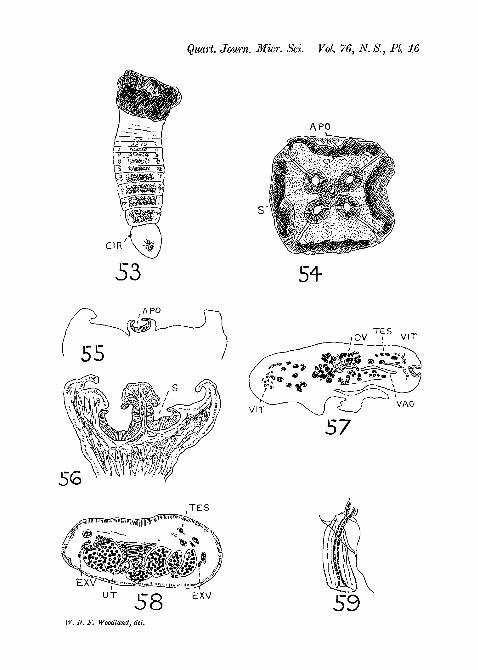
S c i a d o c e p h a l u s mega lod i scus Diesing, 1850.(Pigs. 53-9, PI. 16.)
This small species is, according to Diesing, found in ' C i c h 1 amonocu lus ' , which is the Cichla oce l la r i s of Bloch andSchneider. This fish is known on the Amazon as the ' Tucunare',and although it is abundant I only examined eight examples,and only in one of them (caught at Manaos) did I find fivespecimens of Sc i adocepha lu s mega lod i s cus . Mylargest specimen of this worm is only 3 mm. long (mounted inbalsam) with a maximum breadth of nearly 1 mm. and thi-3 Ihave preserved intact (fig. 53, PL 16). Diesing's largest specimenwas apparently about 9 mm. long. My other four specimensof this worm were slightly smaller than my largest. One con-sisted of but little more than a damaged scolex and the otherthree I either cut into sections (the strobilae and one scolex)or mounted portions (two scoleces) in balsam. I obtained a fewgood sections of the strobila but not sufficient to enable me todescribe the anatomy in detail: hence my account is veryincomplete.
E x t e r n a l F e a t u r e s .My largest worm contained about a dozen recognizable pro-
glottids, the terminal being twice the length of the precedingand the last six showing egg-containing uteri. The matureproglottids are the broadest (0-813 mm.), and the most posterioris 0-132 mm. long; the gravid proglottids decrease in breadthtowards the end of the strobila though they increase in length,but, excepting the terminal, always remain broader than long.
The scolex in one of my smaller specimens was more expandedand therefore more umbrella-shaped (with broad ribs) than the

CBSTODES FROM THE AMAZON 198
contracted head of my largest specimen. When the ' umbrella'is fully expanded it is more than double the breadth of thestrobila. Moderately expanded scoleces (viewed end-on and invertical section) are shown in figs. 54 and 56, PI. 16. In theconcavity of the 'umbrella' are four large (0-182 mm. indiameter) spherical suckers, largely obscured from view byoverlying folds of the umbrella surface, and one smaller(0-132 mm. in diameter) central or apical sucker, with a distinctopening (the 'os orbiculare' of Diesing). There is no trace ofa glandular apical organ. A short thick neck region may be saidto be present. In the short strobila the marginal genital aper-tures open usually a little in front of the midway point of theproglottid and appear to alternate very regularly (judging fronitwo of my specimens and from Diesing's figures). The extrudedcirri are not prominent. Uterine pores are not visible in mymaterial, though I have no doubt they develop in older speci-mens (vide fig. 58, PI. 16).
Some F e a t u r e s of the I n t e r n a l A n a t o m y .The testes are not numerous (I cannot estimate their approxi-
mate number) and are situated at the sides of the proglottid,between the central ovary and the lateral vitellaria. The cirrussac is very small (about 40 X 26 microns) and the extrudedcirrus is short (44 microns) and thick (19 microns) and is slightlyarmed (fig. 59, PI. 16). Both the cirrus sac and vagina open intoa shallow atrium. The ovary occupies the centre of the pro-glottis (in transverse sections) and consists of a large number offollicles (fig. 57, PI. 16). The uterus also consists of a largenumber of swollen diverticula (fig. 58, PI. 16) and doubtlessbursts through to the exterior in the mid-ventral line whensufficient eggs have collected. The vitellaria are arranged, asin Z y g o b o t h r i u m m e g a c e p h a l u m , in two semicircular(in transverse sections) patches lying immediately external tothe lateral excretory canals. It is important to note that adefinite band of longitudinal muscle-fibres is entirely absent,though individual fibres may be scattered in the parenchyma.There is thus no question as to organs being medullary orcortical in position. I was unable to detect calcareous corpuscles.

194 W. N. F. WOODLAND
The excretory system consists, so far as I am able to observe it,of one vessel only on each side of the body, somewhat ventralin position but rather thick-walled.
This species is an undoubted Proteocephalid, and in virtue ofits unusual form of scolex may, like C o r a l l o b o t h r i u ms o 1 i d u m, perhaps be regarded as representative of a distinctgenus.
P r o t e o c e p h a l u s m a c r o p h a l l u s Diesing, 1850(syn. Taen ia m a c r o p h a l l a Diesing.)
(Figs. 18-20, PL 12.)
As La Eue (1914) remarks, this species, like some others,has so far only been known from the brief definition and draw-ings of Diesing, both almost wholly concerned with externalcharacters. Diesing stated that this species is found in ' C i c h 1 aMonocu lus ' (the modern Cichla ocel lar is) , and added(1856) that this fish is called locally the 'Jacunda'. I mayremark that the fish called ' Jharkiindar' which I examined onthe Amazon was greenish-grey in colour, of slender elongatedform, and devoid of the characteristic ocellus at the base ofthe caudal fin and therefore quite distinct from the Tucunarewhich is the native name for Cichla o c e l l a r i s . I foundthirteen examples of P r o t e o c e p h a l u s m a c r o p h a l l u sin a single specimen of Cichla oce l l a r i s caught at Manaos,the largest worm measuring 3-068 mm. in length and 0-94 mm.in maximum breadth (in balsam). In none of these worms arethe proglottids gravid, and there is no evidence that the wormsexamined by Diesing (the largest of which measured about6 mm. in length) bore gravid proglottids. I may also mentionthat, in addition to S c i a d o c e p h a l u s mega lod i s cus andthe present species, I found in the Tucunare two other quitedistinct species of tapeworm which superficially might be mis-taken for P r o t e o c e p h a l u s m a c r o p h a l l u s , since theyare about the same size, but they are distinguished at once bythe fact that in both cases the strobila when not more than3 mm. in length bears fully gravid proglottids. I shall describe.these two forms in a subsequent communication.

CBSTODES FROM THE AMAZON 195
Since the anatomy of P r o t e o c e p h a l u s m a c r o p h a l l u sis typically Proteocephalid and therefore of no particularinterest, I shall content myself with a brief diagnosis of thespecies, necessarily based solely on mature but non-gravidspecimens, adding remarks where necessary.
Distinguishable segments in strobila of non-gravid worms lessthan twenty in number (fig. 18, PL 12) and the non-gravidstrobila may attain a length of 6 mm. All segments broaderthan long save the last one (or two) and all flat in transversesection. The region of maximum breadth usually occurs infront of the middle of the strobila length. There is no neck orline of junction separating the scolex region from the strobilia,the scolex only being indicated by the presence of four typicalhemispherical suckers (c. 0-2 mm. in external diameter). Thereis no apical organ of any description. The marginal cirro-vaginal apertures irregularly alternate. The testes number 30-40in each proglottid and measure (in surface view) about 44microns in diameter. The cirrus sac measures about 146x30microns and contains a long coiled unarmed cirrus. The cirrussac and vagina both open into a small but deep atrium. Thevagina opens ventrally to the cirrus sac and extends on theventral side without dilatation to the ovarian isthmus. Theovary is a massive organ lying ventrally against the posteriorborder of the proglottid and consisting of two lateral lobesunited medianly. The vitellaria are small bodies (c. 22 x 14microns) forming two marginal strands lying, with all otherorgans, internal to the layer of longitudinal muscle-fibres. Thelongitudinal muscle-layer consists of isolated fibres (insufficientin number to form bundles) lying in a row in the vicinity oftransversely disposed fibres, and these, in conjunction withwhat looks like a thickening of the parenchyma, form anobvious, if thin, line of demarcation between the cortex andmedulla. Small calcareous corpuscles appear to be present inthe parenchyma. The cuticle is about 7 microns in thickness.Definite excretory canals are apparently absent. The host andlocality have already been mentioned.
I must add that a number of items of description not includedin the above are only omitted because I was unable, with my

196 W. N. F. WOODLAND
limited and largely immature material, to obtain informationconcerning them.
A PROVISIONAL EECLASSIFICATION OF THE PROTEOCEPHALIDAE
AND THE REDEFINITION OF SOME GENERA FOUNDED BY DIESING.
In view of the fact that I have in my collection of Cestodesfrom the Amazon several scores of new species which I hopeto describe during the next few years, it would seem advisableto lay aside the problem of the classification of forms alreadyknown until these new forms have been examined. But thereare obvious objections to this course, one being that Diesing'sgenera certainly require redefinition, and such is difficult untilit is known to which family they are to be referred. Moreover,there can be no objection to a temporary scaffolding so long asit is recognized as temporary, and so I propose to suggestwithout hesitation rearrangements of existing groups accordingto the information I possess at the time, having due regard,however, to the final taxonomic edifice.
The Order Tetraphyllidea, according to recent authorities(Perrenoud, 1931), comprises seven families—Phyllobothriidae,Onchobothriidae, Proteocephalidae, Lecanicephalidae, Cephalo-bothriidae, Discocephalidae, and Monticelliidae—but for reasonswhich I advanced in 1927 (Woodland, 1927), and for presentpurposes, I prefer to modify this scheme by regarding thePhyllobothriidae and Onchobothriidae as one family—thePhyllobothriidae emend—abolishing the family Monticelliidaefor the reasons about to be given, and including the Tetrarhyn-chidae (usually regarded as a distinct order, the Trypano-rhyncha), the result being six families, three large and wellestablished and the remainder small and problematical. Nowthe majority of species belonging to the family Proteocephahdae—the family with which we are now concerned—answer to thedefinition of the family which La Eue gave in 1911 and 1914,but even at that time there were known to exist three Pro-teocephalid-like worms which showed two types of organ-arrangement both distinct from the Proteocephalid type, viz. the' T e t r a c o t y l u s ' c o r y p h i c e p h a l a first described byMonticelli in 1891, the ' O o r a l l o b o t h r i u m ' l obosum
CESTODBS FROM THE AMAZON 197
(renamed by Mola Bphedrocephalus lobosus) firstdescribed by Eiggenbach in 1896 and Ephedrocephalusmicrocephalus, the anatomy of which was first describedby Mola in 1906. La Bue apparently was only acquainted withthe first species, which he renamed Monticellia coryphice-phala, and, on the basis of its peculiar organ-arrangement,founded a new family for its reception, the Monticellidae. Later,Fuhrmann (1925), who was acquainted with the anatomy of allthree worms and in addition with the anatomy of Marsypo-cephalus tanganyikae (his Loennbergia tangan-y i k a e) which exhibits a third distinct type of organ-arrange-ment, confirmed this new family, amending the definition toinclude all the three types of organ-arrangement which differfrom that characteristic of most Proteocephalids. This is abrief history of the origin of the family Monticelliidae. To pre-pare the ground for the arguments to follow, it is necessarybriefly to state the nature of these four organ-arrangementsknown to Fuhrmann (see diagram on p. 199). In the typicalProteocephalid 'Vitellaria, testes, ovary, and uterus [aresituated] within the inner longitudinal muscle-sheath' (LaEue, 1914); in Mont i ce l l i a and Goezeel la the testes andovary are situated in the dorsal cortex, the vitellaria and theuterus in the ventral cortex: that is, all these organs are cortical;in E p h e d r o c e p h a l u s the testes are in the dorsal cortexand the vitellaria in the ventral cortex, but the ovary anduterus are both in the medulla; in M a r s y p o c e p h a l u s thetestes alone are in the dorsal cortex, all other organs beingmedullary and as in Proteocephalids. La Eue based his newfamily on the characters of the genus M o n t i c e l l i a , butFuhrmann, as already stated, amended the definition to fit thenew facts, the principal character in Fuhrmann's definitionbeing that the 'testes occupy a dorsal or dorso-lateral field inthe cortical parenchyma only, whereas the female genitalia maybe partly in the cortical and partly in the medullary paren-chyma' (Fuhrmann, 1925). Now it is fully evident that theseso-called Monticelliidae would undoubtedly be regarded asorthodox Proteocephalids but for the cortical situation of theorgans mentioned; they show no close affinities with any other

198 W. N. F. WOODLAND
Tetraphyllidean family. It is also evident that the Monti-celliid genera are only grouped together on account of the corticalsituation of the testes, since the other genital organs exhibitmarked differences as regards position, and I cannot think thatthis one common character, definite and peculiar as it may be,is sufficient to serve as the necessary bond for a new family.And I am confirmed in this opinion by the fifth type of organ-arrangement, now described in fresh-water Proteocephalid-likeworms for the first time, seen in Z y g o b o t h r i u m mega-cepha lum and A m p h o t e r o m o r p h u s p e n i e u l u s . Inthese species all the organs are medullary save the vitellaria,which are cortical, and they are arranged in a concentric fashionrather similar to that seen in most Tetrarhynchidae. Accordingto Fuhrmann's definition these worms cannot be Monticelliids,nor are they Proteocephalids according to La Eue. Are wethen to create another new family ?
I believe that the best solution of the difficulty is to abolishthe family Monticelliidae and to amend the definition of thefamily Proteocephahdae so as to include all the aberrant formsmentioned. The definition of the family Proteocephalidae willthen read as follows: Tetraphyllidea1 in which the scolex usuallybears true suckers,2 but never proboscides nor phyllidea;2 theparenchyma is usually subdivided into cortex and medulla byan internal distinct layer of longitudinal muscle-fibres or bya layer of circular fibres or by a difference of parenchymaltexture; the ovary, testes, vitellaria, and uterus are usuallysituated in the medulla but all these organs can in certain generabecome cortical; the ovary is always unilaminate; the vaginaruns dorsally to the uterus sac; ripe terminal proglottids donot usually separate from the strobila. Usual hosts are fresh-water Fishes, Amphibia, and Eeptiles.
Further, regarding ' Monticelliid' species as aberrant formsof Proteocephalidae, it seems best to subdivide the family into
1 I have attempted to define the Tetraphyllidea in my previous paper(1927).
2 For definitions of these structures and of bothridia, bothria, andsuckers I refer the reader to Benham's Chapter on the 'Cestoidea' inLankester's 'A Treatise on Zoology', p. 115.

CESTODES FROM THE AMAZON 119
NORMAL PROTEOCEPHALID
C/ o o o o p o o o o o o o o "~y \
ZYGOBOTHRIUMO O O O O O O O O O O O O O
MARSYPOCEPHALUSO O O O O O O O O O O O O
EPHEDROCEPHALUS0.0.0,0.00 o _o SLS
PELTIDOCOTYLE
THE FIVE TYPES OFORGAN ARRANGEMENT
IN THE PROTEOCEPHALIDAE

200 W. N. F. WOODLAND
groups according to the types of organ-arrangement shown,and these groups can most conveniently be represented by sub-families. I had reached this conclusion when I learnt of thepaper by Mola (1929), who has already proposed the abolitionof the family Monticelliidae, the inclusion of 'Monticelliid'genera in the Proteocephalidae, and the formation of sub-families. But his subdivision of the family is rather differentfrom the one I wish to propose, and the sub-families he foundedwill have to be redefined for my purposes. I propose the sub-division of the family Proteocephalidae into five sub-families,defined as follows:
Sub-family Proteocephalinae Mola, 1929. Proteoeephalids inwhich the testes, ovary, vitellaria, and uterus are not situatedexternal to an internal layer of longitudinal muscles,1 andthe vitellaria are compactly arranged in two marginalstrands.
Genera: P r o t e o c e p h a l u s , S c i a d o c e p h a l u s , &c.Sub-family Zygobothriinae Woodland, 1933. Proteoeepha-
lids in which the vitellaria alone are cortical in position,and are dispersed and placed dorsally and laterally andsometimes ventrally in the proglottid.
Genera: Z y g o b o t h r i u m , A m p h o t e r o m o r p h u s .Sub-family Marsypocephalinae Woodland, 1933. Proteo-
eephalids in which the testes alone are cortical in position,and the medullary vitellaria are compacted to form twomarginal strands.
Genus: M a r s y p o c e p h a l u s .Sub-family Ephedrocephalinae Mola, 1929. Proteoeephalids
in which both the testes and vitellaria are cortical inposition, the vitellaria being dispersed and on the ventralside of the proglottid.
Genus: E p h e d r o c e p h a l u s .Sub-family Monticelliinae Mola, 1929. Proteoeephalids in
which the testes, vitellaria, uterus, and the whole or the1 This mode of expressing the medullary situation of all the organs is
adopted in order to include forms devoid of any distinction between cortexand medulla.

CESTODES FROM THE AMAZON 201
greater part of the ovary are in the cortex. The vitellariaare dispersed and are lateral or ventrolateral in position.
Genera: Monticellia, Goezeella, Peltido-cotyle.
It remains to redefine most of these genera.Proteocephalus Weinland, 1858. Save that it belongs to
the Proteocephalinae I do not propose to redefine thisgenus. Its exact definition cannot be attempted until thevalidity of other Proteocephaline genera, such as 0 p h i o -taenia and Acanthotaenia, has been determined.
Sciadocephalus Diesing, 1850. Proteocephalinae. Scolexumbrella-shaped with four central suckers and a small apicalsucker. Eegularly (?)-alternating cirro-vaginal pores. In-ternal longitudinal muscle-layer absent and no distinctionbetween cortex and medulla.
One species, Sciadocephalus megalodiscus.Zygobothrium Diesing, 1850. Zygobothriinae. Scolex
with four suckers, each with two separate openings. Noapical organ. Irregularly-alternating cirro-vaginal pores.Vitellaria situated laterally and dorsally.
One species, Zygobothrium megacephalum.Amphoteromorphus Diesing, 1850. Zygobothriinae.
Scolex with four bilocular suckers, each with a single opening.No apical organ. Cirro-vaginal pores unilateral, on theright side of the strobila. Vitellaria situated dorsally andventrally.
One species, Amphoteromorphus peniculus.Marsypocephalus Wedl, 1862.Marsypocephalinae.Scolex
with four spherical or ovoid suckers. No apical organ.Cirro-vaginal apertures irregularly alternate. Vitellariacompacted in two marginal strands.
Three species: Marsypocephalus rectangulus,Marsypocephalus heterobranchus, Mar-sypocephalus tanganyikae.
Ephedrocephalus Diesing, 1850. Ephedrocephalinae.Scolex with four simple suckers. No apical organ. Cirro-

202 W. N. F. WOODLAND
vaginal apertures irregularly alternate. Vitellaria ventraland lateral.
Two species: E p h e d r o c e p h a l u s m i c r o c e p h a l u s ,E p h e d r o c e p h a l u s lobosus .
Mont ice l l i a La Bue, 1911. Monticelliinae. Scolex withfour simple suckers. No apical organ. Cirro-vaginalapertures irregularly alternate. Vitellaria ventrolateral.
One species: Mont ice l l i a c o r y p h i c e p h a l a .Goezeella Fuhrmann, 1916. Monticelliinae. Scolex with four
suckers and a massive collar composed of folds of encirclingtissue. No apical organ. Cirro-vaginal apertures irregularlyalternate. Vitellaria ventral and lateral.
One species: Goezeella s i l u r i .P e l t i d o c o t y l e Diesing, 1850. Monticelliinae. Scolex
devoid of typical suckers but possessing two, three, orfour spatulate areas which may serve as suckers. Noapical organ. Cirro-vaginal apertures more or less regu-larly alternating. Vitellaria crescentic (in transversesections) and lateral.
One species: P e l t i d o c o t y l e rugosa .
ADDENDUM.
In a paper ' On the Classification of the Cestoda' (Southwell,1929) Dr. Southwell has devoted more than one-third to alengthy criticism of a number of suggestions which I made in1929 apropos of the classification of the Tetrapbyllidea. I donot propose to reply in detail, but I should like to correct somemisstatements in Dr. Southwell's criticism and to explain moreclearly one or two of my own arguments. The first misstate-ment is on his p. 59:' The characters of the order Tetraphyllidea(sens, nov.) are not given and one is left to guess how this orderdiffers from the other two.' I refer Dr. Southwell to my pp. 539-42 (Woodland, 1927) which, save one-quarter of a page, areentirely devoted to this subject.
With regard to my statement concerning the arrangement ofthe longitudinal muscles of the Phyllobothriidae, Southwellquotes me as follows: 'the parenchyma is not divided by an

CESTODES FEOM THE AMAZON 203
internal layer of longitudinal bundles into cortex and medulla,the longitudinal musculature being restricted to an undividedperipheral zone of fibres and bundle of fibres lying immediatelyinternal to the thin circular muscle-layer underlying the cuticleand more or less coextensive with the subcuticula', and South-well adds that ' It is not clear from this statement whetherWoodland considers that the parenchyma is not divided intocortical and medullary portions at all or whether the paren-chyma is so divided, but that the longitudinal muscles [sic] arenot situated at the junction of the two parts', and he proceedsto show that in certain species I recognize a cortex as distin-guished from the medulla. My reply to these remarks is that insome Phyllobothriidae, e.g. in O r y g m a t o b o t h r i u m mus-t e l i , D i n o b o t h r i u m s e p t a r i a , and many others, thereis no line of distinction between medullary and cortical areas ofparenchyma, the two areas merging into each other, though itis true that the wide band of scattered longitudinal muscle-fibres and bundles is situated in an outer zone, but this outerzone does not define the ' cortex'; in others, e.g. in A n t h o -b o t h r i u m c o r n u c o p i a , there is also no line of divisionbetween cortex and medulla, unless we regard the subcuticula(which contains muscle-fibres) as incorporating the much reducedcortex; in species of P h y l l o b o t h r i u m , on the other hand,an outer zone of parenchyma is definitely separated from aninner zone by a line of circular muscle-fibres, the longitudinalmuscle-fibres or bundles being adjacent and extending through-out the cortex. In short, cortical and medullary areas of paren-chyma may be recognizable or the reverse, but what is certain isthat these two areas in Phyllobothriids are not separated, asthey are in Proteocephalids and Tetrarhynchids, by a well-defined row of longitudinal muscle-fibres or bundles. Southwellprotests against my assumption that these two areas are demar-cated in many Cestodes simply by the band of longitudinalmuscles, and states that these two areas are only to be distin-guished by the presence of a layer of circular and not longitudinalmuscles. But circular muscles are frequently absent, and in suchcases I do not think Southwell would hesitate to label the paren-chyma lying external to a well-defined layer of longitudinal

204 W. N. F . WOODLAND
muscle-bundles as cortex. In other cases, these two areas areonly distinguished by a difference of texture of the parenchyma(as in P e l t i d o c o t y l e r u g o s a , e.g.) and yet the names'cortex' and 'medulla' may justly be retained. In short, theterms cortex and medulla only mean in plain language outerzone and inner zone of parenchyma. In many cases they aresharply distinguished by layers of circular and bundles of longi-tudinal muscle-fibres, and when the circular muscle-layer isclearly visible it certainly affords a more definite line of divisionthan the row of longitudinal muscle-fibres or bundles of fibres,but when circular muscle-fibres are not distinguishable then therow of longitudinal muscles must serve, and, if these areabsent, a difference of parenchymal texture, if such exists; inother cases there is no line of distinction between cortex andmedulla. As regards my suggestions with respect to the positionof the vagina relative to the uterus in the Tetrarhynchidae, onthe one hand, and the Proteocephalidae and Phyllobothriidae,on the other hand, and to the quadripartite or x-shaped formof the ovary in the Tetrarhynchidae and Phyllobothriidae andthe unilaminate form in Proteocephalidae, I may point out thatthese distinctions have recently been incorporated in thedefinitions of these groups by Professor Fuhrmann in his excel-lent account of the Cestoidea in Kukenthal's 'Handbuch derZoologie', from which it would appear that they are not asdevoid of value as Southwell would have us believe. Finally,I may add that Southwell is again mistaken in asserting thatmy suggestions for classifying the Tetraphyllidea are solelybased on the few species examined by me personally.
LITERATURE.
Diesing, C. M. (1850).—'Systema Helminthum', Bd. i. Vindobonae.(1855).—"Sechszehn Gattungen von Binnenwurmern und ihre
Arten", 'Denkschr. d. K. Akad. d. Wissen., Wien. Math.-nat. Cl.',Bd. ix, p. 171.
(1856).—"Zwanzig Arten von Cephalocotylea", ibid., Bd. xii, i, p. 23.Fuhrmann, 0. (1916).—"Eigentiimliche Fischcestoden", 'Zool. Anzeig.',
Bd. xlvi, p. 385.

CESTODES PROM THE AMAZON 205
Fuhrmann, 0., and Baer, J. G. (1925).—"Zoological Results of the thirdTanganyika Expedition, conducted by Dr. W. A. Cunnington, 1904-5.Report on the Cestoda", 'Proc. Zool. Soc. London', pt. i, p. 79.
Goeldi, E. A. (1898).—"Primeira Contribuicao para o Condecimento dosPeixes do Valle do Amazonas e das Guyanas", 'Bol. Mus. Paraense deHist. nat. e Ethnog.', tome ii, 1897-8.
Giinther, A. (1864).—"Catalogue of the Physostomi . . . in the Collectionof the British Museum" in the 'Catalogue of Fishes', vol. v.
Janicki, C. v. (1908).—"Uber den Bau von Amphilina liguloidea Diesing",'Zeit. f. wiss. Zool.', Bd. 89, p. 568.
La Rue, G. R. (1911).—"A Revision of the Cestode Family Proteocephali-dae", 'Zool. Anzeig.', Bd. xxxviii, p. 473.
(1914).—"A Revision of the Cestode Family Proteocephalidae",'Illinois Biological Monographs', vol. i, pp. 1-350.
Linton, E. (1925).—"Notes on Cestode Parasites of Sharks and Skates",'Proc. U.S. Nat. Mus. Wash.', vol. 64, art. 21.
Mola, P. (1906).—"Di alcune specie poco studiate o mal note di Cestodi",'Ann. Mus. Zool. d. R. Univ. d. Napoli' (N.S.), vol. 2, no. 6.
• (1929).—"Descriptio platodorum sine exstis", 'Zool. Anzeig.',Bd. 86, p. 101.
Monticelli, F. S. (1891).—"Notizie su di alcune specie di Taenia", 'Boll.Soc. Nat. Napoli', ser. i, vol. v, p. 151.
(1892).—"Appunti sui Cestodaria", 'Atti r. Accad. d. Sci. Fis. Mat.Napoli' (2) 5, no. 6.
(1892).—"Studii sui Trematodi endoparassiti Monostomum cym-bium Diesing. Contribuzione allo studio dei Monostomidi", 'Mem. r.Accad. Sci. Torino. Cl. Sci. fis. mat. nat.' (2) 42, p. 683.
Perrenoud, W. (1931).—"Recherches anatomiques et histologiques surquelques Cestodes de Selaciens", 'Bull.-Annexe Revue Suisse de Zool.',tome 38, p. 469.
Poche, F. (1925).—"Das System der Platodaria", 'Archiv f. Naturges.Berlin', Bd. 91, Abt. A 2-3.
Riggenbach, E. (1896).—"Das Genus Ichthyotaenia", 'Rev. Suisse Zool.et Ann. Mus. d'Hist. Nat. Geneve', tome iv, p. 165.
Schomburgk, R. H. (1841).—"Fishes of Guiana", Parts 1 and 2. Publishedin the 'Naturalists' Library, Ichthyology', vol. iii, edited by Sir W.Jardine.
Southwell, T. (1929).—"On the Classification of the Cestoda", 'SpoliaZeylanica', vol. xv, pt. i, p. 49.
Spix, J. B. von, and Agassiz, L. (1829).—'Selecta Genera et SpeciesPiscium, quae in itinere per Brasiliam annia 1817-20 . . . ' collegitJ. B. de Spix. Monach.
Woodland, W. N. F. (1925).—"On some remarkable new Montioellia-likeand other Cestodes from Sudanese Siluroids", 'Quart. Journ. Micr. Sci.',vol. 69, pt. iv, p. 703.

206 W. N. F. WOODLAND
Woodland, W. N. F. (1927).—"A revised Classification of the Tetra-phyllidean Cestoda, with Descriptions of some Phyllobothriidae fromPlymouth", 'Proc. Zool. Soe. London', pt. 3, p. 519.
EXPLANATION OF PLATES 11-16.
REFERENCE LETTERS TO FIGURES.
ant, anterior side; apo, apical organ; at, atrium; b, bilocular sucker;bd, spatula-shaped area; cir, cirrus; cort, cortex; cut, cuticle; cs, cirrus sac;due, ductus ejaculatorius; dv, dorsal vessel; exv, excretory vessel; Im,longitudinal muscle-layer; mb, muscles of sucker; med, medulla; n, nerve;ov, ovary; post, posterior side; s, sucker; subcut, subcuticula; sv, sphinctervaginae; tes, testes; up, uterus pore; ut, uterus; vag, vagina; vit, vitellaria;us, vesicula seminalis; w, ventral vessel; wo, ventral opening of ventralexcretory canal.
(All figures drawn under the camera lucida.)
PLATE 11.
Figs. 1-13: Zygobo th r ium megacepha lum Diesing, 1850.Fig. 1.—The entire worm. x2.Fig. 2.—Scolex from side aspect, showing two of the bilocular suckers.
Xl2.Fig. 3.—Scolex, end-on view of. Openings of suckers distended. This
scolex was preserved in an attached condition, whereas that of fig. 2 wasunattached, x 12.
Fig.4.—Scolex, end-on view of. Scolex unattached and contracted, x 12.Fig. 5.—Scolex in vertical section, showing two of the bilocular suckers
in section (the section represented in the diagram of fig. 6). x 18.Fig. 6.—Diagram of plane of section drawn in Fig. 5.Fig. 7.—Scolex of young worm attached to mucosa. The base of the
scolex is distended and flattened, with a sharp edge, x 12.Fig. 8.—Portion of mature strobila, showing overlapping posterior edges
of the proglottids (the dorsal and ventral aspects are similar), x 12.Fig. 9.—Sagittal section (in outline) of young strobila to show the
overlapping posterior edges of the proglottids. The median band is themedulla, x 18.
Fig. 10.—A similar sagittal section through the elongated terminalproglottids of the strobila. x 18.
Fig. 11.—A young mature termmal proglottid of a young worm viewedfrom the ventral aspect, x 39.
Fig. 12.—Gravid proglottids showing the taenoid form of the uterusdiverticula. x 18.
Fig. 13.—Egg shed from uterus pore of a formalin-preserved worm andsketched while in formalin. x395.

CBSTODBS FROM THE AMAZON 2 0 7
PLATE 12.
Figs. 14-17: Zygobo th r ium megacepha lum Diesing, 1850. Pigs.18-20: P r o t e o c e p h a l u s m a c r o p h a l l u s Diesing, 1850.
Fig. 14.—Transverse section through mature proglottid in region of theovary, x 39.
Fig. 15.—The same in the region of the cirrus sac. x 39.Fig. 16.—Transverse section through gravid proglottid in the region of
the cirrus sac. x 28.Fig. 17.—The cirrus sac (with cirrus retracted) and vagina opening into
the atrium in young mature proglottid. x 88.Fig. 18.—Young individual of P r o t e o c e p h a l u s m a c r o p h a l l u s .
xl8 .Fig. 19.—Transverse section through entire proglottid in front of ovary.
x88.Kg. 20.—Transverse section through proglottid in region of ovary, x 88.
PLATE 13.
Figs. 21-30: E p h e d r o c e p h a l u s mic rocepha lus Diesing, 1850.
Fig. 21.—The entire worm. Natural size.Fig. 22.—The scolex in side view, x 12.Fig. 23.—End-on view of the scolex when attached, x 12.Fig. 24.—Side view of scolex when unattached, x 12.Fig. 25.—Portion of gravid strobila showing uterine swellings (previous
to formation of pores), x 12.Fig. 26.—Transverse section (somewhat oblique) through a mature
proglottid in region of future uterine pore. X 28.Fig. 27.—Transverse section through a gravid proglottid in region of
cirrus sac. X 28.Fig. 28.—Horizontal (oblique) section through gravid proglottids to
show form of uterus, x 18.Fig. 29.—Transverse section through an immature proglottid to show
the great thickness of the longitudinal muscle-layer, x 28.Fig. 30.—Egg (drawn in formalin) in optical section. x395.
PLATE 14.
Figs. 31-40: P e l t i d o c o t y l e rugosa Diesing, 1850.
Fig. 31.—The entire worm, x 18.Fig. 32.—The scolex of another worm in side view showing two spatulate
areas, x 28.Fig. 33.—The scolex of a third worm in side view showing two spatulate
areas, x 56.Fig. 34.—Transverse section across scolex in the thick-walled basal
region, x 39.Fig. 35.—Transverse section across scolex in the upper thin-walled
region, showing two spatulate areas, x 39.
NO. 302 p

208 W. N. F. WOODLAND
Fig. 36.—Terminal segment of a worm, x 39.Kg. 37.—Transverse section across a mature proglottid in the region of
the testes. x 56.Fig. 38.—Transverse section across a mature proglottid in the region of
the ovary, x 56.Fig. 39.—Portion of transverse section across an immature proglottid
to show the cortex and medulla, x 250.Fig. 40.—The extended cirrus from a proglottid mounted whole, x 250.
PLATE 15.
Figs. 41-52: A m p h o t e r o m o r p h u s pen icu lus Diesing, 1850.Fig. 41.—The entire worm with contracted (unattached) scolex. x2.Figs. 42, 43.—The contracted (unattached) scoleces. x 12.Figs. 44, 45.—Forms of scoleces when attached to the mucosa. x 12.Fig. 46.—Outline sketch of the four bilocular suckers on the floor of
a scolex. Drawn from a horizontal section, x 28.Fig. 47.—Sagittal section through the scolex to show the bilocular
suckers. X 28.Fig. 48.—Mature proglottid with partly developed uterus. Dorsal
aspect, x 56.Fig. 49.—Gravid proglottid showing the form of the uterus, x 56.Fig. 50.—Transverse section through mature proglottid in region of the
ovary, x 56.Fig. 51.—Transverse section of mature proglottid through the uterine
pore. X 56.Fig. 52.—Intra-uterine eggs drawn in formalin, x 395.
PLATE 16.
Figs. 53-9: Sc iadocepha lus megalodiscus Diesing, 1850.Fig. 53.—Immature entire worm, x 18.Fig. 54.—End-on view of the scolex mounted in balsam, x 39.Kg. 55.—The apical sucker in vertical section, x 56.Fig. 56.—Vertical section through two of the four suckers of the scolex.
x56.Fig. 57.—Transverse section through a mature proglottid in the region
of the ovary (anterior portion). x56.Fig. 58.—Transverse section through a nearly gravid proglottid. x 88.Fig. 59.—Extruded cirrus, x 395.NOTE.—In the text-figure on p. 199 the longitudinal muscle-layer
separating the cortex from the medulla shown in the diagram of the typeof organ-arrangement found in P e l t i d o c o t y l e is of course not presentin P e l t i d o c o t y l e rugosa itself, the cortex only being distinguishableby its different texture.

Quart. Journ. Micr. Sci. Vol. 76, N. S., PI. 11
W. N. F. Woodland, del.

W. N. F- Woodland, del.

Quart. Journ. Micr. Sci. Vol. 76, N. 8., PI. 13
csW. N. F. Woodland, del.

Quart. Journ. Micr, Sci. Vol.

Quart. Joum. Micr. Sci. Vol. 76, N. S., PI. 15
T E S UT 'UP
W. N. F. Woodland, del.
Quart. Journ. Mier. Sci. Vol. 76, N. S., PI. 16
APO
APO
VAG
57
T E S
EXVU T
58W. N. F. Woodland, del.
EXV