Embed Size (px)
Citation preview
Histopathology of Trypanorhyncha plerocercoids (Cestodes) in some... 59
Histopathology of Trypanorhyncha plerocercoids (Cestodes)in some Marine Fish from Waters of the Arabian Gulf
M.M. IBRAHIM
Animal Health Research Institute, Dokki, Cairo, Egypt
ABSTRACT. In March, 1997, 150 fresh fish specimens from the watersof Arabian Gulf, 95 of fish belonging to Groupers, Epinephelus aerola-tus (local name Simman) and the other 55 fish specimens belonging toEmperor lethrinus sp. (local name Sheiry) were subjected to parasito-logical examination. Free floriceps Trypanorhyncha plerocercoids lar-vae were found in the flesh, while encysted forms of these larvae werefound in the body cavity and mesenteries. The incidence of infectionwas higher in the Lethrinus sp. (34.5%) than Epinephelus sp. (24.2%).Intensity of infestation in the muscular tissues was higher in the mus-cles of the caudal peduncle; head and around the vertebral column thanin the abdomen and trunk regions. Histopathology of the infected mus-cular tissues showed pronounced tissues destruction with intensive in-flammatory reaction characterized by focal degenerative and necroticchanges with evidence of hemorrhages and cellular infiltration mainlylymphocytes. Over 350 encysted plerocercoids blastocysts of floricepssp. could be counted from the peritoneal cavity and around the internalorgans in heavy infested fishes with signs of fibrosis and adhesion.
Introduction
The growing importance of fish as a protein source, and interesting in exportingfishery products to the markets with high quality standards requires knowledgeof fish health in the exploited stocks, Palm (1997). In the coastal water of theArabian Gulf, Groupers, Epinephelus aerolatus (local name Simman) and Em-peror, Lethrinus sp. (local name Sheiry) are considered of the most commonlyseen in the markets, and highly prized food fishes. They are predatory fisheswhich eat crustaceans and small fishes. Cestodes parasitic infection involving
59
J. KAU: Mar. Sci., vol. 11, pp. 59-73 (1420 A.H. / 2000 A.D.)
*Present address: Fisheries Research Center, El-Qataif, Eastern Province, Saudi Arabia.
M.M. Ibrahim60
fishes has become host specific. The larval stages of Trypanorhynchs plerocer-coids (cestodes) were a worldwide distribution in many marine fish species(Oppenheimer, 1962; Overstreet, 1977; Reimer, 1981; Petersen et al., 1993 andPalm, 1997). The presence of migrating larval cestodes of Trypanorhyncha inthe flesh of the infected fish pose a potential marketable problems (Deardoff etal., 1984 and Palm, 1997). Infection with Trypanorhyncha induced extensivepathological changes that were evident in the infected host (Moser et al., 1984).There was no much information on Trypanorhyncha cestodes infestation inmarine fish from water of the Arabian Gulf and studies were limited (El-Naffaret al., 1992; Al-Ghais and Kardousha, 1994). In March, 1997, during the courseof routine examination for the fish health condition of some marine fishes fromthe water of the Arabian Gulf at a large fish market in El-Qataif at the EasternProvince of Saudi Arabia, the inspectors noticed heavy infestation with free liv-ing parasitic worms in the flesh of Epinephelus Groupers, Epinephelus aerola-tus (Simman) and Lethrinus sp. So the aim of the present study is to presentdata on the rate and distribution patterns of infestation with Trypanorhynchs lar-val cestodes in the musculature and visceral organs in the infected fish speciesand provide information on the histopathological alteration induced by host �parasitic interaction.
Materials and Methods
In March, 1997, 150 fresh fish samples, 95 of them belonging to Groupers,Epinephelus aerolatus (Simman) and the other 55 fish samples belonging toLethrinus sp. (Sheiry), were harvested from the Coastal water of the ArabianGulf. Fishes were examined carefully, dissected; filleted and slices were takenfrom the infested parts of the flesh, examined carefully and parasites were col-lected and pressed between two glass plates to make the parasites easily to bevisible and examined under a light source. The body cavity as well as internalorgans were carefully searched for the presence of the parasitic worms larvaewith the help of a stereoscopic microscope with 6 × magnification. All post-larval free or encystic plerocercoids parasites were collected, washed in salineand fixed in 70% alcohol and stained with acetic carmine, dehydrated andmounted in Canada balsam (Lucky, 1997) for identification.
For histopathology, representative samples from infected fish flesh as well asinternal organs were fixed in 10% buffered formalin. Routinely processed toparaffin wax, and 5 um sections stained with hematocyline and eosin (H & E)(Carleton et al., 1962).
Examined fish of Lethrinus sp. were range from (55-63 cm) in long and withan average weight (2.150-4 kg) while Epinephelus aerolatus were of (67-80cm) in long and with an average weight (4-9 kg).
Histopathology of Trypanorhyncha plerocercoids (Cestodes) in some... 61
Results
Clinical Observation
The morphological examination of muscular tissues of the freshly Epinephe-lus aerolatus and Lethrinus fish spp., indicated the presence of very conspicu-ous and extensive numbers of free living parasitic worms larvae, while in thebody cavity, mesenteries and around the internal organs as well as gonads, thelarvae were seen either free or forming encysted plerocercoids containing welldevelop scolex. The examined fishes did not show any external visible diseasesign.
Parasitic Identification
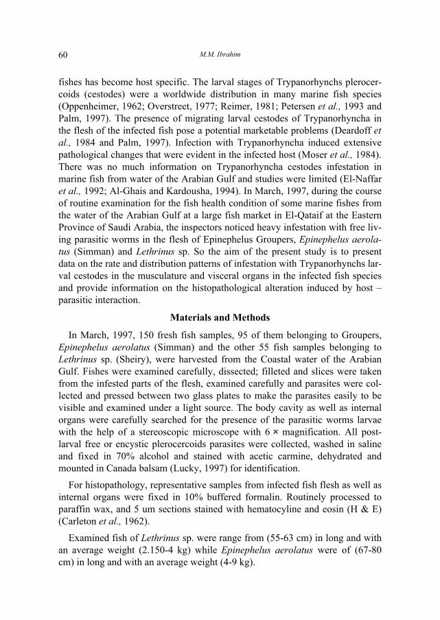
According to the fundamental work and classification of Campbell and Beve-radge (1994); Palm (1977), and based on the assistance of Dr. Palm at the Insti-tut fur Meereskunde, an der Universitat Kiel, Germany, the present parasiticworms larvae, infection was identified as Trypanorhyncha Plerocercoid of Flori-ceps sp. (cestodes), which characterized by a mature scolex bearing four retract-able and reversible strong tentacles armed with hooks and two bothridia (Fig.1).
Incidence of Infection
It was found out of 95 specimens of Epinephelus aereolatus, 23 fish sampleswere found to be infected with an incidence (24.2%), while out of 55 fish sam-ples of Lethrinus sp., 19 samples were found to be infected with an incidence(34.5%).
The Clinical Alteration and Histopathology
1 � In the muscular tissues
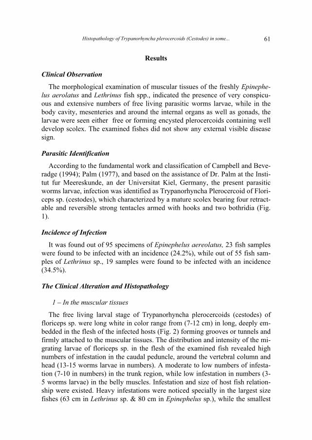
The free living larval stage of Trypanorhyncha plerocercoids (cestodes) offloriceps sp. were long white in color range from (7-12 cm) in long, deeply em-bedded in the flesh of the infected hosts (Fig. 2) forming grooves or tunnels andfirmly attached to the muscular tissues. The distribution and intensity of the mi-grating larvae of floriceps sp. in the flesh of the examined fish revealed highnumbers of infestation in the caudal peduncle, around the vertebral column andhead (13-15 worms larvae in numbers). A moderate to low numbers of infesta-tion (7-10 in numbers) in the trunk region, while low infestation in numbers (3-5 worms larvae) in the belly muscles. Infestation and size of host fish relation-ship were existed. Heavy infestations were noticed specially in the largest sizefishes (63 cm in Lethrinus sp. & 80 cm in Epinephelus sp.), while the smallest
M.M. Ibrahim62
FIG. 1. The parasitic worm larvae of Floriceps plerocercoid Trypanorhynch (cestode), showedwell developed head (Scolexbearing four reversible strong tentacles (T: 1,2,3,4), armedwith hooks (H), and two Bothridia (B). Acetic-carmine stained × 400.
Histopathology of Trypanorhyncha plerocercoids (Cestodes) in some... 63
FIG. 2. Infected fish sp. showed heavy infestation with free white parasitic worms larvae of try-panorhynch plerocercoid (arrows) in the muscles of the caudal peduncle.
size of the same species harbored few in numbers. The average numbers of infes-tation with free parasitic worms in the flesh/fish were ranged from (18-25 larvae/fish) in both kinds of the examined fish species specially in large size hosts.
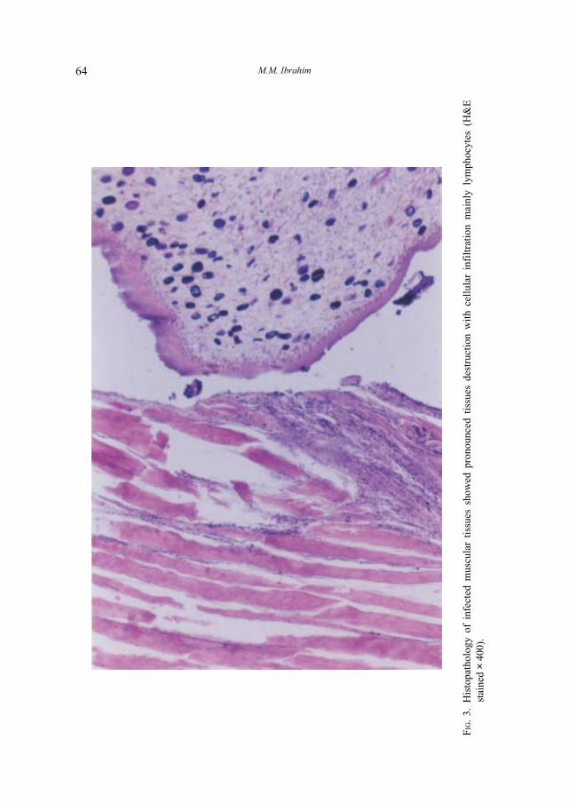
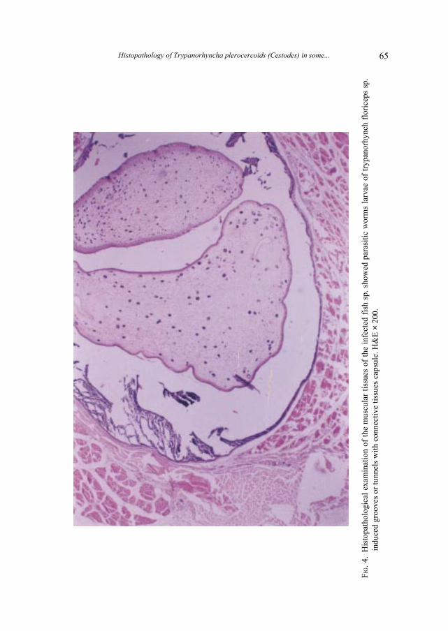
Histopathology of the flesh inhabiting Trypanorhynchan plerocercoids offloriceps sp. larvae showed pronounced tissues destruction and damages withintensive inflammatory reactions specially at the entrance of the motile viableburrowing scolex. Focal degeneration and necrotic changes in the myofibreswith extravasation of erythrocytes and cellular infiltration mainly lymphocytes(Fig. 3). The tunnels induced by the perforating larvae in the muscular tissueswere consisted of slight connective tissues wall around the motile viable larvae,while the adjacent muscular tissues suffered from dystrophic changes (Fig. 4).
2 � In the body cavity and mesenteries
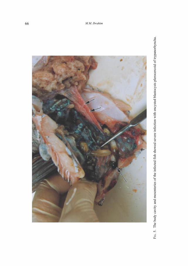
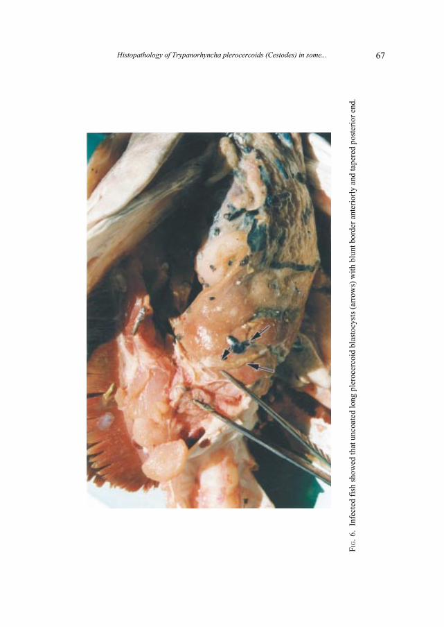
The encysted plerocercoids (blastocysts) of floriceps sp. in the body cavityand mesenteries of the infected fishes species (Fig. 5) were elongated rangefrom small size (few in mm in long) to large size ones up to (12 cm in long).The old and long blastocysts appeared with blunt border anteriorly containinghighly developed head portion of the worm larvae and with tapering posteriorend (Fig. 6). In some cases the blastocysts were seen whitish in color without
M.M. Ibrahim64
FIG.
3.H
isto
path
olog
y of
inf
ecte
d m
uscu
lar
tissu
es s
how
ed p
rono
unce
d tis
sues
des
truc
tion
with
cel
lula
r in
filtr
atio
n m
ainl
y ly
mph
ocyt
es (
H&
Est
aine
d ×
400)
.
Histopathology of Trypanorhyncha plerocercoids (Cestodes) in some... 65
FIG.
4.H
isto
path
olog
ical
exa
min
atio
n of
the
mus
cula
r tis
sues
of
the
infe
cted
fis
h sp
. sho
wed
par
asiti
c w
orm
s la
rvae
of
tryp
anor
hync
h fl
oric
eps
sp.
indu
ced
groo
ves
or tu
nnel
s w
ith c
onne
ctiv
e tis
sues
cap
sule
. H&
E ×
200
.
M.M. Ibrahim66
FIG.
5.T
he b
ody
cavi
ty a
nd m
esen
teri
es o
f th
e in
fect
ed f
ish
show
ed s
ever
e in
fect
ion
with
enc
yste
d bl
asto
cyst
s pl
eroc
erco
id o
f tr
ypan
orhy
ncha
.
Histopathology of Trypanorhyncha plerocercoids (Cestodes) in some... 67
FIG.
6.In
fect
ed f
ish
show
ed th
at u
ncoa
ted
long
ple
roce
rcoi
d bl
asto
cyst
s (a
rrow
s) w
ith b
lunt
bor
der
ante
rior
ly a
nd ta
pere
d po
ster
ior
end.

M.M. Ibrahim68
any covering coat, while other were encapsulated covered with a thick deepbrown to black envelop. Over 300-350 encysted plerocercoid blastocysts couldbe counted specially in large size infected fishes with sings of severe adhesionof the peritoneal wall and internal organs associated with engorgement of themesenteric blood vessels.
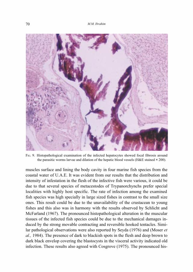
Histologically, the encysted plerocercoids (blastocysts) showed capsules offibrous connective tissues proliferation contained larvae and cellular infiltrationmainly lymphocytes as well as dilation of mesenteric blood vessels (Fig. 7).
3 � Liver
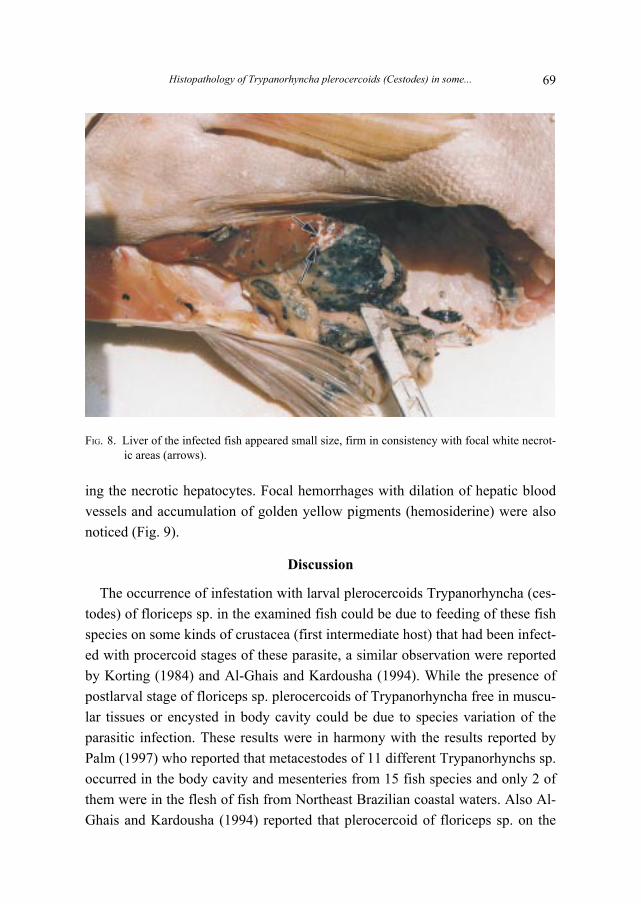
Liver inhabited infection with plerocercoid larvae stage of Trypanorhynchaof floriceps sp. were seen either free or formed encysted plerocercoid blasto-cysts. The infected liver appeared small size and firm in consistency with focalwhitish necrotic areas (Fig. 8). While in other cases, hemorrhages in the liverwas also observed. Histologically, the hepatocytes showed focal fibrosis replac-
FIG. 7. Histopathological examination of the wall of peritoneal cavity of the infected fish showedsevere infection with encysted plerocercoids with signs of fibrosis (H&E stain × 200).
Histopathology of Trypanorhyncha plerocercoids (Cestodes) in some... 69
FIG. 8. Liver of the infected fish appeared small size, firm in consistency with focal white necrot-ic areas (arrows).
ing the necrotic hepatocytes. Focal hemorrhages with dilation of hepatic bloodvessels and accumulation of golden yellow pigments (hemosiderine) were alsonoticed (Fig. 9).
Discussion
The occurrence of infestation with larval plerocercoids Trypanorhyncha (ces-todes) of floriceps sp. in the examined fish could be due to feeding of these fishspecies on some kinds of crustacea (first intermediate host) that had been infect-ed with procercoid stages of these parasite, a similar observation were reportedby Korting (1984) and Al-Ghais and Kardousha (1994). While the presence ofpostlarval stage of floriceps sp. plerocercoids of Trypanorhyncha free in muscu-lar tissues or encysted in body cavity could be due to species variation of theparasitic infection. These results were in harmony with the results reported byPalm (1997) who reported that metacestodes of 11 different Trypanorhynchs sp.occurred in the body cavity and mesenteries from 15 fish species and only 2 ofthem were in the flesh of fish from Northeast Brazilian coastal waters. Also Al-Ghais and Kardousha (1994) reported that plerocercoid of floriceps sp. on the
M.M. Ibrahim70
FIG. 9. Histopathological examination of the infected hepatocytes showed focal fibrosis aroundthe parasitic worms larvae and dilation of the hepatic blood vessels (H&E stained × 200).
muscles surface and lining the body cavity in four marine fish species from thecoastal water of U.A.E. It was evident from our results that the distribution andintensity of infestation in the flesh of the infective fish were various, it could bedue to that several species of metacestodes of Trypanorchynchs prefer speciallocalities with highly host specific. The rate of infection among the examinedfish species was high specially in large sized fishes in contrast to the small sizeones. This result could be due to the unavailability of the crustacean to youngfishes and this also was in harmony with the results observed by Schlicht andMcFarland (1967). The pronounced histopathological alteration in the musculartissues of the infected fish species could be due to the mechanical damages in-duced by the strong movable contracting and reversible hooked tentacles. Simi-lar pathological observations were also reported by Seyda (1976) and (Moser etal., 1984). The presence of dark to blackish spots in the flesh and deep brown todark black envelop covering the blastocysts in the visceral activity indicated oldinfection. These results also agreed with Cosgrove (1975). The pronounced his-
Histopathology of Trypanorhyncha plerocercoids (Cestodes) in some... 71
topathological effects in the liver and mesenteries were characterized by fibro-sis, adhesion of the internal organs and severe degenerative and necrotic chang-es in the hepatocytes. Our results were in harmony to those described by (Mik-haflova et al., 1964) and (Bauer et al., 1977).
In conclusion, since Trypanorhyncha cestodes from the water of the ArabianGulf are still a relatively poor studies, so further studies will be highly recom-mended to give complete understanding of Trypanorhyncha plerocercoids (Ces-todes) infection among the most important commercial marine fish species.This will give us better recommendation and more precise advice with regard tosuch problems.
Acknowledgement
I would like to express my deep gratitude to Mr. F.A. Al-Jame, the generalmanager of Fisheries Research Center, El-Qataif, Eastern Province and all ofmy colleagues in fish diseases unit for their supports. Thanks are also extendedto Dr. H. Palm, in the Institut Fur Meereskunde an der Universitat Kiel, Germa-ny for his assistance in identification of the parasitic infestation.
References
Al-Ghais, S.M. and Kardousha, M.M. (1994) Study on some helminth parasites larvae commonin Arabian Gulf fish: A Comparison between West and East coast of U.A.E., Arab Gulf.Scient. Res., 12(3): 559-571.
Bauer, O.N., Mussellus, V.A., Nikolaeva, V.M. and Strelkov, Y.A. (1977) Fish pathology, IdtPishch Promyshl. (in Russian).
Campbell, R.A. and Beveridge, I. (1994) Order Trypanorhyncha Diesing, 1863. Keys to the Ces-tode Parasites of Vertebrates. In: Khalil, L.F., Jones, A. and Bray, R.A. (Eds.). Walling-ford; CAB International: 51-148.
Carleton, H.M., Drury, R.A., Willington, E.A. and Comeron, H. (1962) cited from Carleton,Histological techniques, 4th Ed., Oxford Univ. Press, N. 4, Toronto.
Cosgrove, G.E. (1975) Parasites in tissue section: Recognition and reaction. In: The pathology offishes (W.E. Ribelin and Magaki, eds.), pp. 205-245.
Deardorff, T., Raybourne, R.B. and Mattis, T.E. (1984) Infection with plerocerci (cestoda) inHawaiian fishes of commercial importance, Sea Grant Quarterly, 6: 1-6.
El-Naffar, M.K.I., Gobashy, A., El-Etreby, S.G., Kardousha, M.M. (1992) General survey ofhelminth parasite genera of Arabian Gulf fishes (Coast of United Arab Emirates), ArabianGulf J. Scient. Res., 10(2): 99-110.
Korting, W. (1984) Larval cyclophyllidean cestodes in carp and tench. Bull. Eur. Ass. Fish Path-ol., 4(3): 40-41.
Lucky, Q. (1977) Methods for the diagnosis of fish diseases. American Publishing Co., Pvt. Ltd.,New York.
Mikhailova, I.G., Prazhikov, E.V. and Prusevich, T.O. (1964) Morphological changes in thefish tissue arround the larvae of some parasitic worms. Tr. Murm. Morsk. Biol. Inst., 5:251-264 (in Russian).
M.M. Ibrahim72
Moser, M., Sakanari, J., Wellings, S. and Lindstrom, K. (1984) Incompatibility between SanFrancisco striped bass (Morone Saxatilis Walbum), and the metacestode, Lacist orhynchustenui (beneaen, 1858), J. Fish Dis., 7: 397-400.
Oppenheimer, C.H.I. (1962) On marine fish diseases �Fish as Food� (G. Borgstrom, ed.), vol.2: 541, Academic Press, New York.
Overstreet, R.M. (1977) Pocllancistrium caryophyllum and other trypanorhynch cestode plero-cercoids from the musculature of cynsion nebulosus and other Sciaeni fish in the Gulf ofMexico. J. Parasitolo., 63: 780-787.
Palm, H.W. (1997) An alterative classification of trypanorchynch of limited importance. System-atic Parasitology, 37: 81-92.
Peterson, E., Palm, H., Moller, M. and Cruz, M.A. (1993) Flesh parasites of fish from centralPhilippine water. Aquatic Org., vol. 15: 81-86.
Reimer, L.W. (1997) Larvaen der Trypanorhynchs in Fischflesish Wiss, Z. pacdagog, Ochach,Liselotte Hermann, Gust row, 2: 207-211.
Schlicht, F.G. and McFarland, W.N. (1967) Incidence of Trypanorhynchan plerocercoids insome Texas Coast sciaenid fishes, Contrib. Mar. Sci., 12: 101-112.
Seyda, M. (1976) On a case of a mass invasion of cestode Gymnorhynchus gigas (Cuvier, 1817)larvae in muscles of brama rall (Bloch, 1791), Acta. Itchthyol. Piscatoria, 6: 59-65.

Histopathology of Trypanorhyncha plerocercoids (Cestodes) in some... 73
ZOK)« ÁUO� s� W�d���« �UL�_« iF� v� WO{d*« WO�O�M�« «dOG��«©«�u���® UJM��u�U��«d��« Ê«b�� U�dO� W�U/û� W�O�� v�dF�«
rO�«d�≈ bL�� vHDB�dB� − �d�UI�« − w�b�U� − Ê«uO(« W�/ Àu�� bNF�
vKOHD�« h�?H�« ¡«d�≈ - ±ππ∑ ÂUF� ��U�� d?N� v� ÆhK���*«vL�M� �UL�_« s� πµ , v�dF�« ZOK)« ÁU?O0 W�d���« �UL�_« s� ±µ∞ ��©ÊUL��« �U?L�Q�® UOK�?� �UL�*«Ë Groupers, Epinephelus aerolatus v�≈Emperor, Lethrinus sp. v�≈ vL?�?M� �U?L?�_« s� Èd?�_« W?MO?� µµ ��«Ë
Æ ©ÍdFA�« �UL�Q�® UOK�� �UL�*«Ë�u� s� f?JM��u�U???��«d???��« Ê«b�� U??�d???O� W�U???/≈ b??�«u?� k�ö�ËÁc� b?�Ë U?LMO� �UL?�_« ö?C?� Âu�K� �d?� ��u?B� f�?���uKH�«�U?L??�_U� U?I��U?�*«Ë v?MD��« n�u?�??��U� WK/u?�??�?� ��u?B� U??�d?O�«
Æ W��UB*«5� U?NM� vK�√ ©%≥¥Ëµ® Íd?FA?�« �UL?�√ 5� W�U?/ù« W�?�� X�U?�Ë U�d?O�« ÁcN� WOKO?HD�« W�U/ù« �bF?� ÊU�Ë Æ© %≤¥Ë≤ ® ÊUL��« �U?L�√U?NM?� Íd?I?H�« �u??L?F�«Ë �√d�«Ë q�c�« W??IDM� ö?C??F� W?O�U??� ��u?B�
Æ �c'«Ë sD��« WIDM� öCF�U??N?� Ê√ b??�Ë W�U???B*« ö??C???FK� v{d*« v?�??O??�?M�« h�???H�U�Ë W??/U?�Ë W??O?�U??�b�« U�ö??)« b?�«u� l� W?��dJM� «d??O?G�Ë �ö??�?L??{«≥µ∞ s� d?��√ b?� - b?�Ë ÆW�u�b?�« W�e�_« iF� p�c?�Ë XO?�u?HL?OK�«s� f�?���uKH?�« �u� s� UJM��u�U?��«d?��« Ê«b�b� v�d?O�« �u?DK� WKB�u?�l� �b�bA�« W�U?/ù« W�U� v� WOK�«b�« ¡U?C�_« �u�Ë vMD��« n�u?���«
Æ rN� nOK��«Ë U�UB��ô« U�ö� �uN�