Embed Size (px)
Citation preview

OGÓLNOPOLSKIE SEMINARIUMSPEKTROSKOPII MÖSSBAUEROWSKIEJ
OSSM-2008
Koninki 8-11 czerwca 2008
POLISH MÖSSBAUER COMMUNITY MEETING
Koninki June 8-11 2008
PROGRAM, ABSTRACTSAND
LIST OF PARTICIPANTS
Organized by:
Mössbauer Spectroscopy Division, Institute of PhysicsPedagogical University of Kraków
Faculty of Physics and Applied Computer ScienceAGH University of Science and Technology, Kraków
Kraków, May 2008

2
HONORARY COMMITTEE
Professor Andrzej Hrynkiewicz – KRAKÓW: ChairmanProfessor Józef Bara – KRAKÓWProfessor Katarzyna Brzózka – RADOMProfessor Mieczysław Budzyński – LUBLINProfessor Jan Chojcan – WROCŁAWProfessor Kazimierz Dziliński – CZĘSTOCHOWAProfessor Janusz Frąckowiak – KATOWICEProfessor Elżbieta Jartych – LUBLINProfessor Michał Kopcewicz – WARSZAWAProfessor Józef Korecki – KRAKÓWProfessor Karol Krop – RZESZÓWProfessor Kazimierz Łątka – KRAKÓWProfessor Antoni Pędziwiatr – KRAKÓWProfessor Mikołaj Rudolf – WROCŁAWProfessor Jan Suwalski – WARSZAWAProfessor Krzysztof Szymański – BIAŁYSTOKProfessor Krzysztof Tomala – KRAKÓWProfessor Józef Zbroszczyk – CZĘSTOCHOWA
ORGANIZING COMMITTEE
Professor Stanisław M. Dubiel – AGH KRAKÓW: Co-chairmanProfessor Krzysztof Ruebenbauer – AP KRAKÓW: Co-chairmanDr. Artur Błachowski – AP KRAKÓW: SecretaryDr. Jakub Cieślak – AGH KRAKÓW: CoordinatorDr. Jan Żukrowski – AGH KRAKÓW: Treasurer
SPONSORS
Rector of the Pedagogical University of Kraków – Professor Henryk Żaliński
Dean of the Faculty of Physics and Applied Computer Science, AGH University of Scienceand Technology, Kraków – Professor Zbigniew Kąkol
Motorola Software Group – PolandPL-30-381 Kraków, ul. M. Bobrzyńskiego 46
Sopockie Towarzystwo Ubezpieczeń Ergo Hestia S.A.PL-81-731 Sopot, ul. Hestii 1
Mr. Kazimierz Czech – AGH KRAKÓW is thanked for his help in maintenance of the OSSM-2008banking account.
OSSM-2008 logo and poster have been designed by Ms Jolanta UrbanikPoster photo by Mr. Kaj Romeyko-Hurko
OSSM-2008 WEB page: www.elektron.ap.krakow.pl/ossm2008Electronic address: [email protected]
Electronic-address for submission of the manuscript for Acta Physica Polonica A:[email protected]
Printed by the Academic Publishing House of the Pedagogical University of Kraków

3
Dear Participants of the Polish Mössbauer Community MeetingOSSM-2008, Koninki June 8-11, 2008
We welcome all participants of the Polish Mössbauer Community Meeting OSSM-2008,being seventh such meeting since resuming our seminars after a long break. The aim of thesemeetings has been to gather Polish scientists using the Mössbauer spectroscopy in theirresearch. The assemblies have been resumed in answer to the questionnaire circulated withinour community in 1995. The first seminar of this new series took place in Lublin (1996), andit has been organized by the Mössbauer-Community of Lublin. Since then the meetings havebeen organized every second year, and our tradition has been to organize them in variousplaces and by various Mössbauer groups. The following assemblies were organized byWrocław Mössbauer Community in Sobótka-Górka (1998), Radom Group in Radom-Zbożenna (2000), Białystok Group in Goniądz (2002), Katowice Community in Wisła (2004),and, the last one, by Częstochowa Mössbauer Group in Koszęcin (2006).
For the current meeting in Koninki we have obtained happily the record number of 47contributions, and we are delighted, as well, with having the record number of 58 registeredparticipants. We have decided for the first time to introduce invited talks to our seminars. Twoinvited talks are to be presented during the current meeting. We hope that this practice will becontinued during future assemblies. We have foreign participants this year as well. ProfessorBogdan Sepiol from the Vienna University, and Professor Zbigniew M. Stadnik from theOttawa University are invited speakers. Dr. Benilde F.O. Costa from the Coimbra Universityin Portugal, and Dr. Viktor I. Mitsiuk from the Bielorussian Academy of Sciences in Mińsk,Bielarus are our participants.
This book gathers all 49 abstracts that have been delivered - either invited talks or regularcontributions. The abstracts are published in the form submitted by their Authors, and neitherediting nor reviewing process of the submitted manuscripts have been performed.
We wish all of you a pleasant and fruitful stay in Koninki.
Stanisław M. DubielKrzysztof Ruebenbauer

4
PROGRAM
Presenting authors/participants are shown in bold.Invited talk: 35 + 5 min.
Contributed talk: 20 + 5, 15 + 5 or 12 + 3 min. (for details see list of lectures)
Sunday June 8th 2008
16.00 – bus departure from the Pedagogical University, Kraków, ul. Podchorążych 2(south-eastern parking)
17.30 – arrival to Koninki18.30 – registration19.30 – dinner

5
Monday June 9th 2008
7.30 – 8.15 – breakfast8.30 – 8.45 OSSM-2008 opening
Session I: Metals and alloys: 8.45 – 10.40Chair: Mieczysław Budzyński
8.45 – 9.25: INVITED TALK (p. 14)Dynamics studies with high-resolution X-ray scattering methodsB. Sepiol, E. Partyka-Jankowska, M. Rennhofer, G. Vogl, J. Korecki, T. Ślęzak, M. Zając, S.Stankov, R. Rüffer
9.25 – 9.50 (p. 16)A dilute-limit heat of solution of aluminum in iron studied with 57Fe Mössbauer spectroscopyJ. Chojcan, A. Ostrasz
9.50 – 10.10 (p. 18)Do 57Fe atoms pin spin-density waves in chromium?J. Żukrowski, S.M. Dubiel, J. Cieślak
10.10 – 10.25 (p. 20)Debye temperature in bcc-Fe-Cr alloysB.F.O. Costa, J. Cieślak, S.M. Dubiel
10.25 – 10.40 (p. 22)On Finemet alloys substituted by 3d - transition elementsK. Brzózka, M. Gawroński, P. Sovák, T. Szumiata, B. Górka
10.40 – 11.00 coffee break

6
Session II: Metals, alloys and oxides: 11.00 – 13.00Chair: Kazimierz Łątka
11.00 – 11.25 (p. 24)Hyperfine interactions and magnetic properties of La0.67Ca0.33Mn1-x
57FexO3 with x=0.1 and0.15J. Przewoźnik, J. Żukrowski, K. Krop, Cz. Kapusta
11.25 – 11.40 (p. 26)Defect structure of Fe-Al systemA. Hanc, J. Kansy, G. Dercz, L Pająk, D. Oleszak
11.40 – 11.55 (p. 28)Ordering process of AlFe28 and CrAlFe 528 alloysA. Hanc, J. Kansy, G. Dercz, L. Pająk
11.55 – 12.10 (p. 30)Mössbauer investigations and photo-emission studies of the Fe 3s spin splitting in some Fe-NialloysM. Kądziołka-Gaweł, W. Zarek, E. Talik, E. Popiel
12.10 – 12.25 (p. 32)Thermal stability and crystallization of iron and cobalt-based bulk amorphous alloysJ. Olszewski, J. Zbroszczyk, K. Sobczyk, W. Ciurzyńska, M. Nabiałek, J. Świerczek, M.Hasiak, A. Łuniewska
12.25 – 12.40 (p. 34)57Fe Mössbauer spectroscopy of the Ni-Cu-Fe amorphous and crystalline alloys based on theP, Si and B glass-forming agentsK. Ziewiec, K. Bryła, A. Błachowski, K. Ruebenbauer
12.40 – 12.55 (p. 36)Early design stage of the MsAa-4 Mössbauer spectrometerA. Błachowski, K. Ruebenbauer, J. Żukrowski, R. Górnicki
13.30 – lunch

7
Session III: Intermetallic compounds: 14.30 – 16.25Chair: Jan Chojcan
14.30 – 14.55 (p. 38)Puzzling magnetism of Gd3Cu4Sn4K. Łątka, A.W. Pacyna, R. Pöttgen, F.M. Schappacher
14.55 – 15.10 (p. 40)Site occupancies in the R2-xFe14+2xSi3 (R = Ce, Nd, Gd, Dy, Ho, Er, Lu, Y) compounds studiedby Mössbauer spectroscopyA. Błachowski, K. Ruebenbauer, J. Przewoźnik, J. Żukrowski, D. Sitko, N.-T.H. Kim-Ngan,A.V. Andreev
15.10 – 15.25 (p. 42)Spin reorientation in the Er2-xFe14+2xSi3 single-crystal studied by Mössbauer spectroscopyJ. Żukrowski, A. Błachowski, K. Ruebenbauer, J. Przewoźnik, D. Sitko, N.-T.H. Kim-Ngan,Z. Tarnawski, A.V. Andreev
15.25 – 15.40 (p. 44)Mössbauer investigations of spin arrangements in Er2-xCexFe14BB.F. Bogacz, A.T. Pędziwiatr
15.40 – 15.55 (p. 46)119Sn Mössbauer spectroscopy of intermetallic HoRhSn compoundJ. Gurgul, K. Łątka, A.W. Pacyna, C.P. Sebastian, R. Pöttgen
15.55 – 16.10 (p. 48)Structural and magnetic properties of Fe3-xTixSn disordered alloysK. Brząkalik
16.10 – 16.25 (p. 50)57Fe hyperfine interactions in Sc(Fe1-xNix)2 Laves phases synthesized under high pressureM. Wiertel, Z. Surowiec, M. Budzyński, A.V. Tsvyashchenko
16.25 – 16.45 coffee break
8
Session IV: Intermetallic compounds and oxides: 16.45 – 18.35Chair: Antoni Pędziwiatr
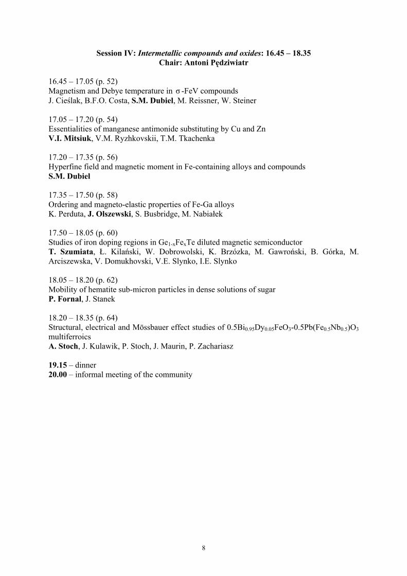
16.45 – 17.05 (p. 52)Magnetism and Debye temperature in σ -FeV compoundsJ. Cieślak, B.F.O. Costa, S.M. Dubiel, M. Reissner, W. Steiner
17.05 – 17.20 (p. 54)Essentialities of manganese antimonide substituting by Cu and ZnV.I. Mitsiuk, V.M. Ryzhkovskii, T.M. Tkachenka
17.20 – 17.35 (p. 56)Hyperfine field and magnetic moment in Fe-containing alloys and compoundsS.M. Dubiel
17.35 – 17.50 (p. 58)Ordering and magneto-elastic properties of Fe-Ga alloysK. Perduta, J. Olszewski, S. Busbridge, M. Nabiałek
17.50 – 18.05 (p. 60)Studies of iron doping regions in Ge1-xFexTe diluted magnetic semiconductorT. Szumiata, Ł. Kilański, W. Dobrowolski, K. Brzózka, M. Gawroński, B. Górka, M.Arciszewska, V. Domukhovski, V.E. Slynko, I.E. Slynko
18.05 – 18.20 (p. 62)Mobility of hematite sub-micron particles in dense solutions of sugarP. Fornal, J. Stanek
18.20 – 18.35 (p. 64)Structural, electrical and Mössbauer effect studies of 0.5Bi0.95Dy0.05FeO3-0.5Pb(Fe0.5Nb0.5)O3multiferroicsA. Stoch, J. Kulawik, P. Stoch, J. Maurin, P. Zachariasz
19.15 – dinner20.00 – informal meeting of the community

9
Tuesday June 10th 2008
7.30 – 8.15 – breakfast
Session V: Thin films and nano-structures: 8.30 – 10.35Chair: Elżbieta Jartych
8.30 – 9.10: INVITED TALK (p. 66)Mössbauer spectroscopy of quasi-crystalsZ.M. Stadnik
9.10 – 9.35 (p. 68)Magnetism of ultra-thin iron films seen by the nuclear resonant scattering of synchrotronradiationT. Ślęzak, S. Stankov, M. Zając, M. Ślęzak, K. Matlak, N. Spiridis, B. Laenens, N.Planckaert, M. Rennhofer, K. Freindl, D. Wilgocka-Ślęzak, R. Rüffer, J. Korecki
9.35 – 9.50 (p. 70)Magnetic nanoparticles in mcm-41 type mesoporous silicaZ. Surowiec, M. Budzyński, M. Wiertel, J. Goworek, B. Bierska-Piech
9.50 – 10.05 (p. 72)Mössbauer study of mechano-synthesized and thermally treated Co-Fe-Ni alloysT. Pikula, D. Oleszak, M. Pękała, M. Mazurek, J.K. Żurawicz, E. Jartych
10.05 – 10.20 (p. 74)Formation and magnetic properties of nanostructured Fe-Pt-B alloysA. Grabias, M. Kopcewicz, D. Oleszak, J. Latuch, M. Pękała, M. Kowalczyk
10.20 – 10.35 (p. 76)Investigation of Fe layers deposited from acetone based electrolyteW. Olszewski, K. Szymański, D. Satuła, L. Dobrzyński
10.35 – 11.00 coffee break

10
Session VI: Biophysics and chemistry: 11.00 – 13.10Chair: Katarzyna Brzózka
11.00 – 11.25 (p. 78)The microstructure and magnetic properties of ferrite nano-particles prepared by wet chemicalmethodD. Satuła, B. Kalska-Szostko, K. Szymański, L. Dobrzyński, J. Kozubowski
11.25 – 11.40 (p. 80)Structural studies by XRD and Mössbauer spectroscopy on nano-crystalline substratesprepared using high-energy ball milling for Bi5Ti3FeO15 synthesisG. Dercz, J. Rymarczyk, A. Hanc, K. Prusik, L. Pająk, J. Ilczuk
11.40 – 12.05 (p. 82)Magnetism of porphyrinsK. Dziedzic-Kocurek, J. Stanek
12.05 – 12.30 (p. 84)Solvent-Fe-tetraphenylporphyrin complexing studied by Mössbauer spectroscopyT. Jackowski, T. Kaczmarzyk, K. Dziliński
12.30 – 12.55 (p. 86)Mössbauer studies of pathological brain tissues affected by PSP diseaseJ. Gałązka-Friedman, E.R. Bauminger, K. Szlachta, Z. Wszolek, D. Dickson, A. Friedman
12.55 – 13.10 (p. 88)Mössbauer study of a reduction process in iron azaporphyrinsT. Kaczmarzyk, T. Jackowski, K. Dziliński
13.30 – lunch14.30 – excursion to Mt. Suhora Astronomical Observatory (Pedagogical University)19.30 – social evening – Andrzej Mróz and his band playing Andrzej Mróz songs

11
Wednesday June 11th 2008
7.30 – 8.15 – breakfast
Session VII: Electron structure calculations and minerals: 8.30 – 10.30Chair: Kazimierz Dziliński
8.30 – 8.55 (p. 90)Calibration of the isomer shift for the 14.4-keV transition in 57Fe and for the 77.34-keVtransition in 197Au using the full-potential linearized augmented plane-wave methodU.D. Wdowik, K. Ruebenbauer
8.55 – 9.20 (p. 92)Analysis of Fe-Cr sigma-phase Mössbauer spectrum. Experimental and theoretical studyJ. Cieślak, J. Toboła, S.M. Dubiel, M. Reissner, W. Steiner, S. Kaprzyk
9.20 – 9.35 (p. 94)Ab initio study of 57Fe hyperfine parameters in (FeAl)1-xTx (T - 3d element) B2-type dilutealloysT. Michalecki, A. Hanc, J. Deniszczyk, W. Borgieł
9.35 – 10.00 (p. 96)57Fe Mössbauer spectroscopy of partially radiation damaged allanitesD. Malczewski, A. Grabias
10.00 – 10.15 (p. 98)Comparison of magnetic and Mössbauer results obtained for paleozoic rocks from SouthernSpitsbergen, ArcticK. Szlachta, M. Olszewska, K. Brzózka, B. Górka, K. Michalski, J. Gałązka-Friedman
10.15 – 10.30 (p. 100)Mössbauer spectroscopy and X-ray diffraction studies on multi-ferroic Bi5Ti3FeO15 ceramicsJ. Rymarczyk, A. Hanc, G. Dercz, J. Ilczuk
10.30 – 11.00 coffee break

12
Session VIII: Ceramic materials, alloys and impurities: 11.00 – 12.10Chair: Józef Zbroszczyk
11.00 – 11.15 (p. 102)The Mössbauer spectroscopy and analytical investigations of the polycrystalline compoundswith general formula Zn1-xSnxCr2Se4 (x=0.1-0.3)I. Jendrzejewska, A. Hanc, P. Zajdel, A. Kita, T. Goryczka, E. Maciążek, J. Mrzigod
11.15 – 11.30 (p. 104)The Mössbauer and X-ray studies of the spinel ferrites Cu1-xFexCr2Se4 and CuCr2-yFeySe4prepared by the ceramic methodE. Maciążek, A. Hanc, R. Sitko, B. Zawisza, I. Jendrzejewska
11.30 – 11.45 (p. 106)The Mössbauer spectroscopy studies of ε to cementite-carbides transformation duringisothermal holding from as-quenched state of high carbon tool steelP. Bała, J. Krawczyk, A. Hanc
11.45 – 12.00 (p. 108)The Mössbauer spectroscopy studies of cementite precipitations during continuous heatingfrom as-quenched state of high carbon Cr-Mn-Mo steelJ. Krawczyk, P. Bała, A. Hanc
12.00 – 12.15 (p. 110)Charge and spin density perturbation on iron nuclei by non-magnetic impurities substituted onthe iron sites in α -FeA. Błachowski, K. Ruebenbauer, J. Żukrowski, J. Przewoźnik
12.15 – 12.35 closing remarks: Krzysztof Szymański13.30 – lunch15.00 – bus departure from Koninki16.30 – arrival to Kraków (Małopolski Autobusowy Dworzec Regionalny and Pedagogical
University later on)

13
Notes

14
Dynamics studies with high-resolution X-ray scattering methods
B. Sepiol 1 , E. Partyka-Jankowska 1 , M. Rennhofer 1 , G. Vogl 1 ,J. Korecki 2 , T. Slezak 2 , M. Zajac 2 , S. Stankov 3 and R. Rüffer 3
1 Dynamics of Condensed Systems, Department of Physics, University of ViennaA-1090 Wien, Strudlhofgasse 4, Austria; [email protected]
2 Solid State Physics Department, AGH University of Science and TechnologyMickiewicza 30, PL-30-059 Cracow, Poland
3 ESRF, BP 220, 38043 Grenoble, France
Detailed information about the microstructure without doubt is the most important and fundamentalinformation necessary to start further analysis of any material. Generally, one can recognize a fewlevels for microstructure analysis: (i) the way the atoms of an alloy are arranged on a theoretical meanstructure, (ii) all kinds of displacements from the mean structure and, (iii) dynamical behavior ofconstituent atoms including their vibrational properties and the motion of atoms between lattice sites.Unfortunately, investigation of any of these levels for a specific alloy is an extremely time-consumingand difficult task. Up to now, only very few materials have been explored in all or in most facets.From the above mentioned analysis levels, the dynamical properties seem to be the most challengingfrom researcher’s point of view, though of considerable importance not only from a fundamentalapproach but also essential for the functionality of nanoscale devices. A detailed characterization ofthe structure of the system on an atomic level is the basis for a thorough understanding of dynamics inthe solid state, where it is necessary to be familiar with both the collective (phonons) and single-particle dynamics (usually called diffusion).
The wavelength of gamma rays, X-rays or neutrons in principle allows to probe the dynamics at lengthscales sufficient to resolve single atoms. Indeed, first diffusion studies on atomistic level wereperformed by neutron scattering and by Mössbauer spectroscopy. X-ray techniques in this area haveonly become feasible with the advent of synchrotron radiation sources. Combining X-ray reflectionwith nuclear resonant scattering of synchrotron radiation results in depth-selectivity of hyperfineparameters, allowing study of resonant atoms motion on the surface or in near-surface layers.A distinct advantage of the technique of nuclear resonant scattering is that it is isotope-specific.Compared to other methods, the signal is essentially free of contributions from surrounding materials.Moreover, probe layers can be selectively deposited to study the magnetic and dynamic propertieswith atomic resolution.Usually only selected features of the investigated system are studied and one uses analogies to similarknown structures to conclude about those properties which are not accessible to direct exploration. Adeep knowledge of some selected model systems therefore appears even more important.
The lecture is intended as a compact survey of gamma and X-ray scattering techniques applied fordynamics studies [1-3] and will be illustrated with most recent experimental results.
1. B. Sepiol and K.F. Ludwig in: Alloy Physics, WILEY-VCH 2007; ed. W. Pfeiler, p. 707.2. G. Vogl and B. Sepiol in: Nuclear Resonant Scattering of Synchrotron Radiation, eds:
G.Langouche and H. de Waard, Hyperfine Interactions 123/124, (1999), p. 595.3. G. Vogl and B. Sepiol in: Diffusion in Condensed Matter, Springer 2005, 2nd ed.; eds: P. Heitjans
and J. Kärger, p. 65.

15
Notes

16
A dilute-limit heat of solution of aluminium in iron studied with 57Fe Mössbauer spectroscopy
J. Chojcan and A. OstraszInstitute of Experimental Physics, Uniwersity of Wrocław
PL-50-204 Wrocław, pl. M. Borna 9, Poland; [email protected]
The room temperature 57Fe Mössbauer spectra for annealed Fe1-xAlx solid solutions were measuredwith constant-acceleration POLON spectrometer of standard design. The spectra were analysed interms of binding energy Eb between two Al atoms in the Fe-Al system [1,2]. It was found that theenergy is positive or Al atoms interact repulsively. The extrapolated value of Eb for x = 0 were usedfor computation of the dilute-limit heat HFeAl of solution of Al atoms in iron [3],
HFeAl = – z·Eb/2,
where z is the coordination number of the crystalline lattice and for α-Fe it is 8.The result was compared with that derived from calorimetric data concerning the heat of formation Hof the system mentioned [4],
HFeAl = [dH/dx]x=0,
as well as with that resulting from the Miedema’s model of alloys [5],
HFeAl = [2·VAl2/3/(nFe
-1/3 + nAl-1/3)]·[ – P(∆φ)2 + Q(∆n1/3)2 – R],
where VAl is the atomic volume of Al, φ is the electronegativity, n1/3 is the cubic root of the electrondensity at the boundary of bulk Wigner-Seitz cells and ∆ denotes the differences in a given parameterfor Fe and Al. The coefficients P, Q, and R are empirical constants; PNA = 14.1 kJ V-2 (d.u.)-1/3 cm-2,Q/P = 9.4 V2/(d.u.)2/3 and R/P = 2V2 for alloys of a transition metal with a non-transition one; NA is theAvogadro’s number, d.u. is about 4.6 1022 electrons per cm3.
The comparison shows that our Mössbauer spectroscopy findings are in a good agreement withboth calorimetric data and the Miedema’s model predictions.
Acknowledgement. The authors would like to thank Honorata Ziemiańska for her assistance in themeasurements.
1. A.Z. Hrynkiewicz and K. Królas, Phys. Rev. B 28 (1983) 1864.2. J. Chojcan, J. Alloys and Comp. 264 (1998) 50.3. J. Stanek, G. Marest, H. Jaffrezic, H. Bińczycka, Phys. Rev. B 52 (1995) 8414.4. R.Hultgren, P.D.Desai, D.T.Hawkins, M.Gleiser and K.K.Kelley, Selected Values of
Thermodynamic Properties of Binary Alloys, American Society for Metals, Metals Park, OH,1973, p.156.
5. A.R. Miedema, Physica B 182 (1992) 1.

17
Notes

18
Do 57Fe atoms pin spin-density-waves in chromium?
J. Żukrowski, S.M. Dubiel and J. CieślakFaculty of Physics and Computer Science, AGH University of Science and Technology, 30-059
Kraków, Poland,[email protected]
Metallic chromium is known as the archetype of antiferromagnet with the Néel temperature of TN~312 K. However, magnetic moment is not constant but it is harmonically modulated with anamplitude of 0.6 µB. This gives rise to call the phenomenon spin-density waves (SDWs). As the entiremagnetic moment is due to a polarization of conduction electrons, the magnetism of chromium is ofitinerant character. Investigation of SDWs is not only of a fundamental meaning, due to their closerelation to the Fermi surface, but also very attracting because of their interesting features. First, theyare incommensurate with the underlying lattice, and their periodicity, being a monotonic function oftemperature, changes between ~ 21 and ~ 28 lattice constant at 4 K and 300 K, respectively. Second,SDWs exist in two phases: (a) high temperature phase i.e. between 312 K and 123 K which has atransverse polarization (TSDW) i.e the wave vector Q is perpendicular to the position vector r, and (b)low temperature phase i.e. below 123 K with a longitudinal polarization (LSDW). The temperature of123 K at which the phase transition occurs is called a spin-flip temperature, TSF. Third, SDWs are verysensitive to impurities, defects and strain. The former is of importance as far as an application of probenuclei techniques, such as the Mössbauer spectroscopy (MS), in the study of the SDWs is used.Following theoretical calculations [1], impurities can influence SDWs. However, the effect ofnonmagnetic impurities is small and it is limited to the LSDW phase while that of magnetic impuritiesis stronger and can effect SDWs in both phases.In MS two isotopes play in practice the major role viz. 57Fe and 119Sn. Luckily, they represent twodifferent cases as far as the influence of foreign atoms on SDWs is concerned. Regarding nonmagnetic119Sn, there is a good deal of evidence that they are almost ideal probe nuclei i.e. all featurescharacteristic of SDWs in a pure chromium (e. g. TN, TSF, sign and amplitude of the third-orderharmonics) measured with 119Sn are in a good accord with the values of the corresponding quantitiesfound with other techniques for a pure chromium. To our knowledge, there is only one report in whichSDWs of chromium were investigated with 57Fe [2]. The author observed only a slight broadening of asingle-line spectrum at 4.2 K relative to the spectrum measured at 300 K. This means that themagnetic 57Fe atoms strongly reduce the amplitude of the SDWs which is known as pinning.In this contribution we will report on a systematic study on a polycrystalline sample of chromiumdoped with < 0.1 at% of iron enriched to ~90% in 57Fe. As can be seen in Fig. 1, the broadening of thespectrum observed versus temperature shows two regions; (a) a high temperature one, where thebroadening is small and virtually temperature independent, and (b) a low temperature one, where asharp increase of the broadening with temperature is observed. The result may be taken as evidencethat the pinning of the SDWS by the magnetic 57Fe atoms depend on the SDWs phase: is stronger forthe TSDW phase, and weaker for the LSDW phase.
Fig.1 (left) 57Fe spectra recorded on 57Fe-doped Cr, and (right) average hyperfine field vs. temperatureas derived from the spectra.[1] Ch. Seidel, Phys. Stat. Sol. (b), 149 (1988); [2] G. K. Wertheim, J. Appl. Phys., 32 (1961) 1105

19
Notes
20
Debye temperature in bcc-Fe-Cr alloys*
B. F. O. Costa 1 , J. Cieślak2 and S.M. Dubiel2
1 CEMDRX Department of Physics, University of Coimbra, 3000-516 Coimbra, Portugal, 2 Faculty ofPhysics and Computer Science, AGH University of Science and Technology, 30-059 Kraków, Poland;
Fe-Cr alloys continue to be of scientific and technological interest. The former stems form the fact thatthey are regarded as a model alloy system for studying various magnetic properties and testingappropriate theories and theoretical models. The letter is related to excellent properties of materialsfabricated from Fe-Cr alloys like heat resistant steels. Chromium steels are regarded, in particular, asgood candidates for the design of structural components in advanced nuclear energy installations likeGeneration IV and fusion reactors.In this contribution we will present results concerning the Debye temperature as determined by meansof the Mössbauer spectroscopy. For that purpose a series of Mössbauer spectra were recorded in atemperature interval of 60 – 300 K on Fe100-cCrx samples with 0 ≤ x ≤ 99.9 prepared by an arc meltingprocess. The spectra were analysed to get the average values of the central shift, <CS>. By fitting itstemperature dependence - which typical plot is shown in Fig. 1 - with the Debye model, we havedetermined the Debye temperature, ΘD, as a function of alloy composition, x. The most strikingfeature we have revealed is a non-monotonic behaviour. Here, in this abstract we want to illustrate thiswith Fig. 1 which shows the behaviour found for the Fe-rich samples. In the range of x between 0 and22, ΘD has a maximum at x ≈ 5 and the relative enhancement is ∼ 30%. It will be shown that such abehaviour of ΘD in that range of composition is paralleled by various physical quantities, and, inparticular, by 57Fe- and 119Sn-site hyperfine field, Curie temperature, magnetic moment per Fe atom,spin-wave stiffness coefficient. Such parallelism can be regarded as evidence for the electron-phononinteraction, which was theoretically predicted to occur in itinerant-electron magnets [1].
Fig.1 (left) Dependence of the average central shift, <CS>, on temperature, T, for the Fe95.15Cr4.85alloy. The solid line represents the best-fit to the experimental data in terms the Debye model, and(right) Debye temperature, ΘD, versus Cr content, x.
[1] D. J. Kim, Phys. Rev. Letter., 47 (1981) 1213; Phys. Rev. B, 25 (1982) 6919
* The results were obtained within the bilateral Polish-Portuguese program 2007/2008

21
Notes

22
On Finemet alloys substituted by 3d - transition elements
K. Brzózka 1 , M. Gawroński 1 , P. Sovák 2 , T. Szumiata 1 , B. Górka 1
1 Department of Physics, Technical University of Radom, 54 Krasickiego Str., 26-600 Radom, Poland;[email protected]
2 Department of Experimental Physics, Faculty of Sciences, P. J. Šafárik University, Park Angelinum9, 041 54 Košice, Slovakia
Excellent soft magnetic properties of two-phase nanocrystalline Finemet alloys, manufactured bycontrolled crystallization of the amorphous precursor, have been the subject of interest for last fifteenyears. Between others, structure and chemical content of nanograins seem to be the key feature whichimplies the macroscopic magnetic properties of those materials. In presented study, we investigatealteration of local structure and magnetic hyperfine fields induced by partial substitution of iron byother 3d transition elements. The greatest attention is paid to manganese-substituted amorphous andnanocrystalline alloys. In case of nanocrystalline materials, Mössbauer spectroscopy gives uniquepossibility to separate information originated from amorphous and crystalline areas as well as tocompare samples of different composition, independently on crystallization degree.Ribbons of nominal composition Fe73.5-xMnxNb3Cu1Si13.5B9 (x = 1, 3, 5, 7, 9, 11, 13, 15) were preparedby melt spinning method. The as-quenched samples were subjected to 1 h annealing under vacuum atselected temperatures. On the basis of DSC results [1, 2], the following temperatures of the isothermalannealing (which are expected to represent different stages of the crystallization process) were chosen:550oC, 575oC and 690oC. Transmission Mössbauer measurements were carried out using 50 mCi57Co(Rh) source of γ - radiation as well as a vibrator working in constant acceleration mode. Thehomogeneity of samples was verified by means of conversion electron spectroscopy using a gas-flowdetector.Mössbauer spectra of as-quenched samples show a smeared shape typical for an amorphous alloy inmagnetically ordered state. All the spectra were analyzed using model independent Le Caër methodtaking into account a linear correlation between magnetic hyperfine field B and local isomer shift δ .The distributions P(B), derived from Mössbauer spectra of amorphous alloys, show a bimodal shapeand therefore they have been separated into two independent components, attributed to different localenvironments of iron atoms. Mean magnetic hyperfine field decreases monotonically with x.Mössbauer spectra collected for annealed alloys reveal partial crystallisation of the samples however
the amorphous remainder constitutes stillnoticeable part of the spectrum area.Depending on composition as well as theannealing temperature, different fraction ofthe discrete component is found and itrepresents various crystalline phases. Insamples annealed at two lower temperatures,DO3-type bcc Fe-Si phase is dominant, whileafter annealing at 690oC also Fe-B crystallitesappear.The Mössbauer results are compared withthose obtained by macroscopic magneticmethods. The influence of substitution withother 3d - transition elements is also analysed.
Figure 1. Mössbauer spectrum of Fe66.5Mn7Nb3Cu1Si13.5B9 alloy annealed for 1 h at 575oC, with high-field and low-field part of amorphous subspectrum as well as a discrete five-sextets component.
1. R. Brzozowski, M. Wasiak, J. Balcerski, P. Sovák and M. Moneta, A. Phys. Pol. A, 113 (2008)117.
2. C. Gomez-Polo, J. I. Pérez-Landazábal, V. Recarte, P. Mendoza Zélis, Y. F. Li, M. Vazquez,J. Magn. Magn. Mat., 290-291 (2005) 1517.
1.00
0.98
0.96
0.94
Rel
ativ
e C
ount
s
-6 -4 -2 0 2 4 6Velocity (mm/s)
-2x10⁻³
0Error

23
Notes

24
Hyperfine interactions and magnetic properties of La0.67Ca0.33Mn1-x57FexO3 with x=0.1 and 0.15
J. Przewoźnik 1 , J. Żukrowski 1 , K. Krop 2 , Cz. Kapusta 1
1 Department of Solid State Physics, Faculty of Physics and Applied Computer Science, AGHUniversity of Science and Technology, Al. Mickiewicza 30, PL-30-059 Kraków, Poland;
[email protected] Department of Physics, Rzeszów University of TechnologyAl. Powstańców Warszawy 6, PL-35-959 Rzeszów, Poland
LaMnO3 is an antiferromagnetic insulator with dominating Mn3+–O–Mn3+ antiferromagneticsuperexchange interaction but the substitution of Ca2+ for La3+ introduces holes into the eg orbitals ofMn promoting Mn3+–O–Mn4+ ferromagnetic (FM) double-exchange (DE) interaction and metallicity inLa1–xCaxMnO3. The optimally doped La0.67Ca0.33MnO3 exhibits a simultaneous first-order metal-insulator (M–I) and FM transitions with maximal temperatures TC and TM-I, which are very similar. Asubstitution of Mn3+ by Fe3+ reduces the number of available hopping sites for the Mn eg electron andsuppresses the DE interaction. This leads to a reduction of the magnetization and to an increase of theresistivity as well as to a decrease of the TC and TM-I temperatures. In order to investigate the effect ofthe competition between the Mn–Mn and Mn–Fe interactions on the magnetic ordering, magnetic andspin-glass (SG) like properties of the strongly Fe doped La0.67Ca0.33MnO3 compound the present studyis undertaken.
Polycrystalline La0.67Ca0.33Mn1-x57FexO3 compounds with x=0.1 (LCMF10O) and 0.15 (LCMF15O)
were studied with AC susceptometry, DC (VSM) magnetometry and 57Fe Mössbauer spectroscopy(MS). The ZFC and FC magnetization measurements in the fields 100 Oe, 1 kOe, 10 kOe and ACsusceptibility (χAC) measurements at different frequencies ranging from 20 Hz to 10 kHz weremeasured. Spin-glass-like irreversibilities between the ZFC and FC magnetization curves occur inLCMF10O and LCMF15O compounds at 100 Oe and are found to decrease with the increase of theapplied field. From FC (100 Oe) DC magnetization curves the ferromagnetic transition temperatures of100.3 K and 62.4 K were found for LCMF10O and LCMF15O compounds, respectively. The SG likebehaviour with a spin glass temperature of 53 K (TSG) and the frequency shift of TSG per decade(defined as ∆TSG/[TSG⋅∆(logf)]) equal to 0.009, a typical value for canonical SG, has been found forLCMF15O compound in the AC susceptibility measurements. The observed irreversibility betweenthe ZFC and FC magnetization curves as well as the frequency independent AC susceptibility suggestthe magnetic behaviour rather different than the SG like in the LCMF10O compound.
The MS measurements were performed between 15 K and 300 K and show a good agreementbetween Curie temperature determined from temperature dependences of the hyperfine field and DCmagnetization for both compounds. The six-line spectra measured in the FM state at lowesttemperatures show a strong broadening and asymmetry of all the lines. This indicates that a staticdistribution of the values of the effective magnetic hyperfine fields (Bhf) exists in LCMF10O andLCMF15O compounds at these temperatures. A gradual broadening of the six-line pattern and adecrease of the magnetic splitting is observed with increasing temperature for both compounds.Eventually, the spectra collapse into a single line paramagnetic spectrum for temperatures higher thanthe corresponding TC. From the analysis of the spectra temperature dependences of the 57Fe hyperfinefield (Bhf) and isomer shift (IS) were obtained. An unusual, nearly linear temperature dependence ofthe mean Bhf in the whole range between 15 K and TC was found in both compounds. The ISparameters shows an usual linear increase with decreasing temperature. The results are compared tothose on the lightly Fe doped La1–xCaxMnO3 and a relation to the magnetotransport properties isdiscussed.

25
Notes

26
Defect structure of Fe-Al system
A. Hanc1, J. Kansy1, G. Dercz1, L Pająk1 and D. Oleszak2
1Institute of Materials Science, Silesian University, Bankowa 12, 40-007 Katowice, Poland;[email protected]
2Faculty of Materials Science and Engineering, Warsaw University of Technology, Wołoska 141,Warsaw, Poland
Iron aluminides represent an intriguing class of materials; they offer a good combination ofmechanical properties, specific weight/strength ratio, corrosion (and oxidation) resistance and low rawmaterial cost, which makes them potential candidates for the substitution of stainless steel inapplications at moderate to high temperature. However, the technical application of these alloys is, atpresent, restricted by low fracture toughness and poor ductility at low temperatures. It is well knownthat upon rapid quenching from elevated temperatures iron aluminides retain a high concentration ofthermal vacancies which, frozen, increase their yield strength and hardness at room temperature.Therefore the development of new, less ductile, Fe-Al alloys depends on better understanding ofproperties and behavior of defect in these materials. Experimental as well as theoretical studies suggestthat iron aluminides present complex point defect, especially triple defect. It is expected that theconcentration of point defects in aluminides is strongly influenced by their composition as well as theheat and mechanical treatment.
In this work, we employed the Mössbauer spectroscopy, positron lifetime spectroscopy, X-raypowder diffraction (XRD), and transmission electron microscopy (TEM) in a study of point defectformation in intermetallic phases of the B2 structure from the Fe-Al system as a function of Alconcentration and their thermal treatment. In the Mössbauer effect investigations the samples wereprepared by melting in spinel Al2O3×MgO crucibles in an induction furnace at vacuum of 10–2 Torr.They were obtained from Armco iron, aluminum of 99,99% purity, and additives. The additives (Mo-0.2, C-0.1, Zr-0.05, B-0.01 at %) were added in order to improve thermal and mechanical properties ofalloys. The ingots were re-melted three times to insure homogeneity and annealed in a vacuum furnacefor 48 h, and then cooled down slowly with the furnace.
The measured spectra were fitted using the transmission integral formula based on the fourcomponent model: (1) a singlet representing the ordered B2 sites; (2) a doublet relating to iron atomsas neighbors of vacancy; (3) a dublet which approximates an unresolved sextet relating to Fe atoms inthe antisite (AS) positions and (4) a dublet relating to the Fe in the corner of AS defects . Theconcentrations of vacancies and Fe-AS antisite atoms was determined from the intensities of thecorresponding sub-spectra in the Mössbauer analysis related to distinct Fe environments. In thenumerical analysis a constraint between areas ( 43 II ) of two latter dublets was used, i.e.
( )[ ]FecII 211843 −−= , where cFe is concentration of Fe atoms.Some correlations between the concentration of point defects and the variation of heat and
mechanical treatment, together with the composition modification are determined.
AcknowledgementsThe work was supported by the State Committee of Scientific Research, Grant no. PB-581/T/2006

27
Notes

28
Ordering process of Fe28Al and Fe28Al5Cr alloys
A Hanc, J. Kansy, G. Dercz, L PająkInstitute of Materials Science, Silesian University, Bankowa 12, 40-007 Katowice, Poland;
Ordered intermetallic alloys based on Fe-Al are considered as material for high temperatureapplications. Interest in alloys from the Fe-Al system are caused by their excellent resistance tooxidation connected with high wear resistance and relatively low densities. According to Fe-Alequilibrium system, two intermetallic phases may constitute the matrix of the alloys: The Fe3Al phaseappears in the alloys including 23-36 % at. of aluminium, while the FeAl phase appears in alloyscontaining 36-50% at. Al. The disadvantage of alloys based on this phases matrix is their low ductilityat ambient temperature caused by influence of environment. The analysis shows that the alloys can begreatly improved by chromium additions. The ductility lowered without any significant changes in themechanical strength. It was found that an adequate engineering ductility can be achieved by alloyingwith 2±6 at.%Cr. This additions supposedly controls ordering process in the system.
Our studies focus on influencing of chromium additions on defect structure and orderingprocess in Fe-Al and Fe-Al-Cr systems. Two alloys on the Fe3Al phase matrix were investigated. Onewith Cr additives in the amount of 5% at. and the second one without this additives.
In the studies 57Fe Mössbauer spectroscopy, positron lifetime spectroscopy, XRD and TEM wereused. The Mössbauer spectra obtained for the investigation alloys were numerically fitted by usinghyperfine magnetic field distribution. The positron lifetime spectroscopy were employed to studyvacancy formation in intermetallic phases of D03 and B2 structures from Fe-Al and Fe-Al-Cr systemsas a function of ternary additive (Cr) and their thermal treatment. Positron annihilation studies indicatelower concentration of vacancies in Fe28Al5Cr alloy samples in comparison with the samples ofFe28Al alloy. Such character of point defect structure can suggest that ordering process during slowcooling is mainly controlled by diffusion mechanism ongoing by vacancy concentration. Low vacancyconcentration in samples with chromium probably makes the atomic ordering process slower whichwas confirmed by the other techniques applied.
AcknowledgementsThe work was supported by the State Committee of Scientific Research, Grant no. PB-581/T/2006

29
Notes

30
ssbaueroM && investigations and photoemission studies of the Fe 3s spin splitting in some Fe-Nialloys
M. Kądziołka-Gaweł, W. Zarek, E. Talik, E. PopielInstitute of Physics, University of Silesia, 4 Uniwersytecka Str., 40-007 Katowice, Poland;
The magnetic properties, crystal and electronic structure for Fe1-xNix ( x = 0.30, 0.325, 0.375)alloys and austenitic steel have been studied using magnetostatic, ssbaueroM && effect, X-ray diffractionand X-ray photoelectron spectroscopy (XPS) methods. The compositions of the studied Fe-Ni alloyswere chosen like that to exist on left, right and in inside of Invar range. The alloy with x = 0.30 hassingle body-centered cubic phase (bcc), alloy with x = 0.375 has single face-centered cubic phase (fcc)and alloy with x = 0.325 has two of these phases.
It was found that investigated Fe-Ni alloys are ferromagnetic with average magnetic momentabout 2 µB/f.u. and Curie temperature above room temperature, austenitic steel is paramagnetic atroom temperature. The discrete analysis, hyperfine magnetic field distribution, local-environmenteffect [1] and fluctuating hyperfine fields method [2] have been applied to analysis the ssbaueroM &&spectra of investigated compounds. The results of these methods indicate that exist Fe atoms whichhave two different magnetic moments (low and high) in Fe0.675Ni0.325 and Fe0.625Ni0.375 alloys. Weassume that the low magnetic moment comes from the Fe atoms which have from eight to twelve Fenearest neighbors. This hypotheses suggested earlier experimental and theoretical works [1, 3].
The presence of the two different magnetic moments in Fe0.675Ni0.325 and Fe0.625Ni0.375 alloyshas been confirmed by XPS measurements. The study of the Fe 3s-spectra in these alloys shows thepresence of three maxima which parameters are closely to γ-Fe (austenitic steel) in contrast toFe0.70Ni0.30 alloy where there are two maxima like for α-Fe. The three maxima can be interpreted astwo multiplet splitting which correspond with two spin states of the Fe atoms in the Fe0.675Ni0.325 andFe0.625Ni0.375 alloys.
In summary, obtained results clearly indicate on dependence between magnetic moment of Featom and local environment.
1. J. B. ller,uM && J. Hesse, Z. Phys. B – Condensed Matter, 54 (1983) 43.2. D. G. Rancourt, S. R. Julian, J. M. Daniels, J. Magn. Magn. Mater., 51 (1985) 83.3. N. Hamada, J. Phys.Soc. Jpn, 46 (1979) 1759.

31
Notes

32
Thermal stability and crystallization of iron and cobalt – based bulk amorphous alloys
J. Olszewski1, J. Zbroszczyk1, K. Sobczyk1, W. Ciurzyńska1, P. Brągiel2,M. Nabiałek1, J. Świerczek1, M. Hasiak3, A. Łukiewska1
1 Institute of Physics, Częstochowa University of Technology,PL-42-200 Częstochowa, Al. Armii Krajowej 19, Poland; [email protected]
2 Institute of Physics, Jan Długosz AcademyPL-42-200 Częstochowa, Al. Armii Krajowej 15, Poland
3 Institute of Materials Science and Applied Mechanics, Wrocław University of Technology,PL-50-370 Wrocław, ul. Smoluchowskiego 25, Poland
Multicomponent systems show a wide super-cooled liquid region which leads to a high glass formingability giving an opportunity to obtain bulk amorphous alloys in the form of thick ribbons, rods, tubesand plates at lower quenching rates than in the case of classical amorphous alloys. In order to receivethe bulk amorphous alloys the material should consist of more than three elements and the differencebetween the atom radii of the main components should be larger than 12 % [1]. The new class ofamorphous alloys exhibits many very promising magnetic and mechanical properties [2].In this paper we present the results of thermal stability, crystallization and high magnetic fieldproperties studies of the bulk Fe61Co10Me7Y2B20 (Me7=Y7, Y6Ti1 or Zr2.5Hf2.5W2) amorphous alloys.Ingots of the proper alloy were prepared by arc melting in a protective argon atmosphere. The bulkamorphous samples in the form of plates 0.5 mm thick were produced by a suction casting method.The microstructure of the as-quenched alloy was investigated by Mössbauer spectroscopy and X-raydiffractometry. Transmission Mössbauer spectra were recorded at room temperature for powderedsamples. However, the X-ray studies were carried out for the as-cast samples and after powdering. Allinvestigations were performed for the as-quenched samples and after annealing below and near thecrystallization temperature determined from DSC curves. Mössbauer spectra in the as-received stateconsist of overlapped lines typical of amorphous ferromagnets. The hyperfine field distributionscorresponding to the spectra show bimodal feature with low and high field components indicating theexistence of at least two regions with different iron concentration. The best thermal stability is shownby the Fe61Co10Y9B20 alloy for which the hyperfine field parameters almost do not change afterannealing at 750 K for 1 h. It seems to be connected with high packing density of atoms which isconfirmed by approach to ferromagnetic saturation studies. Moreover, the replacement of theZr2.5Hf2.5W2 group by Y7 or Y6Ti1 causes the distinct increase of the average hyperfine field from17.17 T to 20.05 T and 20.83 T, respectively. Similar behaviour is also observed for the Curietemperature of these alloys. Early stage of crystallization is already present in the amorphousFe61Co10Y8Ti1B20 alloy after annealing at 750 K for 1 h. It seems to be connected with thecrystallization of the frozen in nuclei during the preparation. The crystalline grains are probably the α-FeCo phase with low cobalt content. Similar behaviour is also observed for all investigated alloys afterannealing at 840 K for 0.5 h.
1. A. Inoue, A. Takeuchi, T. Zhang, A. Murakami, A. Makino, IEEE Trans. Magn. 32 (1996) 4866.2. C.Y. Lin, M.C. Lee, T.S. Chin, J. Phys. D: Appl. Phys. 40 (2007) 310.

33
Notes
34
Fe57 Mössbauer spectroscopy of the Ni-Cu-Fe amorphous and crystalline alloys based on the P,Si and B glass-forming agents
K. Ziewiec 1 , K. Bryła 1 , A. Błachowski 2 , K. Ruebenbauer 2
1 Institute of Technology, Pedagogical UniversityPL-30-084 Kraków, ul. Podchorążych 2, Poland; [email protected]
2 Mössbauer Spectroscopy Division, Institute of Physics, Pedagogical UniversityPL-30-084 Kraków, ul. Podchorążych 2, Poland
Mössbauer spectroscopy is typical shortsighted method seeing the immediate local environment of theresonant atom. Hence, the long-range order has indirect effect on the spectrum mainly via thehyperfine interactions. On the other hand, the short-range order usually strongly affects hyperfineinteractions, and therefore the spectrum shape. Amorphous material is characterized by the short-rangeorder, as completely random structure would require too much energy to occur even for the lowviscosity liquid. Therefore Mössbauer spectra of the glassy state are characterized by some groups ofsub-spectra originating from the particular distinctly different coordination of the resonant atoms.Upon crystallization, i.e., upon setting the long-range order the number of differently coordinated sitesdiminishes, but those remaining are described by similar hyperfine parameters as corresponding sitesin the glassy state. The situation is more complicated for the glass crystallizing into several phases. Forsuch case remnants of the Mössbauer sub-spectra from the glassy state are usually found in the phasescontaining glass-forming agents, but coordination of the resonant atoms surrounded by metal atoms inthe glass does not change much as well. Glass-forming agents contributing significantly to the bindingenergy via the highly localized (narrow band) electrons are essential to retain glassy state of themetallic material upon even very rapid cooling from the low viscosity liquid state.
We have investigated crystallization of the 198964 PFeCuNi metallic glass. Fe57 Mössbauer spectra forthe keV14.41− transition were collected at room temperature on samples annealed at varioustemperatures for one hour (for details see Ref. [1]). Crystallization is completed upon one hourannealing at K 773 . It leads to the formation of the PCu)Fe,(Ni, 3 mixed phosphide ( 4I ) and almostphosphorus-free FCC-Cu)Fe,(Ni, phase. Iron could be found only on the M(II) sites of the mixedphosphide and in the FCC phase. Amorphous phase and resulting phosphide are paramagnetic at roomtemperature, while the magnetically ordered at room temperature FCC-Cu)Fe,(Ni, phase showssome distribution of the hyperfine fields with the maximum centered on the field of iron impurity innickel. Spectra in the amorphous phase show three groups of iron sites. Two of them with thesignificant electric quadrupole splittings are typical for iron coordinated by the phosphorus atoms.Remaining singlet is due to iron having similar environment as iron in the FCC-structure, albeit of thelower electron density on the nucleus, as the isomer (total) shift for this site amounts to
mm/s (3)180.+ in comparison with mm/s )1(040.+ for the FCC-phase versus room temperatureFeα − [1]. Hence, the electron density is lower for this metal coordinated site in the amorphous phase
by 3a.u. 480 −. in comparison with the corresponding site in the FCC-phase [2].
Replacement of the phosphorus by the glass-forming agent having more covalent metal-metalloidbond allows for the amorphous alloy in the magnetically ordered state to occur at room temperature.Such example is the alloy 55104139 SiBPFeNi . Alloys with the composition 551039536 SiBPFeCuNi and
5510322028 SiBPFeCuNi behave similarly, albeit they contain some nearly iron-free Cu-based FCC-phase. The latter alloys are extremely soft magnets in either amorphous or crystalline state as the ironhyperfine field is almost aligned with the ribbon plane.
1. K. Ziewiec, K. Bryła, A. Błachowski, K. Ruebenbauer, J. Przewoźnik, J. Alloys Compd., 429(2007) 133; see also: www.elektron.ap.krakow.pl/glass.pdf
2. U.D. Wdowik, K. Ruebenbauer, Phys. Rev. B, 76 (2007) 155118; see also:www.elektron.ap.krakow.pl/iscal.pdf

35
Notes

36
Early design stage of the MsAa-4 Mössbauer spectrometer
A. Błachowski 1 , K. Ruebenbauer 1 , J. Żukrowski 2 , R. Górnicki 3
1 Mössbauer Spectroscopy Division, Institute of Physics, Pedagogical UniversityPL-30-084 Kraków, ul. Podchorążych 2, Poland; [email protected]
2 Solid State Physics Department, Faculty of Physics and Applied Computer Science, AGH Universityof Science and Technology
PL-30-059 Kraków, Al. Mickiewicza 30, Poland3 RENON
PL-30-732 Kraków, ul. Gliniana 15/15, Poland
Entirely new Mössbauer spectrometer MsAa-4 is currently under design and construction. Newfeatures as compared to the basic features of the MsAa-3 spectrometer [1] could be summarized asfollows:
1. Completely digital processing of the γ-ray detector signal beyond the Gaussian shapefilter/amplifier.
2. Ability to accommodate external multiple detector heads, i.e., one can collect simultaneously up to128 γ-ray spectra in 4096 channels of 32-bit each and up to 128 Mössbauer spectra in 4096channels of 32-bit each provided the proper external multiple detector head is used. The count-rateper single detector is limited to about 510 counts per second total.
3. Improved precision of the reference function from 12-bit to 16-bit. The reference function isstored in 8192 channels per complete cycle.
4. Addition of the random noise to the reference corner prism of the Michelson-Morley calibrationinteferometer.
5. Integrated universal temperature controller being able to use variety of the temperature sensors.6. The spectrometer is now a stand-alone network device as it is equipped with the Ethernet
connection to the outside world.7. Modular design and use of the strict standards allows easy reconfiguration for other applications
than Mössbauer spectroscopy.
Multiple detector heads could be used for mapping of the absorbers or to obtain information on thedirectional dependence of the Mössbauer spectra in a single run. The idea of the directionaldependence mapping is shown schematically in Fig. 1.
Figure 1 Basic layout to be used for mappingof the directional dependence of theMössbauer spectra in a single run. Adirectional dependence is mapped within theabsorber.
Polish Ministry of Science and HigherEducation is acknowledged for financing thisproject under the Grant No. R15 002 03.Support obtained from the Polish MössbauerCommunity while applying for the abovegrant is warmly appreciated.
1. R. Górnicki, A. Błachowski, K.Ruebenbauer, Nukleonika, 52 (2007) S7; seealso: www.elektron.ap.krakow.pl/msaa3.pdf

37
Notes

38
Puzzling Magnetism of Gd3Cu4Sn4
K. Łątka 1 , A. W. Pacyna 2 , R. Pöttgen 3 , F. M. Schappacher 3
1 Radiospectroscopy Division, Marian Smoluchowski Institute of Physics, Jagiellonian University,PL-30-059 Kraków, ul. Reymonta 4, Poland; [email protected]
2 Henryk Niewodniczański Institute of Nuclear Physics, Polish Academy of Sciences,PL-31-342 Kraków, ul. Radzikowskiego 152, Poland
3 Institut für Anorganische und Analytische Chemie, Universität Münster, Corrensstrasse 30,D-48149, Münster, Germany
Gd3Cu4Sn4 crystallizes in the orthorhombic Gd3Cu4Ge4-type structure (space group Immm) [1] withtwo symmetry inequivalent Gd sites 2d (mmm) and 4e (mm) in the unit cell and the nominaloccupational ratio 1:2. A puzzling feature for this compound is only one distinct but broad maximumat about 8.4 K revealed by magnetic susceptibility measurements and interpreted as indicating anantiferromagnetic transition while the transition detected by heat capacity Cp(T) at 13 K has onlyminor influence on susceptibility data [2]. Recent 119Sn Mössbauer spectroscopy measurements [3]show undoubtedly that this last transition has a magnetic character but no evidence of further spinrearrangements were found below TN = 13 K i.e. at 8 K event as well as at another additional 6.5 Ktransition also revealed by heat capacity experiment [2].
The aim of the present work was to get a deeper insight into the magnetic nature of Gd3Cu4Sn4using magnetic and 155Gd Mössbauer spectroscopy measurements. X-ray analysis of ourpolycrystalline sample showed that its structure and lattice parameters are in good agreement withthose previously determined [1, 2]. Bulk magnetic measurements were carried out using a Lake Shore7225 AC susceptometer/DC magnetometer. Above 13 K, the recorded susceptibility obeys fairly wella modified Curie-Weiss law in the form χσ = χ0 + C/(T – Θp), with the temperature independent factorχ0 = –2. 8*10–6 cm3/g, the Curie constant C = 2.118*10–2 K*cm3/g, and the paramagnetic Curietemperature Θp = –56.2 K. The relatively big negative paramagnetic Curie temperature indicates adominant antiferromagnetic exchange interaction among the gadolinium atoms. The effectivemagnetic moment was derived from the formula µeff = 2.83(MC)1/2 where M is the molar mass. Theexperimental value µeff = 8.23 µB is slightly higher than the theoretical free-ion value µeff =gµB[J(J+1)]1/2 = 7.94 µB for the Gd3+ ion. AC zero field magnetic susceptibility χ’(T) data show onlyone broad peak centered at about 8.6 K and a small change of its slope below 13 K. Almost linearbehavior of the magnetization curves versus the external magnetic field obtained below 13 K confirmsthe antiferromagnetic character of our sample. In contrast to 119Sn Mössbauer spectroscopicinvestigations, the 155Gd ones are able to follow directly the temperature evolution of the magnetichyperfine field at both individual Gd sites. The interatomic distances between the Gd atoms in the 2dand 4e sites are relatively large (~4 Å) suggesting that the coupling between the Gd moments at thesetwo sites are weak and therefore one can expect independent ordering of the two magnetic Gdsubstructures. As a matter of fact, Mössbauer spectra obtained above and below TN can be effectivelyfitted by only two magnetic subspectra. The temperature variation of the resulting magnetic hyperfinefield detected below TN points to two magnetic transitions in accordance with magnetic and heatcapacity data. The bigger contribution to the whole spectrum recorded, for example at 4.2 K, has thesubspectrum with the lower magnetic hyperfine field that can be associated with the 4e site having thebigger population of Gd atoms. Then, the observed magnetic transition around 8.6 K can beinterpreted as a transition where the antiferromagnetically coupled 4e substructure disorders andconsequently close to 13 K an antiferromagnetic to paramagnetic transition takes place owing to the2d substructure disordering. These findings confirm that indeed both magnetic Gd substructures orderindependently, which is in contradiction with the expressed suggestion [3] that the transition at 8 Kevent is most likely a spin reorientation.
1. W. Rieger, Monatsh. Chem. 101 (1970) 449.2. S. Singh, S. K. Dhar, P. Manfrinetti, A. Palenzona, J. Alloys Compd. 298 (2000) 68.3. C. J. Voyer, D. H. Ryan, M. Napoletano, P. Riani, J. Phys.: Condens. Matter 19 (2007) 156209.

39
Notes

40
Site occupancies in the 32x14x2 SiFeR +− ( Y Lu, Er, Ho, Dy, Gd, Nd, Ce,R = ) compounds studied byMössbauer spectroscopy
A. Błachowski 1 , K. Ruebenbauer 1 , J. Przewoźnik 2 , J. Żukrowski 2 ,D. Sitko 3 , N.-T. H. Kim-Ngan 3 , A. V. Andreev 4
1 Mössbauer Spectroscopy Division, Institute of Physics, Pedagogical UniversityPL-30-084 Kraków, ul. Podchorążych 2, Poland; [email protected]
2 Solid State Physics Department, Faculty of Physics and Applied Computer Science,AGH University of Science and Technology
PL-30-059 Kraków, Al. Mickiewicza 30, Poland3 Surface Physics Division, Institute of Physics, Pedagogical University
PL-30-084 Kraków, ul. Podchorążych 2, Poland4 Institute of Physics, Academy of Sciences
Na Slovance 2, 18221 Prague, Czech Republic
Intermetallic compounds 32x14x2 SiFeR +− with ( Y Lu, Er, Ho, Dy, Gd, Nd, Ce,R = ) were prepared bythe modified Czochralski method and investigated by means of the 57Fe Mössbauer spectroscopy andpowder X-ray diffraction.
Compounds x217x2 FeR +− , crystallize in the mR3 space group (rhombohedral) and/or in the mmc/P 36space group (hexagonal). For the ideal structure ( 0x = ) within the space group mmc/P 36 there aresix inequivalent crystallographic positions: (2b) , (2d) , (4f) , (6g) , (12j) and (12k) . Positions (2b)and (2d) are filled by the R atoms, while Fe atoms are distributed over remaining positions. For the
mR3 structure there are five sub-lattices: R-(6c) , (6c) , (9d) , (18f) and (18h) . Rare earth atoms Rfill positions R-(6c) , while Fe atoms fill remaining sites. Position (4f) and (6c) form pairs calleddumbbells. Substitution of iron by silicon results in the significant increase of the magnetic transitiontemperature. Compounds with the parameter 0x > are observed as well. Surplus Fe replaces either(2b) or R-(6c) atoms of the R element. These surplus Fe atoms come in pairs called dumbbells andthey generate (4e) sites for the mmc/P 36 structure or (12e) sites for the mR3 structure.Compounds 32x14x2 SiFeR +− order magnetically forming complex ferrimagnetic structures. Themagnetic order breaks symmetry generating more inequivalent sites. For the mmc/P 36 compoundssites (6g) , (12j) and (12k) , and also for the mR3 compounds sites (9d) , (18f) and (18h) split intotwo groups. Hence, iron Mössbauer spectra contain eight components.
Main conclusions of the present contribution could be summarized as follows (for more details seeRef. [1]):
1. Iron atoms partly replace rare earth atoms on the R6c − sites ( mR3 ) forming dumbbells on 12esites, and also partly replace rare earth atoms on the 2b sites ( mmc/P 36 ) forming 4e dumbbells.The non-stoichiometry parameter was determined as )5(250x .= for all compounds investigated.
2. Silicon atoms avoid (4f) and (6g) sites for compounds with the mmc/P 36 structure and avoid(6c) and (9d) sites for the mR3 structure.
1. A. Błachowski, K. Ruebenbauer, J. Przewoźnik, J. Żukrowski, D. Sitko, N.-T.H. Kim-Ngan, A.V.Andreev, J. Alloys Compd., (2008), doi:10.1016/j.jallcom.2007.11.080; see also:www.elektron.ap.krakow.pl/refesi.pdf

41
Notes

42
Spin reorientation in the 32x14x2 SiFeEr +− single-crystal studied by Mössbauer spectroscopy
J. Żukrowski 1 , A. Błachowski 2 , K. Ruebenbauer 2 , J. Przewoźnik 1 , D. Sitko 3 ,N.-T. H. Kim-Ngan 3 , Z. Tarnawski 1 , A. V. Andreev 4
1 Solid State Physics Department, Faculty of Physics and Applied Computer Science, AGH Universityof Science and Technology
PL-30-059 Kraków, Al. Mickiewicza 30, Poland2 Mössbauer Spectroscopy Division, Institute of Physics, Pedagogical University
PL-30-084 Kraków, ul. Podchorążych 2, Poland; [email protected] Surface Physics Division, Institute of Physics, Pedagogical University
PL-30-084 Kraków, ul. Podchorążych 2, Poland4 Institute of Physics, Academy of Sciences
Na Slovance 2, 18221 Prague, Czech Republic
Spin reorientation in the single crystal of 32x14x2 SiFeEr +− with )5(250x .= has been studied in thetemperature range K 3004 − by means of the magnetic measurements and Mössbauer spectroscopy on
Fe57 by using the keV14.41− resonant transition.
The bulk magnetic moment has been measured versus applied field up to T 98.± along the c-axis ofthe mmc/P 36 cell at K 4 . The hysteresis loop has been obtained at K 300 for the external fieldapplied along the c-axis. The bulk moment has been investigated versus temperature in the moderateexternal field of T 10. applied along the c-axis as well. The AC susceptibility has been measured forseveral frequencies and amplitudes of the AC field applied along the c-axis versus temperature eitherin the null external field or in the external field of T 10. along the c-axis. Results of the magneticmeasurements are in agreement with the previous findings and they confirm the presence of the twospin reorientation regions at about K 116 and below K 50 [1, 2].
Mössbauer measurements were performed versus temperature on the powder sample and single crystalwith the radiation beam oriented along the c-axis. The spin reorientation from the b][a − plane ontothe c-axis occurs for all iron sub-lattices except for iron dumbbells substituting erbium (2b) in thetemperature range K 80130 − due to the domain flip mechanism. The reorientation is less perfect forthe sub-lattices containing silicon, i.e. (12j) and (12k) [3]. A gradual second reorientation of the abovesub-lattices occurs below K 50 leading to the partial recovery of the high temperature spin structure.Iron dumbbells substituting erbium do not participate in this recovery. For temperatures below K 20some dipolar contribution to the iron field on (4f) dumbbell sites is seen. It is probably induced by thereorientation of the erbium magnetic moments. Some small discrepancies within the iron hyperfinefields seen at the same temperature for the powder and single crystal sample on the same iron sites areprobably due to the different spin orientation for these two samples caused by different shapeanisotropy, respectively. This conclusion is consistent with the simultaneous change of the effectiveelectric quadrupole interaction.
The analysis of the single crystal and powder sample Mössbauer spectra has been performed withinthe framework of the same model based on the irreducible anisotropy tensor elements 111
11g in theHilbert space of the hyperfine super-Hamiltonians [4].
1. A.V. Andreev, M.I. Ilyn, J. Magn. Magn. Mater., 310 (2007) 1735.2. A.V. Andreev, J. Magn. Magn. Mater., 316 (2007) e383.3. A. Błachowski, K. Ruebenbauer, J. Przewoźnik, J. Żukrowski, D. Sitko, N.-T.H. Kim-Ngan, A.V.
Andreev, J. Alloys Compd., (2008), doi:10.1016/j.jallcom.2007.11.080; see also:www.elektron.ap.krakow.pl/refesi.pdf
4. K. Ruebenbauer, Physica B, 172 (1991) 346.

43
Notes

44
Mössbauer investigations of spin arrangements in Er2-xCexFe14B.
B.F. Bogacz, A.T. PędziwiatrM. Smoluchowski Institute of Physics, Jagiellonian University, Reymonta 4, 30-059 Krakow, Poland;
It was theoretically postulated earlier [ 1 ] that in compounds Er2-xCexFe14B it may be possible toobserve not only axial and planar spin arrangements but also a conical one. In order to experimentallyverify this hypothesis, 57Fe Mössbauer spectroscopy and thermomagnetic analysis have been used tostudy the polycrystalline compounds Er2-xCexFe14B in the postulated composition region (x = 1.0, 1.1,1.2, 1.3, 1.5 ) in the wide range of temperatures. The spin reorientation phenomenon (transition fromplanar to axial spin arrangement) has been investigated mainly by narrow step Mössbauer temperaturescanning in the neighborhood of the spin reorientation temperature, TSR. For temperatures above andbelow the transition, the spectra were analyzed using six Zeeman subspectra associated with sixinequivalent crystal sites in accordance with site occupations (16k1 : 16k2 : 8j1 : 8j2 : 4c : 4e). In theregion of transition each subspectrum splits into two Zeeman sextets. Those sextets are described bydifferent hyperfine magnetic fields and quadrupole splittings. Owing to the applied procedure ofsimultaneous fitting it was possible to obtain consistent fits, to determine the compositiondependencies of hyperfine interaction parameters from fits and to establish the TSR and the temperaturerange of transitions for investigated compounds ( Fig.1 ). Experimental data obtained so far do notclearly support the suggestion of conical arrangement occurrence in the postulated compositions.They indicate that such phenomenon may be shifted towards higher Ce content.
Figure 1. Spin arrangement diagram forEr2-xCexFe14B system. Tc - Curie temperature,TSRM - spin reorientation temperature derived fromMössbauer measurements. The solid and dashedlines represent the theoretically obtained limits ofthe range of the reorientation process. Betweenthese lines the angle of spin orientation is0o < θ < 90o (conical arrangement). The shaded areamarks the experimentally estimated coexistence ofaxial and planar arrangements.
1. A.T. Pedziwiatr, B.F. Bogacz, R. Gargula, Nukleonika 48 (Supp. 1) (2003) S59.
0.0 0.5 1.0 1.5 2.00
100
200
300
400
500
600
conical
axial
planar
TC TSRM TSR theor.
T[K
]
Composition x

45
Notes
46
119Sn Mössbauer spectroscopy of intermetallic HoRhSn compound
J. Gurgul1, K. Łątka2, A. W. Pacyna3, C. P. Sebastian4, R. Pöttgen4
1Institute of Catalysis and Surface Chemistry, Polish Academy of Sciences, Niezapominajek 8,30-239 Kraków, Poland, [email protected]
2M. Smoluchowski Institute of Physics, Jagiellonian University, Reymonta 4, 30-059 Kraków, Poland3H. Niewodniczański Institute of Nuclear Physics, Polish Academy of Sciences, Radzikowskiego 152,
31-342 Kraków, Poland4Institut für Anorganische und Analytische Chemie, Universität Münster, Corrensstrasse 30,
48149 Münster, Germany
The ternary rare earth metal (RE) rhodium stannides RERhSn have intensively been investigatedin recent years with respect to their greatly varying magnetic and electrical properties [1-7]. Anoverview of the literature is given in [4]. It has been shown that the RERhSn compounds crystallize inthe hexagonal ZrNiAl-type structure (space group P 6 2m) [2]. In the course of our systematic studieson magnetic properties and γ-ray resonance absorption spectroscopy of equiatomic RERhSn stannideswe have now investigated the holmium compound in more detail.
Polycrystalline HoRhSn was characterized by means of X-ray diffraction. The hexagonal ZrNiAl-type structure was confirmed with lattice parameters a = 754.5(3) and c = 377.1(1) pm [2]. The titlecompound orders magnetically below TC = 6.3(2) K.
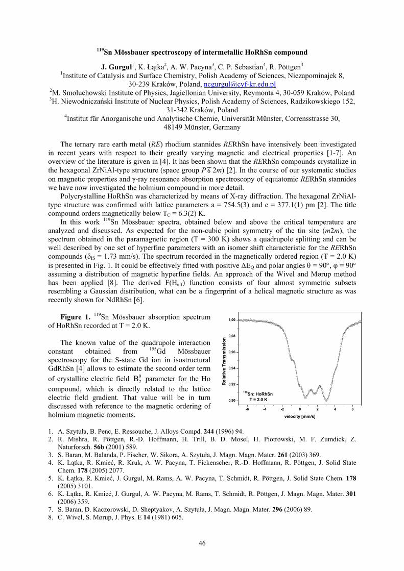
In this work 119Sn Mössbauer spectra, obtained below and above the critical temperature areanalyzed and discussed. As expected for the non-cubic point symmetry of the tin site (m2m), thespectrum obtained in the paramagnetic region (T = 300 K) shows a quadrupole splitting and can bewell described by one set of hyperfine parameters with an isomer shift characteristic for the RERhSncompounds (δIS = 1.73 mm/s). The spectrum recorded in the magnetically ordered region (T = 2.0 K)is presented in Fig. 1. It could be effectively fitted with positive ∆EQ and polar angles θ = 90°, φ = 90°assuming a distribution of magnetic hyperfine fields. An approach of the Wivel and Mørup methodhas been applied [8]. The derived F(Heff) function consists of four almost symmetric subsetsresembling a Gaussian distribution, what can be a fingerprint of a helical magnetic structure as wasrecently shown for NdRhSn [6].
Figure 1. 119Sn Mössbauer absorption spectrumof HoRhSn recorded at T = 2.0 K.
The known value of the quadrupole interactionconstant obtained from 155Gd Mössbauerspectroscopy for the S-state Gd ion in isostructuralGdRhSn [4] allows to estimate the second order termof crystalline electric field 0
2B parameter for the Hocompound, which is directly related to the latticeelectric field gradient. That value will be in turndiscussed with reference to the magnetic ordering ofholmium magnetic moments.
1. A. Szytuła, B. Penc, E. Ressouche, J. Alloys Compd. 244 (1996) 94.2. R. Mishra, R. Pöttgen, R.-D. Hoffmann, H. Trill, B. D. Mosel, H. Piotrowski, M. F. Zumdick, Z.
Naturforsch. 56b (2001) 589.3. S. Baran, M. Bałanda, P. Fischer, W. Sikora, A. Szytuła, J. Magn. Magn. Mater. 261 (2003) 369.4. K. Łątka, R. Kmieć, R. Kruk, A. W. Pacyna, T. Fickenscher, R.-D. Hoffmann, R. Pöttgen, J. Solid State
Chem. 178 (2005) 2077.5. K. Łątka, R. Kmieć, J. Gurgul, M. Rams, A. W. Pacyna, T. Schmidt, R. Pöttgen, J. Solid State Chem. 178
(2005) 3101.6. K. Łątka, R. Kmieć, J. Gurgul, A. W. Pacyna, M. Rams, T. Schmidt, R. Pöttgen, J. Magn. Magn. Mater. 301
(2006) 359.7. S. Baran, D. Kaczorowski, D. Sheptyakov, A. Szytuła, J. Magn. Magn. Mater. 296 (2006) 89.8. C. Wivel, S. Mørup, J. Phys. E 14 (1981) 605.
-6 -4 -2 0 2 4 6
0,90
0,92
0,94
0,96
0,98
1,00
Rel
ativ
e Tr
ansm
issi
on
velocity [mm/s]
119Sn: HoRhSnT = 2.0 K

47
Notes

48
Structural and magnetic properties of Fe3-xTixSn disordered alloys
K. Brząkalik
Institute of Materials Sciences, University of Silesia, Katowice, Bankowa 12, 40-007 Katowice,Poland; [email protected]
A series of the disordered Fe3-xTixSn alloys (where x = 0, 0.25, 0.5, and 0.75) obtained by arc-meltingwere studied. Directly after melting the samples were mechanically crashed and in that form examinedby the X-ray diffraction and Mössbauer effect method. In Fe3Sn based alloy exist three phases: FeSn(B35), FeSn2 (C16) and Fe(Sn) (A2). The contributions of these phases decrease very fast in aid ofternary Fe-Ti-Sn alloy with the D03/L21 type of structure, as Ti atoms concentration increase. It wasshown that this ternary alloy is not homogeneous but consists of four distinct Fe-Ti-Sn phases(different content of the individual atoms) with the contribution depending on the value of the xparameter. The value of the hyperfine magnetic filed parameter at the Fe nuclei is equal to about of110 kG in FeSn and FeSn2 phases. However in alloys with A2, B2 and D03/L21 type of structure thisvalue changes from 0 kG to 335 kG and can be (in the first approach) consider as a function of Featoms number in the nearest neighborhood.

49
Notes

50
57Fe hyperfine interactions in Sc(Fe1−xNix)2 Laves phases synthesized under high pressure
M. Wiertel 1 , Z. Surowiec 1 , M. Budzyński 1 A.V. Tsvyashchenko 2
1 Zakład Metod Jądrowych, Instytut Fizyki, Uniwersytet Marii Curie-SkłodowskiejPL-20-031 Lublin, pl. M. Curie-Skłodowskiej 1, Poland; [email protected]
2 Vereshchagin Institute for High Pressure Physics, Russian Academy of SciencesRU-142190 Troitsk, Moscow Region, Russia
In our earlier work results for ME measurements for quasibinary compounds Sc(Fe1−xNix)2prepared by arc melting under ambient pressure were reported [1]. The synthesis of series ofcompounds Sc(Fe1−xNix)2 of the same composition was carried out in the Institute for High PressurePhysics at Troitsk by application of high temperature at a constant pressure of 8 GPa in a toroid-typehigh pressure chamber [2]. The details of the procedure used in this experiment have been givenelsewhere [3]. The structure of the compounds was checked by XRD method. The samples for
10.00 ≤≤ x have hexagonal C14-type (hP12, the space group P63/mmc) crystal structure as opposedto analogous samples produced under ambient pressure where cubic C15-type (cF24, mFd 3 )structure is stable. In the range of concentration x above 0.20 the samples have C15 structureregardless of the way of preparation.
The Mössbauer measurements have been performed in the range of concentration x up to 0.60 andfor temperatures from RT to temperatures where transitions from ferro- to paramagnetic state occur forindividual samples. In the course of data analysis a consistent fit to the experimental data could beachieved under the assumption of Ni replacing Fe randomly. The relative contributions of individualsubspectra corresponding to different type of a local surrounding {(6-n)Fe+nNi atoms} weredetermined on the basis of the binomial distribution. Only the probabilities higher then 5 % had beentaken into consideration.
Generally the substitution one Ni atom for Fe atom in the near neighbour of nuclear probe reducesthe 57Fe hyperfine magnetic fields by about 2 T. Isomer shift values remain almost constant and equalto -0.30 mm/s for all investigated samples. From the temperature dependences of hyperfine magneticfields Curie temperatures TC were evaluated. It is interesting that TC for the isostructural compoundsobtained under high pressure are by two orders less in comparison with those for samples producedunder ambient pressure even though interatomic distances are practically equal in the both type ofcompounds. A presence of a significant paramagnetic doublet component in the Mössbauer spectra for
50.030.0 ≤≤ x in the wide range of temperature below TC indicate the coexistence of paramagneticand ferromagnetic regions in the samples and occurrence of magnetic clusters with a wide distributionof the Curie temperatures.
1. M. Wiertel, Z. Surowiec, J. Sarzyński, M. Budzyński, A. I. Beskrovny, Nukleonika, 52 (2007) 67.2. L.G. Khvostantsev, L.F. Vereshchagin, and A.P. Novikov, High Temp.-High Press., 9 (1977) 637.3. A.V. Tsvyashchenko, J. Less-Common Met. 99 (1984) L9.

51
Notes

52
Magnetism and Debye temperature in σ-FeV compounds
J. Cieślak1, B. F. O. Costa2, S.M. Dubiel1, M. Reissner3 and W. Steiner3
1 Faculty of Physics and Computer Science, AGH University of Science and Technology,30-059 Kraków, Poland; [email protected]
2 CEMDRX Department of Physics, University of Coimbra, 3000-516 Coimbra, Portugal3 Institute of Solid State Physics, Vienna University of Technology, 1040 Wien, Austria
Sigma-phase has a complex crystallographic structure. Its unit cell is tetragonal (space groupP42/mnm, space group number 136) with five crystallographically non-equivalent lattice sites having ahigh (12-15) coordination number. For this reason it is a member of the Frank-Kasper phase family.The phase is known to exist only in alloy systems and in a certain composition range. As a result ofthis, it is, in general, non-stoichiometric. In the case of Fe-V system, the composition range in whichthe phase can be obtained spans between ∼30 and ∼60 at % V. Consequently, its various physicalproperties can be readily tailored by changing the alloy composition.In this contribution we will present results concerning magnetic and dynamic properties of the phase.The study was carried out with magnetometric (VSM) and Mössbauer spectroscopic methods on aseries of samples with V-content ranging between ∼34 and ∼60 at%. The σ-phase samples wereobtained from α-phase ingots prepared by an arc-melting process. The ingots, after being cold-rolled,were isothermally annealed in vacuum at 973 K for 25 days. Verification of the transformation intothe σ-phase was checked by recording X-ray and neutron diffraction patterns. Measurements on the σ-phase samples were performed both as a function of temperature (4 – 350 K) as well as of an externalmagnetic field (up to 15 T). From the magnetization measurements, several magnetic quantities suchas the Curie temperature, TC, average magnetic moment per Fe atom, <µ>, effective magneticmoment per Fe atom in a paramagnetic phase were determined. From the Mössbauer spectradistributions of the hyperfine field, P(B), average hyperfine field, <B>, the Curie temperature, and theaverage central shift, <CS>, were derived. From the latter, using the Debye model, the Debyetemperature, ΘD, was obtained.The results found and relationships between them will be presented and discussed. In this abstract wewant to mention that in the sample of σ-FeV34 the strongest ever reported magnetic properties havebeen measured. In particular, TC ≈ 320 K and <µ> ≈ 0.9 µB. The former is ∼50 K and the latter ∼0.3µB
greater than their up-to-date known values. A linear relationship revealed between TC and <µ> is alsoworth mentioning. Concerning the Debye temperature, its present determination is to our knowledgethe first one. As shown in Fig. 1, its dependence on vanadium content, x is not a monotonic function.However, due to a lack of any relevant theoretical calculations, interpretation of the ΘD(x) behaviour isnot yet possible.
Fig.1 Debye temperature, ΘD, versus Cr content, x. Solid line is to guide the eye only. Forcomparison, corresponding data for σ-FeCr compounds are added [1].[1] J. Cieślak, B. F. O. Costa, S. M. Dubiel, M. Reissner and W. Steiner, J. Phys.; Condens. Matter, 17(2005) 6889

53
Notes

54
Essentialities of manganese antimonide substituting by Cu and Zn
V.I.Mitsiuk, V.M.Ryzhkovskii, T.M.Tkachenka
Joint Institute of Solid State and Semiconductor Physics National Academy of Sciences of Belarus,Minsk, P. Brovki Str., 19, Belarus; [email protected]
It is known, that manganese antimonide Mn1+хSb, 0≤х≤0.30 [1] with NiAs-type of crystal structure,may form broad regions of solid solutions with transition metals, or with binary compounds of similarstructure, but there is no data published on possibility of its substitution by nonmagnetic Cu or Zn. Thepurpose of present work was to obtain the aforesaid NiAs-type solid solutions, to investigate physicalcharacteristics and to compare the data obtained with those received using Mossbauer Effect Method.The possibility of MnSb substitution by 5% at. Cu or 10% at. Zn was shown earlier [2], and the crystaland magnetic parameters for the solutions were presented. The three of solid solutions obtained havebeen chosen for the Mossbauer study, all have the same cation excess relative to the equiatomic state:Mn1.08Fe0.02Sb; Mn0.98Fe0.02Zn0.1Sb; Mn1.03Fe0.02Cu0.05Sb. Mossbauer study were made at roomtemperature in usual transmission geometry, Fe/Rh was used as a source. All Mossbauer spectra werefitted in the model of two subspectra (Table1).This fitting model agrees with the essentialities ofNiAs-type crystal structure and the magnetic measurement results. There are two different cationpositions in this type of crystal structure: octahedral (MeI) and trigonal-bipyramidal (MeII) and thereare two subspectra, both magnetically splitted, in each Mossbauer spectrum. The hyperfine magneticfields at Fe are not large and have the values 60±2kOe and 81±2kOe, the IS values are similar forboth. The Mossbauer parameters for the unsubstituted sample subspectra and those for the substitutedsample, have similar values. The nonmagnetic Cu or Zn atoms do not appreciable influence the fieldvalues at Fe in MeI and MeII positions in the case of small amounts of Cu or Zn. This result agreeswith the results on magnetic measurements: the saturation magnetization values for the initial Mn1.1Sband for the solid solutions with Mn and Zn have values about 100 Gs×cm3×g-1 and do not show anyappreciable substitution effect. The difference between spectra is in the ratio of subspectra intensitiesthat is directly connected with the population of two cation sublattices in each solid solution.Mossbauer data indicate that the population of MeII cation sublattice for unsubstituted Mn1.1Sb islower than that for the MnZn0.1Sb and MnCu0.1Sb samples as the total cation content in all samples isthe same. Possibly, the defect in sublatice of MeI atoms increases, when the sample is substituted bythe nonmetaallic atoms and the cation population of second sublattice increases owing to the cationsmooving from positions MeI to the positions MeII. Another possible reason of that could be that theFe atoms prefer the MeII positions in substituted sample and behave like Cu and Zn.
Table 1Mossbauer Spectra Parameters of the Mn1.10Sb substituted by Cu and Zn.
Subspectra N 1 2Sample IS,
mm/sQS,
mm/sH,T
W,mm/s
A (%tothe
total)
IS,mm/s
QS,mm/s
H,T
W,mm/s
A (%tothe
total)Mn1.08Fe0.02Sb 0.50 0.19 6.1 0.21 74 0.55 -0.10 8.2 0.17 26
Mn0.98Fe0.02Zn0.1Sb 0.49 0.20 6.1 0.22 49 0.53 -0.11 8.0 0.16 51Mn1.03Fe0.02Cu0.05Sb 0.44 0.33 6.3 0.33 22 0.51 -0.12 7.8 0.15 78
1. I.Teramoto and A.J.G. Van Run. J. Phys. Chem. Solids,. 99 (1968), p.347.2. Mitsiuk V.I., Ryzhkovskii V.M., Tkachenka T.M. Journal of Alloys and Compounds, to be
published in 2008.

55
Notes

56
Hyperfine field and magnetic moment in Fe-containing alloys and compounds
Stanisław M. DubielFaculty of Physics and Computer Science, AGH University of Science and Technology, al.
Mickiewicza 30, 30-059 Krakow, Poland; [email protected]
Hyperfine magnetic field, Bhf, is a very important spectral quantity measured on magnetic sampleswith the Mössbauer Spectroscopy. Bhf arises from combined effects of the orbital, contact and dipolarfields at the nuclear site. For many compounds the orbital moment is quenched, and the dipoleinteraction, although not negligible, is small, hence the so-called Fermi contact term (FCT) related to adensity of s-like electrons at nucleus is regarded as the most important and dominant. Experimentallydetermined Bhf is frequently regarded as a good measure of magnetic properties of an investigatedsample, and it is often rescaled into the underlying magnetic moment, µ, using the simplest possiblerelation between the two quantities i.e. Bhf = A µ, where A is the hyperfine coupling constant.However, according to theoretical calculations [1-3], such procedure is, in general, not justifiedbecause only a part of the FCT due to a polarization of the core electrons (1s, 2s, 3s) is proportional toµ, while the one due to the polarization of conduction electrons (4s) is not. Nevertheless, the linearrelationship between Bhf and µ is often used in practice for various systems but with the same value ofA = 14 T/µB, as determined for a metallic iron.
In this contribution it will be shown, on different examples of various Fe-containing alloy andintermetallic compound systems, that the relationship between Bhf and µ is, in general, not linear, andit is characteristic of a given alloy or compound system. In other words, it is not justified to use onevalue of A even for a given system.
From among several examples, that will be shown to illustrate the above mentioned conclusion,two are presented in this abstract.
101520253035404550
22 24 26 28 30 32 34 36
x [at%]
A [T
/Boh
r mag
neto
n]
Fig 1. (left) Hypefine coupling constant, A, versus Al concentration, x, in an ordered Fe3-xAlx
compound, and (right) average hf. Field, <B>, versus average magnetic moment per Fe atom, <µ>, inthe sigma-phase FeCr and FeV compounds.
[1] R. E. Watson and A. J. Freeman, Phys. Rev., 123 (1961) 2027.[2] M. E. Elzain et al., Phys. Rev. B, 34 (1986) 1430[3] B. Lingren and J. Sjöström, J. Phys. F: Metal. Phys., 18 (1988) 1563

57
Notes

58
Ordering and magnetoelestic properties of Fe-Ga alloys
K. Perduta1, J. Olszewski2, S. Busbridge1, M. Nabiałek2
1 School of Environment and Technology, University of BrightonUK BN2 4GJ Brighton, Lewes Road, United Kingdom; [email protected]
2 Institute of Physics, Częstochowa University of TechnologyPL 42-200, 19 Armii Krajowej Ave., Poland
Magnetic composites represent large group of materials consisting of ferromagnetic orsuperparamagnetic particles disperse in non-ferromagnetic matrix. The mechanical and magneticcharacteristics of these materials depend on the properties of both the particles and the matrix [1]. Inthese paper we present studies of the microstructure and magnetic properties of composite of epoxy-bonded Fe-Ga particles. Ingots of the initial Fe80Ga20 alloy were obtained by arc melting high puritycomponents. In order to achieve the homogeneity the ingots were remelted several times. Subsequentblade-milling in an argon atmosphere and sieving to powders produced particles in the size range 20-50 µm. After blade-milling the powders were annealed at 723K for 2.5, 7.5 and 12.5 hours. Thecomposites were made from powders directly after blade milling and after annealing. The powder wasthen mixed with an epoxy binder and compressed under a pressure of 120 MPa along the shortestdimension to obtain rectangular samples of dimensions 7-10 mm x 10 mm x 40 mm. Compaction wascarried out without an applied magnetic field. The volume fractions of Fe-Ga powder used wasnominally 0.80..
The microstructure of powdered alloy was studied using X-ray diffractometry and Mössbauerspectrometry. These studies were carried out for the powders obtained from as-cast alloy and afterannealing at 723K for 2.5, 7.5 and 12.5 hours. The saturation magnetostriction (λs) of the bulk alloyand its composites were measured using standard strain gauge techniques. All investigations wereperformed at nominal room temperature.
In fig 1 an example of the Mössbauer spectrum obtained for powdered alloy with particles sizerange from 20 to 50 µm is presented.
Figure 1 Mössbauer transmissionspectrum of as-cast Fe80Ga20 alloy (20 to50 µm particle size)
All Mössbauer spectra were decomposedinto five elementary subspectracorresponding to iron atoms having 8, 7,6, 5, 4 iron atoms in their nearestneighbourhood. From the Mössbauerspectroscopy studies we have found thatthe as-cast alloys do not exhibit atomicorder. However after annealing at 723 Kthe DO3 type superstructure appeared.
We have stated that the best magnetoelastic properties are exhibited by composites made fromferromagnetic particles obtained from the as-cast alloy. The measured saturation magnetostriction wasλs= 70 ppm for bulk alloy and λs=100 ppm for composites. , the difference between the two valuesmay be explained by the influence of the epoxy matrix [2]. However composites prepared frompowders annealed at 723 K for different times show lower magnetostriction than composites madefrom as-cast powder.
This work acknowledges support from the European Union (Interreg IIIa grant number 349)
1. A. Clark, U.S. Patent No. 4,378,258, March 29 (1983)2. S. Bednarek, Appl. Phys. A., 68 (1999) 63

59
Notes

60
Studies of iron doping regions in Ge1-xFexTe diluted magnetic semiconductor
T. Szumiata 1 , Ł. Kilański 2 , W. Dobrowolski 2 , K. Brzózka 1 , M. Gawroński 1 , B. Górka 1 ,M. Arciszewska 2 , V. Domukhovski 2 , V. E. Slynko 3, I. E. Slynko 3
1 Department of Physics, Technical University of Radom,ul. Krasickiego 54, Radom, Poland; [email protected]; [email protected]
2 Institute of Physics, Polish Academy of Sciences,Al. Lotników 32/46, 02-668 Warsaw, Poland
3Institute of Material Science Problems, Ukrainian Academy of Sciences5 Wilde Street, 274001, Chernovtsy, Ukraine
Dilute magnetic semiconductors (DMS) fabricated by adding transition metals into nonmagneticclassical semiconductors have recently been recognized as very promising materials forSPINTRONICS [1] and they are intensively investigated in this context. DMS compounds containingiron [2,3] are of special interest because of relatively high magnetic moment and low price of Fe aswell as due to the unique opportunity of the investigations by means of 57Fe Mössbauer spectrometry.In our work we performed Mössbauer and magnetic measurements on Ge1-xFexTe samples in order tostudy nature of magnetism of iron-doped regions. The transmission Mössbauer spectrum ofGe0.96Fe0.04Te sample collected at room temperature (Fig. 1a) consists of one doublet what points toparamagnetic state of magnetic regions. This is consistent with results of macroscopic magnetizationand susceptibility measurements. The presence of only one doublet (of quadrupole splitting valueQS ≈ 0.53 mm/s) not accompanied by singlet line proves that there are no α-Fe phase precipitations inthe alloy and that Fe atoms pass into GeTe crystalline structure (deformed rock salt lattice) taking a
site of the symmetry lower than cubic one. Theseresults are in accordance with X-ray diffraction data.At lowered temperature of the order of T ≈ 90 K aMössbauer spectrum of Ge0.96Fe0.04Te is still ofparamagnetic type (Fig. 1b), what does not coincidewith AC magnetic susceptibility measurements,demon-strating a PM-FM-like transition at the tem-perature about 150 K (similarly to previouslyinvestigated Ge1-xMnxTe systems [4]). A pro-bablereason of this inconsistency is fact, that Mössbauerexperiment was performed in zero external magneticfield, what favored spin relaxation effects - veryimportant in the case of nanometric-size iron-richregions. The existence of real magnetic transition atmentioned temperature is supported by the results ofmeasurements of Hall constant, which reachesmaximum at 140 - 160 K.
Figure 1. Transmission Mössbauer spectrum of the compound Ge0.96Fe0.04Te diluted magneticsemiconductor a) at room temperature and b) at T = 90 K.
1. H. Ohno, Science 281 (1998) 951.2. A. Twardowski, H.J.M. Swagten, W.J.M. de Jonge and M. Demianiuk, Phys. Rev. B 44 (1991)
2220-2226.3. D. R. S. Somayajulu, Narendra Patel, Mukesh Chawda, Mitesh Sarkar and K. C. Sebastian,
Hyperfine Interactions 160 (2005) 241–246.4. W. Dobrowolski, M. Arciszewska, B. Brodowska, V. Domukhovski, V. K. Dugaev, A. Grzęda, I.
Kuryliszyn-Kudelska, M. Wójcik, E. I. Slynko, Science of Sintering, 38 (2006) 109.
5.52e+06
5.56e+06
5.6e+06
5.64e+06
5.68e+06
-8 -6 -4 -2 0 2 4 6 8
Cou
nts
Velosity [mm/s]
exp.Théo
8.185e+06
8.195e+06
8.205e+06
8.215e+06
8.225e+06
-8 -6 -4 -2 0 2 4 6 8
Cou
nts
Velosity [mm/s]
exp.Théo
Room tem perature
T= 90 K
Ge0.96Fe0.04Te
Ge0.96Fe0.04Te
a)
b)

61
Notes

62
Mobility of hematite submicron particles in dense solutions of sugar
P. Fornal1, J. Stanek2
1 Institute of Physics, Cracow University of Technology, Podchorążych Str. 1,30-083 Kraków, Poland; [email protected]
2Marian Smoluchowski Institute of Physics, Jagiellonian University, Reymonta Str. 4,30-059 Kraków, Poland
The nature of the mobility of submicron particles in fluids is Brownian movements. The developed byEinstein [1] and Smoluchowski [2] in the beginning of the previous century formulas are well known.However, their experimental confirmation are in general restricted to the limit where the observationtime is much longer than so called Brownian relaxation time. The characteristic Mössbauerspectroscopy observation time is much shorter than this relaxation time. Thus, the results obtained bythis technique may contribute to the fundamental statistical physic. On the other hand, the livingorganisms consist mainly of nano or micro soft solid objects, like cellular membranes or cellorganelles immersed in the amine gels which sometimes form the mesoscopic three dimensionalstructures (cytoskeleton). The study of their biochemical interactions are frontiers of the currentscience. The results of the study of the study of the mobility of hematite particles in gelatin gel werepresented at ICAME 2007 conference, Kanpur, India.
In the present paper the results of the study of mobility of the submicron hematite particles in watersolutions of sugar (60 % wt. and 80 % wt.) are presented. The dramatic broadening of Mössbauer lineup to few mm/s are observed, see Fig. 1. The broadening, directly related to the distribution of theparticle velocities, measured in the 141 ns time scale, critically depends on the sugar concentration andthe temperature which influence the viscosity of the solutions.
-20 -10 0 10 20velocity [mm/s]
99
100
99
100
Res
onan
t Abs
orpt
ion
[ %
]
-2 C
12 C
99
100
23 C
99
100
33 C
99
100
3 C
-20 -10 0 10 20velocity [mm/s]
98
100
99
100
99
100
Res
onan
t Abs
orpt
ion
[ %
]
2 C
6 C
12 C
99
100
23 C
96
100
dried23 C
Fig. 1. The Mössbauer spectra of hematite in water solution of sugar, 60 % wt., left, and 80 % wt., right,recorded at temperatures as marked.
[1] A. Einstein, Annalen der Phys. 17, 549-560, (1905)[2] M. Smoluchowski, Ann. Phys. 21,756-780 (1906)

63
Notes

64
Structural, electrical and Mössbauer effect studies of 0.5Bi0.95Dy0.05FeO3-0.5Pb(Fe0.5Nb0.5)O3multiferroics
A. Stoch1, J. Kulawik1, P. Stoch2,3, J. Maurin3,4, P. Zachariasz3
1Institute of Electron Technology, Krakow Division , ul. Zabłocie 39, 30-701 Kraków, Poland;[email protected]
2Faculty of Material Science and Ceramics, AGH – University of Science and Technology,al. Mickiewicza 30, 30 – 059 Kraków, Poland
3Institute of Atomic Energy, 05-400 Otwock-Świerk, Poland4National Medicines Institute, ul. Chełmska 30/34, 00-725 Warszawa, Poland
Materials that exhibit ferromagnetic and ferroelectric orderings simultaneously are known asmultiferroics. They are used widely as transducers, actuators, other sensors and as a material for thenext generation computer memories. The first single phased multiferroic perovskites were discoveredin the early 1960s. However they are very rare and limited progress has been made during the lastseveral decades [1].
The 0.5Bi0.95Dy0.05FeO3-0.5Pb(Fe0.5Nb0.5)O3 multiferroic is a solid solution of ceramicsBi0.95Dy0.05FeO3 perovskite and Pb(Fe0.5Nb0.5)O3 complex perovskite. Polycrystalline samples wereprepared by conventional solid-state reaction method. Detailed X-ray studies were used to confirm theformation of required phase. The 0.5Bi0.95Dy0.05FeO3-0.5Pb(Fe0.5Nb0.5)O3 crystallize in P4/mmmcrystal phase with crystal parameters a=c=4,00 Å. Electrical properties of the studied material weremeasured using impedance spectroscopy method in wide temperature range. An equivalent electricalcircuit, composed of various R, C and CPE elements was proposed and fitted to the measured data.The grain boundaries and bulk material resistance was obtained. These resistances vs. temperaturefulfill very well Arrhenius law. Calculated activation energy is about 1 eV for both type of resistances,which is in a good agreement with literature data [2]. The 57Fe Mössbauer effect was measured at 77Kand the hyperfine interaction parameters were obtained. The Mössbauer spectra are composed of twosextets which represents two different Fe ion crystallographic site one represent Bi0.95Dy0.05FeO3 phaseand the second Pb(Fe0.5Nb0.5)O3. The achieved isomer shift and quadruople splitting parameters aretypical for Fe3+ in octahedral symmetry [3]. The obtained magnetic hyperfine fields are about 50 T andare normal for ferric iron. Thus, a Fe3+ charge state for the iron in the 0.5Bi0.95Dy0.05FeO3-0.5Pb(Fe0.5Nb0.5)O3 has been confirmed by the isomer shift and hyperfine field values.
1. J. F. Scott, Nature Materials 6 (2007) 256-257.2. E. J. Abram, D. C. Sinclair, A. R. West, Journal of Electroceramics, 10 (2003) 165-177.3. S. A. Ivanov, P. Nordblad, R. Tellgren, T. Ericsson, H. Rundlof, Solid State Sciences 9 (2007)
440-450.

65
Notes

66
Mössbauer spectroscopy of quasicrystals
Zbigniew M. StadnikDepartment of Physics, University of Ottawa, Ottawa, Ontario K1N 6N5, Canada;
Solids are traditionally divided into two groups: crystalline and amorphous. The dramatic discovery ofan icosahedral Al-Mn alloy by Shechtman et al. [1] extended this dichotomous division by introducingthe notion of quasicrystals (QCs). These are compounds that possess a new type of long-rangetranslational order, quasiperiodicity, and a noncrystallographic orientational order associated with theclassically forbidden fivefold, eightfold, tenfold, and twelvefold symmetry axes [2]. A central problemin condensed matter physics is determining whether quasiperiodicity leads to physical propertieswhich are significantly different from those of crystalline and amorphous materials.
This lecture is aimed at reviewing the contribution of Mössbauer spectroscopy (MS) to elucidatingseveral outstanding problems of the structural and physical properties of QCs. In spite of enormousexperimental and theoretical effort, the atomic structure of QCs, except for the icosahedral binaryalloy YbCd5.7 [3], has not been solved yet. It will be demonstrated how the combination of x-raydiffraction data, electric field gradient calculations, and in-field 57Fe MS data led to the elucidation ofthe atomic structure of the icosahedral Al-Cu-Fe alloy. One of the central questions in the physics ofQCs is that of the possibility of long-range magnetic order in these alloys. Initial intuition suggeststhat quasiperiodicity necessarily leads to geometrical frustration and is therefore incompatible withlong-range magnetic order. Examples of recent 57Fe and 155Gd MS studies of magnetism of QCs willbe presented. There are two types of lattice dynamics in QCs: conventional phonons and, specific to aquasicrystalline structure, phasons [2]. The contribution of 57Fe MS to studies of phason dynamics willbe elaborated. Finally, the application of nuclear inelastic scattering to the determination of the site-specific phonon density of states in QCs will be discussed.
1. D. Shechtman, I. Blech, D. Gratis, J.W. Cahn, Phys. Rev. Lett. 53, 1951 (1987).2. Physical Properties of Quasicrystals, Ed. Z.M. Stadnik, Springer-Verlag, Berlin 1999.3. H. Takakura, C.P Gómez, A. Yamamoto, M. De Boissieu, A.P. Tsai Nature Mater. 6, 58 (2007).

67
Notes

68
Magnetism of ultra-thin iron films seen by the nuclear resonant scattering of synchrotronradiation
T. Ślęzak1, S. Stankov3, M. Zając1, M. Ślęzak1, K. Matlak1, N. Spiridis2, B. Laenens4,N. Planckaert4, M. Rennhofer5, K. Freindl2, D. Wilgocka-Ślęzak2, R. Rüffer3 and J. Korecki1,2
1 Faculty of Physics and Applied Computer Science, AGH University of Science and Technology, Al.Mickiewicza 30, 30-059 Kraków, Poland; [email protected]
2 Institute of Catalysis and Surface Chemistry, Polish Academy of Sciences,ul. Niezapominajek 8, 30-239 Kraków, Poland
3 European Synchrotron Radiation Facility, BP220, F-38043 Grenoble, France4 Instituut voor Kern- en Stralingsfysica, K.U.Leuven, Celestijnenlaan 200D, B-3001 Leuven, Belgium
5 Fakultät für Physik, Universität Wien, Strudlhofgasse 4, A-1090 Wien, Austria
Conversion electron Mössbauer spectroscopy proved in the past to be very useful in studying surfaceand ultrathin film magnetism with monolayer resolution. Twenty years later, its time-domainanalogue, the nuclear resonant scattering (NRS) of synchrotron radiation, showed up to be by orders ofmagnitude faster and more efficient. We discuss the most important features of NRS based onsimulations and experimental data. It is shown how the isotopic sensitivity of NRS, combined with the57Fe probe layer concept, was explored to study influence of the interlayer exchange coupling to FeAumonoatomic superlattices on the magnetic properties of the iron monolayer on Au(001). In the secondexample, combination of UHV conditions and the high brilliance of the third generation synchrotronsource is used to probe the evolution of spin structure in epitaxial Fe films on W(110) via theaccumulation of high quality time spectra directly during the 57Fe film growth. In this way the scenarioof the in-plane spin reorientation transition (SRT) occurring for Fe/W(110) system could be followed.The analysis of NRS data clearly shows that the SRT consists in the formation of the unexpected, non-collinear magnetic structure which mimics the planar domain wall.

69
Notes

70
Magnetic nanoparticles in mcm-41 type mesoporous silica
Z. Surowiec1, M. Budzyński1, M. Wiertel 1 , J. Goworek 2 , B. Bierska Piech3
1 Zakład Metod Jądrowych, Instytut Fizyki Uniwersytet Marii Curie-Skłodowskiej PL -20-031 Lublin,pl. Marii Curie-Skłodowskiej 1 Poland; [email protected]
2 Zakład Adsorpcji Wydział Chemii Uniwersytet Marii Curie-Sklodowskiej PL- 20-031 Lublin, pl.Marii Curie-Skłodowskiej 1
3 Zakład Badań Strukturalnych, Instytut Nauki o Materiałach, Uniwersytet Śląski PL- 40-007Katowice, ul. Bankowa Poland
During the last few years, nanoparticles have attracted much attention due to their importance forfundamental studies and the wide range of potential applications in nanodevices.
One of many methods of nanoparticles producing is embedding of appropriate atoms in themesoporous silica materials. The M41S mesoporous ordered silica materials were invented in 1992 bythe group from Mobil Oil [1]. One of such materials characterized by uniform pore diameter, largepore volume and large surface area is MCM-41. Hexagonal arrangement of the cylindrical pores isobtained in MCM-41 by use of templating technique. Cylindrical micelles formed by surfactant inalkaline medium are used as condensation centres for silica from tetraethylortho-silicate (TEOS), oralkalimetal silicate. Accumulation of micellar rods leads to creation of honeycomb shaped micelle-templated silica. Using templates with alkyl chains of different length allows controlling porediameters in range of 2 to 10 nm.
Mössbauer spectroscopy (MS), X-Ray diffraction (XRD) and Small-Angle X-Ray Scattering(SAXS) studies were performed on mesoporous ordered silica tubes MCM-41 filled by Fe1Ni1-x(x= 0.0 – 0.4).
The profiles of SAXS curves log I(q) versus log(q) plot from pure, empty tubes shows linearbehavior in broad region of scattering angles with the slope – 4. In the outermost part of scatteringcurves the local maximum is present. Such profile of SAXS curve excludes the ordering in mutualarrangement of silica tubes. The curves from filled tubes are more complicated. The linear parts onlog-log plots with slopes –2.7 and 4.0 are observed and local maxima in the outermost part ofscattering curves are still present.
X-ray diffraction patterns reveal the amorphness of silica tubes and diffraction lines from FeNipowders. The pattern of lines changed from spinel structure correspond to Fe3O4 for x = 0 tointermetallic structure Fe0.6Ni0.4 for x = 0.4.
The Mössbauer study of Fe1Ni1-x embedded in the MCM-41 type silicate templates revealedexistence of superparamagnetic phase. The obtained Mössbauer spectra consist of sextets and asuperimposed superparamagnetic doublet. The sextets originate from Fe atoms positions in theferromagnetic phase. The doublet component is related to the superparamagnetic phase of this ironcompounds. The existence of the superparamagnetic nanoparticles results from the relaxationphenomenon due to the intrinsic finite-size effect. The relative contribution of the relaxing componentto the total spectrum at room temperature is about 80%. With the increasing temperature theferrimagnetic component clearly decreases.
1. C.T. Kresge, M.E. Leonowicz, W.J. Roth, J.C. Vartuli, J.S. Beck, Nature 359 (1992) 710.

71
Notes

72
Mössbauer study of mechanosynthesized and thermally treated Co-Fe-Ni alloys
T. Pikula1, D. Oleszak2, M. Pękała3, M. Mazurek1, J.K. Żurawicz1, E. Jartych1
1 Department of Experimental Physics, Institute of Physics, Technical University of Lublin,ul. Nadbystrzycka 38, 20-618 Lublin, Poland, [email protected]
2 Faculty of Materials Science and Engineering, Warsaw University of Technology, ul. Wołoska 141, 02-507 Warsaw, Poland
3 Department of Chemistry, Warsaw University,Al. Żwirki i Wigury 101, 02-089 Warsaw, Poland
Mechanical alloying (MA) is the potential technology of production of nanocrystallinematerials. Such materials exhibit large density of grain boundaries, so their macroscopic propertiesmay be different in comparison with analogical polycrystalline specimens. Moreover, MA can be usedto obtain powdered materials with good soft magnetic properties.
In this work ternary Co-Fe-Ni alloys were prepared in high energy planetary ball mill. Wehave mainly studied Co-rich compositions of the alloys, which have interesting properties in atransitional area in the phase diagram, namely they have good soft magnetic properties [1]. 57FeMössbauer spectroscopy and differential scanning calorimetry were used to characterize the structureand hyperfine interactions of the specimens. Also the magnetic measurements were carried out inorder to determine the effective magnetic moment, Curie temperature, saturation magnetization andcoercive field of the studied alloys. X-ray diffraction proved that during MA process solid solutionswith b.c.c. or f.c.c. lattices were formed with an average grain size about tens of nanometers.However, products of milling are often in the non-equilibrium state. Thermal treatment of the alloysconducted in some cases to decomposition of the suitable solid solution into the mixture of two solidsolutions. In another cases the state of the alloys was stable or the type of the crystalline lattice waschanged during annealing [2].
Mössbauer spectra obtained for the alloys after MA and after thermal treatment in all caseswere six-line patterns. They were numerically fitted by using hyperfine magnetic field distribution assuggested by relatively high values of the width of the spectral lines and disordered structure of thesamples. These distributions reflect the different surroundings of 57Fe isotopes by Co, Fe and Ni atomsdepending on the chemical composition of the alloy. The most probable atomic configurations in thefirst coordination zone were found using the extended binomial distribution. Moreover, the correlationbetween the average value of the hyperfine magnetic field and the effective magnetic moment ofmechanosytnhesized Co-Fe-Ni alloys was visible.
1. S.U. Jen, H.P. Chiang, C.M. Chung, M.N. Kao, J. Magn. Magn. Mater. 236 (2001) 312.2. T. Pikula, D. Oleszak, M. Pękała, E. Jartych, J. Magn. Magn. Mater. 320 (2008) 413.

73
Notes

74
Formation and magnetic properties of nanostructured Fe-Pt-B alloys
A. Grabias1, M. Kopcewicz1, D. Oleszak2, J. Latuch2, M. Pękała3, M. Kowalczyk2
1Institute of Electronic Materials Technology, ul. Wólczyńska 133, 01-919 Warsaw, Poland;[email protected]
2Department of Materials Science and Engineering, Warsaw University of Technology,ul. Wołoska 141, 02-507 Warsaw, Poland
3Department of Chemistry, University of Warsaw, Al. Żwirki i Wigury 101, 02-089 Warsaw, Poland
Nanocomposite materials consisting of hard and soft magnetic phases have been used in thefabrication of efficient exchange-coupled spring magnets. Such a possibility has been discovered forFe-Pt-B alloys in which a fine nanocomposite structure was formed, leading to excellent hardmagnetic properties [1,2].In the present work the (Fe1-xPtx)75B25 alloys were prepared in the form of ribbons by the melt spinningtechnique. The powder samples were obtained by high-energy ball milling of the crystalline melt-spunribbons. Structure of the samples was characterized by Mössbauer spectroscopy in the transmissiongeometry and by X-ray diffraction. Differential scanning calorimetry (DSC) measurements allowedthe determination of annealing temperatures favorable in order to obtain the required nanocompositestructure. Magnetic properties were investigated by using a vibrating sample magnetometer anda Faraday balance equipment.The (Fe0.8Pt0.2)75B25 alloy prepared by the rapid quenching method was fully amorphous. In the DSCcurve a single exothermic peak at about 880 K, related to the crystallization of the amorphous phase,was observed. After the DSC exposure the sample revealed a two-phase structure consisting of thetetragonal FePt and Fe2B phases (Fig. 1a). In the case of the alloys with x = 0.35, 0.4 and 0.45 theas-quenched ribbons were fully crystalline. The samples structures were composed mainly of thetetragonal FePt and of the orthorhombic FeB phases. The high-energy ball milling of the ribbons led tothe gradual refinement of the structure. After milling for 5 h the tetragonal FePt phase transformedinto a disordered cubic FePt solid solution with the average size of the crystallites of about 15 nm.Annealing of the as-milled powder fully recovered the nanocrystalline tetragonal FePt phase whereasthe FeB phase remained unchanged (Fig. 1b).
The hysteresis loop measurements haveshown that the as-quenched amorphous ribbonand the as-milled powders were magneticallysoft whereas the annealed samples exhibitedhard magnetic properties related to theexchange coupling between the hard magnetictetragonal FePt and the soft magnetic Fe-Bphases. The temperature dependence ofmagnetization of the as-milled samplesrevealed the ordering of the FePt phase atabout 650 K.
Figure 1 Mössbauer spectra of the samples after DSC exposure: (a) (Fe0.8Pt0.2)75B25 and(b) (Fe0.65Pt0.35)75B25.
1. W. Zhang, D.V. Louzguine, A. Inoue, Appl. Phys. Lett. 85 (2004) 4998.2. W. Zhang, P. Sharma, K. Shin, D.V. Louzguine, A. Inoue, Scripta Mater. 54 (2006) 431.
Rela
tive
tran
smis
sion
Velocity [mm/s]-6 -3 0 3 6
0.98
1.00
0.96
1.00
a
b
FePt
FePt
Fe2B
FeB

75
Notes

76
Investigation of Fe layers deposited from acetone based electrolyte
W. Olszewski 1 , K. Szymański 1 , D. Satuła 1 , L. Dobrzyński 2,1
1 Faculty of Physics, University of Białystok, Lipowa 41, 15-424 Białystok, Poland;[email protected]
2 The Andrzej Sołtan Institute for Nuclear Studies,05-400 Otwock-Świerk, Poland
From technological point of view iron and its alloys have wide range of application, especially inelectronic industries [1]. It is significant to obtain uniform layer formation with strong connection tothe substrate and small roughness. In most cases the electroplated iron layer is achieved from acidicelectrolytes such as sulphate, chloride, fluoborate and sulfamate, where cation concentration is of theorder of 1 mole/dm3. There were also some attempts to electroplate iron layers from gluconate baths[2], but the surface morphology of the deposits wasn’t satisfying.We showed a method of electroplating shiny layers (Fe, Co, Ni, Cu and Zn) from new type ofelectrolyte [3]. At the beginning of the process electrolyte contains only acetone and small amount ofwater and hydrochloric acid. The source of cation is an anode made of the metal which has to bedeposited. Under DC current mode the anode dissolves and finally the metallic layer is deposited.During this process we obtained shiny layers well connected to the cathode substrate which haveuniform thickness of 400 nm.The deposited Fe layers on Cu substrate were examined using the conversion electron Mössbauerspectroscopy (CEMS) with two geometries: the wave vector of photon k was perpendicular or almostparallel (approximately 2°) to the layer plane. Measurements indicate that the magnetic moments areoriented in preferred direction, on average perpendicular to the sample plane. Those results agreedwith magnetization measurements results.
1. L.J. Gao, P. Ma, K.M. Novogradecz, P.M. Norton, J. Appl. Phys., 81 (1997) 7595.2. E.A. Abd El Meguid, S.S. Abd El Rehim, E.M. Moustafa, Thin Solid Films, 443 (2003) 53.3. W. Olszewski, K. Szymański, M. Biernacka, R. Sobiecki, Materials Science-Poland (in press).

77
Notes

78
The microstructure and magnetic properties of ferrite nanoparticles prepared by wet chemicalmethod
D. Satulab, B. Kalska-Szostkoa, K. Szymańskib, L. Dobrzyńskib,c, J. Kozubowskid
a Institute of Chemistry, University of Bialystok, Hurtowa 1, 15-399 Bialystok, Polandb Institute of Experimental Physics, University of Bialystok, Lipowa 41, 15-424 Bialystok, Poland;
[email protected] The Soltan Institute of Nuclear Studies, 05-400 Otwock-Świerk, Poland
d Warsaw University of Technology, Faculty of Materials Science and Engineering, Wołoska 141, 02-507 Warszawa, Poland
The study of the ferrite nanoparticles prepared by the chemical decomposition of the ironchlorides in various ratio ξ=Fe3+/Fe2+, ranging from 1.25 to 2.25 are presented. The TEM studiesshows that the nanoparticles have spherical shape with diameter about 13 nm for all samples. The X-ray diffractions patterns are composed of lines that could be indexed with a cubic spinel structure andsmall amount of unknown crystalline organic ingredients. For ξ=1.5, 1.75 and 2.0 in addition smallamount of hematite is detected. The influence of ξ ratio on the microstructure and the magneticproperties has been studied by Mössbauer spectroscopy in- and without external magnetic field. Atroom temperature for ξ=1.5, 1.75, 2.0 the measured Mössbauer spectra shows magnetically splittedpattern, while for ξ=1.25 and 2.25 in addition the superparamagnetic doublet in the central part of thespectra was observed. The Mössbauer measurements curried out in the temperature range from RT to13K shows gradually increase of the spin canting on the surface and decrease of the intensities of thesuperparamagnetic doublet. Some samples were subject to oxidation process performed in airatmosphere at elevated temperature. Sample ξ=2.25 lead to the change in the magnetic properties andmicrostructure of the magnetite nanoparticles.

79
Notes

80
Structural studies by XRD and Mössbauer spectroscopy on nanocrystalline substrates preparedusing high-energy ball milling for Bi5Ti3FeO15 synthesis
G. Dercz1, J. Rymarczyk2, A. Hanc1, K. Prusik1, L. Pająk1, J. Ilczuk2
1 Institute of Materials Science, University of Silesia, Bankowa 12, 40-007 Katowice, Poland;[email protected]
2 Department of Material Science, University of Silesia, Żeromskiego 3, 41-200 Sosnowiec, Poland
The Bi5Ti3FeO15 (BTF) multiferroic ceramic can be applied during construction of differentelectronic devices of new generation. This type of ceramics is promising owing to the possibility oftheir applications as different types of memory elements. The Bi5Ti3FeO15 ceramic belongs toferroelectromagnetics characterized by presence of simultaneous magnetic and ferroelectric ordering.This class of materials exhibits a spontaneous magnetization and polarization that can be switched byan applied magnetic and electric field, respectively. Mechanical alloying has emerged as an importantroute for processing extended solid solutions, amorphous, composite and nanocrystalline materials.
In this paper the structural and Mössbauer spectral properties of the substrate powders formultiferroic ceramic Bi5Ti3FeO15 synthesis are presented.
Polycrystalline precursor material (mixture of Bi2O3, TiO2 and Fe2O3 powders) was ground byhigh-energy planetary mill for 1, 3, 5 and 10 hours. It was found that during high-energy millingproceed phase transition and a significant decrease of crystallite size is observed for Bi5Ti3FeO15 andBi2O3 phases. Moreover, the polycrystalline Bi2O3 phase during mechanical alloying (MA) changes toamorphous state. The powder morphology was analyzed by scanning electron microscopy (SEM) andtransmission electron microscopy (TEM) techniques. The X-ray diffraction methods were applied forthe structure analysis of the studied samples. The parameters of diffraction line profiles weredetermined by PRO-FIT Toraya procedure. The crystallite sizes and lattice distortions were analyzedusing Williamson-Hall method. Above methods are standard ones in the studies of nanocrystallinematerials.
Investigations of hyperfine interactions in the studied materials were carried out by Mössbauerspectroscopy. The measurements of the 57Fe Mössbauer spectra were performed in transmissiongeometry by means of a constant spectrometer of the standard design. Hyperfine parameters of theinvestigated spectra were related to the α-Fe standard. The spectra of the samples were measured atroom temperature. Experimental spectrum shape was described with a transmission integral calculatedaccording to the numerical Gauss-Legandre’s procedure. This enables the determination with highprecision of the values of the isomer shift (IS), the quadrupole splitting (QS) and intensities of fittedcomponents representing the investigated phases.

81
Notes

82
Magnetism of porphyrins
K. Dziedzic-Kocurek, J. Stanek
Institute of Physics, Jagiellonian University, 30-059 Krakow, Reymonta Str. 4, Poland;[email protected]
Iron porphyrin dimers were prepared by insertion of iron atoms from 57 FeCl3 into protoporphyrin IX(Sigma) and finally precipitated from water-N,N-dimethylformamide (DMF) solution, according to themodified Adler’s procedure [1]. The specimen and the commercial ferriprotoporphyrin IX chloride(Fe-PPIX-Cl) (Alfa Aesar) were tested by UV/Vis spectroscopy in absorption and fluorescence modes,by Fourier infrared spectroscopy (FTIR) and resonance Raman spectroscopy. The results of the studyof the local dynamical properties of iron in porphyrin monomers and oxo-dimerized forms werereported at ICAME 2007, Kanpur.
In this paper the results of magnetic properties of iron porphyrin monomers and dimers are reported.The powder of Fe-PPIX-Cl and lyophilized samples of Fe-porphyrin dimers were studied between 2 Kand 306 K using Mössbauer spectroscopy and SQUID technique. The magnetization measurementsconfirm pure paramagnetic character of the iron porphyrin sample while the sample of precipitatedantiferromagnetically coupled aggregates contained about 40% of monomers, see Fig. 1. Theestimated value of the exchange coupling constant was J = -76 cm-1, which is typical for diferriccomplexes with atiferromagnetic coupling through an oxo-bridge (Fe-O-Fe) [2]. The Mössabuerspectra, corrected for the finite absorber thickness were fitted to the dynamic line shape profiles withinthe Blume-Tjon model. In the case of the spectra of precipitated specimen two fractions were assumed,see Fig. 2. In monomers, the relaxation rate increases with increasing temperature between 90 and 300K. With decreasing temperature to the LHe region, the relaxation becomes very fast, see Fig. 3. Theaggregation speeds up the spin-spin relaxation and the related spectra are symmetric quadrupoledoublets.
0 50 100 150 200 250 3001,0
1,5
2,0
2,5
3,0
3,5
4,0
4,5
χ*T[e
mu*K
/mol]
T [K]
monomer oxo-dimer
Fig. 1. Magnetic susceptibility of ironporphyrin monomers (above) and ofpartly of aggregated specimen (below)
-6 -4 -2 0 2 4 6
0,84
0,88
0,92
0,96
1,00
Trans
missio
n [%]
Velocity [mm/s]
T = 25 K
Fig. 2. Mössbauer spectrum of ironporphyrin precipitates containingaggregates (doublet) and monomers(relaxation spectrum)
50 100 150 200 250 300-1,5
-1,0
-0,5
0,0
0,5
1,0
1,5
ln τ [
ns]
T[K]
Fig. 3. The temperature depen-dence of the magnetic relaxationtime in iron porphyrin monomers.
References
[1] A. D. Adler et al., J. Inorg. Nucl. Chem. 32, 2443-2445 (1970)[2] R. M. Davydov et al., J. Biol. Inorg. Chem. 2, 242-255 (1997)

83
Notes

84
Solvent-Fe-Tetraphenyloporphyrin complexing studied by Mössbauer spectroscopy
T. Jackowski, T. Kaczmarzyk, K. DzilińskiInstitute of Physics, Częstochowa University of Technology, 42- 200 Częstochowa,
Armii Krajowej 19, Poland; [email protected]
Iron porphyrins are interesting objects for coordination chemistry because of variety of oxidation andspin states of Fe ions. One of the ways of the alternation of these states is binding of additional axialligands. So, axial coordination it is a process that can substantially alter the electronic structure of thewhole complex and its spectroscopic and physical properties. Study of interaction between the ironporphyrins and structurally smaller molecules is focused mainly on porphyrin-oxygen, porphyrin-carbon monoxide or porphyrin-carbon dioxide systems because of they participation in the respiratoryprocess [1]. Less extensively studied solvent-iron porphyrin complexing is a interesting subject forcoordination chemistry not only of liquid solutions but, in certain cases, of powder samples too. It wasfound earlier that tetrahydofuran (THF), which is one of the commonly used solvents in spectroscopicstudy of porphyrins and metalloporphyrins, can form 5-coordinate [2] or 6-coordinate [3] complexeswith the iron porphyrins.
Fig. 1. Molecular structure of Fe(TPP)
The present work reports on Mössbauer study of a Fe-tetraphenylporphyrin Fe(TPP) compound (Fig.1) which contains weakly binding solvent ligands, such as THF and dimethoxyethane (DME) in thesolid state. Electron configurations of Fe ions in the studied complex correspond to the Fe(II) andFe(I) oxidation states. It was found that DME solvent is a weaker complexing agent in comparisonwith THF. Mössbauer parameters for the Fe(II)(TPP) compound containing THF and DME bindingaxial ligands are close one another and indicate high-spin state (S=2) in the both cases. The parametersfor Fe(I)(TPP) complex containing THF axial linands are close to those for Fe(I)(TPP) without axialligands and correspond to the low-spin state (S=1/2) while in the case of the Fe(I)(TPP) complexcontaining the DME axial ligands values of these parameters are significantly higher. The observeddifference is not clear at this stage of the investigation and it is a subject of further study. TheMössbauer results were correlated with electronic absorption spectra and EPR data.
1. K.M Kadish, K.M Smith and R. Guilard (eds), Biochemistry and Binding: Activation of SmallMolecules, Academic Press, London (2000), The Porphyrin Handbook, vol.4.
2. J. Teraoka, S.Hashimoto, H. Sugimoto, M. Mori and T.Kitadawa, J.Am. Chem. Soc., 109 (1987)180.
3. C.A.Reed, T.Mashico, W.R.Scheidt, K.Spartalian and G.Lang., J.Am.Chem.Soc., 102 (1980)2302.

85
Notes

86
Mössbauer studies of pathological brain tissues affected by PSP disease.
J. Gałązka-Friedman1, E.R. Bauminger2, K. Szlachta1, Z. Wszolek3, D. Dickson3, A. Friedman41Faculty of Physics, Warsaw University of Technology, Koszykowa 75, 00-662 Warsaw, Poland,
[email protected],2Racah Institute of Physics, The Hebrew University, 91904 Jerusalem, Israel
3Department of Neurology, Mayo Clinic, Jacksonville, FL, USA4Department of Neurology, Warsaw Medical University, 03-242 Warsaw, Poland
Progressive supranuclear palsy (PSP) is a neurological disease leading to damage of two brainstructures globus pallidus and substantia nigra. The pathomechanism of this disease is still unknown,but one of the hypothesis which is considered is oxidative stress. Oxidative stress is anoverproduction of free radicals in which iron may be involved. To verify the hypothesis that iron mayplay a role in PSP we decided to perform Mössbauer comparative studies of pathological and controltissues.10 samples of PSP globus pallidus, 10 samples of PSP substantia nigra, 12 control samples of globuspallidus and 9 control samples of substantia nigra were measured in the conventional Mössbauerspectrometer at 90 K. All samples measured were fresh frozen. Mössbauer spectra obtained for allsamples showed well resolved doublets with an isomer shift of 0.46±0.01 mm/s and a quadrupolsplitting of 0.70±0.02 mm/s. The main difference observed by Mössbauer spectroscopy between PSPand control samples was in the concentration of iron. The concentration in PSP samples in globuspallidus was found to be 257±19 ng/mg tissue, compared to 183±22 ng/mg in control samples and301±26 ng/mg in substantia nigra compared to 183±22 ng/mg in control samples.An increase in the concentration of iron in PSP substantia nigra (by 70%) was also observed by Dexterat all using inductively coupled plasma spectroscopy [1].Taking into consideration that we did not notice any substantial increase of iron concentration inparkinsonian substantia nigra compared to control substantia nigra [2] and a substantial increase inboth substantia nigra and globus pallidus in PSP, one may suggest that iron plays a different role in thepathomechanism in PSP and in Parkinson’s disease.
1. D.T. Dexter, A. Carayon, F. Javoy-Agid, Y. Agid, F.R. Wells, S.E. Daniel, A.J. Lees, P. Jenner,C.D. Marsden, Brain 114 (1991) 1953
2. A. Friedman, J. Galazka-Friedman, E.R. Bauminger - Iron as a trigger of neurodegeneration inParkinson’s disease. In: Handbook of Clinical Neurology, vol. 83, eds: W. Koller, E. Melamed,Parkinson's disease and related disorders, ISBN 0444519009., Elsevier, Edinburgh 2007; pp.493-506

87
Notes

88
Mössbauer study of a reduction process in iron azaporphyrins
T. Kaczmarzyk, T. Jackowski, K. DzilińskiInstitute of Physics, Częstochowa University of Technology, 42- 200 Częstochowa,
Armii Krajowej 19, Poland; [email protected]
Metalloporphyrins are a class of macrocyclic tetrapyrrole complexes composed of four pyrrole ringsjoined by methine (CH) bridges and coordinated to a metal ion localized near the center of a molecule.Iron porphyrins are known as model systems in investigations of important biological processes, forinstance, the breathing of living organisms. Sufficient background information on the electronicstructure of the iron porphyrins is provided to form a basis for discussion of the more complex hemeproteins. Iron porphyrins are also interesting objects for coordination chemistry because of variety ofoxidation and spin states of the central Fe atom coordinated to the porphyrin ring and different axialligands [1]. The porphyrin macrocycle can be modified for many ways. One of them is the substitutionof the CH bridges by nitrogen atoms (aza-subsitution) which leads to azaporphyrins.
Fig.1. Molecular structure of Fe-diazaoctaethylporphyrin
The azaporphyrins are intermediate compounds between the porphyrins and phthalocyanines and theirelectronic structure has been less extensively studied than the latter compounds. It was establishedearlier that the aza substitution causes changes of trivalent-iron electron configurations[2]. Inparticular, the electron configuration of Fe(III) ion in chloroiron porphyrins with unsubstitutedmethine bridges corresponds to the pure high spin state (S=5/2) while in the case of chloroirontetraazaporphyrin (four methine bridges substituted by nitrogen atoms) - the pure intermediate spinstate (S=3/2) of the Fe(III) ion. The Fe(III) ions in a monoazaporphyrin (one of four methine bridgessubstituted by nitrogen) and diazaporphyrin (Fig.1) complexes indicate the quantum-mechanicallymixed spin states with different contribution of the S=5/2 and S=3/2 components. In this paper weconsider reduced forms of the Fe(III)-azaporphyrins, in particular the Fe(II)- and Fe(I)-complexes ofmono- and diazaporphyrins studied by Mössbauer spectroscopy and compare them with thecorresponding unsubstituted Fe-porphyrins and Fe-phthalocyanines. It was found that the quadrupolesplitting (∆EQ) is more sensitive to the aza substitution than the isomer shift (δ). In the case of theFe(II) complexes the quadrupole splitting values are changed within the range 1.49 – 2.51 mm/s forincreasing number of nitrogen atoms at the bridge positions from the unsubstituted Fe(II)-porphyrin,through mono- di- and tetraaza- substituted compounds to the Fe(II)-phthalocyanine. The values of theisomer shifts are decreased within the range 0.54 – 0.37 mm/s for the same sequence of the Fe(II)complexes. As far as the Fe(I) reduced forms are concerned the quadrupole splittings are increasedwithin the range 1.35 – 2.80 mm/s and the isomer shifts are close to 0.37 mm/s for the correspondingcomplexes. The Mössbauer results are correlated with DFT calculations and EPR data.
1. K.M.Kadish, K.M.Smith, R.Guilard, (eds), Inorganic, Organometallic and CoordinationChemistry, Academic Press, London (2000), The Porphyrin Handbook, vol.3.
2. K.Dziliński, T. Kaczmarzyk, T.Jackowski, et al., Phys. Reports, 37 (2003) 35.

89
Notes

90
Calibration of the isomer shift for the 14.4-keV transition in Fe57 and for the 77.34-keVtransition in Au197 using the full-potential linearized augmented plane-wave method
U.D. Wdowik 1 , K. Ruebenbauer 2
1 Zakład Zastosowań Informatyki, Instytut Techniki, Akademia Pedagogiczna im. KENPL-30-084 Kraków, ul. Podchorążych 2, Poland; [email protected]
2 Zakład Spektroskopii Mössbauerowskiej, Instytut Fizyki, Akademia Pedagogiczna im. KENPL-30-084 Kraków, ul. Podchorążych 2, Poland
Full-potential linearized augmented plane wave method (FLAPW) has been used to calibrate isomershift for the keV414 −. resonant transition in Fe57 . Augmented plane waves and local orbitals wereused for the valence electrons [1-4]. For the correlation and exchange potentials a generalized gradientapproximation has been adopted [5]. Calculations have been performed for the following compounds:
)4( FeF 22 mnm/P , )3( FeCl2 mR , )13( FeBr2 mP , )13( FeI2 mP , )3( FeF3 cR , )3( TiFe mPm and)3( Fe mIm applying fully relativistic approach. Strong on-site Coulomb interactions in the Fe d3
shell of halides were taken into account by applying the Hubbard repulsion parameter U and the on-site exchange interaction constant J [6]. The isomer shift calibration constant of
-13 s mm a.u. )14(2910α .−= has been obtained. The nuclear quadrupole moment of the excited nuclearstate involved was found as b )1(170.Q += . More details could be found in Ref. [7].
The isomer shift calibration constant has been calculated for the keV3477 −. Mössbauer transitionconnecting ground state of the Au197 nucleus with the first excited state of this nucleus as well. TheFLAPW method was used again in the fully relativistic approach. The final assignment of thecalibration constant was based on calculations performed for )6( AuCN mmm/P , )1121( AuCl3 c/P ,
)1121( AuBr3 c/P , )1121( KAuCl4 c/P , )1212( KAuBr4 c/P and metallic gold )3( mFm . It was foundthat the calibration constant takes on the following value 3-1 a.u. s mm )4(06650.+=α . The errorquoted is due to the linear regression fit, and the real error might be as large as % 10 . Thespectroscopic electric quadrupole moment for the ground state of the Au197 nucleus was calculated asthe by-product. It was found that this moment equals b )1(5660.Q += in fair agreement with theaccepted value based on the muonic hyperfine spectroscopy results [8]. The error quoted is again dueto the linear regression fit and the real error might be as large as % 10 . The final assignment of thevalue for the quadrupole moment is based on the calculations for the following compounds
)4( AuCl 1 /amdZI , )4(AuBr 1 /amdZI , )4( AuI 1 /amdZI , )6( AuCN mmm/P and)4( AuMn2 mmm/I . Results for the magnetically ordered )4(Mn Au2 mmm/I were applied to
determine the sign of the quadrupole moment.
1. P. Blaha, K. Schwarz, P.I. Sorantin, S.B. Trickey, Comput. Phys. Commun., 59 (1990) 399.2. E. Sjöstedt, L. Nordström, D.J. Singh, Solid State Commun., 114 (2000) 15.3. G.K.H. Madsen, P. Blaha, K. Schwarz, E. Sjöstedt, L. Nordström, Phys. Rev. B, 64 (2001)
195134.4. K. Schwarz, P. Blaha, G.K.H. Madsen, Comput. Phys. Commun., 147 (2002) 71; P. Blaha, K.
Schwarz, G.K.H. Madsen, K. Kvasnicka, J. Luitz, computer code WIEN2K, Vienna University ofTechnology, Vienna, 2001.
5. J.P. Perdew, K. Burke, M. Ernzerhof, Phys. Rev. Lett., 77 (1996) 3865.6. V.I. Anisimov, I.V. Solovyev, M.A. Korotin, M.T. Czyzyk, G.A. Sawatzky, Phys. Rev. B, 48
(1993) 16929.7. U.D. Wdowik, K. Ruebenbauer, Phys. Rev. B, 76 (2007) 155118; see also:
www.elektron.ap.krakow.pl/iscal.pdf8. R.J. Powers, P. Martin, G.H. Miller, R.E. Welsh, D.A. Jenkins, Nucl. Phys. A, 230 (1974) 413.

91
Notes

92
Analysis of Fe-Cr sigma-phase Mossbauer spectrum. Experimental and theoretical study
J. Cieslak1*, J. Tobola1, S. M. Dubiel1, M. Reissner2, W. Steiner2 and S. Kaprzyk1
1AGH University of Science & Technology, Krakow, Poland; [email protected] University of Technology, Wien, Austria
The σ-phase has a complex close-packed tetragonal structure with thirty atoms in the unit cell (spacegroup P42/mnm, #136). The atoms are distributed over five non-equivalent sites, A, B, C, D and E.57Fe Mössbauer spectroscopy seems to be one of the most suitable method for the investigations ofstructural and magnetic properties of the Fe-Cr σ-phase. This stems from a high sensitivity of thehyperfine parameters on the local configuration of atoms situated within the nearest neighbor (NN)shell of the 57Fe probe nuclei. A typical Mössbauer spectrum of this phase is composed of fivesubspectra with various intensities related to the iron abundance on these sites. Although thecorresponding hyperfine parameters in the paramagnetic phase (isomer shift, IS and quadrupolesplitting, QS) differ from each other, the differences are comparable or smaller then the typicalexperimental linewidths. Consequently, the spectrum is not well resolved, even below the Curietemperature. The relative subspectra intensities have been determined in another experiment [1], butthe hyperfine parameters (IS, QS and linewidths) should be obtained from the fitting procedure.Unfortunately, the decomposition of the overall spectrum is not a unique task, since one can makeseveral mathematically correct fits with different sets of fitting parameters. For that reason, we decidedto calculate electronic structure and resulting hyperfine parameters (IS, QS), for various atomicconfigurations of the σ Fe-Cr system, in order to interpret the experimental Mössbauer spectrum.
The Mössbauer spectrum of the Fe-Cr σ-phase in the paramagnetic state was analysed usingelectronic structure calculations by the KKR technique [2]. Hyperfine interactions, which are thesubject of the Mössbauer investigations, are mainly sensitive to the local NN-configuration changes.For that reason we lowered the symmetry of the unit cell to the simple tetragonal one, and thecalculations were carried out for defined atom configurations using the KKR method adapted toordered systems. In practice, the tetragonal unit cell and atomic positions were unchanged but variableoccupancy made all thirty atomic positions crystallographically nonequivalent. In such specified unitcell each of crystallographic positions was occupied exclusively either by Fe or Cr atom. However, inour numerical attempts we were constrained by the experimental Fe/Cr concentrations on each of fivelattice sites and the considered composition should be as close as possible to the measuredstoichiometry of the σ-Fe53.8Cr46.2. A wide spectrum of Fe/Cr nearest neighbour atom configurations,within the complex thirty atom tetragonal unit cell, was taken into account in order to obtain isomershift parameters for the five lattice sites. Quadrupole splitting values were estimated on the basis of aspecial extended point charge model. The predicted values (IS, ∆IS and QS) combined with themeasured probabilities of Fe occupancy, allowed to successfully fit the Mössbauer spectrum, usingonly five adjustable parameters: background, total intensity, linewidth, IS0 (necessary to adjust therefined spectrum to the used Mössbauer source) and the QS proportionality factor. The theoreticallydetermined ∆IS-values for this Fe-Cr σ-phase remain in line with the corresponding quantitiesmeasured in a Fe-Cr α-phase of similar composition.
[1] J. Cieslak et al., J. M. M. M., 310 (2007) e613[2] A. Bansil et al., Phys. Rev. B, 60 (1999) 13396

93
Notes

94
Ab initio study of 57Fe hyperfine parameters in (FeAl)1-xTx (T - 3d element) B2-type dilute alloys
T. Michalecki1, A. Hanc1, J. Deniszczyk1, W. Borgieł21 Institute of Materials Science, University of Silesia, 40-007 Katowice, Bankowa 12, Poland;
[email protected] A. Chełkowski Institute of Physics, University of Silesia, 40-007 Katowice, Uniwersytecka 4, Poland
The Fe-Al based intermetallic compounds and alloys with the structure of B2-type, although studiedfor decades, are still a subject of both theoretical and experimental investigations, justified by theirhigh temperature applications. It is well known that the stoichiometric, ordered FeAl phase is hard toobtain and, furthermore, the FeAl compound shows low ductility at higher temperatures. Experimentalinvestigations have proven that the transition-metal additions to the base FeAl system can improvegreatly the most important properties of B2-type FeAl alloy.Defect structures and ordering processes in Fe-Al based B2-type intermetallic alloys are decisive fortheir properties. The Mössbauer spectroscopy, which is one of the method capable of measure the localatomic arrangement, is used to study the defect structure of the alloys. However, because of atomicdisorder, the precise assignment of hyperfine interaction parameters (isomer shift, hyperfine magneticfiled and electric field gradient) and, consequently, the unique analysis of Mössbauer spectra isdifficult. The interpretation of the Mössbauer spectra can be made easier when aided by an ab initio allelectron quantum computations which yield the precise information on the local electronic charge andspin distribution for a given local atomic arrangement.I our we considered the transition 3d elements (T = Ti, V, Cr, Mn, Co, Ni and Cu) as single diluteadditions in otherwise ordered FeAl compound of B2-type structure. We have performed the self-consistent band-structure calculations for ternary (FeAl)1-xTx (x ≈ 0,06) systems considering differentlocations of T atoms in the FeAl lattice (i.e. replacing the Fe or Al atoms) and performing the sitepreference analysis. The quantum electronic structure calculations were performed with the use ofFull-Potential Linearized Augmented Plane Wave method implemented in the WIEN2k-code ofP. Blacha, et al. [2]. The exchange-correlation potential was assumed within the Local Spin DensityApproximation with the Generalized Gradient corrections in the form developed by Perdew, et al. [3].For the structures with T-atoms at preferred sites, basing on the calculated charge and spin electronicdensities, the hyperfine interaction parameters for 57Fe nuclide were determined. The calculated resultswe compare with the available experimental data.Separation of the electronic states of Fe into three different groups: core – 1s, 2s, 2p; intermediate –2s, 2p and valence – 3d, 4s, allows us to analyse different partial contributions to the hyperfineparameters. We have found that, even for a given location of T atom (e.g. at Al sublattice – as for theT–additions with atomic number Z < 26), the hyperfine parameters strongly depend on the type ofT atom. The calculations give the ferromagnetic ground state for all considered compositions. Thepresented calculations show that the variation of the electric field gradient (EFG) across the series ofinvestigated alloys is equally due to a change of the asphericity of Fe 2p and 3d charge distribution.Applying the model of V.R. Marathe et al. [4] we relate the variation of EFG with the changes in theelectronic band structures of (FeAl)1-xTx compounds. Because of large valence-electron contribution tothe EFG, the commonly assumed proportionality between the EFG and the second-order crystal-fieldparameter is not justified for investigated alloys.
Acknowledgements
The work was supported by the State Committee of Scientific Research, Grant no. PB-581/T/2006
1. Electronic structure calculations of solids using the WIEN2k package for material scienceK. Schwarz, P. Blaha, G.K.H. Madsen, Computer Physics Communications 147, 71 (2002).
2. J.P. Perdew, K. Burke, M. Ernzerhof, Phys. Rev. Lett., 77, 3865 (1996).3. V.R. Marathe, A. Trautwein, in Advances in Mössbauer Spectroscopy, edited by B.V. Thosar,
P.K. Iyengar, J.K. Srivastava and S.C. Bhargava (Elsevier, Amsterdam, 1983), p. 423.

95
Notes

96
57Fe Mössbauer spectroscopy of partially radiation damaged allanites
D. Malczewski 1, A. Grabias 2
1 Wydział Nauk o Ziemi, Uniwersytet Śląski, 41-200 Sosnowiec, ul. Będzińska 60, Poland;[email protected]
2 Instytut Technologii Materiałów Elektronicznych, 01-919 Warszawa, ul. Wólczyńska 133, Poland
Metamict minerals contain radioactive elements that degrade the crystal structure mainly byprogressive overlap recoil nuclei collision cascades from α-decays of 238U, 232Th and 235U and theirdaughter products [1]. We report the results of 57Fe Mössbauer spectroscopy, gamma-ray spectrometryand microprobe analysis of three partially metamict allanites,(Ca,Ce,REE)2(Fe2+,Fe3+)(Al,Fe3+)2O[Si2O7][SiO4](OH) where REE means rare earth elements [2]. Thesamples were collected in pegmatites from: Reno, Washoe Co., Nevada (USA), Franklin, Sussex Co.,New Jersey (USA) and Nya Bastnas Field (Sweden). The absorbed α-dose for these minerals rangingfrom 5.8 x 10¹⁴ α-decay/mg (allanite from Reno) to 1.9 x 10¹⁵ α-decay/mg (allanite from Franklin).The Mössbauer spectra show decreasing the total content of Fe2+ doublets with absorbed α-dose. Wealso observe an increase of the line widths with absorbed α-dose for these Fe2+ doublets.
Figure 1 57Fe Mössbauer spectrum of allanite from Nya Bastnas Field (Sweden) with correspondingXRD pattern. The sample is dated at 1863 Ma with absorbed α-dose D = 1.2 x 10¹⁵ α-decay/mg.
Table 1Parameters for 57Fe Mössbauer spectrum for allanite from Nya Bastnas Field.
Doublet no. IS (mm/s)* QS (mm/s) Γ (mm/s) Assignment(CN)**
Intensity
1 1.17(2) 2.42(4) 0.16(1) Fe2+ (6) 0.032 1.08(1) 1.67(1) 0.19(1) Fe2+ (6) 0.513 0.35(1) 1.91(1) 0.18(1) Fe3+ (6) 0.364 0.28(2) 0.84(3) 0.29(2) Fe3+ (6) 0.10
*Relative to the α-Fe standard**Coordination number
1. R.C. Ewing, Nucl. Instr. And Meth. In Phys. Res., B91 (1994) 22.2. J. Janeczek, R.K. Eby, Phys. Chem. Minerals 19 (1993) 343.
-4 -3 -2 -1 0 1 2 3 48.53
8.61
8.70
8.78
8.87
8.95
4
3
4
3 2
1
2
1
Meg
a Co
unts
Per
Cha
nnel
Velocity (mm/s)
10 20 30 40 50 60 70 80 900
300
600
900
1200
1500CP
S
2Θ(o)

97
Notes
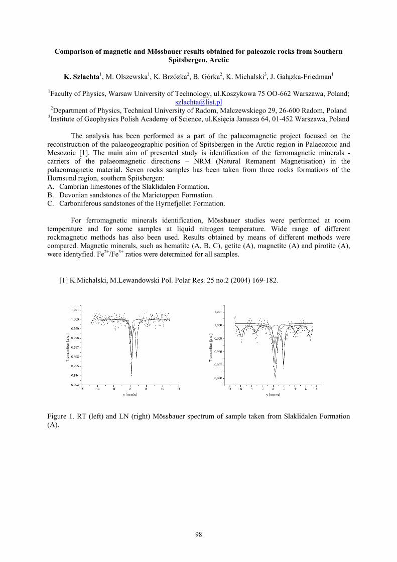
98
Comparison of magnetic and Mössbauer results obtained for paleozoic rocks from SouthernSpitsbergen, Arctic
K. Szlachta1, M. Olszewska1, K. Brzózka2, B. Górka2, K. Michalski3, J. Gałązka-Friedman1
1Faculty of Physics, Warsaw University of Technology, ul.Koszykowa 75 OO-662 Warszawa, Poland;[email protected]
2Department of Physics, Technical University of Radom, Malczewskiego 29, 26-600 Radom, Poland3Institute of Geophysics Polish Academy of Science, ul.Księcia Janusza 64, 01-452 Warszawa, Poland
The analysis has been performed as a part of the palaeomagnetic project focused on thereconstruction of the palaeogeographic position of Spitsbergen in the Arctic region in Palaeozoic andMesozoic [1]. The main aim of presented study is identification of the ferromagnetic minerals -carriers of the palaeomagnetic directions – NRM (Natural Remanent Magnetisation) in thepalaeomagnetic material. Seven rocks samples has been taken from three rocks formations of theHornsund region, southern Spitsbergen:A. Cambrian limestones of the Slaklidalen Formation.B. Devonian sandstones of the Marietoppen Formation.C. Carboniferous sandstones of the Hyrnefjellet Formation.
For ferromagnetic minerals identification, Mössbauer studies were performed at roomtemperature and for some samples at liquid nitrogen temperature. Wide range of differentrockmagnetic methods has also been used. Results obtained by means of different methods werecompared. Magnetic minerals, such as hematite (A, B, C), getite (A), magnetite (A) and pirotite (A),were identyfied. Fe2+/Fe3+ ratios were determined for all samples.
[1] K.Michalski, M.Lewandowski Pol. Polar Res. 25 no.2 (2004) 169-182.
Figure 1. RT (left) and LN (right) Mössbauer spectrum of sample taken from Slaklidalen Formation(A).

99
Notes

100
Mössbauer spectroscopy and X-ray diffraction studies on multiferroic Bi5Ti3FeO15 ceramics
J. Rymarczyk1, A. Hanc2, G. Dercz2, J. Ilczuk1
1 Department of Material Science, University of Silesia, Żeromskiego 3, 41-200 Sosnowiec, Poland;[email protected]
2 Institute of Materials Science, University of Silesia, Bankowa 12, 40-007 Katowice, Poland
Magnetoelectric multiferroics are materials which exhibit both magnetic order andferroelectricity in the same phase. Multiferroic materials with ferroelectric and magnetic properties arenow intensively studied. This class of materials would offers a large application potential for newdevices taking advantage of two coupled degrees of freedom based on local off-centered distortion andelectron spin. Over the last few years the field of multiferroic materials has seen tremendous boom,initiated in part by computational studies of first-principles explaining the basic physics underlyingtheir scarcity.
The present work involves the structure analysis and the determination of hyperfineparameters of multiferroic Bi5Ti3FeO15 ceramics, prepared by the refined pressed mixture of the Bi2O3,TiO2 and Fe2O3 simple oxides. This analysis was performed by X-ray diffraction (XRD), Mössbauerspectroscopy (MS) and scanning electron microscopy (SEM) techniques.The measurements of the 57Fe Mössbauer spectra were performed in transmission geometry by meansof a constant spectrometer of the standard design. The Mössbauer spectra of the sample in theparamagnetic state were calculated by means of a discrete analysis (a single line and a quadrupoledoublet). The magnetically spitted spectra were calculated by means of hyperfine field distributionusing the Hesse-Rubartsch method.

101
Notes

102
The Mössbauer spectroscopy and analytical investigations of the polycrystallinecompounds with general formula Zn1-xSnxCr2Se4 (x=0.1-0.3)
Izabela Jendrzejewska1, Aneta Hanc2, Paweł Zajdel3, Andrzej Kita1, Tomasz Goryczka2,Ewa Maciążek1, Janusz Mrzigod1
1 Institute of Chemistry, University of Silesia, 40-006 Katowice, Szkolna 9, e-mail: [email protected] Institute of Materials Science, University of Silesia, 40-007 Katowice, Bankowa 12
3 University College of London, Department of Chemistry, 20 Gordon Street, WC1H 0AJ London, UK
Chromium zinc selenide ZnCr2Se4 crystallizes in the normal spinel structure. The lattice parameter hasa value a0=10.498Å [1]. Polycrystalline quaternary compounds with spinel structure containing zinc,tin and selenium are not known in the literature. The polycrystalline compounds in the system Zn1-
xSnxCr2Se4 (where x = 0.1, 0.2, 0.3,) were obtained by ceramic technology. The samples were sinteredthree times at 1073K for 7 days. The sintered compounds were again crushed into a powder and usedfor a structural study. The structure and phase analysis were carried out using an X-ray diffractionpatterns registered at room temperature with the Philips X’Pert diffractometer. The lattice parametersincrease with increasing Sn concentration, because the ionic radius of Sn2+ (105 pm) is bigger thanionic radius of Zn2+ (89 pm) [2].Chemical compositions of the obtained samples were determined using ICP-AES method (InductivelyCoupled Plasma – Atomic Emission Spectrometry). Obtained results are presented in Table 1.
Table 1. Phase composition and lattice parameters of spinel phases in the Zn1-xSnxCr2Se4 system.
x Phase composition and chemical composition Lattice parameter [Å]
0.1 Spinel, Zn0.87Sn0.05Cr2..02Se4.0 10.5041
0.2 Spinel, Zn0.87Sn0.06Cr1..98Se4.0 10.5105
0.3 Spinel, Zn0.71Sn0.07Cr1.88Se4.0 10. 5158
The 119Sn Mössbauer spectra were recorded cryostat using a constant-acceleration spectrometer. Thespectra were measured at room temperature. Experimental spectrum shape was described with atransmission integral calculated according to the numerical Gauss-Legandre’s procedure. TheMössbauer spectra of the sample in the paramagnetic state were calculated by means of a discreteanalysis (a single line and a quadrupole doublet). The magnetically spitted spectra were calculated bymeans of hyperfine field distribution using the Hesse-Rubartsch method In order to take into accountthe asymmetry of lines, the linear relation between isomer shift (IS) and hyperfine magnetic field (H)was included in the fitting procedure. In the Mössbauer effect investigation the samples prepared aspellets were used with Li2Co3 as a binder, in which the investigated material was placed uniformly.The uniformity of samples was confirmed by microscopic methods.
AcknowledgmentThe present work is supported by The Ministry of Science and Higher Education projects No. N N204 289134 (2891/B/H03/2008/34).
1. P.K. Baltzer, H.W. Lehmann, M. Robbins, Phys. Rev. Lett., 15 (1965) 493.2. R.D.Shannon, Acta Cryst., A32 (1976) 751.

103
Notes

104
The Mössbauer and X-ray studies of the spinel ferrites Cu1-xFexCr2Se4 and CuCr2-yFeySe4prepared by the ceramic method
E. Maciążek 1 , A. Hanc 2 , R. Sitko 1 , B. Zawisza 1 , I. Jendrzejwska 1
1 Institute of Chemistry, University of Silesia, 40-006 Katowice, Szkolna 9, Poland;[email protected]
2 Institute of Materials Science, University of Silesia, 40-007 Katowice, Bankowa12, Poland
CuCr2Se4 crystallizes in spinel structure, space group F d-3m. It has a normal cationdistribution, the copper ions being located at the tetrahedral interstitial sites of the selenium sublattice,and chromium ions at the octahedral ones [1]. The attempts to substitute copper or chromium by ironwere done. The spinel ferrites were obtained by the ceramic method. As the starting materials, thestoichiometric amounts of metal selenides: CuSe, Cr2Se3 and respectively FeSe or Fe2Se3 were used.
Diffraction patterns were done for all samples using Siemens diffractometer D5000 andfiltered FeKα radiation. Lattice parameters were determined using the computer program HX61S [2].
The chemical composition of the obtained spinels were analyzed by wavelength-dispersiveX-ray spectrometry (WDXRF). The material was digested in HNO3/HCl, pipetted onto the Milliporefilter, dried and measured by WDXRF. The quantitative analysis was performed using severalmultielement standards.
The measurements of the 57Fe Mössbauer spectra were performed in transmission geometry bymeans of a constant spectrometer of the standard design. The 14.4 keV gamma rays were provided bya 50mCi source of 57Co/Rh. The spectra were measured at room temperature. Hyperfine parameters ofthe investigated spectra were related to the α-Fe standard. Experimental spectrum shape was describedwith a transmission integral calculated according to the numerical Gauss-Legandre’s procedure. TheMössbauer spectra of the sample in the paramagnetic state were calculated by means of a discreteanalysis (a single line and a quadrupole doublet). The magnetically spitted spectra were calculated bymeans of hyperfine field distribution using the Hesse-Rubartsch method . In order to take into accountthe asymmetry of lines, the linear relation between isomer shift (IS) and hyperfine magnetic field (H)was included in the fitting procedure.In the Mössbauer effect investigation the samples prepared as pellets were used with Li2Co3 as abinder, in which the investigated material was placed uniformly. The uniformity of samples wasconfirmed by microscopic methods.
AcknowledgmentThe X-ray measurements were carried out at the Department of Solid State Physics, Institute ofPhysics, University of Silesia.The present work is supported by The Ministry of Science and Higher Education projects No. NN204 289134 (2891/B/H03/2008/34) and N N204 1784 33 (1784/B/H03/2007/33)
1. I. Okońska-Kozłowska, J. Kopyczok, H.D. Lutz, Th. Stingl, Acta Cryst. C49(1993)1448.2. F. Ahmed, Instruction to cell dimension program HS60s, Loughborough University, unpublished.

105
Notes

106
The Mössbauer spectroscopy studies of ε to cementite carbides transformation duringisothermal holding from as-quenched state of high carbon tool steel
Piotr Bała1, Janusz Krawczyk1 and Aneta Hanc2
1 Faculty of Metals Engineering and Industrial Computer Science, University of Science and
Technology, Mickiewicza 30, 30-059 Krakow, Poland; [email protected]
2Institute of Materials Science, Silesian University, Bankowa 12, 40-007 Katowice, Poland
In this work, we employed the Conversion Electron Mössbauer in a study new high-carbon steel.The chemical composition high-carbon alloy steel present in Table 1.
Samples, taken from investigated steel, were austenitized at the temperature of 900ºC and hardened inoil. Austenitizing time was 20 minutes. After that seven of eight samples were tempered.Tempering consisted of holding the samples at 200°C by chosen times. All the times mentioned abovewere selected basing on IHT diagram presented in work [2].
The influence of the tempering time on the nucleation and solubility of ε carbides and cementitenucleation and growth, the mechanical stability of retained austenite and on the products of itstransformation, was determined.
The analysis of phase transformations during tempering in different time using CEMS methodmade possible to reveal fine details connected with it. It had been difficult to make during continuousheating from as-quenched state, presented in works [1,3].
1. P. Bała, J. Pacyna and J. Krawczyk, Archives of Metallurgy and Materials 52 (2007) p.113÷120.2. P. Bała, The kinetics of phase transformations during tempering and its influence on the
mechanical properties, PhD thesis, AGH University of Science and Technology, Cracow, Poland(2007). Promotor J. Pacyna (in Polish).
3. J. Krawczyk, P. Bała and J. Frąckowiak, Archives of Materials Science and Engineering 28(2007) p. 633÷636.
Acknowledgements
The work was partially supported by the State Committee of Scientific Research, Grant no. PB-
581/T/2006
mass %C Mn Si Cr Mo V Al
1.22 1.93 0.19 1.52 0.36 0.17 0.04

107
Notes

108
The Mössbauer spectroscopy studies of cementite precipitations during continuous heating fromas-quenched state of high carbon Cr-Mn-Mo steel
J. Krawczyk 1 , P. Bała 1 , A. Hanc 2
1 University of Science and Technology, Faculty of Metals Engineering and Industrial ComputerScience, PL-30-059 Krakow, Al. Mickiewicza 30, Poland;
[email protected] Institute of Materials Science, Silesian University, Bankowa 12, 40-007 Katowice, Poland
This work completes the knowledge concerning the kinetics of cementite precipitation duringtempering. Investigations were performed on 120MnCrMoV8-6-4-2 steel.The samples of investigated steel were austenitized at the temperature of 900ºC and hardened in oil.After that, four of five samples were tempered. Tempering consisted of heating the samples up tochosen temperatures at the heating rate of 0.05ºC/s and fast cooling after reaching desired temperature.All the temperatures mentioned above were selected basing on CHT diagram published in work [1].In this work we presents the results of investigations performed using Conversion Electron MossbauerSpectroscopy and interpretation concerning cementite nucleation and growth during tempering inrelation to previously conducted dilatometric, microscopic and mechanical investigations [2].The values of hyperfine magnetic field on 57Fe atomic nucleuses, determined for the third componentof Mössbauer spectrum as regards its intensity, indicate that these are the components coming fromferromagnetic carbides. Differences in hyperfine magnetic fields coming from Fe atoms existing in thestructure of carbides, measured on samples heated up to the temperatures of 80ºC and 210ºC, from57Fe atoms precipitated from carbides during heating up to the temperatures of 350ºC and 470ºC, allowto state that these are the carbides of different crystal structure. Data from work [3] as well as theresearch on the products of phases transformations during tempering of this steel, performed with theuse of other research methods [2] show that mentioned above spectrum components measured on thesamples heated up to 80ºC (ground surface) and to 210ºC (ground and polished surface) come from ε(Fe2,4C) carbides. However, component spectrum, obtained on the sample heated up to 350ºC and to470ºC come from 57Fe atoms present in the structure of cementite [2-4]. The influence of hardenedsteel heating temperature on cementite precipitation was determined. CEMS was applied not only formagnetic hyperfine filed studies, but also to analyze the values of quadruple splitting and isomer shift,what resulted in significant conclusions concerning the changes in cementite precipitationsmorphology, chemical composition and the level of stresses being present in this research.
3. J. Pacyna, P. Bała, T. Skrzypek, Proceedings of the 13th International Scientific ConferenceAMME’2005, Gliwice-Zakopane, Poland (2005) 509.
4. P. Bała, J. Pacyna and J. Krawczyk, Archives of Metallurgy and Materials, 52 (2007) 113.5. V.A. Shabashov, L.G. Korshunov, A.G. Mukoseev, V.V. Sagaradze, A.V. Makarov, V.P.
Pilyugin, S.I. Novikov, N.F. Vildanova, Materials Science and Engineering A346 (2003) 196.6. J. Krawczyk, P. Bała, J. Frąckowiak, XXXV School of Materials Science, Krakow-Krynica
Poland (2007) 24
Acknowledgements
The work was partially supported by the State Committee of Scientific Research, Grant no. PB-
581/T/2006

109
Notes

110
Charge and spin density perturbation on iron nuclei by non-magnetic impurities substituted onthe iron sites in Feα −
A. Błachowski 1 , K. Ruebenbauer 1 , J. Żukrowski 2 , J. Przewoźnik 2
1 Mössbauer Spectroscopy Division, Institute of Physics, Pedagogical UniversityPL-30-084 Kraków, ul. Podchorążych 2, Poland; [email protected]
2 Solid State Physics Department, Faculty of Physics and Applied Computer Science,AGH University of Science and Technology
PL-30-059 Kraków, Al. Mickiewicza 30, Poland
The pure Feα − crystallizes in the BCC structure with the ferromagnetic ordering of the rather welllocalized iron magnetic moments. This material is extremely soft from the magnetic point of view, andthe easy magnetic axes are aligned with the principal axes of the cubic chemical unit cell. The orbitaland dipolar hyperfine magnetic fields on the iron nucleus are absent. Hence, the hyperfine field is dueto the non-vanishing electron spin density on the iron nucleus. The latter density originates from theatomic core polarization and from the itinerant electron polarization. A dominant contributionoriginates from the s-like electrons of either core or conduction band. The system is not far fromsaturation in the vicinity of the room temperature. The charge (electron) density on the nucleus iscaused predominantly by the s-like electrons as well belonging either to the atomic core or to theconduction band. It is related directly to the Fe57 isomer shift ( keV14.41− transition) via thecalibration constant 13 s mma.u. )2(2910 −−=α . [1]. Relatively diluted non-magnetic impurity on theiron site generates perturbation in the vicinity of the Fermi level, the latter perturbation having short-range influence on the charge and spin density on the adjacent iron nuclei. Lattice relaxation aroundthe impurity has indirect effect on the above densities as well. Usually, no observable electric fieldgradient is generated by the impurity due to the effective screening by the itinerant electrons.
A systematic investigation has been performed by means of the room temperature Mössbauerspectroscopy on Fe57 ( keV14.41− transition) for the following impurities:
Au Ir, Os, Pd, Rh, Ru, Mo, Nb, Ga,X = [2]. Investigations performed versus impurity concentration(randomly distributed over iron sites) indicate additive effect for the charge density perturbation, andadditive in the algebraic sense effect for the corresponding spin density perturbation. Hence, the effectof impurity depends solely on the distance between impurity and the iron nucleus. On the other hand,ferromagnetism is preserved provided the concentration of impurities is kept reasonably low. TheCurie temperature is only weakly perturbed for impurities studied provided the concentration ofimpurities remains relatively low.
It has been found that impurities being further away than a third or in some cases as the secondneighbor do not contribute directly to the charge and spin perturbation. On the other hand, they haveusually some minor effect on the average charge and spin density. Generally, the perturbation to eithercharge or spin density has some oscillatory character versus distance from the impurity. This effect isparticularly strong for ruthenium [3] and iridium [4]. The phase of the charge oscillation is vastlydifferent from the phase of the spin oscillation in the majority of cases.
1. U.D. Wdowik, K. Ruebenbauer, Phys. Rev. B, 76 (2007) 155118; see also:www.elektron.ap.krakow.pl/iscal.pdf
2. For references see: www.elektron.ap.krakow.pl/papers.htm3. A. Błachowski, K. Ruebenbauer, J. Żukrowski, Phys. Rev. B, 73 (2006) 104423; see also:
www.elektron.ap.krakow.pl/feru.pdf4. A. Błachowski, K. Ruebenbauer, J. Żukrowski, J. Alloys Compd., (2007),
doi:10.1016/j.jallcom.2007.09.133; see also: www.elektron.ap.krakow.pl/feir.pdf

111
Notes

112
LIST OF PARTICIPANTS
1. Dr inż. Piotr BałaWydział Inżynierii Metali i Informatyki Przemysłowej, Akademia Górniczo-Hutnicza,Al. Mickiewicza 30, PL-30-059 Kraków; [email protected]. 106, 108
2. Dr inż. Artur BłachowskiZakład Spektroskopii Mössbauerowskiej, Instytut Fizyki, Akademia Pedagogiczna,ul. Podchorążych 2, PL-30-084 Kraków; [email protected]. 34, 36, 40, 42, 110
3. Dr Bogdan BogaczInstytut Fizyki, Uniwersytet Jagielloński,ul. Reymonta 4, PL-30-059 Kraków; [email protected]. 44
4. Dr inż. Krzysztof BryłaInstytut Techniki, Akademia Pedagogiczna,ul. Podchorążych 2, PL-30-084 Kraków; [email protected]. 34
5. Dr Krzysztof BrząkalikInstytut Nauki o Materiałach, Uniwersytet Śląski,ul. Bankowa 12, PL-40-007 Katowice; [email protected]. 48
6. Dr hab. Katarzyna BrzózkaKatedra Fizyki, Wydział Mechaniczny, Politechnika Radomska,ul. Krasickiego 54, PL-26-600 Radom; [email protected] ; [email protected]. 22, 60, 98
7. Prof. dr hab. Mieczysław BudzyńskiInstytut Fizyki, Uniwersytet Marii Curie-Skłodowskiejpl. Marii Curie-Skłodowskiej 5, PL-20-031 Lublin; [email protected]. 50, 70
8. Dr hab. Jan ChojcanInstytut Fizyki Doświadczalnej, Uniwersytet Wrocławski,pl. M. Borna 9, PL-50-204 Wrocław; [email protected]. 16
9. Dr inż. Jakub CieślakWydział Fizyki i Informatyki Stosowanej, Akademia Górniczo-Hutnicza,Al. Mickiewicza 30, PL-30-059 Kraków; [email protected]. 18, 20, 52, 92
10. Dr Benilde F.O. CostaDepartamento da Fisica, Universidade da Coimbra,3000-516 Coimbra, Portugal; [email protected]. 20, 52

113
11. Dr hab. Józef DeniszczykZakład Modelowania Materiałów, Instytut Nauki o Materiałach, Uniwersytet Śląski,ul. Bankowa 12, PL-40-007 Katowice; [email protected]. 94
12. Dr Grzegorz DerczInstytut Nauki o Materiałach, Wydział Informatyki i Nauki o Materiałach,Uniwersytet Śląski, ul. Bankowa 12, PL-40-007 Katowice; [email protected]. 26, 28, 80, 100
13. Prof. dr hab. Stanisław M. DubielWydział Fizyki i Informatyki Stosowanej, Akademia Górniczo-Hutnicza,Al. Mickiewicza 30, PL-30-059 Kraków; [email protected]. 18, 20, 52, 56, 92
14. Dr hab. Kazimierz DzilińskiInstytut Fizyki, Politechnika Częstochowska,Al. Armii Krajowej 19, PL-42-200 Częstochowa; [email protected]. 84, 88
15. Dr Piotr FornalInstytut Fizyki, Politechnika Krakowska,ul. Podchorążych 1, PL-30-084 Kraków; [email protected]. 62
16. Dr hab. Jolanta Gałązka-FriedmanWydział Fizyki, Politechnika Warszawska,ul. Koszykowa 75, PL-02-662 Warszawa; [email protected]. 86, 98
17. Mgr Michał GawrońskiKatedra Fizyki, Wydział Mechaniczny, Politechnika Radomska,ul. Krasickiego 54, PL-26-600 Radom; [email protected]. 22, 60
18. Dr inż. Agnieszka GrabiasInstytut Technologii Materiałów Elektronicznych,ul. Wólczyńska 133, PL-01-919 Warszawa; [email protected]. 74, 96
19. Dr Jacek GurgulInstytut Katalizy i Fizykochemii Powierzchni, Polska Akademia Nauk,ul. Niezapominajek 8, PL-30-239 Kraków; [email protected]. 46
20. Dr Aneta HancZakład Modelowania Materiałów, Instytut Nauki o Materiałach, Uniwersytet Śląski,ul. Bankowa 12, PL-40-007 Katowice; [email protected] ; [email protected]. 26, 28, 80, 94, 100, 102, 104, 106, 108
114
21. Dr hab. Elżbieta JartychInstytut Fizyki, Politechnika Lubelskaul. Nadbystrzycka 38, PL-20-618 Lublin; [email protected]. 72
22. Dr Izabela JendrzejewskaZakład Krystalografii, Instytut Chemii, Uniwersytet Śląski,ul. Bankowa 12, PL-40-007 Katowice; [email protected]. 102, 104
23. Mgr Tomasz KaczmarzykInstytut Fizyki, Politechnika Częstochowska,Al. Armii Krajowej 19, PL-42-200 Częstochowa; [email protected]. 84, 88
24. Dr hab. Jerzy KansyZakład Modelowania Materiałów, Instytut Nauki o Materiałach, Uniwersytet Śląski,ul. Bankowa 12, PL-40-007 Katowice; [email protected]. 26, 28
25. Mgr Mariola Kądziołka-GawełInstytut Fizyki, Uniwersytet Śląski,ul. Uniwersytecka 4, PL-40-007 Katowice; [email protected]. 30
26. Dr inż. Janusz KrawczykWydział Inżynierii Metali i Informatyki Przemysłowej, Akademia Górniczo-Hutnicza,Al. Mickiewicza 30, PL-30-059 Kraków; [email protected]. 106, 108
27. Prof. dr hab. Karol KropKatedra Fizyki, Politechnika Rzeszowska,Al. Powstańców Warszawy 6, PL-35-959 Rzeszow; [email protected]. 24
28. Prof. dr hab. Kazimierz ŁątkaInstytut Fizyki, Uniwersytet Jagielloński,ul. Reymonta 4, PL-30-059 Kraków; [email protected]. 38, 46
29. Dr Ewa MaciążekZakład Krystalografii, Instytut Chemii, Uniwersytet Śląski,ul. Bankowa 12, PL-40-007 Katowice; [email protected]. 102, 104
30. Dr Dariusz MalczewskiWydział Nauk o Ziemi, Uniwersytet Śląski,ul. Będzińska 60, PL-41-200 Sosnowiec; [email protected]. 96
115
31. Mgr Mariusz MazurekInstytut Fizyki, Politechnika Lubelska,ul. Nadbystrzycka 38, PL-20-618 Lublin; [email protected]. 72
32. Dr Viktor MitsiukJoint Institute of Solid State and Semiconductor Physics, National Academy of Sciencesof Belarus, 220072, Minsk, P. Brovki Str., 19, Belarus; [email protected]. 54
33. Dr hab. Marek MonetaKatedra Fizyki Ciała Stałego, Uniwersytet Łódzki,ul. Pomorska 149, PL-90-236 Łódź; [email protected]
34. Dr inż. Dariusz OleszakWydział Inżynierii Materiałowej, Politechnika Warszawska,ul. Wołoska 141, PL-02-507 Warszawa; [email protected]. 26, 72, 74
35. Dr Jacek OlszewskiInstytut Fizyki, Politechnika Częstochowska,Al. Armii Krajowej 19, PL-42-200 Częstochowa; [email protected]. 32, 58
36. Mgr Wojciech OlszewskiWydział Fizyki, Uniwersytet w Białymstoku,ul. Lipowa 41, PL-15-424 Białystok; [email protected]. 76
37. Dr Andrzej OstraszInstytut Fizyki Doświadczalnej, Uniwersytet Wrocławski,pl. Maxa Borna 9, PL-50-204 Wrocław; [email protected]. 16
38. Dr Kamila PerdutaSchool of Environment and Technology, University of Brighton,Lewes Road, Brighton, BN2 4GJ United Kingdom; [email protected]. 58
39. Prof. dr hab. Antoni PędziwiatrInstytut Fizyki, Uniwersytet Jagielloński,ul. Reymonta 4, PL-30-059 Kraków; [email protected]. 44
40. Mgr Tomasz PikulaInstytut Fizyki, Politechnika Lubelska,ul. Nadbystrzycka 38, PL-20-618 Lublin; [email protected]. 72

116
41. Dr Janusz PrzewoźnikWydział Fizyki i Informatyki Stosowanej, Akademia Górniczo-Hutnicza,Al. Mickiewicza 30, PL-30-059 Kraków; [email protected]. 24, 40, 42, 110
42. Prof. dr hab. Krzysztof RuebenbauerZakład Spektroskopii Mössbauerowskiej, Instytut Fizyki, Akademia Pedagogiczna,ul. Podchorążych 2, PL-30-084 Kraków; [email protected]. 34, 36, 40, 42, 90, 110
43. Mgr Jolanta RymarczykKatedra Materiałoznawstwa, Wydział Informatyki i Nauki o Materiałach, UniwersytetŚląski, ul. Żeromskiego 3, PL-41-200 Sosnowiec; [email protected]. 80, 100
44. Dr Jan SarzyńskiUniwersytet Marii Curie-Skłodowskiejpl. Marii Curie-Skłodowskiej 5, PL-20-031 Lublin; [email protected]
45. Dr Dariusz SatułaWydział Fizyki, Uniwersytet w Białymstoku,ul. Lipowa 41, PL-15-424 Białystok; [email protected]. 76, 78
46. Prof. Bogdan SepiolDynamics of Condensed Systems, Department of Physics, University of Vienna,Strudlhofgasse 4, A-1090 Wien, Austria; [email protected]. 14
47. Prof. Zbigniew StadnikDepartment of Physics, University of Ottawa,150 Louis Pasteur, Ottawa, Ontario K1N 6N5, Canada; [email protected]. 66
48. Prof. dr hab. Jan StanekInstytut Fizyki, Uniwersytet Jagielloński,ul. Reymonta 4, PL-30-059 Kraków; [email protected]. 62, 82
49. Dr inż. Agata StochInstytut Technologii Elektronowej Oddział w Krakowie,ul. Zabłocie 39, PL-30-701 Kraków; [email protected]. 64
50. Dr Zbigniew SurowiecInstytut Fizyki, Uniwersytet Marii Curie-Skłodowskiej,pl. Marii Curie-Skłodowskiej 5, PL-20-031 Lublin; [email protected]. 50, 70

117
51. Mgr inż. Karol SzlachtaWydział Fizyki, Politechnika Warszawska,ul. Koszykowa 75, PL-02-662 Warszawa; [email protected]. 86, 98
52. Dr Tadeusz SzumiataKatedra Fizyki, Wydział Mechaniczny, Politechnika Radomska,ul. Krasickiego 54, PL-26-600 Radom; [email protected] ; [email protected]. 22, 60
53. Dr hab. Krzysztof SzymańskiWydział Fizyki, Uniwersytet w Białymstoku,ul. Lipowa 41, PL-15-424 Białystok; [email protected]. 76, 78
54. Dr Tomasz ŚlęzakWydział Fizyki i Informatyki Stosowanej, Akademia Górniczo-Hutnicza,Al. Mickiewicza 30, PL-30-059 Kraków; [email protected]. 14, 68
55. Dr inż. Urszula D. WdowikZakład Zastosowań Informatyki, Instytut Techniki, Akademia Pedagogiczna,ul. Podchorążych 2, PL-30-084 Kraków; [email protected]. 90
56. Dr Marek WiertelInstytut Fizyki, Uniwersytet Marii Curie-Skłodowskiej,pl. Marii Curie-Skłodowskiej 5, PL-20-031 Lublin; [email protected]. 50, 70
57. Dr hab. Józef ZbroszczykInstytut Fizyki, Politechnika Częstochowska,Al. Armii Krajowej 19, PL-42-200 Częstochowa; [email protected]. 32
58. Dr Jan ŻukrowskiWydział Fizyki i Informatyki Stosowanej, Akademia Górniczo-Hutnicza,Al. Mickiewicza 30, PL-30-059 Kraków; [email protected]. 18, 24, 36, 40, 42, 110