Embed Size (px)
Citation preview
Reviewed by:
Date:
Page 1 of 21
T:\FINALSOP\FINAL SOP PDF\G. GAS CHROMATOGRAPHY MASS SPECTROMETRY\B.AMPHET QUANT SPE VER 03-08-2013.DOCX
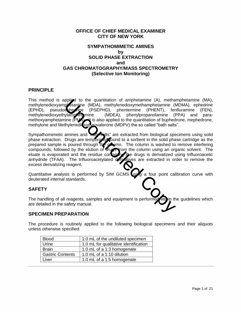
OFFICE OF CHIEF MEDICAL EXAMINER CITY OF NEW YORK
SYMPATHOMIMETIC AMINES by
SOLID PHASE EXTRACTION and
GAS CHROMATOGRAPHY/MASS SPECTROMETRY (Selective Ion Monitoring)
PRINCIPLE
This method is applied to the quantitation of amphetamine (A), methamphetamine (MA), methylenedioxyamphetamine (MDA), methylenedioxymethamphetamine (MDMA), ephedrine (EPHD), pseudoephedrine (PSEPHD), phentermine (PHENT), fenfluramine (FEN), methylenedioxyethylamphetamine (MDEA), phenylpropanolamine (PPA) and para-methoxyamphetamine (PMA). It is also applied to the quantitation of buphedrone, mephedrone, methylone and Methylenedioxypyrovalerone (MDPV) the so called “bath salts”.
Sympathomimetic amines and “bath salts” are extracted from biological specimens using solid phase extraction. Drugs are temporarily bound to a sorbent in the solid phase cartridge as the prepared sample is poured through the column. The column is washed to remove interfering compounds, followed by the elution of drugs from the column using an organic solvent. The eluate is evaporated and the residue containing the drugs is derivatized using trifluoroacetic anhydride (TFAA). The trifluoroacetylated derivatives are extracted in order to remove the excess derivatizing reagent.
Quantitative analysis is performed by SIM GCMS using a four point calibration curve with deuterated internal standards.
SAFETY
The handling of all reagents, samples and equipment is performed within the guidelines which are detailed in the safety manual.
SPECIMEN PREPARATION
The procedure is routinely applied to the following biological specimens and their aliquots unless otherwise specified:
Blood 1.0 mL of the undiluted specimen
Urine 1.0 mL for qualitative identification
Brain 1.0 mL of a 1:3 homogenate
Gastric Contents 1.0 mL of a 1:10 dilution
Liver 1.0 mL of a 1:5 homogenate
Uncontrolled Copy
Reviewed by:
Date:
Page 2 of 21
T:\FINALSOP\FINAL SOP PDF\G. GAS CHROMATOGRAPHY MASS SPECTROMETRY\B.AMPHET QUANT SPE VER 03-08-2013.DOCX
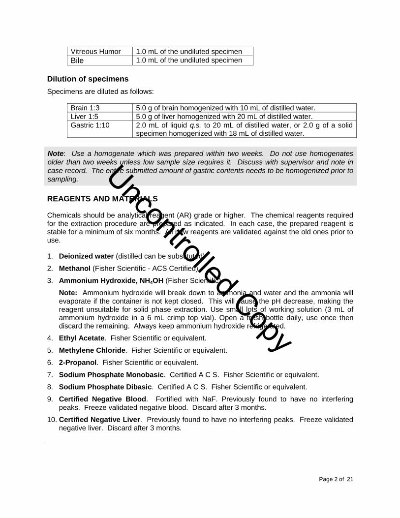
Vitreous Humor 1.0 mL of the undiluted specimen
Bile 1.0 mL of the undiluted specimen
Dilution of specimens
Specimens are diluted as follows:
Brain 1:3 5.0 g of brain homogenized with 10 mL of distilled water.
Liver 1:5 5.0 g of liver homogenized with 20 mL of distilled water.
Gastric 1:10 2.0 mL of liquid q.s. to 20 mL of distilled water, or 2.0 g of a solid specimen homogenized with 18 mL of distilled water.
Note: Use a homogenate which was prepared within two weeks. Do not use homogenates
older than two weeks unless low sample size requires it. Discuss with supervisor and note in
case record. The entire submitted amount of gastric contents needs to be homogenized prior to
sampling.
REAGENTS AND MATERIALS
Chemicals should be analytical reagent (AR) grade or higher. The chemical reagents required for the extraction procedure are prepared as indicated. In each case, the prepared reagent is stable for a minimum of six months. All new reagents are validated against the old ones prior to use.
1. Deionized water (distilled can be substituted)
2. Methanol (Fisher Scientific - ACS Certified)
3. Ammonium Hydroxide, NH4OH (Fisher Scientific)
Note: Ammonium hydroxide will break down to ammonia and water and the ammonia will evaporate if the container is not kept closed. This will cause the pH decrease, making the reagent unsuitable for solid phase extraction. Use small lots of working solution (3 mL of ammonium hydroxide in a 6 mL crimp top vial). Open a fresh bottle daily, use once then discard the remaining. Always keep ammonium hydroxide refrigerated.
4. Ethyl Acetate. Fisher Scientific or equivalent.
5. Methylene Chloride. Fisher Scientific or equivalent.
6. 2-Propanol. Fisher Scientific or equivalent.
7. Sodium Phosphate Monobasic. Certified A C S. Fisher Scientific or equivalent.
8. Sodium Phosphate Dibasic. Certified A C S. Fisher Scientific or equivalent.
9. Certified Negative Blood. Fortified with NaF. Previously found to have no interfering peaks. Freeze validated negative blood. Discard after 3 months.
10. Certified Negative Liver. Previously found to have no interfering peaks. Freeze validated negative liver. Discard after 3 months.
Uncontrolled Copy
Reviewed by:
Date:
Page 3 of 21
T:\FINALSOP\FINAL SOP PDF\G. GAS CHROMATOGRAPHY MASS SPECTROMETRY\B.AMPHET QUANT SPE VER 03-08-2013.DOCX
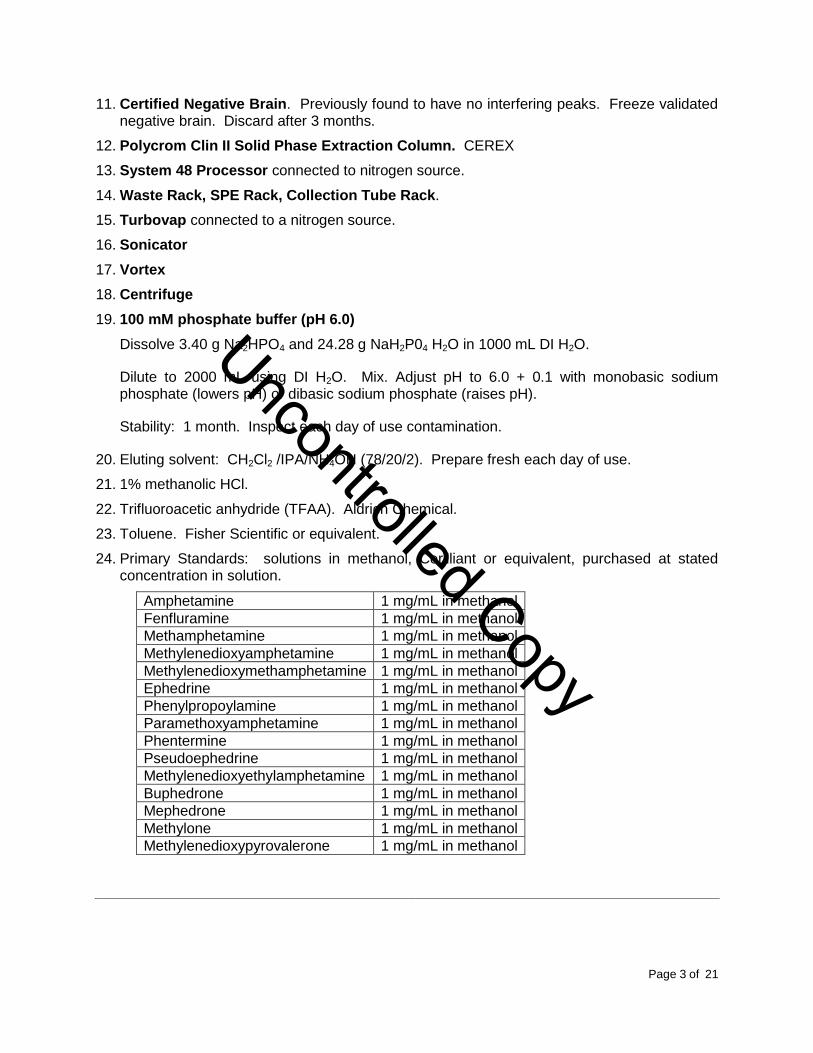
11. Certified Negative Brain. Previously found to have no interfering peaks. Freeze validated negative brain. Discard after 3 months.
12. Polycrom Clin II Solid Phase Extraction Column. CEREX
13. System 48 Processor connected to nitrogen source.
14. Waste Rack, SPE Rack, Collection Tube Rack.
15. Turbovap connected to a nitrogen source.
16. Sonicator
17. Vortex
18. Centrifuge
19. 100 mM phosphate buffer (pH 6.0)
Dissolve 3.40 g Na2HPO4 and 24.28 g NaH2P04 H2O in 1000 mL DI H2O.
Dilute to 2000 mL using DI H2O. Mix. Adjust pH to 6.0 + 0.1 with monobasic sodium phosphate (lowers pH) or dibasic sodium phosphate (raises pH).
Stability: 1 month. Inspect each day of use contamination.
20. Eluting solvent: CH2Cl2 /IPA/NH4OH (78/20/2). Prepare fresh each day of use.
21. 1% methanolic HCl.
22. Trifluoroacetic anhydride (TFAA). Aldrich Chemical.
23. Toluene. Fisher Scientific or equivalent.
24. Primary Standards: solutions in methanol, Cerilliant or equivalent, purchased at stated concentration in solution.
Amphetamine 1 mg/mL in methanol
Fenfluramine 1 mg/mL in methanol
Methamphetamine 1 mg/mL in methanol
Methylenedioxyamphetamine 1 mg/mL in methanol
Methylenedioxymethamphetamine 1 mg/mL in methanol
Ephedrine 1 mg/mL in methanol
Phenylpropoylamine 1 mg/mL in methanol
Paramethoxyamphetamine 1 mg/mL in methanol
Phentermine 1 mg/mL in methanol
Pseudoephedrine 1 mg/mL in methanol
Methylenedioxyethylamphetamine 1 mg/mL in methanol
Buphedrone 1 mg/mL in methanol
Mephedrone 1 mg/mL in methanol
Methylone 1 mg/mL in methanol
Methylenedioxypyrovalerone 1 mg/mL in methanol
Uncontrolled Copy
Reviewed by:
Date:
Page 4 of 21
T:\FINALSOP\FINAL SOP PDF\G. GAS CHROMATOGRAPHY MASS SPECTROMETRY\B.AMPHET QUANT SPE VER 03-08-2013.DOCX
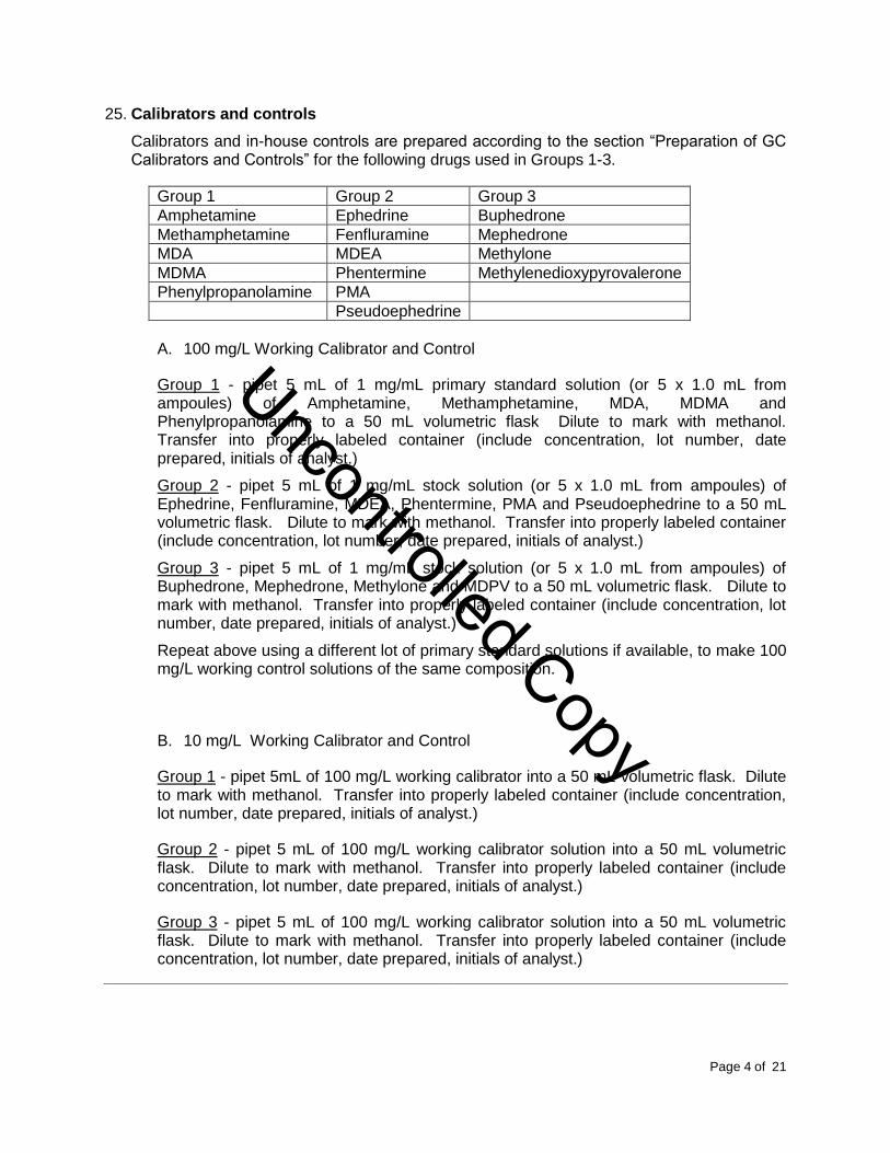
25. Calibrators and controls
Calibrators and in-house controls are prepared according to the section “Preparation of GC Calibrators and Controls” for the following drugs used in Groups 1-3.
Group 1 Group 2 Group 3
Amphetamine Ephedrine Buphedrone
Methamphetamine Fenfluramine Mephedrone
MDA MDEA Methylone
MDMA Phentermine Methylenedioxypyrovalerone
Phenylpropanolamine PMA
Pseudoephedrine
A. 100 mg/L Working Calibrator and Control
Group 1 - pipet 5 mL of 1 mg/mL primary standard solution (or 5 x 1.0 mL from ampoules) of Amphetamine, Methamphetamine, MDA, MDMA and Phenylpropanolamine to a 50 mL volumetric flask Dilute to mark with methanol. Transfer into properly labeled container (include concentration, lot number, date prepared, initials of analyst.)
Group 2 - pipet 5 mL of 1 mg/mL stock solution (or 5 x 1.0 mL from ampoules) of Ephedrine, Fenfluramine, MDEA, Phentermine, PMA and Pseudoephedrine to a 50 mL volumetric flask. Dilute to mark with methanol. Transfer into properly labeled container (include concentration, lot number, date prepared, initials of analyst.)
Group 3 - pipet 5 mL of 1 mg/mL stock solution (or 5 x 1.0 mL from ampoules) of Buphedrone, Mephedrone, Methylone and MDPV to a 50 mL volumetric flask. Dilute to mark with methanol. Transfer into properly labeled container (include concentration, lot number, date prepared, initials of analyst.)
Repeat above using a different lot of primary standard solutions if available, to make 100 mg/L working control solutions of the same composition.
B. 10 mg/L Working Calibrator and Control
Group 1 - pipet 5mL of 100 mg/L working calibrator into a 50 mL volumetric flask. Dilute to mark with methanol. Transfer into properly labeled container (include concentration, lot number, date prepared, initials of analyst.)
Group 2 - pipet 5 mL of 100 mg/L working calibrator solution into a 50 mL volumetric flask. Dilute to mark with methanol. Transfer into properly labeled container (include concentration, lot number, date prepared, initials of analyst.)
Group 3 - pipet 5 mL of 100 mg/L working calibrator solution into a 50 mL volumetric flask. Dilute to mark with methanol. Transfer into properly labeled container (include concentration, lot number, date prepared, initials of analyst.)
Uncontrolled Copy
Reviewed by:
Date:
Page 5 of 21
T:\FINALSOP\FINAL SOP PDF\G. GAS CHROMATOGRAPHY MASS SPECTROMETRY\B.AMPHET QUANT SPE VER 03-08-2013.DOCX
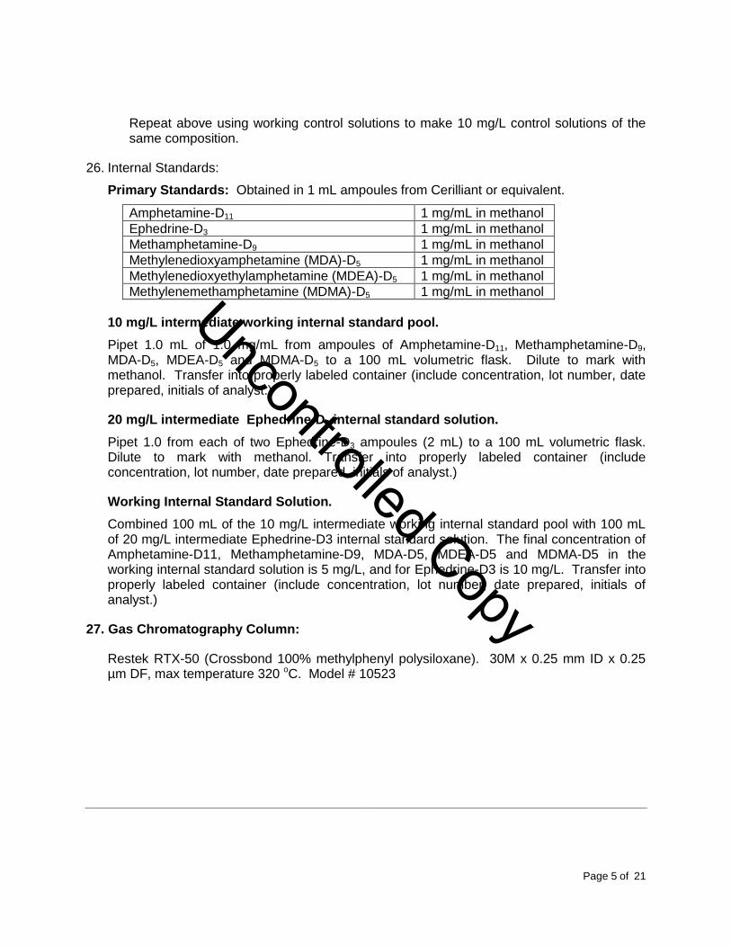
Repeat above using working control solutions to make 10 mg/L control solutions of the same composition.
26. Internal Standards:
Primary Standards: Obtained in 1 mL ampoules from Cerilliant or equivalent.
Amphetamine-D11 1 mg/mL in methanol
Ephedrine-D3 1 mg/mL in methanol
Methamphetamine-D9 1 mg/mL in methanol
Methylenedioxyamphetamine (MDA)-D5 1 mg/mL in methanol
Methylenedioxyethylamphetamine (MDEA)-D5 1 mg/mL in methanol
Methylenemethamphetamine (MDMA)-D5 1 mg/mL in methanol
10 mg/L intermediate working internal standard pool.
Pipet 1.0 mL of 1.0 mg/mL from ampoules of Amphetamine-D11, Methamphetamine-D9, MDA-D5, MDEA-D5 and MDMA-D5 to a 100 mL volumetric flask. Dilute to mark with methanol. Transfer into properly labeled container (include concentration, lot number, date prepared, initials of analyst.)
20 mg/L intermediate Ephedrine-D3 internal standard solution.
Pipet 1.0 from each of two Ephedrine-D3 ampoules (2 mL) to a 100 mL volumetric flask. Dilute to mark with methanol. Transfer into properly labeled container (include concentration, lot number, date prepared, initials of analyst.)
Working Internal Standard Solution.
Combined 100 mL of the 10 mg/L intermediate working internal standard pool with 100 mL of 20 mg/L intermediate Ephedrine-D3 internal standard solution. The final concentration of Amphetamine-D11, Methamphetamine-D9, MDA-D5, MDEA-D5 and MDMA-D5 in the working internal standard solution is 5 mg/L, and for Ephedrine-D3 is 10 mg/L. Transfer into properly labeled container (include concentration, lot number, date prepared, initials of analyst.)
27. Gas Chromatography Column:
Restek RTX-50 (Crossbond 100% methylphenyl polysiloxane). 30M x 0.25 mm ID x 0.25 µm DF, max temperature 320 oC. Model # 10523
Uncontrolled Copy

Reviewed by:
Date:
Page 6 of 21
T:\FINALSOP\FINAL SOP PDF\G. GAS CHROMATOGRAPHY MASS SPECTROMETRY\B.AMPHET QUANT SPE VER 03-08-2013.DOCX
EXTRACTION PROCEDURE
1. Obtain a list of amphetamine cases to be analyzed. (Refer to “Procedure for Work list Printing.”) Review with supervisor for any updates of rush cases or other special instructions.
2. Find and collect all samples on the list and place in an empty rack.
3. Obtain enough 16 x 125 mm disposable culture tubes for calibrators, controls, and all requested cases. Label the tubes appropriately. Tubes should bear the entire toxicology number (e.g., YY-1234, not 1234), the specimen type and any dilution.
4. Pipet 1 mL of validated negative matrix or sample into16 x 125 mm test tube labeled as to the contents.
5. Add appropriate amounts of working calibrator solutions for Group 1, 2 (and group 3 if needed) to negative matrix tubes as indicated below.
Four calibrators of Group 1 amphetamines, four calibrators of Group 2 amphetamines and, four calibrators of Group 3 (bath salts, if needed) and matrix-matched blank(s) should be run with each batch of samples. Positive calibrators are prepared at following concentrations:
0.1 mg/L - add 10 μL of 10 mg/L working calibrator solution 0.5 mg/L - add 50 μL of 10 mg/L working calibrator solution 1.0 mg/L - add 10 μL of 100 mg/L working calibrator solution 1.5 mg/L - add 15 μL of 100 mg/L working calibrator solution
Vortex 15 seconds to mix.
Note: Deionized water is used as the negative matrix for urine and gastric specimens.
6. Spike the control tubes for each matrix for both Group 1 and Group 2 (and Group 3, if required):
0.5 mg/L - add 50 μL of 10 mg/L working control solution 1.0 mg/L - add 10 μL of 100 mg/L working control solution
Vortex 15 seconds to mix.
Note: These samples should be placed randomly in the batch.
7. Add 50 μL of working internal standard pool (5 mg/L for all amphetamines, except for ephedrine at 10 mg/L) to all test tubes. The concentration of the internal standard in each sample is 0.25 mg/L except for Ephedrine-D3, which is 0.5 mg/L.
8. Add 2 mL of 100 mM phosphate buffer (pH 6.0), mix & vortex 15 seconds to mix.
9. Sonicate sample for 20 minutes using an ultrasonic bath.
10. Centrifuge for 10 minutes at ≈ 3000 rpm.
Uncontrolled Copy
Reviewed by:
Date:
Page 7 of 21
T:\FINALSOP\FINAL SOP PDF\G. GAS CHROMATOGRAPHY MASS SPECTROMETRY\B.AMPHET QUANT SPE VER 03-08-2013.DOCX
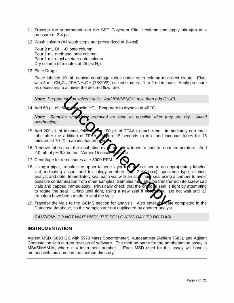
11. Transfer the supernatant into the SPE Polycrom Clin II column and apply nitrogen at a pressure of 2-4 psi.
12. Wash column (All wash steps are pressurized at 2-4psi).
Pour 2 mL DI H2O onto column Pour 1 mL methanol onto column Pour 1 mL ethyl acetate onto column Dry column (2 minutes at 25 psi N2)
13. Elute Drugs
Place labeled 10 mL conical centrifuge tubes under each column to collect eluate. Elute with 3 mL CH2Cl2 /IPA/NH4OH (78/20/2); collect eluate at 1 to 2 mL/minute. Apply pressure as necessary to achieve the desired flow rate.
Note: Prepare elution solvent daily. Add IPA/NH4OH, mix, then add CH2Cl2
14. Add 50 μL of 1% methanolic HCl. Evaporate to dryness at 40 oC.
Note: Samples should be removed as soon as possible after they are dry. Avoid
overheating.
15. Add 200 μL of toluene, followed by 100 μL of TFAA to each tube. Immediately cap each tube after the addition of TFAA. Vortex 15 seconds to mix. and incubate tubes for 15 minutes at 70 oC in an incubation oven.
16. Remove tubes from the incubation oven and allow tubes to cool to room temperature. Add 2.0 mL of pH 9.8 buffer. Vortex 15 seconds to mix.
17. Centrifuge for ten minutes at ≈ 3000 RPM.
18. Using a pipet, transfer the upper toluene layer to a glass insert in an appropriately labeled vial, indicating aliquot and toxicology numbers (ex: 2-YY-xxxx), specimen type, dilution, analyst and date. Immediately seal each vial with an aluminum seal using a crimper to avoid possible contamination from other samples. Samples may also be transferred into screw cap vials and capped immediately. Physically check that the crimped seal is tight by attempting to rotate the seal. Crimp until tight, using a new seal if necessary. Do not wait until all transfers have been made to seal the vials.
19. Transfer the vials to the GCMS section for analysis. Also enter the date completed in the Dataease database, so the samples are not duplicated by another analyst.
CAUTION: DO NOT WAIT UNTIL THE FOLLOWING DAY TO DO THIS!
INSTRUMENTATION
Agilent MSD (6890 GC with 5973 Mass Spectrometer), Autosampler (Agilent 7683), and Agilent Chemstation with current revision of software. The method name for the amphetamine assay is MSnSIMAM.M, where n = instrument number. Each MSD used for this assay will have a method with this name in the method directory.
Uncontrolled Copy
Reviewed by:
Date:
Page 8 of 21
T:\FINALSOP\FINAL SOP PDF\G. GAS CHROMATOGRAPHY MASS SPECTROMETRY\B.AMPHET QUANT SPE VER 03-08-2013.DOCX
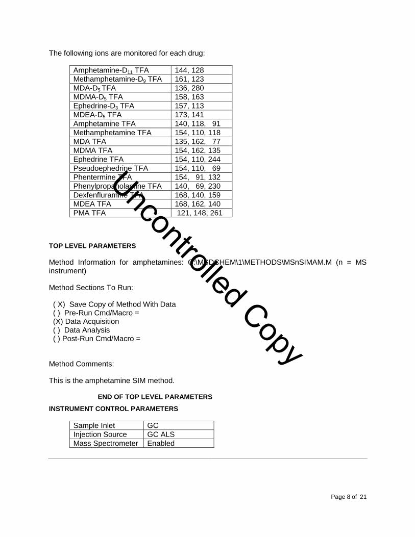
The following ions are monitored for each drug:
Amphetamine-D11 TFA 144, 128
Methamphetamine-D9 TFA 161, 123
MDA-D5 TFA 136, 280
MDMA-D5 TFA 158, 163
Ephedrine-D3 TFA 157, 113
MDEA-D5 TFA 173, 141
Amphetamine TFA 140, 118, 91
Methamphetamine TFA 154, 110, 118
MDA TFA 135, 162, 77
MDMA TFA 154, 162, 135
Ephedrine TFA 154, 110, 244
Pseudoephedrine TFA 154, 110, 69
Phentermine TFA 154, 91, 132
Phenylpropanolamine TFA 140, 69, 230
Dexfenfluramine TFA 168, 140, 159
MDEA TFA 168, 162, 140
PMA TFA 121, 148, 261
TOP LEVEL PARAMETERS
Method Information for amphetamines: C:\MSDCHEM\1\METHODS\MSnSIMAM.M (n = MS instrument)
Method Sections To Run:
( X) Save Copy of Method With Data ( ) Pre-Run Cmd/Macro = (X) Data Acquisition ( ) Data Analysis ( ) Post-Run Cmd/Macro =
Method Comments:
This is the amphetamine SIM method.
END OF TOP LEVEL PARAMETERS
INSTRUMENT CONTROL PARAMETERS
Sample Inlet GC
Injection Source GC ALS
Mass Spectrometer Enabled
Uncontrolled Copy
Reviewed by:
Date:
Page 9 of 21
T:\FINALSOP\FINAL SOP PDF\G. GAS CHROMATOGRAPHY MASS SPECTROMETRY\B.AMPHET QUANT SPE VER 03-08-2013.DOCX
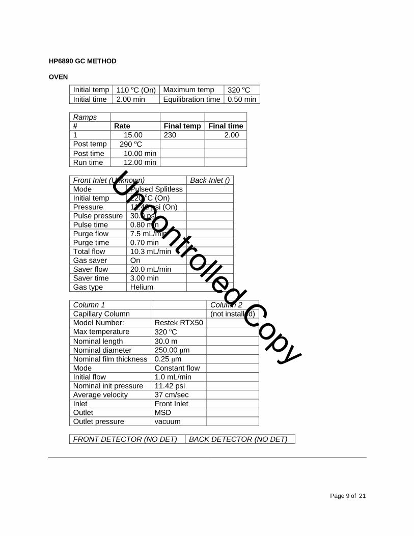
HP6890 GC METHOD
OVEN
Initial temp 110 ºC (On) Maximum temp 320 ºC
Initial time 2.00 min Equilibration time 0.50 min
Ramps
# Rate Final temp Final time
1 15.00 230 2.00
Post temp 290 ºC
Post time 10.00 min
Run time 12.00 min
Front Inlet (Unknown) Back Inlet ()
Mode Pulsed Splitless
Initial temp 220 oC (On)
Pressure 11.40 psi (On)
Pulse pressure 30.0 psi
Pulse time 0.80 min
Purge flow 7.5 mL/min
Purge time 0.70 min
Total flow 10.3 mL/min
Gas saver On
Saver flow 20.0 mL/min
Saver time 3.00 min
Gas type Helium
Column 1 Column 2
Capillary Column (not installed)
Model Number: Restek RTX50
Max temperature 320 ºC
Nominal length 30.0 m
Nominal diameter 250.00 μm
Nominal film thickness 0.25 μm
Mode Constant flow
Initial flow 1.0 mL/min
Nominal init pressure 11.42 psi
Average velocity 37 cm/sec
Inlet Front Inlet
Outlet MSD
Outlet pressure vacuum
FRONT DETECTOR (NO DET) BACK DETECTOR (NO DET)
Uncontrolled Copy
Reviewed by:
Date:
Page 10 of 21
T:\FINALSOP\FINAL SOP PDF\G. GAS CHROMATOGRAPHY MASS SPECTROMETRY\B.AMPHET QUANT SPE VER 03-08-2013.DOCX
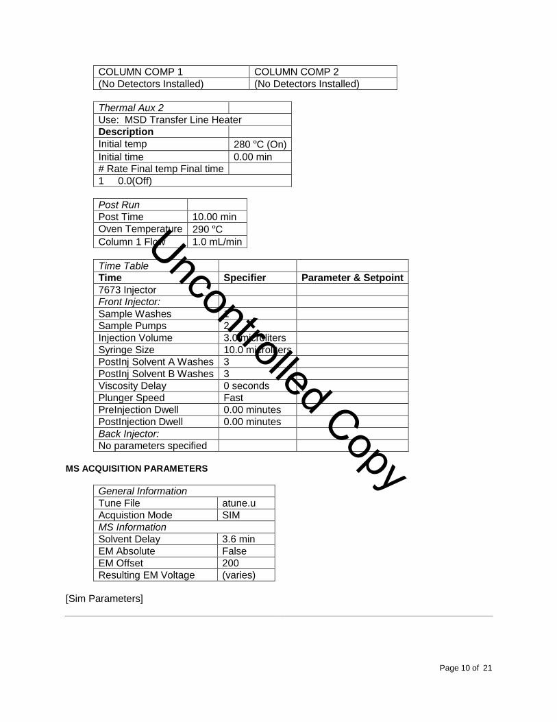
COLUMN COMP 1 COLUMN COMP 2
(No Detectors Installed) (No Detectors Installed)
Thermal Aux 2
Use: MSD Transfer Line Heater
Description
Initial temp 280 ºC (On)
Initial time 0.00 min
# Rate Final temp Final time
1 0.0(Off)
Post Run
Post Time 10.00 min
Oven Temperature 290 ºC
Column 1 Flow 1.0 mL/min
Time Table
Time Specifier Parameter & Setpoint
7673 Injector
Front Injector:
Sample Washes 1
Sample Pumps 2
Injection Volume 3.0 microliters
Syringe Size 10.0 microliters
PostInj Solvent A Washes 3
PostInj Solvent B Washes 3
Viscosity Delay 0 seconds
Plunger Speed Fast
PreInjection Dwell 0.00 minutes
PostInjection Dwell 0.00 minutes
Back Injector:
No parameters specified
MS ACQUISITION PARAMETERS
General Information
Tune File atune.u
Acquistion Mode SIM
MS Information
Solvent Delay 3.6 min
EM Absolute False
EM Offset 200
Resulting EM Voltage (varies)
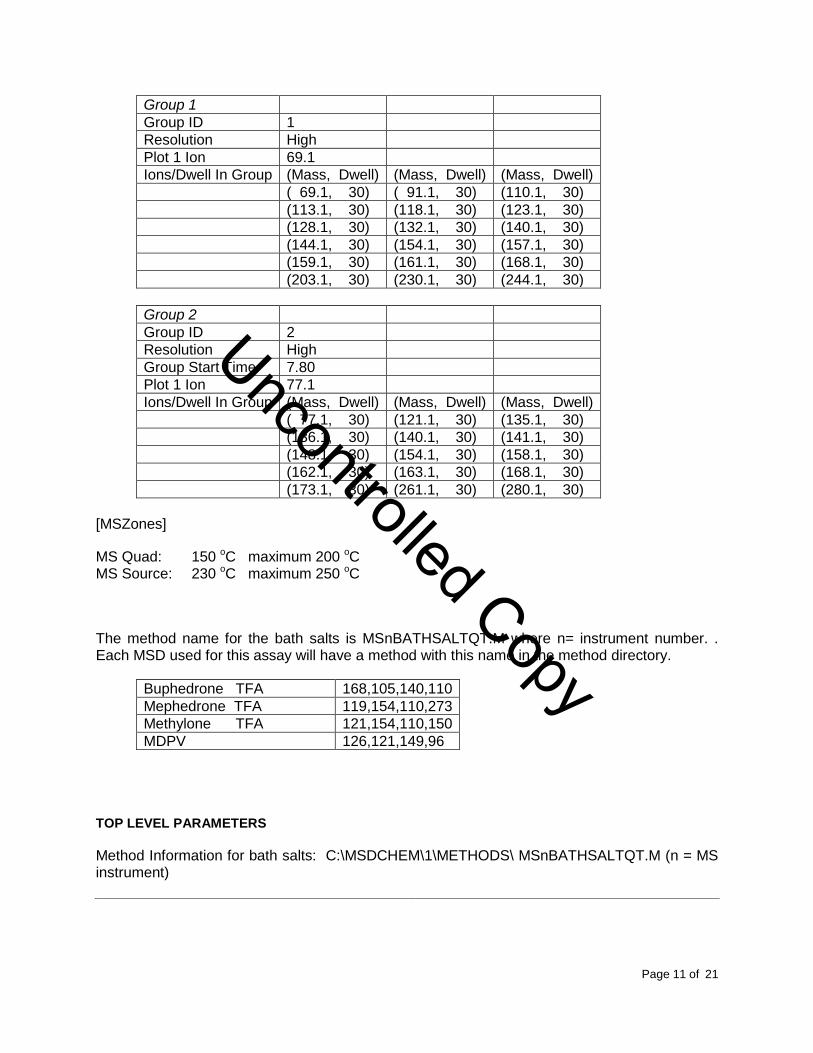
[Sim Parameters]
Uncontrolled Copy
Reviewed by:
Date:
Page 11 of 21
T:\FINALSOP\FINAL SOP PDF\G. GAS CHROMATOGRAPHY MASS SPECTROMETRY\B.AMPHET QUANT SPE VER 03-08-2013.DOCX
Group 1
Group ID 1
Resolution High
Plot 1 Ion 69.1
Ions/Dwell In Group (Mass, Dwell) (Mass, Dwell) (Mass, Dwell)
( 69.1, 30) ( 91.1, 30) (110.1, 30)
(113.1, 30) (118.1, 30) (123.1, 30)
(128.1, 30) (132.1, 30) (140.1, 30)
(144.1, 30) (154.1, 30) (157.1, 30)
(159.1, 30) (161.1, 30) (168.1, 30)
(203.1, 30) (230.1, 30) (244.1, 30)
Group 2
Group ID 2
Resolution High
Group Start Time 7.80
Plot 1 Ion 77.1
Ions/Dwell In Group (Mass, Dwell) (Mass, Dwell) (Mass, Dwell)
( 77.1, 30) (121.1, 30) (135.1, 30)
(136.1, 30) (140.1, 30) (141.1, 30)
(148.1, 30) (154.1, 30) (158.1, 30)
(162.1, 30) (163.1, 30) (168.1, 30)
(173.1, 30) (261.1, 30) (280.1, 30)
[MSZones]
MS Quad: 150 oC maximum 200 oC MS Source: 230 oC maximum 250 oC
The method name for the bath salts is MSnBATHSALTQT.M where n= instrument number. . Each MSD used for this assay will have a method with this name in the method directory.
Buphedrone TFA 168,105,140,110
Mephedrone TFA 119,154,110,273
Methylone TFA 121,154,110,150
MDPV 126,121,149,96
TOP LEVEL PARAMETERS
Method Information for bath salts: C:\MSDCHEM\1\METHODS\ MSnBATHSALTQT.M (n = MS instrument)
Uncontrolled Copy
Reviewed by:
Date:
Page 12 of 21
T:\FINALSOP\FINAL SOP PDF\G. GAS CHROMATOGRAPHY MASS SPECTROMETRY\B.AMPHET QUANT SPE VER 03-08-2013.DOCX
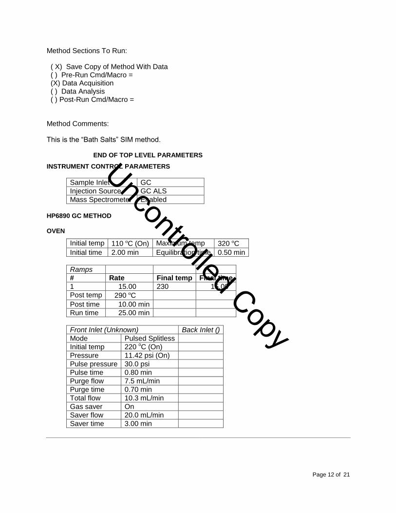
Method Sections To Run:
( X) Save Copy of Method With Data ( ) Pre-Run Cmd/Macro = (X) Data Acquisition ( ) Data Analysis ( ) Post-Run Cmd/Macro =
Method Comments:
This is the “Bath Salts” SIM method.
END OF TOP LEVEL PARAMETERS
INSTRUMENT CONTROL PARAMETERS
Sample Inlet GC
Injection Source GC ALS
Mass Spectrometer Enabled
HP6890 GC METHOD
OVEN
Initial temp 110 ºC (On) Maximum temp 320 ºC
Initial time 2.00 min Equilibration time 0.50 min
Ramps
# Rate Final temp Final time
1 15.00 230 15.00
Post temp 290 ºC
Post time 10.00 min
Run time 25.00 min
Front Inlet (Unknown) Back Inlet ()
Mode Pulsed Splitless
Initial temp 220 oC (On)
Pressure 11.42 psi (On)
Pulse pressure 30.0 psi
Pulse time 0.80 min
Purge flow 7.5 mL/min
Purge time 0.70 min
Total flow 10.3 mL/min
Gas saver On
Saver flow 20.0 mL/min
Saver time 3.00 min
Uncontrolled Copy
Reviewed by:
Date:
Page 13 of 21
T:\FINALSOP\FINAL SOP PDF\G. GAS CHROMATOGRAPHY MASS SPECTROMETRY\B.AMPHET QUANT SPE VER 03-08-2013.DOCX
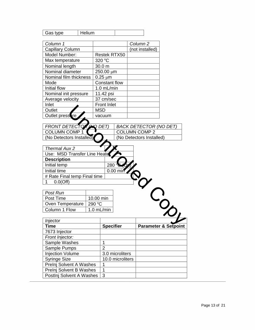
Gas type Helium
Column 1 Column 2
Capillary Column (not installed)
Model Number: Restek RTX50
Max temperature 320 ºC
Nominal length 30.0 m
Nominal diameter 250.00 μm
Nominal film thickness 0.25 μm
Mode Constant flow
Initial flow 1.0 mL/min
Nominal init pressure 11.42 psi
Average velocity 37 cm/sec
Inlet Front Inlet
Outlet MSD
Outlet pressure vacuum
FRONT DETECTOR (NO DET) BACK DETECTOR (NO DET)
COLUMN COMP 1 COLUMN COMP 2
(No Detectors Installed) (No Detectors Installed)
Thermal Aux 2
Use: MSD Transfer Line Heater
Description
Initial temp 280 ºC (On)
Initial time 0.00 min
# Rate Final temp Final time
1 0.0(Off)
Post Run
Post Time 10.00 min
Oven Temperature 290 ºC
Column 1 Flow 1.0 mL/min
Injector
Time Specifier Parameter & Setpoint
7673 Injector
Front Injector:
Sample Washes 1
Sample Pumps 2
Injection Volume 3.0 microliters
Syringe Size 10.0 microliters
PreInj Solvent A Washes 1
PreInj Solvent B Washes 1
PostInj Solvent A Washes 3
Uncontrolled Copy
Reviewed by:
Date:
Page 14 of 21
T:\FINALSOP\FINAL SOP PDF\G. GAS CHROMATOGRAPHY MASS SPECTROMETRY\B.AMPHET QUANT SPE VER 03-08-2013.DOCX
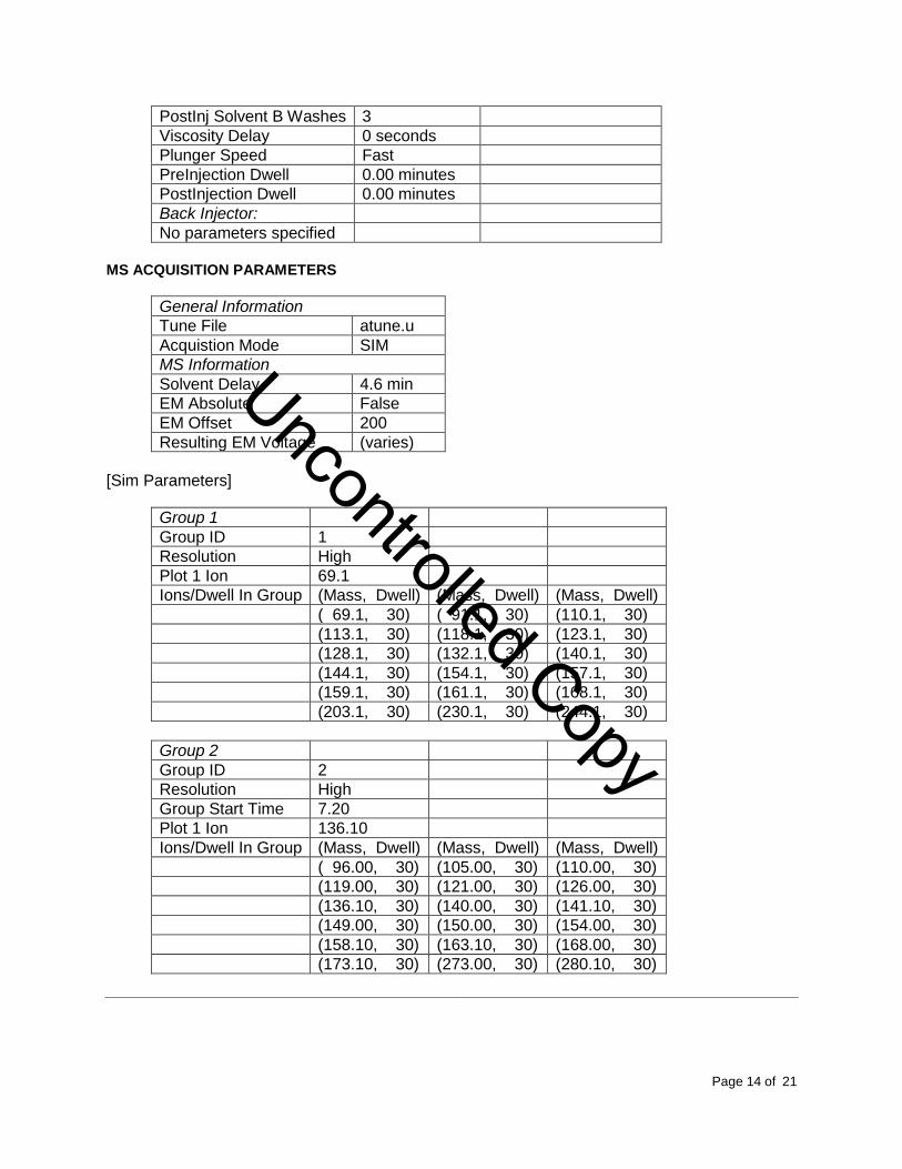
PostInj Solvent B Washes 3
Viscosity Delay 0 seconds
Plunger Speed Fast
PreInjection Dwell 0.00 minutes
PostInjection Dwell 0.00 minutes
Back Injector:
No parameters specified
MS ACQUISITION PARAMETERS
General Information
Tune File atune.u
Acquistion Mode SIM
MS Information
Solvent Delay 4.6 min
EM Absolute False
EM Offset 200
Resulting EM Voltage (varies)
[Sim Parameters]
Group 1
Group ID 1
Resolution High
Plot 1 Ion 69.1
Ions/Dwell In Group (Mass, Dwell) (Mass, Dwell) (Mass, Dwell)
( 69.1, 30) ( 91.1, 30) (110.1, 30)
(113.1, 30) (118.1, 30) (123.1, 30)
(128.1, 30) (132.1, 30) (140.1, 30)
(144.1, 30) (154.1, 30) (157.1, 30)
(159.1, 30) (161.1, 30) (168.1, 30)
(203.1, 30) (230.1, 30) (244.1, 30)
Group 2
Group ID 2
Resolution High
Group Start Time 7.20
Plot 1 Ion 136.10
Ions/Dwell In Group (Mass, Dwell) (Mass, Dwell) (Mass, Dwell)
( 96.00, 30) (105.00, 30) (110.00, 30)
(119.00, 30) (121.00, 30) (126.00, 30)
(136.10, 30) (140.00, 30) (141.10, 30)
(149.00, 30) (150.00, 30) (154.00, 30)
(158.10, 30) (163.10, 30) (168.00, 30)
(173.10, 30) (273.00, 30) (280.10, 30)
Uncontrolled Copy
Reviewed by:
Date:
Page 15 of 21
T:\FINALSOP\FINAL SOP PDF\G. GAS CHROMATOGRAPHY MASS SPECTROMETRY\B.AMPHET QUANT SPE VER 03-08-2013.DOCX
[MSZones]
MS Quad: 150 oC maximum 200 oC MS Source: 230 oC maximum 250 oC
END OF MS ACQUISITION PARAMETERS
END OF INSTRUMENT CONTROL PARAMETERS
INSTRUMENT SETUP
A GCMS autotune must be performed at the start of each day of use.
All autosampler syringe wash vials are filled with methanol.
Prepare a sequence using the following steps.
When Chemstation is opened, the Openlab ECM Login screen appears, Enter the instrument name (ms3, ms4, etc.) as appropriate for username and the current password. Verify that Account field says ”production” and Domain field says “Built-In”. If Chemstation is already running, it may be necessary to log out and re-login. Using the Chemstation software, at the top Method and Run toolbar under ECM, select Logon to ECM. Follow the instructions above to log on.
1. On the Method and Run toolbar, under Sequence, select Load Sequence. Select default.s. Click on Select.
2. In the Method and Run toolbar, under Sequence, select Edit Sequence. At the top of the screen under Data Path, click on Browse. Under Select Data Path, click on the msdchem folder. The click 1 and then click on the Data folder to highlight it. In lower left of dialog box, select Make New Folder. A folder with the name New Folder is created under DATA. Right click on New Folder and Rename or double click to highlight the folder name and change the entry. Rename the file using the format MSnMMDDYYx, where n is instrument number, MM = month, DD = day, YY= year and x = a letter indicating the batch being run, e.g. MS3041111a. Click OK which will take you back to the sample log table.
3. In the Sample column verify the pre-loaded entries. Starting at the first empty fieId, enter sample or QC information. For samples, this would include aliquot number, laboratory number, specimen source, dilution if any (i.e., 2-11-2432 fem). If the sample has been diluted, enter the appropriate dilution factor in the Multiplier column. For QC samples use an appropriate designator. Enter the next sample in the batch in the next open field down the column.
Uncontrolled Copy
Reviewed by:
Date:
Page 16 of 21
T:\FINALSOP\FINAL SOP PDF\G. GAS CHROMATOGRAPHY MASS SPECTROMETRY\B.AMPHET QUANT SPE VER 03-08-2013.DOCX
4. In the Type column, select the corresponding sample type for each vial: Sample, Blank, Calibration or QC.
5. In the Vial column, click in the cell with the number 1, hold down the left mouse button and drag to the last vial number in the sequence (cells will be highlighted). Right click and select Fill Column and Increment. Verify that the vial numbers are correct.
6. In the Method column, verify that the correct method is loaded in the first cell. Then click on the first cell containing the method name, hold down the left mouse and drag to the last vial number in the sequence (cells will be highlighted). Right click and select Fill Column, No Increment. Verify that the method for each vial is correct.
7. In the Data File column, in the cell corresponding to the first vial, enter the data file name in the format MSnMMDDx001, where n = instrument number, MM = month, DD = day, and x = a letter indicating the batch being run, i.e. MS20411a001. Click on this cell, hold down the left mouse and drag to the last vial number of the sequence (the cells will be highlighted). Right click and select Fill Column and Increment. Verify that the data file information for each vial is correct.
8. In the Comment column, enter any additional information for the vials.
9. In the Level column, verify that the correct level numbers are entered for calibrators in this batch.
10. Verify No Update is selected for all vials under Update Rf and Update Rt.
11. Review the information typed for the sequence. Correct any information as needed. Verify that the Data Path is C:MSDCHEM\1\Data\current sequence name. Verify that the Method Path is C:MSDCHEM\1\METHODS. Then Click ok.
Note: Occasionally, it will be desirable to run several subsequences in one batch. Use
the instructions below to accomplish this.
Setting-Up a Subsequence
On the Sample Log Table:
1. Select “Keyword” for “Type”
2. Select “DataPath” for “Method/Keyword”
3. Under “Comment/KeywordString” type in the new data path for your subsequence ie: C:\MSDCHEM\1\DATA\MSnMMDDYYx where “n” is the instrument’s number and “x” is the letter designated to the subsequence (it must be different than that of the original sequence).
4. The suffix of the data files must be different from that of the original; i.e.:
MSNMMDDy001; the subsequence data files must start with 1 again.
5. After typing in the entire sequence, save sequence accordingly.
Uncontrolled Copy
Reviewed by:
Date:
Page 17 of 21
T:\FINALSOP\FINAL SOP PDF\G. GAS CHROMATOGRAPHY MASS SPECTROMETRY\B.AMPHET QUANT SPE VER 03-08-2013.DOCX
6. Go to Sequence Simulate Sequence Run Sequence.
7. A dialog box will pop-up: DataPath C:\MSDCHEM\1\DATA\MSnMMDDYYy does not exist. Edit Sample Log Table? Click No if the sequence was set up correctly.
8. A 2nd dialog box will pop-up: Create C:\MSDCHEM\1\DATA\MSnMMDDYYy? Click
Yes.
9. A 3rd dialog box will pop-up: Sequence Verification Done! View it? Click Yes.
Setting-Up a Subsequence with a Different Method
On the Sample Log Table:
1. After inserting the DataPath keywords and Commands, Insert a Row.
2. Select “Keyword” for “Type”
3. Select “MethodPath” for “Method/Keyword”
4. Check that the method for each sample is changed to the new method.
5. Follow Steps 5-9 in the “Setting-Up a Subsequence” Section (see previous section).
1. On the Method and Run toolbar, under Sequence, select Run Sequence. In the dialog box under Sequence Comment enter the initials of the individual who has entered the sequence in the Operator Field. Under Data File Directory verify that the data file path is C:MSDCHEM\1\DATA\batch name.
2. Under Sequence, select Save Sequence As. Under File Name, type in the name of the folder that the batch will be saved to under DATA, MSnMMDDYYx (e.g. MS2041211a) Select Save. The extension “.s” will automatically be added.
3. Under Sequence select Print Sequence. Verify that Brief Format is selected and click on OK. The sequence will be printed. Apply the preprinted labels for documenting chain-of-custody verification of process steps to the printed sequence list.
4. Load vials into the appropriate autosampler positions as indicated by the order on the printed sequence list. Check vial information against the sequence list and ensure that the vial is inserted in the correct numeric position in the autosampler as indicated on the sequence list.
5. Under Sequence select Run Sequence. Verify that the Sequence comments and Data Field information are correct (i.e. verify that the proper sequence is loaded. If not, load the proper sequence). Click on Run Sequence.
6. After the batch is finished, unload the vials. Compare the vial information to the sequence list as they are removed, to verify that the correct vial was in the correct position. Date and
Uncontrolled Copy
Reviewed by:
Date:
Page 18 of 21
T:\FINALSOP\FINAL SOP PDF\G. GAS CHROMATOGRAPHY MASS SPECTROMETRY\B.AMPHET QUANT SPE VER 03-08-2013.DOCX
initial the sequence list when this is completed. Annotate discrepancies, if necessary. Be sure to enter a copy of the sequence in the Sequence Logbook.
DATA TRANSFER AND PROCESSING
All processing and review are performed on a processing computer.
SAVE METHOD TO ECM
1. After the run finishes, the data files will be in the data subdirectory on the local chemstation and also automatically transferred to ECM. From the acquiring computer, make sure the proper method, the one used to acquire the data, is loaded. On the top toolbar under ECM click on Save Method to ECM.
2. Click on GCMS, the correct instrument name folder, the appropriate month and batch to which the method will be saved.
RETRIEVE BATCH FROM ECM
1. At the processing computer, click on Processing Data Analysis. Log on using your OCME network username and password.
2. On Enhanced Data Analysis screen, click on ECM at the top toolbar and select Retrieve entire sequence from ECM.
3. This will open up Openlab ECM screen. Select GCMS, then the appropriate instrument, the month, and, finally the batch to be retrieved. On the status line at the bottom of the screen that the batch is being retrieved. The batch will be downloaded to the following location: C:\msdchem\1\ECM\Retrieve\”batch name”
LOAD METHOD AND BATCH
1. On the left screen under the C drive, open C:\msdchem\1\ecm\retrieve
2. Under retrieve, click on the batch that was retrieved. When all files have been downloaded to the processing computer, verify that the appropriate method is present in the batch.
3. To load the method, right click on the method under the batch being processed and select load. This will bring up “Be sure changes are saved. Load now?” Click yes. If the method is not present, load the method by retrieving the method from ECM.
4. Click on any file in the batch to load it.
Uncontrolled Copy
Reviewed by:
Date:
Page 19 of 21
T:\FINALSOP\FINAL SOP PDF\G. GAS CHROMATOGRAPHY MASS SPECTROMETRY\B.AMPHET QUANT SPE VER 03-08-2013.DOCX
PERFORM BATCH CALIBRATION
Under enhanced data analysis:
1. Process the calibrators. Select Tools from the toolbar, DoLIST, and Quant, No Report (QT 1). Press Add, and OK. Select the files for this action to be performed on, in this case, calibrators only. Verify that the selected files are located in the correct subdirectory. Change the path if necessary. Click the → Arrow and Process.
2. Review the integrations of the targeted compounds for each calibrator, checking that the ion peaks are present and integrated correctly (i.e. the baseline is the most scientifically accurate one that can be drawn). Select View from the toolbar, QEDIT. Answer appropriately when prompted to save changes made to quantitation results when moving from file to file. Return to Data Analysis by selecting View from the toolbar, return to Data Analysis.
3. Update the existing calibration table (all levels). Select Calibrate, Update, Quick Levels Update. When prompted to clear responses, select YES. When asked to requant files before update, select NO. Select single data file/level option. Select the appropriate data file to associate with calibration level 1 (0.1 mg/L). Click OK. Repeat for remaining calibration levels (0.5, 1.0, 1.5 mg/L). Select level 3 when prompted to update retention times.
4. Load the file associated with level 3 (1.0 mg/L), by selecting File, Load Data File. Select Calibrate, Update One Level. Do NOT requant. Select Update One Level, select only Replace Qualifier Ion Relative Responses, and choose the corresponding existing level ID (#3). Click Do Update.
5. Review the Compound database. Double click on the internal standard listed on the left to reveal the compounds quantitated with it. Select the calibration tab to reveal compound responses, calibration curves, and r2. To disable a point on the calibration curve for a compound, delete its response from the table. Click OK or Cancel when review is complete.
6. Save Method before proceeding. Select Method from the toolbar, Save method, make sure that the path is correct. Save to OpenLab ECM at this time. On Update Calibration screen, select Update Level. Then click on Responses and Replace and on Retention Times and Replace. Then under Existing level ID, select the cal level to update and click on Do Update.
7. Requantitate the calibrators with the updated calibration curve. Select Tools from the toolbar, DoLIST, Requant, no report (QT 2), Add, and OK. Remove any existing commands. Select files to process. Click the → Arrow and Process. Review with QEDIT. Check the responses, retention times and ion ratios.
8. Regression correlation coefficient (r2) for each analyte must be equal to or greater than 0.99.
9. Process controls and cases. Select Tools from the toolbar, DoLIST, Quant, No Report (QT 1), Add, and OK. Select appropriate files. Click the → Arrow and Process. Review with QEDIT. The blank must not contain detectable amounts of target analytes or significant interfering peaks.
Uncontrolled Copy
Reviewed by:
Date:
Page 20 of 21
T:\FINALSOP\FINAL SOP PDF\G. GAS CHROMATOGRAPHY MASS SPECTROMETRY\B.AMPHET QUANT SPE VER 03-08-2013.DOCX
10. When review is complete, return to Data Analysis. Select report format by choosing Quantitate from the toolbar, Report Options. Check SIM style report and uncheck Internal Standards. Press OK.
11. To print reports, select Tools from the toolbar, DoLIST, Profile Quant w/o Calculations (QT 0,1,’P’), Add, and OK. Select files to print, click the → Arrow and Process.
12. Print the calibration table for the current batch by clicking Calibrate on the command line. Select List, Calibrate Report and click OK. The Calibration report will print to the screen. Review the r2 values, then right click on the screen report to print it.
13. Save files to ECM. Select ECM from the toolbar, select “Save multiple data files to ECM”. Select all files.
14. Save method to ECM. Select ECM from the toolbar, Save Method to ECM. Make sure data path is correct.
BATCH CLEAN UP
1. At the processing terminal, select “My Computer”. Find the batch on the C drive at C:\msdchem\1\ecm\retrieve\batch. Right click on the batch to be deleted and select delete. Do not delete a batch that has not been successfully uploaded to ECM.
ACCEPTANCE CRITERIA
1. Review the entire batch , checking that the ion peaks are present and integrated correctly (e.g., that the baseline is the most scientifically accurate one that can be drawn), that the ion ratios are ± 20 % of the average of a calibrator, that the peaks are ± 2 % of the calibrator retention times, and the peaks meet chromatography criteria. The blank must not contain detectable amounts of target analytes or significant interfering peaks. The blood controls must be within ± 20 % of the target value. For tissues, the controls are acceptable up to ± 30 %.
Note: In some instances ratios will be off in exceptionally low or high concentrations. The
operator must evaluate this and schedule proper dilutions or other methodologies, as
needed. See REPORTING section.
2. Make copies of all controls, the r2 report and the sequence list, enough to attach a set to each case in the batch.
REPORTING
After the batch has been reviewed and printed, it must be reported, using the following guidelines:
1. Each case printout must have a copy of the sequence and all controls appended.
Uncontrolled Copy
Reviewed by:
Date:
Page 21 of 21
T:\FINALSOP\FINAL SOP PDF\G. GAS CHROMATOGRAPHY MASS SPECTROMETRY\B.AMPHET QUANT SPE VER 03-08-2013.DOCX
2. Concentrations greater than or equal to 0.1 mg/L are reported in mg/L. Results are truncated and reported to the hundredth of a mg/L (ex: 0.275 mg/L is reported as 0.27 mg/L).
3. Concentrations lower than 0.1 mg/L but meeting all other criteria are reported as “less than 0.1 mg/L”. If the drug is not detected, or the criteria are not met, the drug is reported as “not detected”.
4. Sample concentrations greater than the highest acceptable calibrator must be re-extracted with suitable dilution(s) to bring it within quantitative curve limits.
5. Review other findings in the case, especially GCMS and GC results to see if they are consistent with these GCMS findings. If there are discrepancies, schedule additional testing to resolve them. If in doubt, consult with a supervisor.
6. If the positive matrix controls are greater than ± 30 % of target for non-blood matrices, the samples may be reported qualitatively, as “detected” or “not detected”. If quantitative results are needed, the sample must be repeated.
7. If the concentration of ephedrine is less than 1.0 % of the pseudoephedrine concentration, or conversely, the concentration of pseudoephedrine is less than 1.0 % of the ephedrine concentration, the drug that is less than 1.0 % will be reported as “not detected”.
REFERENCES
Clark’s Identification and Isolation of Drugs in Pharmaceuticals, Body Fluids and Post-Mortem Materials, Ed. A.C. Moffat, The Pharmaceutical Press, London, Second Edition, 1986.
Agilent 6890 GC System Installation Guide.
Agilent 6890 GC System Users Guide.
Agilent 6890 GC System Standard Operating Procedures.
Agilent 5973 & 5973 Network Mass Selective Detector Installation Guide.
Agilent 5973 & 5973 Network Mass Selective Detector Users Guide.
Agilent 5973 & 5973 Network Mass Selective Detector Standard Operating Procedures.
SPEware Corp Cerex Applications Manual.
System 48 Processor Users Guide.
Turbovap Users Guide.
Uncontrolled Copy