Embed Size (px)
Citation preview
July 2014
1
OECD GUIDELINE FOR THE TESTING OF CHEMICALS 1
DRAFT PROPOSAL FOR A NEW TEST GUIDELINE 2
In Vitro Skin Sensitisation: human Cell Line Activation Test (h-CLAT) 3
4
INTRODUCTION 5
1. A skin sensitiser refers to a substance that will lead to an allergic response following skin contact as 6
defined by the United Nations Globally Harmonized System of Classification and Labelling of Chemicals 7
(UN GHS) (1). This test guideline (TG) provides an in vitro procedure called human Cell Line Activation 8
test, or h-CLAT, to be used for supporting the discrimination between skin sensitisers and non-sensitisers 9
in accordance with the UN GHS (1). 10
2. There is general agreement regarding the key biological events underlying skin sensitisation. The current 11
knowledge of the chemical and biological mechanisms associated with skin sensitisation has been 12
summarised in the form of an Adverse Outcome Pathway (AOP) (2), starting with the molecular initiating 13
event and continuing through intermediate events until reaching the adverse effect, namely allergic contact 14
dermatitis in humans or contact hypersensitivity in rodents. The molecular initiating event is the covalent 15
binding of electrophilic substances to nucleophilic centres in skin proteins. The second key event in this 16
AOP takes place in the keratinocytes and includes inflammatory responses as well as gene expression 17
associated with specific cell signalling pathways such as the antioxidant/electrophile response element 18
(ARE)-dependent pathways. The third key event is the activation of dendritic cells (DC), typically assessed 19
by expression of specific cell surface markers, chemokines and cytokines. The fourth key event is T-cell 20
proliferation, which is indirectly assessed in the murine Local Lymph Node Assay (3). 21
3. The assessment of skin sensitisation has typically involved the use of laboratory animals. The classical 22
methods that use guinea-pigs—namely, the Magnusson Kligman Guinea Pig Maximisation Test (GPMT) 23
and the Buehler Test - TG 406 (4)—assess both the induction and elicitation phases of skin sensitisation. 24
The murine tests—the Local Lymph Node Assay (LLNA) - TG 429 (3) and its two non-radioactive 25
modifications, LLNA: DA -TG 442 A (5) and LLNA: BrdU-ELISA - TG 442 B (6)—all assess exclusively 26
the induction response, and have also gained acceptance, since they provide an advantage over the guinea 27
pig tests in terms of animal welfare and by providing an objective measurement of the induction phase of 28
skin sensitisation. 29
4. More recently mechanistically-based in chemico and in vitro test methods have been considered 30
scientifically valid for the evaluation of the skin sensitisation hazard potential of chemicals. However, a 31
combination of non-animal methods (in silico, in chemico, in vitro) within Integrated Approaches to 32
Testing and Assessment (IATA) will be needed to be able to fully substitute for the animal tests currently 33
in use given the restricted AOP mechanistic coverage of each of the currently available non-animal test 34
methods (2)(7). 35
5. The h-CLAT method is proposed to address the third key event (dendritic cell activation) of the skin 36
sensitisation AOP by quantifying changes in the expression of cell surface markers associated with the 37
process of activation of DC (i.e. CD86 and CD54), in the human leukemia cell line THP-1, following 38
exposure to sensitisers (8). The measured expression levels of CD86 and CD54 cell surface markers are 39
July 2014
2
then used for supporting the discrimination between skin sensitisers and non-sensitisers. 1
6. The h-CLAT method has been evaluated in a European Union Reference Laboratory for Alternatives to 2
Animal Testing (EURL ECVAM)-coordinated validation study and subsequent independent peer review 3
by the EURL ECVAM Scientific Advisory Committee (ESAC) and was considered scientifically valid (9) 4
to be used as part of an IATA to support the discrimination between sensitisers and non-sensitisers for the 5
purpose of hazard classification and labelling. Examples of the use of h-CLAT data in combination with 6
other information are reported in the literature (10) (11). 7
7. Definitions are provided in Annex I. 8
9
INITIAL CONSIDERATIONS AND LIMITATIONS 10
8. Skin sensitisers have been reported to induce the expression of cell membrane markers associated with 11
DC activation (2). Consequently test methods such as h-CLAT that are based on DC-like cell lines and 12
which measure markers of DC activation (12) (13) are considered relevant for the assessment of the skin 13
sensitisation potential of chemicals. However, since DC activation represents only one key event of the 14
skin sensitisation AOP, information generated with test methods measuring markers of DC activation may 15
not be sufficient on its own to conclude on the absence of skin sensitisation potential of chemicals. 16
Therefore, data generated with the h-CLAT method should be considered in the context of integrated 17
approaches, such as IATA, and combined with other complementary information e.g. derived from in vitro 18
assays addressing other key events of the skin sensitisation AOP as well as non-testing methods, including 19
read-across from chemical analogues. 20
9. The test method described in this Test Guideline can be used to support the discrimination between skin 21
sensitisers (i.e., UN GHS Category 1) and non-sensitisers in the context of IATA. This Test Guideline 22
cannot be used on its own, neither to sub-categorise skin sensitisers into subcategories 1A and 1B as 23
defined by UN GHS (1), for authorities implementing these two optional subcategories, nor to predict 24
potency for safety assessment decisions. However, depending on the regulatory framework, a positive 25
result with the h-CLAT may be used on its own to classify a chemical into UN GHS category 1. 26
10. The h-CLAT method proved to be transferable to laboratories experienced in cell culture techniques 27
and flow cytometry analysis. The level of reproducibility in predictions that can be expected from the test 28
method is in the order of 80% within and between laboratories (9). Results generated in the validation 29
study (14) and other published studies (15) overall indicate that, compared with LLNA results, the 30
accuracy in distinguishing skin sensitisers (i.e., UN GHS Cat.1) from non-sensitisers is 88% (N=128) with 31
a sensitivity of 95% (87/92) and a specificity of 69% (25/36). The h-CLAT is more likely to under predict 32
chemicals showing a low to moderate skin sensitisation potency (i.e. UN GHS subcategory 1B) than 33
chemicals showing a high skin sensitisation potency (i.e. UN GHS subcategory 1A) (14) (15). Taken 34
together, this information indicates the usefulness of the h-CLAT method to contribute to the identification 35
of skin sensitisation hazards. However, the accuracy values given here for the h-CLAT as a stand-alone 36
test method are only indicative, since the test method should be considered in combination with other 37
sources of information in the context of an IATA and in accordance with the provisions of paragraph 9 38
above. Furthermore, when evaluating non-animal methods for skin sensitisation, it should be kept in mind 39
that the LLNA test as well as other animal tests may not fully reflect the situation in the species of interest, 40
i.e., humans. 41

July 2014
3
11. The term "test chemical" is used in this Test Guideline to refer to what is being tested1 and is not 1
related to the applicability of the h-CLAT to the testing of substances and/or mixtures. On the basis of the 2
current data available, the h-CLAT method was shown to be applicable to test chemicals covering a variety 3
of organic functional groups, reaction mechanisms, skin sensitisation potency (as determined in in vivo 4
studies) and physicochemical properties (9) (15) (16). Limited information is currently available on the 5
applicability of the h-CLAT method to mixtures (16). The test method is nevertheless technically 6
applicable to the testing of multi-constituent substances and mixtures. However, before use of this Test 7
Guideline on a mixture for generating data for an intended regulatory purpose, it should be considered 8
whether, and if so why, it may provide adequate results for that purpose. Such considerations are not 9
needed when there is a regulatory requirement for the testing of the mixture. The h-CLAT method is 10
applicable to test chemicals soluble or that form a stable dispersion (i.e., a colloid or suspension in which 11
the test chemical does not settle or separate from the solvent into different phases) in an appropriate 12
solvent (see paragraph 19). Test chemicals with a Log Kow of up to 3.5 have been successfully assessed by 13
the h-CLAT method (15). However, test chemicals with a Log Kow of greater than 3.5 may still be tested 14
at lower soluble concentrations. In such a case, a positive result could still be used to support the 15
identification of the test chemical as a skin sensitiser, but a negative result should be considered as 16
inconclusive. Furthermore, because of the limited metabolic capability of the cell line and because of the 17
experimental conditions, pro-haptens (i.e. chemicals requiring enzymatic activation to exert their 18
sensitisation activity) and pre-haptens (i.e. chemicals activated by auto oxidation) may also provide 19
negative results in the h-CLAT (16). In the light of the above, negative results should be interpreted in the 20
context of the stated limitations and in the connection with other information sources within the framework 21
of IATA. In cases where there is evidence demonstrating the non-applicability of the h-CLAT method to 22
other specific categories of chemicals, it should not be used for those specific categories of chemicals. 23
12. As described, the h-CLAT method supports the discrimination between skin sensitisers from 24
non-sensitisers. However, it may also potentially contribute to the assessment of sensitising potency (10) 25
(11) when used in integrated approaches such as IATA. Nevertheless, further work, preferably based on 26
human data, is required to determine how h-CLAT results may possibly inform potency assessment. 27
28
PRINCIPLE OF THE TEST 29
13. The h-CLAT method is an in vitro method which quantifies changes of cell surface marker expression 30
(i.e., CD86 and CD54) on a human cell line, THP-1 cells, following 24 hours exposure to test chemical. 31
The changes of surface marker expression are measured by flow cytometry following cell staining with 32
fluorescein isothiocyanate (FITC)-labelled antibodies. Cytotoxicity measurement is also conducted 33
concurrently to assess whether upregulation of surface maker expression occurs at sub-cytotoxic 34
concentrations. The relative fluorescence intensity of surface markers compared to solvent control are 35
calculated and used in the prediction model (see paragraph 31), to support the discrimination between 36
sensitisers and non-sensitisers 37
14. Prior to routine use of the method described in this Test Guideline, laboratories should demonstrate 38
technical proficiency, using the 10 Proficiency Substances listed in Annex II. 39
40
1 In June 2013, the Joint Meeting agreed that where possible, a more consistent use of the term "test chemical"
describing what is being tested should now be applied in new and updated Test Guidelines.
July 2014
4
PROCEDURE 1
15. This test guideline is based on the h-CLAT DB-ALM protocol no. 158 (17) which represents the 2
protocol used for the EURL ECVAM-coordinated validation study. It is recommended that this protocol is 3
used when implementing and using the h-CLAT method in the laboratory. The following is a description of 4
the main components and procedures for the h-CLAT method, which comprises two steps: dose finding 5
assay and CD86/CD54 expression measurement. 6
7
Preparation of cells 8
16. The human leukemia cell line, THP-1, should be used for performing the h-CLAT method. It is 9
recommended that cells (TIB-202) are obtained from a well-qualified cell bank, such as American Type 10
Culture Collection. 11
17. THP-1 cells are cultured, at 37°C under 5% CO2 and humidified atmosphere, in RPMI-1640 medium 12
supplemented with 10% foetal bovine serum (FBS), 0.05 mM 2-mercaptoethanol, 100 units/mL penicillin 13
and 100 µg/mL streptomycin. THP-1 cells are routinely passaged every 2-3 days at the density of 0.1 to 0.2 14
× 106 cells/mL and should be maintained at densities from 0.1 × 10
6 to 0.8 × 10
6 cells/mL. The cell density 15
should not exceed 1 × 10⁶ cells/mL. The reactivity check of the cells should be performed using the 16
positive controls, 2,4-dinitrochlorobenzene (DNCB) (CAS n. 97-00-7, ≥ 99% purity) and nickel sulfate 17
(CAS n. 10101-97-0, 99% purity) and the negative control lactic acid (CAS n. 50-21-5, ≥ 99% purity), two 18
weeks after thawing. Both DNCB and NiSO4 should produce a positive response of both CD86 and CD54, 19
and LA should produce negative response of both CD86 and CD54. Only the cells which passed the 20
reactivity check are to be used for the assay. Cells can be propagated up to two months after thawing 21
(passage number should not exceed 30). 22
18. For testing, THP-1 cells are seeded between 0.1 and 0.2 × 10⁶ cells/mL, and pre-cultured for 48 hours 23
or for 72 hours in culture flasks. In the day of testing, cells harvested from culture flask are resuspended 24
with fresh culture medium at 2 × 10⁶ cells/mL. Then, cells are distributed into a 24 well flat-bottom plate 25
with 500 L (1 × 10⁶ cells/well) or a 96-well flat-bottom plate with 80 L (1.6 × 10
5 cells/well). 26
Dose finding assay 27
28
Preparation of test chemicals and control substances 29
30
19. The test chemicals and control substances are prepared on the day of testing. For the h-CLAT method, 31
test chemicals are dissolved in saline, medium or dimethyl sulfoxide (DMSO, 99% purity) to final 32
concentrations of 100 mg/mL or 500 mg/mL. The test chemicals, which are not soluble in saline, are 33
dissolved in DMSO and diluted. However, other solvents may be used if sufficient scientific rationale is 34
provided. 35
36
20. Based on the 100 mg/mL (in saline) or 500 mg/mL (in DMSO) solutions of the test chemicals, 2-fold 37
serial dilutions are made using corresponding solvent to obtain the stock solutions (eight doses) to be tested 38
in the h-CLAT method. These stock solutions are then further diluted 50-fold (for saline) or 250-fold (for 39
DMSO) into the culture medium (working solutions). These working solutions are finally used for 40
treatment with a further 2-fold dilution factor. 41
42
21. The negative control used in the h-CLAT method is culture medium (for chemicals solubilised either 43
with medium or saline) or DMSO (for chemicals solubilised in DMSO). It undergoes the same dilution as 44
July 2014
5
described for the working solutions in paragraph 20, so that the final concentration of saline is 1%, and that 1
of DMSO is 0.2%. 2
3
Application of test chemicals and control substances 4
5
22. The culture medium or working solutions described in paragraph 20 and 21 are mixed 1:1 (v/v) with 6
the cell suspensions prepared in the 24-well or 96-well flat-bottom plate (see paragraph 18). The treated 7
plates are then incubated for 24 hours at 37°C under 5% CO2. Care should be taken to avoid evaporation of 8
volatile test chemicals and cross-contamination between wells by test chemicals, e.g., by sealing the plate 9
prior to the incubation with the test chemicals. 10
11
Propidium iodide (PI) staining 12
13
23. After 24 hours of exposure, cells are transferred into sample tubes and collected by centrifugation. The 14
supernatants are discarded and the remaining cells are resuspended with 600 L of a phosphate buffered 15
saline containing 0.1% bovine serum albumin (FACS buffer). 200 L of cell suspension is transferred into 16
96-well round-bottom plate and washed twice with 200 uL of FACS buffer. Finally, cells are resuspended 17
in 200 L of FACS buffer and 10 L of PI solution is added (final concentration of PI is 0.625 g/mL). 18
19
Cytotoxicity measurement by flow cytometry and estimation of CV75 value. 20
21
24. The PI uptake is analysed using flow cytometry with the acquisition channel FL-3. A total of 10,000 22
living (PI negative) cells are acquired. The cell viability can be calculated using the following equation by 23
the cytometer analysis program. When the cell viability is low, up to 30,000 cells including dead cells 24
should be acquired. Alternatively, the data acquisition can be finished one minute after the initiation. 25
26
27
28
29
30
The CV75 value, i.e. a concentration showing 75% of THP-1 cell survival (25% cytotoxicity), is calculated 31
by log-linear interpolation using the following equation: 32
33
34
35
36
37
Where: 38
39
A is the minimum value of cell viability over 75% in testing groups 40
B is the maximum value of cell viability below 75% in testing groups 41
C or D is the concentration showing the value of cell viability A or B 42
43
The CV75 value is used to determine the concentration of test chemicals in CD86/CD54 expression 44
measurement. 45
46
CD86/CD54 expression measurement 47
48
Cell Viability = Number of living cells
Total Number of acquired cells
× 100
Log CV75 = A – B
(75 – B) × Log (C) – (75 – A) × Log (D)
July 2014
6
Preparation of the test chemicals and Control Substances. 1
2
25. The appropriate solvent (saline or DMSO; see paragraph 19) is used to dissolve the test chemicals. The 3
test chemicals are first diluted to the concentration corresponding to 100-fold (for saline) or 500-fold (for 4
DMSO) of the 1.2 × CV75 determined in the dose finding assay (see paragraph 24). If the CV75 is not 5
determined (i.e. if sufficient cytotoxicity is not observed in the dose finding assay), the highest soluble 6
concentration of test chemical prepared with each solvent should be used as starting dose. Then, 1.2-fold 7
serial dilutions are made using the corresponding solvent to obtain the stock solutions (eight doses ranging 8
from 0.335 × CV75 to 1.2 × CV75) to be tested in the h-CLAT method. The stock solutions are then further 9
diluted 50-fold (for saline) or 250-fold (for DMSO) into the culture medium (working solutions)). These 10
working solutions are finally used for treatment with a further 2-fold dilution factor. Alternative 11
concentrations may be used upon justification (e.g. in case of poor solubility or cytotoxicity). 12
13
26. The negative (solvent) control is prepared as described in paragraph 21. The positive control used in 14
the h-CLAT method is DNCB (see paragraph 17), for which stock solutions are prepared in DMSO and 15
diluted as described for the stock solutions in paragraph 25. The final concentration of the positive control, 16
4.0 g/mL, should yield approximately 70-90% of cell viability. Alternatively, the CV75 of DNCB, which 17
is determined in each test facility, could be also used as the positive control dose. Other suitable positive 18
controls may be used if historical data are available to derive comparable run acceptance criteria. 19
20
Application of test chemicals and control substances 21
22
27. For each test chemicals and control substances, one experiment is needed to derive a prediction. Each 23
experiment consists of at least two independent runs (n=2) (see paragraphs 31 and 33). Test chemicals and 24
control substances prepared as working solutions are mixed with suspended cells at 1:1 ratio, and cells are 25
incubated for 24 hours as described in paragraphs 25 and 26. 26
27
Cell staining and analysis 28
29
28. After 24 hours of exposure, cells are transferred into sample tubes and collected by centrifugation. The 30
supernatants are discarded and the remaining cells are resuspended with 600 L of FACS buffer. Cells are 31
split in three aliquots of 180 L into a 96-well round-bottom plate. After centrifugation, cells are 32
resuspended in 200 L of blocking solution (FACS buffer containing 0.01% (w/v) globulin) and incubated 33
at 4°C for 15 min. 34
35
29. After centrifugation, cells are stained with 50 L of FITC-labelled anti-CD86, anti-CD54 or mouse 36
IgG1 (isotype) antibodies at 4°C for 30 min. The antibodies described in the h-CLAT DB-ALM protocol 37
no. 155 (17) should be used by diluting 3:25 (v/v, for CD86) or 3:50 (v/v, for CD54 and IgG1) with FACS 38
buffer. After washing with 200 L of FACS buffer three times, cells are resuspended in 200 L of FACS 39
buffer and 10 L of PI solution is added (final concentration of PI is 0.625 g/mL). The expression levels 40
of CD86 and CD54, and cell viability are analysed using flow cytometry. 41
42
DATA AND REPORTING 43
44
Data evaluation 45
46
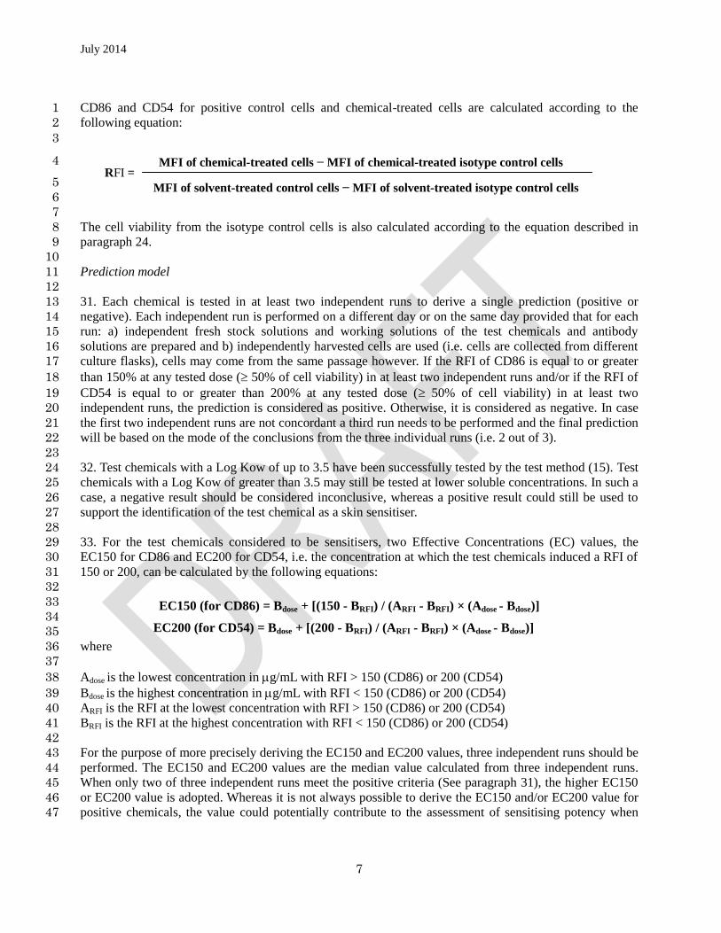
30. The expression of CD86 and CD54 is analysed with flow cytometry with the acquisition channel FL-1. 47
Based on the geometric mean fluorescence intensity (MFI), the relative fluorescence intensity (RFI) of 48
July 2014
7
CD86 and CD54 for positive control cells and chemical-treated cells are calculated according to the 1
following equation: 2
3
4
5
6
7
The cell viability from the isotype control cells is also calculated according to the equation described in 8
paragraph 24. 9
10
Prediction model 11
12
31. Each chemical is tested in at least two independent runs to derive a single prediction (positive or 13
negative). Each independent run is performed on a different day or on the same day provided that for each 14
run: a) independent fresh stock solutions and working solutions of the test chemicals and antibody 15
solutions are prepared and b) independently harvested cells are used (i.e. cells are collected from different 16
culture flasks), cells may come from the same passage however. If the RFI of CD86 is equal to or greater 17
than 150% at any tested dose ( 50% of cell viability) in at least two independent runs and/or if the RFI of 18
CD54 is equal to or greater than 200% at any tested dose ( 50% of cell viability) in at least two 19
independent runs, the prediction is considered as positive. Otherwise, it is considered as negative. In case 20
the first two independent runs are not concordant a third run needs to be performed and the final prediction 21
will be based on the mode of the conclusions from the three individual runs (i.e. 2 out of 3). 22
23
32. Test chemicals with a Log Kow of up to 3.5 have been successfully tested by the test method (15). Test 24
chemicals with a Log Kow of greater than 3.5 may still be tested at lower soluble concentrations. In such a 25
case, a negative result should be considered inconclusive, whereas a positive result could still be used to 26
support the identification of the test chemical as a skin sensitiser. 27
28
33. For the test chemicals considered to be sensitisers, two Effective Concentrations (EC) values, the 29
EC150 for CD86 and EC200 for CD54, i.e. the concentration at which the test chemicals induced a RFI of 30
150 or 200, can be calculated by the following equations: 31
32
33
34
35
where 36
37
Adose is the lowest concentration in g/mL with RFI > 150 (CD86) or 200 (CD54) 38
Bdose is the highest concentration in g/mL with RFI < 150 (CD86) or 200 (CD54) 39
ARFI is the RFI at the lowest concentration with RFI > 150 (CD86) or 200 (CD54) 40
BRFI is the RFI at the highest concentration with RFI < 150 (CD86) or 200 (CD54) 41
42
For the purpose of more precisely deriving the EC150 and EC200 values, three independent runs should be 43
performed. The EC150 and EC200 values are the median value calculated from three independent runs. 44
When only two of three independent runs meet the positive criteria (See paragraph 31), the higher EC150 45
or EC200 value is adopted. Whereas it is not always possible to derive the EC150 and/or EC200 value for 46
positive chemicals, the value could potentially contribute to the assessment of sensitising potency when 47
RFI = MFI of chemical-treated cells − MFI of chemical-treated isotype control cells
MFI of solvent-treated control cells − MFI of solvent-treated isotype control cells
EC200 (for CD54) = Bdose + [(200 - BRFI) / (ARFI - BRFI) × (Adose - Bdose)]
EC150 (for CD86) = Bdose + [(150 - BRFI) / (ARFI - BRFI) × (Adose - Bdose)]
July 2014
8
used in integrated approaches such as IATA (10)(11). 1
2
Acceptance criteria 3
4
34. The following acceptance criteria should be met when using the h-CLAT method. 5
6
- The cell viabilities of medium and solvent control are more than 90%. 7
8
- In the positive control (DNCB), RFI values of both CD86 and CD54 are over the positive criteria 9
(CD86 RFI 150 and CD54 RFI 200) and cell viability is more than 50%. 10
11
- In the solvent control (DMSO), RFI values of both CD86 and CD54 should not exceed the positive 12
criteria (CD86 RFI 150 and CD54 RFI 200). 13
14
- For both medium and DMSO controls, the MFI ratio of both CD86 and CD54 to isotype control 15
should be > 105%. 16
17
- The cell viability of tested chemicals at more than four tested doses in each run should be 50%. 18
19
35. Negative results are acceptable only for test chemicals exhibiting cell viability at 1.2 × CV75 of less 20
than 90%. Negative results with cell viability of 90% or higher are discarded. The dose finding study 21
should be retested to determine the CV75 determination. Positive results for test chemicals of any cell 22
viability at 1.2 × CV75 are acceptable. It should be noted that when 5000 g/mL in saline, 1000 g/mL in 23
DMSO, or the highest soluble concentration is used as the maximal test concentration of a test chemical, 24
the results are acceptable. 25
26
Test report 27
28
36. The test report should include the following information. 29
30
Test Chemical 31
- Mono-constituent substance 32
Chemical identification, such as IUPAC or CAS name(s), CAS number(s), SMILES or InChI 33
code, structural formula, and/or other identifiers; 34
Physical appearance, water solubility, DMSO solubility, molecular weight, and additional 35
relevant physicochemical properties, to the extent available; 36
Purity, chemical identity of impurities as appropriate and practically feasible, etc; 37
Treatment prior to testing, if applicable (e.g., warming, grinding); 38
Concentration(s) tested; 39
Storage conditions and stability to the extent available. 40
July 2014
9
- Multi-constituent substance, UVCB and mixture: 1
Characterisation as far as possible by e.g., chemical identity (see above), purity, quantitative 2
occurrence and relevant physicochemical properties (see above) of the constituents, to the extent 3
available; 4
Physical appearance, water solubility, DMSO solubility and additional relevant physicochemical 5
properties, to the extent available; 6
Molecular weight or apparent molecular weight in case of mixtures/polymers of known 7
compositions or other information relevant for the conduct of the study; 8
Treatment prior to testing, if applicable (e.g., warming, grinding); 9
Concentration(s) tested; 10
Storage conditions and stability to the extent available. 11
Controls 12
- Positive control 13
Chemical identification, such as IUPAC or CAS name(s), CAS number(s), SMILES or InChI 14
code, structural formula, and/or other identifiers; 15
Physical appearance, water solubility, DMSO solubility, molecular weight, and additional 16
relevant physicochemical properties, to the extent available and where applicable; 17
Purity, chemical identity of impurities as appropriate and practically feasible, etc; 18
Treatment prior to testing, if applicable (e.g., warming, grinding); 19
Concentration(s) tested; 20
Storage conditions and stability to the extent available; 21
Reference to historical positive control results demonstrating suitable run acceptance criteria, if 22
applicable. 23
- Negative (vehicle) control 24
Chemical identification, such as IUPAC or CAS name(s), CAS number(s), and/or other 25
identifiers; 26
Purity, chemical identity of impurities as appropriate and practically feasible, etc; 27
Physical appearance, molecular weight, and additional relevant physicochemical properties in the 28
case other negative controls / vehicles than those mentioned in the Test Guideline are used and to 29
July 2014
10
the extent available; 1
Storage conditions and stability to the extent available; 2
Justification for choice of solvent for each test chemical. 3
4
Test method Conditions 5
- Name and address of the sponsor, test facility and study director 6
- Description of test method used 7
- Cell line used, its storage conditions and source (e.g., the facility from which they were obtained) 8
- Passage number and confluence of cells used for testing 9
- Flow cytometry used (e.g., model), including instrument settings, globulin, antibodies and propidium 10
iodide used 11
- If applicable, the procedure used to demonstrate proficiency of the laboratory in performing the test 12
method (e.g. by testing of proficiency substances) or to demonstrate reproducible performance of the 13
test method over time. 14
15
Results 16
- Tabulation of the data, including CV75 (if applicable), individual geometric MFI, RFI, cell viability 17
values, EC150/EC200 values (if applicable) obtained for the test chemical and for the positive control 18
in each run, and an indication of the rating of the test chemical according to the prediction model 19
- Description of any other relevant observations, if applicable. 20
21
Discussion of the Results 22
- Discussion of the results obtained with the h-CLAT method 23
- Consideration of the test method results within the context of an IATA, if other relevant information 24
is available 25
26
Conclusions 27
28
29
July 2014
11
LITERATURE 1
2
1. United Nations UN (2013). Globally Harmonized System of Classification and Labelling of Chemicals 3
(GHS). Fifth revised edition. New York & Geneva: United Nations Publications. ISBN: 4
978-92-1-117006-1. Available at: 5
[http://www.unece.org/trans/danger/publi/ghs/ghs_rev05/05files_e.html]. 6
2. OECD (2012). The Adverse Outcome Pathway for Skin Sensitisation Initiated by Covalent Binding to 7
Proteins. Part 1: Scientific Evidence. Series on Testing and Assessment No. 168. Available at: 8
http://www.oecd.org/officialdocuments/publicdisplaydocumentpdf/?cote=ENV/JM/MONO(2012)10/P9
ART1&docLanguage=En 10
3. OECD (2010), The Local Lymph Node Assay. OECD Guideline for the Testing of Chemicals No. 429, 11
Organisation for Economic Cooperation and Development, Paris. Available at: 12
http://www.oecd.org/env/testguidelines 13
4. OECD (1992). Skin Sensitisation. OECD Guideline for the Testing of Chemicals No. 406, Organisation 14
for Economic Cooperation and Development, Paris. Available at: 15
http://www.oecd.org/env/testguidelines 16
5. OECD (2010), Skin Sensitisation: Local Lymph Node Assay: DA. OECD Guideline for the Testing of 17
Chemicals No. 442A, Organisation for Economic Cooperation and Development, Paris. Available at: 18
http://www.oecd.org/env/testguidelines 19
6. OECD (2010), Skin Sensitisation: BrdU-ELISA: DA. OECD Guideline for the Testing of Chemicals No. 20
442B, Organisation for Economic Cooperation and Development, Paris. Available at: 21
http://www.oecd.org/env/testguidelines 22
7. Adler S, Basketter D, Creton S, Pelkonen O, van Benthem J, Zuang V, Andersen KE, Angers-Loustau A, 23
Aptula A, Bal-Price A, Benfenati E, Bernauer U, Bessems J, Bois FY, Boobis A, Brandon E, Bremer S, 24
Broschard T, Casati S, Coecke S, Corvi R, Cronin M, Daston G, Dekant W, Felter S, Grignard E, 25
Gundert-Remy U, Heinonen T, Kimber I, Kleinjans J, Komulainen H, Kreiling R, Kreysa J, Leite SB, 26
Loizou G, Maxwell G, Mazzatorta P, Munn S, Pfuhler S, Phrakonkham P, Piersma A, Poth A, Prieto P, 27
Repetto G, Rogiers V, Schoeters G, Schwarz M, Serafimova R, Tähti H, Testai E, van Delft J, van 28
Loveren H, Vinken M, Worth A, Zaldivar JM. (2011). Alternative (non-animal) methods for cosmetics 29
testing: current status and future prospects-2010. Archives of Toxicology 85:367-485. 30
8. Ashikaga T, Yoshida Y, Hirota M, Yoneyama K, Itagaki H, Sakaguchi H, Miyazawa M, Ito Y, Suzuki H, 31
Toyoda H. (2006), Development of an in vitro skin sensitization test using human cell lines: The human 32
Cell Line Activation Test (h-CLAT) I. Optimization of the h-CLAT protocol. Toxicol. In Vitro 33
20:767–773. 34
9. EC EURL-ECVAM (2013). Recommendation on the human Cell Line Activation Test (h-CLAT) for 35
skin sensitisation testing. Accessible at: (in preparation) 36
10.Nukada Y, Miyazawa M, Kazutoshi S, Sakaguchi H, Nishiyama N. (2013) Data integration of 37
non-animal tests for the development of a test battery to predict the skin sensitizing potential and 38
potency of chemicals. Toxicol. In Vitro 27:609-618. 39

July 2014
12
11. Tsujita-Inoue K, Hirota M, Ashikaga T, Atobe T, Kouzuki H, Aiba S (2014) Skin sensitization risk 1
assessment model using artificial neural network analysis of data from multiple in vitro assays. Toxicol. 2
In Vitro 28:626-639. 3
12. Sakaguchi H, Miyazawa M, Yoshida Y, Ito Y, Suzuki H. (2007) Prediction of preservative 4
sensitization potential using surface marker CD86 and/or CD54 expression on human cell line, THP-1. 5
Arch. Dermatol. Res. 298:427-437. 6
13. Nukada Y, Ashikaga T, Miyazawa M, Hirota M, Sakaguchi H, Sasa H, Nishiyama N. (2012) Prediction 7
of skin sensitization potency of chemicals by human Cell Line Activation Test (h-CLAT) and an 8
attempt at classifying skin sensitization potency. Toxicol. In Vitro 26:1150-60. 9
14. EC EURL ECVAM (2012). human Cell Line Activation Test (h-CLAT) Validation Study Report 10
Accessible at: in publication 11
15. Takenouchi O, Miyazawa M, Saito K, Ashikaga T, Sakaguchi H. (2013) Predictive performance of the 12
human Cell Line Activation Test (h-CLAT) for lipophilic with high octanol-water partition coefficients. 13
J. Toxicol. Sci. 38:599-609. 14
15
16. Ashikaga T, Sakaguchi H, Sono S, Kosaka N, Ishikawa M, Nukada Y, Miyazawa M, Ito Y, 16
NishiyamaN, Itagaki H. A comparative evaluation of in vitro skin sensitisation tests: the human 17
cell-line activation test (h-CLAT) versus the local lymph node assay (LLNA). (2010) Altern. Lab. 18
Anim. 38:275-284. 19
20
17. DB-ALM (INVITTOX) (2014) Protocol 158: human Cell Line Activation Test (h-CLAT), XXpp. 21
Available: (in publication) 22
23
18.OECD (2005). Guidance Document No.34 on “the Validation and International Acceptance of New or 24
Updated Test Methods for Hazard Assessment”. OECD Series on Testing and Assessment. 25
Organization for Economic Cooperation and Development, Paris, France, 2005, 96 pp. 26
27
19. ECETOC (2003). Contact sensitization: Classification according to potency. European Centre for 28
Ecotoxicology & Toxicology of Chemicals (Technical Report No. 87). 29
30
July 2014
13
ANNEX I 1
DEFINITIONS 2
3
Accuracy: The closeness of agreement between test method results and accepted reference values. It is a 4
measure of test method performance and one aspect of relevance. The term is often used interchangeably 5
with concordance to mean the proportion of correct outcomes of a test method (18). 6
7
AOP (Adverse Outcome Pathway): sequence of events from the chemical structure of a target chemical 8
or group of similar chemicals through the molecular initiating event to an in vivo outcome of interest (2). 9
10
CV75: The estimated concentration showing 75% cell viability. 11
12
EC150: the concentrations showing the RFI values of 150 in CD86 expression 13
14
EC200: the concentrations showing the RFI values of 200 in CD54 expression 15
16
FACS buffer: A phosphate buffered saline containing 0.1% bovine serum albumin. 17
18
Hazard: Inherent property of an agent or situation having the potential to cause adverse effects when an 19
organism, system or (sub) population is exposed to that agent. 20
21
IATA (Integrated Approach to Testing and Assessment): A structured approach used for hazard 22
identification (potential), hazard characterisation (potency) and/or safety assessment (potential/potency and 23
exposure) of a chemical or group of chemicals, which strategically integrates and weights all relevant data 24
to inform regulatory decision regarding potential hazard and/or risk and/or the need for further targeted and 25
therefore minimal testing. 26
27
Medium control: An untreated replicate containing all components of a test system. This sample is 28
processed with test chemical-treated samples and other control samples to determine whether the solvent 29
interacts with the test system. 30
31
Mixture: A mixture or a solution composed of two or more substances in which they do not react. 32
33
Mono-constituent substance: A substance, defined by its quantitative composition, in which one main 34
constituent is present to at least 80% (w/w). 35
36
Multi-constituent substance: A substance, defined by its quantitative composition, in which more than 37
one main constituent is present in a concentration ≥ 10% (w/w) and < 80% (w/w). A multi-constituent 38
substance is the result of a manufacturing process. The difference between mixture and multi-constituent 39
substance is that a mixture is obtained by blending of two or more substances without chemical reaction. A 40
multi-constituent substance is the result of a chemical reaction. 41
42
July 2014
14
Positive control: A replicate containing all components of a test system and treated with a substance 1
known to induce a positive response. To ensure that variability in the positive control response across time 2
can be assessed, the magnitude of the positive response should not be excessive. 3
4
Pre-haptens: chemicals which become sensitisers through abiotic transformation 5
6
Pro-haptens: chemicals requiring enzymatic activation to exert skin sensitisation potential 7
8
Relative fluorescence intensity Relative values of geometric mean fluorescence intensity (MFI) in 9
chemical-treated cells compared to MFI in solvent-treated cells. 10
11
Relevance: Description of relationship of the test to the effect of interest and whether it is meaningful and 12
useful for a particular purpose. It is the extent to which the test correctly measures or predicts the 13
biological effect of interest. Relevance incorporates consideration of the accuracy (concordance) of a test 14
method (18). 15
16
Reliability: Measures of the extent that a test method can be performed reproducibly within and between 17
laboratories over time, when performed using the same protocol. It is assessed by calculating intra- and 18
inter-laboratory reproducibility and intra-laboratory repeatability (18). 19
20
Run: A run consists of one or more test chemicals tested concurrently with a negative control and with a 21
positive control. 22
23
Sensitivity: The proportion of all positive/active chemicals that are correctly classified by the test. It is a 24
measure of accuracy for a test method that produces categorical results, and is an important consideration 25
in assessing the relevance of a test method (18). 26
27
Solvent control: An untreated sample containing all components of a test system, including the solvent 28
that is processed with the test chemical-treated and other control samples to establish the baseline response 29
for the samples treated with the test chemical dissolved in the same solvent. When tested with a concurrent 30
medium control, this sample also demonstrates whether the solvent interacts with the test system. 31
32
Specificity: The proportion of all negative/inactive chemicals that are correctly classified by the test. It is a 33
measure of accuracy for a test method that produces categorical results and is an important consideration in 34
assessing the relevance of a test method (18). 35
36
Substance: Chemical elements and their compounds in the natural state or obtained by any production 37
process, inducing any additive necessary to preserve the stability of the product and any impurities 38
deriving from the process used, but excluding any solvent which may be separated without affecting the 39
stability of the substance or changing it composition. 40
41
Test chemical: The term " test chemical is used to refer to what is being tested. 42
July 2014
15
1
United Nations Globally Harmonized System of Classification and Labeling of Chemicals (UN 2
GHS): A system proposing the classification of chemicals (substances and mixtures) according to 3
standardized types and levels of physical, health and environmental hazards, and addressing corresponding 4
communication elements, such as pictograms, signal words, hazard statements, precautionary statements 5
and safety data sheets, so that to convey information on their adverse effects with a view to protect people 6
(including employers, workers, transporters, consumers and emergency responders) and the environment 7
(1). 8
9
UVCB: substances of unknown or variable composition, complex reaction products or biological 10
materials. 11
12
Valid test method: A test method considered to have sufficient relevance and reliability for a specific 13
purpose and which is based on scientifically sound principles. A test method is never valid in an absolute 14
sense, but only in relation to a defined purpose (18). 15
16
17
18
19

July 2014
16
ANNEX II 1
2
PROFICIENCY SUBSTANCES 3
4
Prior to routine use of a test method that adheres to this Test Guideline, laboratories should demonstrate 5
technical proficiency by correctly obtaining the expected h-CLAT prediction for the 10 chemicals 6
recommended in Table 1.. Proficiency substances were selected to represent the range of responses for skin 7
sensitisation hazards. Other selection criteria were that the substances are commercially available, and that 8
high-quality in vivo reference data as well as high quality in vitro data generated with the h-CLAT method 9
are available. Also, published reference data are available for the h-CLAT method (9)(15). 10
11
Table 1: Recommended substances for demonstrating technical proficiency with the h-CLAT method 12 13
Proficiency substances CASRN Physical
state
In vivo
prediction1
h-CLAT
prediction2
2,4-Dinitrochlorobenzene 97-00-7 Solid Sensitiser
(extreme) Positive
Chloramin T 127-65-1 Solid Sensitiser
(strong) Positive
Nickel sulfate 10101-97-0 Solid Sensitiser
(moderate) Positive
Phenylacetaldehyde 122-78-1 Liquid Sensitiser
(moderate) Positive
hydroxycitronellal 107-75-5 Liquid Sensitiser
(weak) Positive
Imidazoidinyl urea 39236-46-9 Solid Sensitiser
(weak) Positive
1-Butanol 71-36-3 Liquid Non-sensitiser Negative
Glycerol 56-81-5 Liquid Non-sensitiser Negative
Lactic acid 50-21-5 Liquid Non-sensitiser Negative
Vanillin 121-33-5 Solid Non-sensitiser Negative
14
Abbreviations: CAS RN = Chemical Abstracts Service Registry Number 15 1
The in vivo hazard and (potency) prediction is based on LLNA data (9) (15). The in vivo potency is 16
derived using the criteria proposed by ECETOC (19).
17 2
Predictions made with the h-CLAT method should be considered in the framework of IATA and in 18
accordance with the provision of paragraph 9. 19