Embed Size (px)
Citation preview
6/1/13 Odontogenic Cysts of upper jaw an analysis – ENT SCHOLAR
entscholar.com/article/odontogenic-cysts-of-upper-jaw-an-analysis/ 1/10
Odontogenic Cysts of upper jaw an analysisMarch 26, 2013 · Rhinology
This article attempts to analyze all cases of odontogenic cysts involving upper jaw who presented atStanley Medical college during 2007 – 2012. This article analyzes the incidence of these cystsduring the above said period, age of occurence, sex prediliction if any, clinical presentations andoptimal treatment modality. Common complaints with which patients presented to our Institutionwas swelling over jaw, next was loosening of dentition, paresthesia. 30 patients had presented withcysts involving upper jaw out of which 29 were females and one was male. All these patientsunderwent surgical removal of the cystic lesion.
Odontogenic cysts are defined as epithelial cell lined cysts. This lining is derived from the odontogenicepithelium. Most of these odontogenic cysts are defined by their position than by their histology. It isimportant hence to describe even the site of lesion while sending the surgical specimen to apathologist.
Introduction:
International Classification of Diseases (ICD 10) classifies odontogenic cysts involving upper jaw
into:
1. Radicular cysts
2. Dentigerous cysts
3. Primordial cyst
4. Lateral periodontal cyst
5. Residual cyst
6. Odontogenic keratocyst
7. Calcifying odontogenic cyst (Gorlin cyst)
8. Globulomaxillary cyst
9. Eruption cyst
These cysts are the most common cystic lesions involving maxillofacial area . Cystic lesions are
common in the jaw bones than anywhere else in the body because of the presence of epithelial cell
rests which are commonly left behind following odontogenesis
Abstract
Definition:
1
Author
Professor Balasubramanian Thiagarajan Balasubramanian Thiagarajan

6/1/13 Odontogenic Cysts of upper jaw an analysis – ENT SCHOLAR
entscholar.com/article/odontogenic-cysts-of-upper-jaw-an-analysis/ 2/10
Radicular cysts:
Synonyms – Periapical cyst, dental cyst
This is the commonest of all odontogenic cysts . These cysts could also be considered as an
inflammatory cyst originating from Malassez’s cell rests . These cysts are caused by root infections
involving roots of teeth closely related to maxillary sinus antrum. Infections / inflammation
releases toxins at the apex of the tooth leading on to periapical inflammation. They stimulate the
Malassez’s cell rests which can be found in the periodontal ligament resulting in periapical
granuloma which could either be infected or sterile. These cysts could well be sterile if the patient
had received antibiotic therapy for dental infections. Radiological differentiation between
granuloma and cyst could prove to be rather difficult. The general rule of the thumb being if the
lesion is large in radiological imaging then it should be considered as cyst. These cysts increase in
size at the expense of the surrounding bony barrier. This expansion is caused by pressure effects
and effects of inflammatory enzymes over the surrounding bone. These cysts are lined by stratified
squamous epithelium without kerain formation. Evidence of inflammation can be seen along the
cyst wall.
Pathophysiology of Radicular cysts:
1. Inflammatory mediators / enzymes
2. Bacterial toxins
These two factors have been implicated as the probable factors contributing to Radicular cysts.
Among these two Bacterial toxins play a rather vital role. Bacterial endotoxins have been found in
large amounts in and around necrotic tooth. These toxins have been shown to be mitogenic . These
endotoxins also stimulate expression of cytokines and chemokines . Inflammatory mediators and
proinflammatory cytokines released by the host tissue are known to modulate the biochemical
activity of epidermal growth factor (EGF) there by causing increased proliferation of cellular
elements. They also stimulate local fibroblasts into hyperactivity by expressing Keratinocyte
growth factor. The epithelial cell rests of Malassez are usually quiescent / stable cells. These cells
are in the G0 phase of their cell cycle. These cells need to be exposed to extracellular signals to
push them into the cell cycle proper. These extracellular signals are collectively known as Mitogen.
Experimentally a cell can be identified to be in the proliferative phase by their ability to express
markers like PCNA and Ki67. Ki67 marker is present in cells belonging to all phases of cell
division except G0 phase. Studies reveal increased levels of PCNA and Ki67 markers in the
2
3
4
5
7
8

6/1/13 Odontogenic Cysts of upper jaw an analysis – ENT SCHOLAR
entscholar.com/article/odontogenic-cysts-of-upper-jaw-an-analysis/ 3/10
epithelial lining of radicular cysts .
The actual binding of Mitogen (growth factor) to receptors present on the cell membrane surface
initiates a series of intracellular reactions pushing the cell into mitotic phase.
Probable growth factors (Mitogen) involved in the pathogenesis of radicular cysts include:
1. EFG & KGF – released by stromal fibroblast
2. TGFα – released by macrophages and lymphocytes
3. IGF (Insulin like growth factor) – released by stromal fibroblasts
In the pathophysiology of formation of radicular cysts mediators released by inflammatory cells(macrophages and lymphocyts) play a vital role .
Enlargement of radicular cyst:
This invariably occurs at a rather slow pace. Various factors influence the rate of expansion. These
factors include:
1. Mural growth
2. Hydrostatic enlargement
3. Bone resorbing factor
Rapid expansion of radicular cyst is associated with increase in hydrostatic pressure within the cyst.
The hydrostatic pressure within the cyst is higher than that of capillary pressure, causing fluid to
enter from the capillaries into the cyst cavity. This high hydrostatic pressure within the cyst has
been attributed due to the amount of high molecular weight protein present in the cyst fluid. This
protein is released by inflammatory cells in response to inflammatory stimulus.
Role played by mast cells in radicular cyst enlargement:
Mast cells play a significant role in radicular cyst enlargement . Studies reveal that there are
increased number of mast cells in the subepithelial zone of these cysts. Mast cells contribute to
increase in the size of these cysts in the following manner:
1. By directly releasing heparin into the lumen
2. By releasing hydrolyic enzymes
3. By releasing histamine which causes transudation of serum proteins
Bone resorption by radicular cysts:
Radicular cysts causes resorption of alveolar process of maxilla. Osteoclasts have been known to
cause this bone resorption. Osteoclasts need to be activated before it can reabsorb bone matrix.
Osteoclasts can be activated by:
RANKL
8
9
10
11
11

6/1/13 Odontogenic Cysts of upper jaw an analysis – ENT SCHOLAR
entscholar.com/article/odontogenic-cysts-of-upper-jaw-an-analysis/ 4/10
This reaction can be blocked by:
Osteoprotegerin (OPG)
RANKL is the molecule which activates osteoclasts by binding to its receptor RANK which is
expressed on the surface of osteoclast precursor cells, where as OPG blocks this very reaction
preventing activation of osteoclasts.
Inflammatory mediators like cytokines and Interleukins stimulate prolilferation of osteoclasts.
In response to inflammation host cells are known to produce Matrix Metallo Proteinase (MMP).
This molecule is capable of degrading extracellular matrix like collagen, fibronectin and
proteoglycans. Endotoxins released by bacteria also stimualtes release of MMP. This substance
helps osteoclasts in the bone resorption process.
Clinical features:
As the cyst expands it causes erosion of the floor of the maxillary sinus. As soon as it enters themaxillary antrum the expansion starts to occur a little faster because there is space available forexpansion. When it reaches a size wherein it fills up the whole antrum, it can erode the anterior wallof the maxilla (in the canine fossa area). This is the weakest portion of the maxillay bone. Wheniterodes the anterior wall of the maxilla it could cause expansion of the maxilla which could be seenas a swelling in the cheek area. On palpation egg shell crackling may be felt in the anterior wall of themaxilla over the canine fossa. There will be associated tenderness.
Tapping the teeth with a tongue depressor will cause tingling sensation because of involvement of theroot of the teeth.
Management:
If the cyst is small, then it may resolve with endodontic therapy of the involved tooth. If the cyst islarge then it will have to excised / marsupialised through Caldwell Luc approach. With the advent ofnasal endoscopy, the lesion could be accessed using a nasal endoscope. The excised specimenshould be sent for histopathological examination because squamous cell carcinoma could be lurkingwithin the cystic lesion.
Clinical photo of a patient with radicular cyst
6/1/13 Odontogenic Cysts of upper jaw an analysis – ENT SCHOLAR
entscholar.com/article/odontogenic-cysts-of-upper-jaw-an-analysis/ 5/10
Clinical photograph showing radicular cyst maxilla being exposedvia sublabial incision
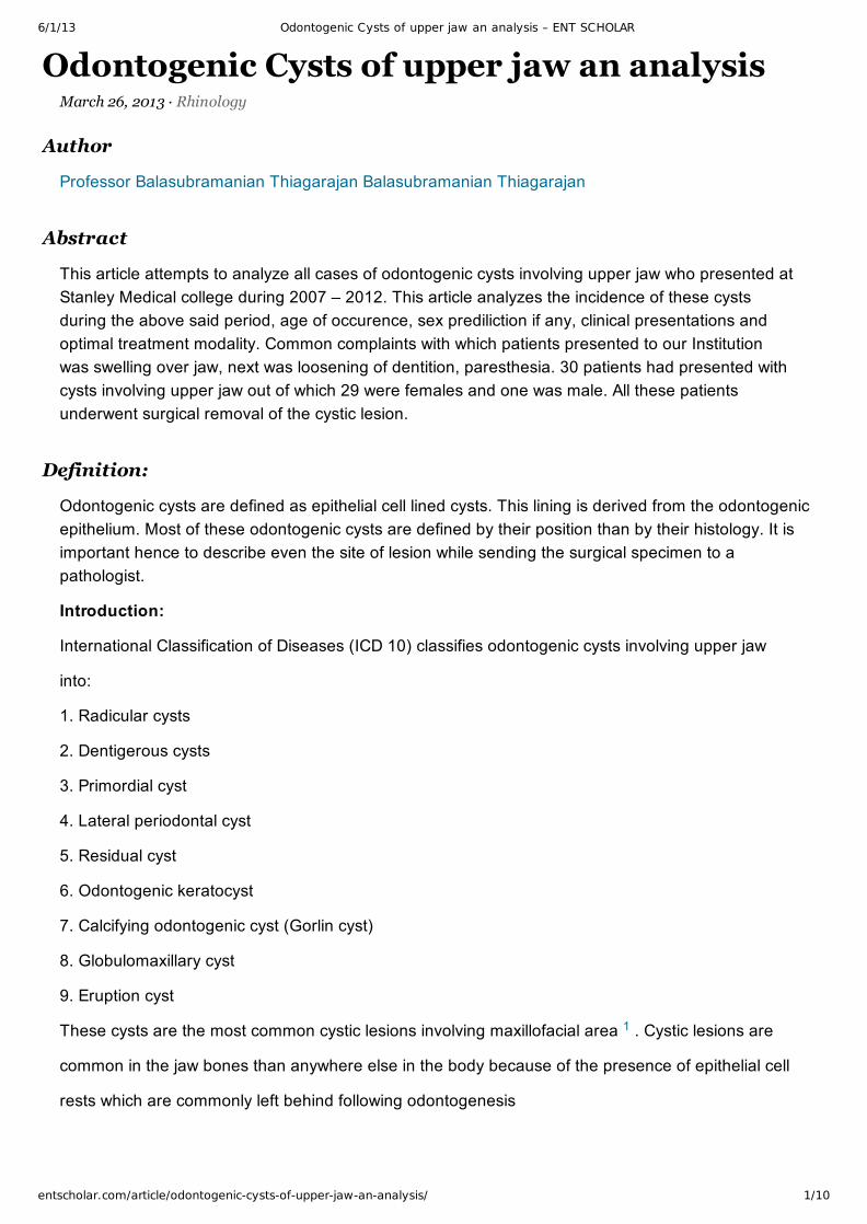
Coronal CT of nose and sinuses showing radicularcyst of upper jaw
CT scan axial cut of nose and sinuses showingglobulomaxillary cyst
CT scan showing dentigerous cyst with unerupted teeth
6/1/13 Odontogenic Cysts of upper jaw an analysis – ENT SCHOLAR
entscholar.com/article/odontogenic-cysts-of-upper-jaw-an-analysis/ 6/10
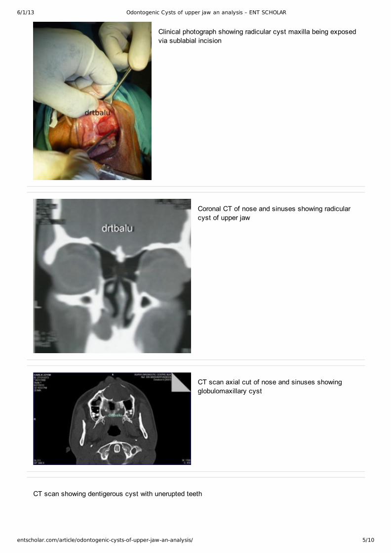
CT scan showing dentigerous cyst with uneruptedteeth
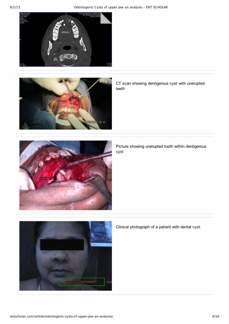
Picture showing unerupted tooth within dentigerouscyst
Clinical photograph of a patient with dental cyst

6/1/13 Odontogenic Cysts of upper jaw an analysis – ENT SCHOLAR
entscholar.com/article/odontogenic-cysts-of-upper-jaw-an-analysis/ 7/10
Dentigerous cyst:
Also known as follicular cyst. This cyst is associated with unerupted tooth. This cyst is formed due
to accumulation of fluid between the enamel epithelium and the completely formed tooth crown.
This overlying cyst prevents teeth from erupting. This cyst is almost always associated with
permanent dentition. In the upper jaw it is common in the canine tooth area. This cyst has its
highest incidence during the 2nd and 3rd decades of life.
Radiologically the presence of pericoronal radiolucency is a diagnostic pointer. This tumor should
be differentiated from ameloblastoma, odontogenic keratocyst and calcifying odontogenic cyst. All
these lesions manifest with pericoronal radiolucency in routine radiographs.
Primordial cyst:
This cyst arises due to cystic changes that occur in a developing tooth bud before the actual
formation of enamel and dentin matrix. Since this cyst arises from developing tooth bud the tooth
would be missing from the dental arch, or if teeth are all present then the presence of
suprenumerary teeth should be suspected.
Lateral periodontal cyst:
This cyst develops from the periodontal ligament close to the lateral surface of erupted / unerupted
teeth. This cyst is asymptomatic. The involved teeth is vital.
Residual cyst:
This cyst arises from remnants of epithelial cell rests left behind after extraction. This can also
occur when a radicular cyst at the apex of the teeth is extracted. This cyst is commonly seen in the
elderly.
Odontogenic keratocyst:
This cyst has a keratinized epithelial lining. Major draw back of this condition is its propensity to recureven after complete removal. This cyst can mimic any of the cysts described above. It needs
to be identified radiologically and pathologically. This cyst is seen between wide age groups.
Calcifying odontogenic cyst (Gorlin’s cyst):
This is a very rare slow growing benign tumor like cyst. This condition manifests the features of
solid mass while displaying features of tumor and cystic lesion. This cyst has equal incidence in
both maxilla and mandible.
Globulomaxillary cyst:
This is actually a fissural cyst arising from epithelial inclusions trapped at the line of fusion between
the globular portion of the median nasal process and the maxillary process. Pathologists consider

6/1/13 Odontogenic Cysts of upper jaw an analysis – ENT SCHOLAR
entscholar.com/article/odontogenic-cysts-of-upper-jaw-an-analysis/ 8/10
this cyst to be odontogenic rather than developmental. Radiographs show these cysts as pear
shaped / circular shaped between the roots of maxillary lateral incisor and canine. Both these teeth
are vital in these patients.
Gingival cysts:
are of two types i.e. adult and new born. In newborn these cysts are multiple, but rarely may also besingle. They are located in the alveolar ridges. In children these cysts originate from the dentallamina. They are asymptomatic and donot cause any problems. In adults these cysts are commonlyfound in the lower premolar area. It is usually single.
Eruption cyst:
Also known as eruption hematoma. This occurs when the erupting tooth bursts through the bone, butis yet to penetrate the overlying gingiva. Bleeding into the cyst lumen may cause discoloration givingan impression of hematoma. These cysts rupture as soon as the tooth completes eruption, henceneed not be treated.
Coronal CT scan of nose and sinuses showingdental cyst right maxilla
Picture showing the site of lesion exposed prior tosurgery
Figure showing Caldwel Luc procedure completed via canine fossa

6/1/13 Odontogenic Cysts of upper jaw an analysis – ENT SCHOLAR
entscholar.com/article/odontogenic-cysts-of-upper-jaw-an-analysis/ 9/10
Inferior meatal antrostomy being performed tofacilitate drainage
Management:
Majority of odontogenic cysts can be removed surgically using sublabial incision and reaching theinterior of maxillary sinus via canine fossa (Caldwel Luc procedure). It should be borne in mind thatthe canine fossa is the thinnest part of the maxilla and can easily be breached.
After removal of the cyst via caldwel luc procedure it is mandatory to perform inferior meatalantrostomy to facilitate drainage of maxillary sinus because its mucociliary clearance mechanism isinadequate / reduced following surgery.
1. Shear M (1994) Developmental odontogenic cysts. An update. J Oral Pathol Med 23,111
2. Nakamura T, Ishida J, Nakano Y, Ishii T, Fukumoto M, Izumi H, Kaneko K (1995) A study of cystsin the oral region. Cysts of the jaw. J Nihon Univ Sch Dent 37, 3340
3. Benn A, Altini M (1996) Dentigerous cysts of inflammatory origin.A cliniccpathologic study. OralSurg Oral Med Oral Pathol Oral Padiol Endod 81, 203209
4. Kiss Csongor. Cell to cell interaction. Endodontic Topic 2004, 8:88103.
5. Muglali M, Komerik N, Bulut E, Yarim GF, Celebi N, Sumer M. Cytokine and chemokine levels inradicular and residual cyst fluid. J Oral Pathol Med 2008, 37: 1859.
References
6/1/13 Odontogenic Cysts of upper jaw an analysis – ENT SCHOLAR
entscholar.com/article/odontogenic-cysts-of-upper-jaw-an-analysis/ 10/10
6. Muglali M, Komerik N, Bulut E, Yarim GF, Celebi N, Sumer M. Cytokine and chemokine levels inradicular and residual cyst fluid. J Oral Pathol Med 2008, 37: 1859.
7. Lin LM, Huang GTJ dan Rosenberg PA. Proliferation of epithelial cell rests, formation of apicalcysts and regression of apical cysts after periapical wound healing. JOE 2007,33(8):90816.
8. Oliveira MG, Lauxen IS, Chaves ACM, Rados PV, Filho MSA. Immunohistochemical analysis of thepattern of p53 and PCNA expression in odontogonec cystic lesions. Med Oral Patol Oral Cir Bucal2008, 13(5):E27580.
9. Hayashi M, Ohshima T, Ohshima M, Yamaguchi Y, Miyata H, Takeichi O, et al. Profiling ofradicular cyst and odontogenic keratocyst cytokine production suggests common growthmechanisms. JOE 2008, 34(1):1421.
10. Nair P, Sundqvist G, Sjogren U. Experimental evidence supports the abscess theory ofdevelopment of radicular cysts. Oral Surg Oral Med Oral Pathol Oral Radiol Endod 2008, 106:294303.
11. Shylaja S. Mast cells in odontogenic cysts. Journal of Clinical and Diagnostic Research [serialonline] 2010 April [cited: 2011 October 15]; 4:22262236.