Embed Size (px)
Citation preview

www.wjpr.net Vol 3, Issue 4, 2014.
529
Sahota et al. World Journal of Pharmaceutical Research
OCCURRENCE OF YERSINIA ENTEROCOLITICA IN DRINKING
WATER IN THE ABSENCE OF INDICATOR ORGANISM
Sahota P*, Sharma N, Kirandip and Pandove G
Department of Microbiology, Punjab Agricultural University, Ludhiana, Punjab,
India.
ABSTRACT
The drinking water quality with respect to bacteriological and
physicochemical examination was done for 238 drinking water
samples from different water utilities (municipal corporation supply
taps, submersible pumps, filter samples, water tanks, storage tanks,
water coolers, etc.) of urban areas of Ludhiana, Punjab, India. Standard
methods were used for the analysis of pH, TDS, while E. coli, Yersinia
spp. determination was carried out using BWTK and Multiple Tube
Technique. Yersinia spp. was detected in 164 (68.91%) water samples.
Statistical correlation was examined between the bacteriological and
physicochemical parameters and found to be independent from each other. Difference
between mean in each case, high standard deviation indicate that the distribution is widely off
normal and exhibit an asymmetric distribution. The occurrence of E. coli was also not found
to be associated with the presence or absence of Yersinia enterocolitica. So, E. coli was not
found to be a suitable indicator for Yersinia spp. The findings of the current study reports the
occurrence of Y. enterocolitica in drinking water and suggesting that there is need for further
surveillance studies to understand the global epidemiology of emerging pathogens.
Keywords: BWTK, correlation, indicator orgainsm, urban areas, drinking water, Yersinia
enterocolitica.
INTRODUCTION
Safe water supplies and environmental sanitation are vital for protecting the environment,
improving health and alleviating property. Deterioration in water quality below the
established standards is mainly due to human activities and untreated or inadequately treated
wastes discharge in the environment. The World Health Organisation (WHO) estimated that
Article Received on 01 April 2014, Revised on 22 April 2014, Accepted on 15 May 2014
*Correspondence for Author
Dr.(Mrs.) Param Pal Sahota,
Sr.Microbiologist, Deptt. of
Microbiology, PAU,Ludhiana-
141004, Punjab.
World Journal of Pharmaceutical ReseaRch SJIF Impact Factor 5.045
Volume 3, Issue 4, 529-542. Research Articl ISSN 2277 – 7105
www.wjpr.net Vol 3, Issue 4, 2014.
530
Sahota et al. World Journal of Pharmaceutical Research
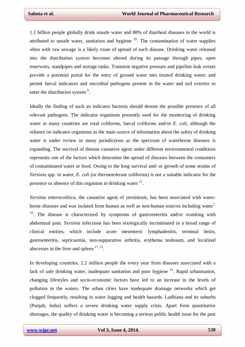
1.1 billion people globally drink unsafe water and 88% of diarrheal diseases in the world is
attributed to unsafe water, sanitation and hygiene 20. The contamination of water supplies
often with raw sewage is a likely route of spread of each disease. Drinking water released
into the distribution system becomes altered during its passage through pipes, open
reservoirs, standpipes and storage tanks. Transient negative pressure and pipeline leak events
provide a potential portal for the entry of ground water into treated drinking water; and
permit faecal indicators and microbial pathogens present in the water and soil exterior to
enter the distribution system 9.
Ideally the finding of such an indicator bacteria should denote the possible presence of all
relevant pathogens. The indicator organisms presently used for the monitoring of drinking
water in many countries are total coliforms, faecal coliforms and/or E. coli, although the
reliance on indicator organisms as the main source of information about the safety of drinking
water is under review in many jurisdictions as the spectrum of waterborne diseases is
expanding. The survival of disease causative agent under different environmental conditions
represents one of the factors which determine the spread of diseases between the consumers
of contaminated water or food. Owing to the long survival and/ or growth of some strains of
Yersinia spp. in water, E. coli (or thermotolerant coliforms) is not a suitable indicator for the
presence or absence of this organism in drinking water 21.
Yersinia enterocolitica, the causative agent of yersiniosis, has been associated with water-
borne diseases and was isolated from human as well as non-human sources including water7,
19. The disease is characterized by symptoms of gastroenteritis and/or vomiting with
abdominal pain. Yersinia infections has been etiologically incriminated in a broad range of
clinical entities, which include acute mesenteric lymphadenitis, terminal ileitis,
gastroenteritis, septicaemia, non-suppurative arthritis, erythema nodosum, and localized
abscesses in the liver and spleen 17, 23.
In developing countries, 2.2 million people die every year from diseases associated with a
lack of safe drinking water, inadequate sanitation and poor hygiene 22. Rapid urbanisation,
changing lifestyles and socio-economic factors have led to an increase in the levels of
pollution in the waters. The urban cities have inadequate drainage networks which get
clogged frequently, resulting in water logging and health hazards. Ludhiana and its suburbs
(Punjab, India) suffers a severe drinking water supply crisis. Apart from quantitative
shortages, the quality of drinking water is becoming a serious public health issue for the past

www.wjpr.net Vol 3, Issue 4, 2014.
531
Sahota et al. World Journal of Pharmaceutical Research
few years. Budha Nala, an important drainage line of Ludhiana district, passes through the
Ludhiana city, parallel to Sutlej and carries large volume of untreated sewerage water and
industrial effluents of the city. Most urban areas are serviced by a municipal water
distribution system that originates from local reservoirs or canals. The Municipal Corporation
is responsible for providing pure and safe drinking water to city dwellers on a regular and
continuous basis, and for regularly disposing of sewerage. However, the city dwellers often
find that the water is contaminated because of leaks in the pipelines, imperfect water taps and
misuse. The only reliable way to link water quality and health problems in urban areas where
a large population is exposed would be to perform the surveillance which contributes to the
protection of public health by promoting improvement of the quality, quantity, accessibility,
coverage, affordability and continuity of drinking water supplies.
The objective of this study was to determine the frequency of contamination as tested for
with indicators of bacterial and physico-chemical water quality parameters, occurrence of
yersiniae in drinking water, evaluating their association with the coliform fecal indicator
bacteria, characterization and study of its virulence factors.
MATERIAL AND METHODS
Study area
Ludhiana is the largest city in Punjab both in terms of area and population. The Municipal
Corporation has divided the whole city into ‘declared areas’ and ‘undeclared areas’. The
municipal corporation supplies domestic water to the declared areas while undeclared areas
have installed their own private water supply systems.
Sampling and physicochemical analyses
A total of 238 drinking water samples were collected from 185 municipal corporation supply
taps, 18 submersible pumps, 11 filter samples and 24 from water tanks, storage tanks, water
coolers, etc. from different water utilities from January 2010 to July 2011. The water samples
used for analysis were collected in sterile 1 L glass bottles, treated with sodium thiosulfate to
inactivate any residual halogen compound present in the sample (Na2S2O3 concentration of
18 mg/L neutralizes upto 5 mg of free (residual) chlorine per litre). The samples containing
high concentration of zinc and copper were treated with EDTA at concentration of 372 mg/L
to reduce metal toxicity 18. The samples were transported in refrigerated containers at 4˚C,
and analyzed within 24 h. The pH and TDS were determined using a portable calibrated pH
meter (Hanna Instruments) and HiMedia’s TDS meter, respectively.
www.wjpr.net Vol 3, Issue 4, 2014.
532
Sahota et al. World Journal of Pharmaceutical Research
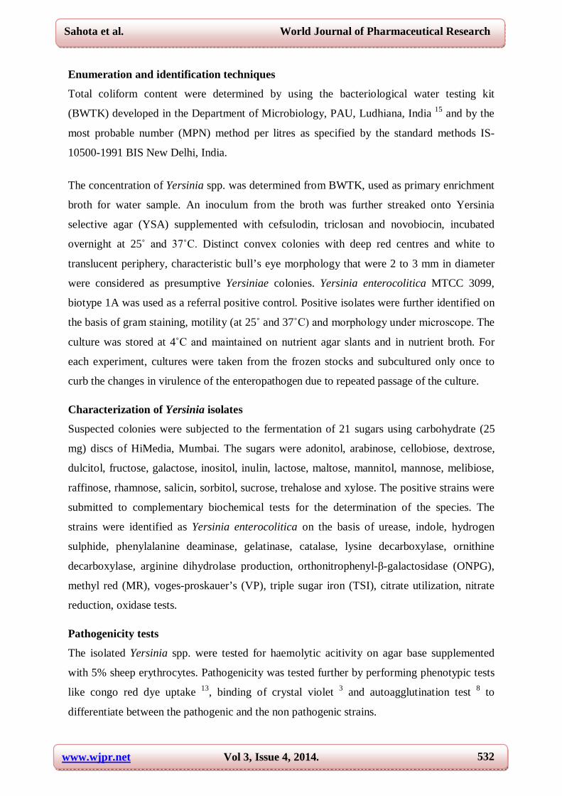
Enumeration and identification techniques
Total coliform content were determined by using the bacteriological water testing kit
(BWTK) developed in the Department of Microbiology, PAU, Ludhiana, India 15 and by the
most probable number (MPN) method per litres as specified by the standard methods IS-
10500-1991 BIS New Delhi, India.
The concentration of Yersinia spp. was determined from BWTK, used as primary enrichment
broth for water sample. An inoculum from the broth was further streaked onto Yersinia
selective agar (YSA) supplemented with cefsulodin, triclosan and novobiocin, incubated
overnight at 25˚ and 37˚C. Distinct convex colonies with deep red centres and white to
translucent periphery, characteristic bull’s eye morphology that were 2 to 3 mm in diameter
were considered as presumptive Yersiniae colonies. Yersinia enterocolitica MTCC 3099,
biotype 1A was used as a referral positive control. Positive isolates were further identified on
the basis of gram staining, motility (at 25˚ and 37˚C) and morphology under microscope. The
culture was stored at 4˚C and maintained on nutrient agar slants and in nutrient broth. For
each experiment, cultures were taken from the frozen stocks and subcultured only once to
curb the changes in virulence of the enteropathogen due to repeated passage of the culture.
Characterization of Yersinia isolates
Suspected colonies were subjected to the fermentation of 21 sugars using carbohydrate (25
mg) discs of HiMedia, Mumbai. The sugars were adonitol, arabinose, cellobiose, dextrose,
dulcitol, fructose, galactose, inositol, inulin, lactose, maltose, mannitol, mannose, melibiose,
raffinose, rhamnose, salicin, sorbitol, sucrose, trehalose and xylose. The positive strains were
submitted to complementary biochemical tests for the determination of the species. The
strains were identified as Yersinia enterocolitica on the basis of urease, indole, hydrogen
sulphide, phenylalanine deaminase, gelatinase, catalase, lysine decarboxylase, ornithine
decarboxylase, arginine dihydrolase production, orthonitrophenyl-β-galactosidase (ONPG),
methyl red (MR), voges-proskauer’s (VP), triple sugar iron (TSI), citrate utilization, nitrate
reduction, oxidase tests.
Pathogenicity tests
The isolated Yersinia spp. were tested for haemolytic acitivity on agar base supplemented
with 5% sheep erythrocytes. Pathogenicity was tested further by performing phenotypic tests
like congo red dye uptake 13, binding of crystal violet 3 and autoagglutination test 8 to
differentiate between the pathogenic and the non pathogenic strains.

www.wjpr.net Vol 3, Issue 4, 2014.
533
Sahota et al. World Journal of Pharmaceutical Research
Statistical analysis
Mean, standard deviation, variance and coefficient of variation of water samples were
calculated statistically of all the bacteriological and physicochemical parameters (MPN
index, pH, TDS, E. coli and Yersinia spp.). MPN index was compared with pH, TDS using
Pearson value of correlation at 1% level of significance. Measurements of bacteria (E. coli
and Yersinia spp.) were dichotomized as 0 for nondetection and 1 when measured as in
detection limits. For comparison between the dichotomous variables, the chi-squared
coefficient as per Yates correction factor was calculated at P <0.01 to evaluate the
relatedness.
RESULTS AND DISCUSSION
A randomized prospective drinking water survey was conducted over a period of 18 months
from the endemic area of gastroenteritis, urban and semi-urban areas of Ludhiana, Punjab,
India. A total of 238 drinking water samples were analyzed, 185 from Municipal Corporation
taps, 18 from submersible pumps, 11 filter samples and 24 from water coolers, water tanks,
storage tanks, etc., of which 66% (157) samples were positive for the occurrence of total
coliforms.
Out of 157 bacteriologically contaminated samples, 28.15% showed highest MPN index from
the adjoining areas of Budha Nala; Dharampura, New Madho Puri, Dhandari Kalan, New
Shakti Nagar, Basti Jodhewal. The large volume of domestic and industrial wastewater has
converted the Budha Nala to a virtually sewage drain and this resulted in the deterioration of
drinking water quality in the surrounding areas. The possible reason of high level of
microbial contamination in adjoining areas could be due to poor water storage conditions,
distribution lines, untreated water, sewage, poor hygiene, crowded living conditions with
inadequate sewage facilities. Other reasons for contamination may be the crossing over of
sewage pipes with fresh water supply lines, wrong alignment of water pipelines, illegal water
and sewerage connections, non chlorination of water reservoir due to lackadaisical attitude of
Municipal Corporation authorities. This suggests the reason for high prevalence of
waterborne diseases such as typhoid fever, diarrhea and dysentery.
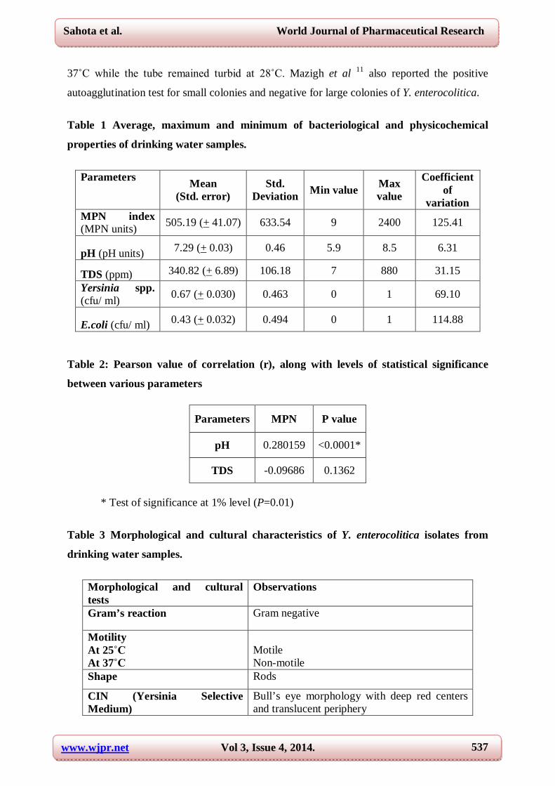
A summary of the bacteriological and physicochemical data for the water samples is shown
in Table 1. The average pH ranged from 6.83 - 7.75 and the average TDS was between
234.64 - 447. The data showed the maximum variation for MPN index ranged from 9 to 2400
followed by level of E. coli, Yersinia spp., TDS and minimum variation was observed in pH.

www.wjpr.net Vol 3, Issue 4, 2014.
534
Sahota et al. World Journal of Pharmaceutical Research
Difference between mean in each case, high standard deviation indicate that the distribution
is widely off normal and exhibit an asymmetric distribution. Measurements of bacteria (E.
coli and Yersinia spp.) were dichotomized as 0 for nondetection and 1 when measured as in
detection limits. Thus to conduct the surveillance of drinking water of a city, random
sampling is desirable as one particular area or locality does not represent the whole
population.
MPN value was statistically compared with pH and TDS using Pearson value of correlation at
1% level of significance (Table 2). pH value showed only upto 28% correlation with the
MPN index at P=0.01 which is not considered to be statistically significant, and TDS also did
not show any significant value. Thus, no correlation was found and it was concluded that the
bacteriological parameter (MPN index) is independent of physicochemical parameters (pH
and TDS). So, to interpret the quality of water, these parameters must be considered and
studied separately.
Identification of E. coli
For the isolation of E. coli, drinking water sample was inoculated in BWTK and MacConkey
tube and presumptive positive results were confirmed based on the ability of the organisms to
ferment by showing colour change in the kit and MPN tubes. Positive water samples were
then streaked on MacConkey lactose agar plates (37˚C, 48 h) and were further confirmed
with MUG medium after incubation for 24 h and observed yellow or yellowish brown
colonies under a UV lamp.
Isolation, identification and biochemical characterization of Yersinia enterocolitica from
drinking water.
For primary enrichment, test water sample (10 ml) was inoculated in BWTK and MacConkey
tubes and incubated at 37°C for 12 h. Water samples positive for Yersinia spp. formed
superficial film on the liquid media in the kit and tube, were streaked on Yersinia Selective
Supplement Medium (Cefsulodin 7.50mg/500ml Medium, Triclosan 2.0mg/500ml Medium,
Novobiocin 1.25mg/500ml) and incubated at 28°C and 37°C for 24 h. The cultural
characterization of presumptive isolates of Yersinia spp. showed convex colonies with 2-3
mm diameter, characteristic bull’s eye morphology with deep red centres and white to
translucent periphery. Typical Y. enterocolitica colonies on selective media that responded
positively to the mannitol and urease tests were counted and scored as Y. enterocolitica. They
were all gram negative rods, motile at 25˚C and non motile at 37˚C when observed under
www.wjpr.net Vol 3, Issue 4, 2014.
535
Sahota et al. World Journal of Pharmaceutical Research
microscope. The morphology of the isolates were also observed on HiChrome UTI agar and
nutrient agar which showed circular, smooth, convex colonies with glistening surface and
were easily emulsifiable. In nutrient broth, some strains of Yersinia enterocolitica produced
uniform turbidity with some green colouration in the medium. On MacConkey’s lactose agar,
all the isolated and test strains produced colourless, raised convex colonies having entire
margin. The colonies were small when incubated at 25˚C and were large when incubated at
37˚C (Table 3).
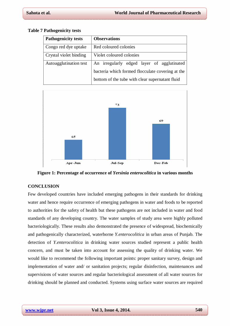
Yersinia spp. were present in 68.91% (164) drinking water samples tested bacteriologically
contaminated, of which Y. enterocolitica was the most frequently isolated. The isolation rate
of Yersiniae was nearly same in rainy (Jul-Sep) months and winter (Dec-Feb) months (Fig
1). The possible reasons may be due to its ability to survive for extended periods and even
multiply when water temperatures are low. The presence of Yersinia tends to be poorly
correlated with the levels of coliforms and HPC bacteria 5, 6. This might to a large extent be
due to the different temperature preference of these bacteria; whereas fecal coliform counts
are lower in winter, the occurrence of Y. enterocolitica is more frequent in colder months 10.
The preference for colder months is also reflected by the fact that the majority of yersiniosis
cases are reported from cooler regions in Europe and North America.
For identification to the species level, presumptive colonies were taken out of the pure culture
obtained and were then subjected to the fermentation of 21 sugars, of which, isolates were
able to ferment the following sugars: cellobiose, dextrose, fructose, mannitol, mannose,
melibiose, raffinose, and sucrose while negative for adonitol, arabinose, dulcitol, inulin,
inositol, lactose, maltose, salicin and xylose (Table 4). All the isolates were then
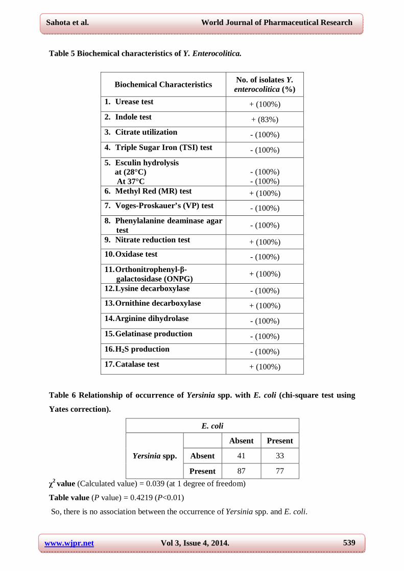
biochemically characterized and were positive for urease, methyl red, nitrate reduction,
orthonitrophenyl-β-galactosidase (ONPG), ornithine decarboxylase and catalase while 83%
of the strains showed indole-positive test. Citrate, triple sugar iron (TSI) utilization,
hydrolysis of esculin, voges-proskauer’s (VP), phenylalanine deaminase, oxidase, lysine
decarboxylase, arginine dihydrolase, gelatinase and H2S were not produced (Table 5). Strains
of Y. enterocolitica can be differentiated from Y. pseudotuberculosis with a positive result for
urease, fermentation of sucrose and negative reactions for rhamnose and melibiose
fermentation 1.

www.wjpr.net Vol 3, Issue 4, 2014.
536
Sahota et al. World Journal of Pharmaceutical Research
Correlation between the occurrence of E. coli and Yersinia spp.
The relationship between the occurrence of Yersinia spp. and E. coli was studied on the basis
of their presence or absence with respect to each other by chi-square test using Yates
correction factor at one degree of freedom. All the drinking water samples divided into 4
sections- samples in which both the organisms were absent and vice-versa, and the samples in
which one organism is absent and other is present as shown in Table 6. For statistical
comparison, chi-square test between presence or absence of the organisms (Yersinia spp. and
E. coli) was used, which is considered a valid hypothesis that there is no association between
the occurrence of 2 populations (or organisms). Calculated value at one degree of freedom
was 0.039 which is less than the one-tailed P value (0.4219) at P<0.01. This result showed
that the association between the occurrence of two organisms (Yersinia spp. and E. coli) is
considered not to be statistically significant, i.e., E. coli is not a suitable indicator to show the
presence of Yersinia spp.
Different authors have also found that coliform count did not correlate with the occurrence of
Yersinia spp. Schiemann 16 in his study on surface and well water also found that many
samples that showed positive results for Y. enterocolitica were negative for coliforms.
Determination of pathogenicity of Y. enterocolitica
Our study revealed that all the strains of Y. enterocolitica showed haemolytic activity, with a
halo diameter. Production of hemolysin was performed in nutrient agar plates with 5% of
sheep blood incubated at 35+0.5˚C for 24 and 48 h. The haemolytic activity is strongly
associated with enterotoxin production 12.
All the isolates of Yersinia enterocolitica were positive for congo red dye uptake and crystal
violet binding (Table 7). The ability to take up dye is associated with the presence of
virulence plasmid. The virulent plasmid associated determinants have been used to
distinguish between virulent and avirulent strains, including colony morphology, crystal
violet binding, congo red uptake, autoagglutination, hydrophobicity, mannose resistant
haemagglutination and serum resistance 2,4,14. The congo red pigmentation assay provides a
simple and efficient means of screening for virulence.
The autoagglutination of Y. enterocolitica is dependent on the virulence plasmid. All the
isolates were tested by inoculating and incubating two tubes of tissue culture medium, one
was incubated at 37˚C and the second at 28˚C. All the strains showed autoagglutination at
www.wjpr.net Vol 3, Issue 4, 2014.
537
Sahota et al. World Journal of Pharmaceutical Research
37˚C while the tube remained turbid at 28˚C. Mazigh et al 11 also reported the positive
autoagglutination test for small colonies and negative for large colonies of Y. enterocolitica.
Table 1 Average, maximum and minimum of bacteriological and physicochemical
properties of drinking water samples.
Parameters Mean
(Std. error) Std.
Deviation Min value Max value
Coefficient of
variation MPN index (MPN units) 505.19 (+ 41.07) 633.54 9 2400 125.41
pH (pH units) 7.29 (+ 0.03) 0.46 5.9 8.5 6.31
TDS (ppm) 340.82 (+ 6.89) 106.18 7 880 31.15 Yersinia spp. (cfu/ ml) 0.67 (+ 0.030) 0.463 0 1 69.10
E.coli (cfu/ ml) 0.43 (+ 0.032) 0.494 0 1 114.88
Table 2: Pearson value of correlation (r), along with levels of statistical significance
between various parameters
Parameters MPN P value
pH 0.280159 <0.0001*
TDS -0.09686 0.1362
* Test of significance at 1% level (P=0.01)
Table 3 Morphological and cultural characteristics of Y. enterocolitica isolates from
drinking water samples.
Morphological and cultural tests
Observations
Gram’s reaction Gram negative
Motility At 25˚C At 37˚C
Motile Non-motile
Shape Rods
CIN (Yersinia Selective Medium)
Bull’s eye morphology with deep red centers and translucent periphery

www.wjpr.net Vol 3, Issue 4, 2014.
538
Sahota et al. World Journal of Pharmaceutical Research
HiChrome UTI agar circular, smooth, convex colonies, with glistening surface
Trypticase soya agar circular, smooth, convex colonies and were easily emulsifiable
Nutrient agar circular, smooth, convex colonies, with glistening surface, 1-2 mm in diameter, entire or slightly crenated edges and were easily emulsifiable
Mac Conkey’s lactose agar At 25˚C At 37˚C
Small colourless, raised convex colonies having entire margin Large colourless, raised convex colonies having entire margin
Nutrient broth Yersinia enterocolitica produced uniform turbidity with some green colouration by some strains
Bacteriological Water Testing Kit (BWTK)
Forms superficial layer on the kit
Table 4 Comparison of fermentation of various sugars by isolates of Y. Enterocolitica.
Sugars utilization No. of isolates Y. enterocolitica (%)
1. Adonitol - (100%) 2. Arabinose + (17%), - (83%) 3. Cellobiose + (67%), - (33%) 4. Dextrose + (50%), - (50%) 5. Dulcitol - (100%) 6. Fructose +(50%), - (50%) 7. Galactose +(17%),- (83%) 8. Inulin - (100%) 9. Inositol - (100%) 10. Lactose - (100%) 11. Maltose +(17%),- (83%) 12. Mannitol + (100%) 13. Mannose +(67%),- (33%) 14. Melibiose +(50%),- (50%) 15. Raffinose +(50%),- (50%) 16. Rhamnose +(17%),- (83%) 17. Salicin - (100%) 18. Sorbitol +(17%),- (83%) 19. Sucrose +(67%),- (33%) 20. Trehalose +(17%),- (83%) 21. Xylose - (100%)
www.wjpr.net Vol 3, Issue 4, 2014.
539
Sahota et al. World Journal of Pharmaceutical Research
Table 5 Biochemical characteristics of Y. Enterocolitica.
Biochemical Characteristics No. of isolates Y. enterocolitica (%)
1. Urease test + (100%) 2. Indole test + (83%) 3. Citrate utilization - (100%) 4. Triple Sugar Iron (TSI) test - (100%) 5. Esculin hydrolysis at (28°C) At 37°C
- (100%) - (100%)
6. Methyl Red (MR) test + (100%) 7. Voges-Proskauer’s (VP) test - (100%) 8. Phenylalanine deaminase agar
test - (100%)
9. Nitrate reduction test + (100%) 10. Oxidase test - (100%) 11. Orthonitrophenyl-β-
galactosidase (ONPG) + (100%)
12. Lysine decarboxylase - (100%) 13. Ornithine decarboxylase + (100%) 14. Arginine dihydrolase - (100%) 15. Gelatinase production - (100%) 16. H2S production - (100%) 17. Catalase test + (100%)
Table 6 Relationship of occurrence of Yersinia spp. with E. coli (chi-square test using
Yates correction).
E. coli
Yersinia spp.
Absent Present
Absent 41 33
Present 87 77 χ2 value (Calculated value) = 0.039 (at 1 degree of freedom)
Table value (P value) = 0.4219 (P<0.01)
So, there is no association between the occurrence of Yersinia spp. and E. coli.
www.wjpr.net Vol 3, Issue 4, 2014.
540
Sahota et al. World Journal of Pharmaceutical Research
Table 7 Pathogenicity tests
Pathogenicity tests Observations
Congo red dye uptake Red coloured colonies
Crystal violet binding Violet coloured colonies
Autoagglutination test An irregularly edged layer of agglutinated
bacteria which formed flocculate covering at the
bottom of the tube with clear supernatant fluid
Figure 1: Percentage of occurrence of Yersinia enterocolitica in various months
CONCLUSION
Few developed countries have included emerging pathogens in their standards for drinking
water and hence require occurrence of emerging pathogens in water and foods to be reported
to authorities for the safety of health but these pathogens are not included in water and food
standards of any developing country. The water samples of study area were highly polluted
bacteriologically. These results also demonstrated the presence of widespread, biochemically
and pathogenically characterized, waterborne Y.enterocolitica in urban areas of Punjab. The
detection of Y.enterocolitica in drinking water sources studied represent a public health
concern, and must be taken into account for assessing the quality of drinking water. We
would like to recommend the following important points: proper sanitary survey, design and
implementation of water and/ or sanitation projects; regular disinfection, maintenances and
supervisions of water sources and regular bacteriological assessment of all water sources for
drinking should be planned and conducted. Systems using surface water sources are required
www.wjpr.net Vol 3, Issue 4, 2014.
541
Sahota et al. World Journal of Pharmaceutical Research
to disinfect to ensure that all bacterial contamination (such as E. coli and Yersinia spp.) is
inactivated.
REFERENCES
1. Bercovier H., Brenner D. J., Ursing J., Steigerwalt A. G., Fanning G. R., Alonso J. M.,
Carter G. P. and Mollaret H. H. Characterisation of Yersinia enterocolitica sensu strict,
Curr. Microbiol. 4, 201-206 (1980).
2. Bhaduri S. Pathogenic Yersinia enterocolitica. In: Guide to Foodborne Pathogens (R. G.
Labbe and S. Garcia, ed.). John Wiley and Sons, New York, NY, USA, pp. 245-255
(2001).
3. Bhaduri S., Conway L. K. and Lachica R. V. Assay of crystal violet binding for rapid
identification of virulent plasmid bearing clones of Yersinia enterocolitica, J. Clin.
Microbiol. 25, 1039-1042 (1987).
4. Carniel E. 2006 Y. enterocolitica and Y. pseudotuberculosis enteropathogenic yersiniae.
In: The Prokaryotes (M. Dworkin, S. Falkow, E. Rosenberg & E. Stackebrandt, ed.).
Springer, New York, USA, pp. 270-398 (2006).
5. Falcão J. P., Dias A. M. G., Correa E. F. and Falcão D. P. Microbiological quality of ice
used to refrigerate foods, Food Microbiol. 19, 269-276 (2002).
6. Health Canada Guidelines for Canadian Drinking Water Quality: Guideline Technical
Document- Bacterial Waterborne Pathogens-Current and Emerging Organisms of
Concern, Water Quality and Health Bureau, Healthy Environments and Consumer Safety
Branch, Health Canada, Ottawa, Ontario (2006).
7. Highsmith A. K., Feeley J. C., Skaliy P., Wells J. G. and Wood B. T. Isolation of Y.
enterocolitica from well water and growth in distilled water, Appl. Environ. Microbiol. 4,
745-750 (1977).
8. Laird W. J. and Cavanaugh D. C. Correlation of autoagglutination and virulence of
yersiniae, J. Clin. Microbiol. 11, 430-432 (1989).
9. LeChavellier M. W., Gullick R. W., Karim M. R., Friedman M. and Funk J. E. The
potential for health risks from intrusion of contaminants into the distribution system from
pressure transients, J. Water Health 1, 3-14 (2003).
10. Massa S., Cesaroni D., Poda G. and Trovatelli L. D. Isolation of Yersinia enterocolitica
and related species from river water, Zentralbl. Mikrobiol. 143, 575-581 (1988).
www.wjpr.net Vol 3, Issue 4, 2014.
542
Sahota et al. World Journal of Pharmaceutical Research
11. Mazigh D., Alonso J. M. and Mollaret H. H. Simple method for demonstration of
differential colony morphology of plasmid-associated virulent clones of Yersinia
enterocolitica, J. Clin. Microbiol. 17, 555-557 (1983).
12. Paniagua C., Rivero O., Anguita J. and Naharro G. Pathogenicity factors and virulence
for rainbow trout (Salmo gairdneri) of motile Aeromonas spp. isolates from river, J. Clin.
Microbiol. 28, 350-355 (1990).
13. Riley G. and Toma S. Detection of pathogenic Yersinia enterocolitica by using Congo
red-magnesium oxalate agar medium, J. Clin. Microbiol. 27, 213-214 (1989).
14. Robins-Browne R. M. Yersinia enterocolitica. In: Food Microbiology, Fundamentals and
Frontiers (M. P. Doyle, L. R. Beuchat & T. J. Montville, ed.). ASM Press, Washington
DC, USA, 2:215-245 (2011).
15. Sahota P., Pandove G., Achal V. and Vikal Y. Evaluation of a BWTK for detection of
total coliforms, E. coli and emerging pathogens from drinking water: comparison with
standard MPN method, Water. Sci. Technol. 62(3):676-683 (2010).
16. Schiemann D. A. Isolation of Y. enterocolitica from surface and well waters in Ontario,
Canad. J. Micrbiol. 24, 1048-1052 (1978).
17. Sonnenwirth A. C. Yersinia. In: Manual of clinical microbiology (E.H. Lennette, E. H.
Spaulding & J. P. Traunt, 2nd ed.). American Society for Microbiology, Washington DC,
pp. 222-229 (1974).
18. Standard Methods for the Examination of Water and Wastewater 17th edition, American
Public Health Association, Washington, DC (1989).
19. Toma S., and Lafleur L. Survey on the incidence of Yersinia enterocolitica infection in
Canada, Appl. Microbiol. 28, 469-473 (1974).
20. WHO Quantifying selected major risks to health, The World Health Report 2002. World
Health Organization, Geneva (2003a).
21. WHO Guidelines for Drinking Water Quality, 4th edition, World Health Organisation,
Geneva (2011).
22. WHO/UNICEF United Nations Children’s Fund, and Water Supply and Sanitation
Collaborative Council, Global Water Supply and Sanitation Assessment 2000 Report
(2000).
23. Worsmer G. P. and Keusch G. T. Yersinia enterocolitica: clinical observations In:
Yersinia enterocolitica (E. J. Bottone ed.). CRC Press, Inc., Boca Raton, Fla., pp. 83-93
(1981).