Embed Size (px)
Citation preview
1
Yersinia enterocolitica behavior in the presence of the bacterivorous Acanthamoeba 1
castellanii 2
Lambrecht E.1, Baré J.1, Van Damme I.1, Bert W.2 , Sabbe K.3, Houf K.1* 3
1Department of Veterinary Public Health and Food Safety, Faculty of Veterinary Medicine, 4
Ghent University, Merelbeke, Belgium 5
2 Nematology Unit, Department of Biology, Ghent University, Ghent, Belgium 6
3 Laboratory of Protistology and Aquatic Ecology, Department of Biology, Ghent University, 7
Ghent, Belgium 8
9
RUNNING TITLE: Yersinia enterocolitica and A. castellanii interactions 10
11
CORRESPONDING AUTHOR 12
Kurt Houf 13
Ghent University 14
Department of Veterinary Public Health and Food Safety 15
Salisburylaan 133 16
9820 Merelbeke-Belgium 17
Tel: +32 09 264 74 51; Fax: +32 09 264 74 91 19
20
KEYWORDS: Acanthamoeba castellanii, Yersinia enterocolitica, Foodborne pathogen21
Copyright © 2013, American Society for Microbiology. All Rights Reserved.Appl. Environ. Microbiol. doi:10.1128/AEM.01915-13 AEM Accepts, published online ahead of print on 9 August 2013
on February 3, 2018 by guest
http://aem.asm
.org/D
ownloaded from
2
Abstract 22
Free-living protozoa play an important role in the ecology and epidemiology of human 23
pathogenic bacteria. In the present study, the interaction between Yersinia enterocolitica, an 24
important foodborne pathogen, and the free-living amoeba Acanthamoeba castellanii was 25
studied. Several cocultivation assays were set up to assess the resistance of Y. enterocolitica to A. 26
castellanii predation and the impact of environmental factors and bacterial strain-specific 27
characteristics. Results showed that all Y. enterocolitica strains persist in association with A. 28
castellanii for at least 14 days, and association with A. castellanii enhanced survival of Yersinia 29
under nutrient-rich conditions at 25 °C and under nutrient-poor conditions at 37 °C. Amoebae 30
cultivated in the supernatant of one Yersinia strain showed a temperature- and time-dependent 31
permeabilization. Intraprotozoan survival of Y. enterocolitica depended on nutrient availability 32
and temperature, with up to 2.8 log cfu/mL bacteria displaying intracellular survival at 7 °C for at 33
least 4 days in nutrient-rich medium. Transmission electron microscopy was performed to locate 34
the Yersinia inside the amoebae. As Yersinia and Acanthamoeba share similar ecological niches, 35
this interaction identifies a role of free-living protozoa in the ecology and epidemiology of Y. 36
enterocolitica. 37
on February 3, 2018 by guest
http://aem.asm
.org/D
ownloaded from
3
Introduction 38
Yersinia enterocolitica is the third most frequently reported foodborne pathogen in the European 39
Union, with 7017 reported human cases in 2011 (1). Yersiniosis occurs mainly in infants and 40
preschoolers and is characterized by acute enteritis with fever and diarrhea. In adolescents and 41
adults, pseudo-appendicitis, sequelae as arthritis and erythema nodosum, and septicemia can also 42
occur (2, 3). Most infections are caused by Y. enterocolitica bioserotypes 4/O:3 and 2/O:9, which 43
all harbor a virulence plasmid (pYV) that is required for full pathogenicity in humans (1, 4). This 44
virulence plasmid is optimally expressed at 37 °C, but can become lost during laboratory 45
cultivation (5, 6). 46
Most Y. enterocolitica infections in humans are associated with the handling and 47
consumption of raw or undercooked pork products (1). Pigs are considered as the primary 48
reservoir of human pathogenic Y. enterocolitica as they harbor these pathogens in the tonsils and 49
gastro-intestinal lymphoid tissue (7, 8). However, many factors related to the epidemiology of Y. 50
enterocolitica, such as environmental sources and transmission routes, remain indistinct (9) . 51
Free-living protozoa are unicellular, heterotrophic, eukaryotic micro-organisms 52
ubiquitous in natural and anthropogenic aquatic and terrestrial environments, including food-53
related environments (10-13). Free-living protozoa, including members of the amoebal genus 54
Acanthamoeba graze on bacteria and are therefore considered as important bacterial predators. 55
However, various bacteria are able to evade protozoan ingestion and/or digestion and can even 56
benefit from the association with protozoa (14). Some bacteria enhance their survival by 57
extracellular association (15), though others are taken up and enter a membrane bound vacuole, 58
resisting protozoan digestion (16-18). Both associations result in survival, and even 59
multiplication, of the bacteria in the presence of its predator. The intraprotozoan location further 60
on February 3, 2018 by guest
http://aem.asm
.org/D
ownloaded from
4
represents a shelter for bacteria against physical and chemical environmental conditions (16, 19). 61
Moreover, free-living protozoa serve as a vehicle for the colonization of new habitats (16) and 62
hosts (20). Free-living protozoa also play a role in the selection of bacterial virulence traits and 63
adaptation to survival in macrophages (21-24). In addition, gene transfer between intraprotozoan 64
bacteria and protozoan host has been reported (25). 65
Yersinia enterocolitica and free-living protozoa share the same ecological niches, such as 66
water (26, 27), vegetables (28, 29) and anthropogenic environments, e.g. domestic refrigerators 67
(13, 30). The role of free-living protozoa in the ecology and epidemiology of foodborne 68
pathogens, such as Campylobacter, Salmonella and E. coli has already been documented (31-33), 69
but for Y. enterocolitica, only a brief description of an intraprotozoan replication, probably 70
followed by the killing and digestion of the bacterial strain (34), and an increased resistance to 71
free chlorine in their early intraprotozoan lifestyle (19) have been reported so far. 72
To further elucidate the ecology and epidemiology of this common pathogen, evaluation 73
of the importance of its association with free-living protozoa, and the influence of environmental 74
factors and Yersinia strain characteristics thereon, is needed. 75
The objectives of the present study were to assess the impact and the nature of the 76
associations between Y. enterocolitica and free-living protozoa. Therefore, in vitro coculture 77
experiments using the model protozoon Acanthamoeba castellanii and Yersinia strains with 78
different bacterial characteristics were performed under different environmental conditions. 79
These coculture experiments (i) evaluated the ability of Y. enterocolitica to survive or grow in 80
association with the bacterivorous Acanthamoeba under different conditions, (ii) assessed if the 81
yersiniae could survive or grow extracellularly on factors released by the amoebae and vice versa, 82
and (iii) investigated if Y. enterocolitica were resistant to amoebal digestion, if they could survive 83
or grow intracellularly, and where they were exactly located inside A. castellanii. 84
on February 3, 2018 by guest
http://aem.asm
.org/D
ownloaded from
5
Materials and methods 85
86
Cultivation of Acanthamoeba castellanii. Acanthamoeba castellanii (American Type Culture 87
Collection ATCC30234) was maintained axenically in Proteose peptone Yeast extract Glucose 88
medium (PYG, ATCC-recipe, http//www.lgcstandards-atcc.org) at 25 °C in 75 cm2 culture flasks. 89
Light microscopic observations and plating of culture samples on Plate Count Agar (PCA, 90
Biorad, Hercules, California, USA), which were incubated at 30 °C for 48 h, were performed to 91
verify axenicity of the culture. For coculture experiments, stationary growth phase amoebae (3.5 92
days old) forming a monolayer were used. The adherent amoebae were harvested by tapping the 93
flasks and subsequent centrifugation of the solution (300 x g, 5 min), washed with Page’s 94
Amoeba Saline (PAS, ATCC-recipe), and resuspended in PYG or PAS medium, depending on 95
the experiment to be performed. For further interaction studies, the Acanthamoeba culture was 96
diluted to c. 5x10⁵ living trophozoites/mL. The actual cell numbers were determined with a 97
Füchs-Rosenthal chamber (Blaubrand, Wertheim, Germany). The membrane integrity and thus 98
also the cell viability of the Acanthamoeba trophozoites was assessed using the trypan blue 99
exclusion assay (35). 100
101
Cultivation of Yersinia enterocolitica strains. Four Y. enterocolitica strains, isolated between 102
2010-2011, were used in this study: strain YeH3+ (bioserotype 4/O:3, pYV+), and its plasmid 103
cured derivate YeH3- were isolated from the stool of a one-year old boy, strain YeH9- 104
(bioserotype 2/O:9, pYV-) from the stool of a 43-year old woman, and strain YeM3+ 105
(bioserotype 4/O:3, pYV+) from minced pork. All strains were maintained in glycerol at -20 °C. 106
Strains were cultivated in Tryptone Soya Broth (TSB, Biorad, Hercules, California, USA) at 37 107
°C for 24 h to activate the pYV-virulence plasmid (36). At the stationary growth phase, bacteria 108
on February 3, 2018 by guest
http://aem.asm
.org/D
ownloaded from
6
were harvested by centrifugation (10,000 x g, 5 min), washed in PAS and resuspended in PYG 109
or PAS (depending on the experimental setup). For use in further experiments, the bacterial 110
suspension was diluted to c. 5x10⁷ CFU/ml by using data from Y. enterocolitica growth curves. 111
The exact number of viable bacteria was determined after plating a serial dilution of the 112
suspension on PCA and 48 h incubation at 30 °C. Before the start of the experiments and after 113
each coculture experiment, the bioserotype and the presence of the virulence plasmid of each Y. 114
enterocolitica strain were verified by biochemical testing and PCR analysis as described by Van 115
Damme et al. (37). 116
117
Coculture experiments. Coculture experiments with A. castellanii and the four Y. enterocolitica 118
strains were performed in both nutrient-rich (PYG) and nutrient-poor (PAS) media (34, 35, 38) at 119
7, 25 and 37 °C. Those nutrient and temperature conditions were used to mimic natural (e.g. 120
aquatic areas (39)), food-related environmental conditions (e.g. refrigerators, temperatures and 121
organic loads in food processing areas (13, 40)), and the mammal body temperature. 122
123
Persistence of Yersinia in coculture with Acanthamoeba castellanii. The ability of Y. 124
enterocolitica to survive in association with the bacterivorous amoebae was assessed by 125
persistence assays. With these assays, no distinction could be made if recovered bacteria were 126
internalized in the amoebae or if it concerned adherent or free-living extracellular bacteria. 127
Acanthamoeba castellanii cells were seeded into 12-well plates (2 mL/well at a concentration of 128
c. 5x10⁵ living trophozoites/mL PYG or PAS) and plates were incubated at 25 °C for 2 h to allow 129
amoebal settlement and adhesion. Then, the medium was gently removed and replaced by 2 mL 130
Y. enterocolitica suspension (c. 5x10⁵ or 5x10⁷ cfu/mL; dissolved in PYG or PAS) to achieve a 131
on February 3, 2018 by guest
http://aem.asm
.org/D
ownloaded from
7
multiplicity of infection of c.1 bacteria per amoeba and c.100 bacteria per amoeba, respectively. 132
Plates were centrifuged at 50 x g for 5 min to enhance cell contact and incubated at 7, 25 or 37 133
°C. Monocultures of A. castellanii and of each Yersinia strain were included as controls. At day 134
1, 2, 3, 6, 7, 10 and 14, the cells were harvested by scraping the wells. The amoebal cell integrity 135
was visually evaluated by light microscopy and the trypan blue exclusion assay. The number of 136
viable Yersinia was determined after plating serial dilutions of the cell suspension on PCA and 137
incubation for 48 h at 30 °C. 138
139
Effect of released factors on microbial survival and growth. The ability of Y. enterocolitica to 140
survive and/or grow in association with A. castellanii without direct interaction with the 141
amoebae, i.e. extracellularly on factors released by the amoebae, and vice versa, was determined 142
by supernatant assays. Therefore, A. castellanii (c. 5x 10⁵ cells/mL) and Yersinia strains (c. 5x 143
107 cfu/mL) were harvested after 5 min centrifugation at 300 x g and 5 min at 10,000 x g, 144
respectively. The supernatant of both the bacterial and amoebal monocultures was filter-sterilized 145
(0.22 µm, Nalgene Syringe cellulose acetate membrane, Thermo Scientific, Langenselbold, 146
Germany) and the pH of the bacterial supernatant was measured (WTW pH 330i, WTW, 147
Weilheim, Germany). Subsequently, the Yersinia were resuspended in the supernatant of the 148
amoeba cultures and vice versa. One mL of each Yersinia or A. castellanii suspension was 149
inoculated in 12 well-plates. As controls, amoebae suspended in TSB-medium (growth medium 150
of the Yersinia cultures) and Y. enterocolitica strains suspended in PYG-medium (growth 151
medium of Acanthamoeba) were included. All plates were incubated at 7, 25 and 37 °C. After 3 h 152
and 1, 2 and 3 days of incubation, cells were harvested by scraping the wells. The number of 153
on February 3, 2018 by guest
http://aem.asm
.org/D
ownloaded from
8
viable Yersinia and the amoebal concentration and cell integrity were determined as described 154
above. 155
156
Intraprotozoan survival of Y. enterocolitica. The ability of Y. enterocolitica to survive inside A. 157
castellanii, and the enumeration of the intracellular Yersinia, was assessed by gentamycin 158
protection assays. Cocultures and amoebal monocultures were set up as described above for the 159
persistence assays. After 2 h of (co)cultivation, the medium was gently removed and the cells 160
were washed with PAS to minimize extracellular bacteria. Afterwards, 4 mL of gentamycin 161
sulphate solution (Sigma-Aldrich, St. Louis, USA) at a final concentration of 100 µg/mL PYG 162
(34) was added to each well and the plates were incubated for 1 h at 7, 25 or 37 °C to kill the 163
extracellular Y. enterocolitica. In previous tests, this gentamycin treatment was effective in 164
killing 99.97 to 100% of the Y. enterocolitica, with no effect on the viability of Acanthamoeba 165
castellanii (data not shown). For the experiments in PAS, also gentamycin suspended in PYG 166
was used for initial killing of the extracellular bacteria, as this treatment was most effective (34). 167
After incubation, wells were washed with PAS and 4 mL gentamycin maintenance solution (20 168
µg gentamycin/mL PAS or PYG, depending on the experimental setup) was added to each well. 169
This moment was defined as the 0 h-time point (i.e. 3 h after initial setup of the (co)culture). The 170
plates were further incubated at 7, 25 or 37 °C, and after 1, 2, 3 and 4 days the number of 171
amoebae and the intra- and extra-amoebal bacterial counts were determined. For the extra-172
amoebal bacterial counts, the supernatant was removed and plated on PCA plates. Afterwards 173
wells were washed with PAS, 4 mL PYG or PAS was added to each well and the amoebae were 174
harvested by cell scraping. This cell suspension was subsampled for further analysis: 0.5 mL was 175
used for enumeration and viability testing of amoebae as described above, 2 mL of the cell 176
suspension was used for transmission electron microscopy analysis (cf. below), and the remaining 177
on February 3, 2018 by guest
http://aem.asm
.org/D
ownloaded from
9
amoebal suspension (1.5 mL) was used to determine the intra-amoebal bacterial counts. For the 178
latter, the amoebae were lysed with 0.5% sodiumdeoxycholate for 5 min at room temperature, 179
which has been shown to result in effective lysis of the amoebae without affecting intracellular 180
bacteria (data not shown). The suspension was plated on PCA plates. All plates were incubated at 181
30 °C for 48 h, after which the extra- and intracellular Yersinia were counted. 182
183
Localization of intraprotozoan Y. enterocolitica. Transmission electron microscopy (TEM) was 184
applied to determine the exact subcellular location of intraprotozoan Y. enterocolitica. The 185
amoebae from the coculture and control wells from the gentamycin protection assays (second 186
aliquot) were harvested by centrifugation at 300 x g for 5 min and serially fixed with 187
glutaraldehyde in sodium cacodylate buffer (pH 7.4) as follows: 0.8%, 1.25% and finally 2.5% 188
glutaraldehyde in 0.05M sodium cacodylate buffer (each concentration includes 20 min 189
incubation at 4 °C under continuous rotation (13 rpm, Dynal MX-1, Life Technologies, 190
Belgium). Cells were then rinsed three times 10 min in 0.05M sodium cacodylate buffer. Post 191
fixation took place in 1% osmium tetroxide in the same buffer for 1 h at room temperature. 192
Afterwards, the cells were rinsed twice with distilled water and were stepwise dehydrated, using 193
ethanol series of increasing concentrations (15-100% ethanol, 10 min each). The cells were 194
subsequently impregnated with a low-viscosity embedding medium (Spurr’s resin (41)), and 195
polymerisation was performed at 70 °C for 8 h. Samples were sectioned on a Reichert Ultracut S 196
(Leica, Vienna, Austria), first semi-thin (0.5 µm) until the region of interest was reached, after 197
which ultra-thin (70 nm) sections were made. Semi-thin sections were studied using a Wild light 198
microscope (Heerbrugg, Switserland). Ultra-thin sections were studied with a Jeol JEM-1010 199
transmission electron microscope (Jeol Ltd., Tokyo, Japan) operating at 60 kV, and pictures were 200
digitized using a Ditabis system (Pforzheim, Germany). 201
on February 3, 2018 by guest
http://aem.asm
.org/D
ownloaded from
10
Statistical analysis 202
All assays (persistence, supernatant, and gentamycin protection) were performed in triplicate, 203
except for the persistence assay with MOI 1:1. Statistical data analysis was performed on the 204
quantitative data using the software Stata 11.0 (Stata Corporation, College Station, Texas USA). 205
A probability of p<0.05 was required for statistical significance. Bacterial counts obtained by the 206
persistence assays were log10 transformed and analyzed for each time point using generalized 207
least squares regressions, with strains as random-effects. Coculture and monoculture counts were 208
compared for every combination of temperature and nutrient condition. To determine strain 209
and/or temperature differences in the absolute number of living amoeba/bacteria (supernatant 210
test) and of intraprotozoan Yersinia (gentamycin protection assays), negative binomial 211
regressions were used, including strains as random effects where necessary. 212
213
Results 214
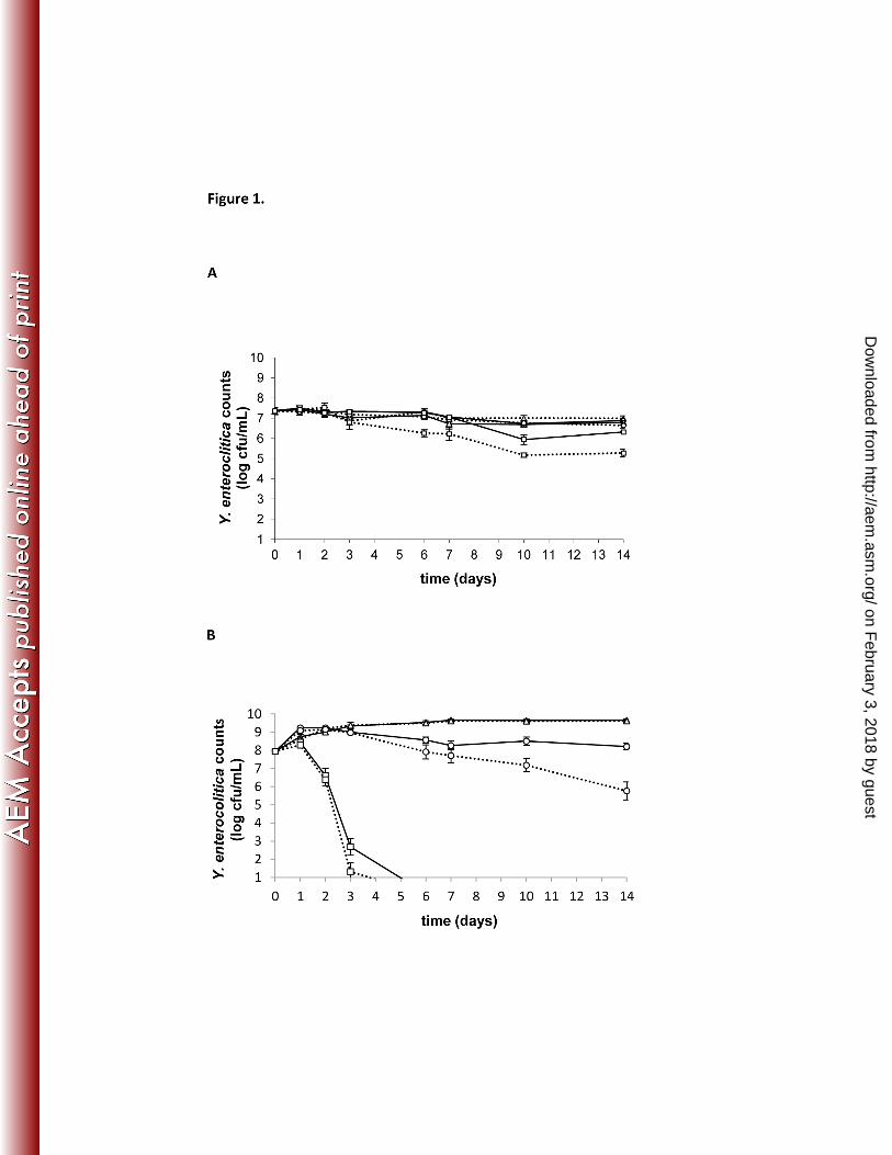
Persistence of Yersinia in coculture with Acanthamoeba castellanii. In general, for each strain 215
under each experimental condition (temperature and media combination), the survival of Y. 216
enterolitica in both cocultures and controls showed not to be strain-dependent. 217
In the cocultures, under nutrient-poor conditions (PAS, Fig. 1A), the number of viable 218
yersiniae did not differ significantly between the different temperatures (p>0.05). The number of 219
viable Yersinia in both coculture and monoculture conditions at 7 and 25 °C remained almost 220
constant during the 14-day monitoring period, with no significant differences between cocultures 221
and monocultures (p>0.05). At 37 °C, the number of viable Yersinia in the monocultures 222
on February 3, 2018 by guest
http://aem.asm
.org/D
ownloaded from
11
decreased earlier than the ones in the cocultures and by day 14 the number of viable Yersinia in 223
the monoculture was significant lower than in the cocultures (p<0.001). 224
Under nutrient-rich conditions (PYG, Fig. 1B), the number of viable yersiniae was 225
significantly higher in cocultures at 7 and 25°C compared to 37 °C after 1 day, and significantly 226
higher at 7 °C compared to 25 °C after 6 days (p<0.001). However, similar differences between 227
temperatures were obtained in the Yersinia monocultures. For each temperature, an initial 228
bacterial growth of 0.5 to 1.5 log cfu/mL was observed, both in cocultures and controls (Fig. 1B). 229
From day 3 onwards, a status quo of viable Yersinia was observed in both co- and monocultures 230
at 7 °C (c. 9.5 log cfu/mL) and in the cocultures at 25 °C (c. 8.5 log cfu/mL), whereas the 231
bacterial viability in the monocultures at 25 °C decreased. At 37 °C, no viable Yersinia could be 232
recovered after day 3, both in the co- and monocultures. At 7 and 37 °C, no significant difference 233
in viable Yersinia counts was detected between the cocultures and the monocultures, whereas at 234
25 °C the number of viable Yersinia in cocultures was significantly higher than in the 235
monocultures from day 6 onwards (p<0.05). 236
For each condition (i.e. specific strain, temperature and media combination), light 237
microscopy revealed no visual difference in amoebal cell integrity and density between 238
cocultures and amoebal monocultures during the 14-day monitoring period. In most conditions, 239
the amoebae stayed adherent and pseudopodia and vacuoles were visible, both in co- and 240
monocultures. By contrast, in PAS-medium at 7 and 25 °C a gradual transformation of 241
trophozoites into resting cyst was observed after 3 and 6 days respectively, in both coculture and 242
monoculture conditions. However, after 14 days, living trophozoites could still be detected. At 37 243
°C, amoebal viability declined and cyst formation and the presence of amoebal cell debris was 244
observed from day 3-4 onwards in both coculture and amoeba monoculture conditions. 245
Furthermore, from day 6 onwards, no trophozoites could be detected at 37 °C. 246
on February 3, 2018 by guest
http://aem.asm
.org/D
ownloaded from
12
To increase the amoebal grazing pressure on Yersinia, persistence assays with an MOI 1:1 247
were also performed at 7, 25 and 37 °C under nutrient-poor conditions, which resulted in similar 248
observations as reported above for MOI 100:1 (data not shown). 249
250
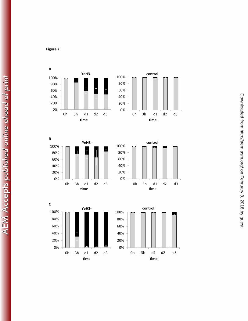
Effect of released factors on microbial survival and growth.. In general, the survival and 251
growth of yersiniae inoculated in amoebal supernatant were similar as seen in the controls. For 252
both, an initial growth of c. 1.0 -1.5 log cfu/mL was observed at each temperature. At 7 and 25°C 253
a slight increase c. 1 log cfu/mL was detected after time, while at 37 °C, a rapid decrease c. 6-8 254
log cfu/mL of yersiniae viability was observed after 1 day (data not shown). 255
No differences in amoebal survival and growth between those cultivated in bacterial 256
supernatant and in control medium were observed, except for amoebae inoculated in the 257
supernatant of Yersinia strain YeH3- (Fig. 2). Here, significantly lower absolute numbers of 258
viable amoebae were detected in bacterial supernatant than in the control after 3 h incubation at 259
37 °C (p<0.001), after 2 days at 25 °C (p<0.05) and after 3 days at 7 °C (p<0.001). From 3 h 260
onwards, the number of living amoebae in the supernatant treatment condition at 37 °C was 261
significantly lower compared to the treatment conditions at 25 °C and 7 °C (p<0.01). The total 262
number of amoebae, i.e. the sum of permeabilized and viable amoebae, remained the same over 263
time (c. 5x10⁵ cells/mL). 264
The initial pH of the TSB-medium before inoculation was 7.2 ± 0.1 units, but it decreased 265
to 5.2 units when YeH3- was cultivated in TSB. The pH-lowering effect of the other strains was 266
less pronounced (5.6-6.5 pH units). Acidified TSB-medium alone (HCl, 5.2 pH units) also caused 267
permeabilization at 7 and 37°C, but to a much lesser extent (i.e. maximum 28% permeabilized 268
cells after d4) than the amoeba cultivated in YeH3- supernatant (data not shown). 269
270
on February 3, 2018 by guest
http://aem.asm
.org/D
ownloaded from
13
Intraprotozoan survival of Y. enterocolitica. For each experimental conditions, amoebal counts 271
and cell integrity remained constant over time, in both cocultures and amoeba monoculture. In 272
nutrient-poor medium (PAS) at 25 and 37 °C, no viable intracellular Yersinia could be recovered 273
after one day. At 7 °C, no extracellular Yersinia were recovered at the 0 h time point, after 274
treatment with 100 µg/mL gentamycin. However, recovery of extracellular Yersinia after one 275
day, probably due to the failure of the gentamycin maintenance solution (20 µg/mL) in killing 276
extracellular bacteria in PAS at low temperature, compromised the exact determination of the 277
amount of intracellular Yersinia. 278
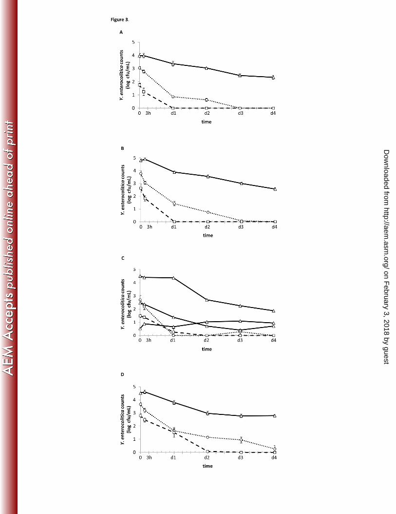
In nutrient-rich medium (PYG), Y. enterocolitica could survive inside A. castellanii (Fig. 279
3). Over time, all Y. enterocolitica strains survived intracellularly for at least 4 days at 7 °C, with 280
0.71 to 2.8 log cfu/mL detected inside the amoebae. At 25 and 37 °C, the number of intracellular 281
Yersinia decreased, with low or no bacterial recovery at the end of the experiment. At each time 282
point, the number of intracellular Y. enterocolitica in coculture with Acanthamoeba was 283
significantly higher at 7 °C than at 25 °C (p<0.001) and 37 °C (p≤0.001). 284
Although the initial inoculation concentration was the same for each experimental 285
condition (strain and temperature combination), counts were already significantly different 286
between temperature treatments at time point 0 (i.e. after a 2 h feeding period followed by a 1 h 287
gentamycin treatment). Strains YeH3+ and YeM3+ had similar concentrations at 0h, whereas the 288
number of viable yersiniae of strain YeH3- was almost 1 log cfu/mL lower. Replicate-counts at 289
the 0 h time point of strain YeH9- at 7 °C varied from 0.53 to 4.50 log cfu/mL (although similar 290
inoculation levels at the start of the experiment), whereas replicates of all other strain/temperature 291
combinations varied much less. 292
293
on February 3, 2018 by guest
http://aem.asm
.org/D
ownloaded from
14
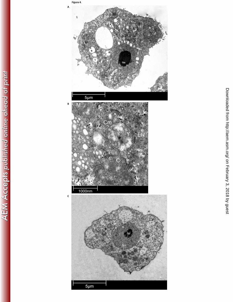
Localization of intraprotozoan Y. enterocolitica. At each time point, transmission electron 294
microscopy was performed to determine the intra-amoebal location of the bacteria. Pseudopodia 295
and food vacuoles were visible, indicating that the experimental coculture conditions favored 296
normal amoebic activity. After 3 days of cocultivation, the intraprotozoan bacteria were located 297
in the cytoplasm of the amoeba, but not visibly surrounded by an amoebal vacuole membrane 298
(Fig. 4). Host endoplasmic reticulum (ER) was located close to the intracellular bacterium. 299
300
on February 3, 2018 by guest
http://aem.asm
.org/D
ownloaded from
15
DISCUSSION 301
The present study showed that Acanthamoeba castellanii enhances Yersinia enterocolitica 302
survival under certain environmental conditions. As Yersinia and Acanthamoeba share similar 303
ecological and anthropogenic niches, this interaction identifies a potential role of free-living 304
protozoa in the ecology and epidemiology of Y. enterocolitica. Though Acanthamoeba is a 305
bacterivorous free-living species that actively grazes on bacteria, with an estimated ingestion rate 306
up to 700 bacteria per amoeba per hour (42), in the persistence assays, no decreased bacterial 307
viability was observed in the cocultures with A. castellanii during the 14-day monitoring period. 308
This proves that Y. enterocolitica could survive in presence of A. castellanii, although no 309
distinction could be made if the recovered bacteria were internalized in the amoebae (i.e. resistant 310
to amoebal digestion) or if it concerned adherent or free-living extracellular bacteria (i.e. resistant 311
to amoebal uptake). 312
Moreover, the persistence assays show that interaction with A. castellanii enhances the survival 313
of Y. enterocolitica under nutrient-rich conditions at 25 °C and under nutrient-poor conditions at 314
37°C. However, the presence of cell debris in both cocultures and amoebal monocultures at latter 315
temperature indicates amoebal lysis, presumably due to nutrient depletion and supra-optimal 316
temperature. A similar decrease in amoebal viability under co- and monoculture conditions has 317
also been reported by Baré et al. and Greub et al. (35, 43). The formed amoebal cell debris may 318
in turn be used by Yersinia as a nutrient source, explaining the better survival in the cocultures at 319
37 °C than in the monocultures. The fact that, after 3 days incubation in nutrient-rich medium at 320
37 °C, no Yersinia could be detected in both cocultures and monocultures, is presumably due to 321
the high metabolic activity of Yersinia under these conditions, which led to rapid nutrient 322
depletion. 323
on February 3, 2018 by guest
http://aem.asm
.org/D
ownloaded from
16
The present study also revealed that low temperatures and high nutrient availability favor 324
intraprotozoan survival. These conditions are met in industrial food processing settings, at home 325
as well as in natural environments. Although Yersinia is a psychrotrophic bacterium and can 326
withstand low temperatures without amoebal protection ((44), cf. bacterial monoculture controls 327
in persistence assays), the association can enhance Y. enterocolitica survival by physically 328
protecting against chlorine treatment. Chlorine is a commonly used compound to disinfect water 329
and food processing equipment, and increased resistance against chlorine of intra-protozoan Y. 330
enterocolitica has been reported (19). 331
In general, higher numbers of intracellular bacteria were recovered at lower temperatures 332
and for a longer period of time. This is in parallel with a recently published study where a 333
Yersinia strain was not recovered from Acanthamoeba polyphaga after 2 days at 30 °C (34). The 334
high intraprotozoan survival observed at 7 °C could be due to slower amoebal phagocytic 335
digestion (45), bacterial circumvention of amoebal digestion by escaping into the amoebal 336
cytosol, as observed by TEM, or both. Further research is needed to elucidate Yersinia’s 337
intracellular survival mechanisms. 338
The number of viable intracellular Yersinia decreased with time. This could be due to 339
(partial) bacterial digestion, bacterial release in the environment and subsequent bacterial killing 340
by gentamycin or to a switch to a viable but non cultivable state (VBNC) of the intracellular 341
Yersinia (46). Partial intraprotozoan digestion has already been described for other bacterial 342
pathogens (35, 47-49). Linking back towards the results of the persistence assay, whereby no 343
reduction in viable bacteria was observed under most cocultivation conditions, another 344
explanation could be that intraprotozoan Yersinia are released or trigger their release in the extra-345
amoebal environment (34). In the gentamycin protection assay these extracellular bacteria are 346
killed by the gentamycin maintenance concentration, whereas extracellular growth/survival is 347
on February 3, 2018 by guest
http://aem.asm
.org/D
ownloaded from
17
possible for yersiniae in the persistence assay. In the gentamycin protection assay, no amoebal 348
growth was observed, which confirmed that the amoebae did not appear to obtain adequate 349
nutrients from Yersinia to increase their concentration. 350
Strain variation in intracellular survival capacity was detected from the start of the 351
experiments onwards, which could be due to variation in the ability of the strains to invade or to 352
become internalized by Acanthamoeba or to their intrinsic intracellular survival capacities. This 353
study indicates that the presence of the Yersinia virulence plasmid favors bacterial uptake and/or 354
intracellular survival, as at time point zero, the number of intracellular yersiniae was higher for 355
the plasmid carrying YeH3+ strain than for its plasmid cured derivate (YeH3-). 356
For the first time, TEM was used to visualize the exact subcellular location of Yersinia 357
inside A. castellanii. Images show that the intracellular yersiniae were located in the cytosol and 358
were not visibly surrounded by an amoebal vacuole membrane. This may indicate that Y. 359
enterocolitica is able to circumvent the normal digestion pathway by escaping from the food 360
vacuole to the cytosol. This post-ingestional adaptation mechanism to avoid protozoan digestion 361
has also been described for members of the genus Rickettsia (18). In addition, the Y. 362
enterocolitica were located close to the host endoplasmic reticulum (ER), which may be 363
advantageous as the bacteria have easy access to newly synthesized host proteins. Further 364
research is necessary to reveal if the co-localization of host ER and intracellular yersiniae is 365
coincidental or if there exists a Yersinia mechanism to repose the host ER, as described for 366
Legionella pneumophila (50, 51). 367
In the supernatant assays, the supernatant of strain YeH3- (4/O:3, pYV-) had a 368
temperature-dependent permeabilizing effect on the amoebae. It is not clear if this reflects a 369
direct or indirect effect on the amoebal cell membrane integrity. During bacterial cultivation at 370
the start of the experiments, the pH of this supernatant had already decreased compared to the 371
on February 3, 2018 by guest
http://aem.asm
.org/D
ownloaded from
18
supernatant of the other strains. However, the permeabilizing effect could not be explained by a 372
pH reduction as such. As this permeabilizing effect was not observed in the persistence assays, a 373
hypothesis could be that direct cell contact between amoebae and bacteria may activate a host 374
defense mechanism to prevent permeabilization. Alternatively, the effect can be concentration-375
dependent, as washing steps in the persistence assay protocol could have lowered the amount of 376
permeabilizing factors. Further research is needed to identify the cause of this permeabilization 377
(influenced by temperature) and the observed pH drop, with attention to the production of a 378
potential bacterial toxin or a metabolite with anti-protozoan activity (14). 379
The association of Y. enterocolitica with protozoa has relevant ecological and 380
epidemiological implications. Besides enhancing Y. enterocolitica survival in their presence, 381
being intra- or extracellular, free-living protozoa have the potential to act as a reservoir, vector, 382
infection route, biological gym and evolutionary crib for intracellular Yersinia enterocolitica, as 383
previously described for other pathogenic bacteria (17, 24, 52, 53). 384
385
Acknowledgements 386
This work was supported by the Research Fund of Ghent University (BOF, Ghent, Belgium; 387
grant 01D07612). Many thanks to M. Claeys for the help with the transmission electron 388
microscopy and to C. Van Lancker for the technical assistance. 389
390
on February 3, 2018 by guest
http://aem.asm
.org/D
ownloaded from
19
Figure 1. Persistence of Y. enterocolitica in coculture with A. castellanii 391
Yersinia enterocolitica were cultivated with (full line) or without A. castellanii (dotted line) in 392
nutrient-poor PAS (A) or nutrient-rich PYG medium (B) at different temperatures (∆ 7 °C, ο 25 393
°C, □ 37 °C). MOI 100:1. Values represent the overall means of the four Y. enterocolitica strains 394
of the three replicate experiments ± standard error of the mean. 395
396
Figure 2. Effect of supernatant from Y. enterocolitica strain YeH3- on A. castellanii 397
Percentage of living (indicated in grey) and permeabilized (in black) amoebae related to the total 398
amoebal count, when cultivated in the cell-free supernatant of a Y. enterocolitica culture (strain 399
YeH3-) and incubated at 7 °C (A), 25 °C (B) and 37 °C (C). Amoebae were cultivated in the 400
growth medium of the Yersinia culture (TSB) as control. Bars represent the mean of three 401
replicate experiments ± standard error of the mean. 402
403
Figure 3. Intracellular viable counts of Y. enterocolitica at different incubation 404
temperatures 405
Acanthamoeba castellanii and Y. enterocolitica strain YeH3- (A), YeH3+ (B), YeH9- (C) and 406
YeM3+ (D) were cocultivated in nutrient-rich medium and incubated at different temperatures 407
( ∆ 7 °C, …ο… 25 °C, - -□- - 37 °C). Values represent the means of three replicate experiments 408
± standard error of the mean. 409
410
Figure 4. Acanthamoeba castellanii trophozoite with internalized Yersinia enterocolitica 411
TEM micrographs of A. castellanii incubated for 3 days with Y. enterocolitica strain YeH3- in 412
nutrient- rich medium at 7 °C (A and B) and of an A. castellanii monoculture control (C). P: 413
pseudopodium, V: food vacuole, C: cytoplasm, Nm: nucleus membrane, N: nucleus, Ne: 414
on February 3, 2018 by guest
http://aem.asm
.org/D
ownloaded from
20
nucleolus, M: mitochondrion, Y: Yersinia enterocolitica, ER: endoplasmic reticulum, G: 415
globules, Cm: bacterial cell membrane. 416
417
on February 3, 2018 by guest
http://aem.asm
.org/D
ownloaded from
21
References 418
1. EFSA. 2013. The European Union summary report on trends and sources of zoonoses, 419
zoonotic agents and food-borne outbreaks in 2011. EFSA journal 11:3129-3379. 420
2. Bottone EJ. 1999. Yersinia enterocolitica: overview and epidemiologic correlates. 421
Microbes Infect 1:323-333. 422
3. Verhaegen J, Charlier J, Lemmens P, Delmee M, Van Noyen R, Verbist L, Wauters 423
G. 1998. Surveillance of human Yersinia enterocolitica infections in Belgium: 1967-424
1996. Clin Infect Dis 27:59-64. 425
4. Bottone EJ. 1997. Yersinia enterocolitica: the charisma continues. Clin Microbiol Rev 426
10:257-276. 427
5. Riley G, Toma S. 1989. Detection of pathogenic Yersinia enterocolitica by using congo 428
red-magnesium oxalate agar medium. J Clin Microbiol 27:213-214. 429
6. Farmer JJ, 3rd, Carter GP, Miller VL, Falkow S, Wachsmuth IK. 1992. 430
Pyrazinamidase, CR-MOX agar, salicin fermentation-esculin hydrolysis, and D-xylose 431
fermentation for identifying pathogenic serotypes of Yersinia enterocolitica. J Clin 432
Microbiol 30:2589-2594. 433
7. Van Damme I, Habib I, De Zutter L. 2010. Yersinia enterocolitica in slaughter pig 434
tonsils: enumeration and detection by enrichment versus direct plating culture. Food 435
Microbiol 27:158-161. 436
8. Nesbakken T, Eckner K, Hoidal HK, Rotterud OJ. 2003. Occurrence of Yersinia 437
enterocolitica and Campylobacter spp. in slaughter pigs and consequences for meat 438
inspection, slaughtering, and dressing procedures. Int J Food Microbiol 80:231-240. 439
9. Fredriksson-Ahomaa M, Stolle A, Korkeala H. 2006. Molecular epidemiology of 440
Yersinia enterocolitica infections. FEMS Immunol Med Microbiol 47:315-329. 441
on February 3, 2018 by guest
http://aem.asm
.org/D
ownloaded from
22
10. Baré J, Sabbe K, Van Wichelen J, van Gremberghe I, D'Hondt S, Houf K. 2009. 442
Diversity and habitat specificity of free-living protozoa in commercial poultry houses. 443
Appl Environ Microbiol 75:1417-1426. 444
11. Baré J, Houf K, Verstraete T, Vaerewijck M, Sabbe K. 2011. Persistence of free-living 445
protozoan communities across rearing cycles in commercial poultry houses. Appl Environ 446
Microbiol 77:1763-1769. 447
12. Vaerewijck MJ, Sabbe K, Baré J, Houf K. 2008. Microscopic and molecular studies of 448
the diversity of free-living protozoa in meat-cutting plants. Appl Environ Microbiol 449
74:5741-5749. 450
13. Vaerewijck MJ, Sabbe K, Van Hende J, Baré J, Houf K. 2010. Sampling strategy, 451
occurrence and diversity of free-living protozoa in domestic refrigerators. J Appl 452
Microbiol 109:1566-1578. 453
14. Matz C, Kjelleberg S. 2005. Off the hook:how bacteria survive protozoan grazing. 454
Trends Microbiol 13:302-307. 455
15. Akya A, Pointon A, Thomas C. 2009. Viability of Listeria monocytogenes in co-culture 456
with Acanthamoeba spp. FEMS Microbiol Ecol 70:20-29. 457
16. Berk SG, Faulkner G, Garduno E, Joy MC, Ortiz-Jimenez MA, Garduno RA. 2008. 458
Packaging of live Legionella pneumophila into pellets expelled by Tetrahymena spp. does 459
not require bacterial replication and depends on a Dot/Icm-mediated survival mechanism. 460
Appl Environ Microbiol 74:2187-2199. 461
17. Greub G, Raoult D. 2004. Microorganisms resistant to free-living amoebae. Clin 462
Microbiol Rev 17:413-433. 463
on February 3, 2018 by guest
http://aem.asm
.org/D
ownloaded from
23
18. Whitworth T, Popov VL, Yu XJ, Walker DH, Bouyer DH. 2005. Expression of the 464
Rickettsia prowazekii pld or tlyC gene in Salmonella enterica serovar Typhimurium 465
mediates phagosomal escape. Infect Immun 73:6668-6673. 466
19. King CH, Shotts EB, Jr., Wooley RE, Porter KG. 1988. Survival of coliforms and 467
bacterial pathogens within protozoa during chlorination. Appl Environ Microbiol 468
54:3023-3033. 469
20. Snelling WJ, Stern NJ, Lowery CJ, Moore JE, Gibbons E, Baker C, Dooley JS. 2008. 470
Colonization of broilers by Campylobacter jejuni internalized within Acanthamoeba 471
castellanii. Arch Microbiol 189:175-179. 472
21. Bruggemann H, Cazalet C, Buchrieser C. 2006. Adaptation of Legionella pneumophila 473
to the host environment: role of protein secretion, effectors and eukaryotic-like proteins. 474
Curr Opin Microbiol 9:86-94. 475
22. Koubar M, Rodier MH, Garduno RA, Frere J. 2011. Passage through Tetrahymena 476
tropicalis enhances the resistance to stress and the infectivity of Legionella pneumophila. 477
FEMS Microbiol Lett 325:10-15. 478
23. Cirillo JD, Falkow S, Tompkins LS. 1994. Growth of Legionella pneumophila in 479
Acanthamoeba castellanii enhances invasion. Infect Immun 62:3254-3261. 480
24. Cirillo JD, Falkow S, Tompkins LS, Bermudez LE. 1997. Interaction of 481
Mycobacterium avium with environmental amoebae enhances virulence. Infect Immun 482
65:3759-3767. 483
25. McCuddin ZP, Carlson SA, Rasmussen MA, Franklin SK. 2006. Klebsiella to 484
Salmonella gene transfer within rumen protozoa: implications for antibiotic resistance and 485
rumen defaunation. Vet Microbiol 114:275-284. 486
on February 3, 2018 by guest
http://aem.asm
.org/D
ownloaded from
24
26. Falcão JP, Brocchi M, Proenca-Modena JL, Acrani GO, Correa EF, Falcão DP. 487
2004. Virulence characteristics and epidemiology of Yersinia enterocolitica and Yersiniae 488
other than Y. pseudotuberculosis and Y. pestis isolated from water and sewage. J Appl 489
Microbiol 96:1230-1236. 490
27. Hausmann K, Hülsmann N, Radek R. 2003. Protistology. Schweizerbart'sche 491
verlagbuchhandlung Stuttgart, Duitsland. 492
28. Cocolin L, Comi G. 2005. Use of a culture-independent molecular method to study the 493
ecology of Yersinia spp. in food. Int J Food Microbiol 105:71-82. 494
29. Vaerewijck MJ, Sabbe K, Baré J, Houf K. 2011. Occurrence and diversity of free-495
living protozoa on butterhead lettuce. Int J Food Microbiol 147:105-111. 496
30. Jackson V, Blair I, McDowella D, Kennedy J, Bolton D. 2007. The incidence of 497
significant foodborne pathogens in domestic refrigerators. Food Control 16:6. 498
31. Axelsson-Olsson D, Svensson L, Olofsson J, Salomon P, Waldenstrom J, Ellstrom P, 499
Olsen B. 2010. Increase in acid tolerance of Campylobacter jejuni through coincubation 500
with amoebae. Appl Environ Microbiol 76:4194-4200. 501
32. Douesnard-Malo F, Daigle F. 2011. Increased persistence of Salmonella enterica 502
serovar Typhi in the presence of Acanthamoeba castellanii. Appl Environ Microbiol 503
77:7640-7646. 504
33. Alsam S, Jeong SR, Sissons J, Dudley R, Kim KS, Khan NA. 2006. Escherichia coli 505
interactions with Acanthamoeba: a symbiosis with environmental and clinical 506
implications. J Med Microbiol 55:689-694. 507
34. Anacarso I, de Niederhausern S, Messi P, Guerrieri E, Iseppi R, Sabia C, Bondi M. 508
2011. Acanthamoeba polyphaga, a potential environmental vector for the transmission of 509
food-borne and opportunistic pathogens. J Basic Microbiology. 510
on February 3, 2018 by guest
http://aem.asm
.org/D
ownloaded from
25
35. Baré J, Sabbe K, Huws S, Vercauteren D, Braeckmans K, van Gremberghe I, 511
Favoreel H, Houf K. 2010. Influence of temperature, oxygen and bacterial strain identity 512
on the association of Campylobacter jejuni with Acanthamoeba castellanii. FEMS 513
Microbiol Ecol 74:371-381. 514
36. Rohde JR, Luan XS, Rohde H, Fox JM, Minnich SA. 1999. The Yersinia 515
enterocolitica pYV virulence plasmid contains multiple intrinsic DNA bends which melt 516
at 37 degrees C. J Bacteriol 181:4198-4204. 517
37. Van Damme I, Berkvens D, Baré J, De Zutter L. 2013. Influence of isolation methods 518
on the occurrence of plasmid-carrying Yersinia enterocolitica serotype O:3 in slaughter 519
pig tonsils, faeces and carcass surface swabs. Int J Food Microbiol 164:32-35. 520
38. Axelsson-Olsson D, Olofsson J, Svensson L, Griekspoor P, Waldenstrom J, Ellstrom 521
P, Olsen B. 2010. Amoebae and algae can prolong the survival of Campylobacter species 522
in co-culture. Exp Parasitol 126:59-64. 523
39. Berry D, Horn M, Xi C, Raskin L. 2010. Mycobacterium avium infections of 524
Acanthamoeba strains: host strain variability, grazing-acquired infections, and altered 525
dynamics of inactivation with monochloramine. Appl Environ Microbiol 76:6685-6688. 526
40. Vaerewijck MJ, Sabbe K, Baré J, Spengler HP, Favoreel HW, Houf K. 2012. 527
Assessment of the efficacy of benzalkonium chloride and sodium hypochlorite against 528
Acanthamoeba polyphaga and Tetrahymena spp. J Food Prot 75:541-546. 529
41. Spurr A. 1969. A low-viscosity epoxy resin embedding medium for electron microscopy. 530
J. Ultrastruct Res. 26:31-43. 531
42. Corsaro D, Müller K-D, Michel R. 2013. Molecular characterization and ultrastructure 532
of a new amoeba endoparasite belonging to the Stenotrophomonas maltophilia complex. 533
Exp Parasitol 133:383-390. 534
on February 3, 2018 by guest
http://aem.asm
.org/D
ownloaded from
26
43. Greub G, La Scola B, Raoult D. 2003. Parachlamydia acanthamoeba is endosymbiotic 535
or lytic for Acanthamoeba polyphaga depending on the incubation temperature. Ann N Y 536
Acad Sci 990:628-634. 537
44. Goverde RL, Huis in't Veld JH, Kusters JG, Mooi FR. 1998. The psychrotrophic 538
bacterium Yersinia enterocolitica requires expression of pnp, the gene for polynucleotide 539
phosphorylase, for growth at low temperature (5 degrees C). Mol Microbiol 28:555-569. 540
45. Khan. 2009. Acanthamoeba: Biology and pathogenesis. Caister Academic Press, Norfolk 541
England. 542
46. Smith JJ, Howington JP, McFeters GA. 1994. Survival, physiological response and 543
recovery of enteric bacteria exposed to a polar marine environment. Appl Environ 544
Microbiol 60:2977-2984. 545
47. Pickup ZL, Pickup R, Parry JD. 2007. Effects of bacterial prey species and their 546
concentration on growth of the amoebae Acanthamoeba castellanii and Hartmannella 547
vermiformis. Appl Environ Microbiol 73:2631-2634. 548
48. Gaze WH, Burroughs N, Gallagher MP, Wellington EM. 2003. Interactions between 549
Salmonella typhimurium and Acanthamoeba polyphaga, and observation of a new mode 550
of intracellular growth within contractile vacuoles. Microb Ecol 46:358-369. 551
49. Tezcan-Merdol D, Ljungstrom M, Winiecka-Krusnell J, Linder E, Engstrand L, 552
Rhen M. 2004. Uptake and replication of Salmonella enterica in Acanthamoeba 553
rhysodes. Appl Environ Microbiol 70:3706-3714. 554
50. Robinson CG, Roy CR. 2006. Attachment and fusion of endoplasmic reticulum with 555
vacuoles containing Legionella pneumophila. Cell Microbiol 8:793-805. 556
on February 3, 2018 by guest
http://aem.asm
.org/D
ownloaded from
27
51. de Felipe KS, Glover RT, Charpentier X, Anderson OR, Reyes M, Pericone CD, 557
Shuman HA. 2008. Legionella eukaryotic-like type IV substrates interfere with organelle 558
trafficking. PLoS Pathog 4:e1000117. 559
52. Siddiqui R, Khan NA. 2012. War of the microbial worlds: who is the beneficiary in 560
Acanthamoeba-bacterial interactions? Exp Parasitol 130:311-313. 561
53. Thomas V, McDonnell G, Denyer SP, Maillard JY. 2010. Free-living amoebae and 562
their intracellular pathogenic microorganisms: risks for water quality. FEMS Microbiol 563
Rev 34:231-259. 564
565
566
567
on February 3, 2018 by guest
http://aem.asm
.org/D
ownloaded from