Embed Size (px)
Citation preview
Obliterating Left Ventricular Mural ThrombosisBy JACOB ZATUCHNI, M.D., AND KEE TON TAN, M.D.
HEREIN is reported a case in which aleft ventricular mural thrombus was
so large as almost completely to obliteratethe left ventricle. Such an occurrence, toour knowledge, has never been reported.
Case ReportA 64-year-old white man, blind since birth,
considered himself well until March 1960. Becauseof progressive weakness, shortness of breath uponeffort, and swelling of the ankles, he enteredEpiscopal Hospital on June 13, 1960. Cardiactherapy was intensified and left thoracentesis wasdone on several occasions. The fluid was sero-sanguineous in character. Pleural biopsy, witha Vim-Silverman needle, disclosed only fibroustissue. He was discharged on July 18, 1960.
Shortly thereafter, shortness of breath againincreased, and ankle edema and left pleuraleffusion reappeared. He was re-admitted to Epis-copal Hospital on August 17, 1960. Findings weresimilar to those previously observed.The blood pressure was 100/80 mm. Hg. The
temperature was normal. There was bilateral opticatrophy. The jugular veins were slightly dis-tended. The cardiac impulse was not palpable.Heart sounds were distant and normal in quality,rate, and rhythm. There were no murmurs. Signsof left pleural effusion were present. The abdo-men was negative; the liver and spleen were notfelt. There was moderate pitting edeama of thelegs and feet.The hemoglobin and electrolytes were normal,
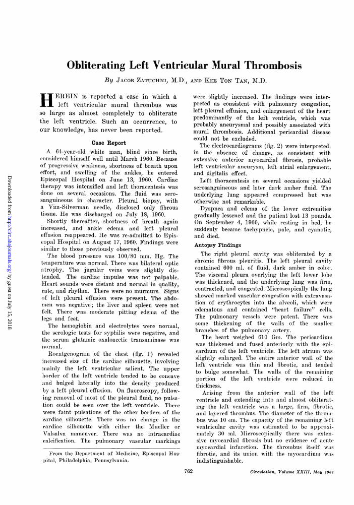
the serologic tests for syphilis were negative, andthe serum glutamic oxaloacetie transamninase wasnormal.Roentgenogram of the chest (fig. 1) revealed
increased size of the cardiac silhouette, involvingmainly the left ventricular salient. The upperborder of the left ventricle tended to be concaveanrd bulged laterally into the density producedby a left pleural effusion. On fluoroscopy, follow-ing removal of most of the pleural fluid, no pulsa-tion could be seen over the left ventricle. Therewere faint pulsations of the other borders of thecardiac silhouette. There was no change in thecardiac silhouette with either the Mueller orValsalva maneuver. There was no intracardiaccalcification. The pulmonary vascular markings
From the Department of Medicine, Episcopal Hos-pital, Philadelphia, Pennsylvania.
were slightly increased. The findings were inter-preted as consistent with pulmonary congestion,left pleural effusion, and enlargement of the heartpredominantly of the left ventricle, which wasprobably aneurysmal and possibly associated withmural thrombosis. Additional pericardial diseasecould not be excluded.The electrocardiograms (fig. 22) were interpreted,
in the absence of change, as consistent withextensive anterior inyocardial fibrosis, probableleft ventricular aneurysm, left atrial enlargement,and digitalis effect.
Left thoracentesis on several occasions yieldedserosanguineous and later dark amber fluid. Theunderlying lung appeared compressed but wasotherwise not remarkable.Dyspnea and edema of the lower extremities
gradually lessened and the patient lost 13 pounds.On September 4, 1960, while resting in bed, lhesuddenly became tachypneic, pale, and cyanotic,and died.Autopsy Findings
The right pleural cavity was obliterated by achronic fibrous pleuritis. The left pleural cavitycontained 600 ml. of fluid, dark amber in color.The visceral pleura overlying the left lower lobewas thickened, and the underlying lung was firm,contracted, and congested. Microscopically the lungshowed marked vascular congestion with extravasa-tion of erythrocytes into the alveoli, which wereedematous and contained "heart failure" cells.The pulmonary vessels were patent. There wassome thickening of the walls of the smn.allerbranches of the pulmonary artery.
The heart weighed 610 Gm. The pericardiuinwas thickened and fused anteriorly with the epi-cardium of the left ventricle. The left atrium wasslightly enlarged. The entire anterior wall of theleft ventricle was thin and fibrotic, and tendedto bulge somewhat. The walls of the reniainingportion of the left ventricle were reduced inthickness.
Arising from the anterior wall of the leftventricle and extending into and almost obliterat-ing the left ventricle was a large, firm,, fibrotic,and layered thrombus. The diameter of the thromi-bus was 10 cim. The capacity of the remnainitig leftventricular cavity was estimated to be approxi-mately 30 ml. Microscopically there was exten-sive myocardial fibrosis but no evidence of acutemyoeardial infarction. The throinbus itself wasfibrotic, and its union with the myocardiuni wasindistinguishable.
Circulation, Volume XXIII. May 1961762
by guest on July 15, 2018http://circ.ahajournals.org/
Dow
nloaded from

LEFT VENTRICULAR THROMBOSIS
No other thrombi were found. The cardiacvalves were normal. The coronary arteries exhibiteda moderate degree of generalized narrowing andsclerosis, and the anterior branch of the leftcoronary artery was completely occluded by anorganized atheroma, 5 ml. in length, beginning2.5 cm. from the ostium. The entire aorta showedmild atherosclerotic changes.
The liver weighed 1,100 Gm. It was nutmeg inappearance and revealed dilatation and congestionof the central veins and adjacent sinusoids. Thesurrounding hepatic cells showed degenerativechange. The spleen was firm and weighed 120Gm. There was marked congestion and a slightincrease in fibrous tissue.
Sections of the heart, liver, and spleen are shownin figure 3.
DiscussionIt was apparent from the chest roentgeno-
gram that there was an aneurysm of the leftventricle.1 The basis for the aneurysm wasconsidered clinically to be myocardial fibrosissecondary to occlusive coronary artery dis-ease, the absence of pain notwithstanding.Indeed, many cases of cardiac aneurysm havebeen reported to sustain a silent myocardialinfarction and to present with congestiveheart failure of insidious onset.2 Interestingly,however, although the patient steadfastlydenied having any pain, his family, followinghis death, related, in response to direct ques-tioning, that he had had bouts of substernalpain for several months prior to his demise.Curiously, it was his wish that no physicianshould be informed of this distress becausehe feared hospitalization.
The aneurysm displayed no motion. Acardiac impulse was not palpable, and theleft ventricular portion of the cardiac sil-houette was seen by fluoroscopy not to pul-sate. The absence of pulsation was thought tobe due to intrinsic myocardial disease andpossibly to mural thrombosis and even peri-cardial restriction, which frequently occurin left ventricular aneurysms arising fromocclusive coronary artery disease. 4 Unfor-tunately, his condition did not permit studyof the heart by contrast roentgenography,by which diagnostic conclusions might havebeen possible.5-7At autopsy, the entire anterior wall of the
Circulation, Volume XXIII, May 1961
Figure 1Posteroanterior chest roentgenogram. The cardiacsilhouette is increased in size and its shape suggestsmainly left ventricular enlargement. The upperborder of the left ventricular portion of the cardiacsilhouette is concave and sweeps outward, due toaneurysmal formation, into the density producedby a left pleural effusion.
left ventricle was reduced to a fibrous shell.Arising from it and almost filling the leftventricle was a huge, organized, and layeredthrombus.We have been unable to find in the litera-
ture a similar occurrence, although in thecase reported recently by Dexter, Lawton,and Raynolds,8 the left ventricular muralthrombus may possibly have been as large.Their case had an aneurysm, 18 cm. indiameter, described as being completely filledwith a mural thrombus. Another might bethe case reported by Shennan and Niven.9In both of these cases, although it is notclear if the thrombus was restricted to theaneurysm or filled the ventricular cavity,illustrations show, indeed, a huge thrombus.
Such an obliterating thrombus of the leftventricle would be anticipated to producesevere restriction of inflow similar to that
763
by guest on July 15, 2018http://circ.ahajournals.org/
Dow
nloaded from

ZATUCHNI, TAN
Figure 2Electrocardiogram showing sinus rhythm and find-ings consistent with left atrial enlargement, exten-sive anterior myocardial fibrosis, probable aneu-rysm, and digitalis effect.
occurring in mitral stenosis. Indeed, the elee-trocardiogram showed signs consistent withleft atrial enlargement, which was found atnecropsy, and the pulmonary arterioles showedchanges consistent with pulmonary hyperten-sion.
Forward flow from the left ventricle wouldalso be expected to be seriously compromised.The fatigue, pallor, and low blood pressurebespoke a low cardiac output.
Similar findings occur, of course, in manyother conditions,'0 including left ventricularaneurysm alone." In the presence of leftventricularianeurysm, a clue to the existenceof mural thrombosis is thromboembolism,which occurs in almost two thirds of thecases.3 It is not always present, however,and it may also occur in the absence of clinicalmanifestations and in the absence of muralthrombosis.3Another clue to the presence of associated
mural thrombosis is absence of pulsationsof the left ventricular aneurysm.' Pulsationsof a left ventricular aneurysm are character-istically paradoxical.'2' 13 The involved wallof the left ventricle is fibrous and bulgeswith each ventricular systole. The presenceof a mural thrombus would be anticipatedto dampen such an outward thrust. Thereliability of this sign in diagnosing asso-ciated mural thrombosis is questionable, how-ever, for other conditions, like dilatation or
Figure 3Gvoss sections of the liver (upper left), spleen(upper right), and heart viewed from above.There was chronic passive congestion of the liverand spleen, which were firm. In the section ofheart, note the thin fibrous residue of the anteriorwall and the huge, fibrous, layered thrombus aris-ing from it and almost obliterating the left ven-tricle. The remaining cavity has been increasedby distortion for photographic purposes. Thesubjacent muscle is the septal portion of the leftventricle.
pericardial effusion, may produce similardampening. Also, to our knowledge, therehas been no systematic study made of thepulsations of left ventricular aneurysm alonecompared with those associated with muralthrombosis. Certain it is that pulsations donot exclude mural thrombosis.14 Other roent-genographic signs suggestive of left ventricu-lar mural thrombosis include increased den-sity and even calcification,15 but these are veryrare.For these reasons, clinical recognition is
difficult. Nevertheless, knowledge of its occur-rence can lead to a strong clinical suspicionof its presence which perhaps may beestablished with certainty by contrast roent-genography.
Summary and ConclusionsA case is recorded of a huge mural throm-
bus that encroached upon and almost oblit-erated the left ventricle. There was under-lying occlusive coronary artery disease withextensive myocardial fibrosis and aneurysmformation. Clinical manifestations, other than
Circulation, Volume XXIII, May 1961
764
by guest on July 15, 2018http://circ.ahajournals.org/
Dow
nloaded from

LEFT VENTRICULAR THROMBOSIS
those produced by a persistent left pleuraleffusion, were similar in effect to any con-dition producing severe restriction of leftventricular inflow and outflow. A clue toits presence was absence of left ventricularpulsations.
References1. PARKINSON, J., BEDFORD, D. E., AND THOMSON,
WV. A. R.: Cardiac aneurysm. Quart. J. Med.31: 455, 1938.
. MOYER, J. B., AND HILLER, G. I.: Cardiacaneurysm. Clinical and electrocardiographicanalysis. Am. Heart J. 41: 340, 1951.
3. SCHLICHTER, J., HELLERSTEIN, H. K., AND KATZ,L. N.: Aneurysm of the heart: A correlativestudy of one hundred and two proved cases.Medicine 33: 43, 1954.
4. PHARES, W. S., EDWARDS, J. E., AND BURCHELL,H. B.: Cardiac aneurysms: Clinicopathologicstudies. Proc. Staff Meet., Mayo Clin. 28:264, 1953.
5'. SOLOFF, L. A., AND ZATUCHNI, J.: The angio-cardiographic diagnosis of left atrial throm-bosis. Circulation 14: 25, 1956.
(;. SOLoFF, L. A., AND ZATUCHNI, J.: The definitivediagnosis of effusive or constrictive pericardi-tis. Am. J. M. Sc. 234: 687, 1957.
7. DOLLY, C. H., DOTTER, C. T., AND STEINBERG, I.:
Ventricular aneurysm in a 29 year old manstudied angioeardiographically. Am. Heart J.42: 894, 1951.
8. DEXTER, M. W., LAWTON, A. H., AND RAYNOLDS,A. H.: Report of an unusually large ventricularaneurysm. Am. J. Cardiol. 2: 372, 1958.
9. SHENNAN, T., AND NIVEN, W.: Unusually largecardiac aneurysm. J. Path. & Bact. 28: 390,1925.
10. BURWELL, C. S., AND ROBIN, E. D.: Diagnosis ofdiffuse myoeardial fibrosis. Circulation 20: 606,1959.
11. COOLEY, D. A., COLLINS, H. A., MoRRis, G. C.,JR., AND CHAPMAN, D. W.: Ventricular aneu-rysm after myocardial infarction. Surgicalexcision with the use of temporary cardio-pulmonary by-pass. J.A.M.A. 167: 557, 1958.
12. ZDANSKY, E. (translated by Boyd, L. J.):Roentgen Diagnosis of the Heart and GreatVessels. New York, Grune and Stratton, Inc.,1953.
13. ROESLER, H.: Clinical Roentgenology of theCardiovascular System. Springfield, Illinois,Charles C Thomas, 1946.
14. DRESSLER, W., AND PFEIFFER, R.: Cardiac aneu-rysm: Report of 10 cases. Ann. Int. Med.14: 100, 1940.
15. SCHWEDEL. J. B., AND GROSS, H.: Ventricularaneurysm. Roentgenographic and post-mortemsurveys. Am. J. Roentgenol. 41: 32, 1939.
The very ingenious Dr. Hales writes me, that, having many years since, tied a ligatureabout a frog's neck, to prevent any effusion of blood, he cut off its head, and, thirtyhours after, observed the blood circulating freely in the web of the foot: the frog alsoat this time moved its body when stimulated: but, on thrusting a needle down throughthe spinal marrow, the animal was strongly convulsed, and, immediately after, becamemotionless.-ROBERT WHYTT. Physiological Essays. Edinburgh, 1755, p. 176.
Circulation. Volume XXIII, May 196'
765)
by guest on July 15, 2018http://circ.ahajournals.org/
Dow
nloaded from

JACOB ZATUCHNI and KEE TON TANObliterating Left Ventricular Mural Thrombosis
Print ISSN: 0009-7322. Online ISSN: 1524-4539 Copyright © 1961 American Heart Association, Inc. All rights reserved.
75231is published by the American Heart Association, 7272 Greenville Avenue, Dallas, TXCirculation
doi: 10.1161/01.CIR.23.5.7621961;23:762-765Circulation.
http://circ.ahajournals.org/content/23/5/762.citationlocated on the World Wide Web at:
The online version of this article, along with updated information and services, is
http://circ.ahajournals.org//subscriptions/
is online at: Circulation Information about subscribing to Subscriptions:
http://www.lww.com/reprints Information about reprints can be found online at: Reprints:
document. Permissions and Rights Question and Answer
of the Web page under Services. Further information about this process is available in thewhich permission is being requested is located, click Request Permissions in the middle columnClearance Center, not the Editorial Office. Once the online version of the published article for
can be obtained via RightsLink, a service of the CopyrightCirculationoriginally published in Requests for permissions to reproduce figures, tables, or portions of articlesPermissions:
by guest on July 15, 2018http://circ.ahajournals.org/
Dow
nloaded from