Embed Size (px)
Citation preview

Nutrit ional Therapyin Crit ical ly I l l andInjured Patients
Rifat Latifi, MDa,b,*
KEYWORDS
� Critically ill � Amino acid � Acute phase protein� Intensive care unit � Trauma � ARDS � Sepsis� Immunonutrition
The vocabulary and practice of nutrition support of critically ill patients have changedsignificantly in recent years and new nutripharmaceuticals and disease-oriented nutri-tional support have become integral components of patient care in the new era ofcomprehensive management. The question to be answered is when to feed andwhat to feed critically ill and injured patients. Critical illness and tissue injury initiatea complex series of rapidly responding homeostatic events in an attempt to preventongoing tissue damage and to activate the repair process. Classically, inflammationhas been recognized as the hallmark of the homeostatic response. But more recently,attention has been focused on defining the complexities of response at the cellular,metabolic, and molecular levels. There is mounting evidence regarding the multiplespecific metabolic changes in critically ill and injured patients and their need for funda-mental nutrients and special substrates. As there are efforts to refine and further definenutritional support for critically ill patients, pursuing a deeper understanding of thisfield is imperative. Specifically, to provide timely and disease-directed and disorder-directed nutritional support, the most crucial changes in acute phase proteins, cyto-kines, and other biochemical indices must be elucidated. Furthermore, it has alreadybecome clear that no 1 care plan or formula fits all situations in all patients. Rather,nutritional support must be based principally on each individual patient’s pathophys-iology and condition.The benefit of early institution of adequate enteral or parenteral nutrition in the over-
all management of critically ill patients has been well established. Early nutritionalsupport has the potential to reduce disease severity, diminish complications, anddecrease the intensive care unit (ICU) length of stay. In general, whenever possible,
Author has no conflicts of interest to declare.a Trauma Division, Department of Surgery, University of Arizona, PO Box 245063, 1501 NorthCampbell Avenue, Tucson, AZ 85724, USAb Trauma Services, Hamad General Hospital, Hamad Medical Corporation, Doha, Qatar* Trauma Division, Department of Surgery, University of Arizona, PO Box 245063, 1501 NorthCampbell Avenue, Tucson, AZ 85724.E-mail address: [email protected]
Surg Clin N Am 91 (2011) 579–593doi:10.1016/j.suc.2011.02.002 surgical.theclinics.com0039-6109/11/$ – see front matter � 2011 Elsevier Inc. All rights reserved.
Latifi580
the gastrointestinal (GI) tract is the optimal route of providing nutrition in critically illpatients. If, on the other hand, a patient cannot receive all the needed nutrientsubstrates and calories enterally, nutrition should be provided parenterally.1 Becauseof the recent advances in identifying and recognizing the fundamental metabolicchanges of crucial nutrient substrates in critically ill patients, nutritional formulas arebeing designed to compensate for these changes and support the organism duringcritical illness, infection, trauma, and/or severe sepsis. One example of such anapproach to the nutritional support of critically ill patients is the provision ofimmune-enhancing formulas (IEFs), which have been shown to improve immuneresponses both in laboratory animals and critically ill patients. These enteral formulascontain increased amounts of peptides; arginine; glutamine; vitamins E, A, and C;nucleotides and nucleosides; branched-chain amino acids (BCAAs); and u-3 fattyacids. It is suggested that these principal nutrients can modulate and affect a varietyof inflammatory, metabolic, and immune processes if given in doses higher than thoseordinarily recommended or required.
STRESS RESPONSE
The sequence of responses to stress and injury has been described as the ebb phase,the catabolic flow phase, and the anabolic flow phase.2–4 Each of these phasesinduces distinct changes that require specific interventions to eliminate or minimizethe untoward consequences of illness and/or injury. The ebb phase is dominated bycirculatory changes that require resuscitation (with fluid, blood, and blood products)over a period of the first 8 to 24 hours. The catabolic flow phase, dominated by highlevels of catabolism, typically lasts 3 to 10 days, but may last longer. The anabolicflow phase emerges more subtly as the patient’s metabolism shifts to synthetic activ-ities and reparative processes.4 The catabolic flow phase is driven by cytokine medi-ators released from lymphocytes and macrophages in the cellular immune reaction,dominated by interleukin (IL)-6.4 The release of these mediators is proportional tothe intensity of the injury, but the release of the individual cytokines is also upregulatedby hormonal and humoral events.4 The early nonspecific reaction to systemic tissueinjury that is responsible for the reprioritization of protein synthesis in the liver istermed the acute phase response (APR).5–7 Depending on the magnitude and severityof the injury, APR is characterized by an exponential increase levels of positive acutephase proteins and a decrease levels of negative acute phase proteins. The regulationof APR, a complex process, depends on many factors. Tissue injury or infection leadsto a local inflammatory response, which in turn leads to the release of many cytokinesat the site of inflammation; the cytokines are eventually carried to the liver, where theyact on the hepatocytes. These proteins reach a maximum concentration within a fewdays after the onset of tissue damage and return to their normal concentrations withina week.8 A variety of cytokines have been implicated in the production of acute phaseproteins from the liver, including IL-1 and IL-6 and tumor necrosis factor-a.8 Thispattern is predictable and reproducible. First, the serum concentration decreasesfor most of the acute phase proteins, both for positive and negative reactants. Later,the hepatic synthesis of negative acute phase proteins decreases, and the concentra-tion of serum albumin remains depressed for days to weeks after the injury. Thealbumin level usually reaches the lowest point by the fifth postinjury day.9 Whetherspecifically tailored nutritional support in the immediate postinjury phase can alteror blunt the APR has not been adequately answered.6
C-reactive protein is the earliest acute phase reactant to respond to stress, and itsserum concentration peaks at 48 hours.8 The serum protein concentration of positive

Nutritional Therapy in Critically Ill Patients 581
acute phase proteins after minor injuries ordinarily returns to normal within the first weekafter injury. Continued and prolonged production of acute phase proteins in critically illpatients may be an indicator of ongoing sepsis and tissue damage and is associatedwith higher mortality rates.9 Perhaps some of the changes at this stage are responsiblefor what is defined as compensatory antiinflammatory response syndrome.10
Positive acute phase proteins seem to act as a protective response to tissue injury.These proteins have diverse functions as antioxidants, proteolytic inhibitors, andmediators of coagulation. The negative acute phase proteins include albumin, preal-bumin (PA), retinol-binding protein, and transferrin. Their serum concentrationsdecrease immediately after the injury, in proportion to the severity of the injury. Theseproteins are used clinically primarily to attempt to monitor the nutritional status ofacutely ill patients, with a moderate degree of success.
PROTEIN AND NITROGEN METABOLISM IN CRITICALLY ILL PATIENTS
Severely injured and critically ill patients characteristically demonstrate significantmuscle losses, negative nitrogen balance, increased requirements of up to 2 to 3 timesthe normal, and redistribution of amino acids from the peripheral tissues to thesplanchnic organs.Metabolic response to injury is characterized by the striking increase in protein
catabolism. Skeletal muscle and nitrogen losses after injury occur as a result. Theprocess of increased nitrogen losses is complex and correlates with the increasedmetabolic rate, which peaks several days after injury and gradually returns towardnormal over several weeks.2,11–14 This phenomenon occurs consistently after bluntinjury, burns, sepsis, and various other major injuries15–19 and is accompanied bymobilization and increased use of nutrient substrates such as fatty acids, amino acids,and glucose.20 Increased muscle protein catabolism after injury has also been consis-tently demonstrated,18,19 seems thus far to be irreversible, and is proportional inmagnitude to that of the injury.
AMINO ACID METABOLISM
Although plasma amino acid levels have been measured in critically ill and injuredpatients in an effort to identify specific changes related to the catabolic response,the results have been inconsistent.19,21 Nonetheless, the adverse consequences forthe critically ill patient are a rapid loss of musclemass and subsequent marked debility.All amino acids in the body cell mass are required for optimal protein synthesis;however, alanine and glutamine are the major carriers of nitrogen frommuscle, consti-tuting as much as 70% of the amino acids released from skeletal muscle after injury.22
Alanine is a major substrate for the production of glucose by the liver. Glutamine hasbeen identified as the primary fuel for enterocytes, and for other rapidly dividingmasses of cells, where it is converted to alanine. The use of glutamine by the intestineas an oxidative fuel has a sparing effect on glucose. During sepsis, glutamine deple-tion is even more severe and lasts longer than the generalized protein depletion asso-ciated with the hypercatabolism after injury. Furthermore, glutamine, a nonessentialamino acid, serves as an important respiratory substrate not only for the enterocytesbut also for other rapidly dividing cells, including the bone marrow, endothelial cells,and proliferating cells in wounds and areas of inflammation.23,24 After surgical inter-ventions, glutamine consumption by the GI tract is greatly increased.25,26
In sepsis, the lungs and kidneys, in addition to the skeletal muscle, become organsof net glutamine release.27,28 Furthermore, during sepsis, the liver increases glutamineuptake and becomes the primary organ for glutamine use.28

Latifi582
In the presence of endotoxemia, glutamine may be used in the liver for gluconeo-genesis, ureagenesis, and synthesis of proteins, nucleotides, and glutathione.29,30
After surgery, severe injury, or sepsis, the rapid decrease in the level of glutamine inthe plasma and in the intracellular pool is greater than that of any other amino acid,and plasma glutamine concentration is inversely correlated with the severity of theunderlying insult. The plasma glutamine concentration is reversed toward normalonly late in the course of recovery.30 Glutamine supplementation has been shown toexert trophic effects on intestinal mucosa. Total parenteral nutrition (TPN) solutions,enriched with glutamine, increase jejunal mucosal weight, nitrogen levels, and DNAcontent, and significantly decrease atrophy of the villi.31 Furthermore, glutamineprevents deterioration of gut permeability and has been proved to be beneficial forpatients with intestinal mucosal injury secondary to chemotherapy and radiation.32–34
Hepatic steatosis, pancreatic atrophy, and bacterial translocation from the gut, whichare sometimes associated with standard TPN solutions, are reduced by the use ofglutamine-supplemented TPN solutions.35–37 Glutamine also improves the nitrogenbalance and reduces the skeletal muscle glutamine loss in patients after electivecholecystectomy and other major surgical procedures.38 Administration of glutaminein TPN as dipeptide glutamine complexes has decreased the incidence of infections inbone marrow transplant patients.39
Arginine is considered a nonessential amino acid in the diet of healthy adultsbecause the endogenous pathways can readily synthesize adequate amounts ofthis amino acid from available substrates in the metabolic pool. Arginine stimulatesthe release of growth hormone and prolactin, and can also induce a marked releaseof insulin.40 Supplementing the diet with arginine has been shown to improve weightgain, increase nitrogen retention, and accelerate wound healing in animals and humanbeings.41 The trophic effects of arginine on the immune system in human beings havealso been demonstrated.41 In both animals and human beings, plasma arginine levelsdecrease promptly and significantly after a burn injury.42,43
IMMUNONUTRITION AND IMMUNOMODULATION
Immunonutrition or immune-enhancing diets (IEDs) continue to gain wider use in thecare of critically ill and injured patients. This trend follows an increasing body of liter-ature supporting the concept that different specific nutrient substrates enhancea depressed immune system or modulate an overreactive immune system. Althoughthe biological properties of immune-enhancing nutritional substrates have been wellstudied, their role in routine clinical care remains controversial. Multiple meta-analyses have shown that IED formulations when compared with standard nutritionalregimens are associated with reductions in ventilator days, infectious morbidity, andhospital length of stay. However, studies have yet to show an increased survivalbenefit with their use. Moreover, most of those studies assessed a combination ofimmune-enhancing substrates, commonly in the form of a commercially availableformulation. Therefore, the evidence documenting the efficacy and effect of each ofthe individual components is much more difficult to identify.
KEY NUTRIENTSGlutamine
As stated earlier, glutamine is an amino acid that serves as the primary fuel for smallbowel enterocytes and other rapidly proliferating cells, such as cells in wounds.24 It isclassified as a nonessential amino acid because the human body can ordinarilysynthesize it in sufficient quantities. However, during periods of stress, the body’s

Nutritional Therapy in Critically Ill Patients 583
accentuated requirements may exceed its capacity to synthesize adequate amountsof glutamine.44–46 Glutamine is involved in many immune functions, including theproduction of heat shock proteins.47 Studies have shown that dietary supplementationwith glutamine may lead to decreases in nosocomial infections in patients withsystemic inflammatory response and a decrease in pneumonia, sepsis, and bacter-emia in trauma patients; other studies, however, have refuted any effect of glutamineon reducing mortality.48–51 Parenterally administered glutamine has been associatedwith a decrease in gram-negative bacteremia.52 Thus, the addition of glutamine toenteral nutrition regimens has been recommended for burn and trauma patients andother patients in the ICU in the 2009 Society of Critical Care Medicine (SCCM)/Amer-ican Society for Parenteral and Enteral Nutrition (A.S.P.E.N.) nutritional guidelines.53
Arginine
Similar to glutamine, arginine is considered a nonessential amino acid in healthy indi-viduals, but its role becomes much more important in critically ill patients becauserequirements for arginine increase during periods of stress.54 Arginine seems to benecessary for normal T-lymphocyte function, and levels of arginine are closely regu-lated by specialized immune cells known as myeloid suppressor cells.53
Arginine may also stimulate the release of hormones such as growth hormone,prolactin, and insulin.55 Moreover, arginine seems to have trophic effects on theimmune system in humans, in addition to increasing weight gain, increasing nitrogenretention, and improving wound healing.56–58 A systematic review of 22 randomizedclinical trials (RCTs) showed in an a priori subgroup analysis that patients fed commer-cial formulations with high arginine levels experienced a significant reduction in infec-tious complications. Furthermore, surgical patients seemed to have the greatestbenefits from arginine when compared with their nonsurgical counterparts.57
The use of arginine supplementation, however, has been linked to a potentially in-creased mortality rate in hemodynamically unstable septic patients when comparedwith those patients who received standard enteral and parenteral nutrition.59,60 Theproposedmechanism for this adverseoutcome is that arginine is abiosynthetic substratefor nitric oxide (NO)productionand that increased levelsofNOcan then lead to increasedvasodilation and further hemodynamic instability.57,60 But other studies have disputedthose findings and, instead, showed a reduced mortality rate in moderately septicpatients.61 Resolution of these differences require additional, carefully controlled andmonitored clinical studies. The idea that nutritional supportwith formulas containing argi-nine increases mortality is simply not logical or plausible; most hemodynamicallyunstable patients do not receive large enough amounts of enteral nutrition during theacute deterioration phase to induce any significant effect on their hemodynamic status.
Nucleotides
Nucleotides are perhaps best known for their roles in the synthesis of DNA and RNAand hence, for their roles in genetic coding. However, nucleotides also play a role inATP metabolism as components of many coenzymes involved in carbohydrate,protein, and lipid syntheses. Nucleotides may be synthesized by some cells, but itis thought that rapidly dividing cells, such as epithelial cells and T lymphocytes, areunable to produce nucleotides, and accordingly, during periods of stress, a relativedeficit of nucleotides develops.62 Nucleotides have been implicated in the modulationof immune function, and exogenous nucleotides have been found to be required forthe helper/inducer T-cell response.63 In the clinical setting, IED-containing nucleotideshave been shown to reduce infections, ventilator days, and length of hospital staysignificantly for both critically ill and postsurgical patients.64 However, those studies
Latifi584
have not addressed the isolated effects of nucleotides as substrates, so that furtherstudies identifying their individual specific therapeutic benefits are needed to clarifythis information.65
Antioxidant Vitamins and Trace Minerals
Oxidative stress has been increasingly recognized as a central component of the path-ophysiology of critical illness. Nutrients with antioxidant properties include vitamins Eand C (ascorbic acid); trace minerals include selenium, zinc, and copper. A meta-analysis of 11 clinical trials showed overall that use of antioxidants was associatedwith a significant reduction in mortality (relative risk [RR], 0.65; 95% confidenceinterval [CI], 0.44–0.97; P 5 .03), but had no effect on infectious complications.66
Among the antioxidants, selenium may be the most effective.67,68
A systematic analysis suggested that selenium supplementation, with or withoutother antioxidants, was associated with a reduction in mortality (RR, 0.59; 95% CI,0.32–1.08; P 5 .09).66 The current recommendation is to provide a combination ofantioxidant vitamins and trace minerals, especially a regimen including selenium, toall critically ill patients receiving specialized nutrition therapy.53,68
u-3 FATTY ACIDS
Dietary u-3 fatty acids are rapidly incorporated into the cell membranes, influencingmembrane stability, membrane fluidity, cell mobility, and cell signaling pathways.Moreover, they are able to mitigate the potency of the inflammatory response andhave thus been implicated in the reduction of cardiovascular disorders. In an animalmodel, these fatty acids protected against bacterial translocation and gut-derivedsepsis whether or not the diet was also supplemented with arginine or glutamine.69
Their role in modulating the immune system in conditions such as acute respiratorydistress syndrome (ARDS) is also well known.
BCAAS
After injury and/or sepsis, an energy deficit that may develop in skeletal muscle is metby increased oxidation of BCAAs. Ample evidence indicates that skeletal muscle is themajor site of BCAA metabolism.70–73
A recent study demonstrated that critically ill patients who were unable to be fedenterally, but who were given TPN fortified with BCAAs at high concentrations (ateither 23% or 45%) had significantly lower morbidity and mortality when comparedwith patients receiving standard TPN (1.5 g/kg/d of protein).73 The decrease inmortality correlated with higher doses of BCAAs (at 0.5 g/kg/d or higher).73
Furthermore, BCAA-rich parenteral nutrition formulas have been shown to correct theplasma amino acid imbalance that consistently exists in critically ill patients. Suchformulas also improve plasma concentrations of PA and retinol-binding protein in septicpatients. In a series of traumapatients, BCAAsupplementation improvednitrogen reten-tion, transferrin levels, and lymphocyte counts. Because the concentration of BCAAs islow inseptic patients, probablyasa result of heavyuseofBCAAs, nutritional supplemen-tationwith BCAAsmay be beneficial. BCAAs are thought to be required for lymphocytesto synthesize protein, RNA, and DNA, as well as to divide in response to stimulation.74
IMMUNE-MODULATING NUTRITION IN ARDS AND SEPSIS
ARDS is a constellation of clinical, radiological, and physiologic abnormalities that isobserved in a variety of medical and surgical patients. It is an entity that occurs in
Nutritional Therapy in Critically Ill Patients 585
a subset of patients with severe acute lung injury (ALI); it may coexist with left arterialor pulmonary capillary hypertension. ARDS is defined by its acute onset, bilateral infil-trates on a chest radiogram, a pulmonary artery wedge pressure less than 18 mm Hg,and a PaO2/FiO2 ratio of less than 200.75
Many clinical conditions and factors, direct or indirect, are associatedwith the devel-opment of ARDS. The direct causes of pulmonary injury include traumatic contusions,aspiration, pneumonia, and inhalation injuries from caustic substances or chemicals.Indirect causes include sepsis, polytrauma, shock, massive blood transfusion, andpancreatitis. The common clinical entity is the underlying inflammatory response inthe lung itself, regardless of whether or not the primary insult occurred in the lung.The management of ARDS has encompassed an array of supportive measures,
including, most recently, low tidal volume mechanical ventilation. Other measuresinclude aggressive pulmonary toilet, pressure control ventilation (with or withoutinverse ratio ventilation), and proning (prone positioning of the patient). Failure of thesemeasures has prompted the use of others, such as tracheal gas insufflation, inhaledNO, extracorporeal membrane oxygenation, high-frequency oscillatory ventilation,and partial liquid ventilation. Most such measures, however, are merely supportivebecause they do not address the main issue in ARDS, which is immunologic derange-ment at the molecular level.Furthermore, evidence from pulmonary lavage indicates that a neutrophil-
predominant pulmonary edema fluid is partly responsible for the development ofARDS.75 Activation of neutrophils and proinflammatory cytokines initiates or exacer-bates the inflammatory response in ARDS and ALI, increasing capillary permeability,resulting in severe acute hypoxemia due to pulmonary edema, and ultimately leadingto fibrosing alveolitis or fibrotic lung injury, which causes a higher mortality rate. Die-tary fish oil and borage oil have been implicated in suppression of the intrapulmonaryinflammatory response by modulating the eicosanoid pathway, leading to overalldecreased levels of neutrophils in bronchoalveolar lavage. Moreover, fish oil andborage oil alter the fatty acid composition of lung phospholipids by decreasing thearachidonic acid level and increasing the eicosapentaenoic acid (EPA) level.76 Whenfish oil and borage oil are used in combination, the dihomo-g-linolenic acid levelincreases as well. It has been suggested that the incorporation of EPA into phospho-lipids modulates the eicosanoid metabolism, causing it to produce the less biologicallyactive 3-series prostaglandins and 5-series leukotrienes (LTs).77,78 In animal models,the inhibition of LTB4 has been shown to reduce vascular permeability in the lungs andto decrease the response of neutrophils to endotoxins.Experiments have also shown that supplementation of parenteral nutrition with g-
linolenic acid (GLA) increased prostaglandin E1 (PGE1) and the plasma arachidonicacid ratio. In an animal model, such supplementation also reduced the ratio of throm-boxane B2 and 6-keto-prostaglandin F1a. Those effects were induced via the increasein levels of dihomo-g-linolenic acid (a precursor to the antiinflammatory eicosanoids)and via the increase in levels of PGE1, which modulates the arachidonic acid pathway.
EVIDENCE-BASED NUTRITIONAL SUPPORT
A prospective, multicenter, double-blinded, randomized controlled trial involving 146patients with ARDS first showed a benefit of the EPA 1 GLA 1 antioxidants diet onpulmonary neutrophil recruitment, gas exchange, mechanical ventilation require-ments, length of ICU stay, and new organ failures. In that trial, patients in the 2randomization arms received, for at least 4 to 7 days, either an enteral formulationwith EPA 1 GLA or an isonitrogenous isocaloric standard diet.79
Latifi586
Subsequent studies by the same authors also showed a decrease in levels of inflam-matory mediators from bronchoalveolar lavage fluids (BALFs), namely, IL-8 and LTB4,as well as an associated decrease in BALF neutrophils and protein permeability, sug-gesting a possible mechanism of the observed benefit.80 A subsequent single-center,prospective, randomized controlled unlabeled study expanded the criteria to includepatients with ALI in addition to those with ARDS; oxygenation and lung complianceimproved.81
Subsequently, another prospective, multicenter, double-blinded, randomizedcontrolled trial involving 165 patients showed a significant decrease in the 28-daymortality rate (absolute mortality reduction, 19.4%, P 5 .037) in patients with sepsisor septic shock, requiring mechanical ventilation, who received the EPA1GLA1 anti-oxidants diet, when compared with the control group. Moreover, in patients receivingthat diet compared with those in the control group, the number of ventilator-free days(13.4 � 1.2 vs 5.8 � 1.0 days) and ICU-free days (10.8 � 1.1 vs 4.6 � 0.9 days) alsoincreased, and new organ dysfunction significantly decreased.82
A recent meta-analysis of 24 RCTs assessed the outcome of critically ill patientsrandomized to an immune-modulating (IM) diet, which included supplementationwith arginine, glutamine, fish oil, and combinations of these components. The IMdiet, when compared with the control diet, had no effect on the mortality rate or thelength of hospital stay. However, a subgroup analysis showed that patients withsystemic inflammatory response syndrome (SIRS), sepsis, or ARDS who receivedfish oil alone had a significantly improved outcome in terms of the mortality rate, therate of secondary infections, and the length of hospital stay.83
The 3 RCTs mentioned in the meta-analysis that addressed fish oil alone aresummarized in Table 1. The SCCM/A.S.P.E.N. guidelines include a grade A recom-mendation, for patients with ARDS and ALI, for an enteral formula with an antiinflam-matory lipid profile (EPA 1 GLA) and with antioxidants, given the consistent evidenceprovided by those 3 large RCTs.53 Grade A designates the strongest recommenda-tion, supported by at least 2 large RCTs with clear-cut results and a low risk offalse-positive (alpha error) or false-negative (beta error) results.
IMMUNONUTRITION IN GI TRACT SURGERY
The role and effectiveness of immunonutrition in patients undergoing upper GI tractsurgery have been studied and debated extensively in the literature.83–93 Accordingto a meta-analysis of 11 randomized controlled clinical trials of enteral nutrition withan IEF that included 1009 patients, nutritional support supplemented with key nutri-ents (arginine, glutamine, BCAAs, nucleotides, and u-3 fatty acids) significantlyreduced the risk of developing infectious complications and reduced the overallhospital stay in critically ill patients and in patients with GI cancer.94 Fukuda andcolleagues87 found that the perioperative administration of immunonutrition in patientsundergoing esophagectomy reduced infectious complications and shortened thelength of hospital stay (P<.05) when compared with the control group. Similarly, Bragaand colleagues,85 in a prospective randomized study of patients with malignancy ofthe upper GI tract demonstrated that immunonutrition improved their outcomes.However, other investigators of the GI tract found no major differences, althoughmajor study design issues have been pointed out.89–91
A study of patients undergoingmastectomy found that preoperative oral IEFs supple-mented with arginine and u-3 fatty acids enhanced the patients’ immune status,reduced the length of SIRS, and reduced the rate of perioperative infections.92 Stillanother study of patients with cancer found that enteral nutrition enriched with
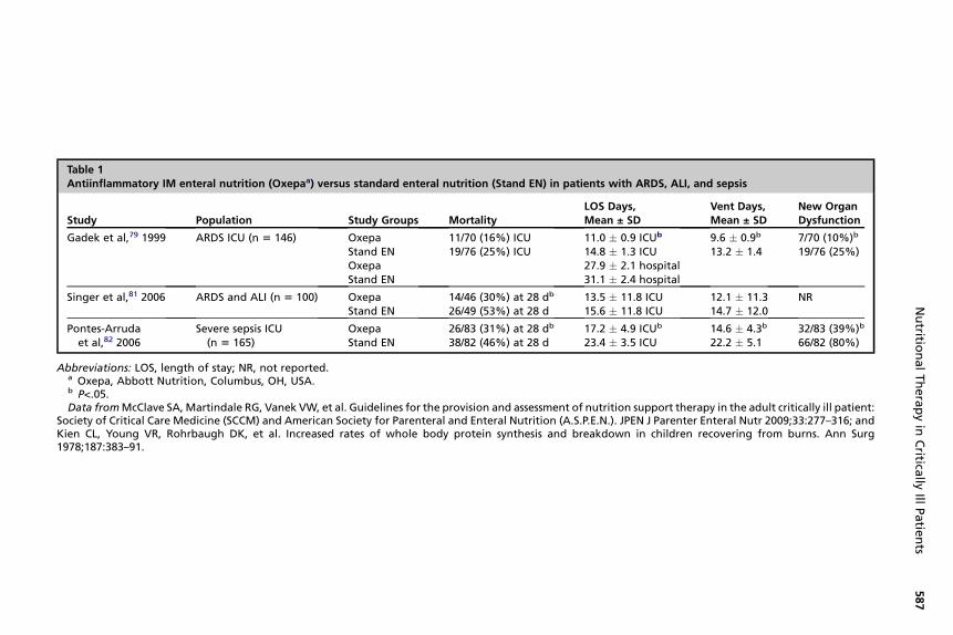
Table 1Antiinflammatory IM enteral nutrition (Oxepaa) versus standard enteral nutrition (Stand EN) in patients with ARDS, ALI, and sepsis
Study Population Study Groups MortalityLOS Days,Mean ± SD
Vent Days,Mean ± SD
New OrganDysfunction
Gadek et al,79 1999 ARDS ICU (n 5 146) OxepaStand ENOxepaStand EN
11/70 (16%) ICU19/76 (25%) ICU
11.0 � 0.9 ICUb
14.8 � 1.3 ICU27.9 � 2.1 hospital31.1 � 2.4 hospital
9.6 � 0.9b
13.2 � 1.47/70 (10%)b
19/76 (25%)
Singer et al,81 2006 ARDS and ALI (n 5 100) OxepaStand EN
14/46 (30%) at 28 db
26/49 (53%) at 28 d13.5 � 11.8 ICU15.6 � 11.8 ICU
12.1 � 11.314.7 � 12.0
NR
Pontes-Arrudaet al,82 2006
Severe sepsis ICU(n 5 165)
OxepaStand EN
26/83 (31%) at 28 db
38/82 (46%) at 28 d17.2 � 4.9 ICUb
23.4 � 3.5 ICU14.6 � 4.3b
22.2 � 5.132/83 (39%)b
66/82 (80%)
Abbreviations: LOS, length of stay; NR, not reported.a Oxepa, Abbott Nutrition, Columbus, OH, USA.b P<.05.Data fromMcClave SA, Martindale RG, Vanek VW, et al. Guidelines for the provision and assessment of nutrition support therapy in the adult critically ill patient:
Society of Critical Care Medicine (SCCM) and American Society for Parenteral and Enteral Nutrition (A.S.P.E.N.). JPEN J Parenter Enteral Nutr 2009;33:277–316; andKien CL, Young VR, Rohrbaugh DK, et al. Increased rates of whole body protein synthesis and breakdown in children recovering from burns. Ann Surg1978;187:383–91.
Nutritio
nalTherapyin
Critica
llyIll
Patie
nts
587

Latifi588
immune-enhancing nutrients was associated with preservation of lean body mass.93
A consensuspanel froma recent conference on immune-enhancing enteral therapy rec-ommended the use of IEDs in the following 2 groups of patients: (1) severely malnour-ished patients (albumin levels <3.5 g/dL) undergoing upper GI tract surgery andpatients with albumin levels less than 2.8 g/dL undergoing lower GI tract surgery and(2) patients with blunt or penetrating torso trauma with an injury severity score greaterthan 18, or an abdominal trauma index less than 20.94
SUMMARY
Nutritional support of critically ill or injured patients has undergone significantadvances in the last few decades. These advances are the direct result of the growingscientific progress and increased knowledge of the biology and biochemistry of keymetabolic and nutrient changes induced by injury, sepsis, and other critical illnesses,both in adults and children. As this knowledge has increased, the science of nutritionalsupport has become more disease based and disorder based. Depending on the indi-vidual patient’s metabolic needs, key nutrients are replenished or added in largeramounts to supplement specific deficiencies or to prevent further deterioration andclinical consequences. However, choosing or constructing the ideal nutrient formula-tions for the various critically ill or injured patients is increasingly complex: the resultantspecifically designed formulas, like manna from Heaven, will be expected to mitigatethe body’s metabolic response to injury, balance oxidative processes, and help regu-late or moderate the immune system, which may be deranged because of the under-lying disease, while additionally providing the balanced nutrients required for normalmaintenance, structure, and function.This is a daunting challenge, indeed, and much more sophisticated, labor-intensive,
and judiciousclinical investigationandcarewill be required toaccomplish thedesiredac-cumulation of the unequivocal knowledge and data leading to optimal patient outcomes.
REFERENCES
1. Ziegler T. Parenteral nutrition in the critically ill patient. N Engl J Med 2009;361:1088–97.
2. Cuthbertson DP. Observations on the disturbance of metabolism produced byinjury to the limbs. Q J Med 1932;1:233–46.
3. Hill AG, Hill GL. Metabolic response to severe injury. Br J Surg 1998;85:884–90.4. Ingenblek Y, Berstein L. The stressful condition as a nutritional adaptive
dichotomy. Nutrition 1999;15(4):305–20.5. Azimuddin K, Latifi R, Ivatury R. Acute phase proteins in critically ill patients. In:
Latifi R, Dudrick SJ, editors. The biology and practice of current nutritionalsupport. 2nd edition. Georgetown (TX): Landes Bioscience; 2003. p. 63–71.
6. Kudlackova M, Andel M, Hajkova H, et al. Acute phase proteins and prognosticinflammatory and nutritional index (PINI) in moderately burned children aged upto three years. Burns 1990;16:53–6.
7. Boosalis MG, Ott L, Levine AS, et al. Relationship of visceral proteins to nutritionalstatus in chronic and acute stress. Crit Care Med 1989;17:741–74.
8. Castell JV, Gomez-Lechon MJ, David M, et al. Acute-phase response of humanhepatocytes: regulation of acute-phase protein synthesis by interleukin-6. Hepa-tology 1990;12:1179–86.
9. Issihiki H, Akira S, Sugita T, et al. Reciprocal expression of NF-IL6 and C/EBP inhepatocytes: possible involvement of NF-IL6 in acute phase protein gene expres-sion. New Biol 1991;3(1):63–70.

Nutritional Therapy in Critically Ill Patients 589
10. Bankey PE, Mazuski JE, Ortiz M, et al. Hepatic acute phase protein synthesis isindirectly regulated by tumor necrosis factor. J Trauma 1990;30:1181–7.
11. Latifi R, Caushaj PE. Nutrition support in critically ill patients: current status andpractice. J Clin Ligand Assay 1999;22:279–84.
12. Wilmore DW, Orcutt TW, Mason AD Jr, et al. Alterations in hypothalamic functionfollowing thermal injury. J Trauma 1975;15:697–703.
13. Birkhahn RH, LongCL, FitkinD, et al. Effects ofmajor skeletal trauma onwhole bodyprotein turnover inmanmeasured by L-[1, 14C]-leucine. Surgery 1980;88:294–308.
14. Kien CL, Young VR, Rohrbaugh DK, et al. Increased rates of whole body proteinsynthesis and breakdown in children recovering from burns. Ann Surg 1978;187:383–91.
15. Levenson SM, Pulaski EJ, del Guercio LR. Metabolic changes associated withinjury. In: Zimmerman LM, Levine R, editors. Physiological principles of surgery.2nd edition. Philadelphia: WB Saunders; 1964. p. 5–7.
16. Young VR, Munro HN. N-methylhistidine (3-methylhistidine) and muscle proteinturnover: an overview. Fed Proc 1978;37:2291–300.
17. Bilmazes C, Kien CL, Rohrbaugh DK, et al. Quantitative contributors by skeletalmuscle to elevated rates of whole-body protein breakdown in burned children asmeasured by 3-methylhistidine output. Metabolism 1978;27:671–6.
18. Williamson DH, Farrell R, Kerr A, et al. Muscle protein catabolism after injury inman as measured by urinary excretion of 3-methylhistidine. Clin Sci Mol Med1977;52:527–33.
19. Long CL, Schiller WR, Blakemore WS, et al. Muscle protein catabolism in theseptic patient as measured by 3-methylhistidine excretion. Am J Clin Nutr1977;30:1349–52.
20. Essen P, McNurlan MA, Gamrin L, et al. Tissue protein synthesis rates in criticallyill patients. Crit Care Med 1998;26:92–100.
21. Latifi R, Dudrick SJ, editors. Surgical nutrition: strategies in critically ill. Austin(TX): Springer-Verlag/R.G.Landes; 1995.
22. Garber AJ, Karl IE, Kipnis DM. Alanine and glutamine synthesis and release fromskeletal muscle. I: glycolysis and amino acid release. J Biol Chem 1976;251:826–35.
23. Souba WW, Wilmore DW. Postoperative alteration of arteriovenous exchange ofamino acids across the gastrointestinal tract. Surgery 1983;94:342–50.
24. Souba WW, Klimberg VS, Plumley DA, et al. The role of glutamine in maintaininga healthy gut and supporting the metabolic response to injury and infection.J Surg Res 1990;48:383–91.
25. Fox AD, Kripke SA, Berman JM, et al. Dexamethasone administration inducesincreased glutaminase specific activity in the jejunum and colon. J Surg Res1988;44:391.
26. Souba WW, Smith RJ, Wilmore DW. Effect of glucocorticoids on glutamine metab-olism in visceral organs. Metabolism 1985;34:450–6.
27. Plumley DA, Souba WW, Hautamaki D, et al. Accelerated lung amino acid releasein hyperdynamic septic surgical patients. Arch Surg 1990;125:57.
28. Austgen TR, Chen MK, Flynn TC, et al. The effects of endotoxin on thesplanchnic metabolism of glutamine and related substrates. J Trauma 1991;6:742–51.
29. Austgen TR, Chen MK, Moore W, et al. Endotoxin and renal glutamine metabo-lism. Arch Surg 1991;126:23.
30. Souba WW, Smith RJ, Wilmore DW. Glutamine metabolism by the intestinal tract.JPEN J Parenter Enteral Nutr 1985;9:608–17.
Latifi590
31. Hwang TL, O’Dwyer ST, Smith RJ, et al. Preservation of small bowel mucosa usingglutamine-enriched parenteral nutrition. Surg Forum 1986;38:56.
32. Zapata-Sirvent RL, Hnasbrough JF, Ohara MM, et al. Bacterial translocation ofvarious diets including fiber-and glutamine enriched enteral formulas. Crit CareMed 1994;22:690–6.
33. Fox AD, Kripke SA, DePaula J, et al. Effect of a glutamine-supplemented enteraldiet on methotrexate-induced enterocolitis. JPEN J Parenter Enteral Nutr 1988;12:325–31.
34. Klimberg VS, Souba WW, Dolson DJ, et al. Prophylactic glutamine protects theintestinal mucosa from radiation injury. Cancer 1990;66:62–8.
35. Li SJ, Nussbaum MS, McFadden DW, et al. Addition of L-glutamine to total paren-teral nutrition and its effects on portal insulin and glucagon and the developmentof hepatic steatosis in rats. J Surg Res 1990;48:421–6.
36. Helton WS, Jacobs DO, Bonner-Weir S, et al. Effects of glutamine-enrichedparenteral nutrition on the exocrine pancreas. JPEN J Parenter Enteral Nutr1990;14:344–52.
37. Burke DJ, Alverdy JC, Aoys E, et al. Glutamine-supplemented total parenteralnutrition improves gut immune function. Arch Surg 1989;124:1396–9.
38. Hammarqvist F, Wernerman J, Ali R, et al. Addition of glutamine to total parenteralnutrition after elective abdominal surgery spares free glutamine in muscle, coun-teracts the fall in muscle protein synthesis, and improves nitrogen balance. AnnSurg 1989;209:455–61.
39. Stein TP, Yoshida S, Yamasaki K, et al. Amino acid requirements of critically illpatients. In: Latifi R, editor. Amino acids in critical care and cancer. Austin(TX): RG Landes Company; 1994. p. 9–25.
40. Barbul A. Arginine and immune function. Nutrition 1990;6:59–62.41. Barbul A, Sisto DA, Wasserkrug HL, et al. Arginine stimulates lymphocyte
immune response in healthy humans. Surgery 1981;90:244–51.42. Stinnett J, Alexander JW, Watanabe C, et al. Plasma and skeletal muscle amino
acids following severe burn injury in patients and experimental animals. Ann Surg1982;195:75–89.
43. Xiao-jun C, Chih-chun Y, Wei-shia H, et al. Changes of serum amino acids inseverely burned patients. Burns 1983;10:109–15.
44. Rose WC. Amino acid requirements of man. Fed Proc 1949;8(2):546–52.45. Roth E, Funovics J, Muhlbacher F, et al. Metabolic disorders in severe abdominal
sepsis: glutamine deficiency in skeletal muscle. Clin Nutr 1982;1(1):25–41.46. Askanazi J, Carpentier YA, Michelsen CB, et al. Muscle and plasma amino acids
following injury. Influence of intercurrent infection. Ann Surg 1980;192(1):78–85.47. Ziegler TR, Ogden LG, Singleton KD, et al. Parenteral glutamine increases serum
heat shock protein 70 in critically ill patients. Intensive Care Med 2005;31(8):1079–86.
48. Conejero R, Bonet A, Grau T, et al. Effect of a glutamine-enriched enteral diet onintestinal permeability and infectious morbidity at 28 days in critically ill patientswith systemic inflammatory response syndrome: a randomized, single-blind,prospective, multicenter study. Nutrition 2002;18(9):716–21.
49. Garrel D, Patenaude J, Nedelec B, et al. Decreased mortality and infectiousmorbidity in adult burn patients given enteral glutamine supplements: a prospec-tive, controlled, randomized clinical trial. Crit Care Med 2003;31(10):2444–9.
50. Houdijk AP, Rijnsburger ER, Jansen J, et al. Randomised trial of glutamine-enriched enteral nutrition on infectious morbidity in patients with multiple trauma.Lancet 1998;352(9130):772–6.

Nutritional Therapy in Critically Ill Patients 591
51. Hall JC, Dobb G, Hall J, et al. A prospective randomized trial of enteral glutaminein critical illness. Intensive Care Med 2003;29(10):1710–6.
52. Wischmeyer PE, Lynch J, Liedel J, et al. Glutamine administration reduces gram-negative bacteremia in severely burned patients: a prospective, randomized,double-blind trial versus isonitrogenous control. Crit Care Med 2001;29(11):2075–80.
53. McClave SA, Martindale RG, Vanek VW, et al. Guidelines for the provision andassessment of nutrition support therapy in the adult critically ill patient: Societyof Critical Care Medicine (SCCM) and American Society for Parenteral andEnteral Nutrition (A.S.P.E.N.). JPEN J Parenter Enteral Nutr 2009;33(3):277–316.
54. Rose WC. The nutritive significance of the amino acids and certain relatedcompounds. Science 1937;86(2231):298–300.
55. Davis SL. Plasma levels of prolactin, growth hormone and insulin in sheepfollowing the infusion of arginine, leucine and phenylalanine. Endocrinology1972;91(2):549–55.
56. Daly JM, Reynolds J, Thom A, et al. Immune and metabolic effects of arginine inthe surgical patient. Ann Surg 1988;208(4):512–23.
57. Heyland DK, Novak F, Drover JW, et al. Should immunonutrition become routine incritically ill patients? A systematic review of the evidence. JAMA 2001;286(8):944–53.
58. Bower RH, Cerra FB, Bershadsky B, et al. Early enteral administration of a formula(Impact) supplemented with arginine, nucleotides, and fish oil in intensive careunit patients: results of a multicenter, prospective, randomized, clinical trial. CritCare Med 1995;23(3):436–49.
59. Bertolini G, Iapichino G, Radrizzani D, et al. Early enteral immunonutrition inpatients with severe sepsis: results of an interim analysis of a randomized multi-centre clinical trial. Intensive Care Med 2003;29(5):834–40.
60. Zhou M, Martindale RG. Arginine in the critical care setting. J Nutr 2007;137(6Suppl 2):1687S–92S.
61. Galban C, Montejo JC, Mesejo A, et al. An immune-enhancing enteral dietreduces mortality rate and episodes of bacteremia in septic intensive care unitpatients. Crit Care Med 2000;28(3):643–8.
62. Kulkarni AD, Rudolph FB, Van Buren CT. The role of dietary sources of nucleo-tides in immune function: a review. J Nutr 1994;124(Suppl 8):1442S–6S.
63. Van Buren CT, Kulkarni AD, Fanslow WC, et al. Dietary nucleotides, a requirementfor helper/inducer T lymphocytes. Transplantation 1985;40(6):694–7.
64. Beale RJ, Bryg DJ, Bihari DJ. Immunonutrition in the critically ill: a systematicreview of clinical outcome. Crit Care Med 1999;27(12):2799–805.
65. Grimble GK, Westwood OM. Nucleotides as immunomodulators in clinical nutri-tion. Curr Opin Clin Nutr Metab Care 2001;4(1):57–64.
66. Heyland DK, Dhaliwal R, Suchner U, et al. Antioxidant nutrients: a systematicreview of trace elements and vitamins in the critically ill patient. Intensive CareMed 2005;31(3):327–37.
67. Crimi E, Liguori A, Condorelli M, et al. The beneficial effects of antioxidant supple-mentation in enteral feeding in critically ill patients: a prospective, randomized,double-blind, placebo-controlled trial. Anesth Analg 2004;99(3):857–63.
68. Angstwurm MW, Engelmann L, Zimmermann T, et al. Selenium in Intensive Care(SIC): results of a prospective randomized, placebo-controlled, multiple-centerstudy in patients with severe systemic inflammatory response syndrome, sepsis,and septic shock. Crit Care Med 2007;35(1):118–26.

Latifi592
69. Gennari R, Alexander JW, Eaves-Pyles T. Effect of different combinations of die-tary additives on bacterial translocation and survival in gut-derived sepsis. JPENJ Parenter Enteral Nutr 1995;19(4):319–25.
70. Blackburn GL, Moldawer LL, Usui S, et al. Branched-chain amino acid adminis-tration and metabolism during starvation, injury, and infection. Surgery 1979;86(2):307–15.
71. Yoshida S, Lanza-Jacoby S, Stein TP. Leucine and glutamine metabolism inseptic rats. Biochem J 1991;276(Pt 2):405–9.
72. Freund HR, James JH, Fischer JE. Nitrogen-sparing mechanisms of singlyadministered branched-chain amino acids in the injured rat. Surgery 1981;90(2):237–43.
73. Garcı́a-de-Lorenzo A, Ortı́z-Leyba C, Planas M, et al. Parenteral administration ofdifferent amounts of branch-chain amino acids in septic patients: clinical andmetabolic aspects. Crit Care Med 1997;25(3):418–24.
74. Calder PC. Branched-chain amino acids and immunity. J Nutr 2006;136(Suppl 1):288S–93S.
75. Ware LB, Matthay MA. The acute respiratory distress syndrome. N Engl J Med2000;342(18):1334–49.
76. Mancuso P, Whelan J, DeMichele SJ, et al. Dietary fish oil and fish and borage oilsuppress intrapulmonary proinflammatory eicosanoid biosynthesis and attenuatepulmonary neutrophil accumulation in endotoxic rats. Crit Care Med 1997;25(7):1198–206.
77. Needleman P, Raz A, Minkes MS, et al. Triene prostaglandins: prostacyclin andthromboxane biosynthesis and unique biological properties. Proc Natl Acad SciU S A 1979;76(2):944–8.
78. Lee TH, Menica-Huerta JM, Shih C, et al. Characterization and biologic proper-ties of 5,12-dihydroxy derivatives of eicosapentaenoic acid, including leukotrieneB5 and the double lipoxygenase product. J Biol Chem 1984;259(4):2383–9.
79. Gadek JE, DeMichele SJ, Karlstad MD, et al. Effect of enteral feeding with eico-sapentaenoic acid, gamma-linolenic acid, and antioxidants in patients with acuterespiratory distress syndrome. Enteral Nutrition in ARDS Study Group. Crit CareMed 1999;27(8):1409–20.
80. Pacht ER, DeMichele SJ, Nelson JL, et al. Enteral nutrition with eicosapentaenoicacid, gamma-linolenic acid, and antioxidants reduces alveolar inflammatorymediators and protein influx in patients with acute respiratory distress syndrome.Crit Care Med 2003;31(2):491–500.
81. Singer P, Theilla M, Fisher H, et al. Benefit of an enteral diet enriched with eico-sapentaenoic acid and gamma-linolenic acid in ventilated patients with acutelung injury. Crit Care Med 2006;34(4):1033–8.
82. Pontes-Arruda A, Aragao AM, Albuquerque JD. Effects of enteral feeding with ei-cosapentaenoic acid, gamma-linolenic acid, and antioxidants in mechanicallyventilated patients with severe sepsis and septic shock. Crit Care Med 2006;34(9):2325–33.
83. Marik PE, Zaloga GP. Immunonutrition in critically ill patients: a systematic reviewand analysis of the literature. Intensive Care Med 2008;34(11):1980–90.
84. Martindale R, Miles J. Is immunonutrition ready for prime time? JPEN J ParenterEnteral Nutr 2003;18:489–96.
85. Braga M, Gianotti L, Nespoli L, et al. Nutritional approach in malnourishedsurgical patients: a prospective randomized study. Arch Surg 2002;137:174–80.
86. Seto Y, Fukuda T, Yamada K, et al. Celiac lymph nodes: distant or regional forthoracic esophageal carcinoma? Dis Esophagus 2008;21:704–7.
Nutritional Therapy in Critically Ill Patients 593
87. Fukuda T, Seto Y, Ymada K, et al. Can immune-enhancing nutrients reduce post-operative complications in patients undergoing esophageal surgery? Dis Esoph-agus 2008;21:708–11.
88. Giger U, Buchler M, Farhadi J, et al. Preoperative immunonutrition suppressesperioperative inflammatory response in patients with major abdominal surgery—a randomized controlled pilot study. Ann Surg Oncol 2007;14(10):2798–806.
89. Helminen H, Raitanen M, Kellosalo J. Immunonutrition in elective gastrointestinalsurgery patients. Scand J Surg 2007;96:46–50.
90. Klek S, Kulig J, Sierzega M, et al. The impact of immuno stimulating nutrition oninfectious complications after upper gastrointestinal surgery: a prospective,randomized, clinical trial. Ann Surg 2008;248(2):212–20.
91. Ryan A, Power D. Letters to the editor: immuno-nutrition in upper gastrointestinalsurgery. Ann Surg 2009;249(6):1062–3.
92. Okamoto Y, Okano K, Izuishi K, et al. Attenuation of the systemic inflammatoryresponse and infectious complications after gastrectomy with preoperative oralarginine and omega-3 fatty acids supplemented immunonutrition. World J Surg2009;33:1815–21.
93. Ryan AM, Reynolds J, Healy L, et al. Enteral nutrition with Eicosapentaenoic Acid(EPA) preserves lean body mass following esophageal cancer surgery: results ofa double-blinded randomized controlled trial. Ann Surg 2009;3(249):355–63.
94. Heys SD, Walker LG, Smith I, et al. Enteral nutrition supplementation with keynutrients in patients with critical illness and cancer. Meta-analysis of randomizedcontrolled clinical trials. Ann Surg 1999;229:446–77, 93.