Embed Size (px)
Citation preview

iCopyright © 2020. Korean Society for Neurorehabilitation
HIGHLIGHTS• We report a patient with hypoxic brain injury following cardiac arrest.• Hypoxic brain injury may leads to cognitive deficit of multiple domain.• Systematic and serial evaluations for cognitive dysfunction are important.
Brain Neurorehabil. 2020 Nov;13(3):e20https://doi.org/10.12786/bn.2020.13.e20pISSN 1976-8753·eISSN 2383-9910
Case Report
Received: Jan 23, 2020Revised: Jun 26, 2020Accepted: Jul 6, 2020
Correspondence toJoo Hyun ParkDepartment of Rehabilitation Medicine, Seoul St. Mary's Hospital, College of Medicine, The Catholic University of Korea, 222 Banpo-daero, Seocho-gu, Seoul 06591, Korea.E-mail: [email protected]
Yongjun Jang, Eunji Lee, Youngkook Kim, Joo Hyun Park
Number Processing Error as a Clinical Manifestation of Hemispatial Neglect Following Hypoxic Brain Injury: a Case Report
Brain & NeuroRehabilitation
02

1/7
ABSTRACT
Hemispatial neglect is a symptom where patients do not show response to stimuli on the contralesional side of their brain lesion. Although it is most common in the context of hemispheric stroke, several pathological processes including neurodegenerative disease, neoplasia, and trauma may cause this. Prevalence of hemispatial neglect is unknown and rarely reported among patients with hypoxic brain injury. Also, hemispatial neglect accompanying neglect dyslexia is rather hard to be recognized and symptoms involving numbers are exceptionally rare. We report a patient with hypoxic brain injury following cardiac arrest who showed signs of neglect dyslexia for numbers that provided a primary clue for the diagnosis of left hemispatial neglect. Early detection of different forms of cognitive dysfunction of hypoxic brain injury is highly essential in providing early neurorehabilitation for better prognosis.
Keywords: Perceptual Disorders; Brain Hypoxia; Dyslexia
INTRODUCTION
Hypoxic brain injury following cardiac arrest is a leading cause of mortality and long-term neurologic disability among survivors [1]. Despite improvements in the pre-hospital and critical care management of patients with hypoxic brain injury, the conditions associated with cognitive impairments still remain problematic for many survivors [2]. The most common cognitive disturbances are disorders of consciousness, attention, speed of processing, memory impairments, and executive function. Disturbance of the visuospatial domain of cognition was rarely reported after hypoxic brain injury [3]. Unfortunately at the acute stage, clinicians tend to focus more on the patient’s level of consciousness since a decreased consciousness follows the neurologic deficit after global cerebral anoxic injury [1]. Lack of awareness of clinicians for multi-domain cognitive deficit may hinder the chance of detecting cognitive dysfunction and delay early neurorehabilitation. We report a patient with hypoxic brain injury following cardiac arrest. This patient presented with neglect in number processing, which led to the diagnosis of combined left hemispatial neglect.
Brain Neurorehabil. 2020 Nov;13(3):e20https://doi.org/10.12786/bn.2020.13.e20pISSN 1976-8753·eISSN 2383-9910
Case Report
Received: Jan 23, 2020Revised: Jun 26, 2020Accepted: Jul 6, 2020
Correspondence toJoo Hyun ParkDepartment of Rehabilitation Medicine, Seoul St. Mary's Hospital, College of Medicine, The Catholic University of Korea, 222 Banpo-daero, Seocho-gu, Seoul 06591, Korea.E-mail: [email protected]
Copyright © 2020. Korean Society for NeurorehabilitationThis is an Open Access article distributed under the terms of the Creative Commons Attribution Non-Commercial License (https://creativecommons.org/licenses/by-nc/4.0) which permits unrestricted non-commercial use, distribution, and reproduction in any medium, provided the original work is properly cited.
ORCID iDsYongjun Jang https://orcid.org/0000-0001-6844-2292Youngkook Kim https://orcid.org/0000-0003-3964-026XJoo Hyun Park https://orcid.org/0000-0001-9257-8704
Conflict of InterestThe authors have no potential conflicts of interest to disclose.
Yongjun Jang ,1 Eunji Lee,1 Youngkook Kim ,2 Joo Hyun Park 1
1 Department of Rehabilitation Medicine, Seoul St. Mary's Hospital, College of Medicine, The Catholic University of Korea, Seoul, Korea
2 Department of Rehabilitation Medicine, Yeouido St. Mary's Hospital, College of Medicine, The Catholic University of Korea, Seoul, Korea
Number Processing Error as a Clinical Manifestation of Hemispatial Neglect Following Hypoxic Brain Injury: a Case Report
Brain & NeuroRehabilitation
02
https://e-bnr.org

CASE REPORT
A 39-year-old male who was diagnosed with hypertension and dyslipidemia was admitted to the emergency department of university affiliated hospital with the status of cardiac arrest. On the day of arrest, he was having dinner with his colleagues and suddenly collapsed. Basic Life Support was provided, and the initial electrocardiography revealed he had ventricular fibrillation. Defibrillation was performed, and he showed regular rhythm 30 minutes after the arrest. After a week of hypothermia regimen, cardiologists concluded that the arrest was due to arrhythmia, and an implantable cardiac device (ICD) was implanted.
His mental status was alert after management regarding his heart condition, but cognitive dysfunction was suspected by a physiatrist at the routine intensive care unit bedside evaluation. The brain magnetic resonance imaging that he underwent before ICD implantation showed diffuse cortical high signals in bilateral hemispheres on susceptibility weighted imaging with dark signal intensity on apparent diffusion coefficient (ADC) map, and also showed symmetric high signals in bilateral basal ganglia and thalami on diffusion weighted image with normal or subtle high signals on ADC map (Fig. 1). Since he received an ICD implantation, single photon emission computed tomography was taken as a replacement for follow up study. The result showed diffuse, mild to moderate hypoperfusion in the bilateral cerebral cortex, most severely in the right frontal and right parietal cortices (Fig. 2). Electroencephalography showed normal brain activity, but his initial Korean Mini-Mental Status Examination score was 16, indicating a cognitive dysfunction of moderate degree.
Ten days after the cardiac arrest, the patient was transferred to the department of rehabilitation. Routine rehabilitation evaluations were performed for a better understanding of the patient's function. Manual muscle test for his body was generally above “Good” grade. Korean Modified Barthel Index (K-MBI) was 5 out of 100, indicating a total dependence in the activity of daily living (ADL).
Cardiac rehabilitation (aerobic exercise targeting Borg dyspnea scale of 10–12) was performed to enhance his motor performance. Table top cognitive training and fine motor training were also preceded.
His motor function showed dramatic improvement, in which he was able to walk independently within a week. However, his cognitive dysfunction did not show much
2/7https://doi.org/10.12786/bn.2020.13.e20
Visuospatial Deficit Following Hypoxic Brain Injury Brain & NeuroRehabilitation
02
https://e-bnr.org
A B C D
Fig. 1. The brain magnetic resonance imaging shows diffuse cortical high signals in bilateral hemispheres on susceptibility weighted imaging (A) with dark signal intensity on ADC (B) map; symmetric high signals in bilateral basal ganglia and thalami on diffusion weighted image (C) with normal or subtle high signals on ADC (D) map. ADC, apparent diffusion coefficient.

improvement. Loewenstein Occupational Therapy Cognitive Assessment was performed to further investigate his cognitive dysfunction. Visuomotor organization, spatial relations on picture, drawing clock, pictorial sequence were severely impaired. Also, the digit trail making test (TMT) was done to evaluate his visual attention and task switching ability.
His digit TMT sheet showed an interesting pattern. He connected 1-2-13, 1-2-3-14, 6-7-18, 14-25 (Fig. 3). When ignoring the first digit of the two-digit numbers, the sequence was correct. The tendency of omitting the first digit of every two-digit number was commonly seen. When simple calculation questions were given to him, he could only solve the one-digit
3/7https://doi.org/10.12786/bn.2020.13.e20
Visuospatial Deficit Following Hypoxic Brain Injury Brain & NeuroRehabilitation
02
https://e-bnr.org
Fig. 2. Single photon emission computed tomography shows diffuse, mild to moderate hypoperfusion in the bilateral cerebral cortex, most severely in the right frontal and right parietal cortices.
Fig. 3. Result of the initially performed trail making test shows a tendency of omitting the first digit of every two-digit number.

calculations. Allocentric neglect syndrome was suspected based on the result of digit TMT sheet. The patient was asked to read an article out loud. To improve his attention, the article was shown to him line by line, and he was not allowed to point each word with his finger. As a result, he committed partial omissions or substitutions for letters at the beginning of the word, usually on the left side (Fig. 4A). However, unlike for numbers, dyslexia was not distinct in words for he could understand the context of reading materials. Further evaluation was performed to validate the presence of neglect or the possibility of combined visual problem. His visual evoked potential, visual field, and acuity showed normal results.
Neglect test batteries including apple cancellation, clock drawing test, Ota test, line bisection test, and scene copying tasks were performed. The result suggested a left side neglect. Moreover, he seemed to have both allocentric and egocentric neglect (Fig. 4B and C).
Despite that he had certain disability of left side spatial neglect, he could manage most of the activities of daily living (K-MBI 92/100 by the time of discharge). However, obvious mistakes were observed whenever he had to read or draw. He was asked to color up the entire picture but was able to fill up the right half of the picture only. After thorough evaluations, the patient was revealed to have a left hemispatial neglect along with a tendency of dyslexia.
Medication such as dopaminergic and adrenergic agents were not possible since the patient had an ICD for controlling his heart rhythm. Throughout months of out-patient clinic-based rehabilitation sessions (5 sessions per week, 40 minutes per session), scanning training,
4/7https://doi.org/10.12786/bn.2020.13.e20
Visuospatial Deficit Following Hypoxic Brain Injury Brain & NeuroRehabilitation
02
https://e-bnr.org
A B C
D E F
Fig. 4. Results of neglect evaluation batteries. (A) A random material reading, (B) apple cancellation, (C) clock drawing test, (D) Ota test, (E) line bisection test, and (F) scene copying tasks were performed, and the results showed left side allocentric and egocentric neglect.

optokinetic stimulation and mirror therapy were performed. Moreover, he was asked to apply right hemispatial eye-patch at home. After three months of rehabilitation sessions, he was able to compensate for his neglect symptoms and was able to return to his social life with minimal post-hypoxic brain injury sequelae only.
DISCUSSION
Hypoxic brain injury first affects the cerebral cortex since it is the far most area which can be directly affected by brain hypoperfusion [4]. The parietal lobe is commonly regarded as a part of a network of brain areas involved in controlling attention throughout feedback connections with the visual cortex [5]. Our patient had a global hypoxic brain injury in bilateral frontal, temporal, and parietal lobes with suspicious involvement of occipital lobes. However, he had a more prominent hypoxic injury in the right frontal and parietal region. This strongly explains his left hemispatial neglect.
To our best of knowledge, there are only a few reports of hemispatial neglect following hypoxic brain injury. Hemispatial neglect can manifest in various forms since it is a very complex disorder that can affect several components of spatial behavior in different ways [6]. It is difficult for a single test to sufficiently provide evaluation of all forms of neglect. Therefore, serial and systematic assessment of patients with cognitive dysfunction is essential in providing appropriate treatment. The evaluations for patients with a hypoxic brain injury requires tests for vigilance or cancellation tasks, continuous performance tasks, and other tests of memory, attention and processing speed such as the TMT. In the bedside setting, simple screening tools such as the Mini-Mental State Examination (MMSE) may be a handy screening tool for detecting the existence of cognitive deficit.
Our patient showed alert consciousness at the time of referral, but MMSE evaluation for his cognitive function revealed cognitive dysfunction of moderate degree mainly due to his problem with attention. Furthermore, cognitive dysfunction of the visuospatial domain was detected throughout evaluation of digit TMT. Digit TMT successfully caught the subtle sign of neglect in two-digit numbers, which might have been missed. He had shown peculiarity in the result of TMT and calculation. Additional neglect screening batteries showed that his left side neglect was not only confined to numbers but also for words, though the severity of neglect was not severe for words. As a possible explanation to this phenomenon, patients with neglect dyslexia shows impairment in visuospatial processing, but not in their linguistic ability [7]. Therefore, the preserved lexical effects and implicit language processing ability may have impact on patients' ability to compensate their visuospatial processing deficit when reading words. As a result, individuals with neglect dyslexia may process words more accurately than nonwords such as numbers and figures [8]. This may be the reason why the dyslexia for words of our patient was not distinct. While hemispatial neglect has been studied extensively, the impact of neglect on the representation of number magnitude has only more recently been focused, which makes this case report more valuable.
The neglect has a negative effect on the patient's motor, cognitive, and social recovery and ADL performance. A systemic review revealed that the poststroke neglect has a significant negative impact on functional outcome, either as an independent predictive factor or in connection with other variables such as age, hemiparesis, severity of stroke, anosognosia, other cognitive impairment [9]. In addition, patients with neglect tends have longer average
5/7https://doi.org/10.12786/bn.2020.13.e20
Visuospatial Deficit Following Hypoxic Brain Injury Brain & NeuroRehabilitation
02
https://e-bnr.org
length of stay in the hospital [10]. Therefore, early detection and neurorehabilitative approach is needed for better outcome and to minimize length of stay.
Early detection of different forms of neglect and proper diagnosis is crucial for providing appropriate goal setting in rehabilitation and predicting the need for intervention as patients with neglect are more dependent on their environment compared to patients without neglect [11,12]. In general, neuropsychological paper-and-pencil tasks, such as cancellation or bisection tasks, are used for diagnosis of neglect [13]. The treatment of choice for hemispatial neglect and dyslexia are still controversial. Our patient also tried eye-patching bilateral right half-fields, so that the right hemisphere stroke stimulates the right hemisphere and reduces the stimulation of the left hemisphere for interhemispheric rebalance [14]. The outcome seems to be positive since the follow-up neglect tests showed much improvement after weeks of eye patching. However, the treatment effect of optokinetic stimulation and the patient's natural recovery cannot be undertaken.
In conclusion, the present case report described the diagnosing process of hemispatial neglect in a patient with hypoxic brain injury. Detecting number processing error of the patient in digit TMT provided an important clinical cue to make a proper diagnosis of hemispatial neglect, thereby providing appropriate rehabilitation intervention. Clinician's critical thinking warrants the attention of clinicians to trace the obscure symptoms of patients to enhance their functional outcome.
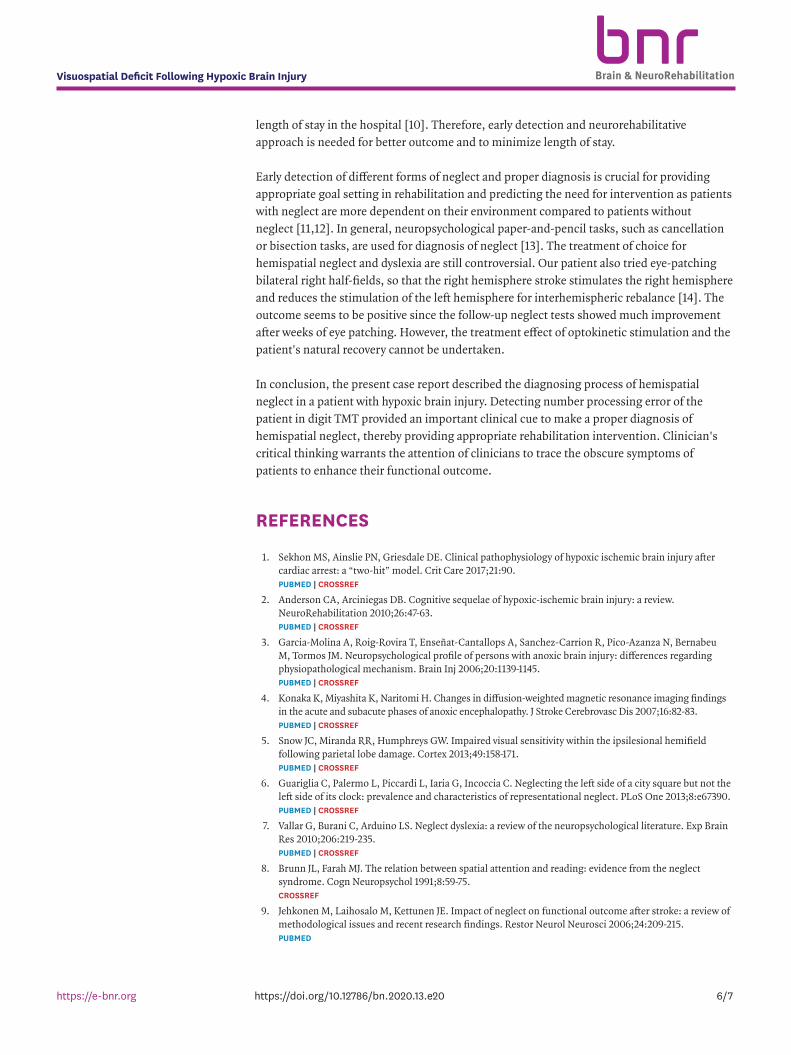
REFERENCES
1. Sekhon MS, Ainslie PN, Griesdale DE. Clinical pathophysiology of hypoxic ischemic brain injury after cardiac arrest: a “two-hit” model. Crit Care 2017;21:90. PUBMED | CROSSREF
2. Anderson CA, Arciniegas DB. Cognitive sequelae of hypoxic-ischemic brain injury: a review. NeuroRehabilitation 2010;26:47-63. PUBMED | CROSSREF
3. Garcia-Molina A, Roig-Rovira T, Enseñat-Cantallops A, Sanchez-Carrion R, Pico-Azanza N, Bernabeu M, Tormos JM. Neuropsychological profile of persons with anoxic brain injury: differences regarding physiopathological mechanism. Brain Inj 2006;20:1139-1145. PUBMED | CROSSREF
4. Konaka K, Miyashita K, Naritomi H. Changes in diffusion-weighted magnetic resonance imaging findings in the acute and subacute phases of anoxic encephalopathy. J Stroke Cerebrovasc Dis 2007;16:82-83. PUBMED | CROSSREF
5. Snow JC, Miranda RR, Humphreys GW. Impaired visual sensitivity within the ipsilesional hemifield following parietal lobe damage. Cortex 2013;49:158-171. PUBMED | CROSSREF
6. Guariglia C, Palermo L, Piccardi L, Iaria G, Incoccia C. Neglecting the left side of a city square but not the left side of its clock: prevalence and characteristics of representational neglect. PLoS One 2013;8:e67390. PUBMED | CROSSREF
7. Vallar G, Burani C, Arduino LS. Neglect dyslexia: a review of the neuropsychological literature. Exp Brain Res 2010;206:219-235. PUBMED | CROSSREF
8. Brunn JL, Farah MJ. The relation between spatial attention and reading: evidence from the neglect syndrome. Cogn Neuropsychol 1991;8:59-75. CROSSREF
9. Jehkonen M, Laihosalo M, Kettunen JE. Impact of neglect on functional outcome after stroke: a review of methodological issues and recent research findings. Restor Neurol Neurosci 2006;24:209-215.PUBMED
6/7https://doi.org/10.12786/bn.2020.13.e20
Visuospatial Deficit Following Hypoxic Brain Injury Brain & NeuroRehabilitation
02
https://e-bnr.org

10. Cherney LR, Halper AS, Kwasnica CM, Harvey RL, Zhang M. Recovery of functional status after right hemisphere stroke: relationship with unilateral neglect. Arch Phys Med Rehabil 2001;82:322-328. PUBMED | CROSSREF
11. Buxbaum LJ, Ferraro MK, Veramonti T, Farne A, Whyte J, Ladavas E, Frassinetti F, Coslett HB. Hemispatial neglect: subtypes, neuroanatomy, and disability. Neurology 2004;62:749-756. PUBMED | CROSSREF
12. Ten Brink AF, Verwer JH, Biesbroek JM, Visser-Meily JM, Nijboer TC. Differences between left- and right-sided neglect revisited: a large cohort study across multiple domains. J Clin Exp Neuropsychol 2017;39:707-723. PUBMED | CROSSREF
13. Plummer P, Morris ME, Dunai J. Assessment of unilateral neglect. Phys Ther 2003;83:732-740. PUBMED | CROSSREF
14. Smania N, Fonte C, Picelli A, Gandolfi M, Varalta V. Effect of eye patching in rehabilitation of hemispatial neglect. Front Hum Neurosci 2013;7:527. PUBMED | CROSSREF
7/7https://doi.org/10.12786/bn.2020.13.e20
Visuospatial Deficit Following Hypoxic Brain Injury Brain & NeuroRehabilitation
02
https://e-bnr.org