Embed Size (px)
Citation preview
NP
--X
yl
NP
--G
lc
NP
--G
al
NP
--M
an
NP
--X
yl
NP
--G
lc
NP
--G
al
NP
--M
an
NP
--X
yl
NP
--G
lc
NP
--G
al
NP
--M
an
NP
--X
yl
NP
--G
lc
NP
--G
al
NP
--M
an
Enz
yme
only
Mar
kers
Xyl
ManGlc
Gal
Lam2Cell2
Lam3Cell3Lam4
Cell5
Cell6
Nitrophenyl glycoside substrate Substrate + enzyme
Figure S1. Differences in the occurrence of transglycosylation during hydrolysis of nitrophenyl glycosides by cauliflower-leaf extract.
Each nitrophenyl (NP) glycoside substrate was incubated at 1.4 mM with cauliflower lamina enzyme (50-60%-saturated ammonium sulphate cut; final concentration 1.2 mg protein ml-1) for an appropriate time, determined in preliminary tests to permit the partial hydrolysis of the substrate (except in the case of NP--Xyl, which is not hydrolysed by cauliflower extracts). Incubation times were: NP--Xyl, 24 h; NP--Xyl, 8 h; NP--Glc, 24 h; NP--Glc, 4 min; NP--Gal, 2 min; NP--Gal, 6 min; NP--Man, 4 min; NP--Man, 8 min. Products were run by TLC in butan-1-ol/acetic acid/H2O, 4:1:1, and stained with thymol/H2SO4. The NP-glycoside substrates were all para- except NP--Gal, which was the ortho-isomer; they are all of high chromatographic mobility (green arrows), and their monosaccharide products are indicated by blue arrows. In two cases (-xylosidase and -glucosidase), there is evidence for the formation of an intermediary transglycosylation product (NP-disaccharide; yellow arrows). The markers (right lane) were run and stained on the same plate, but have not been as highly contrast-enhanced as the rest of the image.
DP2
DP4
DP6
DP7
DP8
DP9
DP10
XXXG
XXG
XG
Xyl
Glc
1h 2h 4h 8h 18h 24h 32h 48h C0h C48h
Markers
0h ¼h 1h 4h 16h 0h
Low activity enzyme High activity enzyme No enzyme
a b
Lam2
Glc
Cello2
Xyl
Lam4
Cello3
Cello4
Lam3
Cello6
XXLG
XLLG
XXXG
Isopr
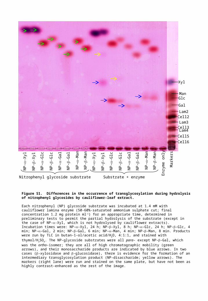
Figure S2. Time-courses for action of cauliflower leaf enzymes on XXXG
Two dialysed preparations of a 50–60%-saturated (NH4)2SO4 cut from cauliflower lamina were incubated with 1.4 mM XXXG for various times and the products were analysed by TLC in BAW. (a) Standard enzyme preparation (as used in other experiments), TLC with two ascents; (b) high-activity enzyme preparation, TLC with three ascents. C = enzyme-free controls incubated for 0 or 48 h. Other details, including the colour-coding of labels, are as in Figure 2.
Distance migrated (cm from origin)
Rad
ioac
tivity
(co
unts
per
85
m c
hann
el)
-1 0 1 2 3 4 5 6 7 8 9 10 11 12 13 14 15 16 1718
0
400
800
1200
1600
2000
2400
2800
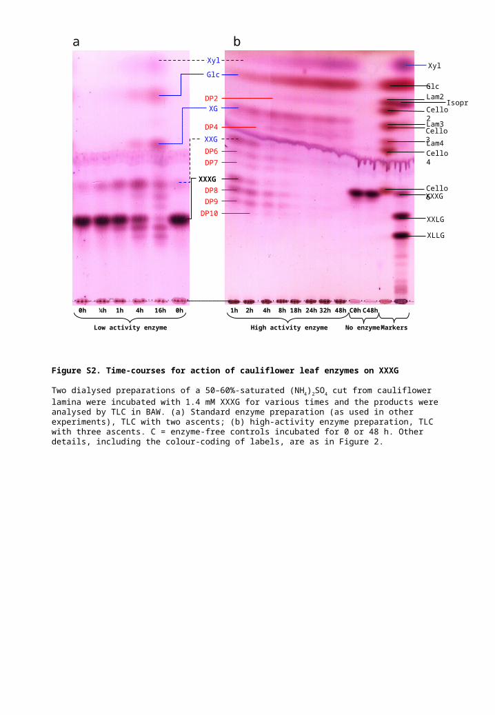
Figure S3 A representative quantitative scan of radioactive oligosaccharides as resolved by thin-layer chromatography
The products formed by TX after 12 h with 2048 µM [3H]XXXGol were resolved by TLC (as shown in Fig. 3d) and then quantified with a LabLogic AR2000 radioisotope imaging scanner.
crud
e
no A
S p
ptn
20%
AS
ppt
30%
AS
ppt
40%
AS
ppt
50%
AS
ppt
60%
AS
ppt
70%
AS
ppt
mar
kerscr
ude
no A
S p
ptn
20%
AS
ppt
30%
AS
ppt
40%
AS
ppt
50%
AS
ppt
60%
AS
ppt
70%
AS
ppt
midrib lamina
XLLG
XXLG
XXXG
XG
IP
Glc
GG
GGG
GGGG
GGGGG
Gal
XLLG
XXLG
XXXG
Glc
Xyl
Gal
XG
XXG
DP9–10
DP10–11
DP8
? DP6
? GXXG? XLG
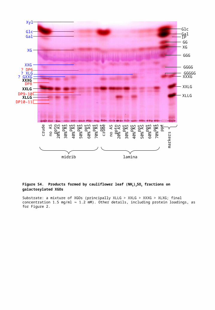
Figure S4. Products formed by cauliflower leaf (NH4)2SO4 fractions on galactosylated XGOs
Substrate: a mixture of XGOs (principally XLLG > XXLG > XXXG > XLXG; final concentration 1.5 mg/ml 1.2 mM). Other details, including protein loadings, as for Figure 2.
MM
a b Ninhydrin c AHPh
DNP-Lys
XGO-NH2
Glc +XXXG
origin
1 2 3 4 MM MM1 2 3 4
d AgNO3
Glc
GlcN
DNP-Lys
XGO-NH2
origin
MM1 2 3 4
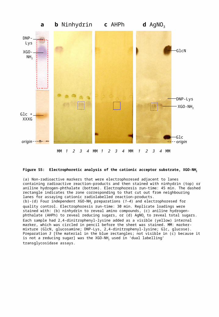
Figure S5: Electrophoretic analysis of the cationic acceptor substrate, XGO-NH2
(a) Non-radioactive markers that were electrophoresed adjacent to lanes containing radioactive reaction-products and then stained with ninhydrin (top) or aniline hydrogen-phthalate (bottom). Electrophoresis run-time: 45 min. The dashed rectangle indicates the zone corresponding to that cut out from neighbouring lanes for assaying cationic radiolabelled reaction-products. (b)-(d) Four independent XGO-NH2 preparations (1-4) and electrophoresed for quality control. Electrophoresis run-time: 30 min. Replicate loadings were stained with: (b) ninhydrin to reveal amino compounds, (c) aniline hydrogen-phthalate (AHPh) to reveal reducing sugars, or (d) AgNO3 to reveal total sugars. Each sample had 2,4-dinitrophenyl-lysine added as a visible (yellow) internal marker, which was circled in pencil before the sheet was stained. MM: marker-mixture (GlcN, glucosamine; DNP-Lys, 2,4-dinitrophenyl-lysine; Glc, glucose). Preparation 3 [the material in the blue rectangles; not visible in (c) because it is not a reducing sugar] was the XGO-NH2 used in ‘dual labelling’ transglycosidase assays.
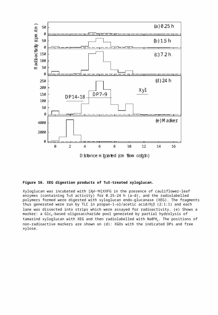
Figure S6. XEG digestion products of TX-treated xyloglucan.
Xyloglucan was incubated with [Xyl-3H]XXFG in the presence of cauliflower-leaf enzymes (containing TX activity) for 0.25-24 h (a-d), and the radiolabelled polymers formed were digested with xyloglucan endo-glucanase (XEG). The fragments thus generated were run by TLC in propan-1-ol/acetic acid/H2O (2:1:1) and each lane was dissected into strips which were assayed for radioactivity. (e) Shows a marker: a Glc8-based oligosaccharide pool generated by partial hydrolysis of tamarind xyloglucan with XEG and then radiolabelled with NaB3H4. The positions of non-radioactive markers are shown on (d): XGOs with the indicated DPs and free xylose.