Embed Size (px)
Citation preview

notes
Dhaval Patel
MD (AIIMS)
1st Edition
i Ophthalmology PG Exam Notes
STRABISMUS

I notes
(Ophthalmology PG Exam Notes)
MD (AIIMS)Dhaval Patel
by inotesforPG.blogspot.com1st edition, February 2014
This is a compilation effort from my preparation notes and other sources, thusany contributions or comments are welcomed in the effort to improve this book.Therefore, feel free to e mail me at-
Thank you GOD
This manual is collection of the notes I made, found in books or internet while
studying for the Final MD exams for ophthalmology.
I have segregated topics just like book chapters to find them back easily. Though these all
might be far less then other preparation notes available, I am proud of what I have made
and I feel nice to present them to my upcoming ophthalmology friends.
Good luck!
-Dhaval Patel MD
February 2014
I notes
(Ophthalmology PG Exam Notes)

I notes Strabismus Dhaval Patel MD
1
STRABISMUS
INDEX Introduction ................................... 3
Applied Aspects .............................. 6
Examination .................................. 10
Evaluation .................................... 11
Amblyopia .................................... 13
Esotropias .................................... 22
Exotropias .................................... 33
DVD ............................................ 40
IOOA ........................................... 43
SOOA ........................................... 46
DRS ............................................. 48
Brown’s Syndrome .......................... 53
Tests ........................................... 55
PRISMS ......................................... 65
Strabismus Surgery ......................... 73
Misc ............................................ 86

I notes Strabismus Dhaval Patel MD
2
Introduction ................................... 3 Classification ................................. 4 Consequences ................................ 5
Applied Aspects .............................. 6 Anatomy ...................................... 6 Muscle Pulleys ............................... 7 Accommodation and Convergence ....... 7 Types of Eye movements ................... 9 Pathways ..................................... 9
Examination .................................. 10 Evaluation .................................... 11
Sensory Evaluation ........................ 11 Sensory System ......................... 11 Sensory Anomalies ...................... 12 Tests ...................................... 12
Muscle Function Evaluation .............. 12 Ocular Torsion ............................. 13 Motor Evaluation .......................... 13
Amblyopia .................................... 13 CHARACTERISTICS OF AMBLYOPIC VISION .................................... 14
Classification ............................... 15 Clinical features ........................... 17 Clinical evaluation ........................ 17 Management ............................... 20 Prognosis ................................... 21
Esotropias .................................... 22 Classification ............................... 22 Etiology ..................................... 23 Accommodative Esotropias .............. 23
Partially Accommodative Esotropia . 26 Essential Infantile Esotropias ............ 27
Etiopathology ........................... 28 Functional Deficits in Infantile Esotropia ................................. 28 EPIDEMIOLOGY AND RISK FACTORS .. 28 CF ......................................... 29 Management ............................. 30 DD ......................................... 30
Monofixation Syndrome .................. 31 Acute Comitant Esotropia ................ 32
Exotropias .................................... 33 Classification ............................... 33 Etiology ..................................... 35 CF ............................................ 35 Examination ................................ 36 Management ............................... 37
DVD ............................................ 40
IOOA ............................................43 SOOA ...........................................46 DRS .............................................48 Brown’s Syndrome ..........................53 Tests ...........................................55
The simultaneous prism cover test .. 55 Prism alternate cover test ............ 55 Krimsky test ............................. 56 Modified Krimsky Test ................. 56 Four prism dioptre test ................ 56 The vertical prism test................. 56 The prism adaptation test ............. 57 Prism under cover test ................. 57
Hess Chart .................................. 58 Squint may result in ....................... 59 Various methods are devised for measuring AC/A ratio ..................... 60 Orthoptic Treatment ...................... 60 convergence insufficiency ............... 60 Definitions .................................. 61 Reading from Books ....................... 62 Nystagmus Examination .................. 65
PRISMS .........................................65 Fresnel principle ........................ 67 Prisms in ................................. 68 Pitfalls in measurement: 8P .......... 68
Occlusion Therapy in AMBLYOPIA ....... 69 Strabismus Surgery .........................73
Preoperative evaluation .................. 73 History of Surgery ......................... 75 Weakening Procedures ................... 75 Strengthening Procedures ................ 76 Conjunctival Incisions ..................... 78 Glue Surgery ................................ 78 Adjustable Sutures ........................ 78 Special Cases ............................... 80
Vertical shift of horizontal muscle ... 80 Slanting Recession-resections ........ 80 Transposition of Muscles ............... 80 Vertical Recti ............................ 81 Inferior Oblique ......................... 82 Superior Oblique ........................ 84
COMPLICATIONS ............................ 85 Intraoperative Surgical ................ 85 Intraoperative Anesthesia related ... 86
Misc ............................................86 Q- Fixation Pattern ........................ 86

I notes Strabismus Dhaval Patel MD
3
Introduction
The term strabismus is derived from the Greek word strabismos, "to squint, to look
obliquely'
Overall prevalence of infantile strabismus is closer to 2%.
Listing’s Planes: 3 planes of eye movement
Fick’s Axis: 3 respective axis perpendicular to three planes
o Horizontal plane: movement aroud Z axis
o Vertical plane: movement aroud X axis
o Torsional: movement aroud Y axis which is antero-posterior
Ductions: uniocular eye movements
o Thus a paresis missed on duction is picked up in a version.
Versions: binocular eye movements in same direction
Vergence: binocular eye movements in opposite direction: disjugate
o Convergence and Divergence
Muscles can be
o Agonists = contralateral synergists= Yoke muscle
o Antagonists
o Hering’s law of equal innervation: which states that for any movement the
synergists receive equal and simultaneous innervation
Clinical application: Secondary deviation, Inhibition palsy of contralateral
antagonist
o Sherrington's law of reciprocal inhibition: In addition the respective antagonists
receives equal and simultaneous inhibition
Exception: Duane‘s retraction syndrome, Retraction nystagmus
Clinical application – Occurrence of strabismus following paralysis of
extra ocular muscle.
I notes Strabismus Dhaval Patel MD
4
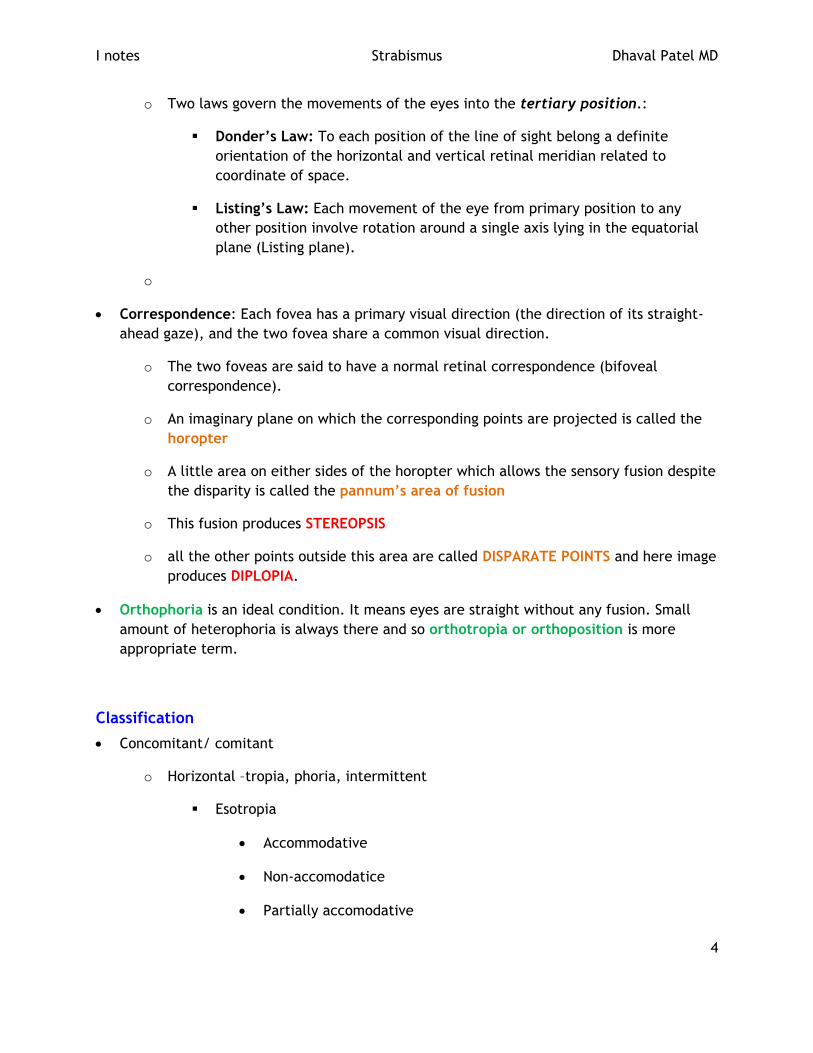
o Two laws govern the movements of the eyes into the tertiary position.:
Donder’s Law: To each position of the line of sight belong a definite
orientation of the horizontal and vertical retinal meridian related to
coordinate of space.
Listing’s Law: Each movement of the eye from primary position to any
other position involve rotation around a single axis lying in the equatorial
plane (Listing plane).
o
Correspondence: Each fovea has a primary visual direction (the direction of its straight-
ahead gaze), and the two fovea share a common visual direction.
o The two foveas are said to have a normal retinal correspondence (bifoveal
correspondence).
o An imaginary plane on which the corresponding points are projected is called the
horopter
o A little area on either sides of the horopter which allows the sensory fusion despite
the disparity is called the pannum’s area of fusion
o This fusion produces STEREOPSIS
o all the other points outside this area are called DISPARATE POINTS and here image
produces DIPLOPIA.
Orthophoria is an ideal condition. It means eyes are straight without any fusion. Small
amount of heterophoria is always there and so orthotropia or orthoposition is more
appropriate term.
Classification
Concomitant/ comitant
o Horizontal –tropia, phoria, intermittent
Esotropia
Accommodative
Non-accomodatice
Partially accomodative

I notes Strabismus Dhaval Patel MD
5
Exotropia
o Vertical
Hypertropia
Hypotropia
o Torsional
Incycotropia
excyclotropia
Incomitant
o Paralytic
Neurogenic: supra, infra, nuclear
Myogenic: nerve paralysis
o Restrictive
o Spastic
Consequences
o Confusion: generally cortex suppresses this foveal rivalry so actually this
confusion produces no confusion
o Diplopia: more troublesome
o Motor adaptations
Head posture
Blind spot mechanism
o Sensory adaptations (possible up to 6-7 years)
Suppression: Suppression is generally possible easily when the other image
is weak, that is imaged in the retinal periphery.
Facultative: under binocular condition
Obligatory: results in AMBLYOPIA
ARC: when image cant be suppressed
I notes Strabismus Dhaval Patel MD
6
Retinal correspondence is abnormal when the fovea of one eye has a
common visual direction with an extrafoveal area in the other eye.
When the amount of shift in the visual directions fully compensates
for the deviation, adaptation is called as harmonious ARC
When the amount of shift in the visual directions does not fully
compensate for the deviation, adaptation is called as unharmonious
ARC
Applied Aspects
Anatomy
Spiral of Tillaux
LR makes angle of 40-45 with sagittal
SR/IR makes 23
SO makes 54
SO: posterior fibres of fan are for depression and anterior are for intorsion
IO make 51
For actions of SR IR SO IO, one tip you can remember is that in abduction, their
primary action increase/ purely primary action whereas in adduction their secondary
action increases
This pulse-step innervation is responsible for a fast eye movement saccade, first a pulse
and then step to maintain the in the net position.
The torsional eye movements occur around the anteroposterior or Fick' s Y axis. This does
not pass through the centre of cornea but at a point on the lateral limbus as shown by
Linwong.
Orbital and Global differentiation of EOM
o all fibre types participate in all activities though to a different extent, based on
the amount of work and are recruited in a sequential order.
Anatomical equator: on maximum diameter of globe
I notes Strabismus Dhaval Patel MD
7
Functional equator: depending on arc of contact of muscles
o 2 mm posterior for LR and 2 mm anterior for MR
o
Muscle Pulleys
the observation of URRETS-ZAVALIA who called attention to the relationship of palpebral
fissure configuration and vertical incomitance in Mongoloid and antimangoloid in A and V
patterns
Improved M.R.I resolution now permits direct visualisation of E.O.M pulleys and paths of
E.O.M are determined using high resolution MRI.
The pulley zone located approximately at the junction between middle and posterior third
of the globe corroborating with LISTING’S LAW which forms the functional origin of E.O.M
the ligament of Lockwood forms pulley of inferior oblique and trochela (ossified) forms
pulley for superior oblique.
The orbital half of E.O.M inserts into pulley complex next to orbital walls while the bulbar
half moves anteriorly to insert into sclera.
Pulley sleeves are continuous with the tough peripheral portion of posterior tenons fascia
becoming slings to the sleeves, these sleeves are stabilised by fibro muscular septa.
Extending from pulleys to orbital wall and to adjacent pulleys by tenons fascia centred at
around equator.
They are composed of collagen, elastin, smooth muscle. Possible function of smooth
muscle in pulleys and tenons fascia is a possible maintance of stiffness and possible
dynamic role in fine tunning their position and a role in vergence mechanisms.
ORBITTM 1.5 extraocular biosimulation program
Accommodation and Convergence
Meter Angle: the vergence of two eyes for an object at 1 meter distance is called one
meter angle. This is Large Meter Angle: Ma

I notes Strabismus Dhaval Patel MD
8
The angle formed between visual axis of one eye and median line is called nagel’s meter
angle or small meter angle: ma
NPA: by a ruler, RAF, Nearest point that get blurred, 100/8 =12 D
Amplitude of accommodation:
o 4 x 4 – (Age/4)
NPC: nearest point that get doubled
o 100-8= 12.5 Ma
Convergence:
o Voluntary
o Involuntary
Proximal
Fusional
Accommodative
Tonic
AC/A: convergence amplitude per unit accommodation: normal 4-5
o Measured in prism diopter per diopter of accomodation
o Stimulus AC/A: as change in vergence is related to change in stimulus
o Response AC/A: 8% higher than stimulus AC/A, in this refractive status is also
taken into account
o Estimation of AC/A
Heterophoria method
Changing distance of fixation from 6m to 33cm
AC/A= IPD + (N-D)/3
Gradient method
Done at fixed distance

I notes Strabismus Dhaval Patel MD
9
6m with concave lens: -3D
33cm with convex lens: +3D
AC/A= (N-D)/3
Graphic method
Fixation disparity method
Types of Eye movements
Saccadic
Persuit
Vergence
Vestibular or tonic neck reflex
Optokinetic eye movements
Position maintenance system (fixation)
Pathways
Supranuclear
voluntary saccades: contralateral frontomotor cortex
pursuit eye movements: ipsilateral parieto-occipital
vergence: temporoparietal cortex
Horizontal eye movement: PPRF
Vertical eye movement: riMLF and PC
Convergence centre: central nucleus of perlia
Infranuclear

I notes Strabismus Dhaval Patel MD
10
Examination
Chief Complaint
o DIPLOPIA
o VISUAL CONFUSION
o ASTHENOPIA
o ABNORMAL EYE MOVEMENTS: wobbly eyes/ nystagmus
History
o Past History
History of patching
History of spectacle wear
Type of strabismus
History of traurna
Previous surnery
o Birth History
o Developmental Milestones
o Family History
o Review of Systems
Physical Examination
o Vision
o Sensory Testing
Near Stereoacuity
I notes Strabismus Dhaval Patel MD
11
Distant Stereoacuity
Retinal Correspondence
o Motor Testing
Bruckner
Krimskey
Hirschberg
Cover uncover
Cover
SPCT
PUCT
APCT
o Other Tests
Red Glass Test
Maddox Rod
Three Step Test
o Cycloplegic Refraction
Evaluation
Sensory Evaluation
Sensory System
Visual Acuity
Normal Retinal Correspondence
Fusion: Central
Fusion: Peripheral

I notes Strabismus Dhaval Patel MD
12
Sensory Anomalies
Amblyopia and Eccentric Fixation
Suppression
Anomalous Retinal Correspondence
Horror Fusionis
Diplopia and Visual Confusion
Central Fusion Disruption
Tests
Refraction
Visuscope
Prism Adaptation Test
o Jampolsky" first described ~lc prism adapta tion lest (PAT) tts dcvclope.d by his
OrlhOptist Woodward. PAT was usc.d to preoperatively predict which pat ients wili
develop res.idual c~otropia aftt•r surgery.
W4DT
Muscle Function Evaluation
Passive Forced Duction Testing
o Office
o Intraoperative
o RESULTS
Qualitative
Absolute Restriction

I notes Strabismus Dhaval Patel MD
13
Uniform Restriction
Leash Phenomenon
Quantitative
o PITFALLS
Patient Apprehension
Pharmacologic Effects
Errors in Techniques
Posterior Restriction
False Negative Tests
Coexisting Paresis and Restrictions
Co-contraction syndromes and Aberrant Regeneration
Force Generation Testing
Ocular Torsion
Motor Evaluation
Amblyopia

I notes Strabismus Dhaval Patel MD
14
Amblyopia is defined as binocular or monocular decrease in best corrected visual acuity
(BCVA) due to pattern visual deprivation and/or abnormal binocular interaction during
visual immaturity for which there is no obvious ocular pathology or visual pathway defect
and which in appropriate cases is reversible.
Incidence – 2- 5%
Prevalence- 1 – 4%
More common unilaterally than bilaterally
Commoner in rural than urban school children
Visual acuity less than 6/12 bilaterally or a difference of 2 or more than 2 lines between
the normal and the amblyopic eye in unilateral amblyopia when measured with the help
of Snellen's Charts and 3 or more than 3 lines when measured with the logMAR charts
NEURAL BASIS OF AMBLYOPIA
o concept of cortical competition: All cortical cells are potentially connected to both
the eyes equally.
o critical period: neural plasticity makes the visual system vulnerable to any abnormal
experience, 7 to 8 years in humans
o It is postulated that strabismic amblyopia is initiated as a maladaptive
differentiation in the ocular dominance columns, whereas the non-strabismic
amblyopia may be initiated from the malfunctioning of the ganglion cell population
of the amblyopic eye.
o Traditionally, it has been postulated that there are 2 mechanisms
Deprivation of form vision
Abnormal binocular interaction
o Lateral Geniculate layers subserving the affected eye have been found to be
atrophic in amblyopia. The cortical ocular dominance columns representing the
amblyopic eye are less responsive to stimulus and show changes microscopically.
o There are two kinds of retinal ganglion cells – the magno cells and the parvo cells.
Dissociation between the parvo and magnocellular systems occurs in amblyopia.
CHARACTERISTICS OF AMBLYOPIC VISION
Single letter acuity is better than linear acuity.
I notes Strabismus Dhaval Patel MD
15
There is a decrease in contrast sensitivity for high spatial frequencies. In strabismic
amblyopia, it improves on decrease illuminance whereas in anisometropic amblyopia, it
does not.
The amblyopic eye performs better in mesopic conditions. (Neutral Density Filters)
Vernier acuity and spatial resolution is disproportionately affected to a lesser degree than
the visual acuity in strabismic amblyopia.
Naso-temporal optokinetic nystagmus (OKN) asymmetry for the resolution of vertical
gratings is seen in strabismic amblyopia.
Abnormalities in pattern visually evoked potential (VEP) are seen in both strabismic and
anisometropic amblyopia. However in sensory deprivation amblyopia changes are seen in
both flash and pattern VEP.
Classification
Strabismic
o Most common type of U/L amblyopia
o Seen in constant tropia with strong fixation preference
o Commoner in esodeviations
o Rare in hypertropia
o Grating acuity is less reduced a/c/to Snellen’s acuity
o Decrease in illumination – drop in visual acuity lesser in amblyopic eye a/c/to
better eye
o
Anisometropic
o Second MC cause
o Significant difference in refractive errors between both eyes
o Commoner in hyperopes
o May be associated with strabismus

I notes Strabismus Dhaval Patel MD
16
o Form vision deprivation and unequal foveal images causing abnormal binocular
interactions
o
Stimulus deprivation (amblyopia ex anopsia)
o Congenital / traumatic total cataract
o Complete ptosis, significant corneal scarring, therapeutic patching
o U/L lesions worse than B/L
o
Isoametropic
o B/L uncorrected high refractive errors
o Hyperopia>+5.0DS
o Myopia> - 8-10 DS
o Astigmatism> +2.5 DC
o Pure form vision deprivation
o More amenable to treatment
o
Meridional
o Uncorrected astigmatic refractive error
o Selective visual deprivation of stimuli along particular spatial orientation
o Astigmatism >2.5 DC in preschool children, >3-4DC in infants
Organic Amblyopia
o subclinical Macular Damage
o Malorientation of cones
o Cone deficiency syndrome
For treatment and prognosis, amblyopia can be classified as
o Severe amblyopia : when the best corrected visual acuity is less than 20/100.

I notes Strabismus Dhaval Patel MD
17
o Moderate amblyopia : best corrected visual acuity 20/40 –
o Mild amblyopia : best corrected visual acuity 20/40 or more
Clinical features
Typically described as ―shimmer effect of hot air over a highway‖
Continuous wavy motion with parts of the image fading in and out of focus
Crowding Phenomena – the isolated letter visual acuity is better than line acuity in amblyopia.
Visual acuity testing should include both line and letter acuity.
Neutral Density Filters: Profoundly reduce vision in eyes with central retinal lesions and glaucoma
but vision in amblyopic eyes is not reduced. This is believed to be due to a relative increase in
mesopic visual acuity in amblyopic eyes.
Clinical evaluation
Visual acuity
o Decrease in best corrected visual acuity (>2 Snellen lines between both eyes)
o Recognition acuity is more impaired than resolution or detection acuity
o Grating acuity is less affected than Snellen acuity in strabismic amblyopia a/c/to
anisometropic
o Visual acuity is more impaired in good eye than amblyopic with neutral density
filter
o Crowding phenomenon- visual acuity is more with isolated optotypes in amblyopia (
decreased lateral inhibition)
o Optokinetic nystagmus
o VEP
o Preferential looking test
o
Neutral density test and crowding test

I notes Strabismus Dhaval Patel MD
18
Thorough ocular and fundus examination
Refraction
o Objective, under full cycloplegia
o Proper fixation ensured
o Proper head positioning avoiding posturing or tilts
o Refraction with loose lens with the examiner’s hand occluding the other eye in
young children
o
Evaluation of fixation
o Angle Kappa method
hand light method
arc perimeter method
synaptophore method
o Visuscopic / ophthalmoscopic method
o Types of eccentric fixation:
parafoveolar
parafoveal
paramacular
peripheral
o Wandering fixation – can be central or eccentric type, occurs on covering sound
eye
o Paradoxical eccentric fixation-surgical overcorrection or spontaneous reversal of
deviation, prolonged occlusion of sound eye
o Localisation of objects – normal in central and eccentric fixation, faulty in
eccentric viewing
o Abnormal colour vision in <6/36 (peripheral eccentric fixation)
o

I notes Strabismus Dhaval Patel MD
19
o
Other sensory abnormalities
o Afterimage test
Each fovea individually marked during monocular viewing with linear strobe
light that bleaches retina, causes linear afterimage shadow for 10sec
Center of light masked to spare fovea line has break in middle
NRC with any type of tropia- see a cross
ARC- each fovea has monocular afterimage but binocularly afterimage
perceived as coming from peripheral visual field
o Four PD base out test
Normally 4 PD base out test induces fusional convergence
2 movements seen normally
1st- version movement of BE in direction of apex of prism
2nd- fusional vergence of eye without prism towards the nose
o Motor fusion and large regional suppression- no movement when prism over
nondominant eye, version in direction of apex when prism over fixing eye
o Monofixation- no movement when prism over nondominant eye, version and
fusional convergence in some when prism over fixing eye
o Amblyoscope
Mirrors placed in front of each eye angled so that RE sees right temporal
side and LE sees left temporal side
Transparent picture slides placed in front of each eye
Can measure fusional vergence amplitude, angle of deviation, area of
suppression, retinal correspondence and torsion
Subjective angle (SA)- amount in degrees examiner must move amblyoscope
to allow patient to see 2 pictures superimposed, measured under binocular
viewing
Objective angle (OA)- measured by alternating target presentaion till there
is no refixation, measured during monocular viewing

I notes Strabismus Dhaval Patel MD
20
Strabismus with NRC and diplopia- SA=OA
NRC and large regional suppression- no SA
Harmonious ARC- significant OA but SA zero, OA = angle of anomaly
Unharmonious ARC- pseudofovea does not compensate for objective
deviation
Management
Refractive correction
o Amblyopia improved with optical correction by 2 or more lines in 77% of the
patients and resolved in 27%.
Occlusion therapy
o Different types
o PEDIG study says full time versus 6 hours has no difference
o Problems
o When to stop?
Penalization
o Its main role is as maintenance therapy and also in patients of moderate
amblyopia, in children uncooperative for occlusion, occlusion failure and possibly n
Manifest-Latent nystagmus (Fusion Maldevelopment syndrome).
Combined therapy
o Combined optical and atropine penalization (COAT) is an effective treatment when
occlusion therapy fails initially, and it might have a more rapid effect than single
modality penalization therapy, but there is an increased risk of reverse amblyopia.
Its effect may be particularly useful in anisometropic amblyopia.
Drug therapy
o Levodopa:
I notes Strabismus Dhaval Patel MD
21
o Citicholine (Cytidin-5-diphposphocholine):
Pleoptics
o Active stimulation of the macula is done using Pleoptophore (Modified Gullstrand’s
ophthalmoscope), till fixation becomes central.
CAM stimulator
Home vision therapy
Surgery to treat the underlying cause of amblyopia
Other modes
o Liquid crystal glasses -Liquid crystal glasses have recently been developed as a
new treatment for amblyopia. They provide an electronic, controlled, intermittent
occlusion of the sound eye allowing for visual stimuli input to the amblyopic fellow
eye.
o Opaque (occluder) contact lenses- Occlusive contact lenses can be used in
treating amblyopia in children who are patch-intolerant and resistant to
conventional treatment.
o Red Filter Treatment - Red filter which excludes wavelengths <640 nm is used to
stimulate cones at the fovea. Red light is ineffective in stimulating the eccentric
point as compared to fovea, due to the lack of cones.
Prognosis
It has been shown that after one year of occlusion therapy, 73% cases show success but this
decreases to 53% after 3 years. Risk factors for failure of occlusion therapy include.
1. Type of amblyopia
–Strabismic amblyopia has the best outcome,
–High anisometropia and organic pathology the worst
2. Age at which therapy began
–Younger age does better
3. Depth of amblyopia
–The better the vision at the start of therapy, the better the prognosis

I notes Strabismus Dhaval Patel MD
22
Esotropias
Classification
A. Depending on Whether the Deviation is Manifest or Not as
• Esophoria: Which is a latent inward deviation of the eye
• Esotropia: Which is a manifest inward deviation of the eye
B. Depending on Whether the Deviation is Concomitant or Not 1. Concomitant a. Primary
• Accommodative 1. Refractive 2. Non-refractive: Hyperaccommodative (High AC/ A ratio) 3. Hypoaccommodative 4. Mixed or partially accommodative
• Non-accommodative 1. Essential infantile 2. Essential late onset (Basic, Convergence excess, Divergence insufficiency) 3. Acute concomitant 4. Microtropia 5. Cyclic esotropia 6. Stress induced esotropia 7. Esotropia due to spasm of the near reflex 8. Esotropia in myopia 9. Nystagmus blockade syndrome
b. Secondmy
• Secondaty • Consecutive
2. Incomitant
a. Paralytic: Lateral rectus palsy b. Restrictive: Duane's Tumour, thyroid, post-operative c. Spastic
I notes Strabismus Dhaval Patel MD
23
Etiology
Uncorrected hypermetropia Blurred Retinal Image
o Some do not accommodate Orthotropic but amblyopic
o Some accommodate
Low AC/A: Orthotropia
Sufficient fusoinal divergence: Esophoria
Insufficient Fusional divergence: Esotropia
Accommodative Esotropias
Classification
Von Noorden classified Accommodative Esotropia on the basis of underlying etiology as
1. Refractive Accommodative Esotropia
2. Non-refractive Accommodative Esotropia
3. Hypo Accommodative Esotropia
4. Partially Accommodative Esotropia
Classification of Eye Movement Abnormalities and Strabismus (CEMAS) group classified
and defined this entity as-
1. Accommodative Esotropia
a. Pure Refractive: esotropia eliminated by hyperopic spectacles
b. Non-Refractive: esotropia at near only and eliminated with plus lenses at near,
e.g. bifocal
c. Mixed: esotropia at distance and greater at near associated with hyperopia and
responds to hyperopic correction at distance with bifocal for near
2. Mixed (Partially Accommodative) Esotropia
a. Hyperopia with incomplete response to spectacles and bifocals.
I notes Strabismus Dhaval Patel MD
24
Clinical Features Refractive
Normoaccommodative • Here, the AC/ A ratio is normal, i.e., 3-4 PD/D • The patient is unable to see for distance, so the patient accommodates more • The deviation for distance and near is more or less the same (within 15 PD) • Generally, they have mild to moderate hyperopia (2-6 D) • They respond well to full hyperopic correction as derived by cycloplegics refraction Hyperaccomodative • Here, the AC/ A ratio is high, i.e., 7-8 PD/D • The patient is unable to see for distance, so the patient accommodates more • The deviation for near is 15 PD more than the deviation for the distance • Generally, they have high hyperopia (> 6 D) • They respond partially to full hyperopic correction as derived by cycloplegic refraction. Some deviation at near still persists
Non-refractive
Hyperaccommodative • The accommodative mechanism is normal • Here, the AC/ A ratio is high i.e. 7-8 PD/D • The patient is able to see for distance • The deviation for near is 15 PD more than the deviation for the distance
• Generally, they have mild (1-2 D) hyperopia
• They have a normal near point of accommodation
• They respond well to bifocal glasses. Hypoaccommodative • The accommodative mechanism is weak, so the patient has to over accommodate • Here, the AC/ A ratio is normal, i.e., 3-5 PD /D, there is convergence excess • The patient is able to see for distance • The deviation for near is 15 PD more than the deviation for the distance • Generally, they have mild (1-2 D) hyperopia • They have a remote near point of accommodation • They respond well to bifocal glasses.

I notes Strabismus Dhaval Patel MD
25
Treatment
Optical correction
• The child should undergo full cycloplegic refraction.
• If the child is > 6 years, then the child is corrected such that mild esodeviation is
present, so that the patient will use his fusional divergence to become orthophoric.
• The frame given to the child has to be light. Harness frames can be given to infants.
• The child should be examined frequently to check for esodeviations both at distance and
near.
If residual esodeviation
Near and distant: power of glasses increased
For near: bifocals
If residual esotropia still persists, then it is of the partially accommodative
type and surge1y will be required to correct the remaining deviation.
Role of miotics
• Miotics are given only if the patient is extremely uncooperative.
• Patient is started with 0.03% Echothiophate Iodide OD increased to BD or with 0.125% till
desirable effect is achieved.
• The other miotic commonly used is Disopropyl Fluorophosphate.
• Miotics have side effects like brow ache, abdominal cramps and nausea.
• Other side effects also include iris cysts, lens opacities, angle closure glaucoma and retinal
detachment.
Orthoptic treatment
Generally, orthoptic treatment has minimal role in management of accommodative esotropias.
Once, the occlusion therapy has improved the visual acuity to the maximum, anti-suppression
exercises are used to overcome the suppression and improve the fusional convergence.
I notes Strabismus Dhaval Patel MD
26
Surgery
• Only, if there is esotropia at the end of full correction with glasses, surgery can be
contemplated.
• Surgery is done more often if there is associated A-V pattern
• Surgery, if planned, should be done only after full adaptation with prisms. Generally, the
residual esodeviation is first corrected with a Fresnel Add on prism worn for 1-2 weeks. If the
esodeviation increases, the power of the add on prism is increased to neutralise the esodeviation.
This increase in the esodeviation after the prism is added is called' eating up the prism'. Surgery
should be planned for the full prism adapted deviation.
Partially Accommodative Esotropia
An esotropia is partially accommodative when accommodative factors contribute to but not
account for entire deviation.1 This entity is also described as decompensated accommodative
esotropia. It is believed to be an evolution of a purely accommodative deviation into one that has
both an accommodative component and a portion that is not corrected by suspending the
accommodative effort. Various risk factors have been implicated in precipitating the
decompensation of a fully controlled acomodative esotropia including an age of onset less than 2
years, untreated hyperopia, poor compliance with glasses, prolonged ill health, aggressive
patching therapy for amblyopia and a high AC/A ratio.
Clinical characteristics
A residual esotropia(>10PD) for distance and near exists despite full correction of the
hypermetropic refractive error or prescription of the bifocal lenses6. It is important to repeat the
cycloplegic refraction to ensure that no residual, uncorrected hypermetropia is present.
Management
Prescribing the full cycloplegic refraction initially will provide the best opportunity for restoring
ocular alignment and maintaining good visual acuity and stereopsis. Amblyopia may be treated
with patching and/or atropine penalization, and amblyopia treatment in accommodative esotropia
is usually successful.

I notes Strabismus Dhaval Patel MD
27
Surgical Management
Surgery is indicated in partially accommodative and deteriorated accommodative esotropia.
Strabismus surgery is generally performed with the goal of only correcting the non-accommodative
component of the esotropia, with the understanding that the child will still need to wear
spectacles postoperatively to correct the accommodative component of the esotropia.Bilateral
medial rectus recession is the procedure of choice for partially accommodative esotropia.
Unilateral recession/resection may also be performed on an amblyopic eye. There is however
controversy regarding how to determine the target angle.
Prism Adaptation
Preoperative prism adaptation[15] is used to predict the patient’s fusional capability and
determine the augmented target angle for surgery. Fresnel prisms, split equally over the two
eyes, are prescribed over the full hypermetropic spectacle correction. Patients are followed at 1
to 2 week intervals and the prism is adjusted until the deviation stabilizes at an esotropia of 8
prism diopters or less. Surgery is done for this amount of esotropia that has been determined by
prolonged prism adaptation. Operating on the larger adapted angle reduces the rate of
undercorrection, with the predictability of a successful surgical outcome at 90%. The disadvantage
of prism adaptation is the time and cost involved in prescribing and adjusting the prisms until the
deviation stabilizes.
Standard surgery
Standard surgical approach is based on the deviation measured with full hypermetropic correction.
The target angle is an average of the near and distance deviation with correction. However, the
standard surgery has a high undercorrection rate of approximately 25%.
Augmented Surgery
In augmented surgery the target angle is determined by averaging the near deviation with
correction and near deviation without correction. Results comparing the standard surgery to this
augmented surgery formula showed 26% undercorrection for standard surgery, while augmented
surgery resulted in 93% success rate with 7% overcorrection
Essential Infantile Esotropias

I notes Strabismus Dhaval Patel MD
28
Etiopathology
The aetiology of Essential Infantile Esotropia is multifactorial.
• Heritable:
• Innervational: Imbalance in the innervation of the medial and the lateral rectus.
• Developmental: Sometimes, the development of the abducens nucleus which supplies the
lateral rectus lags behind the oculomotor nucleus.
• Refractive
Functional Deficits in Infantile Esotropia
Fusion Deficits
o Absence of disparity vergence (motor fusion)
o Lack of two-dimensional fusion and stereopsis
o Alternating monocular suppression
o Subnormal binocular visual evoked potential response
Motion/Pursuit Deficits
o Asymmetric monocular tracking
o Asymmetric monocular motion visual evoked potential
o Asymmetric motion perception
EPIDEMIOLOGY AND RISK FACTORS
20% to 30% of children born to a strabismic parent will themselves develop strabismus
Higher in LBW babies
Risk increases roughly 4% for each 100 g decrease in birth weight under 5 pounds

I notes Strabismus Dhaval Patel MD
29
CF
• Essential infantile esotropia presents within 6 months after birth
• It is characterised by large deviations (30 Degrees).
• There is alternate fixation or cross fixation. The patient sees the left field with the right
eye and the right field with the left eye.
• There is no significant refractive error.
• There is no neurological defect.
• Essential infantile esotropia is associated with inferior oblique overactions (68%),
dissociated vertical deviations (50%) and nystagmus (33%).
There is presence of asymmetric optokinetic nystagmus. Tracking of objects from temporal to
nasal field is smooth while tracking of objects from nasal to temporal field is cogwheel.
With more physiological tests like the polaroid scotometer, there is presence of two-point
scotoma, one at the fovea and the other at the diplopia point. While with more dissociating
tests, a single scotoma is seen.
Variants
• Ciancia syndrome: Here, there is manifest latent jerk nystagmus (Fast phase in the
direction of the fixing eye). The frequency of the nystagmus increases in abduction and
decreases in adduction. The patient thus prefers keeping his eye in the adducted position
with the head turned towards the side.
• Lang syndrome: Here, there is early onset esotropia with nystagmus, DVD and
excyclodeviation of the non-fixing eye. This is associated with Torticollis.

I notes Strabismus Dhaval Patel MD
30
Management
Correction of underlying refractive error
If hypermetropia is present up to 1.5 D, there is no need for correction with glasses.
If hypermetropia is greater than 1.5 D, correct with glasses.
Treatment of amblyopia
Surgical correction
Pre-requisites
a. Deviation should be constant and stable.
b. Eyes should be fixating alternately.
c. Refractive error and amblyopia should have been treated.
d. Any associated deviation should have been picked up.
Goals
Esotropia should be corrected within 10 PD of esotropia.
There should be peripheral fusion to combat diplopia and central suppression to
combat confusion.
Principles
a. In the absence of amblyopia, bilateral medial rectus recessions of 6.5 mm or more
are generally required.
b. In the presence of amblyopia, recess and resection can be tried.
c. Inferior oblique overaction should be corrected by inferior oblique weakening
procedure.
d. Dissociated vertical deviation, if present, can be corrected at a later date.
DD
• 6th nerve palsy: Due to the cross-fixation phenomenon in Essential Infantile Esotropia, the
eye remains adducted mimicking a Lateral Rectus Palsy. To differentiate between Essential
Infantile Esotropia and 6th Ne1ve Palsy, one can use the Doll's eye movements. The patient's

I notes Strabismus Dhaval Patel MD
31
head is rotated to the side the eye is adducted to. If there is 6th Nerve Palsy, the eye will not
abduct but if Essential Infantile Esotropia is the cause, then the eye will abduct. Sometimes,
though the patient might be having Essential Infantile Esotropia, the eye will still not abduct.
In this case, patch the other eye for few hours and then repeat the test. If the eye still does
not abduct, then it is a 6th Nerve Palsy.
• Duane's Syndrome: It can be differentiated from Essential Infantile Esotropia by the
retraction of the globe, changes in the palpebral aperture and associated upshoots and
downshoots.
• Down's syndrome
• Mobius syndrome
• Nystagmus blockade syndrome
• Accommodative esotropias
Monofixation Syndrome
Small angle deviations which can be missed by ordinary methods of examination and have
amblyopia of one eye with variable levels of binocularity are called Microtropias.
Clinical Features
1. Amblyopia of one eye.
2. Anomalous retinal correspondence.
3. Relative scotoma at the fixation spot. This can be tested by placing a 4D Prism diopter in
front of the eye. Though, the image moves from the fovea to a parafoveal point, there is no
refixation movement of the eye as the image lies within the scotoma.
4. Normal or near normal fusional amplitudes.
5. Normal stereoacuity.
I notes Strabismus Dhaval Patel MD
32
Variable Clinical Features
1. Small angle deviation measuring 8 PD or less.
2. Presence of anisometropia. Commonly, these patients have hypermetropic astigmatism.
Management
1. Spectacles for refractive error.
2. Management of amblyopia.
Acute Comitant Esotropia
acquired nature with sudden-onset with diplopia and minimal refractive error, usually in the first
or second decade of life
CF
o Age of onset- 1st or 2nd decade.
o Large-angle comitant esotropia usually >25-30 pd
o Sudden onset esotropia: if the parents are observant enough, they will be able to
tell the clinician the precise hour at which the squinting episode started.
o Diplopia is an inconsistent symptom and its absence does not rule out acute
comitant esotropia
o Good binocular potential
o Refractive error may be present, though its correction does not correct the
esotropia
DD
o Accommodative esotropia
o Basic esotropia
3 types
1. Swan type

I notes Strabismus Dhaval Patel MD
33
a. associated with monocular occlusion for various reasons
b. due to artificial interruption of fusion after a period of monocular occlusion or visual
loss
2. Burian-Franceschetti type.
a. Most common
b. Normal Ocular motility in order to rule out sixth nerve palsy which is more
common.
c. Normal Neuroimaging (MRI) to rule out an associated neurological problem
d. MX: prisms, botox, surgery
3. Bielchowsky
a. sudden onset esotropia in patients with high myopia (>-5.00D).
b. Bielchowsky postulated the presence of esotropia due to increased tonus of
both the medial recti
c. Meyer suggests the presence of esotropia due to direct damage of the lateral
recti in patients with high myopia
Exotropias
Classification
Depending on whether the Deviation is Manifest or Not As
• Exophoria: Which is a latent outward deviation of the eye.
I notes Strabismus Dhaval Patel MD
34
• Exotropia: Which is a manifest outward deviation of the eye.
Depending on whether the Deviation is Concomitant or Not
1. Concomitant
a. Primary
• Infantile exotropia
• Intermittent exotropia
b. Secondmy
• Secondary
• Consecutive
2. Incomitant
a. Paralytic: Medial rectus palsy
b. Restrictive
c. Duane's syndrome Type 2
d. Dissociated horizontal deviation
Calhounz Classification depending on the State of Fusion
a. Exophoria
b. Intermittent exotropia
c. Constant exotropia
Duane's Classification
a. Divergence excess patten: Distance deviation is 15 PD more than the near deviation.
b. Convergence insufficiency pattern: Near deviation is 15 PD more than the distance
deviation.
I notes Strabismus Dhaval Patel MD
35
c. Basic: Distance and near exodeviations are equal.
d. Simulated divergence excess: Basic deviation presenting as divergence excess due to
compensation of near divergence by fusional or accommodative convergence.
Etiology
Mechanical
Shape and axes of the orbit, interpupilla1y distance, size of the eyeball play a role in the
development of exodeviations.
Innervational
Innervational imbalance between the convergence and divergence mechanisms are also
supposed to play a role.
CF
Onset of intermittent exotropia is in childhood. SO% develop intermittent exotropia within
6 months of birth and 70% develop intermittent exotropia within 2 yrs of birth.
Exodeviations are more common in Females (70%).
The exodeviations generally pass through the phases of exophoria, intermittent exotropia
to constant exotropia.
Diplopia-phobia: On exposure to strong light, the exophoria may decompensate into
exotropia resulting in diplopia. To avoid this, the patient squeezes his eye. This is called
as diplopia phobia.
Micropsia: The patient tries to control his exodeviation by using his accommodative
convergence. This leads to micropsia where the objects are seen smaller.
Asthenopia: Patient develops eyestrain, blurring, headache.
Abnormal stereopsis: This starts happening when an intermittent exotropia is becoming
constant and is one of the indications of surgery.
Magician Forceps Phenomenon: On passive adduction of the dominant eye, the other
eye's exodeviation gets corrected. This reflex is present even in the dark. This reflex is
abolished by deep retrobulbar anesthaesia.
I notes Strabismus Dhaval Patel MD
36
Generally, amblyopia never occurs unless the exotropia is unilateral, constant and present
during the period of development of the eye. Sometimes, alternate suppression is present.
The pattern of the suppression depends on the kind of test used to map it. With more
dissociating tests like Prisms and Synaptophore, single large scotoma is produced from the
fovea to the diplopia point. With less dissociating tests like polaroid scotometer and phase
difference haploscope, two different scotoma are seen, one at the fovea and the other at
the diplopia point.
Anomalous retinal correspondence is also known to develop.
Associations
o A-V pattern strabismus
o Comitant vertical deviation
o Dissociated vertical deviation
o Incomitant vertical deviation
Examination
History
Visual acuity
Cycloplegic refraction
Cover test: At near and distance
Measurement of deviation: Should be done with Prism Bar Cover Test in all the cardinal
positions of gaze and both for near and distance. There is Lateral Gaze Comitance in
these patients, i.e. 20% reduction in the angle of squint in lateral gaze
Measurement of Stereopsis
Occlusion test:
o First, do alternate cover uncover test and establish the presence of exotropia.
o Now, if the exodeviation is found to be more for distance than near, then the eyes
are patched for an hour. Sometimes, deviation more for distance than for near is
simulated as the accommodative and fusional convergence compensates for the
near exodeviation.
o So, by patching for an hour, this accommodative convergence is broken.
I notes Strabismus Dhaval Patel MD
37
o After an hour is over, an occluder is placed before the unpatched eye and then the
patch is removed. It is ve1y important to prevent the simultaneous use of both the
eyes as even a brief binocular exposure may decrease the deviation for near by
stimulating the accommodative and fusional convergence.
Measurement of fusional amplitudes
Management
Optical Treatment
The patient is made slightly myopic by over-correcting with concave lens. This stimulates
accommodative convergence and corrects exodeviation
Prismotherapy
Base in prisms are sometimes used to correct the exodeviation. Half of the exodeviation
should be corrected with prisms, so that the remaining exodeviation can stimulate the
fusional convergence. Normally, up to 8 PD can be used in front of each eye and with
Fresnel's prisms upto 15 PD can be used in front of each eye.
Orthoptic Treatment
Anti-suppression exercises
The suppression scotoma should be first treated with flashes of light. Once, the suppression
scotoma decreases, binocular single vision is maintained by anti-suppression exercises like Bar
reading, Synoptophore or Cheiroscope.
Convergence exercises
The convergence exercises cannot affect the basic deviation but can improve the fusional
control thus decreasing the occurrence of exodeviation.
First step is teaching the appreciation of Physiological Diplopia.
Second step, convergence exercises to increase both the tonic and phasic convergence.
Exercises
I notes Strabismus Dhaval Patel MD
38
• Pencil push ups: The pencil is drawn closer to the patient
• SHOT: Simple Home Orthoptic trainer can be used. It consists of dark circles drawn on
either side of a folded paper such that they overlap. The circle on one side has a cross on the
top and the circle on the other side has a cross at the bottom. This paper is slid on the ruler.
The patient has to overlap the two circles such that it appears as if there is only one circle
with a cross both at the top and the bottom. The patient is to overlap the circles and hold it
like that for 40 sees. He does this for 2-3 minutes 21 day. The paper is slid closer and closer
to the eye as the convergence starts improving. It is important to note that convergence
exercises should not be done by those patients who have Intermittent Exotropia at distance
only as this may lead to post-operative over-convergence.
Occlusion Therapy
The occlusion of the preferred eye for 3-5 hours a day is useful in decreasing the angle of
deviation. Exotropic patients become exophoric.
Surgery
Indications
• If constant exotropia is present.
• If intermittent exotropia is present for more than 50% of the waking hours.
• If exodeviation exceeds 20 PD
• If asthenopic symptoms/ diplopia are present
• If diplopia is present
• If the exotropia is progressing from intermittent to constant
• If suppression scotoma is developing
• If there is abnormal stereopsis.
Timing of the surgery
Early surgery
Early surgery is advocated by Knapp. The surgery can be done once the child is 6
months old. The advantage of doing it early is that sensory adaptations have not taken place.

I notes Strabismus Dhaval Patel MD
39
The disadvantage is that if the patient is overcorrected, patient may develop Monofixation
syndrome or consecutive esotropia.
Late surgery
Late surgery is advocated by jampolsky. The surgery can be done once the child is 2 yrs old.
The advantage of doing it late is the diagnosis will be more accurate, child will be more co-
operative for any orthoptic treatment and the chances of consecutive esotropia and mono
fixation syndrome would be less.
The disadvantage is that sensory adaptations like suppression may have developed.
Choice of surgery
The choice of surgery depends on
• The type of exodeviation whether it is a true divergence excess, basic, simulated
divergence excess or convergence insufficiency type
• The presence of lateral gaze inhibition
• The age of the patient
• The presence of A or V pattern
• The presence of Inferior Oblique over-action
General Guidelines
• In true divergence excess, bilateral recession of the lateral recti should be done.
• In convergence insufficiency, bilateral medial rectus resection should be done.
• In basic exotropia and simulated divergence excess, unilateral lateral rectus recession and
medial rectus resection should be done.
• If operating on a child between 6 months 2 yrs old: Slightly undercorrect to avoid
monofixation syndrome and consecutive esotropia.
• If eye is amblyopic, overcorrect by 20 PD to compensate for the slight post-surgical drift
following the surgery.
• If lateral gaze inhibition is present, bilateral lateral rectus recession should be avoided and
there should be slight under correction in the non-preferred eye

I notes Strabismus Dhaval Patel MD
40
• Prior to surgery, the patient's refractive error should be corrected and amblyopia, if
present, should be treated.
• The amount of recession and resection to be done varies from doctor to doctor. Generally,
every 1 mm of medial rectus resection gives around 2.5 PD of correction and every 1 mm of
lateral rectus recession gives around 2 PD of correction.
• In very large squints above 60 PD, sometimes 3 muscles need to be operated
Possible results following surgery
• Orthoposition: Generally, there is a post-operative drift after the surgery. So, if
orthoposition is achieved immediately after the surge1y, orthoptic exercises have to be
prescribed to the patient to increase the positive fusional convergence.
• Consecutive esotropia
In children: Rerefraction, prisms, occlusion therapy
If esotropia> 15 PD persists, to be taken up for surge1y as a fresh case.
In adult: Rerefraction, prisms, occlusion therapy
If esotropia> 20 PD persists for> 6 weeks
Surge1y is required if esotropia persists for> 6 months in spite of the above non-
surgical measures.
Residual exotropia
If residual exotropia is< 15 PD, then just re refraction, cycloplegics and prisms can be
used.
If above exotropia persists at end of 6 months, surgery will be required If residual
exotropia> 15 PD, then surge1y has to be planned within 6- 8 weeks.
DVD

I notes Strabismus Dhaval Patel MD
41
It is a condition wherein, the non-fixing eye moves up and out either on occlusion or
during periods of visual inattention.
When the occluder is removed or the patient regains visual attention, this eye which had
drifted up and out starts coming down and sometimes even becomes hypo trophic. This re-
fixation movement is not associated with a downward movement of the previously fixing
other eye.
As, it does not obey the Herring's law, this phenomenon is called dissociated vertical
deviation. In addition, to an upward movement of the eye, there is an extorsion and
abduction of the eyeball. It is generally bilateral and asymmetrical.
Types
o Manifest DVD: Wherein during periods of fatigue or inattention, one eye deviates
up.
o Latent DVD: Wherein when an occluder is placed in front of the eye, the eye
deviates up.
Associations: Seen in 75% cases of infantile esotropia, commonly after the age of 2 yrs.
Other less common associations include infantile exotropia, heterotropia of sensory origin,
A pattern exotropia with superior oblique overaction.
Theories
o Bielschowsky proposed alternate excitation and suppression of the vertical
convergence centres
o Spielman proposed imbalance of the binocular vision
o Paralysis of the depressors
o Defective mid-brain stimuli
Diagnosis
o Cover-Uncover Test
In a patient with manifest DVD, one eye is already fixating and the other eye is up
and out. Now, when the fixating eye is covered, the non-fixating eye moves down
and sometimes even overshoots. There is no movement of the eye under cover In a
patient with latent DVD, when the eye is covered, the eye moves up and out.
When the cover is removed, the eye which had moved up comes down back to the
original position or sometimes overshoots downwards. There is no movement of the
other eye.
I notes Strabismus Dhaval Patel MD
42
o Prism base down test
If the patient has manifest DVD, one eye is already deviated up and out. Put an
occluder in front of this eye. Tell the patient to fixate at a distance object with
the other eye. Place a base down prism under the occluder. Then, shift the
occluder to the other eye, there will be some downward movement of the
deviated eye. Keep on increasing the power of the prism till there is no movement.
o Modified Krimsky test
This test can be used in those patients who cannot fix with the deviating eye
o Red Glass test
In this test, red glass is held alternately in front of the two eyes. If the eye has a
DVD, then irrespective of in front of which eye, the red glass is held, there red
image will always appear below the white image. If the eye has cyclodeviation,
then red image is seen up or down depending on which eye fixates.
o Bielschowsky phenomenon test
In this test, the eye under the occluder is deviated upwards while the other eye is
fixating. A neutral density filter is slowly introduced in front of the fixating eye
and the density of the filter is gradually increased till the eye under the occluder
assumes central position.
Management
Non-surgical treatment
If the patient has asymmetrical DVD, then patching of the eye with lesser deviation or
occlusion of that eye with +3D will change the fixation to the other eye. So, the DVD will
no longer be a cosmetic problem.
Surgical treatment
Surgical treatment is indicated when the deviation is significant and causing cosmetic
problems.
No surgical treatment is able to correct the deviation completely; it is more of a palliative
measure:

I notes Strabismus Dhaval Patel MD
43
Faden Operation with superior rectus recession: In this operation, the superior rectus is
recessed by 3-5 mm and a posterior fixation suture is taken 10-12 mm behind the insertion
of the muscle. This operation is successful but can lead to recurrences later
Large recession of the superior rectus muscle: Large recession of the superior rectus of
one or both the eyes can be done
Resection of the inferior rectus: If, recurrences occur, resection of the inferior rectus
can be done
Recess-resect procedure: If the patient has a monocular deviation, 4 mm recession of the
superior Rectus along with 6 mm resection of the inferior rectus can be done.
Recession of the inferior oblique and anteriorization of its insertion to a point temporal
to the inferior rectus has also been tried.
IOOA
Classification
Primary
o Mechanical
o Innervational
o Secondary: Paralysis of ipsilateral superior oblique or contralateral superior rectus
Epidemiology
Primary inferior oblique overaction occurs in:
o 72% Congenital esotropes
o 34% Accommodative esotropes
o 32% Intermittent exotropes
Age: PIOO presents around 2-3 yrs of age
Laterality: Unilateral to start with, it soon becomes bilateral

I notes Strabismus Dhaval Patel MD
44
Associations
Association with horizontal deviations in primary position: As shown above, PIOO occurs in
patients with congenital esotropia, intermittent exotropia and accommodative esotropia.
Association with vertical deviation in primary position: Generally less than 5 degrees. Quite
often a V -pattern strabismus is present which has greater exodeviation in upgaze than
downgaze.
Association with upshoots/downshoots: With the eyes in lateral gaze with the abducted eye
fixing, the adducted eye is elevated. With the adducted eye fixing, the abducted eye is
depressed. There is no head tilt and the Parks Bielschowsky test is negative. Though,
objective sign like tilting of the disc may be present, subjective tests like the Double Maddox
rod test, Bagolini test, Hess Screen test and Major Amblyoscope are absent. This happens
because in patients with PIOO, sens01y adaptations take place.
Grading of Inferior Oblique Overaction
• Depending upon the vertical deviation
1. Mild overaction: When vertical deviation is present only on sursum adduction.
2. J'\1oderate overaction: When vertical deviation is present on adduction also.
3. Severe overaction: When vertical deviation is present in primary position.
• Depending on the angle the adducting eye makes with the horizontal line as it elevates
and abducts on lateral version
Grade 1: Angle between the line drawn from the medial canthus to the lateral border of the
eye and from there to the centre of the pupil is up to 15 degrees
Grade 2: Angle is 16-30 degree
Grade 3: Angle is 31-60 degree
Grade 4: Angle is 61-90 degree
DD
o • Dissociated vertical deviation
o • Aberrrant regeneration of the 3rd cranial nerve
I notes Strabismus Dhaval Patel MD
45
o • Rectus rotation in patients who have craniosynostosis
o • Tether effect in patients who have Duane's syndrome
o • Tight lateral rectus muscle syndrome
Treatment
There are various surgeries that can be done for inferior oblique overaction These surgeries
are called inferior oblique weakening procedures.
1. Disinsertion: Here, the muscle is cut from the globe near its insertion and left like
that.
Advantage
• Sim pie and easy procedure
• No suturing of the muscle to the globe required, therefore, no chances of globe
perforation, damage to the macular area.
• Bleeding is minimal.
Disadvantage
The muscle tends to reattach to the globe leading to high recurrence rates. Therefore,
this procedure is not ve1y popular
2. Myectomy: A 8 mm segment of the inferior oblique is excised either nasal to the
inferior rectus or temporal to the inferior rectus. The temporal approach is preferred.
But, due to the high recurrence rates, unpredictable results and occurrence of post-
operative adherence syndrome, this technique is not preferred.
3. Extirption: The excision of the entire muscle with the Tenon's capsule is the most
effective technique but leads to a lot of complications.
4. Recession: Being preferred for Grades 2 and 3 and in cases of secondary inferior
oblique overactlon.
Finks operation
Here, the muscle is reattached to a point which is 6 mm posterior and 6 mm inferior to
the inferior border of the lateral rectus. 8 mm correction is achieved by the above
procedure. If the point of attachment is 2 mm above or below this point, then 6 mm
and 10 mm correction can be achieved.
Parks operation

I notes Strabismus Dhaval Patel MD
46
Here, the muscle is reattached to the globe at a point 2 mm lateral and 3 mm
posterior to aspect of insertion of the inferior rectus muscle. 10 mm correction is
achieved by this procedure. A 10mm Park's operation gives better correction thana
10mmFink's operation.
Elliot and Nankin's operation
It is the anteriorisation of the inferior oblique. Here, the muscle is disinserted and
attached just lateral to the lateral border of the inferior rectus.
Advantages
• Predictable and sustainable results
• Post-operatively, if the desired correction is not achieved, a second surgery is
possible.
Disadvantages
• Violation of the Tenon's capsule may lead to fibrofatty proliferation of the orbital fat
on the sclera or contracture of the radial fibrous septa leading to 'adherence
syndrome'. In this syndrome, there is progressive Hypo tropia, Excyclotropia and
elevation limitation.
• The vortex vein can be damaged leading to severe bleeding.
• Excessive traction on the inferior oblique may damage the Parasympathetic fibres to
the ciliary ganglion leading to transient pupilla1y dilatation and decreased
accommodative tone.
SOOA
Classification
Primary
Mechanical
Innervational
Secondary: Paralysis of ipsilateral inferior oblique or contralateral inferior rectus
Epidemiology
Age: PSOO presents around 2-3 yrs of age

I notes Strabismus Dhaval Patel MD
47
Laterality: Unilateral to start with, it soon becomes bilateral
Associations:
o Association with horizontal deviations in primaty position: Associated with
exotropia and esotropia both.
o Association with vertical deviation in primaty position: Generally less than 5
degrees. Quite often a A pattern strabismus is present which has greater
exodeviation in downgaze than upgaze.
o Association with downshoots/upshoots: With the eyes in lateral gaze with the
abducted eye fixing, the adducted eye is depressed. With the adducted eye fixing,
the abducted eye is elevated.
There is no head tilt and the Parks Bielschowsky test is negative. Though,
objective sign like tilting of the disc may be present, subjective tests like the
Double Maddox rod test, Bagolini test, Hess Screen test and Major amblyoscope are
absent. This happens because in patients with PSOO, sensmy adaptations take
place
Grading is just like IOOA.
Differential Diagnosis:
o Secondary superior oblique overaction: It is rare as the Inferior Oblique rarely gets
paralysed alone
Treatment
Superior oblique weakening procedures are required when there is 'A' pattern strabismus or
the deviation is significant
o Superior oblique weakening procedures
The superior oblique weakening procedures can be done either through the nasal
or the temporal approach
o Nasal approach
When a surgery is done through the nasal approach, the intermuscular septum
attachment to the tendon is undisturbed. This reduces the chance of post-
operative torsional diplopia. Therefore, this approach is preferred when operating
on a patient with Brown's syndrome.
o Temporal approach
This approach is preferred over the nasal approach in all other indications as

I notes Strabismus Dhaval Patel MD
48
o Approach is easier
o The distance at which the tenotomy is to be done can be accurately measured
o Incidence of post-operative ptosis and superior rectus palsy is lesser
o Tenotomy: Here, the muscles is transected at a particular point.
o Recession: Here, the muscle is disinserted and reattached to a point at the medial
border of the superior rectus 4 mm posterior to its insertion
o Anteriorisation: Here, the superior oblique is attached further anteriorly
o Translational recession: Here, the anterior point of the superior oblique
reinsertion is 4 mm nasal from the superior rectus and 12 mm posterior from the
limbus. The advantage of this is that it prevents the depression of elevation in
adduction that occurs with the other two procedures
o Posterior tenotomy of the superior oblique: Here, selective transection is done
of the posterior fibres. It is believed that these fibres are responsible for
depression. Thus, by transecting these fibres a vertical deviation will be corrected
o Superior oblique expander: Here, a 2-2.5 mm silicon band/ silastic band is
inserted between the cut ends of the superior oblique, 3 mm nasal to the superior
rectus.
DRS
It was first described by Stilling and then Turk, so, it is also called as Stilling-Turk-Duane
syndrome.
Duane syndrome is a type of incomitant strabimsus (misalignment of the eyes which varies
with gaze directions).
It also belongs to the roup of extraocular muscle fibrosis syndromes (conditions associated
with restriction of both active and passive movement of the eyeball). But evidence
suggests that DS (and other Congenital Cranial Dysinnervation Disorders-CCDD's) may
actually be prima1y disorders of nerve innervation.

I notes Strabismus Dhaval Patel MD
49
Duane's syndrome (DS) is an eye movement disorder characterised by
o Limited adduction, abduction or both.
o In addition, when the affected eye(s) ad ducts, the eyeball retracts (pulls in) and
the eye opening (palpebral fissure) narrows.
o In some cases, there is also upshoot or downshoot of the eye
Etiology
o Genetics
Majority of cases are sporadic in origin 2-5% of patients showing a familial pattern.
Both dominant and recessive forms of DS have been documented. In some families
with dominant DS, it has skipped a generation (shown reduced penetrance) and
ranged in severity within the same family (shown variable expressivity). Most
familial cases are not associated with other anomalies.
o SALLY Gene: Deletions of Chromosome 4 and 8
o Theory of Innervational Anomaly
Duane syndrome results from an absence of cranial nerve VI (abducens nerve) or its
nucleus and its corresponding alpha motor neurons in the pons and aberrant
innervation of the lateral rectus muscle by a branch of cranial nerve III. When
individuals with DS attempt to adduct, both of these muscles contract at the same
time, resulting in the eyeball retracting inward (pulling in) and the eye opening
(palpebral fissure) narrowing.
o Theory of Structural Anomalies: Some studies suggest that there is fibrosis of
lateral rectus and to some extent of the medial rectus. There is a fibrotic band
running from the orbital apex to the site of insertion of both the lateral and the
medial rectus.
Epidemiology
o Incidence: The frequency of Duane's syndrome in the general population of squint
patients is 1-4%.
o Gender Distribution: Females more commonly affected than the males.
o Laterality: Left eye is more commonly (75%) affected. Both the eyes are affected
in 20% cases.
Clinical Features
o Limitation of abduction/ adduction or both

I notes Strabismus Dhaval Patel MD
50
o Narrowing of the palpebral fissure on adduction and widening on abduction
o Retraction of the globe on adduction
o Eye may be esotropic, orthotropic or exotropic
o Presence ofupshoots and downshoots. Mechanical upshoots tend to be sudden
whereas inne1vational upshoots are progressive
o Abnormal head posture may be present
o 71% of patients have hypermetropia >1.5 D.
o 15% of patients have amblyopia.
o Associated Ocular Anomalies
Congenital ptosis
Microphthalmos
Congenital districhiasis
Nystagmoid movements
Epibulbar dermoids
Keratoconus
Heterochromia iridis
Congenital cataract
Persistent hyaloid artery
Medullated nerve fibres
Optic nerve hypoplasia
Morning glory syndrome
o Associated Non-ocular Findings
Goldenhar's syndrome
Klippei-Feil anomaly: Non-formation of the cervical vertebrae along with
torticollis and facial asymmetry
Wildervanck syndrome :Above with sensorineural deafness

I notes Strabismus Dhaval Patel MD
51
o Variants of Duane's Syndrome
Vertical retraction syndrome
Congenital adduction deficit with synergistic divergence
Huber's Classification of DRS
o Type1
Most common(78%)
Limitation of abduction with normal or near normal abduction
Narrowing of the palpebral fissure and retraction of the globe on adduction
o Type2
7%
Limitation of adduction with normal or near normal abduction
Narrowing of the palpebral fissure and retraction of the globe on adduction
o Type3
15%
Limitation of both abduction and adduction
Narrowing of the palpebral fissure and retraction of the globe on both
adduction and abduction
Khurana's Modification of Huber's Classification
o The three types are further classified as A, Band C depending on the position of
the eye in the primary position
o A: Esotropic(53%)
o B: Exotropic(16%)
o C: Orthotropic(31 %)
Differential Diagnosis
o 6th nerve palsy: Definite onset of new large angle esotropia associated with
diplopia. Also, globe retraction and upshoots/ downshoots are not present.

I notes Strabismus Dhaval Patel MD
52
o Moebius syndrome: Complete paralysis of both the 6th nerves alongwith loss of
olfactory and gustatmy senses.
o Congenital esotropia: Child may cross-fixate.
Management
Indications for Treatment
o The commonest indication is a face turn.
o Other indications include Narrowing of the palpebral fissure, retraction of the
globe, torticollis and limited abduction.
Contra indications
o Less than 5 yrs of age.
o In DRS, late surgery is advised as surgery may disrupt the development of binocular
vision.
Surgical Procedures
o Never resect in Duane's syndrome as it can worsen the narrowing of the palpebral
apertures.
o If esotropia: Medial rectus recession It is the mainstay of treatment of DRS.
o If the esotropia in prima1y positions< 20A, then ipsilateral MR recession is
sufficient.
o If the esotropia>20A, then bilateral MR recession is required
o If exotropia: Lateral rectus recession
o Upshoots/Downshoots: If upshoots/ downshoots are present Y splitting of the
lateral rectus muscle or Faden's operation (posterior fixation suture over the IR
belly). This decreases the slippage of the lateral rectus muscle on the globe during
adduction which is the main cause of the upshoot and downshoot.
o Enophthalmos: If enophthalmos is the main complaint, simultaneous recession of
both the lateral and the medial rectus muscle

I notes Strabismus Dhaval Patel MD
53
Brown’s Syndrome
Brown's syndrome is an ocular motility disorder which simulates Inferior Oblique palsy. It
is caused by the restriction of the action of inferior oblique due to overly taut tendon of
the superior oblique.
Causes
• Congenital: Short SO tendon, tight trochlea, nodule on SO tendon
• Acquired:
Tenosynovitis of the superior oblique tendon
Trauma
Rheumatoid nodule on the tendon
Extraocular surgery: Scleral buckling
Damage to trochlea while giving injection
Acromegaly
Marfan's syndrome
Clinical Features
• Failure of elevation in adduction
• Less severe failure of elevation in midline
• Elevation in abduction is present
• No superior oblique overaction which should be present in a c/o true inferior
oblique palsy
• FDT positive in adduction but negative in abduction
• Presence of 'V' pattern exotropia
Variable Features
• Downshoot in adduction
• Widening of the palpebral fissure on adduction
• Abnormal head posture if hypo tropia is present
I notes Strabismus Dhaval Patel MD
54
Eustis Grading of Brown's Syndrome
• Mild: Restriction of elevation in adduction
• Moderate: downshoot in adduction
• Severe: hypotropia in primary position
DD
• Inferior oblique palsy
• Double elevator palsy
• Fracture of the orbital floor
• Fibrosis of the inferior rectus muscle
Management
Treatment of Amblyopia: If amblyopia is present, then treat that first.
Treatment of the underlying cause, e.g., if Tenosynovitis is the cause of Brown's, then oral
steroids or injection of steroid around trochlea.
Surgical Correction
Indications
• Abnormal head posture
• Hypotropia in prima1y position
• Diplopia in downgaze
Smgeries
Superior oblique tenotomy: 50% cases develop superior oblique paralysis which can be
managed by either recession of the ipsilateral inferior oblique or the contralateral inferior
rectus.
Superior oblique tenectomy: To avoid the complication of superior oblique paralysis
associated with SO tenotomy, 6 mm SO tenectomy within the muscle sheath just nasal to
the superior rectus is advocated.
Silicon band expander: Superior oblique weakening using a silicon band expander.

I notes Strabismus Dhaval Patel MD
55
Tests
The simultaneous prism cover test
Detects the manifest deviation only.
Used in small angle esotropia- microtropia.
Prism over the deviating eye and cover the normal eye at the same time.
Equivalent prism test.
It does not dissociate the eyes.
Sufficient time should be given between changing prisms to allow fusion.
Prism alternate cover test
Most reliable method in adult patients and co-operative children.
Measures the total deviation.
Should be done with prism on both eye.
Eye with out the prism is the fixing eye.
Watch the eye under prism for neutralization. cessation of movement is the end point.
subtract 2 pd after reversal is appreciated.
Cannot measure cyclodeviations.
Performed in the 9 diagnostic positions.
Performed at distance and near.
Subjective prism alternate cover test.

I notes Strabismus Dhaval Patel MD
56
Krimsky test
Prism is placed before the normal fixating eye.
More accurate than the Hirsberg test.
Can be done in infants, patients with poor visual acuity and eccentric fixation.
Can be done only for near.
For distance u have to examine the eyes grossly.
All gaze positions should be examined.
Measures only the manifest deviation.- underestimates the true deviation.
U should see in alignment with the deviated eye.
Value may be affected as the eye is not in primary position.
Modified Krimsky Test
Prism is placed over the deviated eye.
Seen from midline.
Four prism dioptre test
Can be used as base out or as base in.
Placed first over the normal eye and then over the micro tropic eye.
Less sensitive than worth 4 dot test.
Should be done in every patient with anisometropic amblyopia and suspected microtropia.
The vertical prism test
To find out the fixating eye .both eyes move if placed in front of the fixating eye.
A patient should be able to maintain fixation with a 16 pd prism. to find out the
dominance.
I notes Strabismus Dhaval Patel MD
57
To detect malingering. ask the patient , how many images does he see. if he sees 2/3/4.
The prism adaptation test
First described by Dr jampolsky.
To judge which patients may develop residual esotropia postoperatively .
Overcorrecting prisms are placed and reexamined after 1 hr in the clinic.
Prism acceptance-stable exodeviation or a slight convergence movement to attain bifoveal
fixation.
Do a cover test.
The target angle is the prism adapted angle.
There was no over corrections.
Better successful alignment.increased presurgical fusion.
Prism under cover test
Base down prisms are placed in front of the dissociated eye.
Point of neutralization is achieved when no down ward movement is seen.
Spielmans occluder.
To measure the dvd.

I notes Strabismus Dhaval Patel MD
58
Hess Chart
for incomitant strabismus
a. neurogenic such as 3rd, 4th and 6th cranial nerve palsy
b. myogenic causes such as myasthenia gravis, Duane's syndrome, Brown's syndrome,
Thyroid eye disease
c. mechanical such as orbital floor fracture.
In Hess screen, the two eyes are dissociated using lenses of different colours. The fixating
eye looks through the red lens (or filter); the non-fixing eyes through the green lens (or
filter).
In Lees screen, the eyes are dissociated using two opalescent glass screens at right angles to
each other bisected by a two-sided plane mirror.
In mild incomitant strabismus, the central field may appear normal. By analysing the
outer field, small underaction or overaction may become apparent.
stages in the development of muscle sequelae:
1. Overaction of the contralateral synergist according to Hering's law
2. Overaction of the ipsilateral antagonist as its action is unopposed by the paralysed
muscle
3. Secondary underaction of the contralateral antagonist.
This occurs for two reasons:
a} as the ipsilateral antagonist action is unopposed, less impulse is needed to move it
into its desired position and consequently according to Hering's law, its contralateral
synergist receives less impulse and therefore underact.

I notes Strabismus Dhaval Patel MD
59
b} overaction of the contralateral synergist means that its ipsilateral antagonist will
receive equal impulse to relax according to the Sherrington's law.
The chart with the small field is the abnormal one.
If compressed, consider mechanical causes
The greatest negative or inward displacement represents the primary underaction
The muscle with positive or outward displacement indicates an overaction.
Squint may result in Diplopia or Confusion,
Pt. adapts himself to overcome diplopia or confusion by:-
1. Motor adaptation
1) Latent squint
2) Compensatory head posture/ Paralytic squint
3) Purposive squint (blind spot syndrome)
2. Sensory adaptation
1) Supprssion of image from squinting eye
2) Eccentric fixation
3) ARC
Good visual acuity, normal physiological retinal correspondence, proper coordination and
fixation with each eye, formed, are the essential requirements of binocular vision.
I notes Strabismus Dhaval Patel MD
60
The vision in significant anisometropia may be binocular, alternating or exclusively
uniocular.
Various methods are devised for measuring AC/A ratio
a. Heterophoric method
b. Gradient method
c. Fixation-desparity method
d. Haloscopic method
e. Graphic method.
Orthoptic Treatment
1. Antisuppression exercises:
2. Fusion exercises:
3. Ex. diploscope exercise
4. Exercise on Remy separator
5. Exercise with the help of stereogram cards
6. Occlusion to induce use of eye with marked suppression
convergence insufficiency
There are seven types of convergence insufficiency:
1. Primary idiopathic
2. Secondary to primary divergent strabismus (divergence excess type)
3. Secondary to a vertical muscle defect
4. Convergence insufficiency due to refractive error

I notes Strabismus Dhaval Patel MD
61
5. Systemic convergence insufficiency (poor general health)
6. Convergence insufficiency associated with presbyopia
7. Surgically induced convergence insufficiency.
Periodic squint is a special type as reported by Duke-Elder, which differs in degree
depending on far or near fixation. If the squint is greater for near, it is called directly
periodic; if greater for distance, inversely periodic.
Cyclic squint—Cyclic squint is another special type in which the squint appears and
disappears in a rhythmic manner, most frequently at 48 hours intervals.
Definitions
Normally, any point of retinal receptors in one eye corresponds to another point in the
other eye. Such points do not refer to individual retinal receptors but a group of receptors
in a small area—Pannum area. Each eye contains many such areas and the sum of points
in space the images will fall upon corresponding retinal areas is called horopter. In other
words horopter can be considered as a sum total of points in the physical space that
stimulate corresponding elements of two eyes.
The response to a single optotype has been termed angular vision, while the response to
a row of letter is known as cortical vision, the reading of a row of letter involves
interpretation by the cortex, whereas angular vision, or recognizing simple optotypes,
depends simply on the angular magnification of the letter.
The angle subtended by the object at the nodal point of the eye is called the visual angle.

I notes Strabismus Dhaval Patel MD
62
The convergence response of an individual to a unit stimulus of accommodation may be
expressed in a number termed accommodative convergence/accommodation ratio (AC/A
ratio). This ratio which has the dimensions (D/D) is a measure of the responsiveness of
person’s convergence function to a unit of stimulation of accommodation.
In other way, it is how many prism diopters a person’s eyes converge for each diopter that
they accommodate. The normal AC:A ratio is approximately 3–5 prism diopters of
convergence per diopter of accommodation. Values above 5 are considered to denote
excessive accommodative convergence and values under 3 as in sufficiency.
TROPIA: misalignment that is always there, even when both eyes are open and attempting
to work together. Large angle deviations are obvious, if small angle, you can detect by
cover-uncover test.
PHORIA: misalignment that only occurs some of the time, such as when the
synchronization between the eyes is broken by covering one eye. You can break fusion
by cross-cover test.
Horror fusionis: is a rare clinical finding associated with congenital strabismus, commonly
with harmonious ARC. This term literally means ―fear or fusion‖. Patients are unable to
simultaneously perceive images presented to the right and left eye in the same place in
space.
Diplopia is seeing seeing a single object simultaneously in two different visual directions
so that a single object of regard is perceived as two. Visual confusion implies seeing two
different obj ects in the same visual direction.
TNO: Nederlandse Organisatie voor Toegepast Natuurwetenschappelijk Onderzoek or
TNO (Netherlands Organization for Applied Scientific Research)
Reading from Books

I notes Strabismus Dhaval Patel MD
63
The 10-PD test is specifically designed to assess fixation preference in preverbal children
who are not strabismic or who have small deviations. It may be performed either in the
base-down or base-up position.
Assessment of Vision in Nystagmus
When assessing monocular vision in a patient with nystagmus, an occluder placed in front or
one eye may cause nystagmus to worsen leading to a decline in recorded acuity.
Four methods are commonly used:
1. remote occlusion.
2. high plus lenses for fogging.
3. Neutral density filter
4. American Optic (AO) vectograph testing.
Stereoacuity less than 40 seconds of arc suggests peripheral fusion.
TNO Test is based on anaglyphic (red/green dissociation) method.
Binocular lateral critical flicker fusion level is 30Hz.
Normal retinal correspondence occurs in straight eyes (no tropia) under binocular
condition or when the patient's objective and subjective angles of strabismus are the
same. The objective angle is measured by the alternate prism cover test. The subjective
angle is determined by measuring the amount of neutralizing prisms required for
superimposition or fusion.
ARC is binocular phenomenon. There are two types of ARC: harmonious and
unharmonious.
Harmonious ARC occurs when the subjective angle is zero. For example, a patient who
has 20 PD of esotropia on alternate cover testing but who reports a subjective fusion
response with Bagolini lenses (without neutralizing prisms) has harmonious ARC.
Unharmonious ARC occurs when the subjective angle is greater than zero but less than
the objective angle. This result from incomplete sensory adaptation,a test artifact or
change in the original strabismic angle.

I notes Strabismus Dhaval Patel MD
64
Suppression, amblyopia and ARC occur in children who acquire srrabismus before reaching
age 7.
Mean possible angle kappa ranges from 1.4 to 2.8 degrees with emmetropes and
hyperopes tending to have slightly larger angle kappa than myopes.
patient fixating first with one eye and then the other, are of fundamental diagnostic
importance, since a difference between a primary deviation (nonparetic eye fixating)
and a secondary deviation (paretic eye fixating) clearly distinguishes paralytic from
nonparalytic strabismus; the secondary deviation is always greater than the primary
deviation.
Antepodean Strabismus: It is present when the patient has esotropia while fixing with
one eye and exotropia when fixing with the other eye. This occurs with anisometropia and
an improperly corrected refractive error. Some cases are believed to be due to unequal
accomodation.
Park’s Three Step Test:
diagnostic scheme popularized by Parks by asking the following three questions:
(1) Does the patient have a right or left hypertropia in primary position?
(2) Does this deviation increase in dextroversion or levoversion?
(3) Does it increase with the head tilted to the right or left shoulder?
Using this three-step method, one can distinguish a paretic oblique or vertical rectus
muscle in most instances.
For clinical purposes, stereopsis equal to or better than 40 seconds of arc is considered
normal.
Horror fusionis: is a rare clinical finding associated with congenital strabismus, commonly
with harmonious ARC. This term literally means ―fear or fusion‖. Patients are unable to
simultaneously perceive images presented to the right and left eye in the same place in
space.
Diplopia is seeing seeing a single object simultaneously in two different visual directions
so that a single object of regard is perceived as two. Visual confusion implies seeing two
different obj ects in the same visual direction.
Testing for CFDS requires a haploscopic type instrument such as a major amblyoscope or
synoptophore. (central fusion deficiency syndrome)

I notes Strabismus Dhaval Patel MD
65
Nystagmus Examination
Observe the eye for any abnormal head posture which may be used to dampen nystagmus (to achieve null point) especially in those with congenital nystagmus.
Look for any nystagmus in the primary position. Observe for:
its plane: horizontal, vertical, rotatory or see-saw its type: jerk (phases of unequal velocity) or pendular (phases with equal velocity) its direction: direction of the fast phase in jerky nystagmus its amplitude: fine, medium or coarse
Perform the ocular motility by getting the patient to fixate on an object (such as your finger or a picture) placed about 1/2 metres away. Move the object from right to left, upwards and downwards. Place the object at least 5 seconds at each direction to elicit the nystagmus. Observe if the amplitude of the nystagmus is increased or decreased in each direction.
Tip 1: Avoid extreme of gaze in each direction as this may elicit physiological nystagmus Tip 2: In jerky nystagmus, the amplitude is increased when the eyes turn in the direction of the quick phase.
Ask the patient to fixate on a distant object (such as a Snellen's chart) and then on an accommodative target (such as a picture). Observe for any dampening of amplitude (null point) with convergence when the eyes accommodate.
Perform cover test on each eye in turn to elicit manifest latent nystagmus.
You may be asked to perform further examination based on your findings.
Tip 1: In the presence of jerky nystagmus or internuclear ophthalmoplegia, look for cerebellar signs such as pass-pointing or disdiadochokinesia Tip 2 In pendular nystagmus, look for conditions such as ocular albinism, congenital cataract or optic nerve hypoplasia. Tip 3: In see-saw nystagmus, test for bitemporal hemianopia.
PRISMS

I notes Strabismus Dhaval Patel MD
66
A portion of a refracting medium bounded by 2 plane surfaces which are inclined at a
finite angle.
Image is Erect/virtual/displaced towards the apex.
Angle of Deviation depends on
o Refractive index of the material
o Refracting angle
o Angle of incidence.
o Not thickness dependent.
Types of prisms
o Loose prisms
o Prism bar
o Trial frame prisms
o Fresnel prisms/press on prisms
o Prism induced by decentration.
o based on material - Glass , plastic , polyvinyl chloride
Prism notations
o 1 prism dioptre = apparent deviation of the image by 1 cm of an object placed at a
distance of 1 m.
o Centrad- older unit, takes in to consideration the curvature of field.
Prism placement
o Prentice position- parallel to the iris plane. Apex is closer to the eye in prentice
position. glass prisms , trial frame prisms, Fresnel prisms, prisms incorporated in
spectacles.
o Position of minimum deviation:
o Frontal position: back surface of the prism is perpendicular to the fixation target.
plastic prisms
Stacking of prisms-guidelines
o U can stack a horizontal and a vertical prism

I notes Strabismus Dhaval Patel MD
67
o U should not stack two horizontal prisms.
o U can use two horizontal prisms on two eyes.
o U should not place two vertical prisms on same eye.
o U can stack a vertical loose prism on a horizontal prism bar.
Fresnel principle
Augustin fresnel introduced the concept.
Fresnel wafer prism
Wafer prism –woodward and chester.
Fresnel membrane prism
Composed of concentric annular rings.
Principle: the prism apex deviates light just as much as any other part of the lens.
Sheet of prism apices on a thin base sheet used to obtain a prismatic effect across the
lens without creating additional lens thickness.
Method of choice for temporary use. Upto 30° can be applied to either eye but high
powered prisms may not be tolerated. Upto 20° can be worn comfortably
The prism is normally fitted to the back surface of the spectacle lens.
Molded optical grade polyvinylchloride.
Wet – conforms and adheres.
Flexible ,thinner.
Press on prisms.
Maximum thickness- < 2 mm.
Magnification is minimal when compared to conventional prisms.

I notes Strabismus Dhaval Patel MD
68
Prisms in
neuroophthalmology & orbit
Myasthenia gravis
Internulear ophthalmoplegia
Treatment of field defects.
Treatment of nystagmus
Thyroid ophthalmopathy.
CPEO
Glaucoma
Tonometry-applanation tonometry
Gonio- prism
retina
Indirect ophthalmoscopy
Contact lens
Laenders prism for vitreo retinal surgery. 60 prisms.??
Instruments
Slit lamp
Operating microscope.
Keratometer.
Indirect ophthalmoscopy.
Pitfalls in measurement: 8P
1. Position of the head

I notes Strabismus Dhaval Patel MD
69
2. Position of the eye.
3. Position of the prism.
4. Placing of the prism.
5. Presence of high power glasses.
6. Picking the wrong test.
7. Poor co-operation.
8. Prejudiced examiner.
Occlusion Therapy in AMBLYOPIA
Aim of Treatment
o Centralise the fixation
o Achieve optimum V/A
o Equalise vision of 2 eyes
Principles of treatment
o Removal of amblyogenic factor
o Optical correction: To Provide best possible vision
o Occlusion and Penalisation of good eye
French scientist Count de Baffon (1743) introduced occlusion therapy
I notes Strabismus Dhaval Patel MD
70
Patching dates back to days of Thabit Ibn Qurrah AD 900 from Mesopotamia
Occlusion therapy depends on
o Age of patient
o Depth of amblyopia
o Compliance of patient
o Type of amblyopia
o Associated abnormalities
Classification
o Type
o Conventional
Occlusion of sound eye
Decrease in neural signals from sound eye
Most effective, specially in younger children, below 5 years of age
o Inverse
Occlusion of Amblyopic eye (in eccentric fixation)
Helps to break abnormal fixation
Used in initial phase of therapy below 5 years of age
Useful in making child accustomed to patch, before switching it to sound
eye
Not used commonly, as studies show – conventional occlusion best
irrespective of type of fixation below 5 years
o Time
o Full time
All waking hours - At no time both eyes open together during treatment
Advised only when no BSV due to constant squint/ Dense Amblyopia or
difficult to follow schedule

I notes Strabismus Dhaval Patel MD
71
RATIO Normal: Amblyopic Eye = age in years: 1
o Part time
Poor compliance for total occlusion
Parents may stop t/t due to difficulty – Poor outcome
Social and psychological factors
For 2-6 hours patch to sound eye
Well accepted - Better Compliance
Along with one hour near activity / Active vision
therapy shows equal results as total occlusion
Results of ATS encouraging
o Amount of Light Transmitted
o Total - Skin patch - No light, No form vision
1. Direct Skin Patch
2. Spectacle Patch
3. Doyne’s Occluder: Black rubber occluder – sticks by suction to glass
4. Pirate Patch
5. Contact Lens – opaque
o Partial - only form vision obscured
by transparent tape / nail warnish
For mild cases
For maintenance patch
Advantage: Maintains binocularity
Occlusion Amblyopia
o Incidence 4% in 6 hr patch & 10% in full day patch
I notes Strabismus Dhaval Patel MD
72
o Most dangerous complication
Prevented by proper regime & follow-up
o Careful V/A testing in follow-up
o Subclinical ∆ :Pursuit abnormalities in ocular motility recording in sound eye
before V/A ↓
o T/T : Recovers of its own after stopping occlusion after giving period of BSV
Liquid Crystal Glass: Recently developed T/T, with appropriate correction → Provide
electronic controlled, intermittent occlusion of sound eye – intermittent flickering shutter
Follow up
o 1 wk per year of child age
o To examine improvement and To diagnose Occlusion Amblyopia at the earliest
o Continued till V/A improves or No further improvement of 3 mths of occlusion
o Look for Vision (not improving or stabilixed)
Maintenance Occlusion
o V/A improved and stabilised
o To prevent recurrence Must be continued till 9 to 12 years of age
Decision of Surgery
o Occlusion first then surgery in Strabismic Amblyopia, at end point of occlusion
therapy
o Surgery first then occlusion Only If Dense Amblyopia, Large Squint, Does not
tolerate Occlusion
End point of occlusion therapy
o V/A stabilised
o Equal vision in both eyes
o Freely alternating
o Less than 2 line diff. in 2 eyes
o No improvement after 3 consecutive visits with good compliance.
I notes Strabismus Dhaval Patel MD
73
Strabismus Surgery
GOAL:
Cosmetic
Functional
Preoperative evaluation
Assessment of vision, refraction and amblyopia therapy
Measurement of deviation
o Surgery for static angle and not for dynamic angle
o In incomitant paralytic squint, both primary and secondary deviation are noted and
surgery is decided on the basis of fixating eye. If the paralytic eye is dominant,
then secondary deviation needs to be operated.
Important tests
o FDT:
fixation forceps or pierce Hoskins forceps
both direct and reverse leash may be present
o Exaggerated FDT for Obliques
David guyton

I notes Strabismus Dhaval Patel MD
74
Globe is retracted to exaggerate restriction to obliques
For infertor obltque traction test: limbus is held at inferotemporal and
superonasal quadrant positions, the globe is pushed back into the orbit, in
full adduction position maintaining intorsion of the globe. It is then brought
temporally while continuing to push it backwards. A normal or taut IO
muscle would cause the globe to ' pop up" which can be felt and seen. A lax
or weakened muscle would not show this response. After a weakening
procedure positive test indicates insufficient weakening.
For the superior oblique traction test: the limbus is held in the
superotemporal and inferonasal quadrant positions. The eye is pushed back
into orbit, in full adduction positions, maintaining extorsion of the globe. It
is then brought temporally while continuing to push it back in the orbit. A
normal or taut SO muscle would cause the globe to "pop up." A click is felt
by the examiner. After a weakening procedure a positive test indicates an
insufficient weakening done.
o Spring-back Balance Test
Jampolsky
whether surgical adjustment of the muscle has disturbed the balance of
passive muscle forces.
The eye is passively rotated in chosen direction while holding globe at
limbus by two forceps. After removing the forceps, globe should spring back
into primary position or not is noted and necessary readjustments are done.
o Muscle stretch test
simple test for checking the elasticity of the recti muscles,
intraoperatively. muscle to be operated upon after disinsertion and passing
sutures, is pulled toward the opposite side. A normal muscle, can be
advanced up to the centre of cornea with the in straight ahead position. If
this is not permited, it indicates a contracture or fibrosis the muscle.
Ocular deviation is altered in general anesthesia due to relaxation of convergence tone.
The lateral triangle of limbus merges more indistinctly with sclera.
The sclera is thinnest at the insertion of recti. (0.3mm) so at least 0.5 mm muscle stump
should be left for resection surgery.
Nerves to recti are usually safe at 26 mm from insertion site but nerve to IO is at crossing
of IR nasal border and oblique.
I notes Strabismus Dhaval Patel MD
75
In neonate, anterior segment is 80% size of adult and posterior segment is 70% size od
adult.
Symmetric surgery: equal amount of surgery in both eyes.
Symmetrizing surgery: try to establish symmetry where none exists and maintain it
where it exists.
History of Surgery
The history of strabismus surgery starts from the end of the eighteenth century. The first
surgical trials consisted of performing myotomies of the medial rectus.
Although Taylor from Great Britain could be one of the first to be mentioned, it was
Dieffenbach from Germany who accomplished the first official myotomy in 1839. He is
followed by many authors as Roux, Velpeau in Paris, and Bonnet in Lyon, the latter
performing tenotomy instead of myotomy.
In 1849 Guerin performed muscular advancement.
In 1883, de Wecker described the muscular pleating, and Blascowiczs the muscular
resection. Thus, by the end of the nineteenth century, the surgical treatment of
esodeviations was supported by methods aimed to weaken the medial rectus (tenotomies,
myotomies) and to strengthen the lateral rectus (advancement, pleating and resection).
During twentieth century, progress achieved in anesthesiology and the quality of suture
material led Jameson (1922) to substitute tenotomy by muscular recession. Since then,
the surgery of squint has never been modified basically up to 1970 when Cuppers created
the retro-equatorial myopexy. Thus, two kinds of surgical technics are currently available
to surgeons: classic surgery, recession, resection and their variants, dealing with the
static component of the deviation angle, and the Faden Operation of Cuppers struggling
against the dynamic or innervational component.
Weakening Procedures
M2R2F

I notes Strabismus Dhaval Patel MD
76
Limbal incision = aka von noorden style incision
Recession
Conventional: inserting muscle directly at desired point
Hang Back or Hemi Hang Back: inserting muscle with long ends of sutures
hanging, intending supramaximal recession, in hemi-hang back, sutures are passed
through scleral tunnel
Adjustable
Vertical Transposition of Recti
Slanting Recession
Retro Equatorial Myopexy
FADEN = suture in german
Devised by cupper
Muscle sutures posterior to its insertion farther than limit of its arc of contact
(functional equator)
It shortens lever arm drastically and decreases muscle action
Most effective for medial rectus
Marginal Myotomy
Transverse cuts in muscle atleast two thirds of width
Several cuts are made alternately at 2 borders
Myectomy
Done for IO by some surgeons, otherwise its obsolete
Free Tenotomy or Disinsertion
Historical, not done now, highly variable
Strengthening Procedures

I notes Strabismus Dhaval Patel MD
77
1. Resection
Shortens muscle length
Makes muscle taut
Raising it to higher length tension surve
Maximum limit depends on fibrotendinous part of muscle
Maximum for MR 6 mm, LR 9 mm
Minimum for MR 3 mm, LR 4.5 mm
2. Advancement
Makes muscle more taut and effective
Reverse of recession
Ideal in consecutive squint
3. Double breasting or Tucking
Muscle is shortened without excision of muscle mass
In cases of paralytic muscles with thin muscles
4. Cinching
Modified double breasting in which passage of suture is such as to cause double
breasting
5. Transposition of adjacent muscles
I notes Strabismus Dhaval Patel MD
78
Conjunctival Incisions
1. Limbal incision of von Noorden
2. Over the muscle incision of Swan.
3. Paralimbal incision of Prem Prakash
4. Fornix incision of Parks.
5. MISS by Mojo
Glue Surgery
Advantages
o Avoid needle related complications
o Perforations
o VH/ RD/ Endo
o Avoid suture avoid sharing….
o Cheaper drops of glue are used.
o Quicker
o ? Antibiotic properties
o Flexible bonding
Glued muscle can hold upto 200 gms. Actual muscle forces are known to develop forces of
upto 75 gms.
Adjustable Sutures
Jampolsky introduced adjustable suture technique in 1974
Most advantageous in:

I notes Strabismus Dhaval Patel MD
79
Large angle strabismus
Restrictive strabismus
Paralytic strabismus
Multiple muscle surgery
Repeat surgeries
In small children (accurate assessment difficult)
Various techniques introduced commonly
Site modifications
Limbal
Conjunctiva may be recessed till the level of original muscle insertion to
allow access to adjustable sutures.
One corner of limbal incision is left deferring suture closure till the
adjustment is made
Fornix based
Retraction suture can be placed distal to incision at the edge of muscle
insertion prior to end of the procedure
Adjustment can be made on conjunctival surface. Muscle sutures are tied
together and needle of one end is removed. second needle is passed
through conjunctiva
Half bow-knot
Full bow-knot
Cinch Method
Slip-knot Method (Wilmer Eye Inst.) (noose suture)
Ripcord technique: indicated when large amount of adjustment is required and other
methods of adjustment cannot be used
Complications: Post-op
Infection
Slippage of muscle
I notes Strabismus Dhaval Patel MD
80
Suture granuloma
Perforation
Residual strabismus
Persistant diplopia
Special Cases
Vertical shift of horizontal muscle
to correct associated A-V patterns or to correct associated vertical deviations along with
the horizontal deviations.
MR shifted towards apex of A/V patterns
Slanting Recession-resections
Same as above principle ..!! (think and understand)
A eso/exo
V eso/exo
Transposition of Muscles
Indications
1. Paralytic
2. Slipped/ lost muscle
3. DRS
Essential pre-requisite: absence of any restriction in antagonists

I notes Strabismus Dhaval Patel MD
81
Knapp’s procedure:
o in elevator underaction
o both horizontal recti are reinserted close to SR
Hummelsheim's procedure:
o LR palsy
o lateral halves of superior and inferior rectus are dissected up to 14 from their
insertion and reinserted adjacent to the lateral rectus insertion
Jensen's procedure
o LR palsy
o lateral halves of both superior and inferior recti and the upper and lower halves of
lateral rectus dissected free for about 14 mm from their insertion.
o At equator, they are tied with 5-0 mersilene. (SR+LR, LR+IR)
Callahan’s procedure
o Modification of same jansen’s procedure
o used for elevator palsy
peter’s procedure
o for MR in 3rd nerve palsy
o using SO
O' Connor's lateral rectus cinch
Helveston's procedure
Vertical Recti
vertical recti are intricately connected the upper and lower lids. Any surgery more than 5-
6 mm without dissecting these attachments can cause changes in the palpebral aperture,
also causing:
o Inferior rectus recession: lower lid retraction. Ptosis of the lowerlid.
o Inferior rectus resection: elevation of the lower lid.

I notes Strabismus Dhaval Patel MD
82
o Superior rectus recession: upper lid retraction.
o Superior rectus resection: ptosis upper lid.
This is due to the fact that the ligaments for the vertical recti are linked to the Whitnall's
ligament (for superior rectus) and Lockwood's ligament inferior rectus).
inferior rectus should be done conservatively as down gaze is more critical to be
compromised.
Inferior Oblique
Weakening Procedure
Indications
1. SO palsy
2. V pattern with IOOA
3. DEP
4. Upshoot in DRS
5. DVD
6. Torsional kestaunbaum
Initially skin approach by bonnet, now conjunctival approach preferred.
Generalized weakening
o Recession
FINK method: A point 6 mm inferior and 6 mm posterior from the inferior
end of the lateral rectus insertion provides the anterior Point of insertion
for 8 mm recession of IO. (8 mm recession)
I notes Strabismus Dhaval Patel MD
83
PARK method: A point 2 mm lateral and 3 mm posterior to the lateral end
of the insertion of inferior rectus, gives a point of recession of about.
(slight anteroposition also) (10 mm recession)
o Myectomy
o Disinsertion
o Denervation
o Extirpation
Selective weakening
o Anteropositioning with recession
PARK method
Elliot and Nankin's method: anterior end of muscle is positioned at the
lateral end of the inferior rectus insertion with the posterior end being
further down
o Pure anteropositioning (without recession)
Inferior oblique muscle is reinserted just adjacent to the inferior edge of
the lateral rectus with the anterior end of the obliques, being at the
infenor end of the lateral rectus insertion.
o Total anteropositioning
The entire width of the inferior oblique is re-inserted anterior to the
inferior rectus
o Recession of anterior fibres
selectively weaken extorsion without affecting the elevator and abduction
Strengthening Procedure
Generalised strengthening
o Advancement
Advanced 8 mm from its innsertion

I notes Strabismus Dhaval Patel MD
84
o Resection and/ or double breasting
Selective strengthening
o Advancement of anterior fibres
Superior Oblique
Weakening Procedure
Indications
1. Pattern deviation with SOOA
2. Brown’s plus
3. Torsional kestenbaum surgery
Safer to approach from temporal side
Generalised weakening
o Tenotomy (temporal/nasal approalh)
Tendon disinserted from its insertion and left to retract, very unpredictable
o Tenectomy: PTSO
anterior (1 or 2 mm width) fibres are spared and the posterior fibres are
excised.
selectively weakens the and abduction in downgaze, correcting the pattern
deviations, without significantly affecting intorsion.
o Recession
o Translational recesion
o L-lengthening
o Silicon expander lengthening
Selective weakening

I notes Strabismus Dhaval Patel MD
85
o Posterior tenotomy
o Posterior tenectomy
o Recession of anterior fibres.
o Anteropositioning.
Strengthening Procedure
Generalised strengthening
o Tucking (tenoplication)
Caution to prevent ianrogeneic browns
4+4 mm double folding give 8 mmm tucj
Selective strengthening
o Tenting of nasal fibres (Harada Ito and its modifications)
Anterior fibres are selectively advanced towards LR (6-8 mm from SR)
COMPLICATIONS
Intraoperative Surgical
Operating on wrong eye
Operating on wrong muscle
Wrong operation performed
Hemorrhages
Scleral perforations
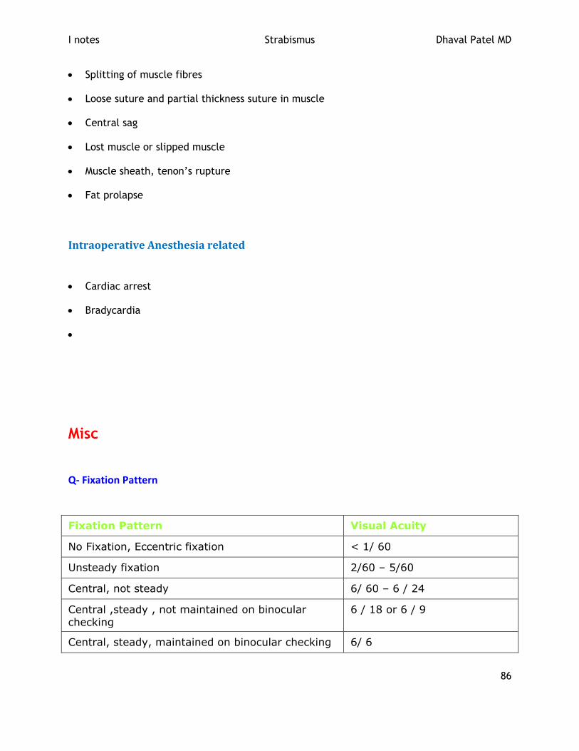
I notes Strabismus Dhaval Patel MD
86
Splitting of muscle fibres
Loose suture and partial thickness suture in muscle
Central sag
Lost muscle or slipped muscle
Muscle sheath, tenon’s rupture
Fat prolapse
Intraoperative Anesthesia related
Cardiac arrest
Bradycardia
Misc
Q- Fixation Pattern
Fixation Pattern Visual Acuity
No Fixation, Eccentric fixation < 1/ 60
Unsteady fixation 2/60 – 5/60
Central, not steady 6/ 60 – 6 / 24
Central ,steady , not maintained on binocular
checking
6 / 18 or 6 / 9
Central, steady, maintained on binocular checking 6/ 6

I notes Strabismus Dhaval Patel MD
87
Alternate spontaneously 6/6 both eyes (equal both
eyes)
Cross fixation 6/ 6 both eyes (equal both
eyes)

I notes Strabismus Dhaval Patel MD
88