Embed Size (px)
Citation preview

Normal Labor and Delivery
Obs&Gyn Department of 1st Clinical Hospital
Wuhan University
Associate Professor Ming lei (明蕾)

Outline
Definition1
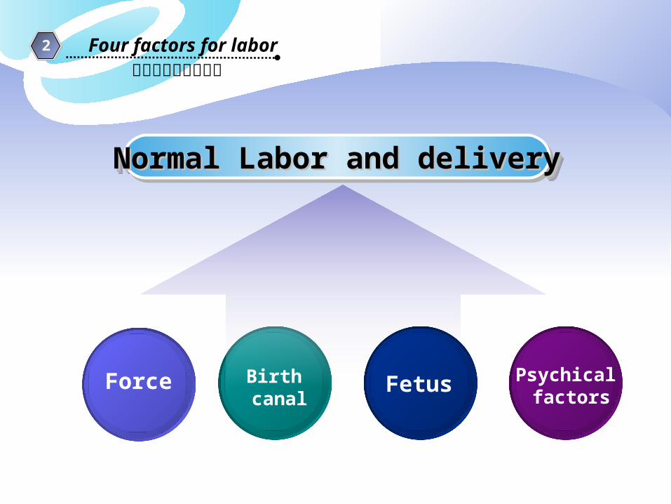
2 Four factors for labor
3 Mechanism of labor

Outline
Diagnosis of threatened labor and labor4
Labor stages5
Clinical course andmanagement of 2nd stages
6Clinical course andmanagement of 1st stages
7
8Clinical course andmanagement of 3rd stages

Definition1
Delivery is the process by which the mature or nearly mature (fetus ,umbilical cord,membranes and placenta) are expelled from the maternal body after 28 weeks
(定义)

Definition1
The last few hours of human pregnancy are characterized by uterine contraction that effect dilatation of the cervix and force the fetus through the birth canal. Much energy is expended during this time, hence the use of term labor to describe this process
Myometrium is unresponsive during pregnancy. After the prolonged period of quiescence, a transitional phase is requires during which myometrial unresponsiveness is suspend and cervix is softened and effaced
(定义)

Definition1
Cause of onset of labor: Precise mechanism in initiation of labor is not defined
Endocrine Mechanical theory Neurological factor
(定义)


Definition1
Term delivery: Term delivery: 37-4237-42 weeks weeks1
Pre-term delivery: Pre-term delivery: 28- 3728- 37 weeks weeks2
Post-term delivery: Post-term delivery: 42 42 weeks weeks3
Abortion:Abortion:4 <28<28 weeks weeks
(定义)
LMP :last menstrual period LMP :last menstrual period 5
6 EDC :expected date of confinement

Definition1
3 Mechanism of labor
2 Four factors for labor
Outline影响分娩的四大因素
Normal Labor and deliveryNormal Labor and deliveryNormal Labor and deliveryNormal Labor and delivery
Force Birth canal
Fetus Psychical factors

影响分娩的四大因素2 Four factors for labor
ForceExpulsive Force
IntraIntraabdominalabdominalpressurepressure
This is created by contraction of the abdominal muscles simutaneously with forced respiratory efforts with the glottis close. this is referred to as pushing
Levator ani muscle
Levator ani muscle is the most important part of pelvic floor
AncillaryForce
uterine contraction
A rhythmic tightening in labor of the upper uterine musculature that contracts the size of the uterus and pushes the fetus toward the birth canal
Major force

RhythmRhythm
SymmetrySymmetry
PolarityPolarity
Retraction
intermissionduration duration
影响分娩的四大因素2 Four factors for labor uterine
Contractionfeature
Force
Four characteristics

Retraction is a phenomenon of the uterus in labor in which the muscle fibres are permanently shortened. Unlike any other muscles of the body
Contraction is a temporary reduction in length of the fibres, which attain their full length during relaxation
retractionretraction

retractionretraction
The effects of retraction in normal labor are: Formation of lower uterine segment and dilatation
and effacement of the cervix Maintain the advancement of the presenting part
made by the uterine contractions and help ultimate expulsion of the fetus
To reduce the surface area of the uterus favoring separation of placenta
Effective haemostasis after the separation of the placenta
影响分娩的四大因素2 Four factors for labor
Birth canal
Normal Labor and deliveryNormal Labor and deliveryNormal Labor and deliveryNormal Labor and delivery
Force Fetus Psychical factors
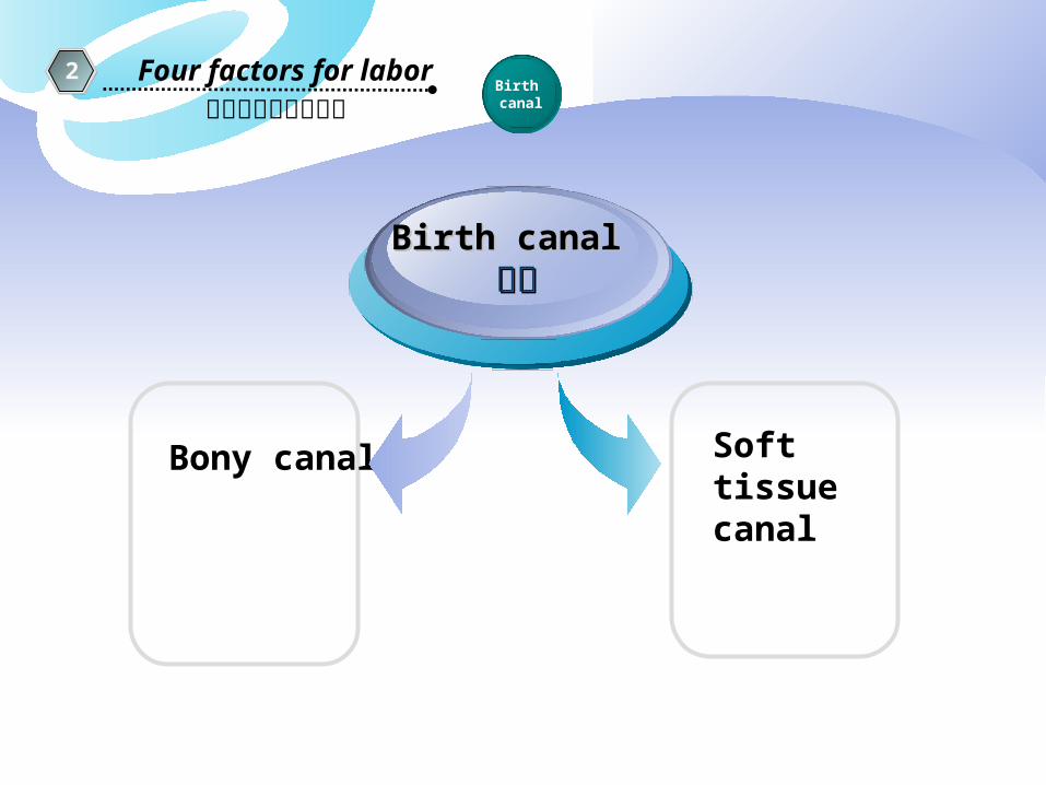
影响分娩的四大因素2 Four factors for labor
Birth canal
Bony canal
Birth canalBirth canal 产道产道
Soft tissue canal

Pelvis anatomical marks
Sacral promontory
Ischial spine (L,R)
Symphysis pubis

影响分娩的四大因素2 Four factors for labor
Birth canal
Bony canal骨产道
Pelvic inlet planeSuperior strait
Pelvic outlet planeInferior strait
Midpelvis outletleast pelvic dimension plane
pelvic axis: A hypothetical line curving through the midpoint of the pelvic planes

4 diameters of the pelvic inletAnteroposterior: 11cmTransverse: 13cm Two obliques: 12.75cm
Pelvic inlet plane
Posteriorly by the promontory and alae of the sacrum, laterallyby the linea terminalis
Anteriorly by the horizontal Rami of the pubic bones and symphysis pubis

4 types in shape – gynecoid (50%), anthropoid, android, platypelloid. Most are intermediate type.
4 diameters – anteroposterior(AP), transverse, and 2 obliques
Obstetrical conjugate – the shortest distance between promontory and symphysis pubis. Estimated by substracting 1.5 to 2 cm from the diagonal conjugate.
True conjugate – the A-P diameter of the pelvic inlet
Pelvic InletPelvic Inlet


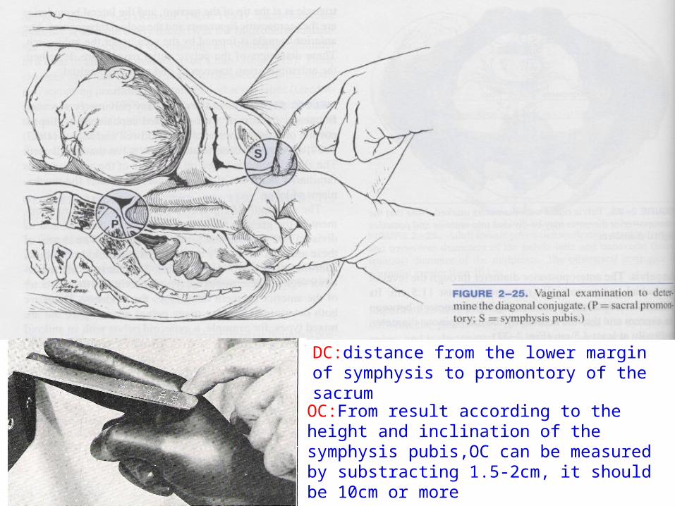
DC:distance from the lower margin of symphysis to promontory of the sacrum
OC:From result according to the height and inclination of the symphysis pubis,OC can be measured by substracting 1.5-2cm, it should be 10cm or more

Anteroposterior diameter Anteroposterior diameter through the level of the ischial through the level of the ischial spinesspines >>11.5cm11.5cm
Interspinous diameter is 10cm or somewhat more
中骨盆平面 mid plane of pelvis
The midpelvis at the level ofIschial spines is of particular importance following engagement of the fetal head in obstructed labor


pelvic outlet plane
Consists of two approximately triangular areas not in the same plane but having a common base, Which is a line drawn between the two ischial tuberosities
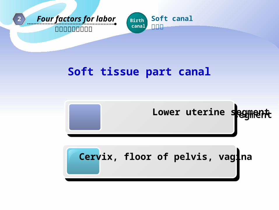
影响分娩的四大因素2 Four factors for labor Birth
canalSoft canal软产道
Lower uterine segment
Lower uterine segment
Cervix, floor of pelvis, vagina
Soft tissue part canal

Soft part of birth canal Formation of uterine lower segmentFormation of uterine lower segment
Cervix effacement and dilatationCervix effacement and dilatation

Isthmus: between anatomical internal OS and histological internal OS
Physiological retraction ring :develops at the junction of the upper and lower uterine segment. As labor progresses, a boundary ridge on the inner uterine surface is marked between the thinning of the lower segment and the concomitant thickening of the upper
Pathological retraction ring: also called Bandl ring, develops from the physiological ring when the thinning of the lower uterine segment is extreme, as in obstructed labor, the ring is very prominent, forming it.

影响分娩的四大因素2 Four factors for labor Birth
canalSoft canal软产道
cervical effacement and dilatationcervical effacement and dilatation
宫颈管

影响分娩的四大因素2 Four factors for labor
Fetus
Normal Labor and deliveryNormal Labor and deliveryNormal Labor and deliveryNormal Labor and delivery
Force Birth canal
Psychical factors
产 力 产 道 胎 儿 精神因素
Fetus 胎儿
Fetal size 胎儿大小Fetal lie 胎产式
Fetal attitude胎姿势
Fetal station胎先露高低
Fetal presentation胎先露
Fetal position胎方位

影响分娩的四大因素2 Four factors for labor
Fetus
Fetal lie Relation of long axis of fetus to that of the mother. Longitudinal (99%), transverse or oblique


影响分娩的四大因素2 Four factors for labor
Fetus
Fetal presentation Fetal part that directly overlies pelvic inletCephalic , breech , or shoulder

影响分娩的四大因素2 Four factors for labor
Fetus
Fetal position
The relation of a chosen portion of the The relation of a chosen portion of the presenting part of the fetus to the right or presenting part of the fetus to the right or left side of the maternal birth canal. left side of the maternal birth canal. For For more accurately more accurately –– anterior, transverse, anterior, transverse, posteriorposterior

影响分娩的四大因素2 Four factors for labor
Fetus
LOA (left occipito-anterior) ROA (right occipito-anterior)
Denominator:bony fixed point on presenting partocciput
影响分娩的四大因素2 Four factors for labor
Fetus
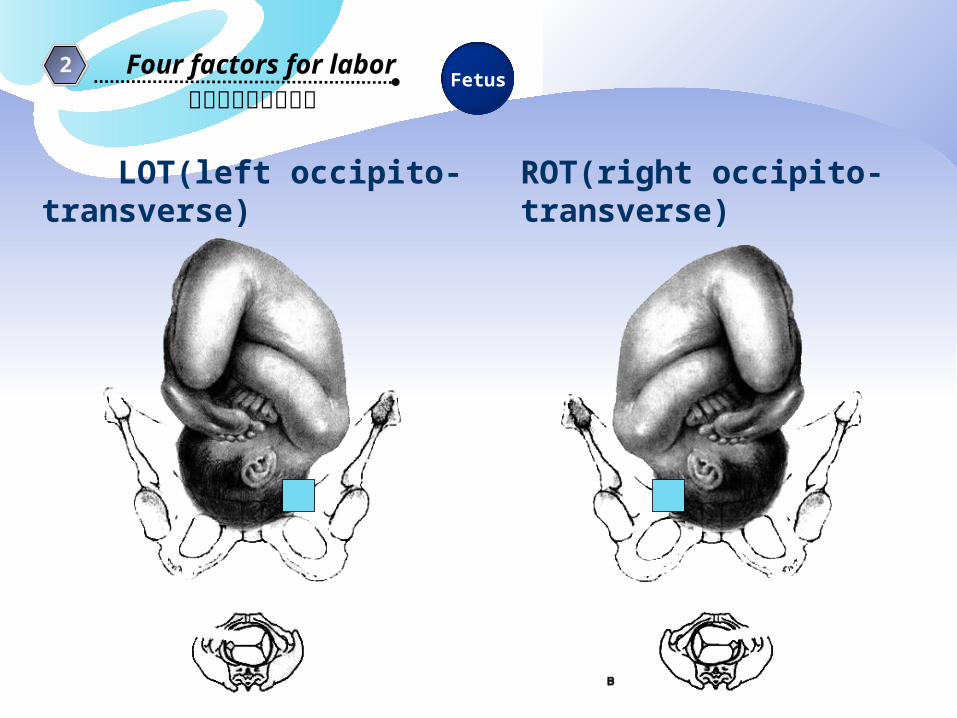
LOT(left occipito-transverse) ROT(right occipito-transverse)

影响分娩的四大因素2 Four factors for labor
Fetus
LOP(left occipito-posterior) ROP(right occipito-posterior)

影响分娩的四大因素2 Four factors for labor
Fetus

影响分娩的四大因素2 Four factors for labor
Fetus
The fetus is in the occiput or vertex presentation in approximately 95% of all labor
In majority of cases, the vertex enters the pelvis with the sagittal suture in the transverse pelvic diameter or the oblique lines
The fetus enters the pelvis in the left occiput transverse(LOT) or LOA position in 40%;in the ROT or ROA in 20%; in OP position in 20%

1 step1 step
Leopold’s maneuvers
4 step
Run hands down maternal abdomen on either side of fetus to determine fetal lie, identifying small parts and fetal spine
Using the thumb and fingers of one hand, the lower portion of the maternal abdomen is grasped just above the symphysis pubis to determine the presentation
With the tips of the fingers of each hand, exerts deep pressure in the direction of the axis of the pelvic inlet to determine if the head or breech descended into the pelvis
Gently palpate the fundus with the tip of the fingers of both hand in order to define which fetal pole is present in the fundus and appropriate size.
2 step
3 step

1. The mother should be supineand comfortable position withher abdomen bared
2.During the first three maneuversthe examiner stands at the sideof the bed that is most convenientand faces the patient
3.The examiner reverses thisposition and face the feet for the last maneuver
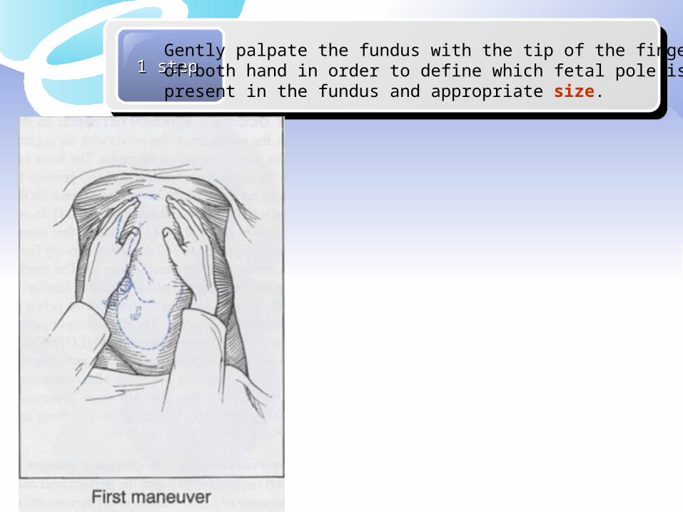
1 step1 stepGently palpate the fundus with the tip of the fingers of both hand in order to define which fetal pole is present in the fundus and appropriate size.

Run hands down maternal abdomen on either side of fetus to determine fetal lie, identifying small parts and fetal spine
2 step

3 step
Using the thumb and fingers of one hand, the lower portion of the maternal abdomen is grasped just above the symphysis pubis to determine the presentation

4 stepWith the tips of the fingers of each hand, exerts deep pressure in the direction of the axis of the pelvic inlet to determine if the head or breech descended into the pelvis

bregma
Adult fetus
Suture: it permits gliding movement of one bone over the other during moulding of the head while the head passes through the pelvis;During Internal examination in labor, palpation of sagittal suture give anidea of the manner of engagement of the head

post fontanelle(lambda)
Bregma(Ant fontanelle)
BPD(parietal bones diameter)
frontal bones
Occipital bone
Sagittal suture
The bones of the vault are not joined thus changes in the shape of the fetal head during labor can occur due to molding
fontanelle:wide gap in suture line

Outline
Definition1
3 Mechanism of labor
2 Four factors for labor
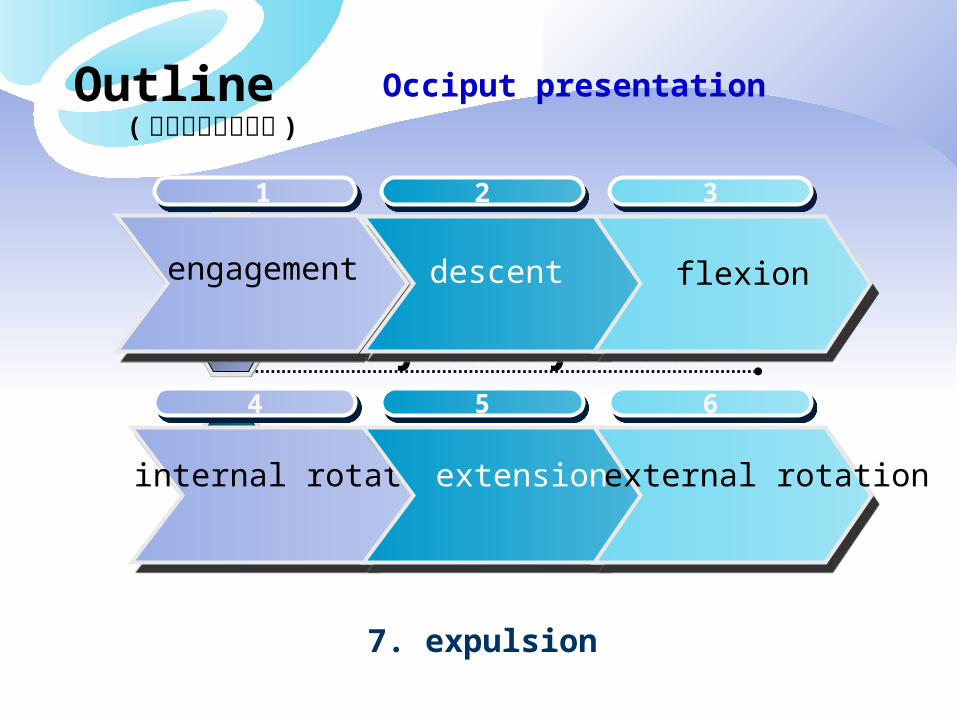
Occiput presentation( 枕先露的分娩机转 )
1. The ability of the fetus to successfully negotiate the pelvis during labor involves changes in position of its head during its passage in labor
2. The mechanisms of labor, also known as the cardinal movements, are described in relation to a vertex presentation, as is the case in 95% of all pregnancies
3. In reality, the mechanism of labor consists of a combination movements that are ongoing simultaneously
4. For purposes of instruction, the various movements often are described as though they occurred separately and independently the cardinal movements are described as 7 discrete sequences, as discussed below
Outline
Definition1
3 Mechanism of labor
2 Four factors for labor
Occiput presentation( 枕先露的分娩机转 )
1 1
engagement
22
descent
33
flexion
44
internal rotation
55
extension
66
external rotation
7. expulsion

Mechanism of labor
( 枕先露的分娩机转 )
Occiput presentation3
1. Engagement
Passage of widest diameter of presenting part to level below the plane of the pelvic inlet
Occurs earlier in primigravidae, usually before the onset of labor while in multiparae, the same may occur in late 1st stage with rupture of the membrane
ischial spines

Mechanism of labor Occiput presentation3
2 、 3. Descent &FlexionDownward passage of presenting part through the pelvisOccurs passively as the head descends due to the shape of the bony pelvis and resistance of pelvic floor soft tissues resulting in passive flexion of the fetal occiput. The chin is brought into contact with the fetal thorax, and the presenting diameter changes from occipitofrontal (12cm) to suboccipitobregmatic (9.5 cm) which is the smallest diameter of fetal head for optimal passage through the pelvis
Occipitofrontal Diameter 12cm
Suboccipitobregmatic Diameter 9.5cm

Occipito frontal diameter 枕额径 12cmOccipito subregmatic diameter 枕下前囟径 9.5cmOccipito mental diameter 枕颏径 13cm

LOT
ROT

Mechanism of labor
( 枕先露的分娩机转 )
Occiput presentation3
4. Internal rotation As the head descends, the presenting part, usually in the
transverse position, is rotated about 45°to anteroposterior (AP) position under the symphysis. Internal rotation brings the AP diameter of the head in line with the AP diameter of the pelvic outlet

Mechanism of labor
( 枕先露的分娩机转 )
Occiput presentation3
5. Extension
1) With further descent and full
flexion of the head, the base
of the occiput comes in contact
with the inferior margin of the
pubic symphysis
2) Upward resistance from the
pelvic floor and the downward
forces from the uterine contractions
cause the occiput to extend and
rotate around the symphysis

Forces Concerned in LaborForces Concerned in Labor
Positive forces * Uterine contractions * Abdominal pressure by rectus muscles * Fundal pressure* Forceps delivery and vacuum extraction
Resistance * The uterine cervix * The muscles of the pelvic floor
Positive forces * Uterine contractions * Abdominal pressure by rectus muscles * Fundal pressure* Forceps delivery and vacuum extraction
Resistance * The uterine cervix * The muscles of the pelvic floor

Mechanism of labor
( 枕先露的分娩机转 )
Occiput presentation3
6. External Rotation When the fetus' head is free of resistance, it
untwists about 45°left or right, returning to its original anatomic position in relation to the body

Mechanism of labor Occiput presentation3
7. Expulsion After the fetus' head is delivered, further descent brings the anterior shoulder to the level of the pubic symphysis
The anterior shoulder is then rotated under the symphysis, followed by the posterior shoulder and the rest of the fetus

Mechanism of labor
( 枕先露的分娩机转 )
Occiput presentation3
Mechanism of labor for left occiput anterior position

4
FalseFalselaborlabor
Diagnosis of threatened labor
As the fetal presenting part descends into the pelvic inlet, the fundal height decreases. Theprimigravida feels comfort of the upper abdomen, eats more, and respires briskly.
Braxon Hicks contractions,during the last 4-8weeks of pregnancy irregular,generally painless uterine contractions occur with slowly increasing frequency
lightening
showCervical effacement, the mucus plug within the cervical canal may be released .And a small amount of blood creating by the ruptured capillary and the mucous in the cervix are mixed together and are discharged , this is called show

Diagnosis of in labor4
1
2
3
Regular uterine contractionRegular uterine contraction
cervical effacementcervical effacement
cervical dilatationcervical dilatation
4 descentdescent

False labor vs. True laborFalse labor vs. True labor
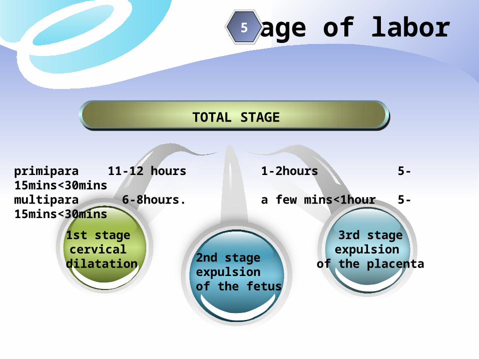
Stage of labor
TOTAL STAGE
2nd stageexpulsion of the fetus
1st stage cervical dilatation
3rd stageexpulsion
of the placenta
primipara 11-12 hours 1-2hours 5-15mins<30minsmultipara 6-8hours. a few mins<1hour 5-15mins<30mins
5

Three stages of labor
The first stage begins when uterine contractions of sufficient frequency, intensity and duration are attained to bring about effacement and progressive dilatation of the cervix and ends when the cervix is fully dilated
The second stage begins when dilatation of the cervix is complete, and ends with delivery of the fetus
The third stage begins immediately after delivery of the fetus, and ends with the delivery of the placenta and fetal membranes
“The fourth stage” is the stage of observation for 2 hours after expulsion of the placenta

The first stage is divided into a relatively flat latent phase and a rapidly progressive active phase. In active phase, there are 3 identifiable component parts: an acceleration phase, a linear phase of maximum slope, and a deceleration phase
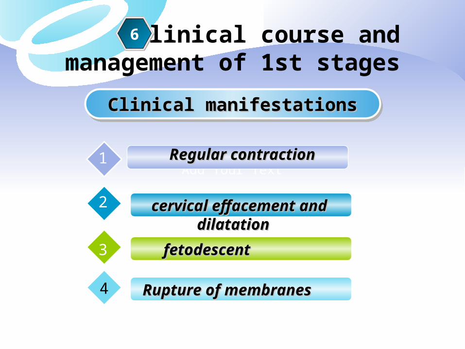
Clinical course andmanagement of 1st stages
Clinical manifestationsClinical manifestationsClinical manifestationsClinical manifestations
Add Your Text
6
Regular contractionRegular contraction1
cervical effacement and dilatationcervical effacement and dilatation2
fetodescentfetodescent3
Rupture of membranes Rupture of membranes 4

2
6 Clinical manifestationsClinical manifestationsClinical manifestationsClinical manifestations
1. At the beginning of the first stage of labor, it is weak
2. intermission lasts a little longer about 5-6mins; duration is about 30s
3. As labor progresses, the intermission lasts 2-3mins; duration is 50-60s
4. intensity increases when the cervix is fully dilated, intermission only last one min or more longer; duration can last more than one min
Uterine contraction1

intensity
duration intermission duration
Intensity: describes the degree of uterine systole, it gradually increases with the advancement of labor until it becomes maximum in the second stage during delivery of the baby
Duration: In the 1st stage,the contraction last for about 30s initially but Gradually increase in duration with the progress of labor. Thus in the 2nd stage, the contractions last longer than in the 1st stage

2
6 Clinical manifestationsClinical manifestationsClinical manifestationsClinical manifestations
Cervical changes and descent2-3
Cervical effacement: the obliteration of the cervix is the shortening of the cervical canal from a length of about 2cm to a mere circular orifice with almost paper-thin edges
Compared with the body of the uterus, the lower segment and the cervix are regions of lesser resistance, these structures are subjected to distention. As the uterinecontraction cause pressure on the membranes, the hydrostatic action of the aminionic sac in turn dilates the cervical canal. In absence of intact membranes, the pressure of the presenting part against the cervix and lower uterine segment is effective. It won’t retard cervical dilatation


2
6 Clinical manifestationsClinical manifestationsClinical manifestationsClinical manifestations
LATENT PHASE
It begins with theregular contractionsafter in labor ends when the cervix dilates to 2cm.8-16h; 1cm/2-3h
ACTIVE PHASE
It refers the cervix dilated from 2cm to complete dilatation.4-8h;
Cervical changes and descent2-3

The first stage is divided into a relatively flat latent phase and a rapidly progressive active phase. In active phase, there are 3 identifiable component parts: an acceleration phase, a linear phase of maximum slope, and a deceleration phase

2
6 Clinical manifestationsClinical manifestationsClinical manifestationsClinical manifestations
The fetal membrane is always ruptured when the cervix is completely dilated and the amnionic fluid runs out , this is called rupture of membranesWhen the membrane rupture before the onset of labor, it is called premature rupture
4 Rupture of membrane

management of 1st stages
education, eating, voidingeducation, eating, voiding1
position(sitting, reclining, recumbent)position(sitting, reclining, recumbent)2
monitoring of the fetal heart ratemonitoring of the fetal heart rate3
dilation of cervix and frequencydilation of cervix and frequency
severity of uterus contractionsseverity of uterus contractions5
6
4

Management principles of 1st stage
Non-interference with watchful expectancy so as to prepare the patient for natural birth
To monitor carefully the progress of labor, maternal conditions and fetal behavior so as to detect any intrapartum complicating early

Actual management of 1st stage
General: antiseptic dressing, encouragement and assurance constant supervision
Bowel: an enema with soap or glycerine suppository is traditionally given in early stage. Reduce infection rate and increase the progress of the laborRest and ambulation:
Diet: food is withheld during active labor; water,fluid juice can be given
Bladder care: full bladder often inhibits uterine contraction and may lead to infection, so encourage the patient to pass urine by herself or catheterisation to be done to her with strict aseptic precautions

Actual management of 1st stage
Relief of pain: analgesia in labor can be used on primigravida. The analgesic drugs should not be given if delivery is anticipated within two hoursMaternal condition: routine check up includes
to record per 2 hours about P,BP, T, cervical dilatation and fetal presentation descent by anal or vaginal examinationto note the urine outputI.V fluid, drugs
Electronic fetal monitoring, auscultationauscultation ,dopplerMonitor the uterus contraction(intensity,duration,frequency)

Clinical course andmanagement of 2nd stages
Clinical manifestationsClinical manifestationsClinical manifestationsClinical manifestations
Add Your TextMore intensive contraction with defecationMore intensive contraction with defecation1
Head visible on vulval gappingHead visible on vulval gapping2
Crowning of headCrowning of head3
7

Head visible on vulval gappingHead visible on vulval gapping Crowning of headCrowning of head
Crowning refers to when the widest part of the baby's head (or their crown) is emerging. At this point, the baby's crown, part of their forehead and the back of the baby's head can be clearly seen.

Crowning refers to when the widest part of the baby's head (or their crown) is emerging. At this point, the baby's crown, part of their forehead (nearly to their eyebrows) and the back of the baby's head can be clearly seen.
As the baby's head crowns, the woman's perineum is stretched to its maximum, being nearly paper-thin. There is usually an intense burning (or stinging sensation) for a few seconds as this occurs, generally easing as the perineum numbs. The burning can trigger panic for some women, causing them to cry out, or scream.

management of 2nd stages
fetal heart ratefetal heart rate1
maternal conditionsmaternal conditions2
PushingPushing3
Head visible on vulval gappingHead visible on vulval gapping
Crowning of head Crowning of head
8
7
Laceration or Episiotomy Laceration or Episiotomy
Delivery of fetus Delivery of fetus
Deal with umbilical cord Deal with umbilical cord
aiding in fetal descent through birth canalaiding in fetal descent through birth canal
5
9
6
7
4

Management principles of 2nd stage
To assist in the natural expulsion of the fetus slowly and steadily
To prevent perineal injuries

Actual management of 2nd stageGeneral: constant supervision is mandatory and the FHR is recorded at
every 5minsvaginal examination is done at the beginning of the 2nd stage not
only to comfirm its onset but to detect any accidental cord prolapse. The position and the station of the head are once more to be reviewed and the progressive descent of the head is ensured
Preparation for delivery: position the accoucheur scrubs up, put on the sterile gown, mask
and gloves and stands on the right side of the tableToileting the external genitaliaTo catheterise the bladder

Actual management of 2nd stage
Conduction of delivery: Episiotomy is done selectively, usually as a routine in ChinaSlowly delivery of the head in between the contractions is to be
regulatedThe mucus and blood in the mouth and pharynx are to be wiped with sterile gauze piece on a little finger or electrical sucker
Prevention of perineal lacerationDelivery by early extension is to be avoidedSpontaneous forcible delivery of the head is to be avoidedTo deliver the head in between contractionsTo perform timely episiotomy(

Actual management of 2nd stage
Immediate care of the newborn air passage should be cleared of mucus and
liquor by gentle suction apgar rating at 1min and at 5mins is to be
recorded clamping and ligature of the cord
baby is wrapped with a dry warm towel, the identification tape is tied both on the wrist of the baby and the mom


Clinical course andmanagement of 3rd stages
Clinical manifestationsClinical manifestationsClinical manifestationsClinical manifestations
Add Your Textthe uterus decreases in sizethe uterus decreases in size1
delivery of placenta(spontaneously, manuallydelivery of placenta(spontaneously, manually2
inspection of the birth canalinspection of the birth canal3
8
evaluated for lacerationsevaluated for lacerations4

Management principles of 3rd stage
To ensure strict vigilance and to follow the management guidelines strictly in practice so as to prevent the complications

Actual management of 3rd stage
Expectant management(traditional) Active management
Control cord traction
Fundal pressure
Oxytocin iv drop
Manual removal

Actual management of 3rd stage
Examination of the placenta membranes and cord
Vulva, vagina and perineum are inspected

Placental SeparationPlacental Separation
• • begins immediately after delivery of fetus,begins immediately after delivery of fetus, involve separation & expulsion of placentainvolve separation & expulsion of placenta••Diminution in Ut sizeDiminution in Ut size PL implantation site areaPL implantation site area ↓ ↓ PL accommodate to reduced PL accommodate to reduced area area thickness because of limited PL elasiticity thickness because of limited PL elasiticity forced to buckle forced to buckleResulting tension Resulting tension weakest layer of decidua (D. spongiosa) cleavage take place weakest layer of decidua (D. spongiosa) cleavage take place at that siteat that siteAs separation proceed As separation proceed hematoma forms between separtating PL & remaining hematoma forms between separtating PL & remaining Decidua Decidua result of separation result of separation

Placental SeparationPlacental Separation
• • begins immediately after delivery of fetus, involve begins immediately after delivery of fetus, involve separation & expulsion of placentaseparation & expulsion of placenta
••Diminution in Ut sizeDiminution in Ut size PL implantation site areaPL implantation site area ↓ ↓ PL PL accommodate to reduced area accommodate to reduced area thickness because of thickness because of limited PL elasiticity limited PL elasiticity forced to buckle forced to buckle
--Resulting tension Resulting tension weakest layer of decidua (D. weakest layer of decidua (D. spongiosa) cleavage take place at that sitespongiosa) cleavage take place at that site
--As separation proceed As separation proceed hematoma forms between hematoma forms between separtating PL & remaining Decidua separtating PL & remaining Decidua result of result of separationseparation

Placental ExtrusionPlacental Extrusion
some casesome case abdominal pr↑ PL be expelled abdominal pr↑ PL be expelled women in recumbent position frequently women in recumbent position frequently
cannot expel placenta spontaneouslycannot expel placenta spontaneously → → artificial means generally requiredartificial means generally required → → compress & elevate fundus while exerting compress & elevate fundus while exerting
minimal traction on umbilical cordminimal traction on umbilical cord

Mechanisms of Placental ExtrusionMechanisms of Placental Extrusion
(1) Schultze mechanism((1) Schultze mechanism(centralcentral separationseparation))
• • PL separation occurs 1st at central areasPL separation occurs 1st at central areas
→ → retroplacental hematomaretroplacental hematoma
→ → push placenta toward uterine cavitypush placenta toward uterine cavity
(2) Duncan mechanism((2) Duncan mechanism(marginalmarginal separationseparation))
① ① placental separation occurs first at peripheryplacental separation occurs first at periphery
② ② blood collects between membranes & uterine blood collects between membranes & uterine wall → escapes from vagina→Maternal surface wall → escapes from vagina→Maternal surface first to appear at vulvafirst to appear at vulva

11
Signs of placental separation
A sudden gush of blood from vagina
The uterus becomes globular and firmer.This sign is the earliest to appear
2
3
The uterus rises in the abdomen because the placenta, having separated, passes down into the lower uterine segment and vagina, where its bulk pushes the uterus upward
4 The umbilical cord lengthens out of the vagina,indicating that the placenta has descended


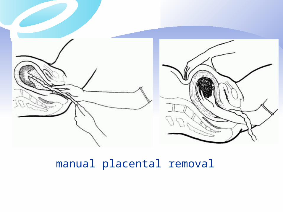
manual placental removal

副胎盘 succenturiateplacenta


Mechanism of control of bleeding
After placental separation,innumerable torn sinuses which have free circulation of blood from uterine and ovarian vessels have to be obliterated. The occlusion is effected by complete retraction where by the arterioles, as they pass tortuously through the interlacing intermediate layer of the myometrium, are literally clamped
Thrombosis occurs to occlude the torn sinuses Apposition of the wall of the uterus following expulsion of
the placenta also contributes to minimize blood loss

Perineum ,Cervical,Vaginal laceration
First degree tear :involves only skin and a minor part of the perineal body

Perineum ,Cervical,Vaginal laceration
Second degree tear :involves perineal body and vaginal wall

Perineum ,Cervical,Vaginal laceration
Third degree tear :involves the anal sphincter and anal canal

Perineum ,Cervical,Vaginal laceration


“The fourth stage”
General condition of the patient and behavior
of the uterus are to be carefully watched postpartum uterine hemorrhage ,1% uterus palpation through the abdominal wall
is repeats the amount of blood on pads are monitored pulse and BP are monitored use of drug : oxytocin

Questions
DefinitionDelivery; physiological retraction ring ; Obstetrical
conjugate; fetal lie; fetal position
Braxon Hicks contractions; crowning of headrowning of head
The first stage; the second stage; the third stage

essays
Mechanism of normal labor Identify false labor or true labor Talk about the signs of placental separation Describe two mDescribe two mechanisms of Placental Extrusionechanisms of Placental Extrusion What is the most common fetal position at onset of
labor? What is active phase? What are you going to do after the 3rd stages?

What is the correct order of the cardinal movements of labor?
A). Flexion, descent, engagement, internal rotation, extension, external rotation
B). Descent, engagement, flexion, internal rotation, external rotation, extension
C). Engagement, flexion, descent, external rotation, extension, internal rotation
D). Engagement, flexion, decent, internal rotation, extension, external rotation