Embed Size (px)
Citation preview

Med Oral Patol Oral Cir Bucal. 2016 May 1;21 (3):e305-15. Diagnostic tools for oral cancer
e305
Journal section: Oral Medicine and PathologyPublication Types: Review
Non-invasive visual tools for diagnosis of oral cancer and dysplasia: A systematic review
Ilaria Giovannacci 1, Paolo Vescovi 1, Maddalena Manfredi 2, Marco Meleti 2
1 DDS, Msci. Department of Biomedical, Biotechnological and Translational Science-Center of Oral Laser Surgery and Oral Pathology, Dental School, University of Parma, Parma, Italy 2 DDS, PhD. Department of Biomedical, Biotechnological and Translational Science-Center of Oral Laser Surgery and Oral Pathology, Dental School, University of Parma, Parma, Italy
Correspondence:Center of Oral Laser Surgery and Oral Medicine Dental School. Department of Biomedical Biotechnological and Translational SciencesVia Gramsci, 14 - 43125 Parma, [email protected]
Received: 11/08/2015Accepted: 14/10/2015
Abstract Background: Gold standard for the diagnosis of oral dysplasia (OD) oral squamous cell carcinoma (OSCC) and malignant lesions is the histological examination. Several adjunctive diagnostic techniques have been proposed in order to increase the sensitivity (SE) and specifi-city (SP) of conventional oral examination and to improve the diagnostic first level accuracy.The aim of this study is to perform a systematic review on non-invasive tools for diagnosis of OD and early OSCC.Material and Methods: Medline, Scopus, Web of Knowledge databases were searched, using as entry terms “oral dysplasia AND diagnosis” / ”oral cancer AND diagnosis”. Data extracted from each study included number of lesions evaluated, histopathological diagnosis, SE, SP, positive and negative predictive values (PPV and NPV), diagnostic accuracy (DA) and the main conclusions.Results: After title and abstract scanning of 11.080 records, we selected 35 articles for full text evaluation. Most evaluated tools were autofluorescence (AF), chemiluminescence (CL), toluidine blu (TL) and chemiluminescence associated with toluidine blue (CLTB). Conclusions: There is a great inhomogeneity of the reported values and there is no significant evidence of supe-riority of one tool over the other. Further clinical trials with a higher level of evidence are necessary in order to assess the real usefulness visual diagnostic tools.
Key words: Oral dysplasia, oral cancer, diagnosis, visual diagnostic tool, systematic review.
Giovannacci I, Vescovi P, Manfredi M, Meleti M. Non-invasive visual tools for diagnosis of oral cancer and dysplasia: A systematic review. Med Oral Patol Oral Cir Bucal. 2016 May 1;21 (3):e305-15. http://www.medicinaoral.com/medoralfree01/v21i3/medoralv21i3p305.pdf
Article Number: 20996 http://www.medicinaoral.com/© Medicina Oral S. L. C.I.F. B 96689336 - pISSN 1698-4447 - eISSN: 1698-6946eMail: [email protected] Indexed in:
Science Citation Index ExpandedJournal Citation ReportsIndex Medicus, MEDLINE, PubMedScopus, Embase and Emcare Indice Médico Español
doi:10.4317/medoral.20996http://dx.doi.org/doi:10.4317/medoral.20996

Med Oral Patol Oral Cir Bucal. 2016 May 1;21 (3):e304-15. Diagnostic tools for oral cancer
e306
IntroductionOral squamous cell carcinoma (OSCC) is the sixth most common malignant tumour, with an incidence of more than 500.000 cases per year (1). The most important prognostic factor influencing the disease-specific survival rate is the tumour stage at diagnosis. Patients with stage I tumours have a 5-year survival rate of 75%, which dramatically decreases in patients with tumours in stage III or IV, being 49% and 30%, respectively (1,2).The diagnostic pathway for oral suspicious lesions usu-ally starts with the conventional objective examination (COE) based on inspection and palpation of the oral mu-cosa with the support of an incandescent light available on the dental chair. It is well known that COE mainly depends on a subjective interpretation, which is a conse-quence of the experience of the operator. Moreover, oral epithelial dysplasia (OED) and early OSCC may already be present within areas of oral mucosa macroscopically normal, as well as within the context of oral potentially malignant disorders such as leukoplakia, erythroplakia, submucous fibrosis and oral lichen planus (3).The gold standard for the diagnosis of oral dysplastic and neoplastic malignant lesions is the histological ex-amination (4). Incisional or excisional biopsy techniques are the most reliable methods to collect a surgical speci-men suitable for microscopic evaluation. However, de-spite the little invasivity of such techniques, they still have some disadvantages in terms of morbidity and pos-sible artifacts induced by the method of collection. In a recent paper, Mehrotra et al. indicated that there are two approaches for detection of oral dysplasia and cancer: 1) oral cancer screening programs that identify asympto-matic patients with suspicious lesions and 2) specific di-agnostic tools to identify dysplasia and early oral cancers in asymptomatic patients with an oral abnormality (5).Several visual diagnostic aids have been developed as adjunctive tools in order to increase the diagnostic ac-curacy (DA) and enhance the specificity (SP) and sen-sitivity (SE) of the conventional diagnostic pathway. However, results of studies on the usefulness of such tools show impressive discrepancies with regard to val-ues such as the positive or negative predictive values (PPV, NPV), when the same tools is evaluated by dif-ferent researchers.The aim of this study is to perform a systematic review on non-invasive tools for the diagnosis of OED and OSCC, taking into account factors as SE, SP, PPV, NPV and DA.
Material and MethodsThe databases Medline, Scopus and Web of Knowledge were searched, using as entry terms “oral dysplasia AND diagnosis” / ”oral cancer AND diagnosis”. No time limits were specified in the present research.
Search flow is shown in figure 1. Papers with abstract unavailable were excluded for further evaluation. Titles and abstract were screened and the following ex-clusion criteria were applied:- papers not in English.- studies ex vivo or based on animal models.- typology of the study: case reports, case series with less than 10 patients, conference proceedings, personal communications, editorials, descriptive studies and re-views.- studies that analyse COE, invasive diagnostic tools (e.g. scalpel biopsy) or minimally invasive diagnostic tools (e.g. brush biopsy, exfoliative cytology) alone. - studies that analyse salivary biomarkers.- studies including also tumours of other head and neck regions (e.g. oropharynx).Papers with equivocal abstracts were included for full-text evaluation. Further studies were excluded after full-text reading, if not pertinent with aim of the present review.Data extracted from each study included authors and publication year, typology of the study, diagnostic tool analysed, number of lesions evaluated, (if present) his-topathological diagnosis, (if present) SE, SP, PPV, NPV, DA and the main conclusions of the study (Tables 1 and 1 continue,2).SE and SP measure the accuracy of a test without any relation to the disease or population, whereas PPV and NPV measure the proportion of people whose test results reflect their health status. DA is the proportion of true positive results (both true positive and true negative) in a selected population, with regard to a specific disease.The mean value of each variable analysed was calcu-lated; range and standard deviation (SD) were indicated for samples having > 2 values. Level of evidence of each study was assessed according to the Oxford Evidence-based Medicine (OEBM) Lev-els for Diagnosis updated in March 2009.
ResultsTwenty-three papers were eventually selected for the present systematic review when using “oral dysplasia AND diagnosis” as entry terms. The use of “oral cancer AND diagnosis” as entry terms allowed the identifica-tion of further 25 full-text manuscripts (6-39).Twenty-three studies were perspective (OEBM level: 2b), 4 studies were pilot (OEBM level: 3b), 3 studies were case-control (OEBM level: 4), 4 studies were cross-sec-tional (OEBM level: 2b). Only one study was a perspec-tive randomized clinical trial (RCT) (OEBM level: 1b).Eight typologies of non-invasive visual diagnostic tools were identified (Table 3).Mean SE and SP (with SD) are shown in figures 2,3.1. Auto fluorescence (AF) - Direct visual fluorescence examination (DVFE)

Med Oral Patol Oral Cir Bucal. 2016 May 1;21 (3):e305-15. Diagnostic tools for oral cancer
e307
Among 12 studies evaluating AF/DVFE, 8 were per-spective (OEBM level: 2b), 2 were cross-sectional (OEBM level: 2b), 1 was a pilot study (OEBM level: 3b) and 1 was a perspective RCT (OEBM level: 1b) (6,10,13,14,16-18,24,29,32,33,35).Data on SE were reported in 10 studies, while informa-tion on SP was available in 11 studies. Mean SE was 72.4% ranging from 20% to 100% (SD = 27.1). Mean SP was 63.79% ranging from 15.3% to 100% (SD = 28.17). Data on PPV were available in 5 studies (mean: 55.74%,
ranging from 15.1% to 92%, SD = 36.71); data on NPV were available in 5 studies (mean: 79.76%, ranging from 61% to 100%, SD = 15.99); DA was reported in 1 study (55%). 2. Chemiluminescence (CL)Among 5 studies evaluating CL, 4 were perspective (OEBM level: 2b) and 1 was observational cross-sec-tional (OEBM level: 2b) (7,15,38).Data on SE and SP were reported in 4 studies. Mean SE was 86.72%, ranging from 69.6% to 100% (SD = 15.65).
Fig. 1. Flow-chart diagram for the selection of the 35 studies included in the present analysis.

Med Oral Patol Oral Cir Bucal. 2016 May 1;21 (3):e304-15. Diagnostic tools for oral cancer
e308
N°
Auth
ors a
nd
year
(ref)
Ty
polog
y of
the s
tudy
Di
agno
stic t
ools
analy
sed
Num
ber
of le
sions
Hi
stopa
tholo
gic d
iagno
sis
Resu
lts
Main
auth
ors c
onclu
sions
1 Pe
truzz
i M
et al
. 201
4 (6)
Doub
le ce
ntre
cross
secti
onal
study
DVFE
vs T
B 56
NS
DVFE
SE:
70%
, SP:
57.7%
, PPV
: 65
.6%, N
PV: 7
0.6%
(mild
dy
splas
ia po
sitive
) DV
FE an
d TB
are bo
th se
nsiti
ve bu
t not
spec
ific i
n OSC
C an
d dy
splas
ia di
agno
sis.
TB S
E: 80
%, S
P: 61
.5%, P
PV:
62.5%
, 72.7
%
2 Kä
mmer
er PW
et
al. 2
013 (
7)
Persp
ectiv
e stu
dy
CL; C
LTB
50
Reac
tive l
esio
ns (4
0),
dysp
lastic
lesio
ns (3
), OS
CC (7
)
CL S
E: 10
0%, S
P: 30
%. P
PV:
26%
, NPV
: 100
%
The a
djun
ct of
TB
to C
L red
uces
the n
umbe
r of f
alse p
ositi
ves
with
out i
ncrea
sing t
he ra
te of
false
nega
tives
. CL
TB S
E: 80
%, S
P: 97
.5%, P
PV:
90%
, NPV
: 95%
3
Palla
gatti
et al
. 20
13 (8
) Pe
rspec
tive
study
TB
37
Be
nign
lesio
ns (1
4),
dysp
lastic
lesio
ns (2
3)
SE: 9
5%, S
P: 71
.45%
, PPV
: 84
.6%, N
PV: 9
0.9%
, DA:
86.48
%
The u
se of
TB
stain
ing w
as ta
ken i
nto c
onsid
eratio
n to i
dent
ify
clini
cally
doub
tful o
ro-p
hary
ngea
l les
ions
. 4
Mitt
al et
al. 2
012
(9)
Pilo
t stu
dy
BR
20
Dysp
lastic
lesio
ns (1
7),
Verru
cous
Ca (
1), O
SCC
(3)
SE an
d DA:
90%
BR
stain
ing c
an be
used
as a
valua
ble d
iagno
stic t
est i
n the
de
tectio
n of o
ral po
tentia
lly m
align
ant a
nd m
align
ant d
isord
ers.
5 M
cNam
ara K
K et
al. 2
012 (
10)
Persp
ectiv
e stu
dy
DVFE
vs C
OE
95
Beni
gn le
sions
(50)
, pr
emali
gnan
t les
ions
(2),
malig
nant
(1) –
52 no
f/u
DVFE
was
stati
stica
lly di
fferen
t fro
m sc
alpel
biop
sy (P
=0.00
01)
COE
is mo
re va
lid th
an D
VFE
at di
scrim
inati
ng be
nign
mu
cosa
l alte
ratio
ns fr
om pr
emali
gnan
cy an
d do n
ot su
ppor
t the
us
e of D
VFE
as or
al ca
ncer
scree
ning
adjun
ct.
6 Aw
an K
H
et al
. 201
2 (11
) Pe
rspec
tive
study
TB
92
Be
nign
lesio
ns (5
1),
Dysp
lastic
lesio
ns (4
1)
SE: 5
6.1%
, SP:
56.9%
TB
is a
usefu
l adj
unct
to cli
nical
visua
l exa
mina
tion b
y aid
ing
in th
e visu
aliza
tion o
f les
ions
.
7 M
ojsa
et al
. 201
2 (1
2)
Persp
ectiv
e stu
dy
CLTB
41
Be
nign
lesio
ns (3
4),
Dysp
lastic
lesio
ns (6
), OS
CC (1
)
SE: 8
1.8%
, SP:
37.5%
, PPV
: 84
.4%, N
PV: 3
3.3%
CL
TB m
ay he
lp to
visu
alize
oral
path
ologic
lesio
ns th
at are
not
readi
ly de
tectab
le wi
th co
nven
tiona
l ope
rator
y ligh
ting.
8 Fa
rah e
t al.
2011
(13)
Pe
rspec
tive
study
DV
FE
118
Non d
yspla
stic
lesio
ns (9
1),
dysp
lastic
lesio
ns (2
4),
OSCC
(3)
SE: 3
0%, S
P: 63
%, P
PV: 1
9%,
NPV:
75%
, DA:
55%
DV
FE ca
nnot
prov
ide a
defin
itive
diag
nosis
rega
rdin
g the
pr
esen
ce of
epith
elial
dysp
lasia.
9 Pa
dern
i C
et al
. 201
1 (14
) Pe
rspec
tive
study
DV
FE
175
Beni
gn le
sions
, dys
plasti
c les
ions
, OSC
C
SE (O
SCC)
: 96.4
%, d
yspla
sia
(60%
), no
dysp
lasia
(71%
) Th
e dev
ice w
as fo
und t
o not
repl
ace t
he hi
stopa
tholo
gy
proc
edur
e. Ho
weve
r, it
is us
eful f
or or
al tis
sue e
xami
natio
n.
10
Awan
KH
et
al. 2
011 (
15)
Persp
ectiv
e stu
dy
CL
126
Non d
yspla
stic l
esio
ns (8
2),
dysp
lastic
lesio
ns (4
4)
SE: 7
7.3%
, SP:
27.8%
CL
does
not h
ave t
he ab
ility
to ac
curat
ely cl
assif
y PM
D by
di
scrim
inati
ng be
twee
n high
-risk
and l
ow- r
isk le
sions
and
there
fore
shou
ld be
used
with
cauti
on.
11
Awan
KH
et al
. 201
1 (16
) Pe
rspec
tive
study
DV
FE
126
Non d
yspla
stic l
esio
ns (8
2),
dysp
lastic
lesio
ns (4
4)
SE: 8
4.1%
, SP:
15.3%
Th
e dev
ice w
as un
able
to di
scrim
inate
high
-risk
from
low-
risk
lesio
ns.
12
Mor
o A et
al.
2010
(17)
Pe
rspec
tive
study
DV
FE
32
Non d
yspla
stic l
esio
ns (2
0),
dysp
lastic
lesio
ns (6
), OS
CC (6
)
SE: 1
00%
, SP:
95%
, PPV
: 92%
, NP
V: 10
0%
Preli
mina
ry re
sults
seem
to in
dica
te th
at au
toflu
ores
cenc
e is a
hi
gh-p
erfor
ming
test
for t
he in
divid
uatio
n of o
ral ca
ncer
in
popu
lation
s at r
isk
Tabl
e 1.
Stu
dies
iden
tified
usi
ng a
s ent
ry te
rms “
oral
dys
plas
ia A
ND
dia
gnos
is”.

Med Oral Patol Oral Cir Bucal. 2016 May 1;21 (3):e305-15. Diagnostic tools for oral cancer
e309
13
Koch
FP
et al
. 20
10 (1
8)
Persp
ectiv
e bl
inde
d CT
DV
FE
78
Non d
yspl
astic
lesio
ns,
dysp
lastic
lesio
ns, O
SCC
SE
: 20%
, SP:
98%
, PPV
: 87%
, NP
V: 61
% (O
SCC/
dysp
lasia)
Re
d au
toflu
ores
cenc
e sho
uld
be an
indi
catio
n fo
r sca
lpel
biop
sy
due t
o a h
igh P
PV fo
r can
cer.
14
Sier
oń A
et a
l. 20
08 (1
9)
Persp
ectiv
e stu
dy
LIFE
14
In
flam
mato
ry le
sions
, dy
splas
tic le
sions
, OSC
C
This
study
dem
onstr
ate a
depe
nden
ce o
f nu
mer
ical c
olor
va
lue (
NCV)
on h
istop
athol
ogica
l gr
ade
Diag
nosti
cs u
sing w
hite-
light
imag
ing w
ith L
IFE
imag
ing i
s no
t onl
y a si
gnifi
cant
faste
r meth
od an
d a b
etter
diag
nosti
cs o
f pr
e-ne
oplas
tic an
d neo
plas
tic le
sions
, but
also
ther
e is a
co
rrelat
ion b
etwee
n NC
V an
d hist
opath
olog
ical g
rade
.
15
Schw
arz R
A
et al
. 200
9 (20
) Ca
se-c
ontro
l stu
dy
DSOS
15
4 No
n dys
plas
tic l
esio
ns (6
6),
dysp
lastic
lesio
ns (4
4),
OSCC
(44)
SE an
d SP
com
para
ble t
o ex
pert
COE
DSOS
has
poten
tial t
o au
gmen
t ora
l can
cer s
cree
ning
effo
rts in
co
mm
unity
setti
ngs.
16
Mall
ia RJ
et a
l. 20
08 (2
1)
Case
-con
trol
study
LI
FE
44
Hype
rplas
tic le
sions
, dy
splas
tic le
sions
, OSC
C
SE: 1
00%
, SP:
96 %
(hyp
erpl
astic
vs
dys
plas
tic) S
E: 95
%, S
P: 86
%
(dys
plas
tic vs
OSC
C)
This
meth
odol
ogy c
ould
act a
s an a
djun
ct fo
r ear
ly
disc
rimin
ation
of o
ral d
yspl
asias
an
d hy
perp
lasias
.
17
Epste
in JB
et a
l. 20
07 (2
2)
Persp
ectiv
e stu
dy
CLTB
97
No
n dys
plas
tic l
esio
ns (4
3),
dysp
lastic
lesio
ns (4
1),
OSCC
(13)
SP
: 55.2
6%, N
PV: 1
00%
Th
e res
ults
sugg
est t
hat u
se o
f thi
s tec
hnol
ogy w
ill fa
cilita
te id
entif
icatio
n of o
ral m
ucos
al les
ions
that
requ
ire fo
llow-
up
18
Du G
F et
al.
2007
(23)
Pi
lot s
tudy
BR
12
8 No
n dys
plas
tic l
esio
ns (9
5),
dysp
lastic
lesio
ns/O
SCC
(33)
SE
: 93.9
%, S
P: 73
.7%
BR
stain
ing m
ay b
e a va
luab
le di
agno
stic t
est i
n dete
ction
of
PMD
and
mali
gnan
t les
ions
.
19
Lane
PM
et a
l. 20
06 (2
4)
Pilo
t stu
dy
DVFE
44
NS
SE
: 98%
, SP:
100%
Th
is de
vice
is a
suita
ble a
djun
ct fo
r ora
l can
cer s
cree
ning
, bi
opsy
guid
ance
and
mar
gin
delin
eatio
n.
20
Shar
wani
A et
al.
2006
(25)
Pe
rspec
tive
study
ES
S 25
No
n dys
plas
tic l
esio
ns (1
4),
dysp
lastic
lesio
ns (1
0),
OSCC
(1)
SE: 7
2%, S
P: 75
%
ESS
may
be a
ble t
o id
entif
y dys
plas
ia in
ora
l tiss
ues.
21
Onof
re M
A et
al.
2001
(26)
Pe
rspec
tive
study
TB
50
No
n dys
plas
tic l
esio
ns (3
7),
dysp
lastic
lesio
ns (6
), OS
CC (7
)
SE: 7
7%, S
P: 67
%, P
PV: 4
3.5%
, NP
V: 88
.9%
TB
stain
ing i
s an
adju
nct t
o cli
nica
l jud
gmen
t and
not a
su
bstit
ute f
or ei
ther
judg
men
t or b
iops
y.
22
Leun
ig A
et a
l. 20
00 (2
7)
Persp
ectiv
e stu
dy
5-AL
A in
duce
d PP
IX fl
uore
scen
ce
58
NS
SE: 9
9%, S
P: 60
%
PPIX
coul
d re
pres
ent a
poss
ible
new
diag
nosti
c too
l to d
etect
early
mali
gnan
t and
seco
ndar
y les
ions
in th
e ora
l cav
ity.
23
Mar
tin IC
et a
l. 19
99 (2
8)
Persp
ectiv
e stu
dy
TB
14
Disp
lastic
lesio
ns/O
SCC
False
neg
ative
rates
: 42%
(Ca i
n sit
u), 5
8% (m
oder
ate d
yspl
asia)
Th
is stu
dy su
gges
ts re
strict
ing t
he u
se o
f TB
in h
igh-
risk
patie
nts a
nd in
case
s of s
uspi
cious
ora
l les
ions
.
Tabl
e 1
Con
tinu
e. S
tudi
es id
entifi
ed u
sing
as e
ntry
term
s “or
al d
yspl
asia
AN
D d
iagn
osis
”.
Abb
revi
atio
ns: B
R=B
enga
l Ros
e; C
L= C
hem
ilum
ines
cenc
e; C
hem
ilum
ines
cenc
e as
soci
ated
with
Tol
uidi
ne B
lue
(CLT
B); C
OE=
conv
entio
nal o
bjec
tive
exam
inat
ion;
DA
=dia
gnos
tic a
ccur
acy;
DSO
S=
dept
h-se
nsiti
ve o
ptic
al sp
ectr
osco
py; D
VFE
=dire
ct v
isua
l fluo
resc
ence
exa
min
atio
n; E
SS=e
last
ic sc
atte
ring
spec
tros
copy
; LIF
E=la
ser i
nduc
ed fl
uore
scen
ce e
xam
inat
ion;
NPV
=neg
ativ
e pr
ogno
stic
va
lue;
NS=
not s
peci
fied;
OSC
C=o
ral s
quam
ous c
ell c
arci
nom
a; P
MD
= po
tent
ially
mal
igna
nt d
isor
ders
; PPV
=pos
itive
pro
gnos
tic v
alue
; PPI
X=p
roto
porp
hyri
n IX
; SE=
sens
itivi
ty; S
P=sp
ecifi
city
; TB
=tol
uidi
ne b
lue;
5-A
LA=5
-am
inol
evul
inic
aci
d.

Med Oral Patol Oral Cir Bucal. 2016 May 1;21 (3):e304-15. Diagnostic tools for oral cancer
e310
Tabl
e 2.
Stu
dies
iden
tified
usi
ng a
s ent
ry te
rms “
oral
can
cer A
ND
dia
gnos
is”.
N°
Auth
ors a
nd
year
(ref)
Ty
polog
y of t
he
study
Diag
nosti
c to
ols
analy
sed
Numb
er of
les
ions
Histo
path
ologic
diag
nosis
Re
sults
M
ain au
thor
s con
clusio
ns
1 Bh
atia N
et
al.20
14 (2
9)
Persp
ectiv
e stu
dy
DVFE
22
2 NS
SE
: 64%
, SP:
54.3%
, PPV
: 15
.1%, N
PV: 9
2.2%
DV
FE m
ay ai
d in t
he de
tectio
n of d
yspla
sia w
hich m
ay no
t be
iden
tified
by C
OE al
one
2 Fr
ancis
co A
L
et al.
2014
(30)
Ca
se-co
ntrol
study
FS
11
6 No
n dys
plasti
c les
ions (
NS),
dysp
lastic
les
ions (
NS),
OSCC
(55)
SE
: 93.8
%, S
P: 88
.5%
FS is
an im
porta
nt too
l for
oral
canc
er an
d PM
D dia
gnos
is.
3 Gu
ze K
et al
. 20
14 (3
1)
Pilot
stud
y RS
18
No
n dys
plasti
c les
ions (
4), d
yspla
stic l
esion
s (3
) OSC
C (1
1)
SE: 1
00%
, SP:
77%
RS
offer
s the
poten
tial to
prov
ide po
int of
care
diagn
osis
of
oral
disea
se us
ing a
non-
invas
ive, c
onve
nient,
and r
elativ
ely
inexp
ensiv
e tec
hnolo
gy.
4 Ha
nken
H et
al.
2013
(32)
Persp
ectiv
e, sin
gle-b
linde
d stu
dy
DVFE
60
No
n dys
plasti
c les
ions (
12),
dysp
lastic
lesio
ns
(47)
OSC
C (1
)
SE: 9
7.9%
, SP:
41.7%
(D
VFE+
COE)
, SE
: 75.9
%, S
P: 33
.3% (C
OE)
DVFE
is a
simple
, non
-inva
sive t
est o
f the
oral
muco
sa th
at ca
n help
the
expe
rienc
ed cl
inicia
n to f
ind or
al pr
emali
gnan
t les
ions.
5 Ra
na M
et al
. 20
12 (3
3)
Persp
ectiv
e ran
domi
zed
study
DV
FE
123
NS
SE: 1
00%
, SP:
74%
(D
VFE+
COE)
, SE
: 17%
, SP:
97%
(COE
)
DVFE
is a
usefu
l de
vice f
or de
tectio
n of o
ral ca
ncer.
6 Sh
arma N
et al
. 20
11*
Obse
rvati
onal,
cro
ss-se
ction
al stu
dy
CL vs
TB
50
NS
TB –
SE: 5
6.6%
, SP:
74.1%
, PP
V: 65
%, N
PV: 6
6.7%
CL
– SE
: 69.6
%, S
P: 81
.5%,
PPV:
76.2%
, NPV
: 75.9
%
Accu
racy,
sens
itivit
y, pr
edict
ive va
lues o
f TB
is su
perio
r to
exfo
liativ
e cyto
logy.
Role
of C
L in
detec
ting d
yspla
sia w
as
sligh
tly su
perio
r but
comp
arable
to T
B.
7 Ca
ncela
-Ro
drìgu
ez P
et
al. 20
11 (3
4)
Persp
ectiv
e stu
dy
TB
160
Non d
yspla
stic l
esion
s (13
1), d
yspla
stic
lesion
s (16
) OSC
C (1
3)
SE: 6
5.5%
, SP:
73.3%
, PPV
: 35
.2%, N
PV: 9
0.6%
The t
est c
an be
a va
luable
adjun
ct to
the di
agno
stic p
roce
ss,
as lo
ng as
it is
caref
ully c
orrel
ated w
ith th
e clin
ical
chara
cteris
tics o
f the
lesio
n and
histo
patho
logica
l diag
nosis
.
8 M
ehro
tra R
et
al. 20
10 (3
5)
Cros
s-sec
tiona
l stu
dy
CLTB
; DVF
E 10
2 (C
LTB)
; 15
6 (DV
FE)
CLTB
- No
n dys
plasti
c les
ions (
98),
dysp
lastic
lesio
ns (3
) OSC
C (1
) DV
FE -
Non d
yspla
stic l
esion
s (14
4),
dysp
lastic
lesio
ns (1
1) O
SCC
(1)
CLTB
- SE
: 0%
, SP:
75.5%
DV
FE -
SE: 5
0%, S
P: 38
.9%
The s
tudy r
esult
s ind
icate
that u
se of
CLT
B or
DVF
E alo
ng
with
a COE
for l
esion
s dee
med c
linica
lly in
nocu
ous w
as no
t be
nefic
ial in
iden
tifyin
g dys
plasia
or ca
ncer.
9 Oh
ES
et al.
20
07 (3
6)
Persp
ectiv
e stu
dy
CL
95
NS
No ad
dition
al les
ions w
ere
detec
ted w
ith C
L.
The o
veral
l dete
ction
rate
was n
ot sig
nifica
ntly i
mpro
ved.
10
Chen
YW
et al
. 20
06 (3
7)
Pros
pecti
ve
study
M
B 58
No
n dys
plasti
c les
ions (
29),
dysp
lastic
les
ions (
13),
OSCC
(16)
SE
: 90%
, SP:
69%
, PPV
: 74%
, NP
V: 84
%
MB
staini
ng m
ay be
usefu
l as a
scree
ning t
ool f
or or
al ca
ncer
in lar
ge, h
igh-ri
sk gr
oups
in a
simila
r way
to th
e mor
e ex
pens
ive T
B.
11
Ram
S et
al.
2005
(38)
Pe
rspec
tive
study
CL
and T
B 31
No
n dys
plasti
c les
ions (
7), d
yspla
stic l
esion
s (1
0) O
SCC
(14)
CL -
SE: 1
00%
, SP:
14.2%
, DA:
80
.6%
TB –
SE: 7
0.3%
, SP:
25%
, DA:
64
.5%
CL is
a mo
re rel
iable
diagn
ostic
tool
than T
B in
the de
tectio
n of
oral
canc
er an
d PM
D.
12
Warn
akula
suriy
a et
al. 19
96 (3
9)
Persp
ectiv
e stu
dy
TB
145
Non d
yspla
stic l
esion
s (88
), dy
splas
tic le
sions
(3
9) O
SCC
(18)
SE: 1
00%
(OSC
C), 7
9.5%
(d
yspla
sia)
SP: 6
2%
TB is
valua
ble fo
r sur
veill
ance
of hi
gh-ri
sk su
bjects
in
addit
ion to
its re
mark
able
sens
itivit
y in t
he de
tectio
n of
invas
ive ca
rcino
ma.
Abb
revi
atio
ns (n
ot in
clud
ed in
Tab
le 1
): FS
=fluo
resc
ence
spec
tros
copy
; MB
=met
hyle
ne b
lue.
* R
efer
ence
Sco
pus i
ndex
ed (S
harm
a N
, Mub
een.
Non
inva
sive
dia
gnos
tic to
ols i
n ea
rly d
etec
tion
of o
ral e
pith
elia
l dys
plas
ia. J
Clin
Exp
Den
t. 20
11;3
(3):e
184-
8.)

Med Oral Patol Oral Cir Bucal. 2016 May 1;21 (3):e305-15. Diagnostic tools for oral cancer
e311
Mean SP was 38.37%, ranging from 14.2% to 81.5% (SD = 29.59). Data on PPV and NPV were available in 2 studies (mean PPV: 74.5%; mean NPV: 63%); DA was reported in 1 study (80.6%). 3. Toluidine Blue (TB)Among 9 studies evaluating TB, 7 were perspective (OEBM level: 2b) and 2 were cross-sectional (1 perspec-tive cross-sectional and 1 observational cross-sectional) (OEBM level: 2b) (6,8,11,26,28,34,38,39).Data on SE and SP were available in 8 studies. Mean SE resulted 72.5%, ranging from 56.1% to 95% (SD = 13.13). Mean SP resulted 61.4%, ranging from 25% to 74.1% (SD=15.95). Data on PPV were available in 5 studies (mean: 58.16%, ranging from 35.2% to 84.6%, SD=19.4); data on NPV were available in 5 studies (mean: 95.3%, ranging from 66.7% to 90.9%, SD=11.42); data on DA were available in 2 studies (mean: 75.49%).
A perspective study evaluating Methylene Blue (MB) was also identified. In this study SE (90%), SP (69%), PPV (74%) and NPV (84%) were available (37).4. Chemiluminescence associated with Toluidine Blue (CLTB)Among 4 studies evaluating CLTB, 3 were perspective (OEBM level: 2b) and 1 was cross-sectional (OEBM level: 2b) (7,12,22,35).Data on SE were available in 3 studies, while data on SP were available in 4 studies. Mean SE was 53.93%, ranging from 0% to 81.8% (SD = 46.72). Mean SP was 66.44%, ranging from 37.5% to 97.5% (SD=25.88). Data on PPV were available in 2 studies (mean: 87.2%); data on NPV was available in 3 studies (mean: 76.1%, ranging from 33.3% to 100%). DA was not reported in any study.5. Bengal Rose (BR)The 2 studies evaluating BR were pilot studies (OEBM level: 3b) (9,23).
Diagnostic tool Number of studies References
Direct visual fluorescence examination (DVFE) – Autofluorescence (AF) 12 (6, 10, 13, 14, 16-18, 24, 29, 32, 33, 35)
Chemiluminescence (CL) 5 (7, 15, 36, 38)
Toluidine Blue (TB) 9 (6, 8, 11, 26, 28, 34, 38, 39)
Chemiluminescence associated with Toluidine Blue (CLTB) 4 (7, 12, 22, 35)
Bengal Rose (BR) 2 (9, 23)
Laser-induced fluorescence examination (LIFE) 2 (19, 21)
5-aminolevulinic acid (ALA) induced protoporphyrin IX (PPIX) fluorescence 1 (27)
Optical spectroscopy (including fluorescence spectroscopy-FS, depth-sensitive optical spectroscopy-DSOS, elastic scattering spectroscopy-ESS and Raman spectroscopy-RS) 4 (20, 25, 30, 31)
Table 3. Typology of not invasive visual diagnostic tools identified in this review and number of related studies.
Fig. 2. Sensitivity with relative standard deviation of non-invasive visual diagnostic tools analysed. DVFE: Direct visual fluo-rescence examination. VL: ViziLite®. TB: Toluidine Blue. VLP: ViziLite Plus®. RB: Bengal Rose. LIFE: Laser-induced fluo-rescence examination. 5-ALA PPIX: 5-aminolevulinic acid (ALA) induced protoporphyrin IX (PPIX) fluorescence.
Med Oral Patol Oral Cir Bucal. 2016 May 1;21 (3):e304-15. Diagnostic tools for oral cancer
e312
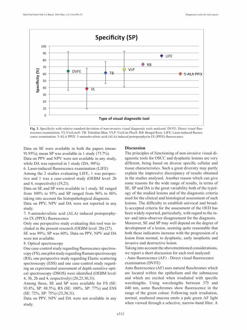
Data on SE were available in both the papers (mean: 91.95%); mean SP was available in 1 study (73.7%). Data on PPV and NPV were not available in any study, while DA was reported in 1 study (DA: 90%).6. Laser-induced fluorescence examination (LIFE)Among the 2 studies evaluating LIFE, 1 was perspec-tive and 1 was a case-control study (OEBM level: 2b and 4, respectively) (19,21). Data on SE and SP were available in 1 study. SE ranged from 100% to 95% and SP ranged from 96% to 86% taking into account the histopathological diagnosis.Data on PPV, NPV and DA were not reported in any study.7. 5-aminolevulinic acid (ALA) induced protoporphy-rin IX (PPIX) fluorescenceOnly one perspective study evaluating this tool was in-cluded in the present research (OEBM level: 2b) (27).SE was 99%; SP was 60%. Data on PPV, NPV and DA were not available.8. Optical spectroscopyOne case-control study regarding fluorescence spectros-copy (FS), one pilot study regarding Raman spectroscopy (RS), one perspective study regarding Elastic scattering spectroscopy (ESS) and one case-control study regard-ing an experimental assessment of depth-sensitive opti-cal spectroscopy (DSOS) were identified (OEBM level: 4, 3b, 2b and 4, respectively) (20,25,30,31).Among these, SE and SP were available for FS (SE: 93.8%, SP: 88.5%), RS (SE: 100%, SP: 77%) and ESS (SE: 72%, SP: 75%) (25,30,31).Data on PPV, NPV and DA were not available in any study.
DiscussionThe principles of functioning of non-invasive visual di-agnostic tools for OSCC and dysplastic lesions are very different, being based on diverse specific cellular and tissue characteristics. Such a great diversity may partly explain the impressive discrepancy of results obtained in the studies analysed. Another reason which can give some reasons for the wide range of results, in terms of SE, SP and DA is the great variability both of the typol-ogy of the studied lesions and of the diagnostic criteria used for the clinical and histological assessment of such lesions. The difficulty to establish univocal and broad-ly-accepted criteria for the assessment of the OED has been widely reported, particularly, with regard to the in-ter- and intra-observer disagreement for the diagnosis.Moreover, SE and SP may well depend on the degree of development of a lesion, seeming quite reasonable that both these indicators increase with the progression of a lesion from normal, to dysplastic, early neoplastic and invasive and destructive lesion.Taking into account the abovementioned considerations, we report a short discussion for each tool analysed:- Auto fluorescence (AF) - Direct visual fluorescence examination (DVFE)Auto fluorescence (AF) uses natural fluochromes which are located within the epithelium and the submucosa and which are excited when irradiated with specific wavelengths. Using wavelengths between 375 and 440 nm, some fluochromes show fluorescence in the range of the green colour. Following such irradiation, normal, unaltered mucosa emits a pale green AF light when viewed through a selective, narrow-band filter. A
Fig. 3. Specificity with relative standard deviation of non-invasive visual diagnostic tools analysed. DVFE: Direct visual fluo-rescence examination. VL:ViziLite®. TB: Toluidine Blue. VLP: ViziLite Plus®. RB: Bengal Rose. LIFE: Laser-induced fluores-cence examination. 5-ALA PPIX: 5-aminolevulinic acid (ALA) induced protoporphyrin IX (PPIX) fluorescence.
Med Oral Patol Oral Cir Bucal. 2016 May 1;21 (3):e305-15. Diagnostic tools for oral cancer
e313
proper filtration is crucial, due to the intense light used for excitation of the fluorochromes (13,15). Areas of reduced AF (dark areas) are suspicious for epithelial dysplasia or OSCC, whereas normal mucosa appears bright green (10).The VELscopeTM (LED Medical Diagnostics Inc., Barnaby, Canada) system consists of a non-invasive de-vice designed to visualise early mucosal changes using the principles of tissue AF. According to such principles, dysplastic changes should be associated with a loss of stromal AF (29,32). It seems of paramount importance to highlight here that benign lesions, or those associated to inflammation, can also be characterized by a loss of stromal AF, which grossly limits the diagnostic specifi-city, especially in low-risk populations.Mean SE and SP for this tool, were 72.4% and 63.79%, respectively. It is opinion of the authors that such values, at the moment, are unacceptable for a tool specifically dedicated to the diagnosis of oral mucosal malignant le-sions. However, it should be stressed that there are ap-parently no other non-invasive visual diagnostic tools significantly better than AF-based tools. It is somewhat surprising that values of SE range from 20% to 100% and value of SP goes from 15.3% to 100%. Level of EBM for the selected studies seem to be quite acceptable, being ≥ 2b for all the studies, except one (3b level) (24). It is worthy mentioning that the study with the highest EBM level (1b) showed high values both of SE and SP (100% and 74%, respectively) (33).- Chemiluminescence (CL)The ViziLite® (VL - Zila Pharmaceuticals, Phoenix, AZ) was the first FDA-approved (2002) adjunctive tech-nology to conventional head and neck examination for improving visualization of early dysplastic or neoplas-tic lesions. This system involves an oral rinse with a 1% acetic acid solution for 1 minute, to remove the glyco-protein barrier and slightly desiccate the oral mucosa. A diffuse chemiluminescent blue/white light with an aver-age wavelength of 490 to 510 nm is then activated and used to examine the oral tissues. Normal cells absorb the light and appear blue, whereas abnormal cells have a higher nuclear/cytoplasmic ratio and should reflect the light appearing whiter with brighter, sharper, more dis-tinct margins (15,36,38).Mean SE and SP resulted 86.72% and 38.37%, respec-tively. All the analysed studies have an EBM level of 2b, but there is a great inhomogeneity especially for SP, which ranges from 14.2% to 81.5%. - Toluidine Blue (TB)Toluidine blue (TB), also known by its chemical name tolunium chloride (TC), is a cationic met achromatic dye that may selectively bind to free anionic groups such as sulfate, phosphate, and carboxyl ate radicals of large molecules. It has been used for decades as aid to
the identification of mucosal abnormalities of the cervix as well as those in the oral cavity (8). TB stains deoxyribonucleic acid and/or may be retained in intracellular spaces of dysplastic epithelium, which clinically appears as royal blue areas. It is postulated that the increased amount of DNA and RNA in neoplas-tic cells and the wider intercellular canals compared to normal epithelial cells are responsible for staining ma-lignant cells (11).Mean SE and SP were 72.5% and 61.4%, respectively. These values are poorly acceptable in oncologic diagno-sis and they seem to be more realistic because standard deviations are lower than those calculated for the other diagnostic tools. - Chemiluminescence associated with Toluidine Blue (CLTB)In order to reduce the high number of false positive cases obtained through VL, the manufacturer added TB (ViziLite Plus® - VLP) (12,22).Data related to the use of this technique are very poor and discordant; mean SE and SP were 53.93% and 66.44%, respectively, but standard deviations were ex-cessively high.- Rose Bengal (RB)Rose Bengal (RB) is the 4,5,6,7-tetrachloro- 2’,4’,5’,7’-tetraiodo-derivative of fluorescein. It has been widely used to diagnose various ocular surface disorders. It has been believed to stain desquamated ocular epithelial cells, dead or degenerated cells but not healthy epithelial cells. RB staining was even used to delineate the extent of corneal and conjunctival neoplasms. Therefore, such findings of RB enlightened us to carry out researches in detection of oral precancerous and malignant lesions (9,23).Data on SE and SP related to this tool are scarce and resulting from studies of low OEBM level (3b). - Laser-induced fluorescence examination (LIFE)This technique is based on AF of the tissue as well as DVFE. The instrumentation proposed by Mallia et al. is comprised of a diode laser (Stocker Yale, Canada, 404 nm, 50 mW, CW) for excitation of tissue fluorophores (21). Light emission from the laser source is guided to the oral mucosa through a 3 µm long bifurcated fiber optic probe that has a central fiber to deliver the excita-tion beam and 6 surrounding fibers (400 µm diameter each) to collect AF emissions. The red to green colour ratio is defined as the numerical color value (NCV).Two studies regarding this tool have been selected for this review. SE and SP values are reported in 1 study only and they are high (SE: 100%-95%; SP: 96%-86%, according to the histopathological diagnosis), but the OEBM level is low (4). Data from further studies with a higher OEBM level are necessary.- 5-aminolevulinic acid (ALA) induced protoporphyrin IX (PPIX) fluorescence

Med Oral Patol Oral Cir Bucal. 2016 May 1;21 (3):e304-15. Diagnostic tools for oral cancer
e314
Only one perspective study describing this technique was selected for this review (29). Topical or systemic administration of 5-ALA results in a selective accumu-lation of PPIX in neoplastic tissue, which is probably due to altered activity levels of the enzymes of the heme biosynthetic pathway within malignant transformed cells. In the protocol of Leuing et al., the patients per-formed a 15-minute continuous rinsing of the oral cav-ity using the 5-ALA solution. After an incubation pe-riod of 1 to 2.5 hours (maximum contrast after 1.5h), fluorescence investigation was performed. In 13.8% of the patients, additional findings like dysplasia, carci-noma in situ, OSCC were found through fluorescence in contrast to COE (27). An evaluation of the biopsy specimens resulted in a SP of 60% and a SE of 99% (29). ALA-induced fluorescence could represent a possible useful new diagnostic tool to detect early malignant le-sions in the oral cavity. However, further studies seem to be necessary.- Optical spectroscopyOptical spectroscopy is a non-invasive diagnostic method that has been investigated in many forms in-cluding fluorescence spectroscopy (FS), elastic or dif-fuse scattering spectroscopy (ESS), and Raman spec-troscopy (RS). Spectroscopic measurements can detect biochemical and architectural alterations in tissue that are related to the carcinogenesis. These alterations may include changes in the concentrations of native fluoro-phores such as collagen, elastin, keratin, nicotinamide adenine dinucleotide (NADH), and flavin adenine di-nucleotide (FAD); changes in hemoglobin concentra-tion and oxygenation; increasing epithelial thickness; increasing nuclear size and nuclear/cytoplasmic ratio; change in vascularization (20,25,30,31).These principles are employed within experimental methods; SE and SP values seem to be high, but there is need for more data. Only one study for each optical spectroscopy method was identified and they had a low OEBM level (4, 3b and 2b, respectively).
References1. Dissanayaka WL, Pitiyage G, Kumarasiri PV, Liyanage RL, Dias KD, Tilakaratne WM. Clinical and histopathologic parameters in survival of oral squamous cell carcinoma. Oral Surg Oral Med Oral Pathol Oral Radiol. 2012;113:518-25.2. Sciubba JJ. Oral cancer. The importance of early diagnosis and treatment. Am J Clin Dermatol. 2001;2:239-51.3. Fedele S. Diagnostic aids in the screening of oral cancer. Head Neck Oncol. 2009;1:5.4. Navone R, Burlo P, Pich A, Pentenero M, Broccoletti R, Mar-sico A, et al. The impact of liquid-based oral cytology on the di-agnosis of oral squamous dysplasia and carcinoma. Cytopathology. 2007;18:356-60.5. Mehrotra R, Gupta DK. Exciting new advances in oral cancer di-agnosis: avenues to early detection. Head Neck Oncol. 2011;3:33.
6. Petruzzi M, Lucchese A, Nardi GM, Lauritano D, Favia G, Ser-pico R, et al. Evaluation of autofluorescence and toluidine blue in the differentiation of oral dysplastic and neoplastic lesions from non dysplastic and neoplastic lesions: a cross-sectional study. J Biomed Opt. 2014;19:76003. 7. Kämmerer PW, Rahimi-Nedjat RK, Ziebart T, Bemsch A, Walter C, Al-Nawas B, et al. A chemiluminescent light system in combination with toluidine blue to assess suspicious oral lesions-clinical evalua-tion and review of the literature. Clin Oral Investig. 2015;19:459-66. 8. Pallagatti S, Sheikh S, Aggarwal A, Gupta D, Singh R, Handa R, et al. Toluidine blue staining as an adjunctive tool for early di-agnosis of dysplastic changes in the oral mucosa. J Clin Exp Dent. 2013;5:e187-91.9. Mittal N, Palaskar S, Shankari M. Rose Bengal staining - diag-nostic aid for potentially malignant and malignant disorders: a pilot study. Indian J Dent Res. 2012;23:561-4. 10. McNamara KK, Martin BD, Evans EW, Kalmar JR. The role of direct visual fluorescent examination (VELscope) in routine screen-ing for potentially malignant oral mucosal lesions. Oral Surg Oral Med Oral Pathol Oral Radiol. 2012;114:636-43. 11. Awan Kh, Yang Y, Morgan P, Warnakulasuriya S. Utility of toluidine blue as a diagnostic adjunct in the detection of potentially malignant disorders of the oral cavity--a clinical and histological as-sessment. Oral Dis. 2012;18:728-33.12. Mojsa I, Kaczmarzyk T, Zaleska M, Stypulkowska J, Zapala-Pos-piech A, Sadecki D. Value of the ViziLite Plus System as a diagnostic aid in the early detection of oral cancer/premalignant epithelial le-sions. J Craniofac Surg. 2012;23:e162-4. 13. Farah CS, McIntosh L, Georgiou A, McCullough MJ. Efficacy of tissue autofluorescence imaging (VELScope) in the visualization of oral mucosal lesions. Head Neck. 2012;34:856-62.14. Paderni C, Compilato D, Carinci F, Nardi G, Rodolico V, Lo Muzio L, et al. Direct visualization of oral-cavity tissue fluorescence as novel aid for early oral cancer diagnosis and potentially malignant disorders monitoring. Int J Immunopathol Pharmacol. 2011;24(2 Suppl):121-8. 15. Awan KH, Morgan PR, Warnakulasuriya S. Utility of chemilumi-nescence (ViziLite™) in the detection of oral potentially malignant disorders and benign keratoses. J Oral Pathol Med. 2011;40:541-4.16. Awan KH, Morgan PR, Warnakulasuriya S. Evaluation of an autofluorescence based imaging system (VELscope™) in the detec-tion of oral potentially malignant disorders and benign keratoses. Oral Oncol. 2011;47:274-7. 17. Moro A, Di Nardo F, Boniello R, Marianetti TM, Cervelli D, Gasparini G, et al. Autofluorescence and early detection of mu-cosal lesions in patients at risk for oral cancer. J Craniofac Surg. 2010;21:1899-903.18. Koch FP, Kaemmerer PW, Biesterfeld S, Kunkel M, Wagner W. Effectiveness of autofluorescence to identify suspicious oral lesions--a prospective, blinded clinical trial. Clin Oral Investig. 2011;15:975-82. 19. Sieroń A, Kościarz-Grzesiok A, Waśkowska J, Kawczyk-Krupka A, Misiak A, Koszowski R, et al. The role of autofluorescence diag-nostics in the oral mucosa diseases. Photodiagnosis Photodyn Ther. 2008;5:182-6. 20. Schwarz RA, Gao W, Redden Weber C, Kurachi C, Lee JJ, El-Naggar AK, et al. Noninvasive evaluation of oral lesions using depth-sensitive optical spectroscopy. Cancer. 2009;115:1669-79. 21. Mallia RJ, Thomas SS, Mathews A, Kumar R, Sebastian P, Madhavan J, et al. Laser-induced autofluorescence spectral ratio reference standard for early discrimination of oral cancer. Cancer. 2008;112:1503-12. 22. Epstein JB, Silverman S Jr, Epstein JD, Lonky SA, Bride MA. Analysis of oral lesion biopsies identified and evaluated by visual examination, chemiluminescence and toluidine blue. Oral Oncol. 2008;44:538-44.

Med Oral Patol Oral Cir Bucal. 2016 May 1;21 (3):e305-15. Diagnostic tools for oral cancer
e315
23. Du GF, Li CZ, Chen HZ, Chen XM, Xiao Q, Cao ZG, et al. Rose bengal staining in detection of oral precancerous and malig-nant lesions with colorimetric evaluation: a pilot study. Int J Cancer. 2007;120:1958-63. 24. Lane PM, Gilhuly T, Whitehead P, Zeng H, Poh CF, Ng S, et al. Simple device for the direct visualization of oral-cavity tissue fluo-rescence. J Biomed Opt. 2006;11:024006. 25. Sharwani A, Jerjes W, Salih V, Swinson B, Bigio IJ, El-Maaytah M, et al. Assessment of oral premalignancy using elastic scattering spectroscopy. Oral Oncol. 2006;42:343-9.26. Onofre MA, Sposto MR, Navarro CM. Reliability of toluidine blue application in the detection of oral epithelial dysplasia and in situ and invasive squamous cell carcinomas. Oral Surg Oral Med Oral Pathol Oral Radiol Endod. 2001;91:535-40. 27. Leunig A, Betz CS, Mehlmann M, Stepp H, Arbogast S, Grevers G, et al. Detection of squamous cell carcinoma of the oral cavity by imaging 5-aminolevulinic acid-induced protoporphyrin IX fluores-cence. Laryngoscope. 2000;110:78-83.28. Martin IC, Kerawala CJ, Reed M. The application of toluidine blue as a diagnostic adjunct in the detection of epithelial dysplasia. Oral Surg Oral Med Oral Pathol Oral Radiol Endod. 1998;85:444-6.29. Bhatia N, Matias MA, Farah CS. Assessment of a decision mak-ing protocol to improve the efficacy of VELscope™ in general dental practice: a prospective evaluation. Oral Oncol. 2014;50:1012-9.30. Francisco AL, Correr WR, Azevedo LH, Kern VG, Pinto CA, Kowalski LP, et al. Fluorescence spectroscopy for the detection of potentially malignant disorders and squamous cell carcinoma of the oral cavity. Photodiagnosis Photodyn Ther. 2014;11:82-90.31. Guze K, Pawluk HC, Short M, Zeng H, Lorch J, Norris C, et al. Pilot study: Raman spectroscopy in differentiating premalignant and malignant oral lesions from normal mucosa and benign lesions in humans. Head Neck. 2015;37:511-7. 32. Hanken H, Kraatz J, Smeets R, Heiland M, Assaf AT, Blessmann M, et al. The detection of oral pre- malignant lesions with an autoflu-orescence based imaging system (VELscope™) - a single blinded clinical evaluation. Head Face Med. 2013;9:23.33. Rana M, Zapf A, Kuehle M, Gellrich NC, Eckardt AM. Clinical evaluation of an autofluorescence diagnostic device for oral cancer detection: a prospective randomized diagnostic study. Eur J Cancer Prev. 2012;21:460-6.34. Cancela-Rodríguez P, Cerero-Lapiedra R, Esparza-Gómez G, Llamas-Martínez S, Warnakulasuriya S. The use of toluidine blue in the detection of pre-malignant and malignant oral lesions. J Oral Pathol Med. 2011;40:300-4. 35. Mehrotra R, Singh M, Thomas S, Nair P, Pandya S, Nigam NS, et al. A cross-sectional study evaluating chemiluminescence and autofluorescence in the detection of clinically innocuous precancer-ous and cancerous oral lesions. J Am Dent Assoc. 2010;141:151-6.36. Oh ES, Laskin DM. Efficacy of the ViziLite system in the identi-fication of oral lesions.J Oral Maxillofac Surg. 2007;65:424-6. 37. Chen YW, Lin JS, Fong JH, Wang IK, Chou SJ, Wu CH, et al. Use of methylene blue as a diagnostic aid in early detection of oral cancer and precancerous lesions. Br J Oral Maxillofac Surg. 2007;45:590-1.38. Ram S, Siar CH. Chemiluminescence as a diagnostic aid in the detection of oral cancer and potentially malignant epithelial lesions. Int J Oral Maxillofac Surg. 2005;34:521-7.39. Warnakulasuriya KA, Johnson NW. Sensitivity and specificity of OraScan (R) toluidine blue mouthrinse in the detection of oral cancer and precancer. J Oral Pathol Med. 1996;25:97-103.
Conflict of InterestNo conflict of interestNo financial disclosures are present for any Authors.