Embed Size (px)
Citation preview

Nodule inception NIN plays a dual role in
the establishment of root nodule symbiosis
in Lotus japonicus
vorgelegt von
Jayne Carol Lambert
München, Oktober 2016
Dissertation zur Erlangung des Doktorgrades
der Naturwissenschaften an der Fakultät für Biologie
der Ludwig-Maximilians-Universität München

2

3
“It always seems impossible until it’s done.”
Nelson Mandela
Dissertation eingereicht am 27. Oktober 2016
Tag der mündlichen Prüfung: 19. Januar 2017
Erstgutachter: Prof. Dr. Martin Parniske
Zweitgutachterin: PD Dr. Cordelia Bolle

4

5
For my family

6

7
Table of contents
1 List of Publications ............................................................................................................. 13
2 List of presentations at scientific conferences ................................................................ 13
3 Abstract ................................................................................................................................ 15
4 Zusammenfassung .............................................................................................................. 17
5 Introduction......................................................................................................................... 19
5.1 The Root Nodule Symbiosis and Arbuscular Mycchorizha Symbiosis ................. 19
5.2 Signal perception and symbiotic calcium signalling ................................................. 20
5.3 Decoding of the calcium signal .................................................................................. 23
5.4 The symbiotic transcriptional network ...................................................................... 25
5.5 NIN - a transcription factor ........................................................................................ 26
5.6 Transcriptional targets of NIN ................................................................................... 27
5.7 NIN domain structure ................................................................................................. 28
5.8 NIN like proteins (NLPs) ........................................................................................... 29
6 Aim of the thesis ................................................................................................................. 31
7 Results .................................................................................................................................. 33
7.1 NIN accumulates in the nucleus ................................................................................ 33
7.2 NIN interacts with CYCLOPS in the nucleus ......................................................... 34
7.2.1 Yeast two-hybrid analysis ..................................................................................... 34
7.2.2 Bimolecular fluorescence complementation (BIFC) ........................................ 35
7.2.3 FLIM–FRET approach ........................................................................................ 37
7.3 NIN inhibits CYCLOPS-DD mediated pNIN activation in N.benthamiana leaf
cells 42
7.4 Development of a Golden Gate cloning system to facilitate co-expression of
different genes in Lotus japonicus ............................................................................................ 44

8
7.5 NIN inhibits CYCLOPS-DD mediated pNIN activation in roots of L. japonicus 45
7.6 NIN inhibits CYCLOPS-DD mediated pNIN activation at the previously
identified CYCLOPS-responsive element ........................................................................... 47
7.7 NIN binds to its own promoter, located in close vicinity to the CYC-box, in a
sequence-specific manner ....................................................................................................... 50
7.7.1 Competition of NIN and CYCLOPS for NIN promoter regulatory elements
53
7.7.2 Co-binding of NIN and CYCLOPS to the NIN promoter regulatory
element 55
7.8 Overexpression of NIN leads to local inhibition of nodulation and infection .... 56
7.9 nin-2 and -8 mutants cannot be complemented by overexpression of NIN ........ 61
7.10 The negative regulatory effect on nodulation caused by overexpression of NIN
is independent of autoregulation of nodulation .................................................................. 63
7.11 Inhibition of nodulation caused by NIN overexpression is downstream of the
common symbiosis pathway .................................................................................................. 65
7.12 NIN overexpression has no effect on the infection with the arbuscular
mycorrhiza fungus Rhizophagus irregularis .............................................................................. 71
7.13 Overexpression of NIN triggers the process of cell division ............................. 73
8 Discussion ............................................................................................................................ 77
8.1 NIN – CYCLOPS – CCaMK: two dimers or a trimer? .......................................... 77
8.2 Dual Role of NIN in symbiosis: Activator and Repressor ..................................... 79
8.3 The NIN promoter - a target for different transcription factors ........................... 82
8.4 Inhibition of nodulation: local or systemic repression caused by NIN
overexpression ......................................................................................................................... 85
8.5 Involvement of NIN in nodule development by inducing cell division ............... 88
8.6 A model for the negative feedback loop caused by NIN ....................................... 89
9 Materials and Methods ....................................................................................................... 93
9.1 Materials ......................................................................................................................... 93

9
9.2 Solutions ........................................................................................................................ 93
9.3 Media .............................................................................................................................. 94
9.4 Chemicals ....................................................................................................................... 95
9.5 Antibodies ..................................................................................................................... 96
9.6 Plant Material ................................................................................................................ 96
9.7 Bacterial Strains ............................................................................................................. 97
9.8 Oligonucleotides ........................................................................................................... 98
EMSA probes and competitors (5´-3´) ................................................................................ 98
9.9 Plasmid construction .................................................................................................... 99
10 Methods ............................................................................................................................. 101
10.1 Molecular biology methods ................................................................................... 101
10.1.1 Polymerase-chain reaction ............................................................................. 101
10.1.2 Electrophoresis of DNA ................................................................................ 102
10.1.3 Extraction of PCR products from agarose gels/PCR clean-up ................ 102
10.1.4 Plasmid isolation and estimation of DNA concentration ......................... 103
10.1.5 Restriction endonuclease digestion of DNA ............................................... 103
10.2 Cloning and Vector construction of NIN ........................................................... 103
10.2.1 Golden gate cloning (cut-ligation reaction) ................................................. 104
10.2.2 Cloning: LR-reaction ...................................................................................... 105
10.2.3 TOPO Cloning ................................................................................................ 105
10.2.4 Transformation of E. coli cells (heat shock method) .................................. 106
10.2.5 Transformation of Agrobacterium cells (Electroporation method) ............ 106
10.2.6 Sequencing ....................................................................................................... 107
10.3 Cultivation methods ............................................................................................... 107
10.3.1 Bacterial growth conditions ........................................................................... 107
10.4 Plant cultivation ...................................................................................................... 107

10
10.4.1 N. benthamiana transformation ....................................................................... 107
10.4.2 Plant germination and growth ....................................................................... 108
10.4.3 Lotus hairy root Transformation .................................................................. 108
10.4.4 Nodulation assay ............................................................................................. 109
10.4.5 AM Assay ......................................................................................................... 109
10.4.6 Nodule sectioning ........................................................................................... 109
10.5 Biochemical methods ............................................................................................. 110
10.5.1 Histochemical GUS Staining ......................................................................... 110
10.5.2 Fluorimetric GUS assay ................................................................................. 110
10.5.1 Protein extraction from N. benthamina leaves .............................................. 110
10.5.2 SDS PAGE ...................................................................................................... 111
10.5.3 Western blot..................................................................................................... 111
10.5.4 Protein expression and purification .............................................................. 111
10.5.5 Electrophoretic mobility shift assay ............................................................. 112
10.5.6 Bradford assay for protein standard curve and protein concentration
measurement ..................................................................................................................... 112
10.5.7 Yeast two-hybrid ............................................................................................. 112
10.6 Microscopy .............................................................................................................. 113
10.6.1 FLIM-FRET .................................................................................................... 114
10.6.2 Statistics ............................................................................................................ 114
11 List of Figures ................................................................................................................... 115
12 List of Tables ..................................................................................................................... 116
13 List of Abbreviations ........................................................................................................ 117
14 Declaration of contribution of other researches .......................................................... 121
15 References .......................................................................................................................... 123
16 Acknowledgement ............................................................................................................ 137

11
17 Curriculum Vitae ............................................................................................................... 139
18 Eidesstattliche Versicherung ........................................................................................... 141
19 Erklärung ........................................................................................................................... 141

12

13
1 List of Publications
Binder, A., Lambert, J., Morbitzer, R., Popp, C., Ott, T., Lahaye, T., and Parniske, M.
(2014). A modular plasmid assembly kit for multigene expression, gene silencing and
silencing rescue in plants. PLoS ONE 9, e88218.
Singh S, Katzer K, Lambert J, Cerri M, Parniske M. CYCLOPS, a DNA-binding
transcriptional activator, orchestrates symbiotic root nodule development, Cell Host
Microbe. 2014 Feb 12;15(2):139-52.
Manuscript:
Lambert J, Rosa Andrade, Katzer K and Parniske M. NIN plays a dual role in the
establishment of the root nodule symbiosis in Lotus japonicus, in preparation
2 List of presentations at scientific conferences
Poster presentation at the ENFC 2014 in Teneriffe, “Lotus japonicus NIN plays a dual role
in the establishment of the root nodule symbiosis”, awarded with 1st Posterprize
Poster presentation at the ENFC 2012 in Munich
Poster presentation at the Botaniker-Tagung 2011 in Berlin
Poster presentation at the Plant Calcium Signaling Meeting 2010 in Münster

14

15
3 Abstract
The symbiosis between legume plants and nitrogen-fixing rhizobia leads to the formation
of a novel organ, the root nodule. Bacterial secreted lipo-chitooligosaccharides, so-called
nodulation factors (NF) are recognized at the surface of plant root hairs, leading to the
initiation of calcium spiking in the nucleus. These calcium oscillations are decoded by a
complex comprising CCaMK, a calcium and calmodulin dependent kinase, and the
transcription factor CYCLOPS, a nuclear-coiled-coil protein. CCaMK phosphorylates
CYCLOPS at two serines (S50 and S154) essential for symbiosis. Activated CYCLOPS
(CYCLOPS-DD) binds to the NIN promoter in a sequence specific manner and in turn
drives its expression. NIN encodes a transcription factor with a highly conserved RWP-
RK DNA-binding domain. Homologous NIN-like proteins control nitrate signalling in
presumably all land plants. Since its discovery, NIN has been considered a positive
regulator in nodulation based on its rapid upregulation after NF perception and its
requirement for root hair infection and the initiation of cortical cell division.
Nevertheless, a negative role of NIN in the process of infection thread formation was
recently described, raising the question how NIN can play a bifunctional role in infection
and nodule organogenesis. In this study, we demonstrated that NIN and CYCLOPS
interact in yeast and in the nucleus in planta. Furthermore, the co-expression of NIN,
CYCLOPS and CCaMK was investigated and the results suggest the formation of a
trimeric complex. Moreover, a negative regulatory role of NIN on the CYCLOPS-DD
mediated activation was observed in transactivation assays using the NIN promoter. In
addition, NIN can efficiently bind to a cis-element of the NIN promoter in close vicinity
of the CYCLOPS binding site in electrophoretic mobility shift assays (EMSA). Strikingly,
both proteins can bind simultaneously to the NIN promoter, resulting in a supershift in
EMSA. Lastly, overexpression of NIN in L. japonicus Gifu wild-type plants and different
nodulation mutants led to an inhibition of nodulation independent of the autoregulation
of nodulation process. In our model NIN regulates its own expression via a negative
feedback loop by binding to the NIN promoter and through protein-protein interaction
between NIN and CYCLOPS thus preventing further activation of the CYCLOPS-
mediated signalling pathway and restricting further infection by rhizobia.

16

17
4 Zusammenfassung
Die Symbiose zwischen Leguminosen und Stickstoff-fixierenden Rhizobien führt zur
Bildung eines neuen Organs, dem Wurzelknöllchen. Bakterielle sekretierte Lipo-
chitooligosaccharide, sogenannte Nodulationsfaktoren (NF), werden an der Oberfläche
von Pflanzenwurzelhärchen erkannt und führen zu symbiotischem „Calcium spiking“ im
Kern. Diese Kalziumoszillationen werden durch einen Komplex dekodiert, der aus
CCaMK, einer Calcium- und Calmodulin- abhängigen Kinase und dem
Transkriptionsfaktor CYCLOPS, einem Zellkern-lokalisierten coiled-coil Protein besteht.
CCaMK phosphoryliert CYCLOPS an zwei Serinen (S50 und S154), die von wesentlicher
Bedeutung für die Symbiose sind. Aktiviertes CYCLOPS (CYCLOPS-DD) bindet
sequenzspezifisch an den NIN Promotor und induziert anschließend die NIN
Expression. NIN kodiert für einen Transkriptionsfaktor mit einer hochkonservierten
„RWP-RK“ DNA-Bindungsdomäne. Homologe NIN-ähnliche Proteine steuern die
Signalweiterleitung in der Nitratassimilation in vermutlich allen Landpflanzen. Seit seiner
Entdeckung galt NIN als ein positiver Regulator in der Knöllchensymbiose, basierend auf
der schnellen Hochregulierung der NIN Genexpression nach NF Wahrnehmung und der
Notwendigkeit von NIN für die Wurzelhaar-Infektion und für die Einleitung der
kortikalen Zellteilung. Für NIN wurde jedoch kürzlich auch eine negative regulatorische
Funktion beim Prozess der Infektionsschlauchbildung demonstriert, was die Frage
aufwirft, wie NIN eine bi-funktionelle Rolle bei Infektion und Knöllchenbildung spielen
kann.
In dieser Studie haben wir gezeigt, dass NIN und CYCLOPS im Zellkern von Pflanzen
und in Hefe miteinander interagieren können. Darüber hinaus wurde die Co-Expression
von NIN, CYCLOPS und CCaMK untersucht und die Ergebnisse zeigen, dass diese
Proteine einen trimeren Komplex bilden. Zudem wurde eine negative regulatorische Rolle
von NIN auf die von CYCLOPS-DD vermittelte Aktivierung in Transaktivierungsassays
unter Verwendung des NIN Promotors beobachtet. Außerdem konnte in Electrophoretic
Mobility Shift Assays (EMSAs) gezeigt werden, dass NIN effizient an ein cis-Element des
eigenen Promotors binden kann, welches sich in unmittelbarer Nähe der CYCLOPS-
Bindungsstelle befindet.

18
Beide Proteine können gleichzeitig nebeneinander an den NIN Promotor binden, was zu
einem Supershift im EMSA führte. Schließlich kommt es durch die Überexpression von
NIN zu einer Hemmung der Knöllchenbildung in Gifu Wildtyp Pflanzen und
verschiedenen nodulierenden Mutanten, die unabhängig vom Prozess der Autoregulation
der Nodulation reguliert ist. In unserem Modell reguliert NIN seine eigene Expression
durch eine negative Feedback-Schleife, indem es an den NIN Promotor bindet und durch
Protein-Protein Interaktion von NIN und CYCLOPS eine weitere Aktivierung des durch
CYCLOPS vermittelten Signalwegs verhindert und dadurch für eine Einschränkung
weiterer Infektionen durch Rhizobien sorgt.

19
5 Introduction
5.1 The Root Nodule Symbiosis and Arbuscular Mycchorizha
Symbiosis
The symbiosis between legume plants and nitrogen-fixing soil bacteria (rhizobia) is a
beneficial interaction for both partners (Den Herder and Parniske, 2009). Rhizobia live in
a newly formed plant organ, named nodule (Brewin et al., 1999; Oldroyd and Downie,
2008; Oldroyd et al., 2011). In this environment, rhizobia fix atmospheric nitrogen under
oxygen-restricted conditions and provide the plant with ammonium, leading to an
improved survival of the plant in nitrogen-poor soil (Gage, 2004). In exchange the
microsymbiont receives carbon sources (including dicarboxylates and amino acids)
deriving from photosynthesis and mineral nutrients (iron, molybdenum, sulphur and
others) from the plant.
Microbial partners efficiently engage in root nodule symbiosis (RNS) with various
flowering plant species, which belong to the Eurosid I clade, specifically those from the
four orders of Fagales, Fabales, Cucubitales and Rosales, also referred to as nodulating
clade (Kistner and Parniske, 2002; Cermak et al., 2011). RNS with rhizobia is restricted to
legumes (Fabaceae), with one exception for the non-legume Parasponia (Op den Camp et
al., 2012; Remigi et al., 2016). There is, however, another RNS between filamentous
actinomycetes of the genus Frankia (Actinobacteria) and so-called “actinorhizal” plants
(e.g. Datisca and Casuarina) (Li et al., 2015; Pawlowski and Sprent, 2008).
In all RNSes, several symbiotic genes were co-opted from the signalling pathway of the
more ancient association between plants and arbuscular mycorrhizal fungi (Delaux et al.,
2013; Delaux et al., 2015). About 80% of all land plants are able to establish the
arbuscular mycorrhizal symbiosis (AMS) with fungi from the phylum Glomeromycota
(Schüβler et al., 2001; Parniske, 2008). The plant receives phosphorus and other inorganic
nutrients from the fungus in exchange for carbon sources (Harrison, 1999; Parniske,
2008). In this association the fungus is able to colonize the plant by invasion of the root
and forms highly branched structures in the cortex, named arbuscules (Gutjahr and
Parniske, 2013).

20
Both symbioses share a common set of genes (common symbiosis genes), which are
required for the initiation of the signalling cascade within the plant (Kistner and Parniske,
2002).
5.2 Signal perception and symbiotic calcium signalling
Under nitrogen limiting conditions, leguminous plants secret flavonoids that attract
compatible symbiosis partners (Peters et al., 1986). In turn, bacteria respond with the
activation of nod genes, leading to the production and secretion of nodulation factors
(NF) into the soil (Fisher and Long, 1992). Variations of NFs exist among different
rhizobial species, which ensures the perfect match of interaction partners between host
plant and microsymbiont (Roche et al., 1991; Denarie et al., 1996). The chemical
compositions of NFs is a chitooligosaccharide backbone with four to five N-
actetylglucosamine residues with β-1,4 linkeages decorated with further species-specific
groups such as methyl, acetyl or sulphate groups (Denarie et al., 1996).
This first signal exchange initiates a complex signalling cascade to activate the common
signalling pathway leading to gene activation (Figure 1). In the past 30 years several genes
required for the two major subsequent events, bacterial infection and nodule formation,
have been identified and characterised (Oldroyd, 2013).
Firstly, NFs are recognised by the two Lysin Motif (LysM)–type receptors Nod Factor
Receptor 1 (NFR1) and NFR5, on the root hair plasma membrane (Madsen et al., 2003;
Radutoiu et al., 2003). NFR1 shows a typical domain structure of a receptor kinase, which
is an active intracellular kinase domain that is capable of auto-phosphorylation and trans-
phosphorylation of other proteins, including NFR5 (Radutoiu et al., 2003; Madsen et al.,
2011). In contrast to NFR1, NFR5 has a pseudokinase that lacks important motifes of a
functional kinase and is not able to phosphorylate other proteins (Madsen et al., 2003;
Madsen et al., 2011). Interestingly, the receptors feature an additional LysM domain in the
extra-cytoplasmic domain exclusively in plants, which has been shown to be crucial for
NF binding (Bateman and Bycroft, 2000; Zhang et al., 2007; Broghammer et al., 2012).
Both receptors localise to the plasma membrane and form a heterodimeric complex
(Madsen et al., 2011; Lefebvre et al., 2012; Pietraszewska-Bogiel et al., 2013).

21
Figure 1: symbiotic signal transduction in root nodule symbiosis
Nod factor (NF) is recognised and bound by the NF receptors NFR1 and NFR5 at the plasma membrane. SYMRK, a plasma membrane bound receptor-like kinase and SYMREM1, a remorin upregulated during symbiosis, are interacting with both NFRs. Calcium channels (CNGCs) are responsible for calcium release from the nuclear envelop. Three components of the nuclear pore complex NUP133, NUP85 and NENA as well as two ion-channels CASTOR and POLLUX are required for the generation of calcium spiking. The nuclear calcium and calmodulin-dependent kinase CCaMK is the decoder of these calcium oscillations. CCaMK and CYCLOPS form a complex and activate NIN expression. Figure modified from (Singh and Parniske, 2012)
SYMRK is a receptor-like kinase (RLK) that is indispensable for both RNS and AMS
(Endre et al., 2002; Stracke et al., 2002; Yoshida and Parniske, 2005; Markmann et al.,
2008; Kosuta et al., 2011; Antolin-Llovera et al., 2014; Ried et al., 2014). SYMRK consists
of an extra-cytoplasmic region including Leucin-rich repeats (LRRs), a malectin-like
domain and a functional intracellular protein kinase domain (Stracke et al., 2002; Yoshida
and Parniske, 2005; Kosuta et al., 2011; Antolin-Llovera et al., 2014).

22
An active role for SYMRK in the signalling pathway of root nodule symbiosis was
demonstrated with the overexpression in hairy root experiments, which lead to the
formation of spontaneous nodules in the absence of rhizobia (Ried et al., 2014). The
interaction of SYMRK and NFR5 places them at an early recognition point for RNS and
suggests a role of SYMRK as a co-receptor (Antolin-Llovera et al., 2014; Ried et al.,
2014).
Furthermore, the interaction between these receptors and a plasma membrane associated
remorin, SYMREM1, was demonstrated. Remorins function as scaffold proteins and are
assumed to be involved in the organization of microdomains and thus may facilitate
bacterial entry (Lefebvre et al., 2010; Jarsch and Ott, 2011; Toth et al., 2012). The
interplay of these receptors is the earliest recognition step between the symbiotic partners
to verify the specificity of the host and allow for colonisation.
Two of the earliest readouts in the plant after contact with bacteria are calcium influx at
the root-hair tip and nuclear calcium spiking (Kosuta et al., 2008; Chabaud et al., 2011;
Sieberer et al., 2012). The same response is triggered with purified NF extracted from
rhizobia (Kosuta et al., 2008; Chabaud et al., 2011; Sieberer et al., 2012). Recently, three
calcium channels have been identified, which encode cyclic nucleotide-gated channels
(CNGCs) and probably account for calcium release from the nuclear envelope
(Charpentier et al., 2016). In the nuclear envelope CASTOR and POLLUX, two cation
channels with a preference for K+ ions, are both required for the establishment of the
symbiosis in Lotus japonicus (Ané et al., 2004; Imaizumi-Anraku et al., 2005; Charpentier et
al., 2008). Moreover, proteins of the nuclear pore complex, NUP85, NUP133 and
NENA, are required for calcium spiking within the plant (Bonfante et al., 2000; Kistner et
al., 2005; Kanamori et al., 2006; Saito et al., 2007; Groth et al., 2010).
Binding of NFs to the receptors leads to a rapid response within the plant, including
calcium spiking, rearrangement of the cytoskeleton and the curling of responsive root
hairs in a typical “shepherd’s crook” structure (Jones et al., 2007; Broghammer et al.,
2012). The first event is an influx of calcium within one minute, actin rearrangement
happens within three to five minutes of NF perception (Weerasinghe et al., 2005),
followed by calcium spiking in the nucleus within 15 minutes and root hair deformation
within one to three hours (Ehrhardt et al., 1996; Miwa et al., 2006; Oldroyd, 2013).

23
Infection is initiated by the attachment of rhizobia to the growing root tip. Then, the
plant cell wall is modified to facilitate rhizobial uptake. The entrapped rhizobium divides
and multiplies to a microcolony in the “shepherd’s crock” curl structure, within an
infection pocket. Within 10 to 20 hours the full process of infection thread formation is
initiated (Fournier et al., 2015). Then, rhizobia enter the plant through a tube like
structure (infection thread) bound by the plant membrane and cell wall, which guides
them to the root cortex (Esseling et al., 2003; Geurts et al., 2005; Fournier et al., 2008;
Murray, 2011). Cortical cell division in close proximity to the infection thread leads to the
formation of a nodule primordium, where the bacteria are released into the cytoplasm of
nodule cells and then differentiate into nitrogen-fixing bacteroids (Timmers et al., 1999;
Bolanos et al., 2004). Genes required for cell-cycle activation and for hormone
biosynthesis are induced during the process of infection thread formation (Yang et al.,
1994; Breakspear et al., 2014).
5.3 Decoding of the calcium signal
In the nucleus there are several proteins that are responsible for inducing gene expression
downstream of calcium spiking. A calcium and calmodulin dependent serine/threonine
kinase (CCaMK) is considered to be the central player in the signalling transduction
pathway and the decoder of calcium spiking (Levy et al., 2004; Mitra et al., 2004; Tirichine
et al., 2006; Hayashi et al., 2010; Singh and Parniske, 2012).
CCaMK harbours a calmodulin binding domain (CaMBD), an active kinase domain and a
C-terminal visinin-like domain (VLD) with three calcium binding EF hands (Gleason et
al., 2006; Tirichine et al., 2006). Interestingly, CCaMK only exists in symbiotic plants
(Hrabak et al., 2003) and it becomes active after calcium binding to the EF hand, leading
to auto-phosphorylation of the kinase (Tirichine et al., 2006; Shimoda et al., 2012;
Swainsbury et al., 2012). Then, binding of CaM to the CaMBD results in a release of auto-
inhibition of the kinase, inducing a conformational change in the protein structure and
activating the kinase for transphosphorylation (Gleason et al., 2006; Shimoda et al., 2012;
Singh and Parniske, 2012).

24
The central role of CCaMK in symbiotic sinalling was demonstrated by the study of an
autoactive version of CCaMK that carries a replacement of threonine at position 265 by
aspartic acid or isoleucine (T265D or I). Plants that express this autoactive CCaMK form
spontaneous nodules in the absence of bacteria (Tirichine et al., 2006; Hayashi et al., 2010;
Madsen et al., 2010). Expression of the kinase alone also leads to spontaneous nodule
formation in ccamk plant mutants; however, infection is not restored (Gleason et al., 2006;
Shimoda et al., 2012; Takeda et al., 2012). Moreover, spontaneous nodulation can also be
triggered by expression of a gain-of-function version of CCaMK in a cyclops mutant
background, with CYCLOPS being a phosphorylation target of CCaMK (Yano et al.,
2008). The fact that an autoactive version of CCaMK is able to induce downstream
signalling, suggestes that the primary purpose for all upstream symbiotic genes is to
activate CCaMK (Gleason et al., 2006; Tirichine et al., 2006; Hayashi et al., 2010; Madsen
et al., 2010).
CCaMK forms a complex with CYCLOPS in the nucleus (Yano et al., 2008; Singh et al.,
2014). CYCLOPS is both required for RNS and AMS (Messinese et al., 2007; Yano et al.,
2008; Horvath et al., 2011; Ovchinnikova et al., 2011). Furthermore, the phosphorylation
of CYCLOPS via CCaMK leads to the activation of downstream transcription factors
(Singh et al., 2014). Two phosphorylation sites of the CYCLOPS protein are essential for
symbiosis, as replacements of S50D and S154D (CYCLOPS-DD) lead to an autoactive
version of CYCLOPS that is able to trigger the nodulation process without the symbiont
partner (Singh et al., 2014). In combination, CYCLOPS-WT and gain-of-function
CCaMKT265D are able to transactivate the NODULE INCEPTION (NIN) promoter in
Nicotiana benthamiana leaves and L. japonicus hairy roots, a gene required for bacterial entry
and nodule formation (Schauser et al., 1999; Singh et al., 2014). Moreover, the same effect
was achieved by only expressing CYCLOPS-DD, suggesting that the two
phosphorylation residues are required for NIN activation (Singh et al., 2014).
Furthermore, the CYCLOPS binding site was pinpointed to a cis-responsive element
“CYC” (CYC-RE) within the NIN promoter (Singh et al., 2014), demonstrating that
CYCLOPS confers both DNA-binding and transactivation abilities. Both functional
domains of CYCLOPS were further analysed, with the activation domain (AD) mapped
to a central part of the protein (aa 267 to 380), while the DNA binding domain (BD) was

25
delimited to the C-terminal region (aa 364 to 518), harbouring a coiled-coil domain (Singh
et al., 2014). A minimal version of CYCLOPS, consisting of only the AD and BD part of
the protein (CYC-min, aa 255 to 518), was demonstrated to be sufficient for mimicking
CYCLOPS-DD behaviour (Singh et al., 2014). The function of CYCLOPS as a
transcriptional activator is dependent on its phosphorylation by CCaMK at the
phosphorylation sites S50 and S154, which induces a structural change, releasing
CYCLOPS from auto-inhibition of the activation domain and promoting its potential for
DNA binding capability (Singh et al., 2014).
5.4 The symbiotic transcriptional network
Furthermore, several nuclear-associated transcriptional regulators, including
NODULATION SIGNALLING PATHWAY 1 and 2 (NSP1 and NSP2) are required
for the expression of downstream genes and initation of nodule development (Kaló et al.,
2005; Smit et al., 2005; Heckmann et al., 2006; Hirsch and Oldroyd, 2009). Both proteins
belong to the family of GRAS transcription factors that are conserved throughout the
plant kingdom and have diverse roles in plant development (Bolle, 2004). A function
during mycorrhorzial colonisation was only demonstrated for NSP2 (Liu et al., 2011).
Moreover, three Ethylene response factors (ERF) required for nodulation (ERN1, ERN2
and ERN3) have been shown to associate with the ENOD11 promoter, an early nodulin
gene encoding a repetitive proline-rich protein (Journet et al., 2001; Charron et al., 2004;
Andriankaja et al., 2007; Fournier et al., 2015).
Interestingly, ERN1 and ERN2 function as transcriptional activators, while ERN3 acts as
a repressor on the identified cis binding element (NF box) on the ENOD11 promoter
(Andriankaja et al., 2007). Expression profiles and cross-complementation studies
suggested a functional reduncancy (Cerri et al., 2012). This was recently corroborated by
ern2 single and ern1ern2 double mutant phenotypic characterisation. ERN1 and ERN2 are
acting together to regulate epidermal infection, but ERN1 seems to be the major factor
orchestrating infection in the cortex (Cerri et al., 2016).

26
NSP1 and NSP2 form a complex, which upregulates symbiosis-related marker-genes such
as ENOD11 or NIN. Only NSP1 demonstrated DNA-binding properties to the target
promoters (Murakami et al., 2006; Hirsch et al., 2009). NSP1 binds to NIN (-893 to -13
bp) on a cis regulatory element containing an AATTT motif (Hirsch et al., 2009). The
heterodimeric complex of NSP1 and NSP2 induces the expression of NIN and ERN1
(Marsh et al., 2007; Middleton et al., 2007; Cerri et al., 2012).
5.5 NIN - a transcription factor
In 1999, the first gene involved in RNS was identified, a putative transcription factor
named NODULE INCEPTION (NIN) (Schauser et al., 1999). A transposon-inactivated
nin was shown to be responsible for a defect in nodulation, while “revertants”, which had
lost the transposon, nodulated normally (Schauser et al., 1999).
NIN is involved in bacterial entry, cortical cell division and nodule formation (Schauser et
al., 1999; Borisov et al., 2003; Schauser et al., 2005). nin mutants show excessive root hair
curling in response to NF treatment, indicating that the NF signal is recognized, but
further signal trandcution is arrested (Schauser et al., 1999; Borisov et al., 2003; Marsh et
al., 2007). Recently, a role of NIN in remodelling the infection pocket was demonstrated
(Fournier et al., 2015).
The NIN cDNA sequence consists of 2,634 bp with a 3’ untranslated region of 140 bp,
coding for a protein of 878 aa (Schauser et al., 1999). The nucleotide identity is around
60% between Lotus japonicus and Pisum sativum NIN (LjNIN and PsNIN, respectively) and
the intron/exon structure is conserved (Borisov et al., 2003).
The open reading frame of PsNIN encodes a protein with 922 aa in comparison to 878 aa
in LjNIN (Borisov et al., 2003). The transcription of NIN was upregulated upon rhizobia
treatment after one day (8 fold) and at 10 days (Marsh et al., 2007; Yano et al., 2008),
suggesting a two-phase NIN response. The expression of PsNIN was localized in the
meristematic cells (zone I) and infection cells (zone II), whereas in the fixation zone (zone
III) no expression was detected (Borisov et al., 2003). In Lotus NIN expression was
shown to be dependent on NFR1, NFR5 and SYMRK (Radutoiu et al., 2003),
furthermore calcium spiking is normal in nin mutant (Miwa et al., 2006). Overexpression

27
of SYMRK, NFR1, NFR5 and gain-of-function versions of CCaMK or CYCLOPS
triggered spontaneous NIN expression in the plant (Ried et al., 2014; Singh et al., 2014).
5.6 Transcriptional targets of NIN
Several genes have been identified in the past years, which are dependent on NIN
expression and respond in a positive or negative regulatory way. 19 putative target genes
of NIN were identified in the transcriptome database and 9 candidates were verified by
real time (RT)-PCR (Soyano et al., 2013).
One transcriptional target is pectate lyase (NPL), a gene involved in infection thread
formation. For the formation of infection treads, cell walls are degraded by a pectate lyase
activity to break down polygalacturonic acid and pectin (methyl esterified PGA) via a
beta-elimination reaction (Xie et al., 2012). Direct regulation of NPL was demonstrated,
as NIN´s DNA binding domain was able to bind to the NPL promoter (Xie et al., 2012).
Other downstream regulation genes are NF-YA and NF-YB, which belong to a hetero-
trimeric complex with NF-YC (Soyano et al., 2013). NIN directly bound to the promoters
of NF-YA1 and NF-YB1 and upregulated their transcription (Soyano et al., 2013). The
heterodimeric NF-Y complex has been described to trigger the entry into the cell cycle
division (Laloum et al., 2013; Laloum et al., 2014).
Soyano and collegues could show that overexpression of NIN induces NF-YA and NF-
YB expression and in turn induces initiation of lateral root organs resembling nodule-like
structures (Soyano et al., 2013).
Although the nitrogen fixing bacteria inside a nodule are beneficial for the plant, a
negative feedback loop exists that prevents hypernodulation, as the bacteria require large
amounts of photosynthate (Reid et al., 2011). The autoregulation of nodulation AON is
dependent on two signals being exchanged, one coming from the root and the other from
the shoot of the plant (Mortier et al., 2012). Furthermore, CLAVATA3/ENDOSPERM
SURROUNDING REGION (CLE)-related peptides, CLE ROOT SIGNAL (CLE-RS1)
and CLE-RS2, are transcriptional targets of NIN (Soyano et al., 2014).

28
These factors are assumed to function as root-derived signals that are perceived by the
shoot acting AON receptor, the HYPERNODULATION ABERRANT ROOT
FORMATION (HAR1) (Wopereis et al., 2000; Okamoto et al., 2009; Soyano et al., 2014;
Kucukoglu and Nilsson, 2015; Nishida et al., 2016). The number of nodules per plant is
strictly regulated via an AON pathway (Magori and Kawaguchi, 2009).
A negative regulatory role of NIN was demonstrated in the daphne mutant, which
harbours a chromosomal translocation 7 kb upstream of NINs transcriptional start site
(Yoro et al., 2014). daphne shows an increased level of infection thread formation, while
nodule formation is inhibited (Yoro et al., 2014). Interestingly, hyperinfection was
suppressed by the overexpression of NIN (Yoro et al., 2014). Downstream of CCaMK,
the autoactivation of Lotus Histidine Kinase (LHK1), encoding a cytokine receptor
protein, also triggers spontaneous nodule formation (snf2) (Murray et al., 2007; Tirichine
et al., 2007). Here, a single nucleotide exchange, leading to the replacemnet of leucine 266
to phenylalanine (L266F) in the receptor, was sufficient to continuously trigger cytokinin
signalling to induce nodule formation. Strikingly, the mutation in LHK1 leads to a local
activation of cell division rather than prolific cell division along the entire root (Murray et
al., 2007). As the daphne snf2 double mutant formed no nodules, the phenotype of daphne is
likely caused downstream of the cytokinin signalling (Yoro et al., 2014).
5.7 NIN domain structure
Six conserved modular domains were identified by alignment of the NIN protein of
legumes and NIN-like proteins from different land plants (Figure 2) (Schauser et al.,
2005). Domains I to III are NIN family specific domains with unknown function. A
transmembrane domain is predicted for domain IV, which is inconsistent with its
localisation in the nucleus in planta (Yokota et al., 2010; Soyano et al., 2013). Domains V
and VI are the RWP-RK, involved in DNA binding and the PB1 domain, which
functions in protein heterodimerization, respectively (Schauser et al., 2005). The
secondary structure predicted for the RWP-RK domain is a helix-turn-helix and a helical
leucine zipper (Schauser et al., 2005).

29
Figure 2: Domain structure of NIN.
Domains I to III are NIN family specific domains with unknown function. A transmembrane region is
predicted for domain IV. NIN harbours two NLS within its conserved RWP-RK domain (V), which
confers DNA binding capability. A protein-protein interaction domain is proposed for domain VI due to
its homology to a PB1 domain. The full-length NIN protein consists of 878 aa and has a molecular weight
of 97 kDa. NIN-C (546-878 aa) is a truncated version of NIN only harbouring both the RWP-RK domain
(571-621 aa) and PB1 domain (780-863 aa).
5.8 NIN like proteins (NLPs)
Due to the conserved DNA binding domain (RWP-RK), NIN belongs to the protein
family of NIN-like proteins (NLPs), which are required in nitrate signalling (Schauser et
al., 2005; Castaings et al., 2009; Suzuki et al., 2013). Strikingly, NLPs feature an additional
functional domain, the GAF domain, which was lost in an ancestor NLP turning into the
symbiotic NIN gene (Schauser et al., 2005).
This 210 bp deletion in the N-terminal part of the gene probably preceded the
recruitment of NIN for its special function in RNS (Schauser et al., 2005). NIN lost its
responsiveness towards nitrate, which is crucial for its role in nodule development, as
nodules are only formed under N-deficient conditions (Streeter and Wong, 1988;
Schauser et al., 2005).
Blast searches of a L. japonicus genomic database identified 4 NLPs, with NLP1 being the
closest homolog to NIN (Suzuki et al., 2013). A 43 bp sequence of a nitrate responsive
cis-element (NRE), which is conserved in nitrate inducible genes, was bound by the RWP-
RK domain of NLPs and NIN (Konishi and Yanagisawa, 2013). In particular the activity
of NLP6 was examined, where the N-terminal part of NLP6 is post-translationally actived
by nitrate signals and in turn can interact with the NRE (Konishi and Yanagisawa, 2013).

30
Another well characterized NLP is NLP7 from Arabidopsis, which is regulated by nitrate
via a nuclear retention mechanism (Marchive et al., 2013). NLP7 is transported actively
from the nucleus to the cytoplasm via exportin, which is consistent with the presence of a
predicted leucine-rich nuclear export signal (Marchive et al., 2013). A ChIP-chip approach
identified 851 targets of NLP7, which were mainly involved in N-metabolism and related
metabolic pathways, including hormone signalling (Marchive et al., 2013). Interestingly,
NLP7 was found to be both an activator and a repressor (Marchive et al., 2013). Nitrate
plays an active role in converting NLPs from an inactive to an active form.
Despite the high homology between NIN and NLPs, in a cross-species transformation
the pNIN-OsNLP1 could not rescue the infection phenotype of the nin-1 mutant (Yokota
et al., 2010). In contrast, expression of the rice homologs of NSP1 and NSP2 could fully
complement the Lotus mutant phenotypes, restoring infection and nodule organogenesis
(Yokota et al., 2010).

31
6 Aim of the thesis
Understanding the molecular crosstalk and the interplay between the symbiotic players
might allow us to transfer this symbiosis to non-legumes with the goal of reducing
fertilizer use in agriculture.
NIN is a nuclear-localised transcription factor that plays an essential role in the root
nodule signalling pathway and is required for both infection and organogenesis (Schauser
et al., 1999). Despite beeing the first symbiosis transcription factor identified in 1999, the
mechanistic action of the NIN protein remains poorly understood.
In the past, NIN was primarily used as a genetic marker to study nodulation during
symbiotic signalling, as it is rapidly induced after inoculation with rhizobia. However, the
function of the NIN protein during nodulation at the molecular level was not well
understood.
My project aimed to characterise NIN at the molecular level and to test whether NIN is
part of the CCaMK/CYCLOPS complex. To do so, I made use of not only the full-
length protein, but also a truncated version referred to hereafter as “NIN-C” comprising
two conserved C-terminal domains, namely RWP-RK and PB1. Both were previously
described as DNA-binding and protein-protein interacting domains respectively (Schauser
et al., 2005). I analysed them in order to better understand how NIN functions as a
transcription factor.
The main goals of the thesis were to I) confirm the protein-protein interaction between
NIN and the CCaMK/CYCLOPS complex, II) understand the molecular mechanism of
this interaction and III) to elucidate the role of NIN in nodulation.

32

33
7 Results
7.1 NIN accumulates in the nucleus
NIN harbours a predicted transmembrane domain and two nuclear localisation signals
(NLS) (Figure 2), indicating that it could localise to a membrane or be targeted to the
nucleus. Due to the predicted domains, a model was proposed were NIN is located at
either the plasma or the nuclear membrane and then undergoes a cleavage process in
which the NIN-C part is released and moves to the nucleus (Schauser et al., 1999; Borisov
et al., 2003).
In order to characterise the subcellular localisation of NIN, I generated YFP-tagged NIN
translational fusions (C-terminal fusion tags) and transiently expressed them under the
control of the constitutive cauliflower mosaic virus 35S promoter in N. benthamiana leaves.
Free YFP was observed in both the cytosol and the nucleus (Figure 3A), in line with the
absence of a NLS. A YFP signal for both the full-length NIN and NIN-C was detected in
the nucleus (Figure 3B and C), indicating that the NLS is functional. However, the
predicted transmembrane domain did not localise NIN to either the plasma or nuclear
membrane.
Figure 3: Localisation of NIN protein in planta.
(A) Free YFP is expressed in the cytosol and the nucleus. NIN-YFP (B) and NIN-C-YFP (C) localised to
the nucleus in N. bentamiana leaf cells. The YFP signal is shown in yellow in an overlay image of the bright
field with the micrograph using an YFP filter. N. benthamiana leaf epidermis cells were transformed by
Agrobacterium-mediated transformation and all proteins were transiently expressed under the 35S promoter.
Protein expression was examined 3 days post infiltration. Bar: 50 µm.

34
7.2 NIN interacts with CYCLOPS in the nucleus
CYCLOPS and CCaMK form a tight complex in the nucleus that activates the
transcription of NIN upon perception of calcium signals. As NIN also localized within
the nuclear compartment, we asked whether NIN could be part of this complex. To test
this hypothesis, three different protein-protein interaction techniques were performed.
7.2.1 Yeast two-hybrid analysis
An deletion series of the CYCLOPS protein was available (Yano et al., 2008) and was
used in yeast two-hybrid (Y2H) assays to further investigate the interaction between
CYCLOPS and NIN. This technique is based on the reconstitution of the split
transcription factor Gal4, which is then able to activate a downstream reporter gene, by
binding to the upstream activated sequence (UAS). Thus, the DNA binding domain (BD)
of GAL4 is fused to one protein (bait) while the activation domain (AD) is fused to the
other protein (prey), in order to test for protein-protein interaction. In this study the yeast
strain AH109 was used, which contains the reporter gene HIS3, required for the
biosynthesis of histidine. Yeast is able to grow on minimal agar plates lacking the amino
acid if the two proteins tested interact. In addition two markers, LEU2 and TRP, are
present on the plasmids for selecting positive transformants.
Using NIN as bait and CYCLOPS as prey, we observed growth on selection media,
indicating an interaction (Figure 4). The co-expression of N-terminal CYCLOPS (1-159
aa) fused to BD and NIN fused to the GAL4 activation domain did not show any yeast
growth on selection media, indicating that the N-terminal part of CYCLOPS is not
sufficient for the interaction with NIN in yeast (Figure 4). For the positive control, the
combination of CYCLOPS-AD and CCaMK-BD demonstrated yeast growth on minimal
agar in all dilutions (Figure 4). The yeast two-hybrid analysis indicates an interaction
between NIN and CYCLOPS.

35
Figure 4: Interaction of NIN and CYCLOPS in yeast
Yeast two-hybrid GAL4 based assay was performed for the identification of interaction domains for NIN
and CYCLOPS. Positive interaction was detected between NIN and CYCLOPS, while CYCLOPS (aa 1-
159) did not interact with NIN. CYCLOPS and CCAMK were used as positive control. Interaction assays
were performed in the yeast strain AH109 cotransformed with prey (AD) and bait (BD) vectors
containing NIN, CYCLOPS variants and CCaMK. The cotransformants and interacting partners were
analysed on synthetic dropout nutrient medium lacking leucine and tryptophan (-LW) and lacking leucine,
tryptophan and histidine (-LWH) plus 10 mM 3-AT respectively. The assay was performed with undiluted,
10-1 and 10-2 diluted suspension of co-transformation with 10 µl spotted on the plate.
7.2.2 Bimolecular fluorescence complementation (BIFC)
Second, a targeted split-YFP experimental approach was performed to test the interaction
of NIN with the CCaMK/CYCLOPS complex. This technique enables in vivo
visualization of protein-protein interaction. Each potential interactor is fused to one half
of the YFP protein, which by itself is non-fluorescent. If our candidates are in close
proximity, an active YFP is reconstituted and a fluorescent signal should be visible upon
excitation. Candidate proteins were either fused to the N-terminal half or C-terminal half
of YFP and were transiently expressed in N. benthamiana leaves using A. tumefaciens
transformation.
When NIN-YFPN was co-expressed with YFPC-CYCLOPS-WT, a nuclear YFP signal
was observed (Figure 5A), suggesting protein-protein interaction. As CCaMK interacts
with different phosphosite variants of CYCLOPS (Singh et al., 2014), we wanted to test if
this is also the case for NIN.

36
Hence, NIN-YFPN was co-expressed with two CYCLOPS phosphosite variants, YFPC-
CYCLOPS-AA and YFPC-CYCLOPS-DD. Both CYCLOPS variants, when co-expressed
with NIN, led to a fluorescent signal in the nucleus, demonstrating that they both interact
with NIN in planta (Figure 5B and C). The interaction between CCaMK-YFPN and YFPC-
CYCLOPS was included as positive control in this assay (Figure 5D). Interestingly, no
fluorescence was observed when NIN-YFPN and CCaMK-YFPC where co-expressed
(Figure 5E), indicating no direct interaction with CCaMK within this approach. However,
in the presence of CYCLOPS-DD, a positive YFP signal for NIN-YFPN and CCaMK-
YFPC was detected in the nucleus, strongly suggesting the formation of a trimeric
complex (Figure 5F).
Figure 5: NIN interacts with different variants of CYCLOPS in N. benthamiana leaf cells.
Analysis of the interaction between NIN and the CCaMK/CYCLOPS complex by bimolecular
fluorescence complementation in N. benthamiana leaf cells. Positive interaction was visualised in the
nucleus between NIN and CYCLOPS-WT (A) as well as with CYCLOPS-AA (B) and with CYCLOPS-
DD (C). As positive control, the interaction between CCaMK and CYCLOPS showed an YFP signal in
the nucleus (D). No YFP fluorescence was detected for the combination of NIN and CCaMK (E),
however, in the presence of CYCLOPS-DD, an interaction between NIN and CCaMK was detected in
the nucleus (F). Candidate proteins were fused to the N- or C-terminal part of YFP (YFPn attached to a
c-Myc tag or YFPc attached to a HA tag) and driven by the cauliflower mosaic virus 35S promoter.
Interaction is indicated by a yellow YFP signal and was visualized 60 hpi. Pictures show an overlay of
bright field with micrographs recorded with an YFP filter. Bars: 10 µm

37
The BiFC technique is often subject to creating false positives, inducing a random
association of the two separated fluorophore halves (Kerppola, 2008). Therefore, it is
necessary to include suitable controls such as non-interacting proteins. In the performed
experiments no interaction was observed between NIN and CCaMK. Still, the lack of
interaction could be due to various reasons. To ensure that this result is not a false
negative, western blot analyses were performed to verify that all proteins are correctly
expressed, at comparable level. The expression level of all proteins was detected with an
immunoblot analysis to verify that the lack of fluorescence was not due to altered
expression of the proteins (data not shown).
7.2.3 FLIM–FRET approach
To validate the previous results and to further quantify the observed interaction between
NIN and CYCLOPS variants, Fluorescence-Lifetime-Imaging-Microscopy (FLIM) was
performed in N. benthamiana epidermal leaf cells.
As this technique was new in the lab, I first established FLIM as a reliable technique for
protein-protein interaction studies using a Leica SP5 confocal microscope and applicable
software from Becker and Hickl.
FLIM is based on the lifetime change of a fluorescent protein due to Förster Resonance
Energy Transfer (FRET). After excitation by laser light and transition into the first
excited state every fluorescent protein has a molecule specific fluorecence lifetime before
it sends out a fluorescent photon and transitions back to the ground state. If two
molecules called donor and acceptor are in close proximity (below 10 nm) the energy of
the first excited state can be transferred from the donor to the acceptor. This energy
transfer results in a decreased lifetime of the donor. By analysing either the lifetime
change or the decrease of the donor fluorescence and increase of the acceptor
fluorescence an energy transfer rate can be calculated.
In this work TSapphire (TS) was used as the donor and mOrange (mO) as the acceptor
molecule. This FLIM-FRET pair was described to be efficient and functional in plant
cells (Bayle et al., 2008) and it allows to directly visualise the proximity of both molecules.

38
In this FLIM-FRET set-up the fluorescence lifetime of the donor was used as probe. A
repetitive pulsing 80 mHz Multi-Photon (MP) laser was tuned to 800 nm and used to
excite the donor fluorophore TSapphire. The excited state of the donor fluorophore was
analysed by a time correlated single photon counting FLIM measurement for a number of
20 scanning cycles (5 sec/cycle), to acquire a suitable photon count rate in our set-up.
First, the functional set-up and the resulted measurements of this technique had to be
verified. So, the lifetime of free TSapphire co-expressed with free mOrange was measured
to estimate the lifetime for non-interacting protein partners. For mimicking the possible
interaction of two proteins we made use of a chimeric construct that separates TSapphire
and mOrange by a 16 aa linker (Bayle et al., 2008). To analyse the fluorescent lifetime
changes of the donor TSapphire in the presence of the acceptor mOrange, N. benthamia
leaves were infiltrated with equimolar amounts of p35S::TSapphire-nos and
p35S::mOrange-nos or with p35S::TSapphire-mOrange-nos, and all proteins were
transiently expressed in the plant. The TSapphire fluorophore lifetime is coded in pseudo
colours ranging from blue (2.6 nanoseconds) to red (1.5 nanoseconds), as depicted in the
representative lifetime images of each measured combination (Figure 6).
A lifetime was successfully and consistently detected with 2.49±0.09 ns (Figure 6) for
TSapphire when co-expressed with free mOrange, showing that the FLIM measurements
were adequate and within the expected lifetime range. As also expected, the lifetime
measured for the TSapphire and mOrange fusion construct resulted in a significant shift
to 2.17±0.11 ns, amounting for an energy transfer of 13% (Figure 6;
Table 1).
To confirm that intermolecular interactions are appropriately detected with our FLIM-
FRET set-up, the donor and acceptor were fused to the known interaction pair CCaMK
and CYCLOPS (Yano et al., 2008; Singh et al., 2014). The coexpression of CCaMK and
CYCLOPS resulted in a lifetime shift of the donor from 2.35±0.09 ns for TS-CYCLOPS
in combination with free mOrange to 2.06±0.10 ns for TS-CYCLOPS in the presence of
CCaMK-mOrange (Figure 6). This amounted for a FLIM-FRET efficiency of around
12% (
Table 1).

39
Figure 6: FRET FLIM analysis of control combinations in N.benthamiana cells.
Lifetime images of TSapphire in a cell co-expressing either free TSapphire and free mOrange (A) or
TSapphire-mOrange fusion (C). Fluorescent lifetimes were significantly reduced for the chimeric contruct,
mimicking interaction (t-test: p=3.76-12). Lifetime images of TSapphire in a cell co-expressing either TS-
CYCLOPS and free mOrange (B) or TS-CYCLOPS and CCaMK-mO (D). Fluorescent lifetimes were
significantly reduced in the presence of CCaMK-mOrange, indicating interaction (t-test: p=5.63-12). In a
colour code ranging from blue (2.6 ns) to red (1.5 ns) representative lifetime images show the analysed
fluorescent lifetime for one nucleus per combination. Below the image, histograms illustrate the
distribution of all measured lifetimes of TSapphire in the nucleus in context of the combination.
Arrowheads point to the lifetime of the depicted nuclei. Insets show mean value and standard deviation.
Measurements were performed in N.benthamiana leaf cells 60 hours post infiltration. TS: TSapphire; mO:
mOrange; Bars: 2 µm

40
For the paper Singh et al., I performed FLIM-FRET analysis for further characterisation
of the CCaMK/CYCLOPS complex. Then, I could show that the complex formation
takes place independent of the phosphorylation status of CYCLOPS, as all analysed
CYCLOPS variants (CYCLOPS-WT, CYCLOPS-AA and CYCLOPS-DD) showed
interaction with CCaMK in FLIM-FRET experiments. Furthermore, the dimerization
properties of CYCLOPS variants were tested, with all combinations resulting in
interactions, and interestingly with CYCLOPS-DD showing a stronger association
compared to CYCLOPS-AA and CYCLOPS-WT. These results were published in
Figure1 (Singh et al., 2014).
To quantify the interaction of CYCLOPS and NIN, they were fused to either TSapphire
at the N-terminus or to mOrange at the C-terminus of the proteins. Expression of all
proteins was driven by the 35S promoter. The averaged mean lifetime was measured for
TS-CYCLOPS-WT/AA/DD in the presence of free mOrange and was found to be 2.33
± 0.08 ns, 2.32 ± 0.08 ns and 2.32 ± 0.05 ns, respectively, in four independent
experiments (Figure 7). These measurements were taken as the control, and used as
reference values for non-interacting proteins. To examine the changes in lifetime in the
presence of a potential interactor, TS-CYCLOPS-WT/AA/DD and NIN-mOrange
(NIN-mO) were co-expressed in N. bethamiana leaf cells. In comparison the average mean
lifetime for TS-CYCLOPS-WT/AA/DD in the presence of NIN-mO resulted in a
significant shift in the lifetime profile of the donor, in detail to 2.08 ns ± 0.09 ns, 2.12 ±
0.07 ns and 2.06 ns ± 0.09 ns, respectively (Figure 7). The overall FRET-FLIM efficiency
for the energy transfer was 11%, 9% and 11 % respectively (Table 1).
In conclusion, FLIM-FRET analysis confirmed the interaction between NIN and
CYCLOPS variants.
Taken together, the results of the BiFC, FRET-FLIM and Yeast-two hybrid experiments
strongly corroborate that NIN interacts with CYCLOPS.

41
Figure 7: FLIM-FRET analysis confirms the interaction between NIN and CYCLOPS variants
(A-C) For control measurements, TSapphire-CYCLOPS and phospho-site mutants were co-expressed
with free mOrange, and the fluorescent lifetime of TSapphire-CYCLOPS variants was measured as
reference for non interacting proteins with no lifetime difference (ANOVA: F2,85=0.195; p=0.823). (D-E)
A significant reduction in lifetime of TS-CYCLOPS-WT/-AA/-DD was detected when co-expressed with
NIN-mO, suggesting an interaction of CYCLOPS phosphorilation-site mutants and NIN in the nucleus
(t-test: p=1.894-13; p= 7.457-9, p=2.626-10) without any difference in lifetime for the variants (ANOVA:
F2,74=0.76; p=0.471). FLIM-FRET measurements were performed in N. benthamina 60 hours post
transformation. In a colour code ranging from blue (2.6 ns) to red (1.5 ns) representative lifetime images
show the analysed fluorescent lifetime for one nucleus per combination. The histogram depicts all
measurements obtained for individual nuclei; the arrowhead shows the lifetime of the nucleus in the
image. For each combination the mean value and standard deviation is indicated below the representative
nucleus. TS: TSapphire; mO: mOrange; Bar: 2 µm

42
expressed proteins average lifetimes (ns)
n FRET efficiency (%)
free TSapphire + free mOrange TSapphire-mOrange fusion
2.49 ± 0.09 2.17 ± 0.11
22 20
13
TSapphire-CYCLOPS-WT+ free mOrange 2.33 ± 0.08 32
TSapphire CYCLOPS-WT + CCaMK-mOrange TSapphire-CYCLOPS-WT+ NIN-mOrange
2.06 ± 0.10 2.08 ± 0.09
21 28
12 11
TSapphire-CYCLOPS-AA+ free mOrange 2.32 ± 0.08 28
TSapphire-CYCLOPS-AA+ NIN-mOrange 2.12 ± 0.07 22 9
TSapphire-CYCLOPS-DD+ free mOrange 2.32 ± 0.05 27
TSapphire-CYCLOPS-DD+ NIN-mOrange 2.06 ± 0.09 28 11
Table 1: Average mean lifetimes and calculated FRET-FLIM efficiencies for protein-protein
interaction
Fluorescent lifetimes of the donor TSapphire in the presence of the acceptor mOrange fused to either a
protein or the free mOrange alone was measured in plants to determine protein-protein interaction
between CCaMK, CYCLOPS variants and NIN. For control measurements, CYCLOPS and free
mOrange or free fluorophores were used. In addition, corresponding FRET efficiencies were calculated.
7.3 NIN inhibits CYCLOPS-DD mediated pNIN activation in
N.benthamiana leaf cells
It was previously shown that the expression of CYCLOPS-DD is sufficient to
transactivate the NIN promoter in planta (Singh et al., 2014). Based on the interaction data
of NIN with CYCLOPS-WT and with two CYCLOPS phosphorylation-site variants in
the nucleus, we asked if this interaction could have an impact on the previously published
CYCLOPS-DD transcriptional properties (Singh et al., 2014).
Therefore, we examined if the interaction of NIN and CYCLOPS can modulate the
transactivation properties of CYCLOPS-DD for the NIN mediated activation in a
transient expression assay in N. benthamiana. Leaves were co-infiltrated with a reporter
construct comprising the NIN promoter (-870 bp) fused to the uidA gene (pNIN:GUS)
and the coding squences of the effector proteins NIN-FL and CYCLOPS-DD (Figure 8).
All combinations were analysed by both fluorimetric and histochemical GUS assays.

43
Figure 8: NIN inhibits CYCLOPS-DD mediated activation of the NIN promoter in N.
benthamiana.
The expression of CYCLOPS-DD alone led to a strong transactivation of the NIN promoter, while NIN
alone showed no transactivation activity of its own transcription. CYCLOPS-DD in combination with
NIN showed a reduction in transcriptional activity compared to the CYCLOPS-DD alone. The
expression of free RFP functioned as an internal control for a possible autoactivation of the reporter
construct. Respective T-DNAs of 35S:NIN-myc and/or 35S:HA-CYCLOPS-DD were co-expressed with a
pNIN:GUS (-870 bp) reporter. GUS activity was quantified with a histochemical and fluorimetric assay
from infiltrated tobacco leave cells 60 hours after infiltration. Mean value and standard deviation were
determined from seven biological replicates. The image of one leaf-disc is representative for each
combination.

44
CYCLOPS-DD strongly induced pNIN:GUS reporter expression, as already shown
previously (Singh et al., 2014). When expressed alone, NIN only induced a faint GUS
expression, indicating that NIN is not able to transactivate its own promoter (Figure 8).
Strikingly, the co-expression of NIN and CYCLOPS-DD resulted in a significant
reduction of the GUS reporter activity, indicating that CYCLOPS-DD, in presence of
NIN is no longer able to transactivate the reporter expression (Figure 8). To confirm that
all proteins were correctly expressed, Western blot analysis were performed with
respective antibodies (data not shown).
7.4 Development of a Golden Gate cloning system to facilitate co-
expression of different genes in Lotus japonicus
In order to confirm our results from the N. benthamiana leaf assays in Lotus roots, it was
necessary to co-express the different proteins in a hairy root system. To facilitate the
assembly of suitable co-expression constructs we developed a Golden Gate based cloning
toolbox, which was successfully published (Binder et al., 2014).
The golden gate system offers the possibility to assemble multiple DNA fragements in a
certain order in a single cut-ligation reaction with high efficiency (Engler et al., 2008;
Engler et al., 2009; Engler and Marillonnet, 2011). For the assembly of a certain construct,
one combines promotors, genes, protein tags, terminators and fluorophore markers. The
correct assembly order of different elements is guaranteed through the use of predefinded
overhangs that link all elements in a certain order. Furthermore, golden gate cloning
exclusively uses type IIS restriction enzymes in the cutting reaction to create sticky
overhangs. In our case, we used BsaI, BpiI and Esp3I, which generate a 4 base-pairs
overhang upon cleavage. This powerful tool has been used already for other applications,
such as the cloning of designer transcription-activator-like effectors (TALEs) (Cermak et
al., 2011; Morbitzer et al., 2011; Weber et al., 2011; Sakuma et al., 2013).

45
Our toolbox of Level I (LI) vectors consisted of a collection of promoters, N-terminal
tags, Gene of interest (GOI), C-terminal tags, terminators and a miscellenous module,
which are all equipted with BsaI restriction sites at both ends. For the successful assembly
of a Level II construct (synthetic gene), you combine six LI vectors and a Level II (LII)
backbone. For co-expression of more than one gene you use a Level III (LIII) backbone
for the assembly of different LII constructs.
I generated serveral contructs for our toolbox, including a promoter, various fluorescent
protein tags for N and C terminal fusion, dummies as placeholders, LI backbones, LII
and LIII backbones.
For reliable co-expression of both NIN and CYCLOPS-DD with equal gene dosage
within one root system, both genes were cloned into the same golden gate LIII backbone.
To enable efficient assembly in a single cut-ligation reaction, the recognition sites of the
Type IIS restriction enzymes BsaI, BpiI and Esp3 were removed from both genes by
introduction of silent point mutations. For the LII construct the genomic sequence of
NIN was recombined with the L. japonicus polyubiquitin promoter (pUb) and with the
sequence of c-myc (coding for a C-terminal tag).
7.5 NIN inhibits CYCLOPS-DD mediated pNIN activation in roots of
L. japonicus
To verify the results of the transactivation assays from N. benthamiana in L. japonicus, hairy
root transformation was performed in a wild-type line harbouring a stable integrated 2 kb
pNIN:GUS transcriptional fusion (Radutoiu et al., 2003).
As expected, CYCLOPS-DD induced a strong and specific GUS activity in 90 % of the
analysed root systems (Figure 9B;
Table 2). In line with the results obtained in N. benthamiana leaf cells previously, no GUS
staining was visible in roots transformed with the empty vector control or expressing
pUb:NIN, demonstrating that NIN, at least when overexpressed, is also not able to
activate its own transcription in Lotus roots (Figure 9C).

46
Figure 9: NIN inhibits CYCLOPS-DD mediated activation of the NIN promoter in Lotus roots.
No GUS activity was observed in transformed roots of the empty vector control (A) and overexpressing
NIN alone (B). Strong GUS activity was detected in roots expressing CYCLOPS-DD alone (C). The co-
expression of CYCLOPS-DD with NIN led to a reduction of GUS staining in approximately 2/3 of the
analysed root systems (D). Some blue-stained patterns were observed with the co-expression of
CYCLOPS-DD and NIN in 1/3 of the analysed root systems (E and F). Analysis of GUS induction in a
pNIN:GUS reporter Lotus line, transformed with constructs expressing pUb:CYCLOPS-DD, pUb:NIN or
co-expressing pUb:NIN and pUb:CYCLOPS-DD (golden gate construct) was performed 4 weeks post
transformation. Reporter activity was determined by GUS staining of transformed plants carrying the
inserted T-DNA. Transformed plants were identified by expression of GFP. Bar: 2 mm
Interestingly, the co-expression of CYCLOPS-DD and NIN led to a reduced induction of
pNIN:GUS. In 72% of the observed transformed root systems, no GUS activity was
detected (referred to as GUS-, Figure 9D;
Table 2), while 28 % of the plants showed some GUS staining in different areas along the
root (referred to as GUS+; Figure 9E and F;
Table 2). In 28% of transformed plants co-expressing NIN and CYCLOPS-DD together
with the pNIN:GUS reporter resulted in a blue stained pattern shown in Figure 9 (E and
F). The positive GUS pattern in the co-expression roots resembles the pattern observed

47
in the CYCLOPS-DD activated GUS in roots, indicating that overexpression of NIN was
not always able to reduce CYCLOPS-DD mediated pNIN activation.
contructs total plants GUS- GUS+
empty vector 91 91 0
CYCLOPS-DD 92 8 84
NIN 74 74 0
CYCLOPS-DD+NIN 50 36 14
Table 2: Quantification of GUS expression in roots of pNIN:GUS line
The total number of hairy roots are listed with respective number of roots presenting GUS activity (GUS
+) or no GUS (GUS-) activity. The data were generated from 5 independent experiments.
Nevertheless, these results are in agreement with the data from N. benthamiana. Again, the
co-expression led to a reduction of GUS staining compared to the CYCLOPS-DD
transactivating activity on the NIN promoter. The pictures illustrate the results of the
histochemical GUS activity that are representative for each construct (Figure 9), which is
consistent with the observations in N. benthamiana (Figure 8). In conclusion, the co-
expression experiments performed in leaf cells and in roots demonstrate that the
transactivation activity of CYCLOPS-DD on the NIN promoter is strongly reduced in
the presence of NIN.
7.6 NIN inhibits CYCLOPS-DD mediated pNIN activation at the
previously identified CYCLOPS-responsive element
We showed that NIN and CYCLOPS interact in planta and that the presence of NIN
reduces the transactivation of the NIN promoter by CYCLOPS-DD. CYC-RE was
demonstrated to be the binding site for CYCLOPS-DD on the NIN promoter and
sufficient to mimic NIN transactivation by CYCLOPS-DD (Singh et al., 2014).

48
We hypothesized that this could be potentially achieved via the the following
mechanisms:
1) As CYCLOPS-DD was demonstrated to interact with the previously identified
CYCLOPS-responsive element (CYC-RE, 30 bp), it is possible that NIN equally binds to
CYC-RE in close proximity to the CYC-box and in turn blocks CYCLOPS-DD from
transactivating the NIN promoter.
CYC-RE: CGATTGCCATGTGGCACGCAGAGAGGAGCC with CYC-box in red
2) Alternativly, since co-expression of NIN and CYCLOPS reduced GUS activity by
inhibiting pNIN activation in Nicotiana and in Lotus, the protein-protein interaction of
NIN and CYCLOPS could prevent the binding of CYCLOPS-DD to DNA and thereby
inhibit CYCLOPS-DD mediated pNIN transactivation.
In order to test if NIN can also reduce CYCLOPS-DD mediated pNIN activation at
CYC-RE, the same set of combinations was performed in transactivation assays as
previously described (Figure 8), but using this time 2xCYC-RE:GUS as a reporter (Figure
10).
The 2xCYC-RE:GUS reporter was transactivated when co-infiltrated with CYCLOPS-
DD, whereas no GUS activity was detected in the presence of NIN alone, as expected
(Figure 10). The co-expression of CYCLOPS-DD and NIN led to a reduction in GUS
activity compared to the CYCLOPS-DD activated 2xCYC-RE (Figure 10), suggesting
that the transactivation activity of CYC-RE can be reduced with NIN co-expression by
either competitive binding of CYCLOPS-DD and NIN or by no binding of either
transcription factor due to their protein-protein interaction capability.
In general the overall GUS activity readout of the 2xCYC-RE:GUS reporter was weaker
for all tested combinations in comparison to the pNIN:GUS promoter in previous
experiments. Still, the same tendency of reporter expression and repression effect was
triggered as with the pNIN:GUS reporter (Figure 8). In conclusion, NIN seems to
function as a transcriptional repressor of the CYCLOPS-DD activated NIN expression in
planta

49
Figure 10: NIN inhibits CYCLOPS-DD transactivation of 2xCYC-RE:GUS in N. benthamiana
Pinpointing the inhibitory site of NIN interfering with the CYCLOPS-DD mediated transactivation to the
cis responsive element CYC-RE. The expression of CYCLOPS-DD alone led to a strong activation of the
2xCYC-RE:GUS, while NIN alone showed no transactivation activity of GUS. Co-expression of both
CYCLOPS-DD and NIN led to a reduction in transactivation of GUS expression. Respective T-DNAs of
35S:NIN-myc and 35S:HA-CYCLOPS-DD were co-expressed with a 2xCYC-RE:GUS (-717 to -683 bp)
reporter. Free RFP was used as control and showed no transactivation activity of 2xCYC-RE:GUS.
.

50
7.7 NIN binds to its own promoter, located in close vicinity to the
CYC-box, in a sequence-specific manner
As NIN can reduce the activation of 2xCYC-RE:GUS expression mediated by
CYCLOPS-DD in planta, we then tested whether NIN could directly bind to DNA on the
CYC-box element or in the vicinity of that element. In turn, NIN binding could prevent
CYCLOPS-DD from binding to the CYC-box or both proteins might simultaneously bind
next to each other on the CYC-RE.
Electrophoretic mobility shift assays (EMSA) provide a powerful tool to study DNA-
protein interactions (Fried, 1989). This method relies on the principle that a DNA-protein
complex (bound probe) will have different mobility during electrophoresis than non-
bound linear DNA (free probe) (Ryder et al., 2008). These shifts can be visualized by a
native acrylamide gel using labeled DNA to form the DNA-protein binding complex
(Ryder et al., 2008). Near infrared (IR) fluorescence EMSA offer a safe and sensitive
alternative to radioactive EMSA techniques. Short linear DNA fragments that contain
consensus binding sequences are used as probe and are 5’ end-labeled with IR dye for the
binding assay.
NIN binding site (NBS) motifs have already been identified (Soyano et al., 2013; Soyano
et al., 2014), so we searched for similar bipartite consensus NBS in silico in order to find
putative binding sites for NIN on the NIN promoter, in particular in the region
surrounding the CYC-RE. One sequence was found in close proximity upstream of the
CYC-RE element, which contains both bipartite consensus elements, a T-rich region and
an AGG separated by 7 bp (named hereafter NIN-RE, Figure 11). Strikingly, another
AGG sequence was found within the CYC-RE with 7 bp downstream of CYC-box
element.
To test direct binding of NIN to the identified cis elements, EMSAs were performed
using different IR-labeled DNA probes (NIN-RE, CYC-RE and PAL) (Figure 11). The
PAL probe was included to distinguish between binding to the palindromic CYC-box
sequence (highlighted in red) and the present AGG sequence (highlighted in green) within
the CYC-RE (Figure 11).

51
Figure 11: NIN can bind to different probes in close proximity and on the CYC-RE.
Different nucleotide binding sites (NBS) within the NIN promoter were used as probes in EMSAs.
Sequences containing the consensus NBS of NIN are highlighted in green, the palindromic region of the
CYC-box is indicated in red. The C-terminal part of NIN (NINC, aa 546-878), the DNA-binding domain
of NIN (RW, aa 546-643) and CYCLOPS (CYCBD, aa 364-518) were tested for ability to bind to different
NBS within the NIN promoter (NIN-RE, CYC-RE and PAL) in EMSAs. CYCBD was able to interact
with all tested elements. NINC did bind to NIN-RE and CYC-RE, but did not interact with PAL. Arrow
point towards the detected shifted bands, while arrow head marks the free probe in the gel. Data and this
figure were generated by Katja Katzer.
We tested these probes with the C-terminal part of NIN (NINC, aa 546-878), which was
already shown to successfully bind to the promoter of LjNF-YA-1 and LjNF-YB-1
(Soyano et al., 2013). This NIN fragment contains the RWP-RK domain and the PB1
domain, which are predicted to function in DNA binding and in protein-protein
interaction, respectively (Schauser et al., 1999; Schauser et al., 2005). This protein was
further reduced to contain only the DNA binding domain (RW, aa 546-643). Two shifted
bands (two bound probes) of the NIN-RE probe were detected upon incubation with
NINC (Figure 11), indicating protein-DNA binding. Interestingly, IR-labeled CYC-RE
was also shifted by NINC, whereas the probe containing the palindromic CYC-box
(Rodríguez-Llorente et al.) alone did not show a mobility shift (Figure 11), sugessting that
the AGG region within the CYC-RE is responsible for NIN-DNA binding. RW also
resulted in a mobility shift with NIN-RE and again with no binding affinity to PAL,
confirming that NIN interacts with DNA in a sequence specific manner via its predicted
DNA binding domain.

52
In conclusion, NIN can interact with DNA on NIN-RE and CYC-RE, but not PAL,
confirming the hypothesis that NIN can bind to DNA on the CYC-RE and on another
site in close vicinity likely via its RWP-RK domain.
It was demonstrated that the binding domain of CYCLOPS (aa 364 – 518, CYCBD) can
bind in a sequence specific manner to a palindromic element named CYC-box (highlighted
in red in Figure 11) (Singh et al., 2014). Hence, we also tested if CYCBD can bind to the
NIN-RE in vitro, although there is no sequence similar to the CYC-box on this element.
Interestingly, CYCBD caused a mobility shift with NIN-RE as with CYC-RE and PAL in
EMSA (Figure 11), suggesting that CYCBD can bind to other elements on DNA other
than the previously identified CYC-box (Singh et al., 2014).
Striking is the observation that NIN binds the probed DNA with two shifted bands while
CYCBD only binds DNA with one specific mobility shift band, suggesting that NIN binds
in different complexes or to additional motifs.
To verify that NINC binds to NIN-RE in a sequence specific manner, different
competition analyses were performed. A NIN binding sequence has already been
described, which is conserved in two parts within the identified consensus sequence,
namely on the left with a richness in T and on the right with the sequence AGG (Soyano
et al., 2013). Consequently, NIN-RE was mutated in the conserved regions of the element
(highlighted in red, Figure 12). Sequence specificity of NIN-RE was demostrated as the
binding can be out-competed by addition of unlabeled NIN-RE DNA in a large
molecular excess (40x and 200x), but not with mutated competitor NIN-RE DNA
(mNIN-RE) with 4 bps substituted on the bipartite NBS or PAL DNA (Figure 12).
Moreover, we confirmed that a higher NINC protein concentration results in a thicker
shifted band detected in EMSAs, refecting the higher amount of bound probe (Figure
12). NIN binds with two mobility shifted bands on NIN-RE; however, we observed that
the lower mobility shift interacts less specifically than the upper one with NIN-RE. One
explanation might be that different stoichiometric complexes of NINC or unspecific
DNA-protein complexes are represented. As expected, unlabeled Pal DNA comprising
the CYC-box did not compete for the binding of NINC to labeled NIN-RE probe (Figure
12), as it was previously shown to not interact with this element (Figure 11).

53
These results demonstrate that NIN has sequence-specific DNA-binding properties and
can directly bind to a NBS in close vicinity to the CYC-box in vitro.
Figure 12: NINC specifically binds to NIN-RE in vitro.
Sequences of NIN-RE and mNIN-RE are shown above the gel. Regions related to the consensus NBS of
NIN and mutated base pairs are highlighted in green and red, respectively. Mobility shifted bands of
NINC bound to NIN-RE are enhanced with increasing concentration of NINC (0-45 pmol). Unlabeled
wild-type or mutated versions of NIN-RE (mNIN-RE) or wild-type PAL were used as competitor DNA
in 40- and 200-fold excess. Arrows point towards the detected shifted bands, while the arrow head marks
the free probe in the gel. Data and this figure were generated by Katja Katzer.
7.7.1 Competition of NIN and CYCLOPS for NIN promoter regulatory elements
As CYCLOPS and NIN can both bind to the same NIN-RE DNA element within the
NIN promoter (Figure 11), we asked about the binding affinity both NIN and CYCLOPS
would have in the same binding reaction with NIN-RE. To test which transcription factor
has a higher affinity for NIN-RE, competition experiments between NINC and CYCBD
were performed (Figure 13).

54
We observed that equal molar amounts of NINC and CYCBD incubated with NIN-RE as
probe caused a shift, which was shifted to an intermediate position, between the mobility
shift of individually bound NINC or CYCBD. Excess of CYCBD over NINC was able to
out-compete the upper specific mobility shift of NINC, whereas the unspecific lower shift
remained bound to NIN-RE. On the other hand, NINC was less effective to prevent
CYCBD from binding to NIN-RE (Figure 13).
While a 5-fold excess of CYCBD over NINC strongly shifted the complex stoichiometry
towards NIN-RE+CYCBD, the same molar excess of NINC over CYCBD only
insufficiently competed for the DNA element. Instead the lower unspecific shift of NINC
bound to NIN-RE increased.
Figure 13: Competition experiments between NINC and CYCBD for NIN-RE.
CYCBD shows a higher affinity for NIN-RE and can out-compete NINC for binding. Equal quantities of
NINC and CYCBD caused a shift of intermediate mobility (CYCBD, NINC)* which is moving towards the
band height of NIN-RE+CYCBD complex with increasing concentration of CYCBD. Molar amounts of
proteins used for each binding reaction are indicated above the gel. Data and this figure were generated by
Katja Katzer.
These results indicate that CYCBD has a higher affinity for NIN-RE and can outcompete
NINC for binding in vitro; and also that once NIN-RE is saturated with CYCBD, high
quantities of NINC are required to replace CYCBD and shift the equilibrium towards NIN-
RE+NINC complex.

55
7.7.2 Co-binding of NIN and CYCLOPS to the NIN promoter regulatory
element
It was demonstrated that NIN binds specifically to NIN-RE, which is in close proximity
to CYC-box, the binding site of CYCLOPS. Next, we asked whether NIN and CYCLOPS
would both interact simultaneously with DNA or would compete for binding to the
tested element, which combines both identified responsive sequences NIN-RE and CYC-
RE (named NC-RE).
The activity of CYCLOPS to mediate DNA binding and transcriptional activation was
previously verified for a minimal CYCLOPS version. So, to better distinguish this band
from the mobility shifts of the individual proteins, the minimal CYCLOPS (CYCMin, aa
255-518) was used, which has a higher molecular weight than CYCBD.
When NC-RE was used as probe, both proteins could individually interact with the
element (Figure 14B). Strikingly, when both proteins were simultaneously present in one
binding reaction, an additional super shift of potentially trimeric NC-RE+CYCMin+NINC
complex was observed (Figure 14).
In accordance, since both NINC and CYCMin bind to different NBS on the CYC-RE,
CYLOPS to the CYC-box and NIN to the 3’ region of the CYC-RE, both proteins can
concurrently bind the probe in EMSA and create an additional super shift, as observed
for NC-RE (Figure 14B). This result can only be explained by simultaneous binding of
CYCMin and NINC.
Interestingly, the auto-active, full-length CYCLOPS (CYCDD) (Singh et al., 2014) did not
cause an additional super shift of CYC-RE in the presence of NINC (Figure 14), which
might be caused by sterical hindrance between the two proteins.
Finally, the results from the competition EMSAs are in agreement with the idea that NIN
inhibits CYCLOPS-DD mediated NIN activation through its co-binding to DNA.

56
Figure 14: Competition analyses using NC-RE and CYC-RE together with CYCLOPS and NINC
(A) Different nucleotide binding sites (NBS) within the NIN promoter were used as probes in EMSAs.
Sequences containing the consensus NBS of NIN are highlighted in green, the palindromic region of the
CYC-box is indicated in red. (B) The minimal CYCLOPS (CYCMin, aa 255-518) and NINC can individually
and simultaneously bind to NC-RE and CYC-RE in EMSA. Super shifted band of trimeric
DNA+CYCMin+NINC complex is indicated. (C) When CYCDD and NINC are incubated together with
CYC-RE only CYCDD can bind and not NINC. NINC does not interfere with binding of CYCDD to CYC-
RE in EMSA.The arrows and the arrowheads show positions of specifically bound and free probe,
respectively. Data and figure were generated by Katja Katzer.
7.8 Overexpression of NIN leads to local inhibition of nodulation and
infection
Due to the observed negative regulatory effect of the NIN protein on its own
transcription in the CYCLOPS-DD mediated activation pathway shown in transactivation
assays in N. benthamiana and L. japonicus roots (Figure 8 and Figure 9), we tested if
overexpression of NIN would cause an effect on the nodulation efficiency in L. japonicus.
Therefore, hairy root experiments were performed, where NIN was expressed under the
strong and constitutive poly-ubiquitin promoter in Gifu wild-type roots. The transformed
plants were inoculated with Mesorhizobium loti symbiont and nodulation was examined 4
weeks post infection (wpi).

57
As depicted in the pictures of representative samples for a successful hairy root
transformation, the root systems often consisted of a mix of transformed (visualized in
green, expressing the GFP transformation marker) and untransformed roots (no green
colour, not expressing the GFP transformation marker)(Figure 15).
Strikingly, nodulation was blocked in GFP positive transformed roots overexpressing
NIN (Figure 15D-F), indicating an inhibitory effect on nodulation. Nevertheless, the
overall root system exhibited normal root architecture (Figure 15D-F).
To validate that this negative effect on nodulation is caused specifically by NIN, the
overexpression of another transcription factor, CYCLOPS, which is relevant for
symbiosis, was tested in Gifu hairy root systems. The overexpression of CYCLOPS in
Gifu wild-type plants led to a normal nodulation efficiency (Figure 15G-I) that was
comparable to the control plants transformed with an empty vector (Figure 15A-C).
Hence, no inhibition of nodulation was observed in this experiment, demonstrating that
overexpression of CYCLOPS has no repressive effect on nodulation and that the
inhibition of nodulation is specific to the overexpression of NIN.
Next, we tested if the inhibitory effect was restricted to nodule organogenesis or if it also
had an impact on the infection process. As observed, infection was also blocked at the
very early microcolony stage, restricted to the GFP positive roots overexpressing NIN
and where nodule organogenesis was not concomitantly triggered (Figure 16B).

58
Figure 15: Nodulation is inhibited by overexpression of NIN in Gifu hairy roots.
(A-C) There were no effects on nodulation along the whole root system in roots transformed with the
empty vector control. (D-F) Nodulation was suppressed in the transformed roots overexpressing NIN,
but nodules were formed on non-transformed roots of the same root system. (G-I) Overexpression of
CYCLOPS did not inhibit nodulation in Gifu hairy roots. Representative brightfield images with
corresponding GFP fluorescent images as a transformation marker and DsRed expressing M.loti were
taken 4 weeks past infection. Bar: 2 mm; Numbers in parentheses show: first number represents the
amount of plants showing a normal wild-type phenotype, the second number represents the number of
total plants tested in 4 independent hairy root experiments.
However, infection threads were normally formed on non-transformed roots of the same
root system overexpressing NIN (Figure 16C). In comparison, the empty vector control
showed normal nodulation and infection, in both GFP negative and GFP positive roots,
on 100% of the observed root systems (Figure 15A-C and Figure 16A).
To further quantify the observed difference on nodulation in GFP positive and negative
roots caused by the overexpression of NIN, the nodule numbers for GFP positive and
GFP negative roots were counted separately for each root system in two Lotus wild-type
ecotypes Gifu and MG-20 (Figure 17).

59
Figure 16: Infection is locally inhibited by overexpression of NIN in Gifu hairy roots.
A typical shepherd’s crook curl is formed by a responsive root hair and an infection thread is formed in
empty vector transformed plants (A). Infection is stopped at the entrapped microcolony stage with
overexpression of NIN (B), however, infection threads are formed in non-transformed roots of the same
root system (C). Representative infection events 14 dai on Gifu hairy roots are depicted. M. loti expressing
dsRED are visualized in red. Bar: 100 µm; Numbers in parentheses represent individual infection events
counted in 4 independent root systems in comparison to the average number of fully developed infection
threads (average from 100 infection events per root system). Infection events were counted 14 dpi with
M.loti.
The mean number of nodules formed on GFP positive roots overexpressing NIN was
significantly lower in comparison to the GFP positive roots expressing the empty vector
control, in both Gifu and MG-20.
In fact, nodule formation was completely inhibited in 80% of the plants overexpressing
NIN, while the nodule number in roots transformed with the empty vector control
showed a median of 13 nodules in Gifu and 8 nodules in MG-20. Consistently, the same
effect on nodulation was observed in both ecotypes. The obtained results are summarized
in Figure 17.
Interestingly the mean nodule number was around 16 in Gifu and 8 in MG-20 in the
untransformed (GFP negative) roots overexpressing NIN in the same roots system,
confirming the local effect of inhibition of nodulation caused via overexpression of NIN
(Figure 17).

60
Figure 17: Local inhibition of nodulation is caused by overexpression of NIN in Gifu and MG-20
hairy roots
An inhibition of nodulation was observed in GFP positive roots transformed with pUb:NIN in Gifu and
MG-20 plants (t-test: Gifu p=7.08-11; ; MG-20 p=1.33-5). In GFP negative roots overexpressing NIN
more nodules are formed compared to roots transformed with the empty vector control in Gifu plants (t-
test: Gifu p=0.001517). In contrast to GFP negative roots overexpressing NIN less nodules are formed
compared to roots transformed with the empty vector control in MG-20 plants (t-test: MG-20
p=0.007161). Gifu and MG-20 were inoculated with the symbiont M.loti and nodule number was scored 4
wpi. Around 30 plants were analysed in each experiment.
As expected, the number of infected nodules along the root was independent of a
transformation event in plants transformed with the empty vector, with nodules formed
in non-transformed roots comparable to GFP positive roots. Looking only at the GFP
negative roots in Gifu, the mean nodule number was higher in NIN overexpressing roots
compared to the empty vector control.

61
The opposite was observed in MG-20, with more nodules in the empty vector control
compared to the ones overexpressing NIN (Figure 17), indicating that there is a
difference in the regulation of nodule inhibition in the different ecotype backgrounds.
These findings indicate that NIN overexpression locally inhibits nodulation in Gifu wild-
type roots, as the number of nodules is solely reduced in GFP positive roots
overexpressing NIN. It can be speculated that expression of NIN is reduced via NIN
overexpression leading to a local inhibition of nodulation and infection. However, the
observation in MG-20 suggests a systemic inhibition of nodulation caused by NIN
overexpression.
7.9 nin-2 and -8 mutants cannot be complemented by overexpression
of NIN
To test the hypothesis that NIN overexpression complements the nin mutant phenotype,
hairy root experiments were performed in nin-2 and nin-8 (Schauser et al., 1999; Perry et
al., 2009). The nin-2 mutant has a transposon insertion that causes a frame shift at C145.
The nin-8 mutant harbours a mutation at C1785 to T that leads to a stop codon and in
addition contains a har-1 mutation.
Both nin mutant alleles cause the same phenotype of non-nodulating and no infection
thread formation and no nodulation after inoculation with the symbiont. Overexpression
of NIN previously causing the repression effect on nodulation and infection in Gifu, was
tested by A. rhizogenes mediated hairy root transformation in both nin mutants.
Representative pictures of the phenotype observed in the nin-8 mutant are depicted in
Figure 18. All plants transformed with the empty vector control showed the typical non-
nodulating phenotype und infection blocked at the microcolony stage (Figure 18A-C).
Nodulation and the infection process was not rescued with overexpression of NIN in nin
hairy roots (Figure 18D-F). Consequently, it was not possible to complement the mutant
phenotype with overexpression of NIN.

62
Figure 18: Overexpression of NIN did not rescue nodulation or infection in nin mutants
(A-C) Hairy roots of nin-8 mutants were transformed with the empty vector control showed the mutant
phenotype without any nodule formation or infection thread formation. (D-F) No complementation of
nodulation and infection growth was observed with overexpression of NIN in hairy roots. The left panels
(A,D) show bright field images, images in the middle panels (B,E) highlight only the transformed roots
expressing free GFP as transformation marker, and the right panels (C,F) show M. loti expressing dsRed
marker in red colour. Roots were analysed under the epifluorescence microscopy 4 weeks after inoculation
with M. loti. Bar: 2 mm in B and E, 500 µm in C and F.
The results from the complementation experiments are in line with the idea of NIN
acting as a repressor and strengthen the hypothesis of a tight regulation of the NIN gene.
As both tested mutants are no genetic null mutants, it would be interesting if
complementation of a null nin mutant might be possible. In nin-2 and nin-8 the N’
terminal part of NIN is still expressed, which could also trigger the inhibitory effect on
infection and nodulation.

63
7.10 The negative regulatory effect on nodulation caused by
overexpression of NIN is independent of autoregulation of
nodulation
Next, we wanted to determine at which point within the symbiosis signalling pathway the
inhibition of nodulation is caused by overexpression of NIN. To pinpoint the negative
mechanistic action caused by overexpression of NIN, several nodulation mutants were
analysed for the effect on nodulation efficiency.
The nodule number is regulated by a key process named autoregulation of nodulation,
preventing the production of an excessive number of nodules along the root (Wopereis et
al., 2000; Magori and Kawaguchi, 2009). This mechanism is based on long-distance cell-
to-cell communication between root and shoot. In Lotus japonicus, one shoot-acting
receptor-like kinase HYPERNODULATION ABERRANT ROOT FORMATION 1
(HAR1) has been identified to be involved in this process (Krusell et al., 2002; Nishimura
et al., 2002; Buzas and Gresshoff, 2007).
Figure 19: Overexpression of NIN inhibited nodulation in har1-3 hairy roots
(A-C) In har1-3 hairy roots transformed with the empty vector, hypernodulation was normal, which is in
agreement with the respective mutant phenotype. (D-F) Nodulation is inhibited in transformed roots
overexpressing NIN (GFP positive) in the har1-3 mutant, but not in non-transformed roots (no GFP
expression) of the same root system. Nodulation was examined after 4 weeks of inoculation with rhizobia.
Bar: 2 mm

64
As HAR1 is required for AON to inhibit nodulation, the mutant phenotype is hyper-
nodulating and thus forms an enormous amount of nodules (Wopereis et al., 2000). To
investigate if the inhibitory effect on nodulation via NIN overexpression is dependent on
HAR1 regulation and thus connected to AON, hairy root experiments were performed in
the har1-3 mutant background. While har1-3 hairy roots transformed with the empty
vector control showed a typical excessive nodulation phenotype (Figure 19A-C), the
overexpression of NIN resulted in the inhibition of nodulation in GFP positive roots
(Figure 19D-E), confirming a local inhibition of nodulation independent of AON.
Strikingly, the root length of transformed roots overexpressing NIN were longer (3.6 ±
0.8 cm) compared to the empty vector control (1.8 ± 0.4 cm) in the absence of rhizobia,
indicating that the cell division process was still active (Figure 20).
Figure 20: har1-3 hairy roots overexpressing NIN were longer in root length
The analysis of har1-3 hairy roots overexpressing NIN showed a longer root length, compared to the roots
transformed with the empty vector control. Root length was measured 4 wpi with M.loti.
Taken together, these data demonstrate that overexpression of NIN is sufficient to block
the process of infection and nodulation within the nodulation signalling pathway in a har-
1 independent manner.

65
7.11 Inhibition of nodulation caused by NIN overexpression is
downstream of the common symbiosis pathway
Based on the previous result that NIN is part of the CCaMK/CYCLOPS complex
(Figure 5), we asked ourselves whether the interference of NIN within the signalling
pathway would be active either upstream or downstream of the common symbiosis
pathway.
Interestingly, the complex nodule organogenesis program can be activated by the
deregulation of CCaMK, which triggers spontaneous nodulation in the absence of
rhizobia (Tirichine et al., 2006). Tirichine reported a spontaneous nodule formation (snf1-
1) mutant, which harbours two mutations causing the induction of nodulation, one is a C
to T transition leading to the replacement of threonine 265 by Isoleucine, and the second
is a mutation in intron 1 (Tirichine et al., 2006).
To investigate if the inhibition of nodulation triggered by overexpression of NIN acts
upstream or downstream of CCaMK, snf1-1 was subjected to the overexpression of NIN
in hairy root experiments and was further cultivated in the absence of rhizobia in order to
test for spontaneous nodulation.
Interestingly, rhizobia-free spontaneous nodules were not formed on hairy roots
overexpressing NIN (GFP positive), while the wild-type roots from the same root system
showed normal nodulation (Figure 21C-F; Figure 22). This is in line with the observation
using Lotus wild-type plants and har1-3 plants, indicating a mechanism independent from
rhizobial infection.
To verfy that overexpression of NIN is causing the inhibition of spontaneous nodule
formation, NSP2, another transcription factor relevant for symbiosis, was equally tested
in this experiment. Overexpression of NSP2 slightly reduced spontaneous nodulation in
snf1-1 (Figure 21G and H; Figure 22), however, still showed nodulation on GFP positive
roots as control plants transformed with the empty vector control (Figure 21; Figure 22).

66
Figure 21: Inhibition of spontaneous nodulation in snf1-1 is caused by overexpression of NIN.
(A and B) In snf1-1 plants transformed with the empty vector control spontaneous nodules developed as
expected for the snf1-1 phenotype. (D-F) Spontaneous nodulation is inhibited in hairy roots
overexpressing NIN in the snf1-1 mutant, but is not blocked in GFP negative roots (non-transformed
roots). (G and H) Overexpression of NSP2 did not inhibit spontaneous nodule formation in snf1-1 hairy
roots. Representative bright field images (on the left panels) with corresponding GFP fluorescent images
highlighting GFP as transformation marker (right panels). The first number in parenthesis depicts the
amount of nodulated plants, the second number represents the total number of plants tested in 4
independent hairy root experiments. Nodulation was assayed 5 weeks post transplantation in pots kept
under sterile conditions. Bar: 2mm

67
Figure 22: Quantification of spontaneous nodules formed in snf1-1 hairy roots overexpressing NIN or NSP2
The number of spontaneous nodules was scored in snf1-1 hairy roots overexpressing either NIN or NSP2 and roots transformed with the empty vector control 5 weeks after transplantation to pots without infection of rhizobia. Statistical analysis showed a significant difference in nodule numbers with overexpression of NIN or NSP2 compared to the empty vector control (ANOVA: F2,54=27.21; p=6.71-9; Tukey HSD: NIN:p=0.0; NSP2:p=0.0001), with less spontaneous nodules formed on hairy roots overexpressing NIN compared to the nodule number counted on hairy roots overexpressing NSP2.

68
Another spontaneous nodulation formation (snf2-2) mutant had been identified that
harbours a gain-of-function mutation in a cytokinin receptor (Lotus Histidine Kinase:
LHK1), caused by a single nucleotide transition C to T, replacing leucine 266 by
phenylalanine (L266F) (Tirichine et al., 2007). Exogenous application of cytokinin to the
plant is solo sufficient to induce non-infected nodule-like structures in a NIN expression
dependent manner (Heckmann et al., 2011).
Due to a strong dependence of LHK1 on NIN, we were investigating if NIN
overexpression would influence the efficiency of spontaneous nodule formation thus
linking the previously observed negative feedback loop to the action of cytokinin
regulation.
To test if cytokinin plays a role in the suppression of nodulation observed by
overexpression of NIN, hairy root experiments were performed in snf2-2 and the
frequency of spontaneous nodulation was analysed.
Again, NIN overexpression restricted nodulation only in the GFP positive roots, while
nodule formation was normal in the non-transformed roots of the same root system in
snf2-2 mutant (Figure 23C-F; Figure 24). snf2-2 plants transformed with the empty vector
control (Figure 23A and B; Figure 24) or overexpressing NSP2 (Figure 23G and H;
Figure 24) exhibited a homogenous distribution of spontaneous nodules on GFP positive
and GFP negative roots of the same root system. These results suggest that the inhibition
of nodulation caused by NIN overexpression is independent of the LHK1 regulation and
cytokinin perception.
These findings support the idea of a local inhibitory effect on nodulation induced by
overexpression of NIN, which acts downstream of CCaMK and LHK1 in the root nodule
symbiosis signalling pathway.

69
Figure 23: Spontaneous nodule formation is inhibited by NIN overexpression in snf2-2.
(A and B) In hairy roots transformed with the empty vector control, spontaneous nodules were formed in
line with the snf2-2 mutant phenotype, regardless of whether the roots were GFP positive or negative. (C-
F) Overexpression of NIN inhibited spontaneous nodulation only in the positive transformed roots of
snf2-2 mutants, and not in the GFP negative roots (non-transformed roots). (G and H) In hairy roots
overexpressing NSP2 spontaneous nodulale were developed like in the empty vector control.
Representative bright field images (left panels) are shown with corresponding GFP fluorescent images
highlighting GFP as transformation marker (right panels). Numbers in parentheses indicate the amount of
plants showing the nodulation phenotype compared with the total number of plants tested in 4
independent hairy root experiments. Bar: 2 mm

70
Figure 24: Quantification of spontaneous nodule development in snf2-2 hairy roots overexpressing NIN or NSP2
Number of spontaneous nodules was scored in hairy roots overexpressing either NIN or NSP2 and in addition transformed roots with the empty vector control in snf2-2 after 5 weeks of transplantation to pots without infection of rhizobia. Statistical analysis showed a significant reduction in nodule numbers only with overexpression of NIN, but not with NSP2 compared to the empty vector control (ANOVA: F2,88=13.7; p=6.62-6; Tukey`s HSD: NIN: p=0.0000122; NSP2: p=0.84;) Furthermore a significant reduction of spontaneous nodule number was detected for the overexpression of NIN in comparison to overexpression of NSP2 in snf2-2 hairy roots (Tukey`s HSD: NSP2 and NIN: p=0.0001403)

71
7.12 NIN overexpression has no effect on the infection with the
arbuscular mycorrhiza fungus Rhizophagus irregularis
NIN is dispensable for the more wide spread AMS among plants (Schauser et al., 1999;
Marsh et al., 2007). Importantly, there is emerging evidence that NIN was specially
recruited for RNS due to its loss in nitrate sensitivity (Suzuki et al., 2013).
Therefore, we expected that the overexpression of NIN would not influence the
symbiosis between AM fungi and L. japonicus. In addition, it was reported that infection
by Rhizophagus irregularis was fully functional in nin mutants (Schauser et al., 1999),
suggesting no function of NIN in AM symbiosis.
Here, we tested our hypothesis by performing hairy root experiments overexpressing
NIN in Gifu wild-type plants and infecting the transformed plants with AM fungi. Hairy
roots transformed with the empty vector control or overexpressing NIN were identified
with the GFP transformation marker. Later, only transformed roots were selected and
stained with ink to highlight structures formed by the fungi within the root.
Both, hairy roots transformed with the empty vector control or roots overexpressing
NIN showed successful AM colonisation in Gifu (Figure 25).
Figure 25: Overexpression of NIN did not inhibit AM colonization in Gifu hairy roots
(A) In Gifu hairy roots transformed with the empty vector control a fully colonized root by the fungus
was also observed. (B) The whole root system overexpressing NIN was colonized by AM in Gifu hairy
roots, showing different AM structures containing fully developed arbuscules and vesicles. Root samples
of Lotus japonicus Gifu wild-type were harvested 4 wpi with R. irregularis, cultivated in chive pots with
BEG195 in controlled climate chambers. AM fungal structures were stained with acid ink and analysed
using a stereomicroscope. Bar: 100 µm

72
Figure 26: Quantification of percentage of root length colonized by AM in Gifu hairy roots
overexpressing NIN.
No significant difference of colonised root length was observed for vesciles or hyphae (Vesicles: p=1;;
hyphae: p=0.9548) between hairyroots transformed with the empty vector control or hairy roots
overexpressing NIN. A higher percentage of arbuscular colonisation was observed in hairy roots
overexpressing NIN (Arbuscules: p=0.02), indicating successful infection. Percentage of root length of
hyphal colonization (hyphae, %), arbuscular colonization (arbuscules, %) and vesicle formation (vesicle,
%) was quantified in 5 plants per combination, using the intersection method. Infection sites per cm per
root were analysed after 5 weeks of cultivation at 24 °C. A t-test was performed to determine the statistical
difference between overexpression of NIN and empty vector for the different structures.

73
To examine the percentage of root length colonisation by the fungus the intersect method
was performed (McGonigle et al., 1990), where the abundance of certain fungal structures
like hyphae, arbuscule and vesicles is taken into account. Gifu hairy roots overexpressing
NIN showed successful infection and were fully colonized with hyphae, arbuscules and
vesicles.
The quantification confirmed a comparable percentage of root length colonization in
hairy roots overexpressing NIN and hairy roots transformed with the empty vector
control for the amount of vesicles and hyphae formed (Figure 26). Interestingly, there was
a slight reduction in the abundance of arbuscles in hairyroots overexpressing NIN
compared to roots transformed with the empty vector control (Figure 26).
As expected, these results confirmed that the molecular mechanism induced by
overexpression of NIN does not interfere with the AM signalling pathway, as all
structures induced by AM fungi are formed in Gifu. Furthermore, these findings suggest a
mechanism interfering with a factor required specifically for RNS, and strengthen the idea
that NIN itself is a likely candidate as it is not required for AM symbiosis.
7.13 Overexpression of NIN triggers the process of cell division
NIN is a transcription factor that is nodulation specific and is crucial for the initiation of
nodule formation (Schauser et al., 1999). NIN functions as transcriptional activator and
one target is LjNF-YA1 that is also required for root nodule organogenesis (Soyano et al.,
2013). NF-YA is part of a heterodimer complex NF-Y, which is involved in cell cycle
regulation and likely triggers cortical cell division in nodule formation (Laloum et al.,
2014).
Interestingly, Soyano and collegues could show that overexpression of NIN induces
nodule like structures in the absence of bacteria. Further the expression of the molecular
nodule marker Enod40 was detected in those structures (Soyano et al., 2013).

74
To investigate whether the published results could be reproduced, I performed hairy root
experiments in L. japonicus Gifu plants overexpressing NIN, under the same conditions as
described in Soyano et al. (2013) and as my previous experiments.
Figure 27: Spontaneous nodule formation and induction of nodule-like structures in Gifu hairy
roots
Spontaneous nodules were not induced in the hairy roots transformed with the empty vector control (A),
while hairy roots overexpressing CCaMK-T265D did form spontaneous nodules (B). Nodule-like
structures were also formed on hairy roots overexpressing NIN (C and D). Spontaneous nodulation was
examined 6 weeks past transplantation to pots, in the absence of rhizobia, using a stereomicroscope in
bright field view. Scale bar: 0.1 mm
Interestingly, the formation of spontanteous structures like bumps and nodules were also
observed in hairy roots overexpressing NIN (Figure 27C and D), indicating a potential of
NIN to induce cell division. In hairy roots tansformed with the empty vector these
spontaneous structures were absent (Figure 27A). Round shaped spontaneous nodules
were developed on hairy roots overexpressing the gain-of-function CCaMK version
carrying a replacement of threonin (T) at position 265 by aspartic acid (D) (Figure 27B)
(Madsen et al., 2010).
Furthermore, longitudinal sections of these stuctures induced by NIN overexpression
show a nodule-like character (Figure 28A). The frequency of such spontaneous structures
induced by NIN overexpression was determined at 14 % (Figure 28B).

75
Figure 28: Overexpression of NIN induced the formation of nodule-like structures in the absence
of rhizobia
(A) Microtome sectioned nodule-like structure were induced in Gifu hairy roots overexpressing NIN were
analysed after 6 weeks of cultivation in the absence of M. loti. Longitudinal 50 µm section; Scale bar: 100
µm. (B) Quantification on the frequency of the development of spontaneous nodule-like structures in
hairy roots experiments in the Gifu hairy roots counted 6 weeks past transplantion in pots in the absence
of rhizobia.
Repetitive inspection of the root systems showed that hairy roots overexpressing NIN
were thicker compared to roots transformed with the empty vector control. To confirm
and quantify this observation, transversal sections of Gifu hairy roots overexpressing
NIN and of roots transformed with the empty vector control were performed. Then the
root diameter was measured, different cell types inspected and the cell layers in those
roots counted.
In comparison to the control plants with a mean diameter of 308 mm, the diameter of
hairy roots overexpressing NIN was significantly increased to 408 nm, showing bigger
sized cortical cells and partly an additional cortical cell layer (Figure 29). Other cell types
were represented equally the hairy roots transformed with the empty vector and in hairy
roots overexpressing NIN.

76
Figure 29: Root diameter was increased in Gifu hairy roots overexpressing NIN
(A) A diameter of 308 mm was measured in light micrographs of cross sections of Gifu hairyroots
transformed with the empty vector (B) While hairy roots overexpressing NIN induced an increase in
diameter of the root to 408 mm. Roots were sectioned 4 weeks past transformation. An average diameter
was calculated from 6 independent root systems in 3 independent transformation events. Sections were
sliced in the region of 1 cm distance to the root origin of the plant. Scale bars: 100 µm
Taken together these results underline the positive role of NIN in root nodule
organogenesis, with its potential to trigger cortical cell division leading to nodule
formation and thicker root diameters.

77
8 Discussion
8.1 NIN – CYCLOPS – CCaMK: two dimers or a trimer?
CYCLOPS and CCaMK form a complex in the nucleus (Yano et al., 2008), raising the
question whether there are additional members in this complex that play an active role in
the signalling pathway (Yano et al., 2008; Singh et al., 2014). The dimer formation of
CCaMK with CYCLOPS was delimited to a functional kinase domain of CCaMK and the
activation domain of CYCLOPS (Yano et al., 2008).
In the current work, NIN was found to interact with CYCLOPS in planta and in yeast
(Figure 5, Figure 4), and that it has a possible function in the nucleus where all three
players are localised (Figure 3) (Yano et al., 2008). The interaction of NIN with
CYCLOPS was independent of the phosphorylation status of CYCLOPS, which is also
the case for the CYCLOPS-CCaMK interaction (Singh et al., 2014). The interaction
between NIN and CYCLOPS was further quantified by FLIM-FRET analysis (Figure 7).
Identification and analysis of the specific interaction domains of the NIN and CYCLOPS
proteins will be required to clarify a possible mode of action for the interplay.
NIN is a transcriptional target of the CCaMK/CYCLOPS complex that activates
downstream genes in the root nodule signalling pathway (Singh et al., 2014). It becomes
increasingly important to understand the molecular mechanism through which the
complex acts and regulates. As overexpression of CCaMK-T265D induces spontaneous
nodule formation in the cyclops-3 mutant, it was believed that another transcription factor
bypasses the action of CYCLOPS (Tirichine et al., 2006; Yano et al., 2008) and which may
be NIN. However, no direct interaction was observed between CCaMK and NIN (Figure
5). Interestingly a gain-of-function version of CCaMK can stimulate the induction of
NIN in the cortex but not in the epidermis tissue, suggesting that the action of NIN is
localized to specific tissues of the root (Tirichine et al., 2006). Therefore, it would be
interesting to further analyse the interaction of NIN with different variants of CCaMK,
especially gain-of-function versions. As all interaction analyses were performed in leaf
cells or in yeast, symbiosis specific factors might also be missing for a positive interaction
between CCaMK and NIN.

78
A direct protein-protein interaction of NIN and CCaMK might require a gain-of-function
CCaMK version or a third protein partner. The observed interaction between NIN and
CCaMK in the presence of CYCLOPS could indicate that CYCLOPS functions as a
scaffold protein in a trimeric complex (Figure 5). A similar interaction was reported about
a protein harbouring the PB1 domain that interacts with a kinase in the presence of a
scaffold protein (Lamark et al., 2003). NIN harbours this conserved PB1 domain at the
very C-terminus, an evolutionary conserved domain that facilitates hetero-dimerization
with other proteins (Ponting et al., 2002; Schauser et al., 2005; Sumimoto et al., 2007).
The formation of a trimer could be easily verified in protein-protein interaction studies
like multicolour BiFC or Co-immunoprecipitation from Nicotiana and with the use of the
golden gate tool box in Lotus hairy root experiments, where co-expression of multiple
proteins is feasible (Binder et al., 2014).
NIN has been shown to be a transcriptional target of the NSP1/NSP2 complex and of
the NSP2 interacting protein IPN2 (Hirsch et al., 2009; Kang et al., 2011), therefore it
would be important to test whether NIN can interact with members of transcriptional
activating complexes other than CCaMK/CYCLOPS. The co-expression of different
complexes could lead to a synergistic effect and further upregulate NIN expression or the
different complexes could interfere with each other and thus downregulate NIN
expression. For example, Laloum and co-workers showed a synergistic mode of action
between the players NSP1 and NSP2 with NF-YA. The trimeric complex triggers a higher
upregulation of ERN1 than that achieved by the NSP1/NSP2 complex or NF-YA alone
(Laloum et al., 2014).
CCaMK has already been reported to be part of a trimeric protein complex (Kang et al.,
2015). In the yeast two-hybrid system the CCaMK interacting protein 73 (CIP73), a
Scythe-N domain containing, ubiquitin like protein with unknown function was identified
to interact with CCaMK (Kang et al., 2011). RNA interference (RNAi)-mediated
knockdown of CIP73 expression impaired nodulation (Kang et al., 2011). Later, the same
approach was applied to find interaction partners of CIP73, identifying a HSC/HSP70-
interacting protein (Raymond et al.; Kang et al., 2015). CCaMK, CIP73 and HIP were
shown to co-localise in the nucleus through the use of a multicolour BiFC system (Waadt
et al., 2008; Kang et al., 2015).

79
Interestingly, HIP expression is downregulated during nodule development and nodule
senescence (Kang et al., 2015). Using RNAi to lower HIP expression levels led to an
increase in nodulation, suggesting a negative role of HIP in nodulation (Kang et al., 2015).
As CYCLOPS and CIP73 are both phosphorylation targets of CCaMK, NIN could also
be a phosphorylation target of CCaMK. Previously it was shown that CCaMK can
phosphorylate NIN in a calcium-dependent manner (Diploma thesis, 2010). The obtained
results suggested that NIN is a phosphorylation target of CCaMK, as is CYCLOPS,
strengthening the hypothesis that CCaMK, CYCLOPS and NIN form a trimeric complex.
This study presents the first evidence of NIN being part of a protein complex, as so far
no protein interaction partners of NIN have been identified. To better understand the
interplay of the regulatory networks will be a task for future research, especially to relate
active networks to specific tissues and time points in symbiosis.
8.2 Dual Role of NIN in symbiosis: Activator and Repressor
When NIN was first identified in L. japonicus, the gene was described as transcriptional
activator and essential for nodule inception (Schauser et al., 1999). Later, NIN was
considered a repressor in symbiotic signalling, due to an extended expression pattern of
ENOD11 in the nin mutant (Marsh et al., 2007). ENOD11 expression is restricted to a
confined region in wild-type plants (Journet et al., 2001; Marsh et al., 2007), leading to the
hypothesis that NIN plays an active negative regulatory role, restricting the expression of
ENOD11 in roots. Here, I demonstrated that the co-expression of NIN and CYCLOPS-
DD lowered the transactivation potential of NIN in comparison to the sole activation by
CYCLOPS-DD in transactivation assays performed both in leaf cells and in roots (Figure
8, Figure 9). These results further demonstrate that NIN functions as a repressor that can
also block its own transcriptional activation. Which molecular mechanism could trigger
this repression mode?
The expression of the transcriptional activator ERN1 is sufficient for the induction of
ENOD11 in the epidermis, as observed in transactivation assays in N. benthamiana and M.
truncatula (Andriankaja et al., 2007; Cerri et al., 2012), similar to CYCLOPS-DD activating
the transcription of NIN (Figure 8, Figure 9) (Singh et al., 2014).

80
ERN transcription factors belong to the family of AP2/ERF proteins. AP2/ERF
activators and repressors join in a coordinated interplay to regulate the fine-tuning of gene
expression in antagonistic signalling pathways (McGrath et al., 2005; Nakano et al., 2006).
ERN family members are involved in the regulation of gene expression of the pre-
infection stage, enabling the association between the bacteria and the plant (Andriankaja
et al., 2007). Interestingly, it was shown that another ERN transcription factor named
ERN3 acts as a repressor of ENOD11 expression, as the co-expression of ERN1 and
ERN3 led to a reduced transactivation activity (Andriankaja et al., 2007). This is in line
with the observation in our experiments where NIN acts as a repressor for the regulation
of NIN transcription via CYCLOPS-DD (Figure 8), pointing towards a similar repression
mechanism.
The DNA binding domain of ERN3 was not required to repress ENOD11 expression,
suggesting a mechanistic action independent of DNA binding (Andriankaja et al., 2007).
Since NIN harbours a conserved RWP-RK domain that is involved in DNA binding
(Schauser et al., 1999; Schauser et al., 2005; Schauser et al., 2005), it would be interesting
to test if a NIN protein without the DNA binding domain would also show a comparable
suppression potential to verify a similar mode of action. While there are several ERN
proteins present in legume plants that function in different roles, NIN is the only known
protein of its family that is involved in the process of nodulation (Schauser et al., 1999;
Andriankaja et al., 2007). This strengthens the idea that NIN might fulfil functions to
both negatively and positively regulate gene expression (Soyano et al., 2013; Soyano et al.,
2014; Yoro et al., 2014).
As other transcription factors bind to the NIN promoter and upregulate its expression,
the question arises if NIN could also prevent the activation by the NSP1/NSP2 complex,
as it does with the CCaMK/CYCLOPS complex (Hirsch et al., 2009; Singh et al., 2014).
No connection between NIN and the NSP complex has been described and it would be
interesting to investigate and maybe find a missing link between different transcriptional
activation protein complexes in the symbiosis pathway. Would the co-expression of NIN
repress the transactivation activity of the NSP1/NSP2 complex on the NIN promoter, as
it did in combination with CYCLOPS-DD?

81
These experiments would further clarify if the observed repression mode is specific for
the CYCLOPS-DD mediated pathway or if it is a general mechanism that is triggered by
any activating transcription factor for the induction of NIN.
In contrast to the negative regulatory function of NIN observed in this work, there are
several publications showing that NIN functions as transcriptional activator (Soyano et
al., 2014; Qiu et al., 2015; Vernie et al., 2015). For example, SCARN expression is
induced by NIN, as was shown in transactivation assays performed in N. benthamiana leaf
cells (Qiu et al., 2015). In addition NIN is able to upregulate both CRE1 and NF-YA1
promoter activity, where CRE1 seems to be more strongly activated compared to NF-YA
(Soyano et al., 2013; Vernie et al., 2015). MtCRE1 gene encodes a cytokinin receptor
(homolog to LjHAR1) required for nodulation (Gonzalez-Rizzo et al., 2006) (Plet et al.,
2011); while NF-YA is involved in cell cycle activation (Soyano et al., 2013; Laloum et al.,
2014). Thus, NIN acts as positive regulator of gene transcription in these cases.
Strikingly, NIN seems to unite both functions as activator and repressor in a bifunctional
transcription factor (this work, (Soyano et al., 2014; Yoro et al., 2014; Qiu et al., 2015;
Vernie et al., 2015). This is in contrast to ERN transcription factors, where each function
is distributed to a separate ERN member, with ERN1 enabling activation of ENOD11
induction, while ERN3 functions as a repressor in Medicago (Andriankaja et al., 2007).
Various NIN-like proteins exist in leguminous plants (4 NLPs in L. japonicus) (Schauser et
al., 2005; Suzuki et al., 2013) and non-leguminous plants (9 NLPs in Arabidopsis) with
probably redundant roles in nitrate signalling (Castaings et al., 2009; Chardin et al., 2014).
NLPs have been shown to function as transcriptional activators with their N-terminal
activation domain inducing nitrate signalling (Konishi and Yanagisawa, 2014). Although
NLPs and NIN are homologous in their conserved C-terminal RWP-RK domain, NIN is
unique to leguminous plants and in symbiosis, due to a deletion within its N-terminal
region leading to a loss in nitrate responsiveness (Schauser et al., 2005; Suzuki et al.,
2013). Hence NIN and NLPs function in different signalling pathways.

82
However, the transactivation activity of NLP1 on nitrate-response element was reduced
in the presence of NIN, showing the same pattern as for the CYCLOPS DD mediated
NIN activation in co-expression experiments with NIN (Figure 8, Figure 9)(Suzuki et al.,
2013). One hypothesis is that NIN and NLPs function in antagonistic ways to regulate
gene expression, repressing or activating the nodulation process (Suzuki et al., 2013;
Soyano et al., 2014; Soyano et al., 2015).
8.3 The NIN promoter - a target for different transcription factors
Transcription factors bind to specific DNA elements to enhance or repress transcription
at a defined locus (Carrera and Treisman, 2008). NIN has been characterised as a
transcription factor due to its conserved RWP-RK domain, which functions in DNA
binding (Schauser et al., 1999; Schauser et al., 2005; Soyano et al., 2015). The RWP-RK
domain consists of an unusual DNA binding domain with a basic region followed by a
heptad leucine or isoleucine repeat (Schauser et al., 1999). Interestingly, this domain also
features structures of bZIP transcriptions factors with an amphipathic leucine-zipper
structure but with an unusual longer stretch between the zipper and the basic sequence
(Schauser et al., 1999; Sornaraj et al., 2016).
Here, we could show that NIN is able to bind to NIN-RE, an element that is in close
proximity to CYC-RE on the NIN promoter (Figure 11, Figure 14). The identified NIN-
RE consists of a bipartite sequence TTTGxxxxxxxAGG, which is similar to the previous
published conserved NIN binding site (NBS) that was found via random binding site
analysis (Soyano et al., 2013). In comparison to this NBS, our identified NBS consists also
of a sequence rich in T (TTT, left part) and of an adenine followed by two guanines
(AGG, right part) (Figure 11) (Soyano et al., 2013; Soyano et al., 2015). In addition,
Soyano and colleagues predicted two NIN binding sites within the NIN promoter (-1849
bp and -338 bp) in ChIP experiments (Soyano et al., 2014), supporting the findings in our
EMSAs (Figure 11, Figure 12).

83
Similar binding sequences for NIN were also identified in the CRE1 promoter. NIN
binding to the CRE1 promoter activates CRE1 expression in cortex cells (Vernie et al.,
2015). Although NIN can bind to its promoter in EMSA, it does not activate its own
transcription (Figure 8, Figure 9, Figure 11), which is supported by the work of Qui and
colleagues (Qiu et al., 2015).
Furthermore, in competition assays we could verify that NIN binds via its DNA binding
domain in a sequence specific manner to the identified NIN-RE binding sequence, as the
unlabelled NIN-RE fully competed with labelled NIN-RE in EMSAs, while a PAL
element and the mutated NIN-RE did not interfere with the binding to NIN-RE (Figure
13). NIN-RE was mutated in the T rich region to an A (left part) and in the AGG region
to TCC (right part), similar to the nucleotide sequence mutated in (Soyano et al., 2013).
Figure 30: alignment of NBS for NIN in this work and Soyano
NIN binding to the NIN-RE resulted in two shifted bands in EMSAs (Figure 11, Figure
12, Figure 13, Figure 14). The higher band showed a specific binding affinity compared to
the lower band, as the upper band was outcompeted with unlabelled NIN-RE and the
lower band was not. Thus, the lower band could be NIN-RE bound in a different
stoichiometric complex or it could be an unspecific DNA-protein association. In other
reports NIN binding properties were also demonstrated in EMSAs, however, only the
higher shifted band was bound specifically by NIN (Soyano et al., 2013; Soyano et al.,
2014, 2015). Moreover, in a recent publication one can clearly see two bands but only the
upper specific band was discussed as bound by NIN (Qiu et al., 2015). To further
determine the specificity in EMSA it would be required to measure relative binding
activity and stoichiometry of NIN to the cis responsive element (Flores et al., 2015).
84
Due to a deletion in the N-terminal part of NIN, NIN has lost its sensitivity towards
nitrate, in contrast to NLPs (Schauser et al., 2005). Nevertheless NIN is still able to bind
to nitrate responsive elements (NRE), although with a weaker affinity compared to
LjNLP1 and AtNIR1 (Suzuki et al., 2013). Interestingly, the same binding pattern with
two shifted bands was shown for these NIN-like proteins, with the upper band presenting
a specific binding and the lower band a non-specific binding in vitro (Suzuki et al., 2013).
There has been no functional explanation for the requirement of this binding to NRE, as
NIN is unable to activate nitrate responsive genes (Suzuki et al., 2013). Maybe NIN binds
to NRE to prevent NLP binding and therefore blocks nitrate signalling, as NIN is highly
upregulated in symbiosis in a N-deficient environment.
Several plant genes have been described to act in cooperation at neighbouring binding
sites, making gene regulation more specific (Buttner and Singh, 1997; Riechmann and
Meyerowitz, 1998; Diaz et al., 2002; Brown et al., 2003). The NIN promoter binding site
for CYCLOPS-DD was identified to be a palindromic sequence of TGCCA, named
“CYC-box”, in the region between –717 bp and – 683 bp upstream of the transcriptional
start site (Singh et al., 2014). Both NINC and CYCLOPSBD were incubated together with
the CYC-RE resulting in a super shift in EMSA, indicating that both proteins can be
simultaneously bound, next to each other (Figure 14).
It seems to be a common feature for transcription factors that the NBSs of two different
DNA binding proteins are located in close proximity to each other or even partially
overlap. Examples for that include CAAT binding TF, DOF TF, HD-ZIP protein and
W-box binding sites for WRKY transcriptions factors (Kusnetsov et al., 1999; Yanagisawa
and Schmidt, 1999; Andriankaja et al., 2007). Accordingly, NIN is also a transcriptional
target of many known players involved in the symbiosis signalling pathway, with different
identified binding elements (Hirsch and Oldroyd, 2009; Singh et al., 2014).
The NIN promoter is also a target for NSP1, which has the preferred binding site
AATTT, and direct binding was verified to the NIN promoter region between -892 bp
and -13 bp (Hirsch et al., 2009). Interestingly, our newly identified NIN-RE harbours an
AATTT sequence for a putative NSP1 binding site only 6 bp downstream from the AGG
recognition site for NIN binding.

85
Figure 31: Putative NSP1 binding site present on NIN-RE
The identified cis-binding element “NIN-RE” of NIN is shown, with the consensus NBS of NIN highlighted in green and a putative NSP1 binding site depicted in blue.
Therefore, it would be interesting to test if NSP1 and NIN could simultaneously bind to
the NIN-RE cis-element, which should result in a supershift in EMSA, similar to the
combined binding of NIN and CYCLOPS to the CYC responsive cis-element (Figure
14). In summary, NIN expression appears to be tightly controlled by the interplay of a
massive regulatory network of different TFs.
8.4 Inhibition of nodulation: local or systemic repression caused by
NIN overexpression
The nin mutant phenotype is non-nodulating, indicating that NIN is required for
nodulation (Schauser et al., 1999). Still, so far no published data could successfully
demonstrate complementation of nin mutants (Figure 18) (Yokota et al., 2010; Yoro et al.,
2014; Clavijo et al., 2015). One reason might be that most nin mutants still produce a
transcriptional product of NIN which might negatively regulate normal nodule
development. While overexpression of a gene under a strong constitutive promoter can
often complement a mutant phenotype (e.g. NSP2), in some cases mutants require
expression of the gene under the native promotor for successful complementation, e.g. in
case of the ern1 mutant (Middleton et al., 2007; Cerri et al., 2012). Partial
complementation of a nin mutant (nin-2) was shown for the infection process, by
transforming a 5 kb (4877 bp) NIN promoter fragment into L. japonicus (Yokota et al.,
2010), suggesting that the expression of NIN has to be tightly regulated to respond in
certain tissues with an adequate expression level. Nodule formation was also partially
complemented in Lotus and Medicago with the native promoter of NIN or the constitutive
poly-ubiqiutin promoter driving NIN expression (Soyano et al., 2014; Yoro et al., 2014;
Clavijo et al., 2015; Vernie et al., 2015).

86
It seems clear that the spatio-temporal expression of NIN has to be tightly regulated to
induce the right signals for a functional mutant complementation of root nodule
symbiosis. Regulatory elements upstream of the utilized 5 kb promoter sequence may be
required for successful complementation, but they have not been identified so far (this
work) (Yoro et al., 2014; Clavijo et al., 2015). The fact that NLPs failed to cross-
complement nin mutants (Yokota et al., 2010), highlights the unique role NIN has
acquired during evolution with a function in symbiosis (Soyano et al., 2014).
NIN overexpression driven by the poly-ubiquitin promoter led to inhibition of nodulation
with arrested infection at the stage of micro-colony formation (Figure 15, Figure 16)
(Maekawa et al., 2009). We therefore concluded that the observed inhibition in
transactivation of the NIN promoter caused by NIN overexpression has a negative
regulatory effect on infection thread formation and nodule number. Interestingly, the
arrested infection phenotype at the micro colony stage observed upon overexpression of
NIN resembles the cyclops mutant phenotype (Figure 16) (Yano et al., 2008). In contrast,
the nin mutant phenotype shows excessive curling of responsive root hairs but no
entrapped microcolony (Schauser et al., 1999). Overexpression of CYCLOPS or NSP2,
both required for RNS, did not trigger the same negative effect on nodulation (Figure 15,
Figure 21, Figure 23) (Kaló et al., 2005; Yano et al., 2008; Yokota et al., 2010), supporting
the hypothesis that the negative regulation observed is a unique feature of NIN
overexpression.
An inhibition of nodulation was only observed in transformed hairy roots (Figure 15),
suggesting a local response due to the overexpression of NIN. In agreement with this is
the observation that overexpression of NIN triggers an inhibition of nodulation in a
mutant background involved in systemic inhibition of nodulation (Figure 19). In the
original paper that identified NIN as an essential player in symbiosis, it was also
hypothesized that NIN might act locally (Schauser et al., 1999). This was due to the
observation of nin revertants showing densely packed nodules in a defined area of the
root (Schauser et al., 1999). Moreover, a local repression effect on rhizobia infection was
detected when NIN was expressed under a cortex specific promoter (Yoro et al., 2014).
NIN has been shown to have a bifunctional role, in both the infection and the nodulation

87
process (Figure 15; Figure 16; Figure 27) (Schauser et al., 1999; Soyano et al., 2014; Yoro
et al., 2014).
Soyano and collegues showed that overexpression of NIN under the 35S promoter
induced the formation of two distinct morphologies in the root architecture, with both
malformed structures and also normal root structures (Soyano et al., 2014). This is in
contrast to my work, as only normal root architecture was observed in all tested mutants
upon overexpression of NIN (Figure 15, Figure 19). The difference in observed root
architecture might be due to the usage of a different promoter (Soyano: 35S promoter;
this work: poly-ubiqutin promoter) and the addition of a glucocorticoid receptor (GR) to
induce the 35S promoter activity (Soyano et al., 2014). In hairy roots showing normal
architecture during overexpression of NIN, both Soyano and I report a reduction of
nodulation only in GFP-positive, NIN overexpressing hairy roots, while the GFP-
negative roots showed normal nodulation, in line with the empty vector control (this
work) (Soyano et al., 2014). However, reports on roots forming malformed structures due
to NIN overexpression demonstrate a systemic repression of nodulation, with no nodule
development on either GFP-positive or GFP-negative roots (Soyano et al., 2014). They
argue for a mechanism whereby NIN overexpression promotes CLE-RS peptide
upregulation, which in turn blocks nodulation in a systemic response (Soyano et al., 2014).
Hence there might be different modes of action regulated by NIN for local and systemic
regulation of nodule development in plants.
Furthermore the domain of NIN that is responsible for the local inhibition of nodulation
still needs to be identified. For example, inhibition of nodule formation was also observed
for another transcriptional target of NIN, SCARN, that is required for root hair infection
(Qiu et al., 2015). The negative regulatory domain of SCARN was pinpointed to the N-
terminal part (Qiu et al., 2015). Interestingly, the C-terminal part of SCARN harbouring a
WA domain with a function in G-acting binding led to an increased number of nodules
on transformed roots in Gifu plants (Qiu et al., 2015). By analysing N- or C-terminal
truncated versions of NIN, the domains of NIN involved in the negative regulation could
be dissected and thus the molecular mechanism underlying the regulation could be
clarified.

88
8.5 Involvement of NIN in nodule development by inducing cell
division
The formation of spontaneous nodules (snf) was first observed with gain-of-function
mutations in CCaMK (T265I, snf1) and LHK1 (L266F, snf2), which are both dependent
on NIN expression (Tirichine et al., 2006; Tirichine et al., 2007; Hayashi et al., 2010). For
the snf2 mutant it was reported that cytokinin signalling is constantly active and triggers
auxin accumulation in nodule primordia (Tirichine et al., 2007). The same effect of local
auxin accumulation was detected by overexpression of NIN, which induced cortical cell
division (Suzuki et al., 2013).
The induction of nodule-like structures was observed upon overexpression of NIN in
14% of transformed Gifu wild-type plants (Figure 27), similary to other works (Suzaki et
al., 2012; Soyano et al., 2013; Soyano et al., 2014). This was observed in less frequent
numbers compared to what was examined with overexpression of gof-CCaMK (67%) or
gof-LHK1 (47%) (Madsen et al., 2010). For example, NF-YA was shown to be a
transcriptional target of NIN and overexpression of NF-YA also induced swelling of root
tips resembling malformed structures (Soyano et al., 2013). The paper lacked, however,
convincing evidence that NIN can transactivate NF-YA in planta (Soyano et al., 2013). In
Medicago, NIN overexpression was confirmed to induce spontaneous nodule-like
structures with a high occurrence rate of 75% on transformed roots (Vernie et al., 2015).
Furthermore Vernie and colleagues demonstrated that NIN expression driven by an
epidermis (pEXPA) and by a cortex (pNRT1.3.) specific promoter still induced cortical
cell division (Vernie et al., 2015). The role of NIN is well characterised in the cortex of
legumes, where it functions as a positive factor for cortical cell division mediated by
cytokinin, which in turn also leads to nodule primordia development (Heckmann et al.,
2011; Soyano et al., 2013).
Transversal sections of Gifu hairy roots overexpressing NIN showed a larger diameter in
comparison to hairy roots transformed with the empty vector control (Figure 29). The
cells in the outer cortex appeared lager and an additional cell layer was visible (Figure 29).
This observation is in line with work, which demonstrated that the outermost cortical
cells contained larger nuclei due to a higher DNA amount, indicating endoreduplication
(Bourdon et al., 2011). In addition, Soyano reported increased cell divisions caused by

89
NIN overexpression in pericycle cells, from which root primordia originate (Soyano et al.,
2013). Based on the role of Arabidopsis RKD proteins in the induction of cell division, the
RWP-RK domain of NIN was proposed to have a similar function (Koszegi et al., 2011;
Waki et al., 2011).
I observed that NIN overexpression also inhibited spontaneous nodulation in both snf1
and snf2 mutants, suggesting that NIN might interfere with cytokinin signalling (Figure
21, Figure 23). Others demonstrated a relationship of NIN with the novel gene tricot (Tco).
Tco, which encodes an AMP1-related carboxypeptidase, might be involved in the
induction of NIN (Suzaki et al., 2013). Furthermore, when Tco was mutated an inhibition
of nodulation in snf2 was observed, supporting the hypothesis that NIN could interfere
with cytokinin signalling (Suzaki et al., 2013).
8.6 A model for the negative feedback loop caused by NIN
When bacteria and plants engage in symbiosis, many plant cells actively respond to NF
perception (Ehrhardt et al., 1996; Journet et al., 2001). However, only a few cells actually
develop into a nodule. This process is independent from AON, as nodules are still
formed in the har1 mutant (Oldroyd and Downie, 2008; Okamoto et al., 2009; Reid et al.,
2011). How this process is regulated and controlled remains unresolved. In the following
model I will present a process that may answer this open question.
Based on the results obtained in this thesis, I propose here the following mechanistic
model. Symbiosis signalling is activated, leading to calcium spiking in the plant (Ehrhardt
et al., 1996). CCaMK, the postulated decoder of calcium spiking, phosphorylates its target
CYCLOPS, forming a complex in the nucleus (Yano et al., 2008). Through the site-
specific phosphorylation of CYCLOPS at serine 50 and 154, CYCLOPS gains DNA
binding properties and is able to transactivate NIN (Singh et al., 2014). For the
transcriptional activation of NIN the active CCaMK/CYCLOPS complex probably
interacts with the basal transcription machinery (Singh et al., 2014) (Figure 32). The
transcription of NIN is then upregulated, leading to an abundance of NIN expression in
the cell. Consequently, NIN accumulates in the nucleus and engages in a trimeric complex
with CCaMK and CYCLOPS.

90
A novel finding of my work is that NIN can bind to its own promoter in close vicinity to
the CYC-box element of CYCLOPS-DD and thereby suppresses further activation of
NIN expression in the CYCLOPS-DD mediated signaling pathway (Figure 11). In
addition, co-binding of NIN and CYCLOPS to the CYC-RE is further stabilized through
protein-protein interaction (Figure 7, Figure 14).
Hence, NIN creates a negative feedback loop on its transcriptional activation mediated by
autoactive CYCLOPS-DD and prevents further pNIN activation, which would otherwise
lead to an inhibition of nodulation (Figure 32).
Figure 32: Model of mechanistic action of NIN creating a negative feedback loop on the
activation of NIN expression mediated by the CCaMK/CYCLOPS complex.
Upon Calcium spiking, CCaMK gets activated and forms a complex with CYCLOPS in the nucleus.
Phosphorylation of CYCLOPS by CCaMK induces a conformational change in CYCLOPS protein that
turns CYCLOPS into a transcriptional activator. Then CYCLOPS is able to bind to DNA and activates
the transcriptional target NIN. Consequently, the transcription of NIN is upregulated and leads to an
increase in NIN protein level, which also accumulates in the nucleus. Hence, NIN engages in the complex
formation with CCaMK/CYCLOPS. In addition, NIN can bind to its own promoter at cis binding
elemts, which are located upstream and downstream of the cis binding element “CYC-box” bound by
CYCLOPS. In turn, this prevents further activation of NIN expression in the CYCLOPS-DD mediated
pathway, by blocking the transactivation property of CYCLOPS. This meachnism might explain the block
of the formation of nodules in Lotus japonicus roots upon overexpression of NIN.

91
In the early phase of infection, NIN expression is highly upregulated, likely through the
identified CYCLOPS-DD mediated pathway (Schauser et al., 1999; Yano et al., 2008;
Madsen et al., 2010; Xie et al., 2012; Singh et al., 2014; Fournier et al., 2015). The
expression pattern of NIN is associated with infected cells in the epidermis following the
process of cortical cell division where nodule primordia are formed (Marsh et al., 2007;
Soyano et al., 2013). Furthermore NIN expression in roots is activated in two stages in
the early infection process (Schauser et al., 1999; Yano et al., 2008).
The observation that the co-expression of NIN and ERN1 leads to a reduction in the
transactivation ability of ERN1 on the NF-responsive element of the ENOD11 promoter
(Vernie et al., 2015) is in line with my data presented in this work concerning CYCLOPS-
DD and NIN (Figure 8). Both experiments observe the suppresion of gene expression
caused by NIN co-expression on the binding cis elements of either ERN1 or CYCLOPS-
DD (Figure 8) (Vernie et al., 2015), suggesting that NIN in combination with either
protein might compete for binding to the cis element, or that co-binding prevents
transactivation capability. However, an interaction between ERN1 and NIN similar to
what was demonstrated for NIN and CYCLOPS in this work (Figure 5; Figure 7; Figure
4), has not yet been tested.
Strikingly, the binding of NIN to the NIN promoter can be outcompeted by addition of
CYCLOPS protein in the EMSA reaction, suggesting that binding of activated CYCLOPS
is more specific or has a higher affinity to the NIN promoter. So given equal protein
amounts of NIN and CYCLOPS, activation of NIN expression is dominant (Figure 13).
Thus NIN would need to be at an excessive amount to compete for binding to the NIN
promoter, which is the case during the second phase of NIN activation in the symbiosis
process (Yano et al., 2008).
In my model I speculate that NIN is upregulated in one cell, leading to an increase of
protein in that cell. Then, NIN is able to move to neighboring cells and there prevents
NIN activation, ensuring that limited specific infection sites are activated and discrete
nodules are formed. This might involve short-distance cell-to-cell communication
mediated by plasmodesmata, which are channels that traverse cell walls enabling transport
through the connected cytoplasm of neighboring cells (Complainville et al., 2003).

92
The ability to move from the cytoplasm to the nucleus has already been demonstrated for
NLPs in Arabidopsis (Konishi and Yanagisawa, 2013; Marchive et al., 2013). Others also
hypothesized about a mobile signal that traffics between epidermis and cortex to ensure a
controlled infection process and nodule development (Hayashi and Parniske, 2014; Held
et al., 2014; van Zeijl et al., 2015).
The negative regulation caused by NIN is likely occuring within the first 24 hours of the
infection process (Marsh et al., 2007; Yoro et al., 2014). A negative regulatory role of NIN
was previously reported for the infection process, as infection was inhibited in the daphne
mutant, harboring a mutation in the NIN promoter (Yoro et al., 2014). Moreover, NIN
cannot activate its own transcription (Figure 8) (Vernie et al., 2015). I believe that the
repressor function of NIN is directed at controlling the number of formed nodules by
creating a negative feedback loop on its own expression at certain stages in symbiosis.
Further evidence to support my model is given by studies of ERN3, which might
represses the association of the ERN1 activator to the DNA binding site on the
ENOD11 promoter by protein-protein interaction (Andriankaja et al., 2007). A negative
feedback-loop triggered by protein-protein interaction is a common way to regulate gene
expression during signaling cascades in plants. This type of regulation has also been
reported for other ERF transcription factors (Fujimoto et al., 2000; Ohta et al., 2001).
Recently, this mechanism has been described in Arabidopsis where two bHLH
heterodimers (LHW and TSL1) form a complex to induce the transcription of a repressor
gene (SACL3). In turn SACL3 prevents DNA binding of the LHW-TSL1 complex to the
SACL3 promoter, via protein-protein interaction with LHW, thus maintaining the root
apical meristem (RAM) size (Katayama et al., 2015).
More and more evidence has been presented that stresses the role of NIN as a
transcription factor with a dual role in gene regulation in the process of RNS, depending
on the stage of symbiosis. Based on the obtained results, I propose that a tight regulation
of NIN expression is required to direct its function in RNS to either exert a positive or a
negative role within the signalling pathway regulating nodule organogenesis.

93
9 Materials and Methods
9.1 Materials
Device Manufacturer
Confocal microscope, Leica TCS SP5 Leica Mikrosystems, Wetzlar
French Pressure cell press SIM-Aminco Spectronic Instruments,
Rochester
Incubators Binder, Tuttlingen
Inverted Microscope Leica DMI6000B Leica Mikrosystems, Wetzlar
LaminAir HB 2448 Heraeus Sepatech GmbH, Osterode
Microtiter plate reader Tecan, Thermo Fisher Scientific
NanoDrop ND-Spectrophotometer Kisker, Steinfurt
Sanyo ultra low (-80 °C freezer) Heraeus Sepatech GmbH, Osterode
Stereomicroscope (Leica MZ16A) Leica Microsystems, Wetzlar
9.2 Solutions
Name Components
Bradford solution (1x) dilution in ddH2O, stored at 4 °C
Blocking solution TBS-T + 5 % Skimmed milk
Coomassie staining solution 10% acidic acid, 10% ethanol, 0,1% Coomassie brilliant blue
CaCl2 solution 75 mM CaCl2, 15 % Glycerol, 10 mM PIPES, pH 7
Extraction buffer (fluorometric Gus assay) 50 mM NaPO4 pH 7; 10 mM EDTA pH 7; 0.1% Laurylsarcosine; 0.1% Triton-X100; 14,7 mM beta-Mercaptoethanol; protease inhibitor (1:1000)
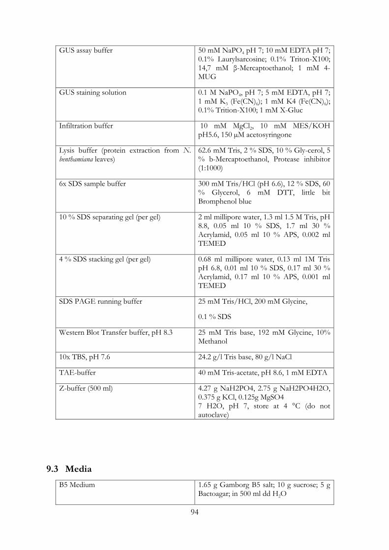
94
GUS assay buffer 50 mM NaPO4 pH 7; 10 mM EDTA pH 7; 0.1% Laurylsarcosine; 0.1% Triton-X100; 14,7 mM β-Mercaptoethanol; 1 mM 4-MUG
GUS staining solution 0.1 M NaPO4, pH 7; 5 mM EDTA, pH 7; 1 mM K3 (Fe(CN)6); 1 mM K4 (Fe(CN)6); 0.1% Trition-X100; 1 mM X-Gluc
Infiltration buffer 10 mM MgCl2, 10 mM MES/KOH pH5.6, 150 µM acetosyringone
Lysis buffer (protein extraction from N. benthamiana leaves)
62.6 mM Tris, 2 % SDS, 10 % Gly-cerol, 5 % b-Mercaptoethanol, Protease inhibitor (1:1000)
6x SDS sample buffer 300 mM Tris/HCl (pH 6.6), 12 % SDS, 60 % Glycerol, 6 mM DTT, little bit Bromphenol blue
10 % SDS separating gel (per gel) 2 ml millipore water, 1.3 ml 1.5 M Tris, pH 8.8, 0.05 ml 10 % SDS, 1.7 ml 30 % Acrylamid, 0.05 ml 10 % APS, 0.002 ml TEMED
4 % SDS stacking gel (per gel) 0.68 ml millipore water, 0.13 ml 1M Tris pH 6.8, 0.01 ml 10 % SDS, 0.17 ml 30 % Acrylamid, 0.17 ml 10 % APS, 0.001 ml TEMED
SDS PAGE running buffer
25 mM Tris/HCl, 200 mM Glycine,
0.1 % SDS
Western Blot Transfer buffer, pH 8.3 25 mM Tris base, 192 mM Glycine, 10% Methanol
10x TBS, pH 7.6 24.2 g/l Tris base, 80 g/l NaCl
TAE-buffer 40 mM Tris-acetate, pH 8.6, 1 mM EDTA
Z-buffer (500 ml) 4.27 g NaH2PO4, 2.75 g NaH2PO4H2O, 0.375 g KCl, 0.125g MgSO4 7 H2O, pH 7, store at 4 °C (do not autoclave)
9.3 Media
B5 Medium 1.65 g Gamborg B5 salt; 10 g sucrose; 5 g Bactoagar; in 500 ml dd H2O

95
Bactoagar 0.8% Bactoagar in 500 ml dd H2O
Hoagland Solution 0,6 ml Hoagland Trace Elements; 1 ml KNO3 (495 mM); 1 ml Ca(NO3)2 4H2O (43 mM); 2 mL MgSO4 7 H2O (200 mM); 2 ml Fe-Citrat (22 mM)
LB Medium 1 % (w/v) Tryptone, 0.5 % (w/v)
LB Medium 1 % (w/v) Tryptone, 0.5 % (w/v) Yeast extract, 1 % (w/v) NaCl, pH 7.0(NaOH)
SD-Medium SD-Medium 0.7 % Yeast nitrogen base without amino acids, 0.1 % Dropout mix, 2 % Glucosemonohydrate, pH 5.8
TY-Medium TY-Medium 0.3 % (w/v) Yeast extract, 0.5 % (w/v) Tryptone, pH 7.0
YPAD Medium (liquid) YPAD Medium (liquid) 1 % Bacto yeast extract, 2 % Bacto peptone, 2 % Glucose monohydrate, 0.004 % Adenine sulfate, pH 6.5
9.4 Chemicals
Chemicals Manufacturer
Acetosyringone Fluka, Buchs, CH
3-AT Sigma-Aldrich, Taufkirchen
Bacterial Protease Inhibitor Sigma-Aldrich, Taufkirchen
Biorad Protein Assay Biorad, München
Carrier DNA (Hering Sperm, 10 mg/ml) Promega, Mannheim
Dropout Supplement Clontech, Mountain View, USA
Gamborg B5 salt Sigma-Aldrich, Taufkirchen
Gelrite Roth, Karlsruhe
LR Clonase II Enzyme Mix Invitrogen Karlsruhe
Luminogen. TMA-6 GE Healthcare UK, Little Chalfont Buckinghamshire
4-MU Sigma-Aldrich Taufkirchen
MUG Sigma-Aldrich Taufkirchen
Plant Protease Inhibitor Sigma-Aldrich Taufkirchen

96
Polyethylenglycol (MW 3350) GE Healthcare Freiburg
9.5 Antibodies
Antibody Description Dilution Source
Anti-HA-HRP primary antibody conjugated
with HRP, IgG,
monoclonal, rat.
1:2000 Roche, Penzberg
Anti-GFPpoly a-GFP primary antibody,
IgG, polyclonal, mouse
1:3000 Rockland
Anti-myc primary antibody 1:1000 Roche, Penzberg
Anti-dsRed primary antibody 1: 5000 Clonetech
Anti-mouse
IRdye800
secondary antibody
conjugated with HRP, IgG,
monoclonal, goat
1:10000 Biomol, Hamburg
Anti-rabbit HRP secondary antibody 1:20000 Amersham
9.6 Plant Material
Plant species ecotype genotype reference
Lotus japonicus Gifu B-129 wild-type Larsen, 1995
Lotus japonicus MG-20 wild-type
Lotus japonicus Gifu snf1-1 Tirichine et al., 2006
Lotus japonicus Gifu snf2-2 Tirichine et al., 2007
Lotus japonicus Gifu har1-3 (Barbulova et al., 2007)

97
Lotus japonicus Gifu nin-2,
nin-8
(Schauser et al., 1999)
(Perry et al., 2009)
Lotus japonicus Gifu (accession number B-129) and ecotype Miyakojima MG-20 were used
as the wild-type along with five symbiotic mutants, nin-2, har1-3, cyclops-3, snf1-1 and snf2-2
in hairy root experiments.
9.7 Bacterial Strains
strain description resistance
Agrobacterium AR1193
MATa trp1-901 leu23,112 ura3-52 his3-200 gal4 gal80 LYS2::GAL1UASGAL1TATA- HIS3 MEL1 GAL2UAS-GAL2TATAADE2, URA3::MEL1UASMEL1TATA-lacZ, Stougaard et al., 1987
rifampicin (100µg/ml), carbenicillin (50 µg/ml)
Agrobacterium agl1
Lazo et al.; 1991 Oct; 963-7
rifampicin (100µg/ml), carbenicillin (50 µg/ml)
Agrobacterium GV3101
Koncz & Schell, 1986
rifampicin (100 mg/ml), gentamycin (50 mg/ml)
E. coli, Top10
F-, mcrA, Δ (mrr-hsdRMS-mcrBC), φ 80lacZ ΔM15, Δ lacX74,recA1, araD139,Δ (araleu) 7697, galU, galKrpsL (StrR), endA1, nupG, λ-
Mesorhizobium loti MAFF 303099
wild-type with RFP transgene (DsRed)
Root transformation was performed with A. rhizogenes (AR1193) and nodulation
experiments were done with M. loti MAFF303099 expressing DsRED constitutively
(Maekawa et al., 2009). E.coli strains DH5alpha, DB3.1 and TOP10 were used for plasmid
transformation and propagation of the plasmid. E.coli Rosetta strain was used for protein
expression of NIN and CYCLOPS. A. tumefaciencs strains agl1 and GV3101 were used for
transient expression in Nicotiana leaf cells. Yeast strain AH109 was used for yeast
transformation and in the yeast-two hybrid analysis.

98
9.8 Oligonucleotides
Oligonucleotides were designed in CLC Main Workbench and the web-based program
primer3plus (Untergasser et al., 2012). They were ordered from Sigma-Aldrich
(Germany).
Primer (5´-3´)
Contruct Primer Sequence forward and reverse
NIN-546-878 coding sequence = NINC ATGGTCTCTCACCATGTCTTCTTATA
CCTTTGGAAGCCG_f
ATGGTCTCACCTTTTAAGATGGGCT
GCTATTGCG_r
NIN-546-643 coding sequence = RW
BsaI
ATGGTCTCTCACCATGTCTTCTTATA
CCTTTGGAAGCCG_f BsaI
ATGGTCTCACCTTTTACTGTATGGC
ACCCTCAGCA_r BsaI
EMSA probes and competitors (5´-3´)
CYC-RE
NIN-RE wild-type
(Singh et al., 2014)
ATATAAGTTTGCATTTTTAGGTACACAAATTT_f AAATTTGTGTACCTAAAAATGCAAACTTATAT_r
NIN-RE mutated ATATAAGATTGCATTTTTTCCTACACAAATTT_f
AAATTTGTGTAGGAAAAAATGCAATCTTATAT_r
NC-RE TAAGTTTGCATTTTTAGGTACACAAATTTTGTACGATTGCCATGTGGCACGCA_f
GCGTGCCACATGGCAATCGTACAAAATTTGTGTACCTAAAAATGCAAACTTAT_r
Pal TACGATTGCCATGTGGCACGC_f
TGCGTGCCACATGGCAATCGT_r

99
9.9 Plasmid construction
pENTR_BsaI:cNIN-546-878 = (NINC) Phusion PCR product of cNIN-546-878
nucleotide sequence amplified from
pENTR:cNIN with NIN-546-
878_fwd/rev cloned into BsaI sites of
pENTR_BsaI-GW-BsaI
pENTR_BsaI:cNIN-546-643 = (RW) Phusion PCR product of cNIN-546-643
nucleotide sequence amplified from
pENTR:cNIN with NIN-546-
643_fwd/rev cloned into BsaI sites of
pENTR_BsaI-GW-BsaI
pDEST17:cNIN-546-878 = (NINC) and
pDEST17:cNIN-546-643 = (RW)
(N-terminal 6xHis-tag)
LR reaction (Invitrogen) of pDEST17
(Invitrogen) and pENTR_BsaI:cNIN-
546-878 or pENTR_BsaI:cNIN-546-643
pDEST15:CYCLOPS, phosphosite and
truncated versions(N-terminal GST-tag)
Singh et al., 2014
pENTR:cNIN Phusion PCR amplified cDNA NIN was
cloned into pENTR/D TOPO via TOPO
reaction (Invitrogen)
pENTR:cNSP2 Phusion PCR amplified cDNA NSP2 was
cloned into pENTR/D TOPO via TOPO
reaction (Invitrogen)
p35S:TSapphire-GW Singh et al., 2014
p35S:Tsapphire-CYCLOPS and
99hosphor-site mutants
Singh et al., 2014

100
p35S:mOrange Singh et al., 2014
Tsapphire-mOrange chimeric fusion Bayle et al., 2008
p35S:mOrange-GW Singh et al., 2014
p35S:CCaMK-mOrange Singh et al., 2014
p35S:NIN-mOrange LR reaction (Invitrogen)
pENTR:cNINΔstop and p35S:GW-
mOrange (Bayle et al., 2008)
pSPYNE35S:CYCLOPS and phosphosite
mutants
Singh et al., 2014
pSPYNE35S:NIN LR reaction (Invitrogen) of
pENTR:cNIN with pSPYNE35S:GW
pSPYCE35S:CCaMK Yano et al., 2008
pGWB735 CYCLOPS and phosphosite
mutants
Singh et al., 2014
pUb:3xHAgCYCLOPS Singh et al., 2014
pUb:cNIN LR reaction (Invitrogen) of
pENTR:cNIN and pUB:GWGFP
(Maekawa et al., 2008)
pUb:cNSP2 LR reaction (Invitrogen) of
pENTR:cNSP2 and pUB:GWGFP
(Maekawa et al., 2008)
pUb:gNIN:myc-
pUb:gCYCLOPSDD:HA-35S:GFP
LIII GG cut-ligation of LII
promUb:gNIN:myc, LII
promUb:gCYCLOPSDD:HA and LII
35S:GFP and insulators

101
Plasmids for the golden gate tool box Binder et al., 2014
p35S:cNIN-YFP LR reaction (Invitrogen) of
pENTR:cNIN and M1( (p35S:GW:YFP)
p35S:cNIN-C-YFP LR reaction (Invitrogen) of
pENTR:cNIN-C and M18
(p35S:GW:YFP)
pNIN:GUS Singh et al., 2014
2xCYC-RE:GUS
Singh et al., 2014
BD-NIN LR reaction (Invitrogen) of
pENTR:cNIN and pBD-GAL4 Cam
(Stratagene)
AD-NIN LR reaction (Invitrogen) of
pENTR:cNIN and pGAD424 (Clontech)
AD-CYCLOPS Yano et al., 2008
BD-CYCLOPS (1-159 aa) Yano et al., 2008
BD-CCaMK Yano et al., 2008
10 Methods
10.1 Molecular biology methods
10.1.1 Polymerase-chain reaction
For cloning experiments the amplification of DNA fragments was performed using
Phusion high fidelity polymerase (NEB) in either HF or GC buffer. Colony PCR was
performed with Taq polymerase in standard buffer (NEB).
These components were mixed as follows:

102
Phusion PCR
component volume
template (200 ng/µl) 1 µl
5x HF buffer 10 µl
dNTPs (5 mM) 1 µl
primer forward (2 µM) 5 µl
primer reverse (2 µM) 5 µl
Phusion(NEB con.) 1 µl
sterile ddH2O 27 µl
total 50 µl
Standard PCR-program for Phusion polymerase:
98 °C:30 sec; (98 °C:10 sec, X °Ca 30 sec,72 °C:15-30s/1 kb) x 35; 72 °C 10 min; 4 °C
10.1.2 Electrophoresis of DNA
1 % Agarose solution was prepared with TAE buffer and boiled. Then, ethidium bromide
was added to the mixture. Gel was left at RT to polymerize. Samples and DNA ladder
were loaded into the sample wells. The gel was run with 120 Volts for 30 minutes. The
separated fragments were visualized by a gel documentation system.
10.1.3 Extraction of PCR products from agarose gels/PCR clean-up
The gel containing DNA sample was placed on a UV table and exposed very shortly to
UV. A scalpel was used to cut out the DNA fragment and transfer it to a 1.5 ml reaction
tube. The extraction procedure of DNA from agarose gels was performed with the Gel
Extraction kit from Fermentas according to the manufacturer´s instructions.
Alternatively, the PCR product was directly cleaned up with the PCR clean-up kit from
Fermentas according to the manufacturer’s instructions. The cleaned PCR product was
further used in cloning reactions.

103
10.1.4 Plasmid isolation and estimation of DNA concentration
Plasmid DNA was isolated from 2 ml of overnight cultures of E.coli grown at 37°C using
the GeneJET Plasmid Miniprep Kit (Thermo Scientific) according to the manufacturer’s
instructions. The concentration of the isolated DNA was measured with a Nanodrop
spectrophotometer according to the manufacturer´s instructions.
10.1.5 Restriction endonuclease digestion of DNA
The restriction enzyme digest-mix was prepared on ice with the following components
(one reaction):
Component Volume
10x NEB buffer 1 µl
BSA (10mg/ml) 1 µl
Enzyme (3 units) 0.3 µl
DNA (250ng/µl) 1 µl
sterile ddH2O 6.7 µl
total volume 10 µl
The reaction was incubated at the recommended temperature for 2 hours. The reaction
was stopped with 10x DNA-loading buffer and run on a 1 % agarose gel to separate the
cut fragments according to its size.
10.2 Cloning and Vector construction of NIN
The coding region of NIN was amplified using cDNA from Gifu wt roots and genomic
NIN was amplified using genomic DNA from Gifu wt roots. The genomic sequence of
NIN was cloned with the Golden Gate strategy (Binder et al.; details see golden gate
cloning). NIN-C (aa) cDNA was cloned from the vector template of NIN-FL. For the
construction of genomic NIN three type IIS sites had to be removed, therefore the gene
was divided into 3 fragments and rejoined in a LI vector. The LII construct for NIN was
combined as pUb:gNIN:myc. For the co-expression of CYCLOPS-DD and NIN together
with a GFP transformation marker, a LIII vector was assembled (pUb:gNIN:myc-
pUb:CYCLOPS-DD:HA-35S:GFP) with insulators separating the genes.

104
10.2.1 Golden gate cloning (cut-ligation reaction)
BsaI, BpiI and Esp3I cut-ligation:
For convenience all plasmids were diluted to a final concentration of 100 ng/μl.
The cut-ligation for Golden Gate cloning was set up as follows:
Compound volume
Destination vector 1 µl
Enzyme (BsaI/BpiI/Esp3I) (5-10 units) 0,5 µl
T4 ligase buffer (10x) 1,5 µl
T4 ligase 0,75 µl
different insert vectors 1 µl
dd H2O ad 15 µl
For BsaI cut-ligations 0.15 μl of bovine serum albumin (10mg/ml) were added. Reactions
were incubated in a thermocycler for 20-40 cycles, cycling between 37°C for 2 min and
16°C for 5 min, followed by 37°C for 5 min, 50°C for 5 minutes and 80°C for 5 minutes.
3-5 μl of the reaction were transformed into E. coli TOP10 or DB3.1 (for insertion of ccdB
cassettes).
Blunt-end cut-ligation (subcloning):
For blunt-end cloning of PCR fragments into pUC57 vector an optimized cut-ligation
protocol was used.
Compound volume
StuI/NruI (5-10 units) 0,5 µl
T4 Ligase 1 µl

105
T4 ligase buffer 2 µl
100 mM ATP 0,2 µl
pUC57 25-50 ng
Insert 6:1 (6x)
H2O up to 20
Standard cut-ligation program: (37 °C:5 min; 20°C:5 min)x 20-40; 80 °C:10 min
0.4 μl of fresh enzyme (StuI/NruI) was added together with 0.4 μl of antarctic
phosphatase (NEB) and 2 μl of phosphatase reaction buffer. Samples were incubated for
another 30 min at 37°C, then heat inactivated at 80°C for 20 min.
5 μl of the reaction was transformed into E. coli Top10 by heat shock and plated on LB
plates containing the appropriate antibiotic supplemented with 40 μg/ml X-Gal (5-Brom-
4-chlor-3-indoxyl-β-D-galactopyranosid) and 100 μM IPTG (Isopropyl β-D-
thiogalactopyranoside). Plasmids were isolated from white colonies and validated by
sequencing.
10.2.2 Cloning: LR-reaction
LR reactions were set up like stated in the table below. The reaction mix was incubated at
25 °C for 1,5 hours.
Component Volume
entry-vector (100 ng) 0.75 µl
destination-vector (300 ng) 0.75 µl
LR clonase Mix 1 µl
The whole reaction mix was transformed into E. coli by heat shock transformation.
10.2.3 TOPO Cloning
The TOPO cloning reaction was set up like listed in the table below, then the reaction
was incubated for 1 hour at 21 °C.

106
Component volume
pENTR-D-TOPO vector (50 ng) 0.5 µl
Salt 0.5 µl
Insert (50 ng) 1 µl
Aqua 1 µl
The whole volume of the TOPO-reaction was transformed into E. coli Top10 by heat
shock.
10.2.4 Transformation of E. coli cells (heat shock method)
Chemically competent E. coli TOP10 or DB3.1 cells were used for transformation. Cells
were thawed on ice for 2 minutes and 50 µl of the suspension was added to a 1.5 ml
microliter tube. 1-5 µl of plasmid DNA or ligation reaction were added and mixed by
gentle pipetting. The reaction was incubated on ice for 2 minutes, and then a heat shock
of 42°C was applied for 30 seconds. The tubes were immediately returned to ice and 500
µl of LB medium was added. After incubation at 37°C on a shaker (200 rpm) for 30 min
to 1 h, 50-200 µl of the suspension was plated on LB plates containing the appropriate
antibiotics and incubated overnight at 37°C. Successful transformations were screened by
colony PCR and/or following plasmid extraction by restriction digestions and sequencing.
10.2.5 Transformation of Agrobacterium cells (Electroporation method)
Electrocompetent A. rhizogenes AR1193 or A. tumefaciens AGL1 cells were used for
transformation. The cells were thawed on ice for 2 minutes and mixed together in chilled
1.5 ml microliter tubes with 1-2 µl of plasmid DNA (10-50 ng) by gentle pipetting. The
mixed suspension was pipetted into chilled electroporation cuvettes. For electroporation
the following settings were used: Voltage 1.2 kV, conductance = 25/25/125 Fd;
resistance = 400 Ω. After the pulse, the cells were immediately mixed with 500 µl of LB
medium and transferred to 1.5 ml microliter tubes. Cells were incubated at 28°C for 1
hour and 20-100 µl were plated on LB plates containing the appropriate antibiotics.
Successful transformations were screened by colony PCR.

107
10.2.6 Sequencing
DNA sequence analysis was performed by the “Sequencing Service of the Faculty of
Biology” with an ABI3730 48 capillary sequencer (Applied Biosystems) using Big Dye
Terminator 3.1. Sequencing runs were set up according to the “cycle, clean & run”
protocol with 100 – 500 ng of plasmid DNA and 3.2 pmol of sequencing primer in a total
volume of 7 µl.
10.3 Cultivation methods
10.3.1 Bacterial growth conditions
E. coli was grown at 37 °C in LB medium overnight. Agrobacterium strains were grown at
28 °C for 1-2 days in LB medium, M. loti strains were grown at 28 °C for 2 to 4 days in
TY medium. Liquid cultures were shaken at 200 rpm.
10.4 Plant cultivation
10.4.1 N. benthamiana transformation
BiFC and localisation stduies were performed in 5 weeks old Nicotiana benthamiana plants
following A. tumefaciens GV3101 pM90RK (Koncz and Schell, 1986) and Agl1 strain
mediated transient transformation, respectively (Lazo et al., 1999; Walter et al., 2004).
Bacteria cultures of 2 ml LB-medium with selective antibiotics were inoculated with 1
Agrobacteria colony carrying the constructs of interest. The cultures were incubated 1-2
days at 28°C shaking at 170-220 rpm, and then the OD600 of the cultures was measured.
Cultures were centrifuged at 4000 rpm for 5 min at RT, the pellet was resuspended in
freshly prepared infiltration buffer and adjusted to a final OD600 of 0.5. Then, the
resuspended cultures were incubated in the dark for 2 hours at RT. Agrobacterium
cultures carrying constructs for testing a putative interaction/localisation were mixed with
an Agrobacterium strain carrying the P19 silencing suppressor (Voinnet et al., 2003;
Lakatos et al., 2004) in equal ratios. Bacterial mixtures were infiltrated into the lower side
of the leaf of N. benthamiana plants via a small wound. After infiltration the plants were
kept in a Binder growth chamber at 22°C for 16 hours day and 8 hours night cycle for 72
hours, which was determined to be the best time point for the expression of NIN. Leaf

108
samples were cut out, and the expression of fluorescent proteins was detected with an
inverted epifluorescence or a confocal laser-scanning microscope, respectively. After
microscopy, 3 leaf discs per sample were harvested and frozen in liquid nitrogen for
protein extraction and western blot analysis.
10.4.2 Plant germination and growth
L. japonicus seeds were scarified with sandpaper, sterilized with 2% NaOCl containing
0.1% sodium dodecylsulfate with an incubation time of 5 minutes, then washed 5 times
with 1 ml sterile ddH2O and incubated for at least 6 hours or overnight in sterile water
rotating on a wheel. Swollen seeds were transferred to 0.8% bactoagar plates and
germinated in the dark (wrapped in aluminium foil) for 3 days (in vertical position). The
plants were unwrapped from the foil and grown at 24°C in a 16 h light / 8 h dark cycle.
Hairy root transformation was performed with 6-7 days old seedlings.
10.4.3 Lotus hairy root Transformation
Following germination L. japonicus seedlings were grown on 0.8% bacto agar plates for 3
days in the dark and 4 days in the light at 24 °C prior to transformation. A. rhizogenes
AR1193 (Stougaard et al., 1987a) carrying the desired plasmids were precultivated on agar
plates containing the appropriate antibiotics for 2-3 days. Bacteria from a single colony
were taken and dissolved in 200 µl dd H2O and grown for 1 day.
For hairy root transformation the bacteria were scraped off the LB plates onto a Petri
dish and diluted in 1 ml of sterile water. The roots were cut at the hypocotyls with a
scalpel that was covered with bacterial suspension. The wound was dipped into the
bacteria and the plants were placed on agar plates containing B5 medium. The plates were
covered with aluminum foil and co-cultivated in the dark at approximately 24 °C in a
growth chamber. After 2 days the aluminum foil was partly removed (two thirds of the
bottom of the plates was still covered to limit light exposure of the roots) and the plants
were incubated for 3 days in 16 h light/8 h dark condition at 24°C in a growth chamber.
The plants were transferred to B5 plates containing Cefotaxime (333 µg/ml) in order to
halt the growth of the Agrobacteria. The plants were then cultivated at 24 °C in 16 h
light/8 h dark conditions until emergence of hairy roots (2 to 3 weeks).
109
10.4.4 Nodulation assay
L. japonicus plants were inoculated with Mesorhizobium loti MAFF303099 expressing dsRed
((Maekawa, 2009 #295)). The bacteria were inoculated from a preculture plate and grown
in 20 ml liquid TY medium containing 10 µg/ml gentamycin for 4 days. Cultures were
centrifuged for 10 minutes at 3000 g, then suspended in 1 ml sterile water. OD600 was
measured and the suspension was diluted in Hoagland medium to an OD600 of 0.05.
For nodulation of transformed L. japonicus hairy roots, plants were transferred from the
B5 agar plates into open pots containing 300 g of sand/vermiculite (1:1) supplemented
with 50 ml Hoagland medium. The plants were grown for one week at 24°C, and then
inoculated evenly with 10 ml of M. loti MAFF303099 dsRED (OD600 of 0.05). Nodulation
was assayed four weeks later.
In sterile assays the plants were cultivated in open pots containing 300 g of
sand/vermiculite (1:1) supplemented with 50 ml Hoagland medium (10-15 plants per
pot). The plants were watered with sterile water twice a week and supplemented with 50
ml Hoagland once a week. Spontanous nodulation was scored 5 weeks after
transplantation.
10.4.5 AM Assay
Seeds of L. japonicus ecotype Gifu B-129 wild-type were scarified and surface sterilized
with 1 % NaClO. Imbibed seeds were germinated on 1 % Bacto Agar (Difco) at 18 °C or
24 °C for 5-6 days. Seedlings were cultivated in chive (Allium schoenoprasum) nurse pots
containing ‘G. intraradices-like’ BEG195 (Stockinger et al., 2009) as described (Kistner et
al., 2005) except that sand/vermiculite (1/1 vol.) was used as substrate. After 5 weeks of
growth in open pots at 24 °C, 16 h light/ 8 h dark cycles, roots were harvested and
cleared with 10 % KOH at 90 °C for 15 min. AM fungal structures were stained with ink
and quantified under the inverted-fluorescence microscope using the intersections
method (McGonigle et al., 1990).
10.4.6 Nodule sectioning
Mature nodules were embedded in 6% low melting agarose and sections (50 µm) were cut
using the vibratome VT100S (Leica). Sections were stained with toluidine blue (0.05%)

110
and pictures were taken with a Leica DM16000 inverted light microscope using
brightfield illumination.
10.5 Biochemical methods
10.5.1 Histochemical GUS Staining
GUS staining was performed with 3 week old transformed L. japonicus roots or with N.
benthamiana leaves that carried a pNIN:GUS reporter 60 hours after transformation.
Samples were infiltrated with a GUS staining solution (1 mM X-Gluc (5-bromo-4-chloro-
3-indolyl-b-glucuronic acid), 0.1 M NaPO4 pH7, 5 mM EDTA, 0,1% Triton X-100, 1mM
K3(Fe(CN)6), 1 mM K4(Fe(CN)6), enzymatic reaction was incubated for 3 hours at 37 °C
and subsequently washed with 100 % Ethanol.
10.5.2 Fluorimetric GUS assay
Leaf samples from infiltrated N. benthamia plants were harvested 72 hours after infiltration
and frozen in liquid nitrogen. Frozen samples were homogenized with beads in a retch
mill (2x40 sec, 30/sec). Then, 200 µl extraction buffer was added to the samples and
vortexed for 1 minute. Samples were rotated for 10 minutes at 4 °C and then centrifuged
at 14000 rpm at 4 °C for 15 minutes. The supernatant was transferred to a new tube and
used for the fluorimetric GUS assay. 10 µl of cell extract was added to GUS buffer and
the reaction was incubated at 37°C for certain time points (3 min, 6 min, 9 min). The
reaction was stopped by addition of 100 µl NaCO3 (0,2 M). The reaction mix was
measured with a tecan reader for fluorescence (4-MU) at an excitation wavelength of 360
nm and a detection wavelength of 465 nm. Protein concentration was estimated with the
Bradford test. 1 µl of cell extract was mixed with 100 µl Bradford reagent and measured at
the absorption of 595 nm.
10.5.1 Protein extraction from N. benthamina leaves
Frozen samples of 3 leaf discs per tested combination was ground with a tissue lyser with
a frequency of 30/sec for 1.5 minutes. Per sample tube 300 µl of lysis buffer were added
and then vortexed for 1 minute. All samples were kept on ice for a few minutes and then
rotated on a wheel for 5 minutes at 4 °C. Then, the protein samples were heated at 95 °C
for 5 minutes and then centrifuged at 16000 rpm for 20 minutes. The protein extracts

111
were separated on an SDS-PAGE and expressed proteins were detected via western blot
analysis.
10.5.2 SDS PAGE
Each protein sample was mixed with 5x SDS sample buffer and incubated at 95 °C for 3
minutes. Then, all samples were shortly centrifuged prior to loading on a gel. A pre-
stained protein ladder was loaded to correlate separated protein sizes from the samples.
Samples were loaded on the gel and run at 150 V for 1 hour. The SDS-gel was then either
stained with Coomassie staining solution or subjected in a western blot with protein
transfer on a membrane.
10.5.3 Western blot
The PVDF-membrane was activated by incubation in 100 % ethanol for 15 seconds and
then transferred to transfer buffer for 5 minutes. A sandwich for the transfer of proteins
from an SDS-gel to the membrane was put together with 4 Whatman filter papers and 2
western sponges. The blot was run at 130V for 90 minutes. After transfer of the proteins
from the gel to the membrane, the PVDF-membrane was washed with TBS once and
then washed with TBS-T twice (each time for 10 minutes). For blocking the membrane
was incubated in TBS-T with 5 % milk for 1 hour at RT on a shaker or overnight at 4 °C.
The membrane was washed 3 times with TBS-T for 10 minutes on a shaker. Antibodies
were diluted in TBS-T with 5 % milk and added to the membrane for 2 hours at RT or
overnight at 4°C. The membrane was washed again 3 times with TBS-T for 10 minutes. A
secondary antibody conjugated with HRP was added to the membrane for the duration of
1 hour. Additional washing was performed 3 times with TBS-T for 10 minutes.
Horseradish peroxidase (HRP) activity was detected using chemiluminescence.
10.5.4 Protein expression and purification
Expression of NIN deletion constructs was induced in E. coli Rosetta pLaqI (Novagen)
for 12 hr at 18 °C by addition of 0.5 mM IPTG. His-tagged proteins were purified by
metal affinity chromatography using TALON resin (Clontech) as described by the
protocol of the manufacturer. Buffer conditions were modified accordingly (binding and
wash buffer: 20 mM PIPES, 500 mM KCl, 2 mM β-mercapto-ethanol, 10 mM imidazole,
pH 7.0; elution buffer: 20 mM PIPES, 200 mM KCl, 2 mM β-mercapto-ethanol, 250 mM

112
imidazole, pH 7.0). Expression and purification of GST-CYCLOPS phosphosite and
truncated versions was performed as described (Sing et al., 2014). Protein concentration
was determined by the Bradford Method (Bio-Rad), using BSA (Sigma) as a standard.
Protein purity was analyzed by SDS-PAGE and Coomassie staining of the gel.
10.5.5 Electrophoretic mobility shift assay
Binding reaction composition and practical application was as described in Sing et al..
2014. 100 fmol 5’ CY5 labeled DNA and 15 pmol NINC and RW, 25 pmol NINN, 20
pmol CYCBD, 35 pmol CYCMin and 75 pmol CYCDD were used (competitor DNA and
variant molar amounts of protein are indicated within the figure). Reactions were solved
on 4 % native polyacrylamide gels. CY5 labeled DNA was visualized with the Typhoon
TriO phosphoimager (Amersham Biosciences). Complementary pairs of labeled and
unlabeled oligonucleotide probes are listed in 9.8.
10.5.6 Bradford assay for protein standard curve and protein concentration
measurement
Bradford reagent was diluted with ddH2O in a ratio of 1:5 and filter sterilized. A dilution
series of 1000 µg, 500 µg, 250 µg and 125 µg BSA was prepared, by diluting a 100mg/l
BSA stock solution. The reaction mixes were incubated for a maximum of 10 minutes at
RT and the OD of each sample was measured at 595 nm. For the determination of
protein concentrations, 20 µl of protein sample were mixed with 980 µl Bradford
solution. Then the concentration of proteins was calculated from the BSA standard curve
using MS Excel.
10.5.7 Yeast two-hybrid
Yeast two-hybrid interaction assay was performed in the the yeast AH109 according to
the user manual (Yeast Protocols Handbook, Clonetech). AH109 yeast strain were
transformed with the lithium acetate method (Woods and Gietz, 2001). Transformants
were screened on SD medium containg 0,67 % yeast nitrogen base, 2% glucose, but
lacking appropriate nutrients.
A single colony of AH109 was inoculated into 20 ml YPAD-medium and grown in a
shaker at 30 °C overnight. The OD of the yeast culture was measured at 546 nm. The
Culture was adjusted to an OD of 0.2 and then grown until an OD of 0.6-0.8. Culture was

113
pelleted for 5 minutes at 700 g and resuspended in 1 ml sterile H2O (an amount sufficient
for 10 single- or co-transformations).
A master mix was prepared with the following components (one transformation) and
vortexed briefly:
component volume
50 % PEG 240 µl
1 M LiAc 36 µl
ssDNAa 30 µl
total 306 µl
Per transformation 300 µl of master mix were used and mixed with 7. 5 µl of plasmid-
DNA (200 ng/µl) and 100 µl yeast cells. The mix was vortexed for 1 minute. The
mixtures were incubated in a water bath at 42 °C for 45 minutes. Then a centrifugation
step was performed at 700 g for 5 minutes. The Pellet was resuspended in 100 µl sterile
0.9 % NaCl and plated onto selective SD(-)LW plates. The plates were incubated at 30 °C
for 2-3 days and grown yeast colonies were subjected to a yeast two hybrid test.
10 independent clones of each combination were picked and inoculated into 500 µl sterile
water in a 12 well plate. Then the cultures were incubated at 30°C shaking at 170-220 rpm
overnight. The next day, all cultures were adjusted to an OD546 of 1.0. For each
construct dilutions of 101, 102 and 103 were prepared in sterile ddH2O. A metallic stamp
was used to transfer 10 µl of each diluted sample to the selective SD plates. All plates
were and incubated at 30 °C for 2-3 days and yeast growth was analysed.
10.6 Microscopy
Confocal laser scanning microcopy (CLSM) was performed with a Leica SP5 microscope.
N. benthamiana leaves were vacuum infiltrated prior to imaging and imaged with a HCX
PL Fluotar 63x objective. For image acquisition the resolution was set to 512 x 512 pixels
and the frame average to 2. Using the argon laser at 20% power, GFP was excited with
the 488 nm laser line and detected at 500-530 nm, YFP with the 514 nm spectral line and
detected at 530-550 nm, mOrange was excited with the 514 nm laser line and detected at
545 nm to 600 nm. TSapphire was excited with a 405 diode laser and detected at 485 nm

114
to 535 nm. Images of L. japonicus root systems were taken using a Leica M165 FC
epifluorescence stereomicroscope equipped with a GFP and RFP filter.
10.6.1 FLIM-FRET
Nicotiana benthamiana leaves were co-infiltrated with Agrobacterium tumefaciens bacteria
carrying the respective plasmid constructs. 60 hpi leave discs were harvested and used for
the interaction study. For CLSM T-Sapphire was excited with a 405 diode laser, whereas
mOrange was excited with a 514 nm argon laser line (Bayle et. al., 2008). The emission of
TSapphire was detected with a 485 nm to 535 nm band pass emission filter, while
mOrange was detected with a 545 nm to 600 nm band-pass emission filter. A Ti-Sapphire
Mai Tai multiphoton (MP) laser from Spectra Physics was tuned at 800 nm for the
excitation of TSapphire fluorescence, which runs at 80 mHz with a 100 fs pulse length.
For a single FLIM measurement a number of 20 scanning cycles (5s/cycle) were applied
to get a suitable photon count rate at a spatial resolution of 256x256 pixels. As the FLIM
setup a Leica SP5 microscope with an implemented FLIM PMT detector from Becker
and Hickl were used. Hence the Becker and Hickl photon count software TCSPC2.80 was
used for recording the light signals. For lifetime calculations a region of interest was set
around the nucleus and the double exponential model was applied for all interaction pairs
in the Becker and Hickl SPCImage software. Both scatter and shift were fixed to zero.
The FRET efficiency was calculated as described in Bayle et al., 2008.
10.6.2 Statistics
All statistical analyses and data plots have been performed and generated with R version
3.0.2. For statistical analysis of the numbers of nodules, AM structures and FLIM-FRET
a t-test and ANOVA was applied.

115
11 List of Figures
Figure 1: symbiotic signal transduction in root nodule symbiosis ........................................ 21
Figure 2: Domain structure of NIN. ......................................................................................... 29
Figure 3: Localisation of NIN protein in planta. ...................................................................... 33
Figure 4: Interaction of NIN and CYCLOPS in yeast ........................................................... 35
Figure 5: NIN interacts with different variants of CYCLOPS in N. benthamiana leaf cells.
........................................................................................................................................................ 36
Figure 6: FRET FLIM analysis of control combinations in N.benthamiana cells. ................ 39
Figure 7: FLIM-FRET analysis confirms the interaction between NIN and CYCLOPS
variants .......................................................................................................................................... 41
Figure 8: NIN inhibits CYCLOPS-DD mediated activation of the NIN promoter in N.
benthamiana..................................................................................................................................... 43
Figure 9: NIN inhibits CYCLOPS-DD mediated activation of the NIN promoter in Lotus
roots. .............................................................................................................................................. 46
Figure 10: NIN inhibits CYCLOPS-DD transactivation of 2xCYC-RE:GUS in N.
benthamiana..................................................................................................................................... 49
Figure 11: NIN can bind to different probes in close proximity and on the CYC-RE. .... 51
Figure 12: NINC specifically binds to NIN-RE in vitro. .......................................................... 53
Figure 13: Competition experiments between NINC and CYCBD for NIN-RE. ................ 54
Figure 14: Competition analyses using NC-RE and CYC-RE together with CYCLOPS
and NINC ...................................................................................................................................... 56
Figure 15: Nodulation is inhibited by overexpression of NIN in Gifu hairy roots. ........... 58
Figure 16: Infection is locally inhibited by overexpression of NIN in Gifu hairy roots. ... 59
Figure 17: Local inhibition of nodulation is caused by overexpression of NIN in Gifu
and MG-20 hairy roots ................................................................................................................ 60
Figure 18: Overexpression of NIN did not rescue nodulation or infection in nin mutants
........................................................................................................................................................ 62
Figure 19: Overexpression of NIN inhibited nodulation in har1-3 hairy roots ................. 63
Figure 20: har1-3 hairy roots overexpressing NIN were longer in root length ................... 64
Figure 21: Inhibition of spontaneous nodulation in snf1-1 is caused by overexpression of
NIN. .............................................................................................................................................. 66

116
Figure 22: Quantification of spontaneous nodules formed in snf1-1 hairy roots
overexpressing NIN or NSP2 .................................................................................................... 67
Figure 23: Spontaneous nodule formation is inhibited by NIN overexpression in snf2-2. 69
Figure 24: Quantification of spontaneous nodule development in snf2-2 hairy roots
overexpressing NIN or NSP2 .................................................................................................... 70
Figure 25: Overexpression of NIN did not inhibit AM colonization in Gifu hairy roots . 71
Figure 26: Quantification of percentage of root length colonized by AM in Gifu hairy
roots overexpressing NIN. ......................................................................................................... 72
Figure 27: Spontaneous nodule formation and induction of nodule-like structures in Gifu
hairy roots ..................................................................................................................................... 74
Figure 28: Overexpression of NIN induced the formation of nodule-like structures in the
absence of rhizobia ...................................................................................................................... 75
Figure 29: Root diameter was increased in Gifu hairy roots overexpressing NIN ............. 76
Figure 30: alignment of NBS for NIN in this work and Soyano .......................................... 83
Figure 31: Putative NSP1 binding site present on NIN-RE .................................................. 85
Figure 32: Model of mechanistic action of NIN creating a negative feedback loop on the
activation of NIN expression mediated by the CCaMK/CYCLOPS complex. ................. 90
12 List of Tables
Table 1: Average mean lifetimes and calculated FRET-FLIM efficiencies for protein-
protein interaction ....................................................................................................................... 42
Table 2: Quantification of GUS expression in roots of pNIN:GUS line ............................. 47

117
13 List of Abbreviations
° C degree(s) Celsius
µ micro
A / ala alanine
aa amino acid
AD activation domain
AM arbuscular mycorrhiza
Amp Ampicilin
ANOVA analysis of variance
AON autoregulation of nodulation
BD binding domain
BiFC bimolecular fluorescence complementation
BF bright field
bp base pair
CaM Calmodulin
CaMBD Calmodulin binding domain
CCaMK Calcium Calmodulin dependent Kinase
CIP CCaMK interacting protein
cDNA complementary DNA
CLE CLAVATA3/ENDOSPERM SURROUNDING REGION
CLE-RS CLE root signal
CLSM confocal laser microscopy
CNCG cyclic nucleotide-gated channel
D / asp aspartic acid
Da Dalton
dd H2O double distilled water / Millipore filtered water
dpi days post infection/inoculation

118
dpt days post transformation
EMSA Electrophoretic mobility shift assay
ENOD11 Early Nodulin11
ERF Ethylene Response Factor
ERN ERF required for nodulation
FLIM Fluorescence Lifetime Imaging Microscopy
FRET Förster Resonance Energy Transfer
g gravitational acceleration / gram(s)
Gent gentamycin
GFP Green Fluorescent Protein
GG golden gate
GOI gene of interest
GUS β-glucuronidase
h hour(s)
HAR1 Hypernodulation aberrant root formation
HRP horse radish peroxidase
IPTG isopropyl β-D-thiogalactopyranoside
IT infection thread
Kan kanamycin
k kilo
LB lysogeny broth / Luria-Bertani broth
L litre(s)
LHK Lotus histidine kinase
Lj Lotus japonicus
LysM lysin motif
M molar
m meter(s) / mili
min minute(s)

119
mO mOrange
n nano
NBS NIN binding site
NF nodulation factor
NFR1 Nod Factor Receptor 1
NF-Y Nuclear Factor-Y subunit
NIN nodule inception
NLS nuclear localization signal
NLP NIN like protein
NPL pectate lyase
NRE Nitrate responsive element
NUP nucleoporin
NSP nodulation signalling pathway
OD optical density
p p-value
p35S cauliflower mosaic virus 35S promoter
PCR polymerase chain reaction
RAM root apical meristem
RFP Red Fluorescent Protein
RLK receptro like kinase
RNS root nodule symbiosis
ROI region of interest
rpm rounds per minute
RT room temperature
s second(s)
S / ser serine
snf spontaneous nodule formation
SYMREM1 Symbiotic Remorin 1
SYMRK Symbiosis Receptor-like Kinase

120
T / thr threonine
TALEs transciption activator-like effectors
TF transcription factor
TM transmembrane domain
TS TSapphier
Tukey’s HSD Tukey’s Honestly Significant Difference
UAS upstream activated sequence
promUb poly Ubiquitin promoter
VLD Visin like domain
wt wild-type
X-Gal 5-Brom-4-chlor-3-indoxyl-β-D-galactopyranosid
X-Gluc 5-bromo-4-chloro-3-indolyl glucuronide
Y2H yeast two-hybrid
YFP Yellow Fluorescent Protein

121
14 Declaration of contribution of other researches
Jayne Lambert, the author of this thesis contributed to the experiments displayed in figure
1-32 as follows:
Figure 1: designed by Andreas Binder and modified by Jayne Lambert
Figure 2-6, 10, 15-32: Experiments were designed, performed and analysed by Jayne
Lambert, statistical analysis was done by Jayne Lambert, who also prepared the
corresponding figures
Figure 7-9: Experiments were designed by Jayne Lambert, performed and analysed by
Jayne Lambert and Isabel Seidler, pictures were taken by Jayne Lambert, and figures were
prepared by Jayne Lambert
Figure 11-14: Experiments were designed by Jayne Lambert and Katja Katzer, EMSAs
were performed by Katja Katzer, and figures were prepared by Jayne Lambert and Katja
Katzer

122

123
15 References
Andriankaja A, Boisson-Dernier A, Frances L, Sauviac L, Jauneau A, Barker DG, de Carvalho-Niebel F (2007) AP2-ERF transcription factors mediate Nod factor dependent MtENOD11 activation in root hairs via a novel cis-regulatory motif. Plant Cell 19: 2866-2885
Ané J, Kiss G, Riely B, Penmetsa R, Oldroyd G, Ayax C, Lévy J, Debellé F, Baek J, Kalo P, Rosenberg C, Roe B, Long S, Dénarié J, Cook D (2004) Medicago truncatula DMI1 required for bacterial and fungal symbioses in legumes. Science 303: 1364-1367
Antolin-Llovera M, Ried MK, Parniske M (2014) Cleavage of the SYMBIOSIS RECEPTOR-LIKE KINASE Ectodomain Promotes Complex Formation with Nod Factor Receptor 5. Current Biology 24: 422-427
Barbulova A, Rogato A, D'Apuzzo E, Omrane S, Chiurazzi M (2007) Differential effects of combined N sources on early steps of the Nod factor-dependent transduction pathway in Lotus japonicus. Mol Plant Microbe Interact 20: 994-1003
Bateman A, Bycroft M (2000) The structure of a LysM domain from E. coli membrane-bound lytic murein transglycosylase D (MltD). J Mol Biol 299: 1113-1119
Bayle V, Nussaume L, Bhat R (2008) Combination of novel green fluorescent protein mutant TSapphire and DsRed variant mOrange to set up a versatile in planta FRET-FLIM assay. Plant Physiol 148: 51-60
Binder A, Lambert J, Morbitzer R, Popp C, Ott T, Lahaye T, Parniske M (2014) A Modular Plasmid Assembly Kit for Multigene Expression, Gene Silencing and Silencing Rescue in Plants. Plos One 9
Bolanos L, Redondo-Nieto M, Rivilla R, Brewin NJ, Bonilla I (2004) Cell surface interactions of Rhizobium bacteroids and other bacterial strains with symbiosomal and peribacteroid membrane components from pea nodules. Mol Plant Microbe Interact 17: 216-223
Bolle C (2004) The role of GRAS proteins in plant signal transduction and development. Planta 218: 683-692
Bonfante P, Genre A, Faccio A, Martini I, Schauser L, Stougaard J, Webb J, Parniske M (2000) The Lotus japonicus LjSym4 gene is required for the successful symbiotic infection of root epidermal cells. Mol Plant Microbe Interact 13: 1109-1120
Borisov A, Madsen L, Tsyganov V, Umehara Y, Voroshilova V, Batagov A, Sandal N, Mortensen A, Schauser L, Ellis N, Tikhonovich I, Stougaard J (2003) The Sym35 gene required for root nodule development in pea is an ortholog of Nin from Lotus japonicus. Plant Physiol 131: 1009-1017
Bourdon M, Coriton O, Pirrello J, Cheniclet C, Brown SC, Poujol C, Chevalier C, Renaudin JP, Frangne N (2011) In planta quantification of endoreduplication using fluorescent in situ hybridization (FISH). Plant J 66: 1089-1099
Breakspear A, Liu C, Roy S, Stacey N, Rogers C, Trick M, Morieri G, Mysore KS, Wen J, Oldroyd GE (2014) The Root Hair “Infectome” of Medicago truncatula Uncovers changes in cell cycle genes and reveals a requirement for auxin signaling in rhizobial infection. The Plant Cell Online: tpc. 114.133496

124
Brewin B, Woodley P, Drummond M (1999) The basis of ammonium release in nifL mutants of Azotobacter vinelandii. J Bacteriol 181: 7356-7362
Broghammer A, Krusell L, Blaise M, Sauer J, Sullivan JT, Maolanon N, Vinther M, Lorentzen A, Madsen EB, Jensen KJ, Roepstorff P, Thirup S, Ronson CW, Thygesen MB, Stougaard J (2012) Legume receptors perceive the rhizobial lipochitin oligosaccharide signal molecules by direct binding. Proc Natl Acad Sci U S A 109: 13859-13864
Brown RL, Kazan K, McGrath KC, Maclean DJ, Manners JM (2003) A role for the GCC-box in jasmonate-mediated activation of the PDF1.2 gene of Arabidopsis. Plant Physiol 132: 1020-1032
Buttner M, Singh KB (1997) Arabidopsis thaliana ethylene-responsive element binding protein (AtEBP), an ethylene-inducible, GCC box DNA-binding protein interacts with an ocs element binding protein. Proc Natl Acad Sci U S A 94: 5961-5966
Buzas DM, Gresshoff PM (2007) Short- and long-distance control of root development by LjHAR1 during the juvenile stage of Lotus japonicus. J Plant Physiol 164: 452-459
Carrera I, Treisman JE (2008) Message in a nucleus: signaling to the transcriptional machinery. Curr Opin Genet Dev 18: 397-403
Castaings L, Camargo A, Pocholle D, Gaudon V, Texier Y, Boutet-Mercey S, Taconnat L, Renou J, Daniel-Vedele F, Fernandez E, Meyer C, Krapp A (2009) The nodule inception-like protein 7 modulates nitrate sensing and metabolism in Arabidopsis. Plant J 57: 426-435
Cermak T, Doyle EL, Christian M, Wang L, Zhang Y, Schmidt C, Baller JA, Somia NV, Bogdanove AJ, Voytas DF (2011) Efficient design and assembly of custom TALEN and other TAL effector-based constructs for DNA targeting. Nucleic Acids Research 39: e82
Cerri MR, Frances L, Kelner A, Fournier J, Middleton PH, Auriac MC, Mysore KS, Wen J, Erard M, Barker DG, Oldroyd GE, de Carvalho-Niebel F (2016) The symbiosis-related ERN transcription factors act in concert to coordinate rhizobial host root infection. Plant Physiol 171: 1037-1054
Cerri MR, Frances L, Laloum T, Auriac MC, Niebel A, Oldroyd GE, Barker DG, Fournier J, de Carvalho-Niebel F (2012) Medicago truncatula ERN transcription factors: regulatory interplay with NSP1/NSP2 GRAS factors and expression dynamics throughout rhizobial infection. Plant Physiol 160: 2155-2172
Chabaud M, Genre A, Sieberer BJ, Faccio A, Fournier J, Novero M, Barker DG, Bonfante P (2011) Arbuscular mycorrhizal hyphopodia and germinated spore exudates trigger Ca2+ spiking in the legume and nonlegume root epidermis. New Phytologist 189: 347-355
Chardin C, Girin T, Roudier F, Meyer C, Krapp A (2014) The plant RWP-RK transcription factors: key regulators of nitrogen responses and of gametophyte development. J Exp Bot 65: 5577-5587
Charpentier M, Bredemeier R, Wanner G, Takeda N, Schleiff E, Parniske M (2008) Lotus japonicus CASTOR and POLLUX are ion channels essential for perinuclear calcium spiking in legume root endosymbiosis. Plant Cell 20: 3467-3479
Charpentier M, Sun J, Vaz Martins T, Radhakrishnan GV, Findlay K, Soumpourou E, Thouin J, Very AA, Sanders D, Morris RJ, Oldroyd GE (2016) Nuclear-localized cyclic nucleotide-gated channels mediate symbiotic calcium oscillations. Science 352: 1102-1105

125
Charron D, Pingret JL, Chabaud M, Journet EP, Barker DG (2004) Pharmacological evidence that multiple phospholipid signaling pathways link Rhizobium nodulation factor perception in Medicago truncatula root hairs to intracellular responses, including Ca2+ spiking and specific ENOD gene expression. Plant Physiol 136: 3582-3593
Clavijo F, Diedhiou I, Vaissayre V, Brottier L, Acolatse J, Moukouanga D, Crabos A, Auguy F, Franche C, Gherbi H, Champion A, Hocher V, Barker D, Bogusz D, Tisa LS, Svistoonoff S (2015) The Casuarina NIN gene is transcriptionally activated throughout Frankia root infection as well as in response to bacterial diffusible signals. New Phytol
Complainville A, Brocard L, Roberts I, Dax E, Sever N, Sauer N, Kondorosi A, Wolf S, Oparka K, Crespi M (2003) Nodule initiation involves the creation of a new symplasmic field in specific root cells of medicago species. Plant Cell 15: 2778-2791
Delaux PM, Radhakrishnan GV, Jayaraman D, Cheema J, Malbreil M, Volkening JD, Sekimoto H, Nishiyama T, Melkonian M, Pokorny L, Rothfels CJ, Sederoff HW, Stevenson DW, Surek B, Zhang Y, Sussman MR, Dunand C, Morris RJ, Roux C, Wong GK, Oldroyd GE, Ane JM (2015) Algal ancestor of land plants was preadapted for symbiosis. Proc Natl Acad Sci U S A 112: 13390-13395
Delaux PM, Sejalon-Delmas N, Becard G, Ane JM (2013) Evolution of the plant-microbe symbiotic 'toolkit'. Trends Plant Sci 18: 298-304
Den Herder G, Parniske M (2009) The unbearable naivety of legumes in symbiosis. Curr Opin Plant Biol 12: 491-499
Denarie J, Debelle F, Prome JC (1996) Rhizobium lipo-chitooligosaccharide nodulation factors: signaling molecules mediating recognition and morphogenesis. Annu Rev Biochem 65: 503-535
Diaz I, Vicente-Carbajosa J, Abraham Z, Martinez M, Isabel-La Moneda I, Carbonero P (2002) The GAMYB protein from barley interacts with the DOF transcription factor BPBF and activates endosperm-specific genes during seed development. Plant J 29: 453-464
Ehrhardt D, Wais R, Long S (1996) Calcium spiking in plant root hairs responding to Rhizobium nodulation signals. Cell 85: 673-681
Endre G, Kereszt A, Kevei Z, Mihacea S, Kaló P, Kiss G (2002) A receptor kinase gene regulating symbiotic nodule development. Nature 417: 962-966
Engler C, Gruetzner R, Kandzia R, Marillonnet S (2009) Golden gate shuffling: A one-pot DNA shuffling method based on type IIs restriction enzymes. PLoS ONE 4: e5553
Engler C, Kandzia R, Marillonnet S (2008) A one pot, one step, precision cloning method with high throughput capability. PLoS ONE 3: e3647
Engler C, Marillonnet S (2011) Generation of Families of Construct Variants Using Golden Gate Shuffling. In C Lu, J Browse, JG Wallis, eds, cDNA Libraries, Vol 729. Humana Press, pp 167-181
Esseling JJ, Lhuissier FG, Emons AM (2003) Nod factor-induced root hair curling: continuous polar growth towards the point of nod factor application. Plant Physiol 132: 1982-1988
Fisher RF, Long SR (1992) Rhizobium--plant signal exchange. Nature 357: 655-660

126
Flores JK, Kariawasam R, Gimenez AX, Helder S, Cubeddu L, Gamsjaeger R, Ataide SF (2015) Biophysical Characterisation and Quantification of Nucleic Acid-Protein Interactions: EMSA, MST and SPR. Curr Protein Pept Sci 16: 727-734
Fournier J, Teillet A, Chabaud M, Ivanov S, Genre A, Limpens E, de Carvalho-Niebel F, Barker DG (2015) Remodeling of the infection chamber before infection thread formation reveals a two-step mechanism for rhizobial entry into the host legume root hair. Plant Physiol 167: 1233-1242
Fournier J, Timmers AC, Sieberer BJ, Jauneau A, Chabaud M, Barker DG (2008) Mechanism of infection thread elongation in root hairs of Medicago truncatula and dynamic interplay with associated rhizobial colonization. Plant Physiol 148: 1985-1995
Fried MG (1989) Measurement of protein-DNA interaction parameters by electrophoresis mobility shift assay. Electrophoresis 10: 366-376
Fujimoto SY, Ohta M, Usui A, Shinshi H, Ohme-Takagi M (2000) Arabidopsis ethylene-responsive element binding factors act as transcriptional activators or repressors of GCC box-mediated gene expression. Plant Cell 12: 393-404
Gage D (2004) Infection and invasion of roots by symbiotic, nitrogen-fixing rhizobia during nodulation of temperate legumes. Microbiol Mol Biol Rev 68: 280-300
Geurts R, Fedorova E, Bisseling T (2005) Nod factor signaling genes and their function in the early stages of Rhizobium infection. Curr Opin Plant Biol 8: 346-352
Gleason C, Chaudhuri S, Yang T, Muñoz A, Poovaiah B, Oldroyd G (2006) Nodulation independent of rhizobia induced by a calcium-activated kinase lacking autoinhibition. Nature 441: 1149-1152
Gonzalez-Rizzo S, Crespi M, Frugier F (2006) The Medicago truncatula CRE1 cytokinin receptor regulates lateral root development a nd early symbiotic interaction with Sinorhizobium meliloti. Plant Cell 18: 2680-2693
Groth M, Takeda N, Perry J, Uchida H, Draxl S, Brachmann A, Sato S, Tabata S, Kawaguchi M, Wang TL, Parniske M (2010) NENA, a Lotus japonicus homolog of Sec13, is required for rhizodermal infection by arbuscular mycorrhiza fungi and rhizobia but dispensable for cortical endosymbiotic development. Plant Cell 22: 2509-2526
Gutjahr C, Parniske M (2013) Cell and developmental biology of arbuscular mycorrhiza symbiosis. Annual Review of Cell and Developmental Biology, Vol 29 29: 593-617
Harrison MJ (1999) Molecular and cellular aspects of the arbuscular mycorrhizal symbiosis. Annu Rev Plant Physiol Plant Mol Biol 50: 361-389
Hayashi M, Parniske M (2014) Symbiosis and pathogenesis: What determines the difference? Current Opinion in Plant Biology 20: V-VI
Hayashi T, Banba M, Shimoda Y, Kouchi H, Hayashi M, Imaizumi-Anraku H (2010) A dominant function of CCaMK in intracellular accommodation of bacterial and fungal endosymbionts. Plant J 63: 141-154
Heckmann A, Lombardo F, Miwa H, Perry J, Bunnewell S, Parniske M, Wang T, Downie J (2006) Lotus japonicus nodulation requires two GRAS domain regulators, one of which is functionally conserved in a non-legume. Plant Physiol 142: 1739-1750
Heckmann AB, Sandal N, Bek AS, Madsen LH, Jurkiewicz A, Nielsen MW, Tirichine L, Stougaard J (2011) Cytokinin induction of root nodule primordia in

127
Lotus japonicus is regulated by a mechanism operating in the root cortex. Mol Plant Microbe Interact 24: 1385-1395
Held M, Hou H, Miri M, Huynh C, Ross L, Hossain MS, Sato S, Tabata S, Perry J, Wang TL, Szczyglowski K (2014) Lotus japonicus cytokinin receptors work partially redundantly to mediate nodule formation. Plant Cell 26: 678-694
Hirsch S, Kim J, Munoz A, Heckmann AB, Downie JA, Oldroyd GE (2009) GRAS proteins form a DNA binding complex to induce gene expression during nodulation signaling in Medicago truncatula. Plant Cell 21: 545-557
Hirsch S, Oldroyd GE (2009) GRAS-domain transcription factors that regulate plant development. Plant Signal Behav 4: 698-700
Horvath B, Yeun LH, Domonkos A, Halasz G, Gobbato E, Ayaydin F, Miro K, Hirsch S, Sun J, Tadege M, Ratet P, Mysore KS, Ane JM, Oldroyd GE, Kalo P (2011) Medicago truncatula IPD3 is a member of the common symbiotic signaling pathway required for rhizobial and mycorrhizal symbioses. Mol Plant Microbe Interact 24: 1345-1358
Hrabak EM, Chan CW, Gribskov M, Harper JF, Choi JH, Halford N, Kudla J, Luan S, Nimmo HG, Sussman MR, Thomas M, Walker-Simmons K, Zhu JK, Harmon AC (2003) The Arabidopsis CDPK-SnRK superfamily of protein kinases. Plant Physiol 132: 666-680
Imaizumi-Anraku H, Takeda N, Charpentier M, Perry J, Miwa H, Umehara Y, Kouchi H, Murakami Y, Mulder L, Vickers K, Pike J, Downie JA, Wang T, Sato S, Asamizu E, Tabata S, Yoshikawa M, Murooka Y, Wu GJ, Kawaguchi M, Kawasaki S, Parniske M, Hayashi M (2005) Plastid proteins crucial for symbiotic fungal and bacterial entry into plant roots. Nature 433: 527-531
Jarsch IK, Ott T (2011) Perspectives on remorin proteins, membrane rafts, and their role during plant-microbe interactions. Mol Plant Microbe Interact 24: 7-12
Jones K, Kobayashi H, Davies B, Taga M, Walker G (2007) How rhizobial symbionts invade plants: the Sinorhizobium-Medicago model. Nat Rev Microbiol 5: 619-633
Journet EP, El-Gachtouli N, Vernoud V, de Billy F, Pichon M, Dedieu A, Arnould C, Morandi D, Barker DG, Gianinazzi-Pearson V (2001) Medicago truncatula ENOD11: a novel RPRP-encoding early nodulin gene expressed during mycorrhization in arbuscule-containing cells. Mol Plant Microbe Interact 14: 737-748
Kaló P, Gleason C, Edwards A, Marsh J, Mitra R, Hirsch S, Jakab J, Sims S, Long S, Rogers J, Kiss G, Downie J, Oldroyd G (2005) Nodulation signaling in legumes requires NSP2, a member of the GRAS family of transcriptional regulators. Science 308: 1786-1789
Kanamori N, Madsen L, Radutoiu S, Frantescu M, Quistgaard E, Miwa H, Downie J, James E, Felle H, Haaning L, Jensen T, Sato S, Nakamura Y, Tabata S, Sandal N, Stougaard J (2006) A nucleoporin is required for induction of Ca2+ spiking in legume nodule development and essential for rhizobial and fungal symbiosis. Proc Natl Acad Sci U S A 103: 359-364
Kang H, Xiao A, Huang X, Gao X, Yu H, He X, Zhu H, Hong Z, Zhang Z (2015) A Lotus japonicus cochaperone protein interacts with the ubiquitin-like domain protein CIP73 and plays a negative regulatory role in nodulation. Mol Plant Microbe Interact 28: 534-545

128
Kang H, Zhu H, Chu X, Yang Z, Yuan S, Yu D, Wang C, Hong Z, Zhang Z (2011) A novel interaction between CCaMK and a protein containing the Scythe_N ubiquitin-like domain in Lotus japonicus. Plant Physiol 155: 1312-1324
Katayama H, Iwamoto K, Kariya Y, Asakawa T, Kan T, Fukuda H, Ohashi-Ito K (2015) A negative feedback loop controlling bHLH complexes is involved in vascular cell division and differentiation in the root apical meristem. Curr Biol 25: 3144-3150
Kerppola TK (2008) Bimolecular fluorescence complementation (BiFC) analysis as a probe of protein interactions in living cells. Annu Rev Biophys 37: 465-487
Kistner C, Parniske M (2002) Evolution of signal transduction in intracellular symbiosis. Trends Plant Sci 7: 511-518
Kistner C, Winzer T, Pitzschke A, Mulder L, Sato S, Kaneko T, Tabata S, Sandal N, Stougaard J, Webb K, Szczyglowski K, Parniske M (2005) Seven Lotus japonicus genes required for transcriptional reprogramming of the root during fungal and bacterial symbiosis. Plant Cell 17: 2217-2229
Konishi M, Yanagisawa S (2013) Arabidopsis NIN-like transcription factors have a central role in nitrate signalling. Nature Communications 4: 1617
Konishi M, Yanagisawa S (2014) Emergence of a new step towards understanding the molecular mechanisms underlying nitrate-regulated gene expression. Journal of experimental botany 65: 5589-5600
Kosuta S, Hazledine S, Sun J, Miwa H, Morris RJ, Downie JA, Oldroyd GE (2008) Differential and chaotic calcium signatures in the symbiosis signaling pathway of legumes. Proceedings of the National Academy of Sciences 105: 9823-9828
Kosuta S, Held M, Hossain MS, Morieri G, Macgillivary A, Johansen C, Antolin-Llovera M, Parniske M, Oldroyd GE, Downie AJ, Karas B, Szczyglowski K (2011) Lotus japonicus symRK-14 uncouples the cortical and epidermal symbiotic program. Plant J 67: 929-940
Koszegi D, Johnston AJ, Rutten T, Czihal A, Altschmied L, Kumlehn J, Wust SE, Kirioukhova O, Gheyselinck J, Grossniklaus U, Baumlein H (2011) Members of the RKD transcription factor family induce an egg cell-like gene expression program. Plant J 67: 280-291
Krusell L, Madsen LH, Sato S, Aubert G, Genua A, Szczyglowski K, Duc G, Kaneko T, Tabata S, de Bruijn F, Pajuelo E, Sandal N, Stougaard J (2002) Shoot control of root development and nodulation is mediated by a receptor-like kinase. Nature 420: 422-426
Kucukoglu M, Nilsson O (2015) CLE peptide signaling in plants - the power of moving around. Physiol Plant 155: 74-87
Kusnetsov V, Landsberger M, Meurer J, Oelmuller R (1999) The assembly of the CAAT-box binding complex at a photosynthesis gene promoter is regulated by light, cytokinin, and the stage of the plastids. J Biol Chem 274: 36009-36014
Lakatos L, Szittya G, Silhavy D, Burgyan J (2004) Molecular mechanism of RNA silencing suppression mediated by p19 protein of tombusviruses. Embo j 23: 876-884
Laloum T, Baudin M, Frances L, Lepage A, Billault-Penneteau B, Cerri MR, Ariel F, Jardinaud MF, Gamas P, de Carvalho-Niebel F, Niebel A (2014) Two CCAAT-box-binding transcription factors redundantly regulate early steps of the legume-rhizobia endosymbiosis. Plant J 79: 757-768

129
Laloum T, De Mita S, Gamas P, Baudin M, Niebel A (2013) CCAAT-box binding transcription factors in plants: Y so many? Trends Plant Sci 18: 157-166
Lamark T, Perander M, Outzen H, Kristiansen K, Overvatn A, Michaelsen E, Bjorkoy G, Johansen T (2003) Interaction codes within the family of mammalian Phox and Bem1p domain-containing proteins. J Biol Chem 278: 34568-34581
Lefebvre B, Klaus-Heisen D, Pietraszewska-Bogiel A, Herve C, Camut S, Auriac MC, Gasciolli V, Nurisso A, Gadella TW, Cullimore J (2012) Role of N-glycosylation sites and CXC motifs in trafficking of medicago truncatula Nod factor perception protein to plasma membrane. J Biol Chem 287: 10812-10823
Lefebvre B, Timmers T, Mbengue M, Moreau S, Hervé C, Tóth K, Bittencourt-Silvestre J, Klaus D, Deslandes L, Godiard L, Murray JD, Udvardi MK, Raffaele S, Mongrand S, Cullimore J, Gamas P, Niebel A, Ott T (2010) A remorin protein interacts with symbiotic receptors and regulates bacterial infection. Proc Natl Acad Sci U S A 107: 2343-2348
Levy J, Bres C, Geurts R, Chalhoub B, Kulikova O, Duc G, Journet EP, Ane JM, Lauber E, Bisseling T, Denarie J, Rosenberg C, Debelle F (2004) A putative Ca2+ and calmodulin-dependent protein kinase required for bacterial and fungal symbioses. Science 303: 1361-1364
Li HL, Wang W, Mortimer PE, Li RQ, Li DZ, Hyde KD, Xu JC, Soltis DE, Chen ZD (2015) Large-scale phylogenetic analyses reveal multiple gains of actinorhizal nitrogen-fixing symbioses in angiosperms associated with climate change. Sci Rep 5: 14023
Liu W, Kohlen W, Lillo A, Op den Camp R, Ivanov S, Hartog M, Limpens E, Jamil M, Smaczniak C, Kaufmann K, Yang WC, Hooiveld GJ, Charnikhova T, Bouwmeester HJ, Bisseling T, Geurts R (2011) Strigolactone biosynthesis in Medicago truncatula and rice requires the symbiotic GRAS-type transcription factors NSP1 and NSP2. Plant Cell 23: 3853-3865
Madsen E, Madsen L, Radutoiu S, Olbryt M, Rakwalska M, Szczyglowski K, Sato S, Kaneko T, Tabata S, Sandal N, Stougaard J (2003) A receptor kinase gene of the LysM type is involved in legume perception of rhizobial signals. Nature 425: 637-640
Madsen EB, Antolin-Llovera M, Grossmann C, Ye J, Vieweg S, Broghammer A, Krusell L, Radutoiu S, Jensen ON, Stougaard J, Parniske M (2011) Autophosphorylation is essential for the in vivo function of the Lotus japonicus Nod factor receptor 1 and receptor-mediated signalling in cooperation with Nod factor receptor 5. Plant J 65: 404-417
Madsen LH, Tirichine L, Jurkiewicz A, Sullivan JT, Heckmann AB, Bek AS, Ronson CW, James EK, Stougaard J (2010) The molecular network governing nodule organogenesis and infection in the model legume Lotus japonicus. Nat Commun 1: 10
Maekawa T, Maekawa-Yoshikawa M, Takeda N, Imaizumi-Anraku H, Murooka Y, Hayashi M (2009) Gibberellin controls the nodulation signaling pathway in Lotus japonicus. Plant J 58: 183-194
Magori S, Kawaguchi M (2009) Long-distance control of nodulation: molecules and models. Mol Cells 27: 129-134
Marchive C, Roudier F, Castaings L, Bréhaut V, Blondet E, Colot V, Meyer C, Krapp A (2013) Nuclear retention of the transcription factor NLP7 orchestrates the early response to nitrate in plants. Nature Communications 4: 1713

130
Markmann K, Giczey G, Parniske M (2008) Functional adaptation of a plant receptor-kinase paved the way for the evolution of intracellular root symbioses with bacteria. PLoS Biol 6: e68
Marsh J, Rakocevic A, Mitra R, Brocard L, Sun J, Eschstruth A, Long S, Schultze M, Ratet P, Oldroyd G (2007) Medicago truncatula NIN is essential for rhizobial-independent nodule organogenesis induced by autoactive calcium/calmodulin-dependent protein kinase. Plant Physiol 144: 324-335
McGrath KC, Dombrecht B, Manners JM, Schenk PM, Edgar CI, Maclean DJ, Scheible WR, Udvardi MK, Kazan K (2005) Repressor- and activator-type ethylene response factors functioning in jasmonate signaling and disease resistance identified via a genome-wide screen of Arabidopsis transcription factor gene expression. Plant Physiol 139: 949-959
Messinese E, Mun JH, Yeun LH, Jayaraman D, Rouge P, Barre A, Lougnon G, Schornack S, Bono JJ, Cook DR, Ane JM (2007) A novel nuclear protein interacts with the symbiotic DMI3 calcium- and calmodulin-dependent protein kinase of Medicago truncatula. Mol Plant Microbe Interact 20: 912-921
Middleton P, Jakab J, Penmetsa R, Starker C, Doll J, Kaló P, Prabhu R, Marsh J, Mitra R, Kereszt A, Dudas B, VandenBosch K, Long S, Cook D, Kiss G, Oldroyd G (2007) An ERF transcription factor in Medicago truncatula that is essential for Nod factor signal transduction. Plant Cell 19: 1221-1234
Mitra R, Gleason C, Edwards A, Hadfield J, Downie J, Oldroyd G, Long S (2004) A Ca2+/calmodulin-dependent protein kinase required for symbiotic nodule development: Gene identification by transcript-based cloning. Proc Natl Acad Sci U S A 101: 4701-4705
Miwa H, Sun J, Oldroyd G, Downie J (2006) Analysis of calcium spiking using a cameleon calcium sensor reveals that nodulation gene expression is regulated by calcium spike number and the developmental status of the cell. Plant J 48: 883-894
Morbitzer R, Elsaesser J, Hausner J, Lahaye T (2011) Assembly of custom TALE-type DNA binding domains by modular cloning. Nucleic Acids Res 39: 5790-5799
Mortier V, Holsters M, Goormachtig S (2012) Never too many? How legumes control nodule numbers. Plant Cell Environ 35: 245-258
Murakami Y, Miwa H, Imaizumi-Anraku H, Kouchi H, Downie J, Kawaguchi M, Kawasaki S (2006) Positional cloning identifies Lotus japonicus NSP2, a putative transcription factor of the GRAS family, required for NIN and ENOD40 gene expression in nodule initiation. DNA Res 13: 255-265
Murray J, Karas B, Sato S, Tabata S, Amyot L, Szczyglowski K (2007) A cytokinin perception mutant colonized by Rhizobium in the absence of nodule organogenesis. Science 315: 101-104
Murray JD (2011) Invasion by invitation: rhizobial infection in legumes. Mol Plant Microbe Interact 24: 631-639
Nakano T, Suzuki K, Ohtsuki N, Tsujimoto Y, Fujimura T, Shinshi H (2006) Identification of genes of the plant-specific transcription-factor families cooperatively regulated by ethylene and jasmonate in Arabidopsis thaliana. J Plant Res 119: 407-413
Nishida H, Handa Y, Tanaka S, Suzaki T, Kawaguchi M (2016) Expression of the CLE-RS3 gene suppresses root nodulation in Lotus japonicus. J Plant Res 129: 909-919

131
Nishimura R, Hayashi M, Wu G, Kouchi H, Imaizumi-Anraku H, Murakami Y, Kawasaki S, Akao S, Ohmori M, Nagasawa M, Harada K, Kawaguchi M (2002) HAR1 mediates systemic regulation of symbiotic organ development. Nature 420: 426-429
Ohta M, Matsui K, Hiratsu K, Shinshi H, Ohme-Takagi M (2001) Repression domains of class II ERF transcriptional repressors share an essential motif for active repression. Plant Cell 13: 1959-1968
Okamoto S, Ohnishi E, Sato S, Takahashi H, Nakazono M, Tabata S, Kawaguchi M (2009) Nod Factor/Nitrate-Induced CLE Genes that Drive HAR1-Mediated Systemic Regulation of Nodulation. Plant and Cell Physiology 50: 67-77
Oldroyd GE, Downie JA (2008) Coordinating nodule morphogenesis with rhizobial infection in legumes. Annu Rev Plant Biol 59: 519-546
Oldroyd GE (2013) Speak, friend, and enter: signalling systems that promote beneficial symbiotic associations in plants. Nature Reviews Microbiology 11: 252-263
Oldroyd GE, Murray JD, Poole PS, Downie JA (2011) The rules of engagement in the legume-rhizobial symbiosis. Annual review of genetics 45: 119-144
Op den Camp RH, Polone E, Fedorova E, Roelofsen W, Squartini A, Op den Camp HJ, Bisseling T, Geurts R (2012) Nonlegume Parasponia andersonii deploys a broad rhizobium host range strategy resulting in largely variable symbiotic effectiveness. Mol Plant Microbe Interact 25: 954-963
Ovchinnikova E, Journet EP, Chabaud M, Cosson V, Ratet P, Duc G, Fedorova E, Liu W, den Camp RO, Zhukov V, Tikhonovich I, Borisov A, Bisseling T, Limpens E (2011) IPD3 controls the formation of nitrogen-fixing symbiosomes in pea and Medicago Spp. Mol Plant Microbe Interact 24: 1333-1344
Pawlowski, K., and Sprent, J.I. (2008). Comparison between actinorhizal and legume symbiosis, in Nitrogen-fixing Actinorhizal Symbioses, eds. K. Pawlowski & W. Newton. Springer Netherlands), 261-288.
Parniske M (2008) Arbuscular mycorrhiza: the mother of plant root endosymbioses. Nat Rev Microbiol 6: 763-775
Perry J, Brachmann A, Welham T, Binder A, Charpentier M, Groth M, Haage K, Markmann K, Wang TL, Parniske M (2009) TILLING in Lotus japonicus identified large allelic series for symbiosis genes and revealed a bias in functionally defective ethyl methanesulfonate alleles toward glycine replacements. Plant Physiol 151: 1281-1291
Peters NK, Frost JW, Long SR (1986) A plant flavone, luteolin, induces expression of Rhizobium meliloti nodulation genes. Science 233: 977-980
Pietraszewska-Bogiel A, Lefebvre B, Koini MA, Klaus-Heisen D, Takken FL, Geurts R, Cullimore JV, Gadella TW (2013) Interaction of Medicago truncatula lysin motif receptor-like kinases, NFP and LYK3, produced in Nicotiana benthamiana induces defence-like responses. PLoS One 8: e65055
Plet J, Wasson A, Ariel F, Le Signor C, Baker D, Mathesius U, Crespi M, Frugier F (2011) MtCRE1-dependent cytokinin signaling integrates bacterial and plant cues to coordinate symbiotic nodule organogenesis in Medicago truncatula. Plant J 65: 622-633
Ponting C, Ito T, Moscat J, Diaz-Meco M, Inagaki F, Sumimoto H (2002) OPR, PC and AID: all in the PB1 family. Trends Biochem Sci 27: 10

132
Qiu L, Lin JS, Xu J, Sato S, Parniske M, Wang TL, Downie JA, Xie F (2015) SCARN a novel class of SCAR protein that is required for root-hair infection during legume nodulation. PLoS Genet 11: e1005623
Radutoiu S, Madsen L, Madsen E, Felle H, Umehara Y, Grønlund M, Sato S, Nakamura Y, Tabata S, Sandal N, Stougaard J (2003) Plant recognition of symbiotic bacteria requires two LysM receptor-like kinases. Nature 425: 585-592
Raymond J, Siefert J, Staples C, Blankenship R (2004) The natural history of nitrogen fixation. Mol Biol Evol 21: 541-554
Reid DE, Ferguson BJ, Hayashi S, Lin YH, Gresshoff PM (2011) Molecular mechanisms controlling legume autoregulation of nodulation. Ann Bot 108: 789-795
Remigi P, Zhu J, Young JP, Masson-Boivin C (2016) Symbiosis within symbiosis: evolving nitrogen-fixing legume symbionts. Trends Microbiol 24: 63-75
Riechmann JL, Meyerowitz EM (1998) The AP2/EREBP family of plant transcription factors. Biol Chem 379: 633-646
Ried MK, Antolin-Llovera M, Parniske M (2014) Spontaneous symbiotic reprogramming of plant roots triggered by receptor-like kinases. Elife 3
Roche P, Lerouge P, Ponthus C, Prome JC (1991) Structural determination of bacterial nodulation factors involved in the Rhizobium meliloti-alfalfa symbiosis. J Biol Chem 266: 10933-10940
Rodríguez-Llorente I, Pérez-Hormaeche J, El Mounadi K, Dary M, Caviedes M, Cosson V, Kondorosi A, Ratet P, Palomares A (2004) From pollen tubes to infection threads: recruitment of Medicago floral pectic genes for symbiosis. Plant J 39: 587-598
Ryder SP, Recht MI, Williamson JR (2008) Quantitative analysis of protein-RNA interactions by gel mobility shift. Methods Mol Biol 488: 99-115
Saito K, Yoshikawa M, Yano K, Miwa H, Uchida H, Asamizu E, Sato S, Tabata S, Imaizumi-Anraku H, Umehara Y, Kouchi H, Murooka Y, Szczyglowski K, Downie J, Parniske M, Hayashi M, Kawaguchi M (2007) NUCLEOPORIN85 is required for calcium spiking, fungal and bacterial symbioses, and seed production in Lotus japonicus. Plant Cell 19: 610-624
Sakuma T, Ochiai H, Kaneko T, Mashimo T, Tokumasu D, Sakane Y, Suzuki K, Miyamoto T, Sakamoto N, Matsuura S, Yamamoto T (2013) Repeating pattern of non-RVD variations in DNA-binding modules enhances TALEN activity. Sci Rep 3: 3379
Schauser L, Roussis A, Stiller J, Stougaard J (1999) A plant regulator controlling development of symbiotic root nodules. Nature 402: 191-195
Schauser L, Wieloch W, Stougaard J (2005) Evolution of NIN-like proteins in Arabidopsis, rice, and Lotus japonicus. J Mol Evol 60: 229-237
Schüβler A, Schwarzott D, Walker C (2001) A new fungal phylum, the Glomeromycota: phylogeny and evolution *Dedicated to Manfred Kluge (Technische Universität Darmstadt) on the occasion of his retirement. Mycological Research 105: 1413-1421
Shimoda Y, Han L, Yamazaki T, Suzuki R, Hayashi M, Imaizumi-Anraku H (2012) Rhizobial and fungal symbioses show different requirements for calmodulin binding to calcium calmodulin-dependent protein kinase in Lotus japonicus. Plant Cell 24: 304-321

133
Sieberer BJ, Chabaud M, Fournier J, Timmers AC, Barker DG (2012) A switch in Ca2+ spiking signature is concomitant with endosymbiotic microbe entry into cortical root cells of Medicago truncatula. The Plant Journal 69: 822-830
Singh S, Katzer K, Lambert J, Cerri M, Parniske M (2014) CYCLOPS, a DNA-binding transcriptional activator, orchestrates symbiotic root nodule development. Cell Host Microbe 15: 139-152
Singh S, Parniske M (2012) Activation of calcium- and calmodulin-dependent protein kinase (CCaMK), the central regulator of plant root endosymbiosis. Curr Opin Plant Biol 15: 444-453
Smit P, Raedts J, Portyanko V, Debelle F, Gough C, Bisseling T, Geurts R (2005) NSP1 of the GRAS protein family is essential for rhizobial Nod factor-induced transcription. Science 308: 1789-1791
Sornaraj P, Luang S, Lopato S, Hrmova M (2016) Basic leucine zipper (bZIP) transcription factors involved in abiotic stresses: A molecular model of a wheat bZIP factor and implications of its structure in function. Biochim Biophys Acta 1860: 46-56
Soyano T, Hirakawa H, Sato S, Hayashi M, Kawaguchi M (2014) Nodule Inception creates a long-distance negative feedback loop involved in homeostatic regulation of nodule organ production. Proceedings of the National Academy of Sciences 111: 14607-14612
Soyano T, Kouchi H, Hirota A, Hayashi M (2013) Nodule inception directly targets NF-Y subunit genes to regulate essential processes of root nodule development in Lotus japonicus. PLoS Genet 9: e1003352
Soyano T, Shimoda Y, Hayashi M (2015) Nodule Inception antagonistically regulates gene expression with nitrate in Lotus japonicus. Plant Cell Physiol 56: 368-376
Stockinger H, Walker C, Schussler A (2009) 'Glomus intraradices DAOM197198', a model fungus in arbuscular mycorrhiza research, is not Glomus intraradices. New Phytol 183: 1176-1187
Stracke S, Kistner C, Yoshida S, Mulder L, Sato S, Kaneko T, Tabata S, Sandal N, Stougaard J, Szczyglowski K, Parniske M (2002) A plant receptor-like kinase required for both bacterial and fungal symbiosis. Nature 417: 959-962
Streeter J, Wong PP (1988) Inhibition of legume nodule formation and N2 fixation by nitrate. Critical Reviews in Plant Sciences 7: 1-23
Sumimoto H, Kamakura S, Ito T (2007) Structure and function of the PB1 domain, a protein interaction module conserved in animals, fungi, amoebas, and plants. Sci STKE 2007: re6
Suzaki T, Kim CS, Takeda N, Szczyglowski K, Kawaguchi M (2013) TRICOT encodes an AMP1-related carboxypeptidase that regulates root nodule development and shoot apical meristem maintenance in Lotus japonicus. Development 140: 353-361
Suzaki T, Yano K, Ito M, Umehara Y, Suganuma N, Kawaguchi M (2012) Positive and negative regulation of cortical cell division during root nodule development in Lotus japonicus is accompanied by auxin response. Development 139: 3997-4006
Suzuki W, Konishi M, Yanagisawa S (2013) The evolutionary events necessary for the emergence of symbiotic nitrogen fixation in legumes may involve a loss of nitrate responsiveness of the NIN transcription factor. Plant Signal Behav 8

134
Swainsbury DJ, Zhou L, Oldroyd GE, Bornemann S (2012) Calcium ion binding properties of Medicago truncatula calcium/calmodulin-dependent protein kinase. Biochemistry 51: 6895-6907
Takeda N, Maekawa T, Hayashi M (2012) Nuclear-localized and deregulated calcium- and calmodulin-dependent protein kinase activates rhizobial and mycorrhizal responses in Lotus japonicus. Plant Cell 24: 810-822
Timmers A, Auriac M, Truchet G (1999) Refined analysis of early symbiotic steps of the Rhizobium-Medicago interaction in relationship with microtubular cytoskeleton rearrangements. Development 126: 3617-3628
Tirichine L, Imaizumi-Anraku H, Yoshida S, Murakami Y, Madsen L, Miwa H, Nakagawa T, Sandal N, Albrektsen A, Kawaguchi M, Downie A, Sato S, Tabata S, Kouchi H, Parniske M, Kawasaki S, Stougaard J (2006) Deregulation of a Ca2+/calmodulin-dependent kinase leads to spontaneous nodule development. Nature 441: 1153-1156
Tirichine L, Sandal N, Madsen L, Radutoiu S, Albrektsen A, Sato S, Asamizu E, Tabata S, Stougaard J (2007) A gain-of-function mutation in a cytokinin receptor triggers spontaneous root nodule organogenesis. Science 315: 104-107
Toth K, Stratil TF, Madsen EB, Ye J, Popp C, Antolin-Llovera M, Grossmann C, Jensen ON, Schussler A, Parniske M, Ott T (2012) Functional domain analysis of the Remorin protein LjSYMREM1 in Lotus japonicus. PLoS One 7: e30817
Untergasser A, Cutcutache I, Koressaar T, Ye J, Faircloth BC, Remm M, Rozen SG (2012) Primer3--new capabilities and interfaces. Nucleic Acids Res 40: e115
van Zeijl A, Op den Camp RH, Deinum EE, Charnikhova T, Franssen H, Op den Camp HJ, Bouwmeester H, Kohlen W, Bisseling T, Geurts R (2015) Rhizobium Lipo-chitooligosaccharide signaling triggers accumulation of cytokinins in Medicago truncatula roots. Mol Plant 8: 1213-1226
Vernie T, Kim J, Frances L, Ding Y, Sun J, Guan D, Niebel A, Gifford ML, de Carvalho-Niebel F, Oldroyd GE (2015) The NIN transcription factor coordinates diverse nodulation programs in different tissues of the Medicago truncatula root. Plant Cell 27: 3410-3424
Voinnet O, Rivas S, Mestre P, Baulcombe D (2003) An enhanced transient expression system in plants based on suppression of gene silencing by the p19 protein of tomato bushy stunt virus. Plant J 33: 949-956
Waadt R, Schmidt LK, Lohse M, Hashimoto K, Bock R, Kudla J (2008) Multicolor bimolecular fluorescence complementation reveals simultaneous formation of alternative CBL/CIPK complexes in planta. Plant J 56: 505-516
Waki T, Hiki T, Watanabe R, Hashimoto T, Nakajima K (2011) The Arabidopsis RWP-RK protein RKD4 triggers gene expression and pattern formation in early embryogenesis. Curr Biol 21: 1277-1281
Weber E, Gruetzner R, Werner S, Engler C, Marillonnet S (2011) Assembly of designer TAL effectors by golden gate cloning. PLoS One 6: e19722
Weerasinghe RR, Bird DM, Allen NS (2005) Root-knot nematodes and bacterial nod factors elicit common signal transduction events in Lotus japonicus. Proc Natl Acad Sci U S A 102: 3147-3152
Woods RA, Gietz RD (2001) High-efficiency transformation of plasmid DNA into yeast. Methods Mol Biol 177: 85-97

135
Wopereis J, Pajuelo E, Dazzo FB, Jiang Q, Gresshoff PM, De Bruijn FJ, Stougaard J, Szczyglowski K (2000) Short root mutant of Lotus japonicus with a dramatically altered symbiotic phenotype. Plant J 23: 97-114
Xie F, Murray JD, Kim J, Heckmann AB, Edwards A, Oldroyd GE, Downie JA (2012) Legume pectate lyase required for root infection by rhizobia. Proceedings of the National Academy of Sciences 109: 633-638
Yanagisawa S, Schmidt RJ (1999) Diversity and similarity among recognition sequences of Dof transcription factors. Plant J 17: 209-214
Yang W, de Blank C, Meskiene I, Hirt H, Bakker J, van Kammen A, Franssen H, Bisseling T (1994) Rhizobium nod factors reactivate the cell cycle during infection and nodule primordium formation, but the cycle is only completed in primordium formation. Plant Cell 6: 1415-1426
Yano K, Yoshida S, Müller J, Singh S, Banba M, Vickers K, Markmann K, White C, Schuller B, Sato S (2008) CYCLOPS, a mediator of symbiotic intracellular accommodation. Proceedings of the National Academy of Sciences 105: 20540-20545
Yokota K, Soyano T, Kouchi H, Hayashi M (2010) Function of GRAS proteins in root nodule symbiosis is retained in homologs of a non-legume, rice. Plant Cell Physiol 51: 1436-1442
Yoro E, Suzaki T, Toyokura K, Miyazawa H, Fukaki H, Kawaguchi M (2014) A positive regulator of nodule organogenesis, Nodule inception, acts as a negative regulator of rhizobial infection in Lotus japonicus. Plant physiology 165: 747-758
Yoshida S, Parniske M (2005) Regulation of plant symbiosis receptor kinase through serine and threonine phosphorylation. J Biol Chem 280: 9203-9209
Zhang XC, Wu X, Findley S, Wan J, Liba ult M, Nguyen HT, Cannon SB, Stacey G (2007) Molecular evolution of lysin motif-type receptor-like kinases in plants. Plant Physiol 144: 623-636

136

137
16 Acknowledgement

138

139
17 Curriculum Vitae

140

141
18 Eidesstattliche Versicherung
Ich versichere hiermit an Eides statt, dass die vorliegende Dissertation von mir
selbstständig und ohne unerlaubte Hilfe angefertigt ist.
München, den 27. Oktober 2016
Jayne Carol Lambert
19 Erklärung
Hiermit erkläre ich, dass die Dissertation nicht ganz oder in wesentlichen Teilen einer
anderen Prüfungskommission vorgelegt worden ist. Ich habe nicht versucht, anderweitig
eine Dissertation einzureichen oder mich einer Doktorprüfung zu unterziehen.
München, den 27. Oktober 2016
Jayne Carol Lambert