Embed Size (px)
Citation preview

R
N
FAa
b
c
C
0d
Coordination Chemistry Reviews 255 (2011) 2764– 2784
Contents lists available at ScienceDirect
Coordination Chemistry Reviews
journa l h o me page: www.elsev ier .com/ locate /ccr
eview
itroxyl (azanone) trapping by metalloporphyrins
abio Doctorovicha,∗, Damian Bikiel a, Juan Pellegrinoa, Sebastián A. Suáreza,nna Larsenc, Marcelo A. Martí a,b,∗
Departamento de Química Inorgánica, Analítica y Química Física / INQUIMAE-CONICET, Universidad de Buenos Aires, Ciudad Universitaria, Pab. II (1428), Buenos Aires, ArgentinaDepartamento de Química Biológica, Facultad de Ciencias Exactas y Naturales, Universidad de Buenos Aires, Ciudad Universitaria, Pab. II (1428), Buenos Aires, ArgentinaDepartment of Chemistry, CNS 359, Ithaca College, Ithaca, NY 14850, USA
ontents
1. Introduction: the chemistry of nitroxyl . . . . . . . . . . . . . . . . . . . . . . . . . . . . . . . . . . . . . . . . . . . . . . . . . . . . . . . . . . . . . . . . . . . . . . . . . . . . . . . . . . . . . . . . . . . . . . . . . . . . . . . . . . . . . 27651.1. Structure and nomenclature . . . . . . . . . . . . . . . . . . . . . . . . . . . . . . . . . . . . . . . . . . . . . . . . . . . . . . . . . . . . . . . . . . . . . . . . . . . . . . . . . . . . . . . . . . . . . . . . . . . . . . . . . . . . . . . . . 27651.2. Pharmacology and toxicology of HNO . . . . . . . . . . . . . . . . . . . . . . . . . . . . . . . . . . . . . . . . . . . . . . . . . . . . . . . . . . . . . . . . . . . . . . . . . . . . . . . . . . . . . . . . . . . . . . . . . . . . . . . 27651.3. HNO production in vivo . . . . . . . . . . . . . . . . . . . . . . . . . . . . . . . . . . . . . . . . . . . . . . . . . . . . . . . . . . . . . . . . . . . . . . . . . . . . . . . . . . . . . . . . . . . . . . . . . . . . . . . . . . . . . . . . . . . . . . 27661.4. HNO donors . . . . . . . . . . . . . . . . . . . . . . . . . . . . . . . . . . . . . . . . . . . . . . . . . . . . . . . . . . . . . . . . . . . . . . . . . . . . . . . . . . . . . . . . . . . . . . . . . . . . . . . . . . . . . . . . . . . . . . . . . . . . . . . . . . . 2767
1.4.1. Angeli’s salt . . . . . . . . . . . . . . . . . . . . . . . . . . . . . . . . . . . . . . . . . . . . . . . . . . . . . . . . . . . . . . . . . . . . . . . . . . . . . . . . . . . . . . . . . . . . . . . . . . . . . . . . . . . . . . . . . . . . . . . . . 27671.4.2. Piloty’s acid . . . . . . . . . . . . . . . . . . . . . . . . . . . . . . . . . . . . . . . . . . . . . . . . . . . . . . . . . . . . . . . . . . . . . . . . . . . . . . . . . . . . . . . . . . . . . . . . . . . . . . . . . . . . . . . . . . . . . . . . . 27671.4.3. NONOates (diazenium diolates) . . . . . . . . . . . . . . . . . . . . . . . . . . . . . . . . . . . . . . . . . . . . . . . . . . . . . . . . . . . . . . . . . . . . . . . . . . . . . . . . . . . . . . . . . . . . . . . . . . . . 27681.4.4. Cyanamide . . . . . . . . . . . . . . . . . . . . . . . . . . . . . . . . . . . . . . . . . . . . . . . . . . . . . . . . . . . . . . . . . . . . . . . . . . . . . . . . . . . . . . . . . . . . . . . . . . . . . . . . . . . . . . . . . . . . . . . . . . 27681.4.5. Hydroxylamine and related compounds . . . . . . . . . . . . . . . . . . . . . . . . . . . . . . . . . . . . . . . . . . . . . . . . . . . . . . . . . . . . . . . . . . . . . . . . . . . . . . . . . . . . . . . . . . . 27681.4.6. Acyloxy nitroso compounds . . . . . . . . . . . . . . . . . . . . . . . . . . . . . . . . . . . . . . . . . . . . . . . . . . . . . . . . . . . . . . . . . . . . . . . . . . . . . . . . . . . . . . . . . . . . . . . . . . . . . . . . 2769
1.5. Reactions of HNO and NO with biologically relevant small molecules . . . . . . . . . . . . . . . . . . . . . . . . . . . . . . . . . . . . . . . . . . . . . . . . . . . . . . . . . . . . . . . . . . . . . . 27691.5.1. Spin states and acidity . . . . . . . . . . . . . . . . . . . . . . . . . . . . . . . . . . . . . . . . . . . . . . . . . . . . . . . . . . . . . . . . . . . . . . . . . . . . . . . . . . . . . . . . . . . . . . . . . . . . . . . . . . . . . . 27691.5.2. Dimerization . . . . . . . . . . . . . . . . . . . . . . . . . . . . . . . . . . . . . . . . . . . . . . . . . . . . . . . . . . . . . . . . . . . . . . . . . . . . . . . . . . . . . . . . . . . . . . . . . . . . . . . . . . . . . . . . . . . . . . . . 27691.5.3. Oxidation . . . . . . . . . . . . . . . . . . . . . . . . . . . . . . . . . . . . . . . . . . . . . . . . . . . . . . . . . . . . . . . . . . . . . . . . . . . . . . . . . . . . . . . . . . . . . . . . . . . . . . . . . . . . . . . . . . . . . . . . . . . 27701.5.4. Reactivity towards NO . . . . . . . . . . . . . . . . . . . . . . . . . . . . . . . . . . . . . . . . . . . . . . . . . . . . . . . . . . . . . . . . . . . . . . . . . . . . . . . . . . . . . . . . . . . . . . . . . . . . . . . . . . . . . . 2770
2. Azanone reactivity towards metalloporphyrins . . . . . . . . . . . . . . . . . . . . . . . . . . . . . . . . . . . . . . . . . . . . . . . . . . . . . . . . . . . . . . . . . . . . . . . . . . . . . . . . . . . . . . . . . . . . . . . . . . . . . 27722.1. Metalloporphyrin nitrosyl complexes of Mn, Fe and Co . . . . . . . . . . . . . . . . . . . . . . . . . . . . . . . . . . . . . . . . . . . . . . . . . . . . . . . . . . . . . . . . . . . . . . . . . . . . . . . . . . . . . 27722.2. Reactions of HNO with Fe porphyrins . . . . . . . . . . . . . . . . . . . . . . . . . . . . . . . . . . . . . . . . . . . . . . . . . . . . . . . . . . . . . . . . . . . . . . . . . . . . . . . . . . . . . . . . . . . . . . . . . . . . . . . . 27722.3. Stabilization of {M(Por)HNO}8 complexes by Fe and Ru porphyrins. . . . . . . . . . . . . . . . . . . . . . . . . . . . . . . . . . . . . . . . . . . . . . . . . . . . . . . . . . . . . . . . . . . . . . . . 27732.4. Characterization of {Fe(Por)HNO}8 complexes by electronic structure methods . . . . . . . . . . . . . . . . . . . . . . . . . . . . . . . . . . . . . . . . . . . . . . . . . . . . . . . . . . . 27742.5. Reactivity of HNO towards Mn and Co porphyrins . . . . . . . . . . . . . . . . . . . . . . . . . . . . . . . . . . . . . . . . . . . . . . . . . . . . . . . . . . . . . . . . . . . . . . . . . . . . . . . . . . . . . . . . . . 2775
2.6. Comparison of the reactivity of HNO and NO towards metalloporphyrins . . . . . . . . . . . . . . . . . . . . . . . . . . . . . . . . . . . . . . . . . . . . . . . . . . . . . . . . . . . . . . . . . 27763. Detection of nitroxyl . . . . . . . . . . . . . . . . . . . . . . . . . . . . . . . . . . . . . . . . . . . . . . . . . . . . . . . . . . . . . . . . . . . . . . . . . . . . . . . . . . . . . . . . . . . . . . . . . . . . . . . . . . . . . . . . . . . . . . . . . . . . . . . . . 27793.1. Spectroscopic methods . . . . . . . . . . . . . . . . . . . . . . . . . . . . . . . . . . . . . . . . . . . . . . . . . . . . . . . . . . . . . . . . . . . . . . . . . . . . . . . . . . . . . . . . . . . . . . . . . . . . . . . . . . . . . . . . . . . . . . . 2779
3.1.1. Colorimetric methods . . . . . . . . . . . . . . . . . . . . . . . . . . . . . . . . . . . . . . . . . . . . . . . . . . . . . . . . . . . . . . . . . . . . . . . . . . . . . . . . . . . . . . . . . . . . . . . . . . . . . . . . . . . . . . 27793.1.2. Fluorescent detection . . . . . . . . . . . . . . . . . . . . . . . . . . . . . . . . . . . . . . . . . . . . . . . . . . . . . . . . . . . . . . . . . . . . . . . . . . . . . . . . . . . . . . . . . . . . . . . . . . . . . . . . . . . . . . . 27793.1.3. EPR detection . . . . . . . . . . . . . . . . . . . . . . . . . . . . . . . . . . . . . . . . . . . . . . . . . . . . . . . . . . . . . . . . . . . . . . . . . . . . . . . . . . . . . . . . . . . . . . . . . . . . . . . . . . . . . . . . . . . . . . . 2780
. . . . . . . . . . . . . . . . . . . . . . . . . . . . . . . . . . . . . . . . . . . . . . . . . . . . . . . . . . . . . . . . . . . . . . . . . . . . . . . 2780
3.2. Electrochemical methods . . . . . . . . . . . . . . . . . . . . . . . . . . . . . . . . . . . . .Abbreviations: Mb, myoglobin; MbNO, nitrosyl myoglobin; MbO2, oxymyoglobin; deoxyMb, deoxymyoglobin; metMb, metmyoglobin; Hb, hemoglobin; HbNO, nitrosohe-moglobin; Gb, Globin; SOD, superoxide dismutase; CuZnSOD, SOD enzyme with copper and zinc; MnSOD, SOD enzyme with manganese; NOS, NO synthase; P450nor,fungal nitric oxide reductase; RNS, reactive nitrogen species; ROS, reactive oxygen species; HA, hydroxylamine; Por, Porphyrin; TTP, tetratolylporphyrinate; TMPyP,meso-tetrakis(N-methylpyridinium-4-yl)porphyrinate; TPPS, meso-(tetrakis(4-sulfonato-phenyl)) porphyrinate; TEPyP, meso-tetrakis (N-ethyl pyridinium-2-yl) porphyrin;MP11, microperoxidase-11; Br8TPPS, b-octabromo-tetrakis(4-sulfonato-phenyl) porphyrin; Co(P), cobalt 5,10,15,20-tetrakis[3-(p-acetylthio-propoxy)phenyl]-porphyrin;TFPPBr8, 2, 3, 7, 8, 12, 13, 17, 18-octa-�-bromo-5, 10, 15, 20-tetrakis-(pentafluorophenyl) porphyrin; ORS, oxygen reactive species; NRS, nitrogen reactive species;DBHC, N,O-dibenzoyl-N-hydroxycyanamide; GSH, reduced glutathione; PARP, poly(ADPRibose) polymerase; GAPDH, glyceraldehyde 3-phosphate dehydrogenase; SERCA2a,sarco/endoplasmic reticulum Ca2+-ATPase; DTT, dithiothreitol; sGC, soluble guanylate cyclase; cGMP, guanosine cyclic 3′ ,5′-monophosphate; AS, Angeliıs salt; PA, Pilotyısacid; TSHA, toluensulfohydroxamic acid; EPR, electron paramagnetic spectroscopy; NHE, normal hydrogen electrode; SCE, saturated calomel electrode; DFT, densityfunctional theory; EXAFS, extended X-ray absorption fine structure; HPLC, high-performance liquid chromatography.
∗ Corresponding author. Tel.: +54 11 4576 3378x116; fax: +54 11 4576 3341.E-mail addresses: [email protected] (F. Doctorovich), [email protected] (M.A. Martí).
010-8545/$ – see front matter © 2011 Elsevier B.V. All rights reserved.oi:10.1016/j.ccr.2011.04.012

a
ARAA
KHNNANNIHMC
1
1
aciaib2
sowtDc
hanc
F. Doctorovich et al. / Coordination Chemistry Reviews 255 (2011) 2764– 2784 2765
4. Summary, outlook and perspectives . . . . . . . . . . . . . . . . . . . . . . . . . . . . . . . . . . . . . . . . . . . . . . . . . . . . . . . . . . . . . . . . . . . . . . . . . . . . . . . . . . . . . . . . . . . . . . . . . . . . . . . . . . . . . . . . . 27814.1. What can we expect for in vivo HNO reactivity? . . . . . . . . . . . . . . . . . . . . . . . . . . . . . . . . . . . . . . . . . . . . . . . . . . . . . . . . . . . . . . . . . . . . . . . . . . . . . . . . . . . . . . . . . . . . . 27814.2. Can we design a reliable, sensitive, selective and physiologically compatible HNO sensor? . . . . . . . . . . . . . . . . . . . . . . . . . . . . . . . . . . . . . . . . . . . . . . . 27814.3. Can we provide a definite answer to the endogenous HNO production paradox? . . . . . . . . . . . . . . . . . . . . . . . . . . . . . . . . . . . . . . . . . . . . . . . . . . . . . . . . . . 2782Acknowledgements . . . . . . . . . . . . . . . . . . . . . . . . . . . . . . . . . . . . . . . . . . . . . . . . . . . . . . . . . . . . . . . . . . . . . . . . . . . . . . . . . . . . . . . . . . . . . . . . . . . . . . . . . . . . . . . . . . . . . . . . . . . . . . . . . . 2782Appendix A. Supplementary data. . . . . . . . . . . . . . . . . . . . . . . . . . . . . . . . . . . . . . . . . . . . . . . . . . . . . . . . . . . . . . . . . . . . . . . . . . . . . . . . . . . . . . . . . . . . . . . . . . . . . . . . . . . . . . . . . 2782References . . . . . . . . . . . . . . . . . . . . . . . . . . . . . . . . . . . . . . . . . . . . . . . . . . . . . . . . . . . . . . . . . . . . . . . . . . . . . . . . . . . . . . . . . . . . . . . . . . . . . . . . . . . . . . . . . . . . . . . . . . . . . . . . . . . . . . . . . . . 2782
r t i c l e i n f o
rticle history:eceived 4 February 2011ccepted 26 April 2011vailable online 5 May 2011
eywords:NOOitroxylzanoneitroxyl anionitrosyl
ron porphyrinemeanganese porphyrin
obalt porphyrin
a b s t r a c t
The present review starts describing nitroxyl (azanone, 1HNO) biological relevance, in relation with NOphysiology, from a chemical reactivity perspective. After a description of commonly used azanone donorsand their characteristics, the overlapping molecular targets of HNO and NO are presented with an empha-sis on heme models and proteins. We present also a brief description of metalloporphyrins and the maincharacteristics of their nitrosyl complexes, and then describe the reactivity of azanone towards Fe, Ru, Mnand Co porphyrins, briefly mentioning heme proteins, and focusing on 1HNO trapping and its discrimina-tion from NO. A comparison of reaction kinetics and/or nitrosyl product stability with non-heme modelsis also described. We illustrate the promiscuity of iron porphyrins, the stabilization properties of Ru andthe discriminating behavior of Mn and Co porphyrins, which allows the design of optical and electro-chemical selective 1HNO sensors. Finally, a comparative analysis and future perspectives are presented,focusing on the in vivo reactivity of azanone and its putative endogenous production.
© 2011 Elsevier B.V. All rights reserved.
. Introduction: the chemistry of nitroxyl
.1. Structure and nomenclature
Nitroxyl, (azanone, HNO), a highly reactive compound is quiten intriguing molecule whose nature has not been completely elu-idated so far. Although nowadays it is accepted that singlet 1HNOs the most stable form, triplet 3NOH is also a viable molecule,nd both have been observed spectroscopically under rather atyp-cal conditions such as an Argon matrix [1–3]. The energy gapetween these species is relatively low, estimated to be only around0 kcal/mol [4–6].
From the structural viewpoint (see Fig. 1), 1HNO displays a benttructure with the N atom as the central one and an H N O anglef about 109◦. The N O bond distance is predicted to be 1.211 A,hich is, as expected, larger than the N O bond distance in NO due
o the addition of electron density to the � antibonding orbital.ue to this fact, the NO infrared stretching frequency for 1HNO isa. 200 cm−1 smaller than for NO.
Apart of the colloquial and widely used name “nitroxyl”, HNOas been also known as nitrosyl hydride [8], which does notppear to be appropriate since HNO is a weak acid but definitelyot a hydride. According to the Red Book of inorganic nomen-lature rules, published by IUPAC in 2005 [10], its compositional
name is azanone.1 This name is derived from the IUPAC namefor ammonia (azane) and related to the isoelectronic compoundHN NH (diazene). The other name recommended by IUPAC, basedon additive nomenclature, is hydridooxidonitrogen, which has thedisadvantage that it also makes reference to a hydride.2 The mainproblem with the widely spread name “nitroxyl” is that aminoxylradicals (of the general formula R2NO•) are usually known asnitroxyl radicals, although there is no close structural relationshipamong HNO and these organic radicals. Therefore, we suggest theuse of the IUPAC name “azanone” for HNO.
The anion of azanone, 3NO−, with a triplet ground state (seebelow), has been called nitroxyl anion [13], nitroside anion [14],and oxonitrate(1−) [15]. The name suggested in the Red Book isoxidonitrate (1−). There seems to be a lack of consistency in thenames suggested by IUPAC, since the acid/base relationship amongHNO and NO− is not represented at all. Considering the above,“azanone anion” seems to be the most appropriate name for NO−.
1.2. Pharmacology and toxicology of HNO
Intense interest in the biological effects of HNO has emergedmainly due to the wide variety of studies of its closely related siblingnitrogen oxide (NO). Nitrogen oxides, especially NO, play an impor-
Fig. 1. Structural and vibrational data for 1HNO [7–9].
tant role in many physiological processes, specifically in cardio-
vascular functions where it regulates blood flow, by transmittingthe relaxation signal from the endothelium (where NO is produced)to the NO responsive vascular smooth muscle cells [16,17].1 As proposed by IUPAC, compositional nomenclature is formally based on com-position, not structure, and may thus be the (only) choice if little or no structuralinformation is available or a minimum of structural information is to be conveyed[11].
2 As proposed by IUPAC, additive nomenclature was originally developed forWerner-type coordination compounds, which were regarded as composed of a cen-tral atom (or atoms) surrounded by added groups known as ligands, but many othertypes of compound may also be conveniently given additive names. Such names areconstructed by placing the names of the ligands (sometimes modified) as prefixesto the name(s) of the central atom(s) [12].

2 hemis
osopfytt
tWso
sae[ntt(oct
tio
vstHd[tmmiwaHuspiabvil
iafbiactidtg
766 F. Doctorovich et al. / Coordination C
The almost 20 years of sustained research in the field of nitrogenxide releasing drugs development has yielded important nitrova-odilators such as NONOates (see below). Interestingly, and in spitef the closely related reactivity of NO and HNO, the biochemicalathways of HNO action have been recently shown to be quite dif-erent from those of NO. This understanding was achieved about 10ears ago, when several studies showed clear demonstrations thathe presence of NO donors in vivo results in different effects thanhose observed for HNO donors [18].
A number of recent reviews have compared and contrasted bothhe pharmacology and toxicology of HNO and NO, see for example
ink, Miranda and coworkers [18–20], thus, we will not tackle thisubject in depth here, and will outline only briefly the bio-relevancef HNO from a primarily chemical standpoint.
Historically, HNO came into light as a possible biologically activeubstance in the mid-eighties, in a course of studies related to thenti-alcoholism drug cyanamide (H2NCN) [21,22]. Cyanamide gen-rates HNO in an oxidative reaction (with catalases or peroxidases)23], which in turn would inhibit the enzyme aldehyde dehydroge-ase. This last inhibition step proceeded via reaction at the cysteinehiolate active site residue by the released HNO, and was thus one ofhe first evidences indicating a high reactivity of 1HNO for thiolatesRS−) [24,25]. Later, other examples of HNO targeting reactive thi-lates have been reported with cysteine protease, where it inhibitsathepsin B functionality, as well as with yeast copper-thiolateranscription factor protein [23–25].
Another biological key feature of HNO reactivity is its high reac-ivity for ferric and ferrous heme proteins, which will be discussedn subsequent sections. Also interestingly, HNO is able to react withther metalloproteins such as CuZnSOD and MnSOD [18,26].
From the physiological viewpoint, HNO is known to cause aasorelaxation response as shown in several studies. HNO donorsuch as Angeli’s salt and cyanamide act in a fashion similar tohat of NO [27,28]. However, the difference between the effects ofNO donors and NO, is that the former shows preferential venousilation while the latter tends to affect arterial and venous sides19]. HNO donors also cause an increase in cardiac muscle con-ractility, via a combined positive effect on the force related with
uscle contraction, with simultaneous relaxation of the cardiacuscles (inotropic and lusitropic effects, respectively), resulting in
ncreased cardiac output. A number of studies have been reportedhere several models are proposed in order to explain the mech-
nism of 1HNO effect on the cardio-vascular system. For example,NO targets a critical cysteine residue in sarco/endoplasmic retic-lum Ca2+-ATPase (cysteine 674 in SERCA2a) [29]. It has also beenhown, that HNO modifies critical thiols in phosphorylating phos-holamban (PLN) and increases SERCA2a activity by removing an
nhibition of the Ca2+ pump [30]. In summary, to the present date, significant body of evidence has been collected pointing to possi-le positive effects of HNO in preventing heart failure, by acting onarious potential targets via diverse mechanisms. This is a strongncentive for the development of new HNO donors for pharmaco-ogical applications.
Another therapeutic application of HNO may stem from the find-ng that it may be a powerful preconditioning agent that helps tolleviate the negative consequences of an ischemic event and reper-usion injury, characterized by blood flow deprivation followedy hypoxia and eventual heart tissue necrosis. The correspond-
ng studies show that a dramatic decrease in the infarct size waschieved by pre-treating heart tissues with Angeli’s salt, (the mostommon HNO donor). However, it has been shown that HNO simul-aneously increases the infarction size if administered during an
schemic event. In this case, the pharmacological effect of NO israstically different from that of HNO, since NO was has pro-ective properties in cardiac ischemia reperfusion injuries wheniven during the reperfusion phase [31]. Finally, HNO pro-drugstry Reviews 255 (2011) 2764– 2784
may also be effective as anticancer agents, as HNO irreversiblyhampers activity of glyceraldehyde 3-phosphate dehydrogenase(GAPDH)—a critical enzyme in glycolysis, which most solid tumorsutilize as their energy source. Also, studies show that breast cancercells are affected by Angeli’s salt presence via its inhibitive inter-action with poly(ADPRibose) polymerase (PARP). PARP function isconsidered critical in DNA repair process, and thus ability to inhibitPARP may potentially furnish an effective approach to disruptionof cancer cell reproduction [32]. In summary, HNO releasing agentshave interesting perspectives as potential therapeutic compounds.However, more work is needed to understand the chemical mech-anisms underlying the observed physiological effects.
1.3. HNO production in vivo
A key question related with HNO and its biological role, concernsthe possibility of in vivo endogenous formation of HNO. The studies atthis time are contradictory and a definite answer is still missing. Themost accepted pathway for in vivo HNO production has been spec-ulated to result from the enzymatic activity of nitric oxide synthase(NOS) under particular cofactor conditions [33–35]. In the proposedreaction, the substrate arginine is reduced by six electrons to yieldHNO according to the following reaction 1:
(1)
In the corresponding studies and based on N2O and NH2OHdetection, the authors suggested that the product from NOS enzy-matic turnover in the absence of the cofactor biopterin, should beHNO and not NO [34,36]. Additional evidence for endogenous HNOgeneration included detection of Fe(II) NO coordination in NOS,rather than the usual ferric resting state [34,37]. Very recently, Mar-letta and coworkers argued that in the last step of NO productionby NOS, a biopterin centered radical oxidizes the {FeNO}7 to an{FeNO}6 species, and then NO is released from the ferric iron. Itremains however to be proven and seems unlikely that the {FeNO}7
intermediate can actually release 3NO− or 1HNO [38,39]. Althoughthese and other results have been presented as evidence for HNOintermediacy in NOS metabolism, the HNO production mechanismhas not been established conclusively and the results remain opento interpretation.
Another endogenous HNO source, relies on the oxidation ofhydroxylamine (HA), or other alcohol amine, such as hydrox-yurea or N-hydroxy arginine. In vivo such a process is postulatedto depend on the activity of several heme proteins, which areable to stabilize oxo-ferryl species, (compound I and compoundII), like peroxidases [26,40,41]. Recently, Donzelli et al. evaluated1HNO production by this mechanism [42], with a newly devel-oped selective assay in which the reaction products, GS(O)NH2, inthe presence of reduced glutathione (GSH) are quantified by HPLC.Their results showed that metmyoglobin, horseradish peroxidaseand mieloperoxidase where efficient HNO producers using hydrox-ylamine as a substrate. Other peroxidases and catalases are able tooxidize in vitro N-containing substrates with lower nitrogen oxida-tion states such as hydroxylamine, hydroxyurea, hydroxamic acid,azide and cyanamide, with the concomitant production of HNO asrecently reviewed by King and coworkers [23]. However, there areseveral remaining unresolved questions concerning the proposedmechanism (which is outlined below, reaction 2).
(2)

hemis
asiot
tlbmettf
t[erw
eni
pRHs
mpHsctb
pbttit
1
rispitw
F. Doctorovich et al. / Coordination C
For example, one point of debate concerns whether the gener-ted HNO molecule may escape from the ferric heme pocket, whicheems unlikely given the HNO reactivity towards ferric hemes. Alsomportant is the way in which the protein is driven to the formationf the oxo-ferryl species in vivo, which in vitro was performed byhe addition of H2O2 [42–45].
In the case of porphyrin models [46], it was proposedhat for [FeIII(TPPS)]3+ the formation of HNO from hydroxy-amine occurred by a two-electron trans-disproportionation of ais-coordinated NH2OH complex producing a low spin ferric inter-ediate [FeIII(Por)(NH3)(HNO)]. Based on the experimental kinetic
vidence, and the obtained N2/N2O ratios from the reaction mix-ures, a different mechanism was proposed for FeIIIMP11, in whichhe formation of [FeIII(Por)(NHis)(HNO)] was suggested to resultrom the reaction of free HA towards iron bound HA.
Another suggested endogenous source of azanone stems fromhe reduction of NO to HNO by Mn or Fe superoxide dismutases43,47] or by xanthine oxidase (XO) [42]. Although both types ofnzymes have been reported as capable of HNO production, the cor-esponding studies relied on indirect methods for HNO detectionhich are sometimes difficult to interpret or unreliable.
Finally, there is at least one non-enzymatic proposed route forndogenous HNO production, which follows the decomposition ofitrosothiols (RSNOs) by other thiols (such as glutathione) accord-
ng to the reaction 3 shown below:
(3)
Although this mechanism has not been established for an in vivorocess, in vitro studies show that the reactivity of excess thiol withSNOs leads to disulfide and HNO formation [48,49]. Moreover,NO is also generated from the nitrosation of dithiol compounds
uch as dithiothreitol (DTT) and lipoic acid [42].Last but not least, it should be stressed that none of the
echanisms described above have been undoubtedly confirmed,rimarily due to the difficulties with the unequivocal detection ofNO. Currently, the formation of N2O, NH2OH, or ferrous nitro-
yl species is used as indirect evidence of HNO production, butlearly the development of reliable analytical methods for quan-itative HNO detection is in high demand for the advancement ofiomedical research in this area.
To summarize, HNO appears to have promising biochemicalrospects. Three areas are emerging with regard to the potentialiomedical applications of HNO releasing molecules as therapeu-ic agents: cardiovascular therapy, reperfusion injury-preventiveherapy, and anti-cancer treatment. On the other hand, endogenousn vivo HNO production still remains as an interesting hypothesishat needs to be proved or rejected.
.4. HNO donors
1HNO and 3NO− are, as will be shown in next chapters, veryeactive molecules. One of the key reactions is 1HNO reaction withtself (i.e. dimerization) to yield water and N2O at nearly diffusionalpeed. Therefore, nitroxyl solutions are not stable and HNO, if not
roduced continuously, is readily lost. In addition, HNO cannot besolated in the solid state, 3NO− is even more unstable, and its reac-ivity is less understood. To overcome these problems, workingith 1HNO (or 3NO−) has always relied on the use of azanone donor
try Reviews 255 (2011) 2764– 2784 2767
molecules, i.e. compounds that under certain specific conditionsspontaneously release 1HNO or 3NO−.
A number of reviews, tackling the subject of 1HNO or 3NO−
generation have appeared over the past decade. Specifically, werecommend two reviews published in 2005 by Miranda et al.[50,51], which include a deep description of the donors reactivity.Hence, here we will only briefly outline the key methods of HNOgeneration and refer the reader to the above reviews for furtherinformation and original references, and cite explicitly only morerecent publications.
1.4.1. Angeli’s saltHistorically, the first method for in situ generation of HNO
was described by Angeli in 1896, from decomposition of Na2N2O3[52,53]. Angeli’s salt (AS) is still routinely used for this purpose.The generally accepted mechanism for spontaneous decomposi-tion of AS to yield HNO and nitrite, involves monoprotonation ofAngeli’s salt anion between pH 4 and 8, as shown below (Reaction4) [44,50,54].
(4)
In strongly acidic media, (i.e. below pH 3), the main nitrogencontaining product of the decomposition of Angeli’s salt is NO (andnot HNO) [55]. Equation 5 below describes the sequence of pro-posed events leading to NO formation via an oxygen-diprotonatedspecies:
(5)
The close comparison of the experimentally determined and cal-culated rate constants values for the above equations supports theplausibility of this mechanism [54] NO can also be generated ina direct redox process with hexaammineruthenium(III) or hexa-cyanoferrate(III) in basic media.
In summary, Angeli’s salt is commonly regarded as a reliableHNO donor in near-neutral media, with a pH-dependent and first-order thermal decomposition. From pH 4 to 8 the rate constantis practically invariable, being 6.8 × 10⁻⁴ s−1 at 25 ◦C, equivalentto a half-life of 17 min [50,52–55] (correspondingly, this value is5 × 10−3 s−1 at 37 ◦C, equivalent to ∼2 min half-life) [58].
1.4.2. Piloty’s acidThe second method of significance is generation of HNO from
an organic compound [60–64], Piloty’s acid (PA), via heterolysis inbasic medium, according to the equation 6 shown below [65,66]:
(6)
The reaction is spontaneous once the sulfohydroxamic moiety isdeprotonated. Therefore, PA will spontaneously and continuouslyproduce HNO above certain pH, which is related to the correspond-ing pKa. Several PA derivatives have been synthesized recently [64],

2 hemis
stao
afnh3ace7
cb
hh(r
esuPttd
1
tt
[
ds(iTu
768 F. Doctorovich et al. / Coordination C
uch as the p-nitro; 2,4,6-triisopropyl; p-methoxy and other deriva-ives. These compounds also produce HNO and start to decomposet pH values as low as 2, therefore covering, as HNO donors, mostf the pH range [Doctorovich, F., private communication].
The rate constants for decomposition of Piloty’s acid at pH 13nd Angeli’s salt at pH 4–8 are comparable, with the values for bothalling in the 10−3 to 10−4 s−1 range (at 25–35 ◦C). Similar to AS ineutral and slightly acidic range, PA under basic conditions exhibitsalf-lives on the order of minutes (27 min at 25 ◦C and 6 min at5 ◦C) [66,67]. All PA derivatives show similar decomposition rates,lthough at different pH ranges [Doctorovich, F., private communi-ation]. One possible complication with Piloty’s acid stems from itsasy oxidation under aerobic conditions—according to the equation
below, resulting in generation of NO instead of 1HNO:
(7)
The above process is predominant under typical physiologicalonditions (aerobic and near neutral pH), where generation of HNOecomes significantly slower [68].
Other characterized HNO donors of this class areydroxylamine-N-sulfonic acid (HOSO2NHOH) and N-ydroxymethanesulfonamide (methanesulfohydroxamic acidMSHA), methylsulfonyl hydroxylamine; CH3SO2NHOH), seeeaction (8):
(8)
Both Piloty’s acid and Angeli’s salt are still widely used for gen-ration of HNO for pharmacological studies. Of the two, Angeli’salt has a comparative advantage of reliable generation of HNOnder oxidative aerobic conditions and in neutral pH, as opposed toiloty’s acids giving off NO as main product in this case. However,he structure of Angeli’s salt anion is not amenable to derivatiza-ion. Hence, there is less flexibility in tailoring the function of HNOelivery for specific biomolecular targets.
.4.3. NONOates (diazenium diolates)NONOates (diazenium diolates) are a class of compounds related
o Angeli’s salt, which release HNO according to the general equa-ion 9 shown below [69]:
RNH N(O) NO]− � [RN N(O) NHO]− → HNO + RNNO¯ (9)
There have been also investigations reported for the photoin-uced decomposition of NONOates by laser kinetic spectroscopy,pecifically for diazen-1-ium-1,2,2-triolate (Angeli’s anion) and
Z)-1[N-(3-aminopropyl)-N-(3-aminopropyl)amino]diazen-1-um-1,2-diolate (DPTA NONOate, shown below in Compound #1).he analyzed distributions of the primary flash photolysis prod-cts showed that neither diazeniumdiolate is a highly selectivetry Reviews 255 (2011) 2764– 2784
photochemical generator of azanone or nitric oxide [70].
Several publications report the use of secondary alkyl-aminesfor the successful production of HNO [71]. There is also reportedexample for HNO generation at neutral pH from a NONOate deriva-tive with primary isopropyl amine [49].
1.4.4. CyanamideIn biomedical context, during the investigation of the
metabolism of alcohol deterrent cyanamide, HNO generation wasdetected as one of the byproducts of the mitochondrial enzymecatalase functioning in peroxidative mode. The reaction is thoughtto occur according to the equation 10 shown below [25]:
(10)
HCN generation was detected directly in this enzymatically acti-vated process, and HNO formation was initially only postulated.Later, nitroxyl generation was supported by N-15 and C-13 labelingstudies. Although clearly cyanamide cannot be thought as a spon-taneous HNO donor, production of HNO and HCN by catalase couldoccur in vivo if conditions are satisfactory [72].
Numerous HNO releasing prodrugs are currently being devel-oped, in pursuit of the stabilization of N-hydroxycyanamide,via N,O-bis-acylation. One of these prodrugs N,O-dibenzoyl-N-hydroxycyanamide (DBHC) was synthesized and is in fact a potentinhibitor of acetaldehyde enzymatic oxidation in yeast. DBHCexhibits HNO–releasing behavior similar to that of cyanamide –according to the reaction 11 below.
(11)
1.4.5. Hydroxylamine and related compoundsA general strategy of employing hydroxylamine, or its’ substi-
tuted version with a good leaving group on N atom has also beendiscussed in terms of its’ potential for HNO generation, accordingto the general equation 12 shown below:
(12)
For example, the production of HNO from dehydrogenation ofN-acylhydroxylamines has been documented and the investiga-tion showed that periodate oxidation of benzohydroxamic acidproduced benzoic acid and N2O, derived presumably from HNO,according to the reaction 13 below:
(13)

hemis
ec
bHgm
1
ye
srtdidnrrata
c(tgi
sDfipv
ral
F. Doctorovich et al. / Coordination C
In addition, King and collaborators [73] achieved HNO gen-ration by oxidation of hydroxyurea with periodate via thearbamylnitroxyl intermediate (Eq. 14).
(14)
Consequently, hydroxyurea is one of the two clinically approvedioactivated HNO donors (along with Pilotyıs acid, vide infra).owever, it should not be overlooked that hydroxyurea can alsoenerate NO under certain conditions in vivo through catalase-ediated oxidation [74].
.4.6. Acyloxy nitroso compoundsIn 2006, King and coworkers also reported synthesis and hydrol-
sis of acyloxy nitroso compounds to yield HNO according to thequation 15 below [75]:
(15)
The HNO releasing ability of these compounds was studied bypectroscopic methods (chemiluminescence and gas chromatog-aphy) and estimated in chemical and bio-essays. Hydrolysis ofhese compounds produces nitrous oxide, the dimerization andehydration product of HNO, which provides evidence for the
ntermediacy of HNO. The rate of hydrolysis in the above reactionepends on pH and the structure of the acyl group of the acyloxyitroso compound. Two compounds – 1-nitrosocyclohexyl trifluo-oacetate and 1-nitrosocyclohexyl acetate – relax a pre-constrictedat aortic ring in a fashion comparable to that of Angeli’s salt. Thus,cyloxy nitroso compounds represent a new class of HNO donorshat may provide a viable alternative to Angeli’s salt and Piloty’scid for biological systems.
Benzohydroxamic acid can be dehydrogenated by the azodi-arboxamide to benzoylnitroxyl, which solvolyzes to give HNOEq. 16). The same reagent can also be used to dehydrogenatehe aliphatic N-t-butyloxycarbonylhydroxylamine to form an HNO-enerating nitrosocarbonyl intermediate. In both cases, HNO isnvoked as an intermediate based on N2O detection [76].
(16)
Recently a number of other acyl nitroso compounds have beenynthesized by Miyata and coworkers to generate HNO via retroiels-Alder reactions [77]. In a very recent example, HNO release
rom one such compound has been achieved in a controllable fash-on upon photo-induction [78], with the change of the solventolarity in this case controlling the selective generation of NOersus HNO, according to Scheme 1 shown below.
In summary, we concur with the conclusions of the previouseviewers of this field, that in spite of a multitude and variety ofvailable HNO donors, many of them have several and importantimitations. The interest of the HNO potential for biomedical appli-
try Reviews 255 (2011) 2764– 2784 2769
cations is very high, and thus active research in this area is bound toproduce new viable HNO donor pathways flexible enough to be cor-related with specific targets for HNO delivery. Table 1 summarizesall the abovementioned HNO donors.
1.5. Reactions of HNO and NO with biologically relevant smallmolecules
In a relatively recent review by Miranda [51], the fundamentalchemistry of 1HNO and 3NO− has been described in a careful andexhaustive manner. We will briefly describe its chemistry, makenote of new advances regarding this topic, and focus on the com-parison with NO reactivity. Rate constants and some reductionpotentials for the reactions mentioned below are summarized inTable 2.
1.5.1. Spin states and acidityWhile NO is a free radical with a doublet ground state, 1HNO has
a singlet ground state [87,88]. It is a weak acid with an accepted pKa
of 11.4 [79,89]. Interestingly enough, its anion, 3NO−, has a tripletground state [90–93], the same as the isoelectronic O2 molecule.Therefore, loss of a proton from HNO is not a simple acid/base equi-librium but a spin-forbidden slow deprotonation (Table 2, Eq. 17)[79]:
1HNO + OH− � 3NO- + H2O (17)
Reprotonation of 3NO− to 1HNO is also slow (Table 2). In princi-ple, coordination should lower the pKa of HNO. In agreement withthis assumption, Olabe and coworkers have reported that the com-plex [Fe(II)(CN)5(HNO)]3− has a pKa value of 7.7 [94]. However,Farmer and coworkers have suggested that the pKa value for theHNO-Fe(II)Mb adduct is close to 11, probably due to the effect ofhydrogen-bonding interactions in the vicinity of the active site [95].NO acts as base by coordinating to a large variety of metal centers,and many nitrosyl complexes are known [96], with NO+, NO• orNO- character (see chapter 2.1). Its redox partner NO+ is on theother hand, a strong electrophile and acid.
1.5.2. DimerizationHNO dimerizes with a second order rate constant of ca. 107 to
produce hyponitrous acid which finally decomposes to producenitrous oxide (Table 2, Eq. 18) [79,97,98].
2HNO → HONNOH → N2O + H2O (18)
Between pH 7.5 and 10.5, where the species HN2O2− accounts
for most of the hyponitrous acid in solution (pKa = 10.9), itsdecomposition rate exhibits a plateau with k (25◦) = 5.0 × 10⁻⁴ s−1
(half-life 23 min) [80]. Outside this pH range the decompositionrate becomes even slower up to values of around 10−6 s−1. BelowpH 5, and in the absence of radical scavengers hyponitrous acidcan decompose by a radical chain mechanism producing N2 andNO3
− [99], so it has to be taken into account that below pH 5ethanol or other radical scavengers should be added to the reac-tion mixtures to avoid complications arising from the radical chainmechanism.
Regarding 3NO−, although it has been suggested that to dimer-ize with k > 8 × 10⁶ M−1 s−1 [51], there is no experimental evidenceto support this asseveration. According to Bonner and cowork-ers, “Coulomb barrier considerations lead one to expect inhibitionof the dimerization reaction with increasing deprotonation ofHNO” [100]. However, it has been recently reported by Lymar andcoworkers that the spin forbidden reaction of 3NO− and 1HNO
takes place with a rate constant of 6.6 × 10⁹ M−1 s−1 (Eq. 19)[70].3NO- + 1HNO → N2O + OH− (19)
2770 F. Doctorovich et al. / Coordination Chemistry Reviews 255 (2011) 2764– 2784
oto-in
hdt�i1teps(cro(a
1
gkut[
1
t
3
Scheme 1. Selective generation of HNO and NO by ph
This result is based on indirect kinetic observations. On the otherand, it is a surprising fact – even for the authors – that a spin forbid-en reaction would be so fast; according to Lymar this could be dueo the very large driving force for this reaction (with an estimated
G of −80 kcal/mol). Although 3NO– is isoelectronic with O2, due tots negative charge is expected to be more nucleophilic, so reaction9 could be thought as a nucleophilic addition of nitroxyl anion tohe N atom of 1HNO to produce the intermediate HONNO− (afterlectronic rearrangement and a 1,2 H shift), which decomposes toroduce N2O (ec. 20). In principle, 1HN O could be expected touffer this type of nucleophilic attack in a similar way to carbonylR2C O) compounds. However, this particular reaction is compli-ated by the different spin states of azanone. Possibly, further theo-etical studies are needed to understand the reaction details. On thether hand and unlike 1HNO, NO has little tendency to dimerize toNO)2 with a small equilibrium constant K = 8.360 × 10⁻² (120 K),nd therefore is rather stable in solution [101].
(20)
.5.3. OxidationThe reaction of 1HNO with O2, which has been studied in the
as phase [102] is rather slow, due to their different spin states, ≈ 103 M−1 s−1 (see Table 2). Strikingly, the reaction product is stillnknown although it has been proposed that the reaction proceedshrough Eq. 21, leading to NO and a radical hydroperoxy species79].
HNO + O2 → NO• + HO2• (21)
On the other hand, 3NO− reacts with O2 at a second-order rateo produce peroxynitrite (Eq. 24) [79].
NO− + O2 → ONOO− (22)
duced cleavage of an acyl nitroso compound [77,78].
in a reaction isoelectronic with the also second-order reaction 25[51,82,83].
NO + O2− → ONOO− (23)
NO, on the contrary, reacts with O2 following third-order kinet-ics, and at a slower rate. The mechanism of this reaction is shownin a simplified manner in Eqs. 24–26 [84].
2NO + O2 →→→ 2NO2 (24)
NO2 + NO → N2O3 (25)
N2O3 + H2O → 2NO2– + 2H+ (26)
1.5.4. Reactivity towards NOThe accepted standard reduction potential for the NO/3NO−
couple is −0.8 V [89]. At physiological pH 1HNO is expected tobe the main nitroxyl-related species, and it has an estimated E◦
(NO,H+/1HNO) ≈ −0.14 V, becoming −0.55 V at pH 7 (all valuesagainst NHE) [79]. As a result, it is currently under discussion if NOcould be reduced to 3NO− or 1HNO in mammalian systems sinceon one hand the reduction potentials mentioned above (−0.8 and−0.55 V) are in the limit of biological reducing agents, and otherspecies which are present in higher concentrations, such as O2, aremore favorable to be reduced. On the contrary, 3NO− should func-tion as a strong reducing agent yielding NO, while 1HNO usuallyacts as an electrophile, as mentioned for reaction 19.
Last but not least, both 3NO− and 1HNO react with NO, with quitedistinct second-order rate constants which differ in three orders ofmagnitude favoring reaction with 3NO− (Table 2, Eqs. 27 and 28)[81]:
3NO− + NO → N2O2− (27)
1HNO + NO → N2O2− + H+ (28)
The resulting N2O2− radical extremely rapidly acquires another
NO molecule, producing the closed shell N3O3− anion, which

F. Doctorovich et al. / Coordination Chemistry Reviews 255 (2011) 2764– 2784 2771
Table 1HNO donors.
Parent donor name Structure Product after HNO release Mechanism of HNO release Ref.
Angeli’s salt NO2– Protonation (pH 4–8, aq.) [54,56–59]
Piloty’s acid (Aromaticsulfo-hydroxamic acid)
ArSO2H Deprotonation (pH 2–14,aq. or organic solvents)
[66,67]
Aliphatic sulfo-hydroxamicacid
RSO2H Deprotonation [66,67]
NONOates(diazeniumdiolates)
RN = NO– Thermal [69,71]
Cyanamide R2NCN HCN Enzymatic [72]
Hydroxylamine HX Oxidative (HIO4) [73]
Acyloxy nitroso CH3COOH + RC(O)R Hydrolytic [75]
Acyl nitroso R1COOH Photochemical [77,78]
Table 2Rate constants and reduction potentials for reactions of azanone, azanone anion and nitric oxide with biologically relevant small molecules.
Eq. # Reaction Rate constanta Reference
17 1HNO + OH– → 3NO– + H2O 4.9 × 10⁴ M−1 s−1 [67]18 1HNO + 1HNO → HONNOH 8 × 10⁶ M−1 s−1 [79]
HONNOH → N2O + H2O 5.0 × 10⁻⁴ s−1 [80]21 1HNO + O2 → NO + HO2 (?) 3–8 × 10³ M−1 s−1 [18,26,79]28 1HNO + NO → N2O2
– + H+ 5.8 × 10⁶ M−1 s−1 [81]-17 3NO– + H2O → 1HNO + OH– 1.2 × 10² s−1 [79]19 3NO– + 1HNO → N2O + OH– 6.6 × 10⁹ M−1 s−1 [70]22 3NO– + O2 → ONOO– 2.7 × 10⁹ M−1 s−1 [79]27 3NO– + NO → N2O2
– 3.0 × 10⁹ M−1 s−1 [81]23 NO + O2
– → ONOO– 4-7 × 10⁹ M−1 s−1 [82,83]24 2NO + O2 → 2NO2 2.54 × 10⁶ M−2 s−1 [84]
Potential vs NHE Reference
NO + H+ + e- → 1HNO E◦ ≈ −0.14 V [85]NO + e- → 3NO- E◦ < −0.8 V [86]
a Rate constants are given at room temperature and pH 7.

2772 F. Doctorovich et al. / Coordination Chemistry Reviews 255 (2011) 2764– 2784
and N
dc
m
2
2
oamfmrpditotFMnt
fecascptCmF[rrcntNosFr
Scheme 2. Reactions of Nitroxyl
ecays to the final products N2O + NO2− with a rate constant of
a. 300 s−1.A summary of the reactions of NO and HNO/NO− with small
olecules is shown in Scheme 2.
. Azanone reactivity towards metalloporphyrins
.1. Metalloporphyrin nitrosyl complexes of Mn, Fe and Co
Besides the small molecules, described above, the main targetsf NO and HNO in biological systems are thiols [51,103], and met-lloproteins, principally heme proteins [104,105]. In this context,uch of the knowledge about the reaction of NO with hemes comes
rom the study of its reactivity with isolated heme models, id est.,etalloporphyrins, mainly of iron, but also manganese, cobalt and
uthenium [106]. There are two salient issues about metallopor-hyrin nitrosyl complexes. The first concerns their formation andissociation rates, the second is related to the NO geometry and
ts effect on other trans ligands. Usually NO binds to the metal viahe nitrogen atom, and its character ranges (formally) from thatf NO+ to that of NO− [104,107]. A good and useful description ofhe metal-NO moieties was originally presented by Enemark andeltham in the 70 s [108–110], and is depicted as {MNO}n, where
is the corresponding metal center, and the key parameter is theumber n, corresponding to the sum of the metal d electrons plushe nitrosyl �* electrons.
Accordingly, for example {FeNO}6 complexes correspond toerric nitrosyls, displaying NO+ character, a linear Fe–N–O geom-try and NO stretching mode �NO ≈ 1937 cm−1, while {FeNO}7
omplexes are ferrous nitrosyls with NO• character, a bent Fe–N–Ongle and �NO ≈ 1670 cm−1, as observed for several crystaltructures of the corresponding Fe metalloporphyrin nitrosylomplexes [111]. Besides the above-mentioned complexes, ironorphyrins can yield also {FeNO}8 complexes, which are supposedo have NO− (nitroxyl) character, as will be described later [112].oncerning their stability, the {FeNO}7 complexes are by far theost stable ones, with Kd ≈ 10−14 due to fast NO association to
e(II) porphyrins (ca. 109 M−1 s−1) and very slow NO dissociation113]. On the other hand {FeNO}6 species show lower associationates (ca. 106 M−1 s−1), and significantly higher NO dissociationates (between 1 and 500 s−1) [113]. A final very important pointoncerning the {FeNO}6 complexes is their reactivity towardsucleophiles, usually OH− and nitrite, which results in the reduc-ion of the porphyrin metal center and the oxidation of NO• toO2
− in a process called reductive nitrosylation. The net outcome
f the reductive nitrosylation reaction is the formation of {FeNO}7pecies, when excess NO reacts with ferric porphyrins [114,115].or a recent review of the above-described reactions, see theeview by Ford and coworkers [104].
itric Oxide [18,26,67,70,79–86].
Manganese porphyrins also form nitrosyl complexes. Fast (ca.106 M−1 s−1) reaction of Mn(II) with NO• yields stable {MnNO}6
complexes [116], that show a linear Mn–N–O geometry [117]. Inter-estingly, no reductive nitrosylation is observed for Mn(III) reactionwith NO [116]. Cobalt porphyrins also form stable nitrosyl com-plexes. The {CoNO}8 species obtained for example by reaction of NOwith Co(II) porphyrins have been explored as isoelectronic mod-els for oxygenated heme [118]. The {CoNO}8 complexes are verystable, with high association and low NO dissociation rates (withvalues of ca. 109 M−1 s−1 and ca. 10−4 s−1, respectively) [113,119].On the other hand, the {CoNO}7 complexes are less stable, withsignificantly lower association rates [119].
A final interesting point in relation to iron nitrosyl complexesdirectly related to HNO/NO− chemistry concerns the nitric oxidereductase (NOR) reaction mechanism, as recently reviewed by Kar-lin [120]. The NOR reaction is proposed to occur by the bindingof two NO molecules to two closely positioned ferrous hemes,which leads to N N coupling and N2O2
2− formation (i.e. NO−
dimerization), which finally leads to N2O, H2O and two ferrichemes [121,122]. Collman and coworkers succeeded in the syn-thesis of an inorganic functional model of NOR, containing an ironporphyrin center and a non-heme iron tri/tetra coordinated to imi-dazole and/or pyridine ligands, which reacts with two equivalentsof NO to yield N2O in the fully reduced state [123,124]. Analy-sis of possible intermediates by EPR, Resonance Raman and IRspectroscopy lead to suggest the existence of two different nitro-syl intermediates, assigned to mono and dinitrosyl species [123].The NOR reaction, as described, can therefore be interpreted as acoordination-mediated HNO/NO− dimerization, depending on theorder of ligand reduction, protonation and N N bond formationsteps.
In summary, several metalloporphyrin nitrosyl complexes havebeen obtained, and kinetically characterized. In the case of Fe,Mn, and Co as metal centers, the same nitrosyl complexes can beobtained by reaction of the corresponding M(III) porphyrins withHNO as will be described in the following sections.
2.2. Reactions of HNO with Fe porphyrins
The first studied reactions of azanone with Fe porphyrins, werenot with isolated porphyrins, but directly with myoglobin, thebenchmark of the heme proteins. Studies by Farmer and cowork-ers showed in 2004 that HNO can bind to ferrous deoxymyoglobinforming a stable Fe(II)(Mb)HNO adduct [125]. More recent studiesalso showed that the Fe(II)(Gb)HNO complex can be obtained withglobins such as hemoglobin, leghemoglobin and the SH2 binding
globin from the clam L. pectinata [95,126]. Although the differencein the Soret and Q visible bands between the Fe(II)(Prot)NO and thecorresponding Fe(II)(Prot)HNO complexes are relatively small, thelast complexes are clearly identified by the characteristic peaks at
hemis
ctHtsfp
pom(aippoai
c1
cice
aFva{e
2p
ptcvtatocwpowaurs
TadpwwpT
F. Doctorovich et al. / Coordination C
a. 15 ppm in the 1H NMR spectra. Further evidence for the forma-ion of Fe(II)(Prot)HNO is obtained by generating the corresponding15NO adducts, which produce splitting of the HNO resonance in
he 1H NMR spectra, since 15N is an NMR-active nucleous with = 1/2. Surprisingly enough, until 2003 no study had been doneor the reaction of ferric porphyrins with HNO donors, althoughreliminary studies with heme proteins had been reported [127].
In this context, the reaction of azanone with isolated ironorphyrins seemed to be a relevant study to be carriedut. The first experiments involved the reaction of com-on, previously described HNO donors such as trioxodinitrate
Angeli’s Salt, AS) and toluensulfohydroxamic acid (a Piloty’scid derivative, TSHA), with several model porphyrins, includ-ng the water soluble anionic meso-tetrakis(4-sulfonatophenyl)orphyrinate [Fe(III)TPPS]3−, the cationic meso-tetrakis-N-ethylyridinium-2yl porphyne [Fe(III)TEPyP]5+, as well as the pentaco-rdinated heme-protein model microperoxidase-11 (Fe(III)MP11)nd the neutral meso-tetraphenyl porphyrinate (Fe(III)TPP) whichs soluble in organic media [128–130].
As expected, all porphyrins yielded the corresponding {FeNO}7
omplexes, according to the general reaction 29.
HNO + Fe(III)Por → Fe(II)PorNO + H+ (29)
The reactions can be followed spectroscopically based on theorresponding reported spectral changes characteristic of the start-ng ferric and nitrosyl porphyrins, respectively. The observedhanges in the position of the Soret and Q bands are however mod-rate.
Although ferrous heme proteins form stable Fe(II)(Prot)HNOdducts, as described above, isolated porphyrins such as FeTPPS oreTPP do not, clearly suggesting that without the protection pro-ided by the protein matrix the Fe(II)(Prot)HNO (or NO−) adductsre unstable. We will describe below the obtention of the first stableFe(Por)NO}8 porphyrin model, thanks to the presence of stronglylectron-withdrawing substituents present in the porphyrin ring.
.3. Stabilization of {M(Por)HNO}8 complexes by Fe and Ruorphyrins
The first evidences of the existence of {Fe(Por)HNO}8
orphyrinate complexes were obtained from the pioneering spec-roelectrochemical experiments with Fe(TPP)NO and Fe(OEP)NOarried out by Kadish and coworkers [131,132]. Although cyclicoltammetry showed a well-defined, reversible one electron reduc-ion of Fe(Por)NO, bulk electrolysis in CH2C12 or pyridine did notllow the isolation of [Fe(Por)NO]−, even after the addition of morehan 10 equiv of electrons, and the electronic spectra showednly the starting material Fe(Por)NO. From the analysis of theurrent–time curves it was proposed that a catalytic reaction inhich Fe(Por)NO− reacted with solvent, supporting electrolyte, orossibly trace amounts of oxygen was the reason for the instabilityf the reduced product. However, cyclic voltammetry experimentsith an OTTLE cell showed reversible one-electron transfers that
llowed UV–vis characterization of the reduced [Fe(Por)NO]− prod-cts. Since there were minor changes in the Soret bands aftereduction, it was concluded that the porphyrin ring was not theite of electron-transfer.
Almost ten years later, Ryan et al. prepared [Fe(TPP)NO]− inHF solution by both electrochemical and chemical reduction, andfforded further structural and reactivity insight [133]. While theichloromethane solutions of [Fe(TPP)NO]− gave back the {FeNO}7
recursor within one hour even at low temperatures, the product
as indefinitely stable in carefully deoxygenated and dried THF,hich allowed Raman spectroscopic characterization. The por-hyrin bands were consistent with an Fe(II) diamagnetic product.he �NO decreased 151 cm−1 while the �Fe-N(O) increased 24 cm-1try Reviews 255 (2011) 2764– 2784 2773
after reduction, consistent with the addition of the electron to thehalf-filled iron dz
2 + NO �* orbital. More recently, FTIR spectro-electrochemical measurements and computational studies weredone for [Fe(OEP)(NO)], confirming that the first reduction is highlycentered on the FeNO moiety [134]. The reaction of [Fe(TPP)NO]−
with weak acids was also studied; the protonated product, theputative HNO complex, was not stable and oxidized back to the{FeNO}7 complex, as judged by UV–vis spectra. The addition of2-chlorophenol to a solution of Fe(TPP)(NO)− generated 1 equiva-lent of hydrogen, as determined by gas chromatographic headspaceanalysis, Eqs. (30) and (31).
[Fe(TPP)NO]– + 2-ClC6H4OH → Fe(TPP)HNO + 2-ClC6H4O− (30)
Fe(TPP)HNO → Fe(TPP)NO + (½)H2 (31)
Similar reactivity studies were carried out in aqueous solu-tions using the water-soluble iron porphyrin Fe(TPPS) [135–137].The electrocatalytic reduction of nitrite in the presence of theFe(III) porphyrin in acid solutions yielded ammonia, N2O and smallamounts of N2. The formation of N2O was attributed to the protona-tion and dimerization of [Fe(TPPS)NO]−, obtained from reduction ofFe(TPPS)NO at a potential of −0.65 vs. SCE. Additional studies withthe less electron-rich porphyrin Fe(TMPyP) showed a very similarreactivity.
Although, as mentioned above, isolated iron porphyrins seemedunable to present stable {Fe(Por)HNO}8 complexes, a fairly stable{Ru(Por)HNO}8 complex, [Ru(TTP)(HNO)(1-MeIm)], was obtained,without the support of a protein environment, suggesting addi-tional source of stabilizations of the HNO-porphyrin moiety [138].Remarkably, this complex is the only Ru(Por)HNO adduct reported,although there are a variety of non-porphyrinic HNO complexeswith Re, Os, Ir, Ru and Fe metals [139,140]. The {Ru(Por)HNO}8
complex was obtained by hydride attack of the {Ru(Por)NO}6 pre-cursor and was characterized by IR and 1H NMR spectroscopies.The HNO signal in the 1H NMR spectrum and the �NO matchedwell with the values obtained for (Mb)HNO described previously[196,197].
Apparently, the main reason for the elusive nature of the{Fe(Por)HNO}8 complexes previously reported is the high ease ofoxidation to the stable {Fe(Por)NO}7 form. However, in a recentwork, authors were able to synthesize an FeIII(TFPPBr8)Cl por-phyrin complex (TFPPBr8 = 2, 3, 7, 8, 12, 13, 17, 18-octa-�-bromo-5,10, 15, 20-tetrakis-(penta-fluorophenyl) porphyrin), the corre-sponding {Fe(Por)NO}7 nitrosyl iron complex FeII(TFPPBr8)NO, andstrikingly, also its one-electron reduction species, a fairly stable{Fe(Por)NO}8 complex which could be isolated as a solid andkept indefinitely under inert atmosphere [112]. The {Fe(Por)NO}8
complex, [Co(C5H5)2]+ [Fe(TFPPBr8)NO]−, was obtained fromreduction of FeII(TFPPBr8)NO with the one-electron reductantcobaltocene, and according to FTIR, UV–vis, 15N NMR and DFTresults, its electronic structure was assigned as intermediatebetween FeII(Por)NO− and FeI(Por)NO, which is in sharp contrastwith the predominant FeII(Por)NO− character of known non-heme{Fe(Por)NO}8 complexes [94,141]. The enhanced stability withrespect to [Fe(TPP)NO]− and [Fe(OEP)NO]−, the only previouslyreported nitroxyl-iron porphyrin complexes, was achieved due tothe electron withdrawing groups present in the porphyrin ring.Moreover, since there is a second, well reversible reduction wavein the cyclic voltammogram of FeII(TFPPBr8)NO, its second reduc-tion product, [Fe(NO)(TFPPBr8)]2−, was also characterized [142].The product is stable enough in solution so that the FTIR andUV–vis spectra could be measured. Although, in principle, the for-
mation of an NO2− ligand could be expected, the N–O stretching IRband shifted to higher frequencies (instead of lower). DFT calcula-tions predict this observed shift to higher frequencies only for theintermediate- and high-spin states of [Fe(NO)(TFPPBr8)]2−.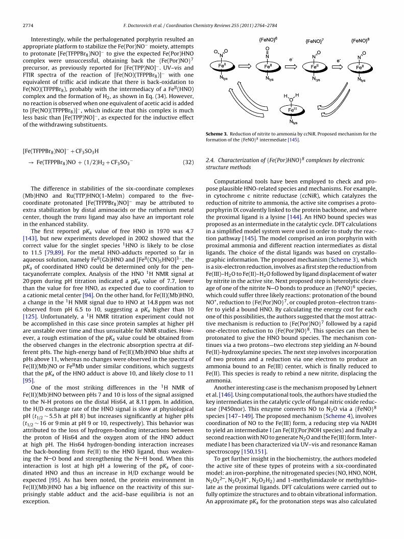
2 hemistry Reviews 255 (2011) 2764– 2784
atcpFeFcntlo
[
(ceci
[ctapt2taao[baetfpFt[
Fttp(atatiideFpe
774 F. Doctorovich et al. / Coordination C
Interestingly, while the perhalogenated porphyrin resulted anppropriate platform to stabilize the Fe(Por)NO− moiety, attemptso protonate [Fe(TFPPBr8)NO]− to give the expected Fe(Por)HNOomplex were unsuccessful, obtaining back the {Fe(Por)NO}7
recursor, as previously reported for [Fe(TPP)NO]−. UV–vis andTIR spectra of the reaction of [Fe(NO)(TFPPBr8)]− with onequivalent of triflic acid indicate that there is back-oxidation toe(NO)(TFPPBr8), probably with the intermediacy of a FeII(HNO)omplex and the formation of H2, as shown in Eq. (34). However,o reaction is observed when one equivalent of acetic acid is addedo [Fe(NO)(TFPPBr8)]−, which indicate that this complex is muchess basic than [Fe(TPP)NO]−, as expected for the inductive effectf the withdrawing substituents.
Fe(TFPPBr8)NO]− + CF3SO3H
→ Fe(TFPPBr8)NO + (1/2)H2 + CF3SO3− (32)
The difference in stabilities of the six-coordinate complexesMb)HNO and Ru(TTP)HNO(1-MeIm) compared to the five-oordinate protonated [Fe(TFPPBr8)NO]− may be attributed toxtra stabilization by distal aminoacids or the ruthenium metalenter, though the trans ligand may also have an important rolen the enhanced stability.
The first reported pKa value of free HNO in 1970 was 4.7143], but new experiments developed in 2002 showed that theorrect value for the singlet species 1HNO is likely to be closeo 11.5 [79,89]. For the metal HNO-adducts reported so far inqueous solution, namely FeII(Gb)HNO and [FeII(CN)5HNO]3−, theKa of coordinated HNO could be determined only for the pen-acyanoferrate complex. Analysis of the HNO 1H NMR signal at0 ppm during pH titration indicated a pKa value of 7.7, lowerhan the value for free HNO, as expected due to coordination to
cationic metal center [94]. On the other hand, for Fe(II)(Mb)HNO, change in the 1H NMR signal due to HNO at 14.8 ppm was notbserved from pH 6.5 to 10, suggesting a pKa higher than 10125]. Unfortunately, a 1H NMR titration experiment could note accomplished in this case since protein samples at higher pHre unstable over time and thus unsuitable for NMR studies. How-ver, a rough estimation of the pKa value could be obtained fromhe observed changes in the electronic absorption spectra at dif-erent pHs. The high-energy band of Fe(II)(Mb)HNO blue shifts atHs above 11, whereas no changes were observed in the spectra ofe(II)(Mb)NO or FeIIMb under similar conditions, which suggestshat the pKa of the HNO adduct is above 10, and likely close to 1195].
One of the most striking differences in the 1H NMR ofe(II)(Mb)HNO between pHs 7 and 10 is loss of the signal assignedo the N-H protons on the distal His64, at 8.11 ppm. In addition,he H/D exchange rate of the HNO signal is slow at physiologicalH (t1/2 ∼ 5.5 h at pH 8) but increases significantly at higher pHst1/2 ∼ 16 or 9 min at pH 9 or 10, respectively). This behavior wasttributed to the loss of hydrogen-bonding interactions betweenhe proton of His64 and the oxygen atom of the HNO adductt high pH. The His64 hydrogen-bonding interaction increases
he back-bonding from Fe(II) to the HNO ligand, thus weaken-ng the N O bond and strengthening the N H bond. When thisnteraction is lost at high pH a lowering of the pKa of coor-inated HNO and thus an increase in H/D exchange would bexpected [95]. As has been noted, the protein environment ine(II)(Mb)HNO has a big influence on the reactivity of this sur-risingly stable adduct and the acid–base equilibria is not anxception.Scheme 3. Reduction of nitrite to ammonia by ccNiR. Proposed mechanism for theformation of the {FeNO}8 intermediate [145].
2.4. Characterization of {Fe(Por)HNO}8 complexes by electronicstructure methods
Computational tools have been employed to check and pro-pose plausible HNO-related species and mechanisms. For example,in cytochrome c nitrite reductase (ccNiR), which catalyzes thereduction of nitrite to ammonia, the active site comprises a proto-porphyrin IX covalently linked to the protein backbone, and wherethe proximal ligand is a lysine [144]. An HNO bound species wasproposed as an intermediate in the catalytic cycle. DFT calculationsin a simplified model system were used in order to study the reac-tion pathway [145]. The model comprised an iron porphyrin withproximal ammonia and different reaction intermediates as distalligands. The choice of the distal ligands was based on crystallo-graphic information. The proposed mechanism (Scheme 3), whichis a six-electron reduction, involves as a first step the reduction fromFe(III)–H2O to Fe(II)–H2O followed by ligand displacement of waterby nitrite in the active site. Next proposed step is heterolytic cleav-age of one of the nitrite N O bonds to produce an {FeNO}6 species,which could suffer three likely reactions: protonation of the boundNO+, reduction to {Fe(Por)NO}7, or coupled proton–electron trans-fer to yield a bound HNO. By calculating the energy cost for eachone of this possibilities, the authors suggested that the most attrac-tive mechanism is reduction to {Fe(Por)NO}7 followed by a rapidone-electron reduction to {Fe(Por)NO}8. This species can then beprotonated to give the HNO bound species. The mechanism con-tinues via a two protons—two electrons step yielding an N-boundFe(II)-hydroxylamine species. The next step involves incorporationof two protons and a reduction via one electron to produce anammonia bound to an Fe(III) center, which is finally reduced toFe(II). This species is ready to rebind a new nitrite, displacing theammonia.
Another interesting case is the mechanism proposed by Lehnertet al. [146]. Using computational tools, the authors have studied thekey intermediates in the catalytic cycle of fungal nitric oxide reduc-tase (P450nor). This enzyme converts NO to N2O via a {FeNO}8
species [147–149]. The proposed mechanism (Scheme 4), involvescoordination of NO to the Fe(III) form, a reducing step via NADHto yield an intermediate I (an Fe(II)(Por)NOH species) and finally asecond reaction with NO to generate N2O and the Fe(III) form. Inter-mediate I has been characterized via UV–vis and resonance Ramanspectroscopy [150,151].
To get further insight in the biochemistry, the authors modeledthe active site of these types of proteins with a six-coordinatedmodel: an iron-porphine, the nitrogenated species (NO, HNO, NOH,N O 2 , N O H , N O H ) and 1-methylimidazole or methylthio-
2 2 2 2 2 2 2late as the proximal ligands. DFT calculations were carried out tofully optimize the structures and to obtain vibrational information.An approximate pKa for the protonation steps was also calculated
F. Doctorovich et al. / Coordination Chemistry Reviews 255 (2011) 2764– 2784 2775
nvers
tsaFpit
(stcNpesfiph{niwbotbofs
apThtsNogtFzt
Scheme 4. Proposed mechanism for the co
aking into account the solvation effects using an implicit solventcheme (PCM model). The most interesting feature proposed by theuthors is that in contrast with the (Mb)HNO structure reported byarmer, they suggested that the intermediate comprises a doublyrotonated species. This Fe(IV) (Por)NHOH complex can react eas-
ly with the second NO molecule. The thiolate role is to stabilizehis species and allows the double protonation.
The details of the heme-thiolate nitric oxide reductaseP450nor) catalytic mechanism are still controversial. One theory,upported by computational results [152], assumes two sequen-ial one-electron transfers from NAD(P)H to an initial {Fe(Por)NO}6
omplex. The {Fe(Por)NO}8 species thus formed would react withO, eventually releasing the ONNO2− anion (most probably in itsrotonated form), which decomposes to N2O and water. How-ver, more recent experimental results [153] suggest the firsttep of the mechanism (Scheme 4) to be direct hydride trans-er from NAD(P)H to {Fe(Por)NO}6, presumably resulting in anron-bound HNO unit. DFT geometry optimization of all the pro-osed reaction intermediates was reported, suggesting that theydride transfer to {Fe(Por)NO}6 could produce {Fe(Por)NOH}8 orFe(Por)HNO}8. Subsequent addition of NO to {Fe(Por)NOH}8 (butot to {Fe(Por)HNO}8 or {Fe(Por)N(H)OH}8 is predicted to lead to
mmediate liberation of HN2O2−, without any stable intermediates,
hich finally decomposes to H2O and N2O. Contrary to what woulde predicted according to the “thiolate push effect” dogma, the thi-late ligand at the heme active site obstructs NO reduction, ratherhan facilitate it. It is in fact shown that replacement of the thiolatey a neutral nitrogen ligand (i.e., lysine, as found in the active sitef cytochrome c nitrite reductase, mentioned above) clearly favors,rom a thermodynamic point of view, NO reduction at the hemeite [154].
Regarding the structural characterization of (Mb)HNO, Lindernd Rodgers used DFT calculations on a model system to study theotentials implicants of the different protonation schemes [155].he calculations were performed for a [Fe(Por)HNO(ImH)] as aeme model for (Mb)HNO. One of the key questions that the authorsried to solve was where was the proton located by calculatingeveral different coordination (O or N) and protonation (HNO orOH) isomers. The most stable isomer at the used level of the-ry corresponded to the N coordinated and protonated one. Theeometrical parameters are almost insensitive to the rotation of
he imidazole ring, suggesting a decoupling of the Fe-N(H)O ande-ImH � bonding. Moreover, in the EXAFS structure the imida-ole ring remains almost in an eclipsed conformation respect tohe pyrrolic 15N atoms [155]. The calculation of this rotated con-ion of NO into N2O by P450nor [136–138].
formation, is only 3.4 kJ/mol higher than the ground state structure.The barrier of rotation around the Fe N bond is low, suggesting thatthe ligand conformation can be easily stabilized in the protein dueto influences of the environment. Another interesting issue regard-ing the EXAFS structure of (Mb)HNO was the particular observationof the long Fe N(HNO) bond despite the high experimental IR fre-quency for Fe N. The authors identified a normal mode with a highFe N character, which agrees with the experimental shift observa-tion upon N isotopic labeling. The rationalization of the long bondand high IR frequency was attributed to a low effective mass.
Recently, Zhang and coworkers focused on the importanceof the computational tools to study compounds involving HNOand porphyrins [156]. Using a quantitative structure observablerelationship, the authors performed a large number of quantumcalculations on heme models in order to evaluate potential method-ologies to predict geometrical parameters: 1H NMR displacements,15N NMR displacements and �NO stretching frequencies of HNO andRNO bound moieties. In particular, the exploration involved Fe andRu porphyrins with methylimidazole and pyridine as axial ligandsand also some non-heme models. Several DFT functional and basissets were employed, selecting different combinations for each oneof the observables proposed. The best method to predict geometri-cal parameters was a pure DFT functional (mPWVWN). Among themost interesting results, the authors explored the potential effectof water interaction with the HNO bound to an Fe(II) porphyrin,as a model for (Mb)HNO. The calculations suggest that the IR fre-quency of the bound HNO in myoglobin can be explained by dualH2O HNO hydrogen bonding.
2.5. Reactivity of HNO towards Mn and Co porphyrins
Besides the reactivity of azanone with iron porphyrins, othermetalloporphyrins, namely with Co or Mn as the metal cen-ter, also react with HNO giving raise to interesting applications.When aqueous solutions of AS (at pH 7) or TSHA (at pH 10) areadded to [Mn(III)TEPyP]5+ under inert atmosphere, in equimolar orslight donor excess ratios, total conversion of [Mn(III)TEPyP]5+ to[Mn(III)TEPyP-NO]4+ is observed. Interestingly, and in opposition towhat is observed for the Fe3+ Porphyrins, there is a significant blueshift (more than 30 nm) of the Soret band, potentially providing asensitive tool for HNO detection and quantification, as will be dis-
cussed in the next section. Similar spectral changes are observedfor HNO donor reactions with [Mn(III)TPPS]3−, although in thesecase excess donor is needed for the reaction to be completed, dueto kinetic reasons that will be explained below.
2776 F. Doctorovich et al. / Coordination Chemistry Reviews 255 (2011) 2764– 2784
S fts of t
wwftncss
N
H
N
H
5[pepvntTTptsofrsr
HppaOstS
cheme 5. HNO versus NO reactivities of Mn, Co and Fe porphyrins and UV–vis shi
However, neither [Mn(III)TEPyP]5+ nor [Mn(III)TPPS]3− reactith NO(g) or NO donors (such as SNAP) under similar conditions,hich means on one hand that the equilibrium constant for the
ormation of the Mn(III) nitrosyl product is not favorable, and onhe other hand that these Mn(III) porphyrins do not suffer reductiveitrosylation to produce the Mn(II)(Por)NO complex as easily as theorresponding Fe(III) porphyrins do. Therefore, Mn(III) porphyrinshow selective reactivity towards HNO, while Mn(II) porphyrins areelective for NO (Eqs. 33–36) [128,129].
O + Fe(III)Por → Fe(II)(Por)NO (33)
NO + Fe(III)Por → Fe(II)(Por)NO + H+ (34)
O + Mn(III)Por → Slowreaction/N.R. (35)
NO + Mn(III)Por → Mn(II)(Por)NO + H+ (36)
Regarding Co porphyrins, the reaction with HNO of cobalt,10,15,20-tetrakis[3-(p-acetylthio-propoxy)phenyl]-porphyrinCo(P)], which is soluble in organic media, was studied [157]. Thisorphyrin has four sulfur anchors that allow it to be attached tolectrodes, as will be shown later. The reaction of NO(g) with CoII(P)roduces Co(III)(Por)NO− in a few minutes, in agreement with pre-ious data for other cobalt porphyrins [158]. In a similar timescale,o spectral changes are observed for Co(II)Por in the presence ofhe nitroxyl donor TSHA. On the other hand, adding to Co(III)PorSHA and 1,8-diazabicyclo(5.4.0)undec-7-ene—that acceleratesSHA decomposition in organic solvent by deprotonation-, slowlyroduces Co(III)(Por)NO−, as confirmed by UV–vis and IR spec-roscopy (�NO = 1679 cm−1). The UV–vis spectral changes are quitemall, similarly to what happens with the corresponding reactionsf iron porphyrins. In a similar timescale, no reaction takes placeor Co(III)Por in the presence of NO(g) or any studied NO donor. Theesults, similarly to what is observed for Mn porphyrins, clearlyhow that CoIIPor reacts with NO, and not with HNO, while CoIIIPoreacts with HNO and not with NO [157].
In summary, while iron porphyrins cannot discriminate NO fromNO due to the reductive nitrosylation reaction, both Mn and Coorphyrins tend to differentiate NO from HNO: Co(II) and Mn(II)orphyrins react rapidly with NO but not with HNO, while Co(III)nd Mn(III) porphyrins react rapidly with HNO but not with NO.
n the other hand, Mn porphyrins tend to show an importanthift in the UV–vis spectra (Soret band) when going from Mn(III)o Mn(II)NO porphyrins, while Co and Fe porphyrins do not (seecheme 5).
he Soret bands for the corresponding M(Por)NO products [128,129,157,158].
2.6. Comparison of the reactivity of HNO and NO towardsmetalloporphyrins
A key determinant of the fate of nitroxyl in any given media,either in vivo or in vitro, will depend upon all the competing reactionrates. To study the 1HNO-to-metalloporphyrin association kinetics,the UV–vis spectral changes corresponding to the formation of anitrosyl porphyrin can be followed as a function of time for eachreaction conditions, such as: different donors (SA or TSHA), pH, andrelative porphyrin to donor concentration ratios. Plots of the corre-sponding traces allow the determination of the initial observed rate�obs for the nitrosylation reaction. Even for reactions performedin strictly anaerobic media to avoid 1HNO/3NO− reactions withO2, trace amounts of oxygen can be present since it is extremelydifficult to remove O2 from water below 10−7 M. The obtainedexponential traces of nitrosyl product formation (when extremeca. 100 donor to porphyrin ratios are used) are strongly indicativethat for all cases the reaction is first order in porphyrin. Strik-ingly, two significantly different reaction times and stoichiometriesare observed for peripherical negatively or positively charged ironand/or manganese porphyrins. For negatively charged porphyrinssuch as [Fe(III)TPPS]3− and [Mn(III)TPPS]3− the reaction with ASat pH 7 (where AS spontaneous decomposition has a half-life ofabout 900 s) [127,159], requires a large excess of AS to drive thereaction to completion and for an equimolar ratio the reaction half-life is ca. 120 min. On the other hand, the reaction of positivelycharged porphyrins such as [Fe(III)TEPyP]5 and [Mn(III)TEPyP]5
total conversion to the nitrosyl metalloporphyrin is obtained withan equimolar porphyrin to donor ratio in less than 10 s. A sim-ilar behavior is observed for the reactions with the HNO donorTSHA. These results clearly point to different reaction mechanismsoperating depending on the porphyrin peripheral charge. The factthat the overall reaction rate for positively charged porphyrins byfar exceeds the donor spontaneous decomposition strongly sug-gests that a direct porphyrin-donor interaction is taking place andthat these porphyrins accelerate their decomposition. Therefore,Scheme 6 was proposed for the reactions of HNO donors with met-alloporphyrins.
In Scheme 6, kon(Donor) represents the bimolecular associa-tion rate constant of the metalloporphyrin with the HNO donor,
kcat(Donor) represents the porphyrin-accelerated donor decom-position rate constant, kd represents the spontaneous donordecomposition rate constant to yield HNO, and kon(HNO) is thebimolecular metalloporphyrin-to-HNO association rate. In order
F. Doctorovich et al. / Coordination Chemis
Scheme 6. Proposed mechanism for the reactions of HNO donors with metallopor-phyrins [129].
Frw
tbt(ptwacsisrtHdtHac
[
t
ig. 2. Nitrosyl product formation rate (diamonds) and estimated HNO productionate (squares) as a function of AS concentration for the reaction of [Mn(III)TPPS]3−
ith HNO.
o obtain the rate constants the following limiting cases haveeen analyzed: case (i) for negatively charged metalloporphyrins,he rate of 1HNO production due to spontaneous decompositionkdonor) exceeds the reaction rate of HNO with the metallopor-hyrin (kon(HNO)), and the dimerization of HNO competes withhe nitrosyl product formation. This is clearly shown in Fig. 2,here the observed nitrosyl product formation rate (diamonds)
nd the HNO production rate (squares) are plotted against donoroncentration. The shape of the �obs against AS plot is not linear,ince when the HNO production is increased (due to an increasen donor concentration) the dimerization rate increases with thequare of [HNO], as described by equation 37, and therefore theelation between [HNO] and [Donor] is not linear. However, usinghe steady state approximation for [HNO] (i.e. assuming that allNO produced by the donor either reacts with the porphyrin orimerizes—kdim = dimerization rate constant), the HNO concentra-ion can be estimated according to equation 39. Using the estimatedNO concentration, a plot of �obs against [HNO] yields as expected
linear plot, which allows determining the bimolecular HNO asso-iation rate constant kon(HNO).
HNO] =[
(kcat(Donor)[Donor] − �obs)]1/2
(37)
kdimOn the other hand, (case ii)) if the reaction of the donor withhe metalloporphyrin is faster than its spontaneous decomposition
try Reviews 255 (2011) 2764– 2784 2777
rate, the metalloporphyrin reacts directly with the donor, accelerat-ing its decomposition rate and forming the corresponding nitrosylcomplex by azanone transfer to the metal center. For these cases,almost no free HNO is produced, as evidenced by the quantitativeformation of the nitrosyl product even for equimolar metallopor-phyrin to donor ratios. In these cases, the �obs against [Donor]plot gives a straight line from which the bimolecular kcat(Donor) rateconstant can be obtained.
Further evidence for direct azanone transfer from AS to themetal center was obtained by the kinetic analysis of the reactionat pH 10, where AS is stable. Even under these conditions, thereaction with positively charged Mn porphyrins produces the nitro-syl product stoichiometrically in less than 5 s, while adding AS tothe negatively charged [Mn(III)TPPS]3− at pH 10 does not produceany reaction at all. Finally, DFT studies on the direct interactionof AS with both Mn and Fe porphyrins showed that AS (N2O3
2−,(ONa = NbO2)2−) is able to coordinate to either Mn(III) or Fe(III)by the Na nitrogen, which results in a significant weakening ofthe Na Nb bond, therefore partially explaining the observed rateacceleration. It is expected that, being negatively charged, AS coor-dination to the metal center will be faster for positively chargedmetalloporphyrins.
Using the kinetic analysis described above, kon(HNO) and/orkon(Donor) were obtained for several Mn and Fe porphyrins, andare shown in Table 3.
The data shown in Table 3 is consistent with two alternativepathways for the reactivity of nitroxyl donors (AS, TSHA) in aque-ous solutions: HNO transfer to the metal center through reaction ofthe porphyrin with azanone or with the HNO donor (see Scheme 6above). Interestingly, and despite the fact that kon for HNO bind-ing are faster than kon for the donors, donor concentrations areusually four orders of magnitude higher than those of HNO. There-fore, depending on the donor and porphyrin concentrations, one orthe other pathway will predominate. At relatively high concentra-tions (10−4, assuming a 1:1 = donor:porphyrin ratio) a faster rate isobserved for the direct reaction with the donor, as initially observedfor Mn(III) cationic porphyrins [129] and later also for the ferricanalogues [130].
Concerning the direct reaction between the donor and the met-alloporphyrin, a redox mechanism could be operative for TSHA.Piloty‘s acid are oxidized by several iron complexes yielding thecorresponding radical, which decomposes to produce NO and thereduced metal complex [160]. This mechanism would imply thatthe reaction proceeds through an intermediate state with thereduced porphyrin (Fe(II)), TSHA radical and free NO. Afterwards,NO would react very fast with the reduced iron porphyrin yield-ing the corresponding nitrosyl complex as the final product, asobserved here. However, a mechanism involving an intermedi-ate formed by TSHA and the metalloporphyrin followed by NO−
transfer to the porphyrins cannot be discarded and both mecha-nisms are compatible with the experimental observations. Moreinterestingly, recent obtained data for a negatively charged bro-mated porphyrin [MnBr8TPPS]3−, indicates that the actual followedreaction mechanism (reaction of the porphyrin with nitroxyl orwith the HNO donor) depends more likely on the reduction poten-tial of the porphyrin metal center, than on its peripheral charge.This is evidenced from the data in Table 3 showing that thenegatively charged [MnBr8TPPS]3−, having a positive reductionpotential due to the presence of the electron-attracting brominesubstituents, reacts directly with the HNO donor, the same as thepositively charged [MnTEPyP]5+. However, the negatively charged[MnTPPS]3−, with a reduction potential of −160 mV, reacts via the
free HNO pathway.The abovementioned scenario becomes even more complexin the presence of dioxygen, due to the reactivity of HNO andthe M(Por)NO porphyrins towards O2. These reactions give place

2778 F. Doctorovich et al. / Coordination Chemistry Reviews 255 (2011) 2764– 2784
Table 3Kinetic rate constants for the reactions of Mn(III) and Fe(III) porphyrins with AS and TSHA.
Porphyrin (III) E1/2 M3+/2+ vs NHE* kon (donor) (M−1 s−1) kon (HNO) (M−1 s−1) Ref.
AS (pH 7) TSHA (pH 10) AS (pH 7) TSHA (pH 10)
[MnTEPyP]5+ +220 mV 1.2 × 10⁴ 1.0 × 10⁴ – – [106][MnBr8TPPS]3− +209 mV 3.7 × 10³ 7.9 × 10³ – – PCa
[FeTEPyP]5+ +380 mV 5.4 × 10³ 1.1 × 10 4 – – [46,130][FeTPPS]3− +23 mV 0.5 – 1.0 × 10⁶ – [97][MnTPPS]3− −160 mV – – ∼4.0 × 10⁴ ∼9.0 × 10⁴ [129][FeMP11] −360 mV – – 6.4 × 10⁴ 3.1 × 10⁴ [46,130]
a PC = Doctorovich and coworkers, personal communication.
Table 4Kinetic rate constants for the relevant reactions of [Mn(TPPS)]3− with AS.
# Reaction Value Ref.
1 HONNOH → N2O 5 × 10⁻⁴ s−1 [67]2 HNO + HNO → HONNOH 8 × 10⁶ M−1 s−1 [66]3 AS → HNO + NO2
− 2.30 × 10⁻³ s−1 [54,56–59]4 [Mn3+(TPPS)]3− + HNO → [Mn2+(TPPS)NO]3− 1.0 × 10⁵ M−1 s−1 [129]5 AS + [Mn3+(TPPS)]3− → [Mn2+(TPPS)NO]3− + NO2
− 20 M−1 s−1 PCa
6 O2 + [Mn2+(TPPS)NO]3− → [Mn3+(TPPS)]3− + NO3− 5 M−1 s−1 PCa
7 O2 + HNO → H+ + NO3− 5 × 10³ M−1 s−1 (b)
a MP = Doctorovich and coworkers, personal communication.b 3 −1 s−1 (average of lit. rate constants) [18,26]; (7b) HO2 + NO → ONOOH k7b = 5 × 10⁹ M−1 s−1
[ fast, and the intermediates have been obviated, we decided to use k7a as the value forr
tOtpsIitsktrf
dA[
potnrurSitOpfpat
t[o(
Scheme 7. Reactions involved in the kinetic simulations which describe appro-
This reaction has to be considered as: (7a) O2 + HNO → HO2 + NO k7a = 5 × 10 M82]; (7c) ONOOH → H+ + NO3
− k7c = 1.2 s−1 [161]; As the reactions 2 and 3 are veryeaction 7.
o oxygen and nitrogen reactive species (ORS and NRS) such asNOO−, NO, NO2, HO2, and others. The most important effects due
o the presence of oxygen are the oxidation of the M(II)(Por)NOroduct back to the M(III)(Por) starting material [129] and the con-umption of HNO, therefore lowering its effective concentration.n order to analyze this scenario a set of more than 20 reactionsnvolving the porphyrins as well as the ORS and NRS have to beaken into consideration (see Appendix A). By using numericalimulations which take into consideration all the rate laws andinetic constants involved, it can be demonstrated that in ordero describe correctly the kinetic reaction profiles at pH 7, only 7eactions are needed, which are shown in Table 4 and Scheme 7or [Mn(TPPS)]3− with AS as the HNO donor.
Scheme 7 Reactions involved in the kinetic simulations whichescribe appropriately the reaction of metalloporphyrins withS (HN2O3
−) in the absence and presence of dioxygen, at pH 754,56–59,66,67,129].
Under anaerobic conditions, AS in solution decomposes toroduce only nitrite and HNO, which dimerizes yielding nitrousxide. However, if O2 is present at a high concentration comparedo AS, nitrate and nitrate are the main reaction products anditrous oxide is practically not formed due to the competingeaction of HNO with O2 to presumably produce initially HO2 andltimately NO3
− and NO2− (only the formation of NO3
– by thisoute was considered, see footnote b in Table 4 and dotted lines incheme 7). Therefore, when the metalloporphyrin and AS coexistn solution under anaerobic conditions, the reaction of HNO withhe M(III)Por competes not only with its dimerization but also with2. Moreover, since the M(II)PorNO product also reacts with O2 toroduce the starting material back, little or no product is observedor relative low concentrations of AS and metalloporphyrin com-ared to O2. On the contrary, when the concentrations of both ASnd metalloporphyrin are at least one order of magnitude higherhan [O2], the M(II)PorNO product is formed and stays in solution.
All the above observations could in principle be applied
o other metalloporphyrins and/or HNO donors different fromMn(TPPS)]3− and AS. The kinetic profiles should heavily dependn the most important rate constants involved, which are k4kon(HNO)), k5 (kon(donor)) and k6 (kox(MPorNO). The rate constantpriately the reaction of metalloporphyrins with AS (HN2O3−) in the absence and
presence of dioxygen, at pH 7 [54,56–59,66,67,129].
k4 ranges from 104 to 106 M−1 s−1, k5 from 1 to 104 M−1 s−1 and k6still has to be determined for a set of metalloporphyrins, althoughit can be estimated that for the metalloporphyrins with positivereduction potentials its value should be below 5 M−1 s−1 (Table 4).
Finally, Table 5 shows a comparison of the rate constants for thereactions of HNO and NO with metalloporphyrins and heme pro-teins. It is clear from the data presented, that although lacking the

F. Doctorovich et al. / Coordination Chemis
Table 5Bimolecular rate constants for HNO and NO binding to metalloporphyrins and hemeproteins.
Target Reactant kon (M−1 s−1) Ref.
Fe(III)MP11 HNO 3.1–6.4 × 10⁴ [130]Fe(III)MP11 NO 1.1 × 10⁶ [163][Fe(III)TPPS]3− NO 4.5 × 10⁵ [113]metMb(Fe(III)) HNO 8 × 10⁵ [18]metMb(Fe(III)) NO 4.9 × 10⁴ [18,164,165]catalase(Fe(III)) HNO 3 × 10⁵
[FeIITPPS]4− NO 1.5 × 10⁹ [113]5
pthtllmodut[tr
3
3
3
opdfncsaoiiptfmdferwdfi(ftalH
op
binding site, thereby assuring strong fluorescence quenching inthe probe off-state. While after excitation at 518 nm BOT1 showsemission at 526 nm and ˚fl = 0.12, by coordination to Cu(II) thefluorescence intensity decreases to ˚fl = 0.01 and the lifetime by
Mb(Mn(III)) HNO 3.4 × 10 [166]Mb(Fe(II)) HNO 1.4 × 10⁴ [125]O2 HNO 3 × 10³ [18]
rotein scaffold, the soluble porphyrins and MP11 display reactivityowards HNO comparable to the observed reactivity of HNO witheme proteins [18]. The bimolecular rate constants for HNO bindingo ferric heme systems are in the order of 105 to 106 M−1 s−1, simi-ar to the values obtained for NO and probably represent an upperimit for the reaction with ferric hemes. However, these rates are
ore than 1000 times lower than the corresponding values for NOr CO binding to ferrous hemes [113]. This trend can be explainedue to the fact that, for ferric hemes, the ligand binding rate issually dependent on the lability of the leaving water ligand, in con-rast with ferrous hemes, where the water ligand is weakly bound113]. This fact plays a key role in ligand binding rates in heme pro-eins [162], therefore it is also expected to be determinant for hemeactivity with HNO.
. Detection of nitroxyl
.1. Spectroscopic methods
.1.1. Colorimetric methodsThe high efficiency by which ferric porphyrins trap HNO, and the
bserved stability of {Mn(Por)NO}6 but not {Mn(Por)NO}5 com-lexes, suggested to test Mn(III) porphyrins as agents for HNO/NOiscrimination. As mentioned in a previous chapter, differentlyrom Fe(III) porphyrins, Mn(III) porphyrins do not suffer reductiveitrosylation in the timescale of the reaction with HNO, and, theonversion of Mn(III) porphyrins to Mn(II)(Por)NO results in a largehift of the UV–vis Soret band. Moreover, since water-soluble met-lloporphyrins (such as Mn(III)TPPS) or others which are soluble inrganic solvents (such as Mn(III)TPP) can be used and show a sim-lar shift, the method is useful for organic or aqueous solutions. Anmportant disadvantage for this reaction is that the Mn(II)(Por)NOorphyrin obtained as a product reacts with O2 to reproducehe starting Mn(III) back. Based on these data, Dobmeier et al.ound a way to overcome this inconvenience [167], and designed a
ethod for quantitative detection of nitroxyl with an estimatedynamic range of 24–290 nM. This optical sensor film, suitableor the quantitative determination of HNO, was obtained throughncapsulation of Mn(III)TPPS within the anaerobic local envi-onment of an aminoalkoxysilane xerogel membrane decoratedith trimethoxysilyl-terminated poly(amidoamine-organosilicon)endrimers, which are poorly O2-permeable. This HNO-sensinglms were tested with the HNO donors AS and sodium 1-isopropylamino)diazene-1-ium-1,2-diolate (IPA/NO), and wereound to provide a rapid means for determining HNO concentra-ions in aerobic solution. However, the rapid dimerization of HNOnd relatively slow rate of HNO complexation in the xerogel filmimit optimal sensor performance to environments with restricted
NO scavenging conditions and renders a narrow dynamic range.Another way to circumvent the problem raised by the reactivityf the nitrosyl product towards O2 is to protect the metallopor-hyrin by a protein matrix which slows down the oxidation rate.
try Reviews 255 (2011) 2764– 2784 2779
MnIII protoporphyrinate IX recombined into apomyoglobin reactswith AS, while it remains indifferent towards NO or NO donors,either under anaerobic or aerobic conditions [166]. The associationrate constant for the reaction of the reconstituted globin with thenitroxyl donor is practically the same than that for the free por-phyrinate, suggesting that the protein environment is not involvedin the association reaction mechanism. However, in contrast towhat happens to the free porphyrinate, the Mn(II)(Por)NO reactionproduct results to be significantly stable in the presence of oxygenwhen the porphyrinate is included in the protein matrix; this fea-ture is ascribed to the role of the distal aminoacidic residues on themetal center.
Another colorimetric method uses the reaction of HNOwith nitrosobenzene to form the indicator cupferron (N-nitrosophenylhydroxylamine) [168]. However, this method islimited to organic solvents.
A main disadvantadge to be taken into account for all thesecolorimetic methods is that, due to the fact that the UV–vismeasurements are done in a wavelength range where biologicalmaterials strongly absorb, these optical methods cannot be usedfor most in vitro or in vivo studies.
3.1.2. Fluorescent detectionSeveral proposed methods use non-heme fluorescent probes for
HNO detection. Oxidation of dihydrorhodamine by two-electronoxidants produces rhodamine, which is fluorescent and can bedetected at 570 nm with excitation at 500 nm. This method hasbeen used for the indirect detection of NO− by reaction withO2, being peroxynitrite the proposed intermediate which oxidizesdihydrorhodamine [169].
More recently, Lippard and coworkers developed two com-pounds for fluorescent detection of nitroxyl. The first one is abithiophene-substituted poly(p-phenyleneethynylene) derivative(CP1) which forms a Cu(II) complex that becomes fluorescent uponexposure to excess NO in unbuffered solutions, but under simulatedbiological conditions (pH 7.4 buffer) it shows a small decrease inemission upon treatment with NO, probably as a consequence ofprecipitation of the probe [170]. A 2-fold increase in CP1–Cu(II)integrated emission occurs upon exposure to AS, indicating thatCP1–Cu(II) can sense HNO selectively over NO in buffered aqueoussolutions, taking into account its apparent insensitivity to NO.
The second probe, BOT1 (see Fig. 3), comprises a BODIPYreporter site, which has optical properties that are well suited forcellular imaging experiments and a tripodal coordination environ-ment provided by a N-(triazolylmethyl)-N,N-dipicolyl framework,both separated by a triazole bridge as a rigid spacer [171]. Thisdesign minimizes the distance between fluorophore and metal
Fig. 3. Structure of BOT1.

2 hemistry Reviews 255 (2011) 2764– 2784
3cwspNsaoc
3
ricAs“daimKdNfaab
taodwdahh–rpnr
3
imwAauooNmfpgStsi
780 F. Doctorovich et al. / Coordination C
0-fold. However, if Cu(II) is reduced to Cu(I) by an excess ofysteine, the emission is restored. When Cu(II)[BOT1] is treatedith excess AS in buffered solutions, a ca. 4-fold increase in emis-
ion was observed with the concomitant formation of NO(g), andossibly Cu(I)[BOT1]. NO2
− and other RNS and ROS such as NO,O3
−, ONOO−, H2O2 and OCl−, failed to induce significant emis-ion enhancement of the Cu(II)[BOT1] complex. The complex waslso tested in HeLa cells: addition of AS to this cells increased thebserved intracellular red fluorescence over the course of 10 min,onsistent with an HNO-induced emission response.
.1.3. EPR detectionXia and coworkers examined the trapping specificity of different
edox forms of Fe-MGD (N-methyl-D-glucamine dithiocarbamateron). According to the authors, with Fe(II)-MGD, NO generatesharacteristic triplet NO•-Fe(II)-MGD signals, whereas HNO fromS is EPR silent. Both NO and NO– give rise to NO•-Fe(II)-MGDignals when Fe(III)-MGD is used. The authors asseverate thatspin trapping with Fe-MGD can distinguish NO and NO− and thisepends on the redox status of the iron center” [172]. However,n EPR signal would be observed if only NO is present, as well asf both NO and HNO are coexisting in the solution. Therefore, this
ethod cannot discriminate HNO from NO. In another publication,omarov and coworkers [173] showed that in the presence ofioxygen Fe(II)-MGD reacts with AS or AS-derived NO− to yieldO•-Fe(II)-MGD, possibly by reaction of NO– with Fe(III)-MGD
ormed by aerobic oxidation of the iron center. Since the param-gnetic NO•-Fe(II)-MGD complex is produced by AS (and/or NO−),nd also by NO, dithiocarbamate iron traps do not distinguishetween NO and NO−.
More recently, the nitronyl nitroxides 2-phenyl-4,4,5,5-etramethylimidazoline-l-oxyl 3-oxide (PTIO) and its water solublenalogue 2-(4-carboxyphenyl)-4,4,5,5-tetramethylimidazoline-1-xyl 3-oxide (C-PTIO) have been investigated as NO/HNOiscriminating agents [174]. Nitronyl nitroxides are known to reactith NO producing the corresponding imino nitroxides, which areetectable by EPR [175]. C-PTIO and PTIO are reduced by HNOnd converted into their respective imino nitroxides or iminoydroxylamines, both detectable by EPR. The yield of the iminoydroxylamine increases at the expense of the imino nitroxide
also produced by reaction with NO – with an increase in theatio [AS]0/[nitronyl nitroxide]0, where [AS]0 represents the totalroduction of HNO. Therefore, nitronyl nitroxide can discrimi-ate between NO and HNO only at high [AS]0/[nitronyl nitroxide]0atios.
.2. Electrochemical methods
Metalloporphyrins, are widely used in technical applications,ncluding gas sensors [176,177] when coupled to a surface. Ordered
onolayers of metalloporphyrins can be easily built, speciallyhen surface molecule linking is based on the establishment ofu S bonds, and a number or thiol bearing porphyrins are avail-ble. Furthermore, the use of S-acetyl protecting groups, thatndergo in situ cleavage on the surface allows the obtentionf single porphyrin layers in a straightforward manner. Basedn the previously described cobalt porphyrins reactivity towardsO and HNO (section 2.5), and the common use of surface-odified electrodes as electrochemical sensors, a porphyrin with
our anchors, Co(II)-5,10,15,20-tetrakis[3-(p-acetylthiopropoxy)henyl]porphyrin (Co(Por), shown in Fig. 4), was immobilized on aold surface by the formation of Au S bonds [157]. Both XPS and
TM techniques show that most Co(Por) molecules are adsorbed inhe lying down configuration by multiple linker binding to the goldurface, and that vacant gold sites are present that can be occupiedn some cases by smaller lateral size adsorbates.Fig. 4. Co(II)-5,10,15,20-tetrakis[3-(p-acetylthiopropoxy) phenyl]porphyrin.
The reactions of HNO and NO with electrode bound Co(Por),were studied by electrochemical techniques. Previous studiesby Kadish and coworkers showed that for nitrosyl complexesof Co(Por) in solution, four redox couples are found corre-sponding to the equilibria between [Co(Por)NO]+2, [Co(Por)NO]+1,CoIII(Por)NO−, [Co(Por)NO]− and [Co(Por)NO]2− states [178].
Electrochemical measurements of Co(Por) bound to electrodesshow the presence of three redox couples, as well as the electrodebound nitrosyl porphyrin complex. The most significant featureof the electrochemical data is that for CoIII/CoII couple (Eq. 37),the obtained E1/2 value is shifted ca. 400 mV to lower potentialscompared to the E1/2 in solution, strongly suggesting that Co(Por)adsorption on the gold electrode facilitates CoII oxidation. On theother hand, for the nitrosyl porphyrin the shift due to the gold sur-face effect is much smaller than before, and of only 60 mV (Eq. 38).This is ascribed to charge donation from the gold surface which sta-bilizes the Co(III) state in [Co(III)(Por)]+. This Co Au interaction isweakened by NO coordination to the metal center.
[Co(III)(Por)]+ + e− � Co(II)(Por) (37)
[Co(III)(Por)NO]+ + e− � [Co(III)(Por)NO−] (38)
Both NO and HNO reactivity of Co(Por) modified electrodes wastested in both the Co(II) and Co(III) oxidation states by electro-chemical methods. While CoIII(Por) reacts efficiently with HNO,it does not so with NO. This is evidenced by the presence of the[CoIII(Por)NO]+/[CoII(Por)NO] oxidation peak at 0.8 V, after HNOaddition to the Co(Por) electrode, and the lack of any electrochem-ical signal when NO donors are used instead. On the contrary, NOreacts rapidly with CoII(Por) modified electrodes while HNO doesnot. In summary, Co(Por) attached to gold retains the same selectivereactivity behavior towards HNO and NO, as observed in solu-tion. Based on the redox dependent selective reactivity observedfor the Co(Por) bound electrodes towards NO and HNO, and theredox potential shift due to a surface effect, reaction Scheme 8 wasproposed to selectively detect HNO amperometrically. Accordingto Scheme 8, the resting state electrode potential is set to 0.8 V,a value where the porphyrin is stable in the CoIII(Por) state andno current flow is observed. Reaction with HNO yields, accordingto previous observations, the CoIII(Por)NO− complex, which underthe described conditions is oxidized to CoIII(Por)NO. The resultingCoIII(Por)NO complex releases the NO ligand in a fast manner andyields CoIII(Por), which allows the catalytic cycle to start again. Inthis scheme the current intensity should be proportional to theamount of HNO that binds the CoIII(Por). Consistent with the pro-
posed scheme, measurement of the current vs. time plot at 0.8 V,where CoIII(Por) is stable, does not show any measurable currentchange. However, a few seconds after the addition of AS the currentintensity increases, and is maintained during several minutes, due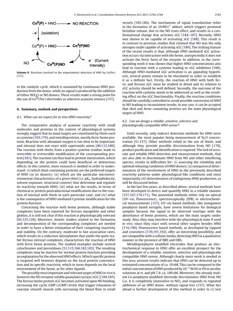
F. Doctorovich et al. / Coordination Chemis
S[
tdot
4
4
msataTrtdesorsictip
cg[aiawbwccaite
hTiv
cheme 8. Reactions involved in the amperometric detection of HNO by Co(Por)157].
o the catalytic cycle, which is sustained by continuous HNO pro-uction from the donor, while no signal is produced by the additionf either NO(g) or NO donors. These results make a strong point forhe use of CoIII(Por) electrodes as selective azanone sensors [157].
. Summary, outlook and perspectives
.1. What can we expect for in vivo HNO reactivity?
The comparative analysis of azanone reactivity with smallolecules and proteins in the context of physiological systems
trongly suggest that its main targets are constituted by thiols suchs cysteine [103,179], and metalloproteins, mostly ferric heme pro-eins. Reaction with abundant oxygen is too slow to be important,nd nitroxyl does not react with superoxide anion [40,112,180].he reaction with thiols, from a protein cysteine residue, leads toeversible or irreversible modification of the corresponding pro-ein [181]. The reaction can thus lead to protein inactivation, whichepending on the protein could have beneficial or deleteriousffects. In this context, more research is needed in order to under-tand: (i) which thiol-containing proteins are the preferred targetsf HNO (or its donors); (ii) which are the particular microenvi-onmental characteristics of a given thiol (i.e. pKa, hydrophobicity,olvent exposure, structural context) that determine and regulatets reactivity towards HNO; (iii) what are the results, in terms ofhemical or protein postraductional modifications due to the reac-ion of nitroxyl with thiols in each particular case; and (iv) whats the consequence of HNO mediated Cysteine modification for therotein function.
Concerning the reaction with heme proteins, although stableomplexes have been reported for ferrous myoglobin and otherlobins, it is still not clear if this reaction is physiologically relevant95,125,126]. Moreover, kinetic studies related to the formationnd decomposition of the corresponding complexes are neededn order to have a better estimation of their competing reactivitynd stability. On the contrary, moderate to fast association rates,hich results in a reductive nitrosylation that yields the quite sta-
le ferrous nitrosyl complexes, characterizes the reaction of HNOith ferric heme proteins. The studied examples include several
ytochromes and peroxidases [51,115,166,182,183]. The resultingomplexes may be inactive for normal protein function providingn explanation for the observed HNO effects. Which specific proteins targeted will however depend on the local protein concentra-ion and its specific reactivity, which in turns depends on the localnvironment of the heme, as for other ligands.
The possibly most important and relevant target of HNO in vivo is
owever the NO receptor soluble guanylate cyclase (sGC) [184,185].he ferrous heme protein sGC becomes activated upon NO binding,ncreasing the cyclic GMP (cGMP) levels that trigger relaxation ofascular smooth muscle cells increasing the blood flow in smalltry Reviews 255 (2011) 2764– 2784 2781
vessels [185,186]. The mechanism of signal transduction reliesin the formation of an {FeNO}7 adduct, which triggers proximalhistidine release, due to the NO trans effect, and results in a con-formational change that activates sGC [184–187]. Recently, HNOwas shown to be capable of activating sGC [188]. This result isin contrast to previous studies that claimed that NO was the onlynitrogen oxide capable of activating sGC [189]. The striking featureof the recent results is that, although HNO mediated sGC activa-tion occurs via interaction with the heme, unexpectedly it does notactivate the ferric form of the enzyme. In addition, in the corre-sponding work it was shown that higher HNO concentrations alsolead to reaction with a cysteine leading to sGC inhibition [188].Although HNO-mediated sGC activation is an appealing hypoth-esis, several points remain to be elucidated in order to establishit as a definite fact. Firstly, the reaction of HNO with both fer-ric and ferrous sGC must be studied in detail and its relation tosGC activity should be well defined. Secondly, the outcome of thereaction with cysteine needs to be addressed as well as the result-ing effect on the sGC functionality. Finally, the reaction conditionsshould be carefully controlled to avoid possible conversion of HNOto NO leading to inconsistent results. In any case, it can be acceptedthat thiol and heme containing proteins are the main physiologicaltargets of HNO.
4.2. Can we design a reliable, sensitive, selective andphysiologically compatible HNO sensor?
Until recently, only indirect detection methods for HNO wereavailable, the most popular being measurement of N2O concen-tration [51,157]. Other methods relied in the use of thiols, andalthough they provide possible discrimination from NO [179],product purification and identification is required. The lack of accu-rate and reliable HNO detection and measurement methods, thatare also able to discriminate HNO from NO and other interferingspecies, results in difficulties for: (i) assessing the reliability andoptimal releasing conditions of HNO donors; (ii) unequivocal deter-mination of the involvement of HNO in the previously describedreactivity patterns under physiological-like conditions and mostimportantly (iii) determination of endogenous HNO production, aswill be discussed below.
In the last few years, as described above, several methods havebeen developed to detect and quantify HNO in a reliable manner[157,167,170,171]. The presented methods either rely on optical(UV–vis, Fluorescence), spectroscopically (EPR) or electrochemi-cal measurements [157]. UV–vis based methods, like manganeseporphyrin based xerogels, have severe limitations for biologicalsamples because the signal to be observed overlaps with theabsorbance of heme proteins, which are the main targets understudy. Also, they may interfere with the physiological state if usedin vivo, since they react with several other targets besides HNO[116,190]. Fluorescence based methods, as developed by Lippardand coworkers [170,191,192], offer an interesting possibility, andare compatible with a cellular media, detecting HNO in a qualitativemanner in the presence of NRS and ORS.
Metalloporphyrin modified electrodes that produce an elec-trochemical response to HNO offer an excellent prospect for thedevelopment of a reliable, sensitive, selective and physiologicallycompatible HNO sensor. Although clearly more work is needed inthis area, present results indicate that HNO can be detected up toestimated concentrations of ca. 10 nM. This can be compared to theinitial concentration of HNO produced by 10−3 M AS or PA in aerobicsolutions at rt. and pH 7.4: ca. 100 nM. Moreover, the already stud-
ied Co-porphyrin modified electrode discriminates HNO from NO(i.e. it is completely insensitive to NO), and responds to repeatedadditions of an HNO donor, without signal loss [157]. What liesahead is further development of this method in order to (i) test
2 hemis
tNttetitb
4p
tptimayb(cfmirtsrtrtopaerotnttds
itpb
A
Aabg
A
t
782 F. Doctorovich et al. / Coordination C
he response in the presence of possible interfering species, likeO2
−, O2, O2−, OONO−, NH2OH, thiols, etc. (ii) calibrate the quan-
itative response to increasing HNO concentrations, (iii) improvehe electrode by the use of nano-structured materials such as poly-lectrolyte brushes [193] in order to increase the sensitivity tohe pM level, and iv) achieve electrode miniaturization for in vivo,n situ, and inside-cell measurements. In summary, a reliable, sensi-ive, selective and physiological compatible HNO sensing device shoulde available in the near future.
.3. Can we provide a definite answer to the endogenous HNOroduction paradox?
Given the unique biological properties described for HNO andhe numerous in vitro assays suggesting the existence of severalotential biosynthetic mechanisms for endogenous HNO produc-ion, it is tempting to envisage that this small molecule, likets cousin nitric oxide, could serve as an endogenous signaling
olecule. The best candidates for endogenous HNO productionre either the oxidation of hydroxylamine (or possibly hydrox-urea) by heme peroxidases [45], the conversion of NO to HNOy Mn or Fe superoxide dismutases [47] or by xanthine oxidaseXO) [194], or the production of HNO by NOS under certain spe-ial conditions [34,195]. From these options, the most reliable soar is the production of azanone by peroxidases, since its for-
ation was evaluated with a recently developed selective assayn which the reaction products, GS(O)NH2, in the presence ofeduced glutathione (GSH) were quantified by HPLC [45]. Key forhis reaction is the availability of active peroxidases and sub-trates in vivo. Also, further insights on the mechanism and theole played by the protein will contribute to clarify this mat-er. Concerning reduction of NO by SOD or XO, both studieselied on indirect methods for HNO detection which are some-imes difficult to interpret. Moreover, the low reduction potentialf the NO,H+/1HNO(3NO−) couple of ca. −0.55 to −0.8 V com-ared to 0.3 V for MnSOD makes NO reduction highly endergonicnd unlikely. Clearly, the proposed HNO production by thesenzymes has to be revised. Finally, HNO production by NOS alsoemains to be definitively proven, as the obtained evidence reliesn the formation of {FeNO}7 in the NOS active site, instead ofhe {FeNO}6 species formed when NO is produced, or in theeed for addition of Mn(III)SOD (that reduces HNO to NO) tohe reacting media, to recover full NOS dependent sGC activa-ion under low biopterin conditions [34,195]. In this context, airect HNO detection method will surely contribute to definitelyolve this issue.
However, the definitive proof for endogenous production of HNOn vivo, should be obtained by in situ measurements of HNO concen-ration in cell culture or tissues. A key element that will determine theossibility of performing such an experiment is the development of aiocompatible and selective HNO sensor.
cknowledgements
This work was financially supported by the University of Buenosires UBACYT X065 and UBACyT2010-12ANPCyT (PICT 2006-2396nd PICT-07-1650) and CONICET PIP1207. MAM and FD are mem-ers of CONICET AL thanks Fulbright foundation for support viarant 49418515.
ppendix A. Supplementary data
Supplementary data associated with this article can be found, inhe online version, at doi:10.1016/j.ccr.2011.04.012.
try Reviews 255 (2011) 2764– 2784
References
[1] H.W. Brown, G.C. Pimentel, J. Chem. Phys. 29 (1958) 883.[2] G. Maier, H.P. Reisenauer, M. De Marco, Angew. Chem. Int. Ed. 38 (1999)
108–110.[3] F. Dalby, J. Phys. 23 (1968) 136–143.[4] P. Bruna, Chem. Phys. Lett. 67 (1979) 109–114.[5] R. Guadagnini, G.C. Schatz, S.P. Walch, J. Chem. Phys. 102 (1995) 774.[6] A. Luna, M. Merch, B. Srn, O. Roos, Chem. Phys. 196 (1995) 437–445.[7] F.W. Dalby, Can. J. Phys. 36 (1958) 1336–1371.[8] J.W.C. Johns, A.R.W. McKellar, J. Chem. Phys. 66 (1977) 1217.[9] W. Anderson, Combust. Flame 117 (1999) 394–403.
[10] N.G. Connelly, T. Damhus, in: Nomenclature Of Inorganic Chemistry: IUPACRecommendations 2005 (Red Book), Royal Society of Chemistry, 2005, p. 300.
[11] N.G. Connelly, T. Damhus, in: Nomenclature Of Inorganic Chemistry: IUPACRecommendations 2005 (Red Book), Royal Society Of Chemistry, 2005, p. 98.
[12] N.G. Connelly, T. Damhus, in: Nomenclature Of Inorganic Chemistry: IUPACRecommendations 2005 (Red Book), Royal Society Of Chemistry, 2005, p. 111.
[13] D. Parsonage, A.J. Greenfield, S.J. Ferguson, BBA-Bioenerget. 807 (1985) 81–95.[14] V. Barone, Chem. Phys. Lett. 262 (1996) 201–206.[15] V. Gutmann, H. Tannenberger, Monatsh. Chem. 87 (1956) 421–424.[16] S. Moncada, R.M. Palmer, E.A. Higgs, Pharm. Rev. 43 (1991) 109–142.[17] L.J. Ignarro, Kidney Int. 55 (1996) S2.[18] K.M. Miranda, N. Paolocci, T. Katori, D.D. Thomas, E. Ford, M.D. Bartberger,
M.G. Espey, D.A. Kass, M. Feelisch, J.M. Fukuto, D.A. Wink, Proc. Natl. Acad.Sci. 100 (2003) 9196–9201.
[19] N. Paolocci, W.F. Saavedra, K.M. Miranda, C. Martignani, T. Isoda, J.M. Hare,M.G. Espey, J.M. Fukuto, M. Feelisch, D.A. Wink, D.A. Kass, Proc. Natl. Acad.Sci. 98 (2001) 10463–10468.
[20] C.H. Switzer, W. Flores-Santana, D. Mancardi, S. Donzelli, D. Basudhar, L. aRidnour, K.M. Miranda, J.M. Fukuto, N. Paolocci, D.A. Wink, Biochim. Biophys.Acta 1787 (2009) 835–840.
[21] E.C. DeMaster, F.N. Shirota, H.T. Nagasawa, Biochem. Biophys. Res. Commun.122 (1984) 358–365.
[22] E. DeMaster, H. Nagasawa, F. Shirota, Pharmacol. Biochem. Behav. 18 (1983)273–277.
[23] J.A. Reisz, E. Bechtold, S.B. King, Dalton Trans. (Cambridge, England: 2003) 39(2010) 5203–5212.
[24] E.C. DeMaster, F.N. Shirota, H.T. Nagasawa, Alcohol 2 (1985) 117–121.[25] E.G. DeMaster, B. Redfern, H.T. Nagasawa, Biochem. Pharm. 55 (1998)
2007–2015.[26] S.I. Liochev, I. Fridovich, Free Radic. Biol. Med. 34 (2003) 1399–1404.[27] J.M. Fukuto, R. Hszieh, P. Gulati, K.T. Chiang, H.T. Nagasaw, Biochem. Biophys.
Res. Commun. 187 (1992) 1367–1373.[28] J. Fukuto, P. Gulati, H.T. Nagasawa, Biochem. Pharm. 47 (1994) 922–924.[29] A.J. Väänänen, P. Salmenperä, M. Hukkanen, P. Rauhala, E. Kankuri, Free Radic.
Biol. Med. 41 (2006) 120–131.[30] S. Lancel, J. Zhang, A. Evangelista, M.P. Trucillo, X. Tong, D.A. Siwik, R.A. Cohen,
W.S. Colucci, Circ. Res. 104 (2009) 720–723.[31] X.L. Ma, A.S. Weyrich, D.J. Lefer, A.M. Lefer, Circ. Res. 72 (1993) 403–412.[32] O. Sidorkina, M.G. Espey, K.M. Miranda, D.A. Wink, J. Laval, Free Radic. Biol.
Med. 35 (2003) 1431–1438.[33] S. Adak, Q. Wang, D.J. Stuehr, J. Biol. Chem. 275 (2000) 33554–33561.[34] H.H. Schmidt, H. Hofmann, U. Schindler, Z.S. Shutenko, D.D. Cunningham, M.
Feelisch, Proc. Natl. Acad. Sci. 93 (1996) 14492–14497.[35] M. Feelisch, M. Te Poel, R. Zamora, A. Deussen, S. Moncada, Nature 368 (1994)
62–65.[36] Y. Ishimura, Y.T. Gao, S.P. Panda, L.J. Roman, B.S.S. Masters, S.T. Weintraub,
Biochem. Biophys. Res. Commun. 338 (2005) 543–549.[37] A.J. Hobbs, J.M. Fukuto, L.J. Ignarro, Proc. Natl. Acad. Sci. 91 (1994)
10992–10996.[38] J.J. Woodward, Y. Nejatyjahromy, R.D. Britt, M.A. Marletta, Control (2010)
5105–5113.[39] S. Stoll, Y. NejatyJahromy, J.J. Woodward, A. Ozarowski, M.A. Marletta, R.D.
Britt, J. Am. Chem. Soc. 132 (2010) 11812–11823.[40] K.M. Miranda, R.W. Nims, D.D. Thomas, M.G. Espey, D. Citrin, M.D. Bartberger,
N. Paolocci, J.M. Fukuto, M. Feelisch, D.A. Wink, J. Inorg. Biochem. 93 (2003)52–60.
[41] M.G. Espey, K.M. Miranda, D.D. Thomas, D.A. Wink, Free Radic. Biol. Med. 33(2002) 827–834.
[42] S. Donzelli, M.G. Espey, D.D. Thomas, D. Mancardi, C.G. Tocchetti, L. a Ridnour,N. Paolocci, S.B. King, K.M. Miranda, G. Lazzarino, J.M. Fukuto, D.A. Wink, FreeRadic. Biol. Med. 40 (2006) 1056–1066.
[43] D.A. Wink, J.A. Cook, S.Y. Kim, Y. Vodovotz, R. Pacelli, M.C. Krishna, J.B. a Russo,D. Mitchell, Jourdheuil, a M. Miles, M.B. Grisham, J. Biol. Chem. 272 (1997)11147–11151.
[44] K.M. Miranda, A.S. Dutton, L. a Ridnour, C. a Foreman, E. Ford, N. Paolocci, T.Katori, C.G. Tocchetti, D. Mancardi, D.D. Thomas, M.G. Espey, K.N. Houk, J.M.Fukuto, D.A. Wink, J. Am. Chem. Soc. 127 (2005) 722–731.
[45] S. Donzelli, M.G. Espey, W. Flores-Santana, C.H. Switzer, G.C. Yeh, J. Huang, D.J.Stuehr, S.B. King, K.M. Miranda, D.A. Wink, Free Radic. Biol. Med. 45 (2008)
578–584.[46] S.E. Bari, V.T. Amorebieta, M.M. Gutiérrez, J.A. Olabe, F. Doctorovich, J. Inorg.Biochem. 104 (2010) 30–36.
[47] V. Niketic, S. Stojanovic, A. Nikolic, A.M. Michelson, Free Radic. Biol. Med. 27(1999) 992–996.

hemis
F. Doctorovich et al. / Coordination C[48] D.R. Arnelle, J.S. Stamler, Arch. Biochem. Biophys. 318 (1995) 279–285.[49] D.A. Stoyanovsky, Y.Y. Tyurina, V.A. Tyurin, D. Anand, D.N. Mandavia, D.
Gius, J. Ivanova, B. Pitt, T.R. Billiar, V.E. Kagan, J. Am. Chem. Soc. 127 (2005)15815–15823.
[50] K.M. Miranda, H.T. Nagasawa, J.P. Toscano, Curr. Top. Med. Chem. 5 (2005)649–664.
[51] K.M. Miranda, Coord. Chem. Rev. 249 (2005) 433–455.[52] A. Angeli, Gazz. Chim. Ital. 26 (1896) 17–25.[53] A. Angeli, F. Angelico, Gazz. Chim. Ital. 33 (1903) 245.[54] M.N. Hughes, P.E. Wimbledon, J. Chem. Soc. Dalton Trans. 8 (1976) 703–707.[55] F. Bonner, B. Ravid, Inorg. Chem. 14 (1975) 558–563.[56] J. Veprek-Siska, V. Pliska, F. Smirous, F. Visely, Collect. Czech. Chem. Commun.
24 (1959) 687–693.[57] M.N. Hughes, P.E. Wimbledon, Chem. Ind. (1975) 742–743.[58] C.M. Maragos, D. Morley, D.A. Wink, T.M. Dunams, J.E. Saavedra, A. Hoff-
man, A.A. Bove, L. Isaac, J.A. Hrabie, L.K. Keefer, J. Med. Chem. 34 (1991)3242–3247.
[59] F.T. Bonner, B. Ravid, Inorg. Chem. 14 (1976) 548–553.[60] O. Piloty, Ber. Dtsch. Ges. 29 (1896) 1559.[61] P.C. Wilkins, H.K. Jacobs, M.D. Johnson, A.S. Gopalan, Inorg. Chem. 43 (2004)
7877–7881.[62] M. Blackburn, B. Mann, B. Taylor, A. Worrall, Eur. J. Biochem./FEBS 153 (1985)
553.[63] J. Scholz, P. Engel, C. Glidewell, K. Whitmire, Tetrahedron 45 (1989) 7695.[64] A. Porcheddu, L. De Luca, G. Giacomelli, Synlett 2009 (2009) 2149–2153.[65] A. Angeli, F. Angelico, F. Scurti, Chem. Central-Blatt. 73 (1902) 691–693.[66] F.T. Bonner, Y.H. Ko, Inorg. Chem. 31 (1992) 2514–2519.[67] F. Seel, C. Bliefert, Z. Anorg, Allg. Chem. 394 (1972) 187–196.[68] R. Zamora, A. Grzesiok, H. Weber, M. Feelisch, Biochem. J. 312 (1995) 333.[69] K.M. Miranda, T. Katori, C.L. Torres de Holding, L. Thomas, L. a Ridnour, W.J.
McLendon, S.M. Cologna, A.S. Dutton, H.C. Champion, D. Mancardi, C.G. Toc-chetti, J.E. Saavedra, L.K. Keefer, K.N. Houk, J.M. Fukuto, D.A. Kass, N. Paolocci,D.A. Wink, J. Med. Chem. 48 (2005) 8220–8228.
[70] S.V. Lymar, V. Shafirovich, J. Chem. Phys. B 111 (2007) 6861–6867.[71] L. Keefer, R. Nims, K. Davies, D. Wink, Methods Enzymol. 268 (1996) 281.[72] H. Nagasawa, E. DeMaster, B. Redfern, F. Shirota, D. Goon, J. Med. Chem. 33
(1990) 3120–3122.[73] B.-B. Zeng, J. Huang, M.W. Wright, S.B. King, Bioorg. Med. Chem. Lett. 14 (2004)
5565–5568.[74] S.B. King, Curr. Top. Med. Chem. 5 (2005) 665–673.[75] X. Sha, T.S. Isbell, R.P. Patel, C.S. Day, S.B. King, J. Am. Chem. Soc. 128 (2006)
9687–9692.[76] T.F. Emery, J. Neilands, J. Org. Chem. 27 (1962) 1075–1077.[77] Y. Adachi, H. Nakagawa, K. Matsuo, T. Suzuki, N. Miyata, Chem. Commun.
(2008) 5149–5151.[78] K. Matsuo, H. Nakagawa, Y. Adachi, E. Kameda, H. Tsumoto, T. Suzuki, N.
Miyata, Chem. Commun. (Cambridge, England) 46 (2010) 3788–3790.[79] V. Shafirovich, S.V. Lymar, Proc. Natl. Acad. Sci. 99 (2002) 7340–7345.[80] J.R. Buchholz, R.E. Powell, J. Am. Chem. Soc. 85 (1963) 509–511.[81] S.V. Lymar, V. Shafirovich, G.A. Poskrebyshev, Inorg. Chem. 44 (2005)
5212–5221.[82] S. Goldstein, G. Czapski, Free Radic. Biol. Med. 19 (1995) 505–510.[83] R.E. Huie, S. Padmaja, Free Radic. Res. Commun. 18 (1993) 195–199.[84] S. Goldstein, G. Czapski, J. Am. Chem. Soc. 118 (1996) 3419–3425.[85] H.M. Smallwood, J. Am. Chem. Soc. 51 (1929) 1985–1999.[86] S. King, H. Nagasawa, Methods Enzymol. 301 (1999) 211.[87] L.E. Orgel, J. Chem. Soc. (Resumed) (1953) 1276.[88] A.D. Walsh, J. Chem. Soc. (Resumed) (1953) 2288.[89] M.D. Bartberger, W. Liu, E. Ford, K.M. Miranda, C. Switzer, J.M. Fukuto, P.J.
Farmer, D.A. Wink, K.N. Houk, Proc. Natl. Acad. Sci. 99 (2002) 10958–10963.[90] M. Tronc, A. Huetz, M. Landau, F. Pichou, J. Reinhardt, J. Phys. B: At. Mol. Phys.
8 (1975) 1160–1169.[91] D. Teillet-Billy, F. Fiquet-Fayard, J. Phys. E: At. Mol. Phys. 10 (1977) Llll-7.[92] C. Szmytkowski, K. Maciag, J. Phys. B: Atom. Mol. Phys. (1991) 4273.[93] J. Tennyson, C. Noble, J. Phys. B: At. Mol. Phys. 19 (1986) 4025.[94] A.C. Montenegro, V.T. Amorebieta, L.D. Slep, D.F. Martín, F. Roncaroli, D.H.
Murgida, S.E. Bari, J.A. Olabe, Angew. Chem. Int. Ed. 48 (2009) 4213–4216.[95] M.R. Kumar, J.M. Fukuto, K.M. Miranda, P.J. Farmer, Inorg. Chem. 49 (2010)
6283–6292.[96] L. Cheng, G.B. Richter-Addo, in: K.M. Kadish, K. Smith, R. Guilard (Eds.), Aca-
demic Press, San Diego, 2000, pp. 219–291.[97] F.C. Kohout, F.W. Lampe, J. Am. Chem. Soc. 87 (1965) 5795.[98] P.A.S. Smith, G.E. Hein, J. Am. Chem. Soc. 82 (1960) 5731–5740.[99] J.R. Buchholz, R.E. Powell, J. Am. Chem. Soc. 87 (1965) 2350–2353.
[100] F.T. Bonner, L.S. Dzelzkalns, J.A. Bonucci, Inorg. Chem. 17 (1978) 2487–2494.[101] A.L. Smith, H.L. Johnston, J. Am. Chem. Soc. (1952) 12–14.[102] M.G. Bryukov, A.A. Kachanov, R. Timonnen, J. Seetula, J. Vandoren, O.M. Sark-
isov, Chem. Phys. Lett. 208 (1993) 392–398.[103] M.D. Bartberger, J.M. Fukuto, K.N. Houk, Proc. Natl. Acad. Sci. 98 (2001)
2194–2198.[104] P.C. Ford, Inorg. Chem. 49 (2010) 6226–6239.
[105] M. Hoshino, L. Laverman, P.C. Ford, Coord. Chem. Rev. 187 (1999) 75–102.[106] K.M. Kadish, The Porphyrin Handbook, Academic Press, 2000.[107] G.B. Richter-Addo, P. Legzdins, Metal Nitrosyls, Oxford University, New York,1992.[108] J.H. Enemark, R.D. Feltham, J. Am. Chem. Soc. 96 (1974) 5002–5004.
try Reviews 255 (2011) 2764– 2784 2783
[109] B.L. Westcott, J.H. Enemark, Inorganic Electronic Structure and Spectroscopy,John Wiley & Sons, New York, 1999.
[110] J.H. Enemark, R.D. Feltham, Coord. Chem. Rev. 13 (1974) 339–406.[111] G.R.A. Wyllie, W.R. Scheidt, Chem. Rev. 102 (2002) 1067–1090.[112] J. Pellegrino, S.E. Bari, D.E. Bikiel, F. Doctorovich, J. Am. Chem. Soc. 132 (2010)
989–995.[113] L.E. Laverman, P.C. Ford, J. Am. Chem. Soc. 123 (2001) 11614–11622.[114] B.B. Wayland, L.W. Olson, J. Am. Chem. Soc. 96 (1974) 6037–6041.[115] M. Hoshino, M. Maeda, R. Konishi, H. Seki, P.C. Ford, J. Am. Chem. Soc. 118
(1996) 5702–5707.[116] I. Spasojevic, I. Batini-Haberle, I. Fridovich, Nitric Oxide 4 (2000) 526–533.[117] Z.N. Zahran, J. Lee, S.S. Alguindigue, M.A. Khan, G.B. Richter-Addo, Dalton
Trans. (Cambridge, England: 2003) (2004) 44–50.[118] W.R. Scheidt, J.L. Hoard, J. Am. Chem. Soc. 95 (1973) 8281–8288.[119] F. Roncaroli, R.V. Eldik, J. Am. Chem. Soc. 128 (2006) 8042–8053.[120] M.P. Schopfer, J. Wang, K.D. Karlin, Inorg. Chem. 49 (2010) 6267–6282.[121] M.R.A. Blomberg, P.E.M. Siegbahn, Biochim. Biophys. Acta 1757 (2006)
969–980.[122] V.K.K. Praneeth, C. Näther, G. Peters, N. Lehnert, Inorg. Chem. 45 (2006)
2795–2811.[123] J.P. Collman, A. Dey, Y. Yang, R.A. Decréau, T. Ohta, E.I. Solomon, J. Am. Chem.
Soc. 130 (2008) 16498–16499.[124] J.P. Collman, Y. Yang, A. Dey, R. a Decréau, S. Ghosh, T. Ohta, E.I. Solomon, Proc.
Natl. Acad. Sci. 105 (2008) 15660–15665.[125] F. Sulc, C.E. Immoos, D. Pervitsky, P.J. Farmer, J. Am. Chem. Soc. 126 (2004)
1096–1101.[126] M.R. Kumar, D. Pervitsky, L. Chen, T. Poulos, S. Kundu, M.S. Hargrove, E.J.
Rivera, A. Diaz, J.L. Colón, P.J. Farmer, Biochem 48 (2009) 5018–5025.[127] D.A. Bazylinski, T.C. Hollocher, J. Am. Chem. Soc. 107 (1985) 7982–7986.[128] S.E. Bari, M. a Martí, V.T. Amorebieta, D. a Estrin, F. Doctorovich, J. Am. Chem.
Soc. 125 (2003) 15272–15273.[129] M.A. Martí, S.E. Bari, D.A. Estrin, F. Doctorovich, J. Am. Chem. Soc. 127 (2005)
4680–4684.[130] S.A. Suárez, M.A. Martí, P.M. De Biase, D.A. Estrin, S.E. Bari, F. Doctorovich,
Polyhedron 26 (2007) 4673–4679.[131] L.W. Olson, D. Schaeper, D. Lancon, K.M. Kadish, J. Am. Chem. Soc. 104 (1982)
2042–2044.[132] D. Lancon, K.M. Kadish, J. Am. Chem. Soc. 105 (1983) 5610–5617.[133] I.K. Choi, Y. Liu, D. Feng, K.J. Paeng, M.D. Ryan, Inorg. Chem. 30 (1991)
1832–1839.[134] Z. Wei, M.D. Ryan, Inorg. Chem. 49 (2010) 6948–6954.[135] M.H. Barley, K. Takeuchi, W.R. Murphy, T.J. Mayer, J. Chem. Soc., Chem. Com-
mun. (1985) 507.[136] J.N. Younathan, K.S. Wood, T.J. Meyer, Inorg. Chem. 31 (1992) 3280–3285.[137] M.H. Barley, T.J. Meyer, J. Am. Chem. Soc. 108 (1986) 5876–5885.[138] J. Lee, G.B. Richter-Addo, J. Inorg. Biochem. 98 (2004) 1247–1250.[139] P.J. Farmer, F. Sulc, J. Inorg. Biochem. 99 (2005) 166–184.[140] P.J. Farmer, M.R. Kumar, E. Almaraz, Comments Inorg. Chem. 31 (2010)
130–143.[141] R.G. Serres, C.A. Grapperhaus, E. Bothe, E. Bill, T. Weyhermüller, F. Neese, K.
Wieghardt, J. Am. Chem. Soc. 126 (2004) 5138–5153.[142] J. Pellegrino, R. Hübner, F. Doctorovich, W. Kaim, Chem. Eur. J. (2011),
doi:10.1002/chem.201003516.[143] M. Graetzel, S. Taniguchi, A. Henglein, Berichte Der Bunsen-Gesellschaft Bot.
Acta 74 (1970) 1003–1010.[144] O. Einsle, A. Messerschmidt, P. Stach, G.P. Bourenkov, H.D. Bartunik, R. Huber,
P.M. Kroneck, Nature 400 (1999) 476–480.[145] O. Einsle, A. Messerschmidt, R. Huber, P.M.H. Kroneck, F. Neese, J. Am. Chem.
Soc. 124 (2002) 11737–11745.[146] N. Lehnert, V. Praneeth, F. Paulat, J. Comput. Chem. 27 (2006) 1338–1351.[147] D.J. Richardson, N.J. Watmough, Curr. Opin. Chem. Biol. 3 (1999) 207–219.[148] S.J. Ferguson, Curr. Opin. Chem. Biol. 2 (1998) 182–193.[149] I. Moura, J.J. Moura, Curr. Opin. Chem. Biol. 5 (2001) 168–175.[150] M. Fujii, J. Biol. Chem. 270 (1995) 1617–1623.[151] E. Obayashi, S. Takahashi, Y. Shiro, J. Am. Chem. Soc. 120 (1998) 12964–12965.[152] D.L. Harris, Int. J. Quantum Chem. 88 (2002) 183–200.[153] A. Daiber, T. Nauser, N. Takaya, T. Kudo, P. Weber, C. Hultschig, H. Shoun, V.
Ullrich, J. Inorg. Biochem. 88 (2002) 343–352.[154] M. Vincent, I. Hillier, J. Ge, Chem. Phys. Lett. 407 (2005) 333–336.[155] D.P. Linder, K.R. Rodgers, Inorg. Chem. 44 (2005) 8259–8264.[156] Y. Ling, C. Mills, R. Weber, L. Yang, Y. Zhang, J. Am. Chem. Soc. 132 (2010)
1583–1591.[157] S.A. Suárez, M.H. Fonticelli, A.A. Rubert, E. de La Llave, D. Scherlis, R.C. Sal-
varezza, M.A. Martí, F. Doctorovich, Inorg. Chem. 49 (2010) 6955–6966.[158] D.G. Whitten, E.W. Baker, A.H. Corwin, J. Org. Chem. 28 (1963) 2363–2368.[159] A.S. Dutton, J.M. Fukuto, K.N. Houk, J. Am. Chem. Soc. 126 (2004) 3795–3800.[160] P.C. Wilkins, H.K. Jacobs, M.D. Johnson, A.S. Gopalan, Inorg. Chem. 43 (2004)
7877.[161] W. Koppenol, J. Moreno, W.A. Pryor, H. Ischiropoulos, J. Beckman, Chem. Res.
Toxicol. 5 (1992) 834–842.[162] E.E. Scott, Q.H. Gibson, J.S. Olson, J. Biol. Chem. 276 (2001) 5177.
[163] V.S. Sharma, R.A. Isaacson, M.E. John, M.R. Waterman, M. Chevion, Biochem22 (1983) 3897.[164] M. Hoshino, F. Matsuzaki, Y. Nabeshima, C. Hama, Neuron 10 (1993) 395–407.[165] L.E. Laverman, A. Wanat, J. Oszajca, G. Stochel, P.C. Ford, R. van Eldik, J. Am.
Chem. Soc. 123 (2001) 285–293.

2 hemis
[
[
[[
[
[[
[[[
[[[
[
[
[
[195] J.J. Woodward, Y. Nejatyjahromy, R.D. Britt, M.A. Marletta, J. Am. Chem. Soc.
784 F. Doctorovich et al. / Coordination C
166] I. Boron, S.A. Suárez, F. Doctorovich, M.A. Martí, S.E. Bari, J. Inorg. Biochem.105 (2011) 1044–1049.
167] K.P. Dobmeier, D.A. Riccio, M.H. Schoenfisch, Anal. Chem. 80 (2008)1247–1254.
168] D.W. Shoeman, H.T. Nagasawa, Nitric Oxide–Biol. Ch. 2 (1998) 66–72.169] K.M. Miranda, M.G. Espey, K. Yamada, M. Krishna, N. Ludwick, S.M. Kim, D.
Jourd’heuil, M.B. Grisham, M. Feelisch, J.M. Fukuto, D.A. Wink, J. Biol. Chem.276 (2001) 1720.
170] A.G. Tennyson, L. Do, R.C. Smith, S.J. Lippard, Nitric Oxide 26 (2007)4625–4630.
171] J. Rosenthal, S.J. Lippard, J. Am. Chem. Soc. 132 (2010) 5536–5537.172] Y. Xia, A. Cardounel, A.F. Vanin, J.L. Zweier, Free Radic. Biol. Med. 29 (2000)
793–797.173] A. Komarov, Free Radic. Biol. Med. 28 (2000) 739–742.174] U. Samuni, Y. Samuni, S. Goldstein, J. Am. Chem. Soc. 132 (2010) 8428–8432.175] T. Akaike, M. Yoshida, Y. Miyamoto, K. Sato, M. Kohno, K. Sasamoto, K.
Miyazaki, S. Ueda, H. Maeda, Biochem 32 (1993) 827–832.176] G. Guillaud, Coord. Chem. Rev. 178–180 (1998) 1433–1484.177] I.R. Davies, X. Zhang, Methods Enzymol. 436 (2008) 63–95.178] G.B. Richter-Addo, S.J. Hodge, G.B. Yi, M.A. Khan, T. Ma, E. Van Caemelbecke,
N. Guo, K.M. Kadish, Inorg. Chem. 35 (1996) 6530–6538.179] S. Donzelli, M.G. Espey, D.D. Thomas, D. Mancardi, C.G. Tocchetti, L.A. Ridnour,
N. Paolocci, S.B. King, K.M. Miranda, G. Lazzarino, J.M. Fukuto, D.A. Wink, FreeRadic. Biol. Med. 40 (2006) 1056–1066.
180] J.C. Irvine, R.H. Ritchie, J.L. Favaloro, K.L. Andrews, R.E. Widdop, B.K. Kemp-Harper, Trends Pharmacol. Sci. 29 (2008) 601–608.
181] B. Shen, A.M. English, Biochem 44 (2005) 14030–14044.
try Reviews 255 (2011) 2764– 2784
[182] D.E. Bikiel, L. Boechi, L. Capece, A. Crespo, P.M. De Biase, S. Di Lella, M.C.González Lebrero, M.A. Martí, A.D. Nadra, L.L. Perissinotti, D.A. Scherlis, D.A.Estrin, Phys. Chem. Chem. Phys.: PCCP 8 (2006) 5611–5628.
[183] M.P. Doyle, S.N. Mahapatro, R.D. Broene, J.K. Guy, J. Am. Chem. Soc. 110 (1988)593–599.
[184] J.W. Denninger, M.A. Marletta, BBA-Bioenerget. 1411 (1999) 334–350.[185] T.L. Poulos, Curr. Opin. Struct. Biol. 16 (2006) 736–743.[186] A.J. Hobbs, L.J. Ignarro, Methods Enzymol. 269 (1996) 1996–11996.[187] L. Capece, D.A. Estrin, M.A. Marti, Biochemistry 47 (2008) 9416–9427.[188] T.W. Miller, M.M. Cherney, A.J. Lee, N.E. Francoleon, P.J. Farmer, S.B. King,
A.J. Hobbs, K.M. Miranda, J.N. Burstyn, J.M. Fukuto, J. Biol. Chem. 284 (2009)21788–21796.
[189] E.A. Dierks, J.N. Burstyn, Biochem. Pharm. 51 (1996) 1593–1600.[190] I. Batinic-Haberle, I. Spasojevic, P. Hambright, L. Benov, A.L. Crumbliss, I.
Fridovich, Inorg. Chem. 38 (1999) 4011–4022.[191] M.H. Lim, S.J. Lippard, Nitric Oxide 43 (2004) 2071–2075.[192] L.E. McQuade, S.J. Lippard, Curr. Opin. Chem. Biol. 14 (2010) 43–49.[193] E.-Y. Choi, O. Azzaroni, N. Cheng, F. Zhou, T. Kelby, W.T.S. Huck, Langmuir 23
(2007) 10389–10394.[194] M. Saleem, H. Ohshima, Biochem. Biophys. Res. Commun. 315 (2004)
455–462.
132 (2010) 5105–5113.[196] R. Lin, P.J. Farmer, J. Am. Chem. Soc. 122 (2000) 2393–2394.[197] C.E. Immoos, F. Sulc, P.J. Farmer, K. Czarnecki, D.F. Bocian, A. Levina, J.B. Aitken,
R.S. Armstrong, P. Lay, J. Am. Chem. Soc. 127 (2005) 814–815.