Embed Size (px)
Citation preview

NIH Stroke Scale WWW.RN.ORG®
Reviewed August, 2017, Expires August, 2019 Provider Information and Specifics available on our Website
Unauthorized Distribution Prohibited
©2017 RN.ORG®, S.A., RN.ORG®, LLC By Wanda Lockwood, RN, BA, MA
The purpose of this course is to explain how to
administer the NIH Stroke Scale, consistently and
accurately.
Upon completion of this course, the healthcare provider should be able to:
• Explain the purpose of the NIH Stroke Scale.
• Explain the 11 items in the stroke scale. • Explain visual field testing.
• Discuss scoring parameters for all 11 items. • Discuss methods of administering each item.
Introduction Most stroke patients are seen initially by first responders and then
emergency room nurses and doctors rather than neurologists or neurosurgeons. Numerous different assessment scales for strokes
have been used, but this made consistent evaluation of condition and
progress difficult. The National Institutes of Health developed to NIH Stroke Scale to provide a common language and method of
assessment for all healthcare providers.
This scale focuses on impairment of function and ensures that the patient will be thoroughly examined in a consistent manner. The
results of testing are easily communicable and understandable when patient care is transferred and when patient’s progress is being
assessed.
Studies have shown that the patient’s results on the NIH Stroke Scale correlate with long-term progress. That is, those with low scores of 4
to 5 usually have positive outcomes and those with higher score have
Purpose
Goals

more severe impairment and more guarded outcomes. The NIH stroke scale can be easily administered in about 5 minutes.
Elements of the NIH Stroke Scale
Healthcare providers administering the NIH Stroke Scale should be specifically trained to use the scale and should have a copy of the
scale available when administering the assessment. Without training, the results are likely to be inconsistent.
Instructions Administer stroke scale items in the order listed. Record performance in each category after each subscale exam. Do not go back and change scores.
Follow directions provided for each exam technique. Scores should reflect what the patient does, not what the clinician thinks the patient can do. The clinician should record answers while administering the exam and work
quickly. Except where indicated, the patient should not be coached (i.e., repeated requests to patient to make a special effort).
It’s especially important to do the scale in order, starting with item 1 and moving through the evaluation to item 11. No question should be
skipped because the examiner assumes he or she knows the answer. Generally, the patient’s first response is the one scored, and once the
healthcare provider has entered a score, that score should not be changed. Additionally, the patient should be scored for actual
performance, not the healthcare provider’s expectations of what the
patient can or should be able to do.
The examiner should not tell the patient the purpose of the testing as a whole or the individual items as this may increase patient anxiety.
Before beginning the evaluation, the examiner should ensure that he or she has a copy of the scale and the picture and word cards
necessary for testing. The examiner should score after each item rather than relying on memory to fill out the scores after completion of
testing.
1a Level of Consciousness The investigator must choose a response if a full evaluation is
prevented by such obstacles as an endotracheal tube, language barrier, orotracheal
trauma/bandages.
0 = Alert; keenly responsive.
1 = Not alert; but arousable by minor stimulation to obey, answer, or respond.
2 = Not alert; requires repeated

A 3 is scored only if the patient
makes no movement (other than reflexive posturing) in response to noxious stimulation.
stimulation to attend, or is
obtunded and requires strong or painful stimulation to make movements (not stereotyped).
3 = Responds only with reflex
motor or autonomic effects or totally unresponsive, flaccid, and areflexic.
Level of Consciousness is assessed by greeting the patient, introducing
yourself, and asking simple questions, such as “How are you feeling”
and “Do you have any pain?” A patient with no impairment should respond readily; and, if speech is not impaired, verbally. Those with
speech impairment may attempt to speak, speak slowly and deliberately, or use body language or gestures, so careful observation
is necessary.
Remember that this is testing level of consciousness and not speech ability, so one should not assume that a person is not alert just
because the person can’t speak.
Scoring: • A patient who answers readily and appears to comprehend is
scored as 0. • If the examiner must repeat the question because the patient
doesn’t appear to understand or touch or otherwise stimulate the
patient to get a response, then this is scored as 1 or 2, depending on the degree of stimulation needed.
• A person who responds by movement only to painful stimuli, such as pinching of the nail beds, is scored as a 2.
• Those who are totally non-responsive, such as comatose patients, or have only reflexive posturing to noxious stimuli (rubbing chest
or pinching nailbed) are scored as 3.
Note: While medical translators are more appropriate than family members for translation during history and physical, for this scale
family members or friends may be asked to translate for the
healthcare provider and patient as the instructions and responses are usually simple.
1b. LOC Questions
The patient is asked the month and his/her age. The answer must be
0 = Answers both questions correctly.
correct - there is no partial credit
for being close. Aphasic and stuporous patients who do not comprehend the questions will
score 2.
Patients unable to speak because of endotracheal intubation, orotracheal trauma, severe
dysarthria from any cause, language barrier, or any other
problem not secondary to aphasia are given a 1. It is important that only the initial answer be graded
and that the examiner not "help" the patient with verbal or non-
verbal cues.
1 = Answers one question correctly.
2 = Answers neither question correctly.
Part 1b tests the level of consciousness as well as the ability to
comprehend and answer questions. Two questions are posed: • What month is it?
• How old are you?
The examiner should not coach the patient (“almost,” “try again”) or signal by facial expression that an answer is right or wrong but should
simply score the answers given (or NOT given); however, if a patient
spontaneously corrects an answer, such as “I’m 72, no….uh….73,” then the correction is accepted as the response.
If the patient cannot speak, the examiner should ask the patient if he
or she is able to write and provide a pencil/pen and paper. Spoken or written responses are equally valid. Misspellings should be ignored as
long as writing can be understood.
Scoring: • Patients who answer both questions correctly are scored as 0.
• Patients who are not able to respond verbally or in writing because of intubation, trauma or other problems (excluding
aphasia) are given a score of 1. • Patients who answer one question correctly are scored as 1 as
well.
• However, aphasic patients who cannot respond or comatose patients are given a score of 2.
1c. LOC Commands
The patient is asked to open and
close the eyes and then to grip and release the non-paretic hand. Substitute another one step
command if the hands cannot be used. Credit is given if an
unequivocal attempt is made but not completed due to weakness.
If the patient does not respond to command, the task should be
demonstrated to him or her (pantomime), and the result scored (i.e., follows none, one or two
commands). Patients with trauma, amputation, or other physical
impediments should be given suitable one-step commands. Only the first attempt is scored
0 = Performs both tasks correctly.
1 = Performs one task correctly.
2 = Performs neither task correctly.
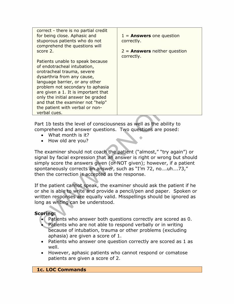
When asking the patient to carry out a physical action, make sure the
patient is focusing on you and whenever possible demonstrate as well as direct the patient. For example, state, “I want you to open your
eyes wide and then close them tightly” and demonstrate the action you want.
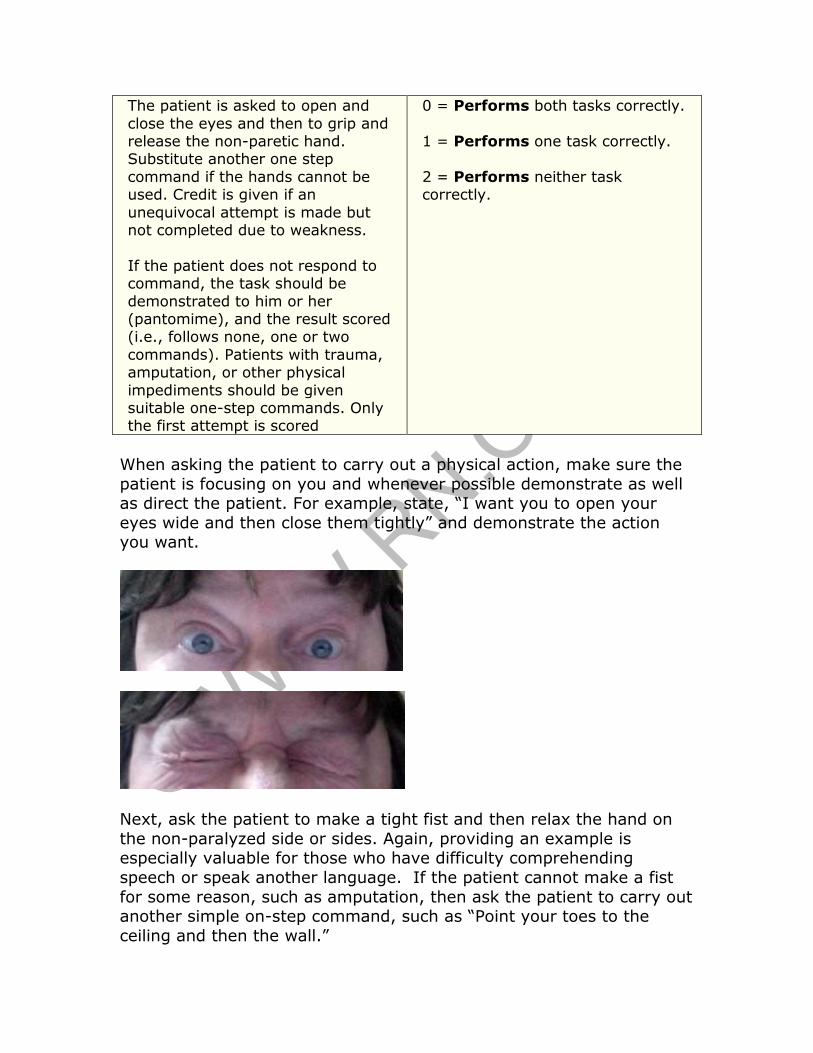
Next, ask the patient to make a tight fist and then relax the hand on
the non-paralyzed side or sides. Again, providing an example is especially valuable for those who have difficulty comprehending
speech or speak another language. If the patient cannot make a fist
for some reason, such as amputation, then ask the patient to carry out another simple on-step command, such as “Point your toes to the
ceiling and then the wall.”

Scoring:
It’s important to score the patient’s first attempt. This is fairly simple to score as the patient is scored according to the ability to do both,
one, or no tasks. • Both tasks correctly done, score 0.
• One task correctly done, score 1. • Neither task done correctly, score 2.
2. Best Gaze
Only horizontal eye movements will be tested. Voluntary or reflexive (oculocephalic) eye movements will
be scored, but caloric testing is not done. If the patient has a
conjugate deviation of the eyes that can be overcome by voluntary
or reflexive activity, the score will be 1. If a patient has an isolated peripheral nerve paresis (CN III, IV
or VI), score a 1.
Gaze is testable in all aphasic patients. Patients with ocular trauma, bandages, pre-existing
blindness, or other disorder of visual acuity or fields should be
tested with reflexive movements, and a choice made by the investigator.
Establishing eye contact and then
moving about the patient from side to side will occasionally clarify the presence of partial gaze palsy.
0 = Normal. 1 = Partial gaze palsy; gaze is
abnormal in one or both eyes, but forced deviation or total gaze
paresis is not present.
2 = Forced deviation, or total gaze paresis not overcome by the oculocephalic maneuver.
This item is done only to evaluate the horizontal movement of the eyes.
In some types of strokes, the eyes may have a forced deviation to the side of the stroke (most often with right-sided strokes). The examiner
may need to hold the eyelids open by pulling them toward the eyebrows with the thumbs.
This item is usually tested by simply asking the patient to follow a
finger with his/her eyes (while demonstrating), but for a patient who is less alert or has difficulty with that, you can ask the patient to look at
your face and move from one side of the patient to the other.
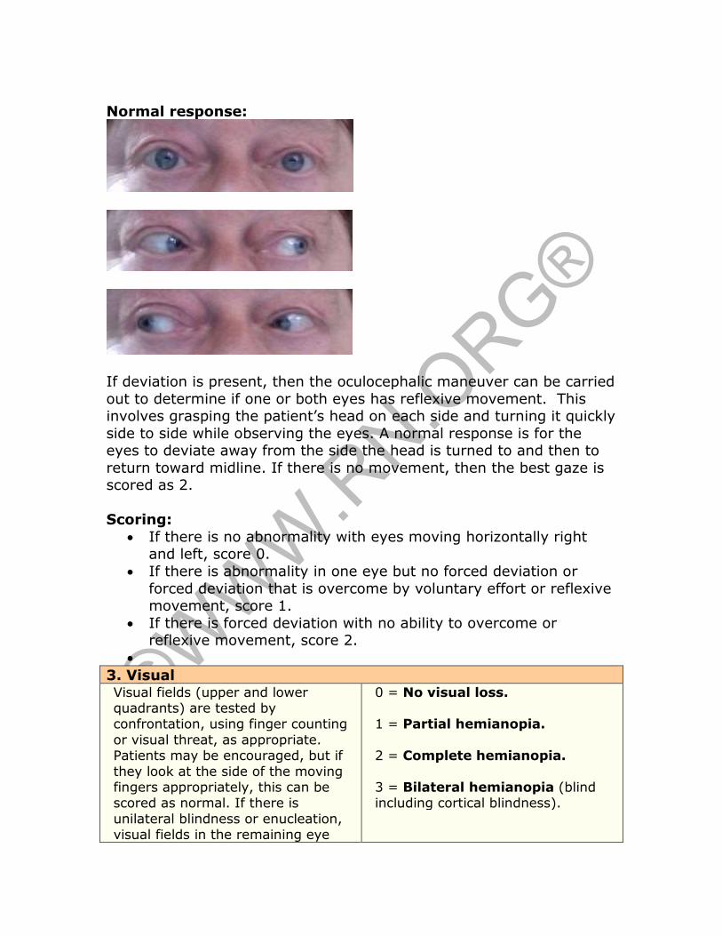
Normal response:
If deviation is present, then the oculocephalic maneuver can be carried
out to determine if one or both eyes has reflexive movement. This involves grasping the patient’s head on each side and turning it quickly
side to side while observing the eyes. A normal response is for the eyes to deviate away from the side the head is turned to and then to
return toward midline. If there is no movement, then the best gaze is scored as 2.
Scoring:
• If there is no abnormality with eyes moving horizontally right and left, score 0.
• If there is abnormality in one eye but no forced deviation or
forced deviation that is overcome by voluntary effort or reflexive movement, score 1.
• If there is forced deviation with no ability to overcome or reflexive movement, score 2.
•
3. Visual Visual fields (upper and lower quadrants) are tested by confrontation, using finger counting
or visual threat, as appropriate. Patients may be encouraged, but if
they look at the side of the moving fingers appropriately, this can be scored as normal. If there is
unilateral blindness or enucleation, visual fields in the remaining eye
0 = No visual loss. 1 = Partial hemianopia.
2 = Complete hemianopia.
3 = Bilateral hemianopia (blind including cortical blindness).

are scored.
Score 1 only if a clear-cut asymmetry, including
quadrantanopia, is found. If patient is blind from any cause,
score 3. Double simultaneous stimulation is
performed at this point. If there is extinction, patient receives a 1,
and the results are used to respond to item 11.
The visual field includes all the eyes can see when looking forward, including peripheral vision. There is a left visual field and a right visual
field with an overlap at the center of vision. Damage to the right or left visual cortex or optic nerves can impair vision.
The visual field of each eye is divided into 4 quadrants—with the
horizontal visual field wider than the vertical. Test 3 assesses the visual fields for impairment.
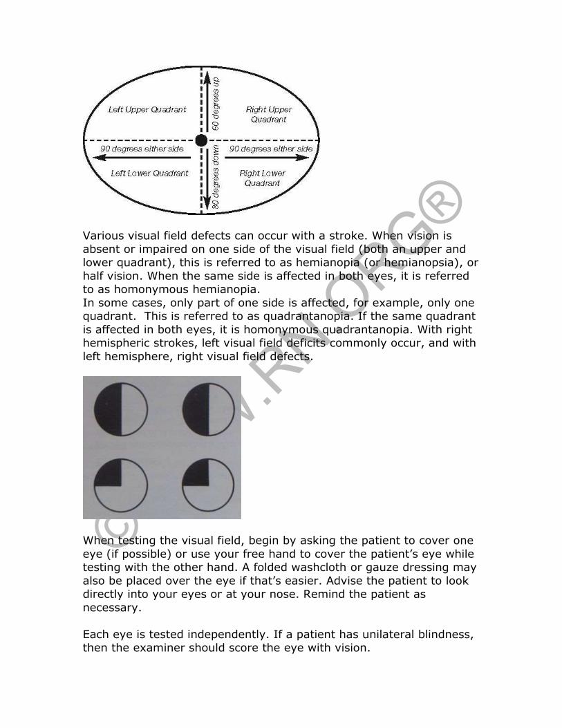
Various visual field defects can occur with a stroke. When vision is
absent or impaired on one side of the visual field (both an upper and lower quadrant), this is referred to as hemianopia (or hemianopsia), or
half vision. When the same side is affected in both eyes, it is referred to as homonymous hemianopia.
In some cases, only part of one side is affected, for example, only one quadrant. This is referred to as quadrantanopia. If the same quadrant
is affected in both eyes, it is homonymous quadrantanopia. With right hemispheric strokes, left visual field deficits commonly occur, and with
left hemisphere, right visual field defects.
When testing the visual field, begin by asking the patient to cover one
eye (if possible) or use your free hand to cover the patient’s eye while testing with the other hand. A folded washcloth or gauze dressing may
also be placed over the eye if that’s easier. Advise the patient to look directly into your eyes or at your nose. Remind the patient as
necessary.
Each eye is tested independently. If a patient has unilateral blindness, then the examiner should score the eye with vision.

Testing usually begins by starting
with one hand beside one of the patient’s ears, two fingers extended
and wiggling. The examiner slowly moves the fingers forward in an arc
around the face, asking the patient to point or tell you when he or she
sees the fingers.
This helps to establish the right position for testing. The same thing
is done on the opposite side. Once the visual field is established, the examiner moves the fingers up to
examine the upper quadrants and down for the lower quadrants.
Positioning of the hand may vary
slightly depending on the patient’s visual field. The testing hand is usually
positioned about 6 inches lateral to the nose (or with the tips of the fingers
aligned with the side of the face) and anterior to the ear for outer quadrants
at about the level of the temples for upper quadrant evaluation and the
mouth or chin for lower quadrant evaluation. (When testing inner
quadrants, the fingers need to come
forward a few inches to compensate for the bulk of the hand covering the opposite eye.)
The visual field of each eye is evaluated in random order—for example
RUQ, RUQ, LLQ, LUQ, RLQ and so on. You should explain the finger counting exercise: “I’m going to hold up different numbers of fingers,
and I’d like you to tell me (or show me) how many you see.”
In some cases, a patient may be able to write the number or indicate the number with fingers if the patient is not able to speak. Sometimes,
if patients are unable to talk, you may note the eyes moving toward the fingers on movement—an indication the patient can see them.
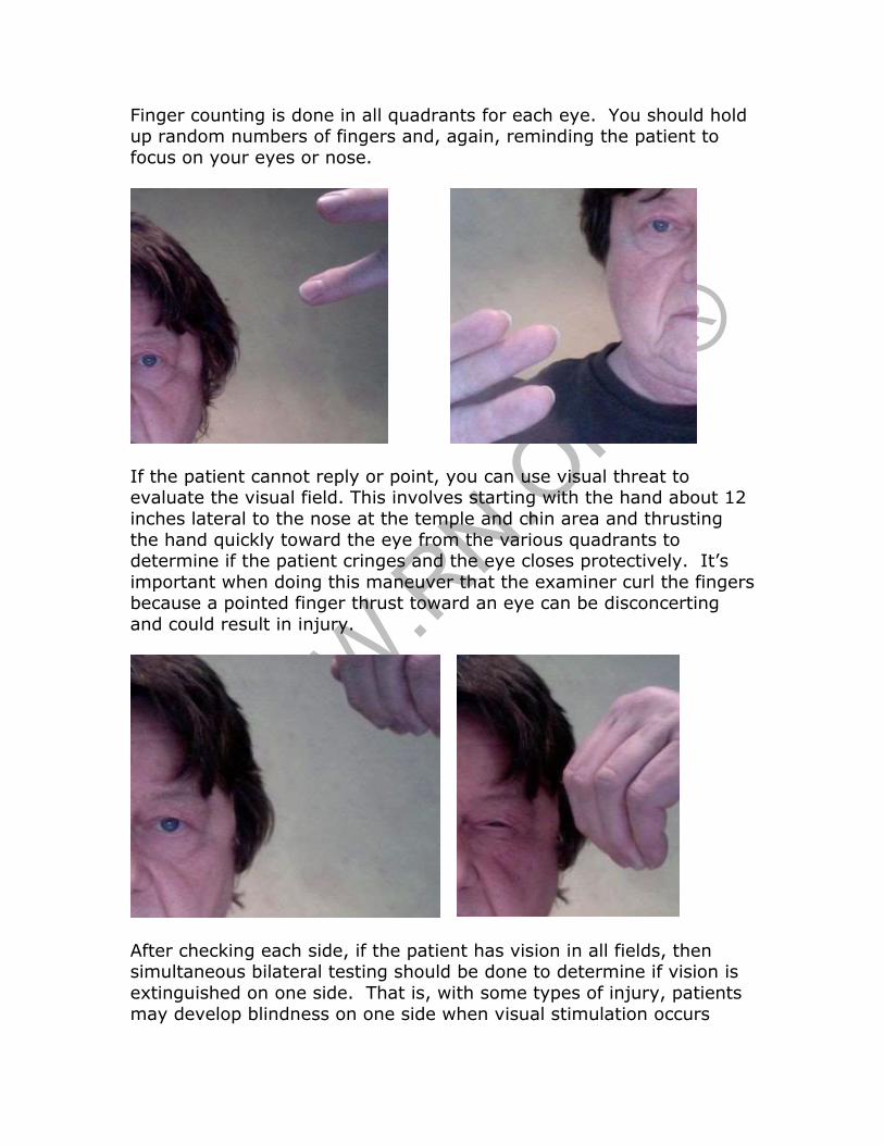
Finger counting is done in all quadrants for each eye. You should hold up random numbers of fingers and, again, reminding the patient to
focus on your eyes or nose.
If the patient cannot reply or point, you can use visual threat to evaluate the visual field. This involves starting with the hand about 12
inches lateral to the nose at the temple and chin area and thrusting
the hand quickly toward the eye from the various quadrants to determine if the patient cringes and the eye closes protectively. It’s
important when doing this maneuver that the examiner curl the fingers because a pointed finger thrust toward an eye can be disconcerting
and could result in injury.
After checking each side, if the patient has vision in all fields, then simultaneous bilateral testing should be done to determine if vision is
extinguished on one side. That is, with some types of injury, patients
may develop blindness on one side when visual stimulation occurs
simultaneously on both sides. If this occurs, the visual field item is scored as 1.
Scoring:
• If vision is intact in all quadrants, score 0. • If there is partial hemianopia/quadrantanopia or if extinction
occurs with bilateral simultaneous testing, score 1. • If there is complete hemianopia (half blindness in one eye),
score 2. • If there is bilateral hemianopia (half blindness in both eyes) or
total blindness, score 3.
4. Facial palsy Ask – or use pantomime to
encourage – the patient to show teeth or raise eyebrows and close eyes. Score symmetry of grimace
in response to noxious stimuli in the poorly responsive or non-
comprehending patient.
If facial trauma/bandages, orotracheal tube, tape or other physical barriers obscure the face,
these should be removed to the extent possible.
0 = Normal symmetrical
movements. 1 = Minor paralysis (flattened
nasolabial fold, asymmetry on smiling).
2 = Partial paralysis (total or
near-total paralysis of lower face). 3 = Complete paralysis of one or
both sides (absence of facial movement in the upper and lower
face).
If a patient has a pronounced paralysis of one side of the face, that
may be quite evident, but paresis or partial paralysis may be more difficult to recognize. The healthcare provider may ask the patient to
raise the eyebrows and close the eyes, similar to 1c, but the focus here is on symmetry rather than the ability to follow commands.
Upper palsy may be exhibited by drooping of the eyelid or smoothing
of wrinkles on the affected side and unequal lifting of the eyebrows. The lower part of the face, especially about the mouth, is usually the
best place to focus because paresis may be most evident there. Ask
the patient to show his or her teeth or make a big smile (giving a demonstration).
If patients are edentulous, they should be asked to show their gums;
however, if they have dentures available and can put them into their mouths, they should be examined with dentures.

If there is no facial palsy, the smile should be relatively even with the lips in basically the same position on both sides. Notice in the photo
below that the same numbers of teeth are visible on both sides, an indication that there is no palsy.
With palsy, the mouth may appear skewed and lips elevated more on one side than the other.
In some cases, there may be very little movement of the lips and few
teeth showing.
If a patient is aphasic or responds poorly, you can use noxious stimuli, such as pinching the nail bed, to elicit a grimace and observe the
grimace closely for asymmetry.
Scoring: • If movements are normal and symmetrical, score 0.
• If there is minor paralysis or asymmetry, score 1.
• If there is partial paralysis with total or near-total paralysis of the lower face, score 2.
• If there is complete paralysis of one or both sides with absence of facial movement in the upper and lower face, score 3.

5. Motor Arm The limb is placed in the appropriate position: extend the
arms (palms down) 90 degrees (if sitting) or 45 degrees (if supine). Drift is scored if the arm falls
before 10 seconds.
The aphasic patient is encouraged using urgency in the voice and
pantomime, but not noxious stimulation. Each limb is tested in turn, beginning with the non-
paretic arm.
Only in the case of amputation or joint fusion at the shoulder, the examiner should record the score
as untestable (UN), and clearly write the explanation for this
choice.
0 = No drift; limb holds 90 (or 45) degrees for full 10 seconds.
1 = Drift; limb holds 90 (or 45) degrees, but drifts down before full
10 seconds; does not hit bed or other support.
2 = Some effort against gravity;
limb cannot get to or maintain (if cued) 90 (or 45) degrees, drifts down to bed, but has some effort
against gravity.
3 = No effort against gravity; limb falls.
4 = No movement.
UN = Amputation or joint fusion, explain: _____________________
NOTE: Score each arm separately: 5a. Left Arm
5b. Right Arm
This test evaluates the patient’s ability to hold the arm in a stable
position without drift (falling). Each limb is scored separately. The left arm is usually tested first and then the right; however, if paralysis or
paresis is present, the examination should begin with the non-paretic arm.
If the patient is sitting, position the arm palm down at 90 or at 45 if
the patient is supine and ask the patient to hold it at that position until told to lower the arm. If the patient is aphasic, demonstrate holding
the arm up to show the patient what is expected.
Once the arm is in the correct position, release the arm. A slight dip is
normal upon release, but then the arm position should stabilize. You should count down 10 seconds, verbally and showing fingers.
Stand to the side and use an environmental marker, such as a window,
poster, or curtain, behind the limb to help determine if the arm is drifting. It can be difficult to see drift if you are standing over and
looking down at the arm.

45 position:
Drift:
If the patient can’t hold the arm up and it falls onto the bed, ask the
patient to try to lift the arm and note any proximal movement.
Flaccid:
Proximal lift:
Scoring should always be done, even though the patient is paralyzed.
If patients are limited in mobility because of disease, or disability, such as arthritis, the examiner must use best judgment when evaluating.
Scoring:
• If there is no drift (normal), the score is 0. • If the arm drifts downward but doesn’t hit a support, such as the
bed or arm of a chair, the score is 1. • If there is some effort to maintain the arm but it falls onto
support before the 10 seconds elapse, the score is 2. • UN for untested should only be used with amputation or joint
fusion. • If the arm immediately falls onto support, and there is no effort
against gravity, the score may be 3 or 4. In this case, you
should ask the patient to try to lift the arm and note any proximal movement of the shoulder. If movement is evident, the
score is 3, even if the movement is minimal.
• If there is no voluntary movement at all of if the patient is comatose, the score is 4.
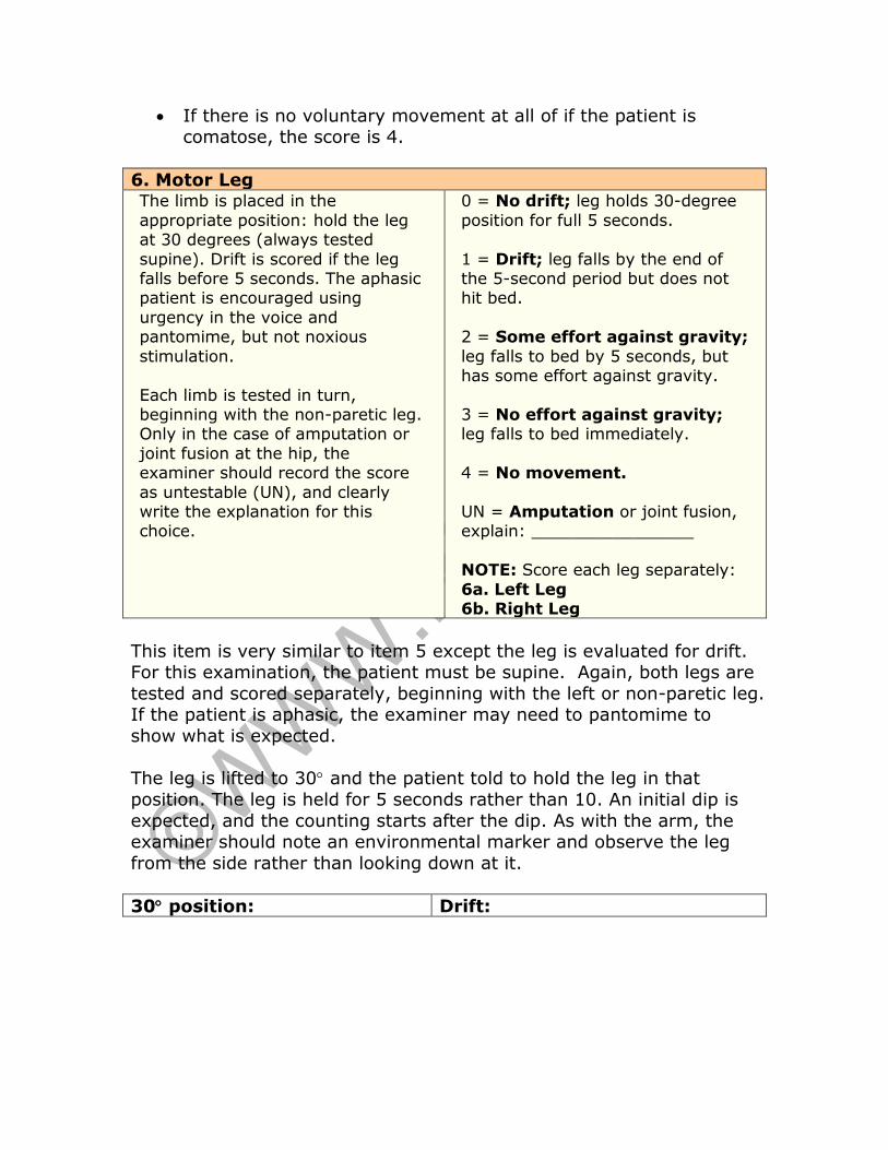
6. Motor Leg The limb is placed in the
appropriate position: hold the leg at 30 degrees (always tested
supine). Drift is scored if the leg falls before 5 seconds. The aphasic patient is encouraged using
urgency in the voice and pantomime, but not noxious
stimulation.
Each limb is tested in turn, beginning with the non-paretic leg. Only in the case of amputation or
joint fusion at the hip, the examiner should record the score
as untestable (UN), and clearly write the explanation for this choice.
0 = No drift; leg holds 30-degree
position for full 5 seconds.
1 = Drift; leg falls by the end of the 5-second period but does not hit bed.
2 = Some effort against gravity;
leg falls to bed by 5 seconds, but has some effort against gravity.
3 = No effort against gravity; leg falls to bed immediately.
4 = No movement.
UN = Amputation or joint fusion, explain: ________________
NOTE: Score each leg separately:
6a. Left Leg 6b. Right Leg
This item is very similar to item 5 except the leg is evaluated for drift. For this examination, the patient must be supine. Again, both legs are
tested and scored separately, beginning with the left or non-paretic leg. If the patient is aphasic, the examiner may need to pantomime to
show what is expected.
The leg is lifted to 30 and the patient told to hold the leg in that
position. The leg is held for 5 seconds rather than 10. An initial dip is
expected, and the counting starts after the dip. As with the arm, the examiner should note an environmental marker and observe the leg
from the side rather than looking down at it.
30 position: Drift:
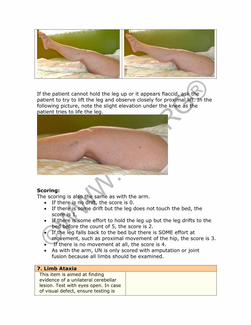
If the patient cannot hold the leg up or it appears flaccid, ask the patient to try to lift the leg and observe closely for proximal lift. In the
following picture, note the slight elevation under the knee as the patient tries to life the leg.
Scoring:
The scoring is also the same as with the arm. • If there is no drift, the score is 0.
• If there is some drift but the leg does not touch the bed, the score is 1.
• If there is some effort to hold the leg up but the leg drifts to the bed before the count of 5, the score is 2.
• If the leg falls back to the bed but there is SOME effort at movement, such as proximal movement of the hip, the score is 3.
• If there is no movement at all, the score is 4. • As with the arm, UN is only scored with amputation or joint
fusion because all limbs should be examined.
7. Limb Ataxia This item is aimed at finding
evidence of a unilateral cerebellar lesion. Test with eyes open. In case
of visual defect, ensure testing is

done in intact visual field. The
finger-nose-finger and heel-shin tests are performed on both sides, and ataxia is scored only if present
out of proportion to weakness.
Ataxia is absent in the patient who cannot understand or is paralyzed. Only in the case of amputation or
joint fusion, the examiner should record the score as untestable
(UN), and clearly write the explanation for this choice. In case of blindness, test by having the
patient touch nose from extended arm position.
0 = Absent.
1 = Present in one limb.
2 = Present in two limbs.
UN = Amputation or joint fusion, explain: ________________
The test for ataxia evaluates muscle control and coordination, differentiating these from general weakness. In this case, ataxia does
not refer to gait (a common usage of the term). Two tasks are required:
• Finger-nose-finger test.
• Heel-shin test. •
Note: Patients who are blind should extend their arms and touch their noses, repeating the action 3 or 4 times.
When explaining the finger-nose-finger task, state, “I want you to put touch your finger to mine” and reach out and touch the patient’s finger
as a demonstration, “and then touch your nose,” again demonstrating, and then, “Good, now repeat that a few
times.”
If a visual field defect was identified, then the examiner should be sure to place his or
her finger within the patient’s visual field. If the patient has difficulty with the first
task, the examiner should observe the patient carefully to determine if the
problem seems to be weakness or ataxia. If, for example, an arm is quite weak, the
patient may have some difficulty directing
his or her finger toward the examiner’s finger and the patient’ own nose and
movement may be unsteady. If the limb is

extremely weak, the examiner should usually assume the score is 0.
For the heel-shin test, unless the patient is very alert and responsive,
it’s often best to help position the heel on the shin (starting below the
knee) when explaining to the patient to run the heel of one foot
down and back up the shin of the opposite leg. If the patient has
difficulty understanding, you can move the heel down and back up the shin in demonstration. Both legs
are examined. If a patient is paralyzed on one side or in one limb, then
the nonparetic limbs are tested.
Scoring: Ataxia is scored only if it’s present:
• If a patient cannot do the tasks because of coma, paralysis, or lack of ability to understand the directions, then the score is 0.
Example: o Left-sided paralysis with no ataxia on the right = 0.
• If ataxia is found in one limb, then the score is 1. Example: o Right-sided paralysis with ataxia in the left arm but not
the left leg = 1. • If ataxia is found in two or more limbs (arms, legs, or a
combination), the score is 2. • The only circumstances in which UN for untested can be scored
is if the patient has amputation or joint fusion that prevents
completion of the task.
8. Sensory Sensation or grimace to pinprick when tested, or withdrawal from
noxious stimulus in the obtunded or aphasic patient. Only sensory
loss attributed to stroke is scored as abnormal and the examiner should test as many body areas
(arms [not hands], legs, trunk, face) as needed to accurately check
for hemisensory loss. A score of 2, “severe or total
0 = Normal; no sensory loss.
1 = Mild-to-moderate sensory loss; patient feels pinprick is less
sharp or is dull on the affected side; or there is a loss of superficial pain with pinprick, but patient is
aware of being touched.
2 = Severe to total sensory loss; patient is not aware of being touched in the face, arm, and leg.

sensory loss,” should only be given
when a severe or total loss of sensation can be clearly demonstrated.
Stuporous and aphasic patients
will, therefore, probably score 1 or 0. The patient with brainstem stroke who has bilateral loss of
sensation is scored 2. If the patient does not respond and is
quadriplegic, score 2. Patients in a coma (item 1a=3) are automatically given a 2 on this
item.
The pinprick test should be done with a sterile safety needle, being
careful not to break the skin. Testing should be on bare skin because
testing through clothing blunts the sensation. Testing should not be done on the hands or feet because preexisting neuropathy may impair
sensation in those areas. Testing is usually done on each side of the face and in the proximal portions of the limb and above wrists and
ankles.
Sites for testing may include: • Sides of face (in front of the ears).
• Above the wrists. • Trunk.
• Slightly below the knees (medial aspect). • Above the ankles.
At the beginning of the test, ask the patient to close his or her eyes and
tell the patient he or she may feel small pricks to the skin. The patient
can indicate where he or she feels the needle pricks by saying “right”
or “left,” pointing, or writing. You should avoid asking the patient if
the needle prick feels sharp or dull as this may confuse some patients
but should ask, “Which side feels sharper?”
When testing, usually start at the top and work down, pricking the skin
in random order, such as right face, left face, right face, right face, left face, right face, and left face. If a regular pattern is used, the patient
may indicate a sensation on a side because of an expectation that the same pattern is persisting.
If the patient responds to one side and not the other, You should prick
the side the patient is not responding to and ask the patient directly, “Do you feel that?” and then prick the other side, “Do you feel that?”
If the patient cannot respond verbally, look carefully at the patient’s
face and observe for grimace, which indicates discomfort and sensation.
Scoring: The patient is scored as 1 or 2 only is sensory loss is clearly
demonstrated. Thus, patients who are aphasic or • If the patient can feel pinpricks normally and equally, score 0.
• If the patient appears to grimace equally for pinpricks on both sides, score 0.
• If the patient can detect touch but doesn’t feel pain or feels one side is duller than the other, score 1. Patients who are in a
stupor or aphasic are usually assumed to be 0 or 1. • If the patient has no sensation of touch on one or both sides or
is non-responsive because of coma, score 2.
9. Best Language A great deal of information about
comprehension will be obtained during the preceding sections of the examination. For this scale
item, the patient is asked to describe what is happening in the
attached picture, to name the items on the attached naming
sheet and to read from the attached list of sentences.
Comprehension is judged from responses here, as well as to all of
the commands in the preceding general neurological exam.
If visual loss interferes with the
0 = No aphasia; normal.
1 = Mild-to-moderate aphasia; some obvious loss of fluency or
facility of comprehension, without significant limitation on ideas
expressed or form of expression. Reduction of speech and/or
comprehension, however, makes conversation about provided materials difficult or impossible. For
example, in conversation about provided materials, examiner can
identify picture or naming card content from patient’s response.
2 = Severe aphasia; all

tests, ask the patient to identify
objects placed in the hand, repeat, and produce speech. The intubated patient should be asked to write.
The patient in a coma (item 1a=3)
will automatically score 3 on this item. The examiner must choose a score for the patient with stupor or
limited cooperation, but a score of 3 should be used only if the patient
is mute and follows no one-step commands.
communication is through
fragmentary expression; great need for inference, questioning, and guessing by the listener. Range
of information that can be exchanged is limited; listener
carries burden of communication. Examiner cannot identify materials provided from patient response.
3 = Mute, global aphasia; no
usable speech or auditory comprehension.
When doing any testing that involves vision, it’s important to find out if the patient normally wears glasses and needs them to read or look at
images. Glasses are often removed by first responders, especially if patients use oxygen masks during transit, but glasses should be
returned to a patient for this examination if possible.
Patients may experience various visual defects because of the stroke, including diplopia, impaired visual memory, and visual hallucinations,
and these defects may interfere with the patient’s ability to complete
this task.
Even if the examiner believes he or she has an adequate understanding of the patient’s language skills by this point of the
exam, this part should still be completed for confirmation. If a patient is blind or has severe visual impairment, this part of the test can be
done by asking the patient to feel and describe common items, such as a glass, ballpoint pen, magazine, comb, or toothbrush. If a patient
cannot speak for any reason (aphasia, trauma, intubation), but is able to write, the patient can write out answers to demonstrate language
ability.
Note: Patients with a left hemisphere stroke, characterized by right
sided paralysis or paresis, may have expressive, receptive, or global aphasia.
For a patient with vision, show the patient the following
picture and ask the patient to describe what is in the picture: “Can you tell me what you see in this picture?” As
the patient responds, listen very carefully to the patient’s articulation
and note any slurring or difficulty expressing ideas.
Task 1

The primary elements of the picture include:
• The boy is taking cookies from the cookie jar while the girl reaches for a cookie.
• The mother is washing dishes. • The boy’s stool is falling.
• The sink is overflowing.
If the patient hesitates or speaks slowly but eventually does an adequate job of describing the picture, this is scored as normal. If, for
example, the patient states the boy is reaching for a cookie and the woman is washing dishes, the examiner should NOT coach by asking
“What is happening to the stool” or “What do you notice about the sink,” but encourage the patient to give more details by asking, “What
else do you see?”
The next task involves showing the patient the following
drawings of common items and asking the patient to identify those items you point to. Items include:
• Glove. • Key.
• Feather. • Cacti (or cactuses).
Task 2

• Chair. • Hammock.
People with visual impairment often identify the glove as a “hand” and the feather as a “leaf,” and these answers are considered correct.
Additionally, many people (especially those who don’t live in desert areas) have trouble identifying cacti and may perceive them as
cartoon animals, and that is considered correct if it seems reasonable.
Hammocks are not common in all parts of the world, so some people may not recognize the hammock or know the word and may describe it
as a swing or describe the tree trunks/stumps or grass. The examiner should allow some leeway in naming because the primary purpose is
to evaluate the patient’s ability to use language and speak clearly.
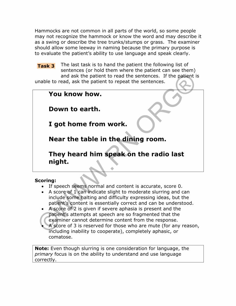
The last task is to hand the patient the following list of sentences (or hold them where the patient can see them)
and ask the patient to read the sentences. If the patient is unable to read, ask the patient to repeat the sentences.
You know how.
Down to earth.
I got home from work.
Near the table in the dining room.
They heard him speak on the radio last
night. Scoring:
• If speech seems normal and content is accurate, score 0.
• A score of 1 can indicate slight to moderate slurring and can include some halting and difficulty expressing ideas, but the
patient’s content is essentially correct and can be understood. • A score of 2 is given if severe aphasia is present and the
patient’s attempts at speech are so fragmented that the examiner cannot determine content from the response.
• A score of 3 is reserved for those who are mute (for any reason, including inability to cooperate), completely aphasic, or
comatose.
Note: Even though slurring is one consideration for language, the
primary focus is on the ability to understand and use language correctly.
Task 3

10. Dysarthria If the patient is thought to be normal, an adequate sample of
speech must be obtained by asking the patient to read or repeat words from the attached list.
If the patient has severe aphasia, the
clarity of articulation of spontaneous speech can be rated. Only if the
patient is intubated or had other physical barriers to producing speech, the examiner should record
the score as untestable (UN) and clearly write an explanation for this
choice. Do not tell they patient why he or
she is being tested.
0 = Normal.
1 = Mild-to-moderate dysarthria; patient slurs at least some words and, at worst, can be understood
with some difficulty.
2 = Severe dysarthria; patient’s speech is so slurred as to be
unintelligible in the absence of or out of proportion to any dysphasia, or is mute/anarthric.
UN = Intubated or other physical
barrier, explain.___________
This item of the scale directly evaluates dysarthria, or slurring. As with
previous items of the scale, even if the examiner feels he or she knows the patient’s score, this testing should be completed. If a patient has a
strong foreign accent, the examiner should try to focus on the clarity
of the words and sounds rather than the pronunciation. If family members are present, the examiner can ask if the patient’s speech
sounds normal or somewhat different.
Note: This part of the test involves reading, but if a patient is aphasic, a non-reader, or cannot read for any reason, then the patient should
be asked to repeat the words. If the patient cannot repeat words, evaluate any responses or spontaneous speech for clarity.
The examiner asks the patient to read the following words, and listens very closely for evidence of slurring, observing the patients lips and
mouth for positioning. Each word presents a different challenge. “Mama” requires the patient to bring the lips together twice to make
the repetitive sounds. Note the ability to pronounce ending “p” sounds, such as with “tip-top” and the “f” sound occuring in “fifty-fifty.” The “ft”
combination can be particularly difficult if dysarthria is present.
“Th” sounds require positioning of the tongue that may be difficult with paresis or paralysis, so the patient may pronounce “th” as with a “d,”
or “z” sound. “Huckleberry” is a 4-syallable word that requires
repositioning of the mouth and tongue for each syllable, so slurring
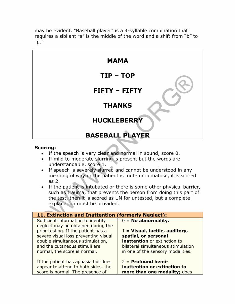
may be evident. “Baseball player” is a 4-syllable combination that requires a sibilant “s” is the middle of the word and a shift from “b” to
“p.”
MAMA
TIP – TOP
FIFTY – FIFTY
THANKS
HUCKLEBERRY
BASEBALL PLAYER
Scoring: • If the speech is very clear and normal in sound, score 0.
• If mild to moderate slurring is present but the words are understandable, score 1.
• If speech is severely slurred and cannot be understood in any meaningful way or the patient is mute or comatose, it is scored
as 2.
• If the patient is intubated or there is some other physical barrier, such as trauma, that prevents the person from doing this part of
the test, then it is scored as UN for untested, but a complete explanation must be provided.
11. Extinction and Inattention (formerly Neglect):
Sufficient information to identify neglect may be obtained during the prior testing. If the patient has a
severe visual loss preventing visual double simultaneous stimulation,
and the cutaneous stimuli are normal, the score is normal.
If the patient has aphasia but does appear to attend to both sides, the
score is normal. The presence of
0 = No abnormality. 1 = Visual, tactile, auditory,
spatial, or personal inattention or extinction to
bilateral simultaneous stimulation in one of the sensory modalities.
2 = Profound hemi-inattention or extinction to
more than one modality; does

visual spatial neglect or
anosognosia may also be taken as evidence of abnormality. Since the abnormality is scored only if
present, the item is never untestable.
not recognize own hand or
orients to only one side of space.
Item 11 basically determines if simultaneous bilateral testing blocks sensation on one side—in any modality. This may have been noted
during the visual field examination or with earlier sensory testing, so
this part of the test includes information gained from other items of the scale—one of the reasons it’s so important to complete each
section.
Additional testing here usually includes asking the patient to close his or her eyes and then lightly touching both sides of the body (face,
above wrists, below knees, above ankles), first on one side and then the other and finally both sides together. The patient indicates which
side or sides are being touched as above. Painful stimuli, such as pinching of nailbeds, may be used to elicit response in aphasic patients.
With strokes in the right hemisphere, patients may develop left-sided
neglect to the extent that they ignore or can’t perceive items or people on their left side. Denial of impairment may also characterize this
condition. If the patient does not appear to respond when you are
standing on one side, you should move to the other side to determine if there is a difference.
Scoring:
• A patient who exhibits no indications of neglect or extinction is scored as 0.
• If neglect or extinction occurs in one sensory modality, then the score is 1.
• A patient who is profoundly paralyzed on one side and cannot feel sensations or is comatose is scored as 2. The patient with
extinction in more than one modality (such as to both visual and sensory stimuli) is also scored as 2.
Conclusion When all of the items are tested, the total scores are added to arrive at a final score. Please note, in some earlier versions of the scale,
amputations were scored as “9,” but this score is omitted when scoring as it has been replaced by UN for untested.
The NIH Stroke Scale is not difficult, but administration and accuracy
improves with practice and experience. For example, ataxia and muscle weakness can be difficult to differentiate for those with little
experience caring for or observing stroke patients; however, most of the scale if fairly straight-forward and can be mastered easily. Even
skilled examiners may vary slightly in scoring, but most people who are trained have similar scores. One may score a patient as a 4 and
another as a 5, but a significant variance suggests a need for review.
Ideally, when first using the scale, a healthcare provider should be teamed with someone who is more experienced. The beginner should
observe first and score the patient, comparing his or her results with
the examiner’s score, and then score a patient while an experienced observer also scores, and again compare results.
References • Berger, MF, Prob, RD, Ilg, UJ, & Karnath, H-O. (2006, June 26).
Deviation of eyes and head in acute cerebral stroke. BMC
Neurology. Retrieved September 20, 2011, from http://www.ncbi.nlm.nih.gov/pmc/articles/PMC1543655/
• Know Stroke: NIH Stroke Scale [booklet]. NINDS. Retrieved September 20, 2011 from
http://www.ninds.nih.gov/doctors/NIH_Stroke_Scale_Booklet.pdf
• NIH Stroke Scale. (n.d.) NINDS. Retrieved September 20, 2011,
from http://www.ninds.nih.gov/doctors/NIH_Stroke_Scale.pdf • NIH Stroke Scale computer course. (2008). American Heart
Association. Retrieved September 20, 2011, from http://learn.heart.org/ihtml/application/student/interface.heart2
/nihsscomputer.html • Windsor, LK, & Windsor, RL (n.d.). Hemianopsia: Loss of half of
the visual field after stroke or traumatic brain injury. The Low Vision Centers of Indiana. Retrieved September 20, 2011, from
http://www.eyeassociates.com/images/visual_field_impairment.htm
• NIH Stroke Scale Training: Parts 1-8. YouTube. Retrieved September 20, 2011, from
http://www.youtube.com/watch?v=x4bjXqtfn6k