Embed Size (px)
Citation preview

Common variants at 7p21 are associated with frontotemporallobar degeneration with TDP-43 inclusions
Vivianna M. Van Deerlin*,1,2, Patrick M. A. Sleiman*,6, Maria Martinez-Lage*,1,8, Alice Chen-Plotkin*,1,3, Li-San Wang1, Neill R Graff-Radford9, Dennis W. Dickson10, RosaRademakers10, Bradley F. Boeve11, Murray Grossman3, Steven E. Arnold3,4,5, David M.A.Mann13, Stuart M. Pickering-Brown12,13, Harro Seelaar14, Peter Heutink15, John C. vanSwieten14, Jill R. Murrell16, Bernardino Ghetti16,17, Salvatore Spina16,18, JordanGrafman19, John Hodges20, Maria Grazia Spillantini21, Sid' Gilman22, Andrew P.Lieberman23, Jeffrey A. Kaye24, Randall L. Woltjer25, Eileen H Bigio26,27, MarselMesulam27, Safa al-Sarraj28, Claire Troakes29, Roger N. Rosenberg30, Charles L. WhiteIII31, Isidro Ferrer32, Albert Lladó33, Manuela Neumann34, Hans A. Kretzschmar35, ChristineMarie Hulette36, Kathleen A. Welsh-Bohmer37,38, Bruce L Miller39, Ainhoa Alzualde40,Adolfo Lopez de Munain41, Ann C. McKee42,43, Marla Gearing44,46,47, Allan I. Levey45,46,James J. Lah45, John Hardy48,49, Jonathan D. Rohrer50, Tammaryn Lashley49,51, Ian R.A.Mackenzie52, Howard H. Feldman53,54, Ronald L. Hamilton55, Steven T. Dekosky56, Julievan der Zee57,58, Samir Kumar-Singh57,58, Christine Van Broeckhoven57,58, RichardMayeux59, Jean Paul G. Vonsattel59,60,61, Juan C. Troncoso62, Jillian J Kril63, John B.J.Kwok64, Glenda M. Halliday64,65, Thomas D. Bird66,67, Paul G. Ince68, Pamela J. Shaw68,Nigel J. Cairns69,70, John C. Morris69,70, Catriona Ann McLean71, Charles DeCarli72,William G. Ellis73, Stefanie H. Freeman74, Matthew P. Frosch74, John H. Growdon74, DanielP. Perl75, Mary Sano75,76, David A. Bennett77, Julie A. Schneider77, Thomas G. Beach78,Eric M. Reiman79,80, Bryan K. Woodruff81, Jeffrey Cummings82, Harry V. Vinters83,84, CarolA. Miller85, Helena C. Chui85, Irina Alafuzoff86,87,88, Päivi Hartikainen88, DanielleSeilhean89, Douglas Galasko90, Eliezer Masliah90,91, Carl W. Cotman92, M. TeresaTuñón93,94, M. Cristina Caballero Martínez94,95, David G. Munoz96, Steven L. Carroll97,
Users may view, print, copy, download and text and data- mine the content in such documents, for the purposes of academic research,subject always to the full Conditions of use:http://www.nature.com/authors/editorial_policies/license.html#terms
Correspondence and requests for materials should be addressed to VVD ([email protected]) or JQT([email protected])..*Co-first authors with equal contributionsAuthor ContributionsVVD, PS, MML, and ACP contributed equally to this manuscript. The overall study was designed and implemented by VMYL, JQT,VVD, HH and MML and discussed with GDS, LSW, and ACP. Coordination, sample handling, DNA extraction, genetic analysis anddata management were done primarily by VVD and MML. Genotyping and additional testing, including immunohistochemical analysis,was done by PS, HH, VVD, or MML. Data analysis and quality control were performed by PS, LSW, and ACP and discussed andreviewed by VVD, HH, GS, VMYL, JQT, and MML. Expression analysis was designed and implemented by ACP. The manuscript wasprepared by ACP, VVD, and PS and reviewed by MML, GDS, HH, JQT, VMYL, and LSW. The members of the International FTLDCollaboration, which includes all other authors, contributed cases, evaluated pathology, performed genetic studies and reviewed themanuscript.Supplementary Information is available onlineAccession NumbersGRN: NM_002087.2TMEM106B: NM_018374.3Competing Financial InterestA patent application on TMEM106B has been submitted. Dr. James Lah is currently involved in clinical trials involving: Elan, Janssen,Medivation, and Ceregene. Dr. Howard Feldman has been a full time employee in Neuroscience Global Clinical Research andDevelopment at Briston-Myers Squibb.
NIH Public AccessAuthor ManuscriptNat Genet. Author manuscript; available in PMC 2010 September 1.
Published in final edited form as:Nat Genet. 2010 March ; 42(3): 234–239. doi:10.1038/ng.536.
NIH
-PA Author Manuscript
NIH
-PA Author Manuscript
NIH
-PA Author Manuscript

Daniel Marson98,99, Peter F. Riederer100, Nenad Bogdanovic101, Gerard D.Schellenberg1,2, Hakon Hakonarson6,7, John Q. Trojanowski1,2, and Virginia M.-Y. Lee1,2
1Center for Neurodegenerative Disease Research, Department of Pathology and LaboratoryMedicine, University of Pennsylvania School of Medicine, Philadelphia, PA USA 2Institute on Aging,University of Pennsylvania School of Medicine, Philadelphia, PA USA 3Department of Neurology,University of Pennsylvania School of Medicine, Philadelphia, PA USA 4Department of Psychiatry,University of Pennsylvania School of Medicine, Philadelphia, PA USA 5Penn Memory Center,University of Pennsylvania School of Medicine, Philadelphia, PA USA 6The Center for AppliedGenomics, Division of Human Genetics, University of Pennsylvania School of Medicine,Philadelphia, PA USA 7Division of Pulmonary Medicine and Department of Pediatrics, TheChildren's Hospital of Philadelphia, University of Pennsylvania School of Medicine, Philadelphia,PA USA 8Department of Neurology, Hospital de la Santa Creu i Sant Pau, Universitat Autonoma deBarcelona, Barcelona, Spain 9Department of Neurology Jacksonville, Mayo College of Medicine,Jacksonville, FL, USA 10Department of Neuroscience, Mayo Clinic, Jacksonville, FL, USA11Department of Neurology, Mayo Clinic, Rochester, MN, USA 12Clinical Neuroscience ResearchGroup, United Kingdom 13University of Manchester, United Kingdom 14Erasmus Medical CentreRotterdam, The Netherlands 15Section of Medical Genomics, Department of Clinical Genetics, VUUniversity Medical Center Amsterdam, The Netherlands 16Department of Pathology and LaboratoryMedicine, Indiana University School of Medicine, Indianapolis, IN, USA 17Indiana Alzheimer DiseaseCenter, Indiana University School of Medicine, Indianapolis, IN, USA 18Department of Neurological,Neurosurgical and Behavioral Sciences, University of Siena, Siena, Italy 19Cognitive NeuroscienceSection, National Institute of Neurological Disorders and Stroke, Bethesda, MD, USA 20Prince ofWales Medical Research Institute, New South Wales, Australia 21Cambridge Centre for BrainRepair, Dept of Clinical Neurosciences, University of Cambridge, Cambridge, United Kingdom22Department of Neurology, University of Michigan, Ann Arbor, MI, USA 23Department of PathologyUniversity of Michigan, Ann Arbor, MI, USA 24Department of Neurology and Biomedical Engineering,Oregon Health and Science University, Portland, OR, USA 25Department of Pathology, OregonHealth and Science University, Portland, OR, USA 26Department of Pathology, NorthwesternUniversity Feinberg School of Medicine, Chicago, IL, USA 27Cognitive Neurology and AlzheimerDisease Center Northwestern University Feinberg School of Medicine, Chicago, IL, USA28Department of Clinical Neuropathology, Institute of Psychiatry, Kings College Hospital, London,United Kingdom 29MRC London Neurodegenerative Diseases Brain Bank, Institute of Psychiatry,Kings College Hospital, London, United Kingdom 30Alzheimer's Disease Center, University of TexasSouthwestern Medical Center, Dallas, TX, USA 31Department of Pathology, University of TexasSouthwestern Medical Center, Dallas, TX, USA 32Institut de Neuropatologia, IDIBELL-HospitalUniversitari de Bellvitge, Barcelona, Spain 33Alzheimer's Disease and Cognitive Disorders Unit,Service of Neurology. ICN. Hospital Clínic Barcelona, Barcelona, Spain 34Institute forNeuropathology, University Hospital Zurich, Zurich, Switzerland 35Center for Neuropathology andPrion Research, Ludwig Maximilians University, Munich, Germany 36Department of Pathology,Duke University Health Sciences Center, Durham, NC, USA 37Dept of Psychiatry, Duke UniversityHealth Sciences Center, Durham, NC, USA 38Bryan ADRC, Duke University Health SciencesCenter, Durham, NC, USA 39Department of Neurology, University of California at San Francisco,San Francisco, CA, USA 40Neurogenetic Unit, Instituto Biodonostia, San Sebastián, Spain41Servicio de Neurología, Hospital Donostia, San Sebastián, Spain. 42Departments Neurology andPathology, Boston University School of Medicine, Boston MA 43Bedford Veterans AdministrationMedical Center, GRECC, Bedford MA, USA 44Department of Pathology and Laboratory Medicine,Emory University School of Medicine, Atlanta, GA, USA 45Department of Neurology, EmoryUniversity School of Medicine, Atlanta, GA, USA 46Alzheimer's Disease Research Center, EmoryUniversity School of Medicine, Atlanta, GA, USA 47Center for Neurodegenerative Disease, EmoryUniversity School of Medicine, Atlanta, GA, USA 48Reta Lila Laboratories, UCL Institute ofNeurology, London, United Kingdom 49Department of Molecular Neuroscience, UCL Institute of
Van Deerlin et al. Page 2
Nat Genet. Author manuscript; available in PMC 2010 September 1.
NIH
-PA Author Manuscript
NIH
-PA Author Manuscript
NIH
-PA Author Manuscript

Neurology, London, United Kingdom 50Dementia Research Centre, UCL Institute of Neurology,London, United Kingdom 51Queen Square Brain Bank for Neurological Disorders, UCL Institute ofNeurology, London, United Kingdom 52Department of Pathology, University of British Columbia,Canada 53Division of Neurology Vancouver General Hospital and the University of British Columbia,Canada 54Neuroscience, Bristol-Myers Squibb, University of Pittsburgh, Pittsburgh PA, USA55Department of Pathology, University of Pittsburgh, Pittsburgh PA, USA 56Department ofNeurology, University of Virginia School of Medicine, Charlottesville, VA, USA 57NeurodegenerativeBrain Diseases Group, Department of Molecular Genetics, VIB, Antwerpen, Belgium 58InstituteBorn-Bunge and University of Antwerp, Antwerpen, Belgium 59Taub Institute for Research onAlzheimer's Disease, Columbia University, New York, NY, USA 60New York Brain Bank, ColumbiaUniversity, New York, NY, USA 61Department of Pathology, Columbia University, New York, NY,USA 62Departments of Pathology and Neurology, Johns Hopkins University School of Medicine,Baltimore, MD, USA 63Disciplines of Medicine and Pathology, University of Sydney, Australia64Prince of Wales Medical Research Institute, Australia 65University of New South Wales, Australia66GRECC, VA Puget Sound Health Care System, University of Washington, Seattle, WA, USA67Department of Neurology, University of Washington, Seattle, WA, USA 68Department ofNeuroscience, University of Sheffield 69Alzheimer's Disease Research Center, WashingtonUniversity School of Medicine, St Louis, MO, USA 70Department of Neurology, WashingtonUniversity School of Medicine, St Louis, MO, USA 71Department of Anatomical Pathology, TheAlfred Hospital, Australia 72Alzheimer's Disease Center, Imaging of Dementia and Aging (IDeA)Laboratory, Department of Neurology, Center for Neuroscience, University of California at Davis,Sacramento, CA, USA 73Department of Pathology, University of California at Davis, Sacramento,CA, USA 74C.S. Kubik Laboratory for Neuropathology, Massachusetts General Hospital & HarvardMedical School, Boston, MA, USA 75Department of Pathology, Mount Sinai School of Medicine,New York, NY, USA 76James J Peters VA Medical Center, New York, NY, USA 77Rush Alzheimer'sDisease Center, Rush University Medical Center, Sun City, AZ, USA 78Sun Health ResearchInstitute, Sun City, AZ, USA 79Banner Alzheimer's Institute, Translational Genomics ResearchInstitute, University of Arizona, Phoenix, AZ, USA 80Arizona Alzheimer's Consortium, Phoenix, AZ,USA 81Mayo Clinic Arizona, Scottsdale, AZ, USA 82Mary S. Easton Center for Alzheimer's DiseaseResearch, Los Angeles, CA, USA 83Department of Pathology and Laboratory Medicine, LosAngeles, CA, USA 84Department of Neurology David Geffen School of Medicine at UCLA, LosAngeles, CA, USA 85Keck School of Medicine, University of Southern California, Los Angeles, CA,USA 86Department of Genetics and Pathology, Uppsala University, Uppsala, Sweden 87Departmentof Clinical Medicine, Kuopio University, Kuopio Finland 88Department of Neurology, KuopioUniversity, Kuopio Finland 89UPMC-Univ Paris 06 and APHP, France 90Department ofNeurosciences, University of California, San Diego, San Diego, CA, USA 91Department ofPathology, University of California, San Diego, San Diego, CA, USA 92Department of Neurology,University of California, Irvine, Irvine, CA, USA 93Hospital de Navarra Pathology Department, Spain94Brain Bank of Navarra, Spain 95Biomedical Research Center, Navarra Health Service-Osasunbidea, Spain 96Department of Laboratory Medicine and Pathobiology, Li Ka ShingKnowledge Institute of St. Michael's Hospital, University of Toronto, Canada 97Department ofPathology, University of Alabama at Birmingham, Birmingham, AL, USA 98Department of Neurology,University of Alabama at Birmingham, Birmingham, AL, USA 99Alzheimer's Disease ResearchCenter, University of Alabama at Birmingham, Birmingham, AL, USA 100Clinical NeurochemistryClinic and Policlinic of Psychiatry, Psychosomatic and Psychotherapy of the University ofWuerzburg, Germany 101Geriatric Medicine and Neuropathology at Department of GeriatricMedicine, Karolinska University Hospital, Stockholm Sweden
Abstract
Van Deerlin et al. Page 3
Nat Genet. Author manuscript; available in PMC 2010 September 1.
NIH
-PA Author Manuscript
NIH
-PA Author Manuscript
NIH
-PA Author Manuscript

Frontotemporal lobar degeneration (FTLD) is the second most common cause of presenile dementia.The predominant neuropathology is FTLD with TAR DNA binding protein (TDP-43) inclusions(FTLD-TDP)1. FTLD-TDP is frequently familial resulting from progranulin (GRN) mutations. Weassembled an international collaboration to identify susceptibility loci for FTLD-TDP, usinggenome-wide association (GWA). We found that FTLD-TDP associates with multiple SNPsmapping to a single linkage disequilibrium (LD) block on 7p21 that contains TMEM106B in a GWAstudy (GWAS) on 515 FTLD-TDP cases. Three SNPs retained genome-wide significance followingBonferroni correction; top SNP rs1990622 (P=1.08×10⁻¹¹; odds ratio (OR) minor allele (C) 0.61,95% CI 0.53-0.71). The association replicated in 89 FTLD-TDP cases (rs1990622; P=2×10⁻⁴).TMEM106B variants may confer risk by increasing TMEM106B expression. TMEM106B variantsalso contribute to genetic risk for FTLD-TDP in patients with GRN mutations. Our data implicateTMEM106B as a strong risk factor for FTLD-TDP suggesting an underlying pathogenic mechanism.
FTLD manifests clinically with progressive behavioral and/or language deficits with aprevalence of 3.5-15/100,000 in 45 to 64 year olds2-5. The clinical presentation of cases withFTLD pathology varies depending on the referral base6 and among these cases, ~50% arediagnosed as FTLD-TDP1. A family history of a similar neurodegenerative disease may bepresent in up to 50% of FTLD cases, supporting the existence of a genetic predisposition7.Autosomal dominant GRN mutations occur in ~20% of FTLD-TDP cases8-11. GRN mutationsare loss-of-function mutations with most resulting in premature termination of the mutanttranscript invoking nonsense-mediated RNA decay and with the ensuing haploinsufficiencycausing disease11,12. However, a substantial number of familial FTLD-TDP cases are notexplained by GRN mutations. Further, patients with the same GRN mutation show variableclinical phenotypes or ages of disease onset which likely reflect additional genetic andenvironmental factors13.
The GWA phase of the study included 515 cases of FTLD-TDP and 2509 disease-freepopulation controls genotyped on the Illumina HH550 or 610-Quad BeadChips asdescribed14 (Table 1). A large population control cohort was acceptable since the generalpopulation incidence of FTLD is low4,5,15. Cases were obtained under institutional reviewboard approval by members of the International FTLD Collaboration consisting ofinvestigators from 45 clinical centers and brain banks representing 11 countries (United States,Canada, United Kingdom, The Netherlands, Belgium, Spain, Germany, Australia, Finland,France, and Sweden). All cases met either pathological (n=499) or genetic (n=16) criteria forFTLD-TDP which was confirmed by detecting TDP-43 inclusions usingimmunohistochemistry (IHC)1,16. A genetic criterion for inclusion (i.e. presence of a knownpathogenic GRN mutation) was used since GRN mutation cases are always diagnosed as FTLD-TDP 8,9,17,18. All cases were checked for relatedness using identity by state (IBS). The resultsconfirmed that although some GRN-associated FTLD-TDP cases share the same mutation onchromosome 17 with similarity in the immediate vicinity of GRN, they are no more related inthe remainder of the genome than individuals without GRN mutations. Detailed inclusioncriteria are provided in Methods; cohort features are in Supplementary Table 1.
Cochran-Armitage trend test statistics were calculated at all markers following quality controlfiltering. In addition to self-reported ancestry, all cases and controls were initially screened atancestry informative markers (AIM) using the STRUCTURE software package19 to reducethe risk of population stratification from self-reported ancestry alone. Each case wassubsequently matched to four controls by ‘genetic matching’ by smartPCA20 as previouslydescribed21. The genomic inflation factor (λ) for this study was 1.05 indicating thatbackground stratification was minimal as demonstrated in the quantile-quantile (Q-Q) plots(Supplementary Fig. 1).
Van Deerlin et al. Page 4
Nat Genet. Author manuscript; available in PMC 2010 September 1.
NIH
-PA Author Manuscript
NIH
-PA Author Manuscript
NIH
-PA Author Manuscript

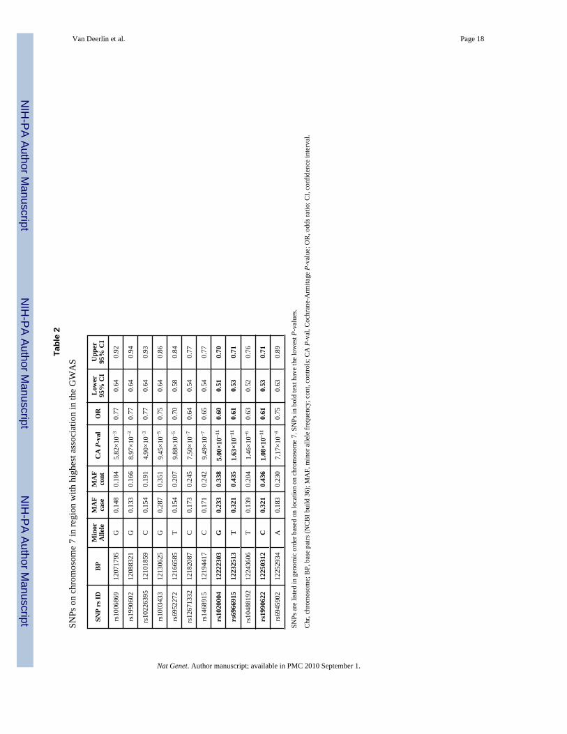
Three SNPs reached genome-wide significance following Bonferroni correction (Figures 1aand b). All three SNPs (rs6966915, rs1020004, and rs1990622) mapped to a 68 kb interval(Supplementary Fig. 2) on 7p21.3 (top marker, rs1990622, minor allele frequency (MAF)32.1% in cases and 43.6% in controls, OR = 0.61, [95% CI 0.53 – 0.71], P=1.08×10⁻¹¹). Forrs1990622, the more common (T) allele confers risk with an OR of 1.64 [95% CI 1.34-2.00].The interval contained nine additional markers in strong LD (r2 >0.45) that were also associatedwith FTLD-TDP (P-value range = 8.9×10⁻³ - 7.5×10⁻⁷; OR range 0.63-0.77) (Table 2). All12 associated SNPs map to a single LD block spanning TMEM106B, which encodes anuncharacterized transmembrane protein of 274 amino acids (Figures 1b and c). SNPsrs1020004 and rs6966915 lie within introns 3 and 5, respectively, of TMEM106B, whilers1990622 is 6.9 kb downstream of the gene. These findings argue strongly for the associationof the 7p21 locus, and the gene TMEM106B, with FTLD-TDP.
The association with FTLD-TDP in the GWA was replicated by TaqMan SNP genotyping in89 independent FTLD-TDP cases and 553 Caucasian control samples at two of the genome-wide significant SNPs (rs1020004 and rs1990622) (Table 1 and Supplementary Table 1). Apolymorphic variation adjacent to rs6966915 interfered with interpretation of TaqMangenotyping therefore precluding its use in the replication. The replication set was selected basedon the same pathological criteria and had similar characteristics as the GWA phase cohort(Supplementary Table 1). In this replication cohort, the top SNPs again showed significantassociation (P=0.004 for rs1020004 and P=0.0002 for rs1990622) with the same directions ofassociation as those found in the GWA phase (Supplementary Table 1). These results suggestthat in the 7p21 locus, encompassing the gene TMEM106B, we have identified a commongenetic susceptibility factor for FTLD-TDP. Of interest, this association was not confirmed ina cohort of 192 living patients with unselected FTLD (Supplementary Table 2). This likelyreflects heterogeneity in neuropathological substrates underlying FTLD, with only ~50% ofunselected clinical FTLD cases expected to have FTLD-TDP. Assuming that TMEM106Bgenetic variants confer risk of FTLD-TDP specifically, the power to detect this association in192 clinical FTLD cases and 553 controls is ~30% for an alpha-value of 0.05. To have >90%power to detect this association, a clinical FTLD cohort would require more than 1400 clinicalFTLD cases and an equal number of controls.
We next evaluated TMEM106B gene expression in different human tissues to identifyphenotype-associated differential expression and also any potential genetic regulators ofexpression. We queried the mRNA-by-SNP browser(http://www.sph.umich.edu/csg/liang/asthma/, last accessed June 6, 2009), for geneticregulators of TMEM106B expression (eSNPs) in lymphoblastoid cell lines22. The top SNP,rs1990622, was significantly correlated with TMEM106B average expression levels (LOD6.32; P=6.9×10⁻⁸), as was SNP rs1020004 (LOD 5.16, P=1.10×10⁻⁶). The risk allele (T) ofrs1990622 was associated with a higher level of mRNA expression, indicating thatTMEM106B may be under cis-acting regulation by either the FTLD-TDP associated SNPs oranother SNP(s) in LD with the associated variants. As the expression data in the publiclyavailable database is derived from lymphoblastoid cell lines from normal individuals22, andthe diseased organ in FTLD-TDP is brain, we asked if a similar correlation between genotypeand expression phenotype for TMEM106B is also present in tissue types affected by disease,and in diseased individuals themselves. Accordingly, we used total RNA isolated from FTLD-TDP postmortem brains (n=18) and neurologically normal control brains (n=7) to evaluateTMEM106B expression in frontal cortex, which is severely affected in FTLD-TDP, byquantitative reverse-transcription PCR (QRT-PCR). All RNA samples used were confirmedto be of equivalent high quality as described23 (Supplementary Table 3). For the sameindividuals for which we obtained expression data, we genotyped SNPs rs1020004 andrs1990622 using allelic discrimination assays.
Van Deerlin et al. Page 5
Nat Genet. Author manuscript; available in PMC 2010 September 1.
NIH
-PA Author Manuscript
NIH
-PA Author Manuscript
NIH
-PA Author Manuscript

Corroborating results from the cell lines, expression of TMEM106B was significantlycorrelated with TMEM106B genotype, with risk allele carriers showing higher expression(overall P=0.027, TT vs. TC P=0.017, TT vs. CC P=0.03, for rs1990622, Fig. 2a andSupplementary Fig. 3a). Strikingly, however, expression of TMEM106B was >2.5 times higherin FTLD-TDP cases compared to normal controls (P=0.045, Fig. 2b). In addition, the effectsof genotype and TMEM106B expression on risk of developing disease are at least partlyindependent, as are the effects of genotype and disease status on TMEM106B expression(Supplementary Table 4a and b). Thus, these data suggest that increased TMEM106B brainexpression might be linked to mechanisms of disease in FTLD-TDP, and that risk alleles atTMEM106B confer genetic susceptibility by increasing gene expression.
The primary criterion for inclusion in the GWAS was a neuropathological diagnosis of FTLD-TDP; therefore we studied all cases together regardless of GRN mutation status. Nevertheless,a priori it was difficult to predict whether additional genetic susceptibility loci would beidentified in a group with Mendelian inheritance of highly penetrant mutations. We thereforeseparately evaluated FTLD-TDP cases with (n=89) and without (n=426) GRN mutations.Association to the 7p21 locus persisted in both the GRN negative and positive clusters andthere was no significant heterogeneity in the ORs for the disease/SNP association between theclusters (Fig. 3 and Supplementary Tables 2 and 5). Using family history status as a covariatein a logistic regression showed that the 7p21 association is independent of family history(Supplementary Table 6). Thus, TMEM106B variants may act as a modifier locus in thepresence of GRN mutations, as the APOE locus has been shown to modify age of onset inpatients with PSEN124 or PSEN225 mutations.
Additionally, in the whole GWA cohort, we observed a correlation between rs1020004genotype and disease duration (P=0.03) with homozygotes for the risk allele (AA, wild-type)having shorter duration of disease (i.e. more severe disease) than individuals homozygous forthe minor allele (GG, Supplementary Figure 4). These results provide strong confirmatoryevidence for association of the 7p21 locus with increased risk for FTLD-TDP in both GRNpositive and negative cases.
In addition to the 7p21 locus, analysis of the GRN cases alone showed highly significantassociation with SNPs near the GRN locus on 17q21 (Fig. 3 and Supplementary Table 7). Notunexpectedly, haplotype analysis of the cases indicated that the chromosome 17 associationwas driven by a shared haplotype among the p.R493X (NM_002087.2:c.1477C>T) mutationcarriers which represented 20.2% (18/89) of the GRN mutation cases. To determine if theobserved association at the GRN locus was dependent on the association at the TMEM106Blocus we carried out a logistic regression analysis conditioning on the most significantlyassociated SNP at the 7p21 locus, rs1990622, in the patients with GRN mutations. Theconditional analysis had no effect on the association at the GRN locus suggesting that theassociations with 17q21 and 7p21 are independent. IBS analysis confirms that these individualsare unrelated and therefore the identified association on chromosome 7 cannot be discountedin GRN mutation carriers. Indeed, conditioning on the top SNP at the GRN locus, rs8079488,also had no effect on the TMEM106B association (results not shown). In addition to the 7p21locus, the GWAS of GRN negative cases showed a trend for association at five other loci(Supplementary Table 8) including a locus on chromosome 9p21.2 that falls within a 7.7 Mbcritical interval defined from five previous linkage studies, representing a potential refinementof that region26. We observed no association at the GRN locus in the cases without GRNmutations.
We then evaluated mRNA expression of TMEM106B in FTLD-TDP with and without GRNmutations separately, and the GRN mutants showed increased expression (overall P=0.0009),compared to controls (P=0.0005) and FTLD-TDP without GRN mutations (P=0.002) (Figure
Van Deerlin et al. Page 6
Nat Genet. Author manuscript; available in PMC 2010 September 1.
NIH
-PA Author Manuscript
NIH
-PA Author Manuscript
NIH
-PA Author Manuscript

2c). Furthermore, controlling for rs1990622 genotype and focusing on heterozygotes (n=14),the presence of a GRN mutation remained significantly associated with increasedTMEM106B expression (P=0.039, Fig. 2d) compared to normal controls. These results arecompatible with a model in which mutations in GRN are upstream of increased TMEM106Bexpression in increasing risk for FTLD-TDP.
A mechanistic understanding of the pathogenesis of FTLD has been hampered theheterogeneity in clinical and pathological features. With the discovery of TDP-43 as a majorFTLD disease protein, the pathologically-defined entity of FTLD-TDP emerged16.Identification of GRN mutations as a major genetic cause of FTLD-TDP, led to definition ofa genetic subgroup of FTLD-TDP. This study identifies TMEM106B as a genetic risk factorfor FTLD-TDP. We speculate that the homogeneous pathologically-defined study populationused here enabled us to detect a robust signal with relatively small case numbers.
Our data suggest a potential disease mechanism in which risk-associated polymorphisms at7p21 increase TMEM106B expression, and elevated TMEM106B expression increases risk forFTLD-TDP. Additionally, we show that TMEM106B genotypes are a significant risk factorfor FTLD-TDP even in GRN mutation carriers implying that GRN mutations may act upstreamof TMEM106B in a pathogenic cascade. Future directions of research on this novel genetic riskfactor will include a detailed evaluation of the TMEM106B locus by sequencing, collection ofmore pathologically-defined FTLD-TDP cases for a genome-wide replication, and studies ofexpression profiles in additional tissues and brain regions. A better understanding of this genemay in turn provide an opportunity to intervene in an otherwise fatal and devastatingneurodegenerative disease.
METHODSInclusion criteria
Individuals of European descent with dementia clinically +/− motor neuron disease (MND)and an autopsy diagnosis of FTLD-TDP confirmed by TDP-43 IHC were included. Mixedpathologies were not excluded. Living individuals with a pathogenic GRN mutation were alsoincluded18. Only a single proband per family was permitted. Appropriate informed consentwas obtained. 598 unique FTLD-TDP cases met inclusion criteria; 515 were used for theGWAS after PCA matching to controls. Characteristics described in Supplementary Table 1.Whole genome amplification (WGA), performed in duplicate and pooled, was used for 15(Repli-g Mini, Qiagen), but only 6 cases ultimately passed quality control parameters for theGWAS. The replication, using SNP genotyping, included cases of insufficient quality orquantity for the GWA phase (n=27), cases available only as formalin-fixed paraffin-embeddedtissue (n=6), and cases randomly not used for GWA phase (n=56). Three FTLD-TDP caseswith mutations in valosin-containing protein (VCP) gene were included (two in GWA and onein replication)18.
ControlsGWAS controls consisted of 2509 samples, including 1297 self-reported Caucasian childrenof European ancestry recruited from CHOP Health Care Network and 1212 samples from the1958 birth cohort genotyped by the WTCCC27. Although the controls were not selected forabsence of neurodegenerative disease, the large size of the cohort relative to the low populationfrequency of FTLD overrides this potential concern. Furthermore, the minor allele frequenciesat the 7p21 loci are very similar (<1-2% variation) between CHOP and WTCCC cohortssuggesting they accurately reflect the control allelic frequencies in the general population(Supplementary Table 9). To reduce the risk of population stratification all internal controlswere screened using the STRUCTURE package19 at 220 AIMs. To improve clustering the
Van Deerlin et al. Page 7
Nat Genet. Author manuscript; available in PMC 2010 September 1.
NIH
-PA Author Manuscript
NIH
-PA Author Manuscript
NIH
-PA Author Manuscript

samples were spiked with 90 CEPH, Yoruban and Chinese/Japanese individuals genotyped aspart of the HapMap project. Cases were excluded if their inferred proportion of ancestry wasless than 90% that of the CEU cluster.
For the replication 553 controls were as follows: 275 from Coriell Institute (NeurologicallyNormal Caucasian control panels, Camden, NJ), 155 clinical controls from neurology clinicsat University of Pennsylvania (UPenn), 28 brain samples of neurologically normal individuals> 60 years from the UPenn Center for Neurodegenerative Disease Research (CNDR), and 95population controls from CHOP.
DNA extraction and quality assessmentSamples sent as DNA from external sites were extracted using different methods. Remainingsamples (376) were extracted at UPenn from frozen brain tissue or blood. Genomic DNA wasextracted from frozen brain tissue (50 mg) by the Qiagen MagAttract DNA Mini M48 Kit onthe M48 BioRobot. Genomic DNA was purified from whole blood using FlexiGene kit(Qiagen). High quality DNA was required for the Illumina genotyping. All DNA samples wereevaluated for purity by spectrophotometric analysis (Nanodrop) and for degradation by 1%agarose gel electrophoresis (Invitrogen).
TDP-43 IHCAutopsy cases were confirmed to have TDP-43 pathology by IHC performed by the sendinginstitution or at UPenn CNDR as previously described16. TDP-43 negative cases wereexcluded.
GRN sequencingTo stratify the analysis according to GRN mutation status, exons 1-13 (with exon 1 representingexon 0 in Gass et al.10) and adjacent intronic regions were sequenced as described13 in casesnot previously evaluated. GRN sequencing was not possible due to limited sample quantity ina few cases (n=13 in GWA, n=15 in replication). Novel variants identified in this study notpredicted to cause a frameshift or premature termination and previously described variants ofuncertain significance were grouped with GRN mutation negative cases. The most commonmutations identified are given in Supplementary Table 1.
Illumina genotyping and quality controlThe FTLD-TDP cases and CHOP control samples were genotyped on either the IlluminaHH550 BeadChip or the Illumina human610-quad BeadChip at the Center for AppliedGenomics at CHOP as previously described14. The 1958 birth cohort samples were genotypedon the HH550 BeadChip by the WTCCC27. Sixteen individuals, 13 cases and 3 controls, wereexcluded from GWA phase for low genotyping (<98% chip-wide genotyping success). Wefurther rejected 13,316 SNPs with call rates <95%, 23,552 SNPs with MAF < 1% and 1,940SNPs with Hardy Weinberg equilibrium P<10−5 in the controls samples; the λ was 1.05. Casesand controls were screened for relatedness using the IBS estimations in plink(http://pngu.mgh.harvard.edu/~purcell/plink/index.shtml) on 100,000 randomly distributedmarkers throughout the genome. Pairwise Pi-hat values in excess of 0.01 were indicative ofrelatedness.
Following the quality control measures cases were matched to controls by ‘genetic matching’as previously described21. We computed principal components for our dataset by runningsmartpca, a part of the EIGENSTRAT package, on 100,000 random autosomal SNPs andapplied a matching algorithm implemented in MATLAB to the output. The matching algorithmassigns each sample a coordinate based on k eigenvalue-scaled principal components. It then
Van Deerlin et al. Page 8
Nat Genet. Author manuscript; available in PMC 2010 September 1.
NIH
-PA Author Manuscript
NIH
-PA Author Manuscript
NIH
-PA Author Manuscript

matches each case to m unique controls within a distance d, keeping only cases that matchexactly m controls. The distance thresholds were manually optimized to minimize λ andmaximize power (i.e. number of cases). We matched each case to four controls, using the firstthree principal components and a distance threshold of 0.025.
Statistical Analysis for AssociationStatistical tests for association were performed using plink. Single marker analyses for thegenome-wide data were done using the Cochran-Armitage trend test. The genomic inflationfactors were 1.05 for the complete case set and 1.03 for the GRN mutation carriers, indicatingonly minor background stratification. The Breslow-Day test within plink was used to test forheterogeneity of odds ratio for the disease/SNP association between GRN mutation carriersand non-carriers. Conditional SNP regression analyses were completed in plink, the alleledosages of the conditioning SNP were included as covariates in the logistic regression models.To determine if the association at the TMEM106B locus was dependent on family history weincluded family history status as a covariate in a logistic regression model using plink.Haplotypes were reconstructed and population frequencies estimated using the EM algorithmimplemented in the program fastPHASE28. For the age of onset and disease duration analyseswe performed an analysis of variance (ANOVA) with the general linear models procedure inR (www.r-project.org). Independent variables for each ANOVA were the log transformed ageof onset or disease duration in years and the individual SNP genotype with additive encoding(ie three categories where 0 is homozygous for the ancestral allele, 1 is heterozygous and 2 ishomozygous for the minor allele). Power calculations were based on the rs1990622 allelefrequencies observed for cases and controls in the GWAS, using a two-tailed test. We assumedthat clinical FTLD cases without TDP-43 pathology as the neuropathological substrate wouldhave allele frequencies similar to controls.
SNP Genotyping for ReplicationFor the replication, genotyping was performed using TaqMan chemistry-based allelicdiscrimination assays (Applied Biosystems (ABI), Foster City, CA) on the ABI 7500 Fast Real-Time System followed by analysis with SDS 7500 software v2.0.1. The ABI assays used were:rs1020004, C_7604953_10 and rs1990622, C_11171598_10. A nearby novel genetic variation(possible deletion) was found to interfere with correct genotyping of the T allele of SNPrs6966915 using ABI reagents C_31573289_10 (as well as by DNA sequencing), thus thisSNP was not used further.
Human samples for expression analysisFrontal cortex human brain samples from the CNDR Brain Bank characterized followingconsensus criteria1,3 were dissected as previously described23. Neurologically normal controls(n=7), FTLD-TDP cases with (n=8), and without (n=10) GRN mutations were sampled(Supplementary Table 10). GRN mutations were confirmed to be absent from control cases.RNA quality was verified using an Agilent 2100 Bioanalyzer (RIN>6 for inclusion) aspreviously described23. mRNA expression was quantified by QRT-PCR on the ABI7500 usingthe delta-delta CT method, and the geometric mean of two housekeeping genes (β-actin andCyclophilin A), shown to have stable expression in frontal cortex samples from FTLD-TDPand normal individuals23. Detailed information on primers is available on request.
Statistical analyses of expression data and replication cohortFor all brain expression and replication cohort analyses, statistical tests were performed usingopen source R software packages. R-scripts are available upon request. For evaluations of theeffect of disease status, SNP genotype, and gender on TMEM106B expression, linearregressions were used to compute p-values in univariate models. We evaluated assumptions
Van Deerlin et al. Page 9
Nat Genet. Author manuscript; available in PMC 2010 September 1.
NIH
-PA Author Manuscript
NIH
-PA Author Manuscript
NIH
-PA Author Manuscript
of linearity by checking QQ plots (observed vs. predicted under normal distribution). Forpairwise comparisons within the linear models, risk allele homozygotes and GRN mutants,respectively, were designated the reference group for marginal t-tests evaluating genotypeeffects and the effects of GRN mutations on expression. Normalized gene expression samplegenotype and gender data are provided in Supplementary Data 1 and 2. For evaluations of theindependent contributory effects of SNP genotype and TMEM106B expression on disease state,logistic regressions were used to compute AIC values in multivariate vs. univariate models(Supplementary Table 4a). For evaluations of the independent contributory effects of SNPgenotype and disease state on TMEM106B expression, linear regressions were used inmultivariate vs. univariate models (Supplementary Table 4b). For analyses of association ofSNP genotypes with disease in our TaqMan replication cohort, Cochran-Armitage trend testswere used to compute P-values under a codominant model.
Supplementary MaterialRefer to Web version on PubMed Central for supplementary material.
AcknowledgmentsThis project was enabled by the contributions and efforts of many individuals in several supportive capacities. Mostimportantly, we extend our appreciation to the patients and families who made this research possible. Extensivetechnical assistance was provided by R. Greene, T. Unger, and C. Kim and study coordination by E. McCarty Wood.The following individuals contributed through sample ascertainment, epidemiology, coordination, and/or clinicalevaluation of cases: R.C. Petersen, D.S. Knopman, K.A. Josephs, D. Neary, J. Snowden, J. Heidebrink, N. Barbas, R.Reñe, J.R. Burke, K. Hayden, J. Browndyke, P. Gaskell, M. Szymanski, J.D. Glass, M. Rossor, F. Moreno, B.Indakoetxea, M. Barandiaran, S. Engelborghs, P.P. De Deyn, W.S Brooks, T. Chow, V. Meininger, L. Lacomblez, E.Gruenblatt. The following individuals contributed through pathological characterization and evaluation of cases: L.Kwong, J.E. Parisi, W. Kamphorst, I. Ruiz, T. Revesz, J.-J.Martin, R. Highley, C. Duyckaerts. The followingindividuals contributed through general technical assistance and/or genetic studies: E. Moore, M. Baker, R. Crook, S.Rollinson, N. Halliwell, S. Usher, R.M. Richardson, M. Mishra, C. Foong, J. Ervin, K. Price Bryan, J. Ervin, C.Kubilus, A. Gorostidi, M. Cruts, I. Gijselinck, H. McCann, P.R. Schofield, G. Forster, K. Firch, J. Pomaician, I. Leber,V. Sazdovitch, I. Volkmann. The following individuals also contributed: D. Clark, S. Weintraub, N. Johnson, A. King,I. Bodi, C. Shaw, J. Kirby, V. Haroutunian, D. Purohit. We also thank the Brain Bank of University of Barcelona/Hospital Clinic, Clinic for Alzheimer's Disease and Related Disorders University of British Columbia, AustralianBrain Donor Programs supported by the National Health and Medical Research Council of Australia, Biobank at theInstitute Born-Bunge, and the French clinical and genetic research network on FTD/FTD-MND. Many grant fundingagencies provided financial support for this study, including the National Institutes of Health (AG10124, AG17586,AG16574, AG03949, AG17586, NS44266, AG15116, NS53488, AG10124, AG 010133, AG08671, NS044233,NS15655, AG008017, AG13854, P3AG12300, AG028377, AG 019724, AG13846, AG025688, AG05133, AG08702,AG05146, AG005136, AG005681, AG03991, AG010129, AG05134, NS038372, AG02219, AG05138, AG10161,AG19610, AG19610, AG16570, AG 16570, AG05142, AG005131, AG5131, AG18440, AG16582, AG 16573, andNIH Intramural Program). Additional funds were provided by: Robert and Clarice Smith and Abigail Van BurenAlzheimer's Disease Research Program, the Pacific Alzheimer's disease Research Foundation (PARF) grant #C06-01,the Alzheimer's Research Trust, Alzheimer's Society, Medical Research Council (Programme Grant and ReturningScientist Award), Stichting Dioraphte(07010500), Hersenstichting (15F07.2.34), Prinses Beatrix Fonds (006-0204),Winspear Family Center for Research on the Neuropathology of Alzheimer Disease, the McCune Foundation, InstitutoCarlos III, Federal Ministry of Education and Research (01GI0505), SAIOTEK Program (Basque Government),Department of Innovation, Diputación Foral de Gipúzkoa (DFG 0876/08), ILUNDAIN Fundazioa, CIBERNED,Wellcome Trust, Canadian Institutes of Health Research (75480), Fund for Scientific Research Flanders (FWO-V),IAP P6/43 network of the Belgian Science Policy Office (BELSPO), the Joseph Iseman Fund, the Louis and RachelRudin Foundation, National Health and Medical Research Council of Australia, Veteran's Affairs Research Funds,Arizona Department of Health Services (contract 211002, Arizona Alzheimer's Research Center), the ArizonaBiomedical Research Commission (contracts 4001, 0011 and 05-901 to the Arizona Parkinson's Disease Consortium),the Prescott Family Initiative of the Michael J. Fox Foundation for Parkinson's Research, the Daljits and Elaine SarkaraChair in Diagnostic Medicine, BrainNet Europe II, and MMCYT Ref SAF 2001-4888.
REFERENCES1. Cairns NJ, et al. Neuropathologic diagnostic and nosologic criteria for frontotemporal lobar
degeneration: consensus of the Consortium for Frontotemporal Lobar Degeneration. Acta Neuropathol2007;114:5–22. [PubMed: 17579875]
Van Deerlin et al. Page 10
Nat Genet. Author manuscript; available in PMC 2010 September 1.
NIH
-PA Author Manuscript
NIH
-PA Author Manuscript
NIH
-PA Author Manuscript

2. Neary D, et al. Frontotemporal lobar degeneration: a consensus on clinical diagnostic criteria.Neurology 1998;51:1546–54. [PubMed: 9855500]
3. McKhann GM, et al. Clinical and pathological diagnosis of frontotemporal dementia: report of theWork Group on Frontotemporal Dementia and Pick's Disease. Arch Neurol 2001;58:1803–9.[PubMed: 11708987]
4. Mercy L, Hodges JR, Dawson K, Barker RA, Brayne C. Incidence of early-onset dementias inCambridgeshire, United Kingdom. Neurology 2008;71:1496–9. [PubMed: 18981371]
5. Ratnavalli E, Brayne C, Dawson K, Hodges JR. The prevalence of frontotemporal dementia. Neurology2002;58:1615–21. [PubMed: 12058088]
6. Forman MS, et al. Frontotemporal dementia: clinicopathological correlations. Ann Neurol2006;59:952–62. [PubMed: 16718704]
7. Goldman JS, et al. Frontotemporal Dementia: Genetics and Genetic Counseling Dilemmas. Neurologist2004;10:227–234. [PubMed: 15335440]
8. Baker M, et al. Mutations in progranulin cause tau-negative frontotemporal dementia linked tochromosome 17. Nature 2006;442:916–9. [PubMed: 16862116]
9. Cruts M, et al. Null mutations in progranulin cause ubiquitin-positive frontotemporal dementia linkedto chromosome 17q21. Nature 2006;442:920–4. [PubMed: 16862115]
10. Gass J, et al. Mutations in progranulin are a major cause of ubiquitin-positive frontotemporal lobardegeneration. Hum Mol Genet 2006;15:2988–3001. [PubMed: 16950801]
11. Gijselinck I, Van Broeckhoven C, Cruts M. Granulin mutations associated with frontotemporal lobardegeneration and related disorders: an update. Hum Mutat 2008;29:1373–86. [PubMed: 18543312]
12. Cruts M, Van Broeckhoven C. Loss of progranulin function in frontotemporal lobar degeneration.Trends Genet 2008;24:186–94. [PubMed: 18328591]
13. Van Deerlin VM, et al. Clinical, genetic, and pathologic characteristics of patients with frontotemporaldementia and progranulin mutations. Arch Neurol 2007;64:1148–53. [PubMed: 17698705]
14. Hakonarson H, et al. A genome-wide association study identifies KIAA0350 as a type 1 diabetesgene. Nature 2007;448:591–4. [PubMed: 17632545]
15. McCarthy MI, et al. Genome-wide association studies for complex traits: consensus, uncertainty andchallenges. Nat Rev Genet 2008;9:356–69. [PubMed: 18398418]
16. Neumann M, et al. Ubiquitinated TDP-43 in frontotemporal lobar degeneration and amyotrophiclateral sclerosis. Science 2006;314:130–3. [PubMed: 17023659]
17. Cairns NJ, et al. TDP-43 in familial and sporadic frontotemporal lobar degeneration with ubiquitininclusions. Am J Pathol 2007;171:227–40. [PubMed: 17591968]
18. Mackenzie IR, et al. Nomenclature for neuropathologic subtypes of frontotemporal lobardegeneration: consensus recommendations. Acta Neuropathol 2009;117:15–8. [PubMed: 19015862]
19. Falush D, Stephens M, Pritchard JK. Inference of population structure using multilocus genotypedata: linked loci and correlated allele frequencies. Genetics 2003;164:1567–87. [PubMed: 12930761]
20. Patterson N, Price AL, Reich D. Population structure and eigenanalysis. PLoS Genet 2006;2:e190.[PubMed: 17194218]
21. Luca D, et al. On the use of general control samples for genome-wide association studies: geneticmatching highlights causal variants. Am J Hum Genet 2008;82:453–63. [PubMed: 18252225]
22. Dixon AL, et al. A genome-wide association study of global gene expression. Nat Genet2007;39:1202–7. [PubMed: 17873877]
23. Chen-Plotkin AS, et al. Variations in the progranulin gene affect global gene expression infrontotemporal lobar degeneration. Hum Mol Genet 2008;17:1349–62. [PubMed: 18223198]
24. Pastor P, et al. Apolipoprotein Eepsilon4 modifies Alzheimer's disease onset in an E280A PS1kindred. Ann Neurol 2003;54:163–9. [PubMed: 12891668]
25. Wijsman EM, et al. APOE and other loci affect age-at-onset in Alzheimer's disease families with PS2mutation. Am J Med Genet B Neuropsychiatr Genet 2005;132B:14–20. [PubMed: 15389756]
26. Le Ber I, et al. Chromosome 9p-linked families with frontotemporal dementia associated with motorneuron disease. Neurology 2009;72:1669–76. [PubMed: 19433740]
27. Genome-wide association study of 14,000 cases of seven common diseases and 3,000 shared controls.Nature 2007;447:661–78. [PubMed: 17554300]
Van Deerlin et al. Page 11
Nat Genet. Author manuscript; available in PMC 2010 September 1.
NIH
-PA Author Manuscript
NIH
-PA Author Manuscript
NIH
-PA Author Manuscript

28. Scheet P, Stephens M. A fast and flexible statistical model for large-scale population genotype data:applications to inferring missing genotypes and haplotypic phase. Am J Hum Genet 2006;78:629–44. [PubMed: 16532393]
Van Deerlin et al. Page 12
Nat Genet. Author manuscript; available in PMC 2010 September 1.
NIH
-PA Author Manuscript
NIH
-PA Author Manuscript
NIH
-PA Author Manuscript
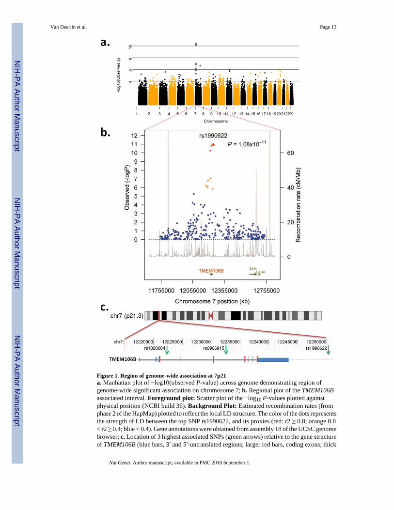
Figure 1. Region of genome-wide association at 7p21a. Manhattan plot of −log10(observed P-value) across genome demonstrating region ofgenome-wide significant association on chromosome 7; b. Regional plot of the TMEM106Bassociated interval. Foreground plot: Scatter plot of the −log10 P-values plotted againstphysical position (NCBI build 36). Background Plot: Estimated recombination rates (fromphase 2 of the HapMap) plotted to reflect the local LD structure. The color of the dots representsthe strength of LD between the top SNP rs1990622, and its proxies (red: r2 ≥ 0.8; orange 0.8< r2 ≥ 0.4; blue < 0.4). Gene annotations were obtained from assembly 18 of the UCSC genomebrowser; c. Location of 3 highest associated SNPs (green arrows) relative to the gene structureof TMEM106B (blue bars, 3′ and 5′-untranslated regions; larger red bars, coding exons; thick
Van Deerlin et al. Page 13
Nat Genet. Author manuscript; available in PMC 2010 September 1.
NIH
-PA Author Manuscript
NIH
-PA Author Manuscript
NIH
-PA Author Manuscript

gray line, intronic regions; gray dashed line, downstream chromosome sequence) andchromosome 7 location.
Van Deerlin et al. Page 14
Nat Genet. Author manuscript; available in PMC 2010 September 1.
NIH
-PA Author Manuscript
NIH
-PA Author Manuscript
NIH
-PA Author Manuscript

Figure 2. TMEM106B expression variation by genotype and disease statea. TMEM106B mRNA expression by QRT-PCR in frontal cortex differed significantly bygenotype at rs1990622 (overall P=0.027, genotype TT vs. TC P=0.017, TT vs. CC P=0.03).Black circles, FTLD-TDP (n=18); open squares, normal (n=7); horizontal lines, group mean.Significance of P-values are denoted by the numbers of asterisks. b. TMEM106B mRNAexpression in frontal cortex was significantly higher in samples from FTLD-TDP patientscompared to normal controls (P=0.045). c. TMEM106B expression in frontal cortex samplesin FTLD-TDP with (GRN pos, n=8) or without (GRN neg, n=10) GRN mutations compared tonormals (n=7). GRN mutation carriers had significantly higher levels of TMEM106Bexpression (overall P =0.0009, GRN pos vs. controls P =0.0005, GRN pos vs. GRN neg P=0.002). d. When only cases heterozygous at rs1990622 (n=14) were evaluated, GRNmutations remained significantly associated with a higher level of TMEM106B expression(P =0.039) in frontal cortex. QRT-PCR was performed in triplicate for all expression studies.Expression values were normalized to the geometric mean of two housekeeping genes and areshown relative to a single reference normal control sample23. Error bars represent the standarderror of the mean. Normalized gene expression data and sample genotype and gender data usedfor these analyses are provided online in Supplementary Material.
Van Deerlin et al. Page 15
Nat Genet. Author manuscript; available in PMC 2010 September 1.
NIH
-PA Author Manuscript
NIH
-PA Author Manuscript
NIH
-PA Author Manuscript

Figure 3. Manhattan plot in cases with and without GRN mutationsManhattan plot of −log10(observed P-value) across genome in cases with (a) and without (b)GRN mutations. The subset of cases with GRN mutations demonstrates regions of genome-wide significant association on chromosomes 7 and 17. The chr 17 association is confirmed tobe driven by a shared haplotype in c.1477C>T (p.R493X) GRN mutation carriers representing~20% of mutation positive cases, however the chromosome 7 association is not related to anysingle GRN mutation and remains when the cases with c.1477C>T are removed(P=1.446×10⁻¹⁰). The same locus on chr 7 identified in the GRN mutation cases is also thestrongest signal in the GRN negative cases, although it does not reach genome-widesignificance. A list of the SNPs with the highest signals in b is given in Supplementary Table8.
Van Deerlin et al. Page 16
Nat Genet. Author manuscript; available in PMC 2010 September 1.
NIH
-PA Author Manuscript
NIH
-PA Author Manuscript
NIH
-PA Author Manuscript

NIH
-PA Author Manuscript
NIH
-PA Author Manuscript
NIH
-PA Author Manuscript
Van Deerlin et al. Page 17
Tabl
e 1
Sum
mar
y of
sam
ples
and
con
trols
use
d fo
r GW
A a
nd re
plic
atio
n ph
ases
Phas
eC
ase
Num
bers
Cas
e St
udy
Sour
ceC
ontr
olN
umbe
rsC
ontr
ol S
tudy
Sou
rce
Met
hod
ofT
estin
gλ
GW
A51
5In
tern
atio
nal
FTLD
Con
sorti
um25
0912
97 C
HO
P Eu
rope
an-
Cau
casi
an,
1212
WTC
CC
Illum
ina
HH
550
or61
0-Q
uad
Bea
dChi
ps1.
05
Rep
licat
ion
89In
tern
atio
nal
FTLD
Con
sorti
um55
3Pe
nn A
utop
sy, P
enn
AD
C,
Cor
iell
Neu
rolo
gica
llyN
orm
al p
anel
, CH
OP
Euro
pean
-Cau
casi
an
TaqM
ange
noty
ping
of
2 SN
Ps
CH
OP,
Chi
ldre
n's H
ospi
tal o
f Phi
lade
lphi
a; P
enn
AD
C, U
nive
rsity
of P
enns
ylva
niaA
lzhe
imer
's D
isea
se C
ente
r; W
TCC
C, W
ellc
ome
Trus
t Cas
e C
ontro
l Con
sorti
um; λ
,gen
omic
con
trol i
nfla
tion
fact
or.
Nat Genet. Author manuscript; available in PMC 2010 September 1.
NIH
-PA Author Manuscript
NIH
-PA Author Manuscript
NIH
-PA Author Manuscript
Van Deerlin et al. Page 18
Tabl
e 2
SNPs
on
chro
mos
ome
7 in
regi
on w
ith h
ighe
st a
ssoc
iatio
n in
the
GW
AS
SNP
rs ID
BP
Min
orA
llele
MA
Fca
seM
AF
cont
CA
P-v
alO
RL
ower
95%
CI
Upp
er95
% C
I
rs10
0686
912
0717
95G
0.14
80.
184
5.82
×10−
30.
770.
640.
92
rs19
9060
212
0883
21G
0.13
30.
166
8.97
×10−
30.
770.
640.
94
rs10
2263
9512
1018
59C
0.15
40.
191
4.90
×10−
30.
770.
640.
93
rs10
0343
312
1306
25G
0.28
70.
351
9.45
×10−
50.
750.
640.
86
rs69
5227
212
1665
85T
0.15
40.
207
9.88
×10−
50.
700.
580.
84
rs12
6713
3212
1820
87C
0.17
30.
245
7.50
×10−
70.
640.
540.
77
rs14
6891
512
1944
17C
0.17
10.
242
9.49
×10−
70.
650.
540.
77
rs10
2000
412
2223
03G
0.23
30.
338
5.00
×10−
110.
600.
510.
70
rs69
6691
512
2325
13T
0.32
10.
435
1.63
×10−
110.
610.
530.
71
rs10
4881
9212
2436
06T
0.13
90.
204
1.46
×10−
60.
630.
520.
76
rs19
9062
212
2503
12C
0.32
10.
436
1.08
×10−
110.
610.
530.
71
rs69
4590
212
2529
34A
0.18
30.
230
7.17
×10−
40.
750.
630.
89
SNPs
are
list
ed in
gen
omic
ord
er b
ased
on
loca
tion
on c
hrom
osom
e 7.
SN
Ps in
bol
d te
xt h
ave
the
low
est P
-val
ues.
Chr
, chr
omos
ome;
BP,
bas
e pa
irs (N
CB
I bui
ld 3
6); M
AF,
min
or a
llele
freq
uenc
y; c
ont,
cont
rols
; CA
P-v
al, C
ochr
ane-
Arm
itage
P-v
alue
; OR
, odd
s rat
io; C
I, co
nfid
ence
inte
rval
.
Nat Genet. Author manuscript; available in PMC 2010 September 1.