Embed Size (px)
Citation preview

p53/CEP-1 Increases or Decreases Lifespan, Depending on Levelof Mitochondrial Bioenergetic Stress
Natascia Ventura1,2,@, Shane L. Rea1,3, Alfonso Schiavi2, Alessandro Torgovnick2,Roberto Testi2,4, and Thomas E. Johnson1
1Institute for Behavioral Genetics, University of Colorado at Boulder, Box 447, Boulder, CO80309, USA.2Department of Experimental Medicine and Biochemical Sciences, University of Rome “TorVergata”, Rome, Italy3Sam and Ann Barshop Institute for Longevity and Aging Studies and the Department ofPhysiology, University of Texas Health Science Center at San Antonio, San Antonio, Texas, USA4Fondazione Santa Lucia, Rome, Italy
SUMMARYMitochondrial pathologies underlie a number of life-shortening diseases in humans. In thenematode Caenorhabditis elegans, severely reduced expression of mitochondrial proteinsinvolved in electron transport chain-mediated energy production also leads to pathologicalphenotypes, including arrested development and/or shorter life; in sharp contrast, mild suppressionof these same proteins extends lifespan. Here we show that the C. elegans p53 ortholog cep-1mediates these opposite effects. We find that cep-1 is required to extend longevity in response tomild suppression of several bioenergetically relevant mitochondrial proteins, including frataxin -the protein defective in patients with Friedreich’s Ataxia. Importantly we show that cep-1 alsomediates both the developmental arrest and life shortening induced by severe mitochondrial stress.Our findings support an evolutionarily conserved function for p53 in modulating organismalresponses to mitochondrial dysfunction and suggest that metabolic checkpoint responses may playa role in longevity control and in human mitochondrial-associated diseases.
Keywordsp53/cep-1; mitochondria; frataxin; C. elegans; Mit mutants; aging; metabolic checkpoint
INTRODUCTIONMutations in genes that affect mitochondrial functionality lead to a variety of life-shortening, degenerative disorders in humans (Wallace 2005). At the root of most of thesediseases are reduced energy production and increased free radical production, resulting fromdirect or indirect impairment of the mitochondrial electron transport chain (ETC). Tissues
@To whom correspondence should be addressed: [email protected], phone:+39-06-72596540, fax:+39-06-72596505.AUTHOR CONTRIBUTIONSN.V. designed and performed most of the experiments, analyzed the data and co-wrote the manuscript. S.R. participated withexperimental design and execution, interpretation of results and co-wrote the manuscript. A.T. and A.S. performed some of theexperiments. R.T. and T.J. equally helped support this work financially and edited the manuscript.COMPETING INTERESTThe authors have declared that no competing interests exist.
NIH Public AccessAuthor ManuscriptAging Cell. Author manuscript; available in PMC 2010 August 1.
Published in final edited form as:Aging Cell. 2009 August ; 8(4): 380–393. doi:10.1111/j.1474-9726.2009.00482.x.
NIH
-PA Author Manuscript
NIH
-PA Author Manuscript
NIH
-PA Author Manuscript
most affected in these diseases are those with high oxidative energy requirements such asneuronal, cardiac, endocrine and renal systems. Friedreich’s Ataxia (FRDA), the mostfrequently inherited ataxia, is one such disorder (Campuzano et al. 1996), and it is ascribedto severely defective expression of frataxin, a nuclear-encoded mitochondrial protein thatplays a role in the biogenesis of Fe-S cluster containing proteins and thus in the functionalityof mitochondrial proteins such as aconitase and complexes I, II and III of the ETC (Puccio etal. 2001).
In the nematode Caenorhabditis elegans, a collection of mutants defined by RNAi orgenetic disruption of proteins involved in Mitochondrial ETC functionality (hereafterreferred to as Mit mutants) are unexpectedly long-lived; they also have slower rates ofdevelopment and decreased fertility (Rea 2005;Ventura et al. 2006). In our attempts togenerate a nematode model of FRDA, we surprisingly found that decreasing the expressionof the frataxin C. elegans ortholog, frh-1, also prolonged animal longevity (Ventura et al.2005). Reduced expression of the NDUSF3 subunit of complex I of the ETC also leads tolifespan extension in C. elegans. In humans, mutation in complex I subunits includingNDUSF3, cause Leigh Syndrome, another degenerative disorder. How can loss of genescritical for both cellular energy production and proper mitochondrial function, and whichwhen mutated in humans lead to life-shortening debilitative diseases, result in life extensionin C. elegans? To reconcile this human-nematode paradox we recently suggested that downto a certain threshold, cells have the ability to counteract a reduction in mitochondrial ETCfunction by invoking compensatory mechanisms, but beyond this threshold cell viability isseverely compromised (Ventura et al. 2006). Consistent with this model, we previouslyshowed that C. elegans Mit mutants exhibit lifespan extension only in a defined window ofmitochondrial protein suppression; indeed when mitochondrial dysfunction becomes toosevere and passes a critical threshold, these mutants exhibit pathological phenotypesincluding arrested development and lifespan reduction, perhaps similar to mitochondrialdisease states in humans (Rea et al. 2007;Ventura & Rea 2007). The molecular mechanismsunderlying this transition in phenotype in response to different levels of mitochondrial stressare still unknown.
Here we show that, cep-1, the C. elegans p53 ortholog, modulates Mit mutant phenotypes.Our findings are in agreement with a growing body of evidence that reveal a role for p53 asa metabolic checkpoint sensor (Bensaad & Vousden 2007), and support an evolutionarilyconserved function for the p53 family in specifying organismal response to mitochondrialstress. Importantly they suggest that p53 in humans may also play a role in controlling thepresentation of FRDA, Leigh Syndrome and possibly other Human MitochondrialAssociated Diseases (HMADs).
RESULTSFrataxin modulation of stress response genes and longevity
Previous findings suggest that the C. elegans Mit mutants experience endogenous stress(Rea et al. 2007; Ventura & Rea 2007). We tested if hormetic-like responses are responsiblefor life extension in these worms. Feeding frh-1 RNAi to wild type animals for threeconsecutive generations results in a 75% decrease in frataxin mRNA level and a maximalincrease in mean lifespan (Ventura et al. 2005). We found that this treatment also robustlyinduced the expression of multiple stress-responsive reporter genes (including gst-4::GFP,hsp-16.2::GFP (Link et al. 1999) and hsp-6::GFP (Yoneda et al. 2004)) when comparedwith animals fed empty vector (Fig. 1A). We quantified this induction by western blottingand found that, relative to control-treated animals, frh-1 RNAi increased the expression ofGST-4 and of HSP-6, 30 and 25 fold, respectively. HSP-16.2 is only expressed after heatshock. In control animals, HSP-16.2 was induced 30 fold by heat shock but almost 50 fold in
Ventura et al. Page 2
Aging Cell. Author manuscript; available in PMC 2010 August 1.
NIH
-PA Author Manuscript
NIH
-PA Author Manuscript
NIH
-PA Author Manuscript

frh-1 RNAi-treated animals (Fig. 1B and Fig. S1A). frh-1 RNAi progressively increased theexpression of the GST-4 and the HSP-6 throughout the three generations of feeding (datanot shown). frh-1 RNAi also weakly induced the expression of the antioxidant gene sod-3over control (Fig. S1B). These data clearly indicate that mild reduction of frataxinexpression induces a robust stress response.
We next sought to determine if known genetic modulators of reaction to stress and lifespanin worms, also controlled frh-1 RNAi-mediated longevity. SKN-1 is the C. elegansfunctional ortholog of the mammalian redox transcription factor Nrf2. SKN-1 is activatedfollowing oxidative stress and, like Nrf2 in other species, it controls the induction ofantioxidant responsive element (ARE)-containing genes, such as the Phase II enzymes gcs-1(An & Blackwell 2003), and gst-4 (Kahn et al. 2008). Mild oxidative stress increases C.elegans lifespan ((Cypser et al. 2006) and Fig. S2A). We tested whether frh-1 RNAi-induced oxidative stress might extend lifespan through skn-1. While a strain carrying askn-1(zu67) mutant allele is short-lived compared to wild-type (An & Blackwell 2003), wefound that frh-1 suppression by RNAi still increased its lifespan (Fig. 2A and Table I).Moreover, while the weak increase in lifespan induced by mild oxidative stress iscompletely blocked in skn-1 mutants, these animals were still long-lived after treatment withfrh-1 RNAi, even when exposed to oxidative stress (Fig. S2B and Table I). The skn-1 geneencodes three different SKN-1 isoforms, SKN-1A, SKN-1B and SKN-1C, which areexpressed in different tissues. The skn-1(zu67) mutant allele does not knock out isoform B(Fig. S2C), which is primarily expressed in the ASI neurons and which was recently shownto specifically mediate lifespan extension by caloric restriction, via effects on mitochondrialrespiration (Bishop & Guarente 2007). Nonetheless, even in a skn-1(zu135) mutantbackground, which disrupts the expression of all three SKN-1 isoforms, frh-1 RNAi was stillable to evoke a lifespan increase (Table I).
Daf-16, a C. elegans homolog of the human forkhead redox transcription factor FOXOfamily, is part of the insulin/IGF-1-like pathway and is required for increased stressresistance and longevity in many C. elegans Age mutants (Henderson et al. 2005). Daf-16modulates lifespan in parallel with skn-1 (Tullet et al. 2008), and along with other Daf genesis involved in the hormetic response to heat shock (Cypser et al. 2006). Compared tocontrol-fed animals, frh-1 RNAi increased lifespan of the daf-16(mgDf50) short-livedmutant (Fig. 2B and Table I). This result is in agreement with previous work showing othermitochondrial mutants increase longevity independently of daf-16 (Dillin et al. 2002).Moreover, consistent with frh-1 RNAi specifying longevity independently of the IGF/insulinlike pathway, we also found that RNAi against frh-1 further increased the lifespan of thelong-lived PI3K mutant age-1(hx546) (Table I).
SIRT1 and FOXO interact to modulate longevity and gene expression in response tooxidative stress and caloric restriction both in mammalian cells and in C. elegans (Wang &Tissenbaum 2006; Wang et al. 2007). We found that frh-1 RNAi still increased longevity intwo different sirtuin knock-out mutants, sir-2.1(ok434) and sir-2.3(ok444) (Fig. 2C andTable I). Our findings indicate that known lifespan-modulating pathways, which are oftenactivated in response to stress, do not mediate the lifespan extension induced by reducedexpression of frataxin.
Mit mutant longevity is dependent upon p53/cep-1The tumor suppressor p53 is activated in response to a broad range of stressors, includinggenotoxic, metabolic and oxidative challenges (Vousden & Lane 2007). In C. elegans, thereis one p53 homolog, cep-1, already known to be required for germ-line apoptosis inresponse to DNA damage and for normal resistance in the soma to environmental stressorssuch as hypoxia and starvation (Derry et al. 2001). Consistent with the role of p53 in
Ventura et al. Page 3
Aging Cell. Author manuscript; available in PMC 2010 August 1.
NIH
-PA Author Manuscript
NIH
-PA Author Manuscript
NIH
-PA Author Manuscript

modulating cell senescence and aging in different species (Bauer & Helfand 2006), it wasrecently shown that loss of cep-1 in C. elegans increases lifespan (Arum & Johnson 2007).We have now confirmed that a different cep-1 allele, cep-1(lg12501) (Schumacher et al.2001) is also long lived (Table I).
We tested if p53/cep-1-modulated C. elegans longevity following reduced frh-1 expression.We fed the C. elegans p53 knock-out strain carrying the cep-1(gk138) allele, previously out-crossed ten times (Arum & Johnson 2007), with control or frataxin RNAi for threeconsecutive generations, and found that the absence of cep-1 significantly suppressed theincrease in lifespan induced by frh-1 RNAi (Fig. 2D and Table I). We next investigatedwhether our observation with p53/cep-1 could be translated to other Mit mutants. Many ofthese mutants display life extension only in a discrete range of mitochondrial proteinsuppression. When mitochondrial protein expression is reduced too severely, lifespan beginsto shorten and other pathological phenotypes appear. We previously demonstrated thisphenomenon using an RNAi feeding dilution strategy ((Rea et al. 2007) and Fig.S3). Wetherefore followed the lifespan of wild-type and cep-1(gk138) strains cultured on RNAi toone of three Mit targets, at concentrations found to maximally increase lifespan: wedecreased the expression of the following mitochondrial components - ATP-3, the ATP5O/OSCP subunit of the F1FoATPase; ISP-1, the Rieske iron-sulfur protein of ubiquinol-cytochrome c oxidoreductase; CCO-1, the COX5B subunit of cytochrome c oxidoreductase.Wild-type animals fed with a 1/10 dilution of atp-3, isp-1 and cco-1 RNAi are all long livedcompared to control RNAi-treated animals (Rea et al. 2007). Interestingly, the lifespanextension induced by the same Mit RNAi concentration was significantly suppressed in thecep-1(gk138) strain (Fig. 3A–C and Table II). Nevertheless, these Mit mutants remainedsignificantly longer-lived than vector-treated animals. These results indicate a role for cep-1in sensing mitochondrial alterations and consequently modulating C. elegans longevity.
Janus-faced role of p53 is evolutionary conservedp53 responds to different levels of stress by inducing opposite cellular outcomes. This effectis mediated, in part, by the action of differential gene activation and p53 modification(Vousden & Lane 2007). We wondered whether cep-1 would have opposite effects on thespecification of Mit mutant longevity, depending on the severity of mitochondrialbioenergetic stress. We thus fed wild-type and cep-1(gk138) strains with more severepreparations (undiluted) of RNAi against atp-3, isp-1 or cco-1 and found that cep-1 mutantsdisplayed longer lifespan than wild-type animals (Fig. 3 D–F and Table II). Specifically,undiluted atp-3 RNAi decreased the lifespan of the wild-type strain below that of controlRNAi-treated animals ((Rea et al. 2007) and Fig. S3A) while in the cep-1 mutant strainlifespan was increased with the same RNAi treatment (Fig. 3D and Fig. S4A). We alsotested a 1:2 atp-3 to empty-vector RNAi ratio and consistently found the same results (Fig.S4A). Undiluted isp-1 RNAi shortened lifespan of the wild-type strain when compared tothe 1/10 dilution, though to control-treated animals this treatment still resulted in some lifeextension ((Rea et al. 2007) and Fig. S3B). Undiluted cco-1, more severely affectedanimals’ phenotype, which appeared more fragile and almost sterile, but it did not shortenanimal lifespan compared to the 1/10 dilution. Nonetheless, consistent to what with saw withmore severe mitochondrial damage induced by undiluted atp-3 RNAi, treatment of cep-1mutant strain with undiluted isp-1 and cco-1 RNAi increased lifespan relative to wild-typeanimals fed with the same RNAi concentration (Fig. 3 E, F and Fig. S4B).
One trivial explanation for the differential effects of cep-1(gk138) on Mit mutant longevityis that the efficacy of RNAi in this line is reduced relative to wild type (N2) animals. RT-PCR was used to compare atp-3 mRNA levels in cep-1 and N2 animals following RNAitreatment (Fig 4A). No difference in RNAi knockdown efficacy was observed. Importantly,
Ventura et al. Page 4
Aging Cell. Author manuscript; available in PMC 2010 August 1.
NIH
-PA Author Manuscript
NIH
-PA Author Manuscript
NIH
-PA Author Manuscript
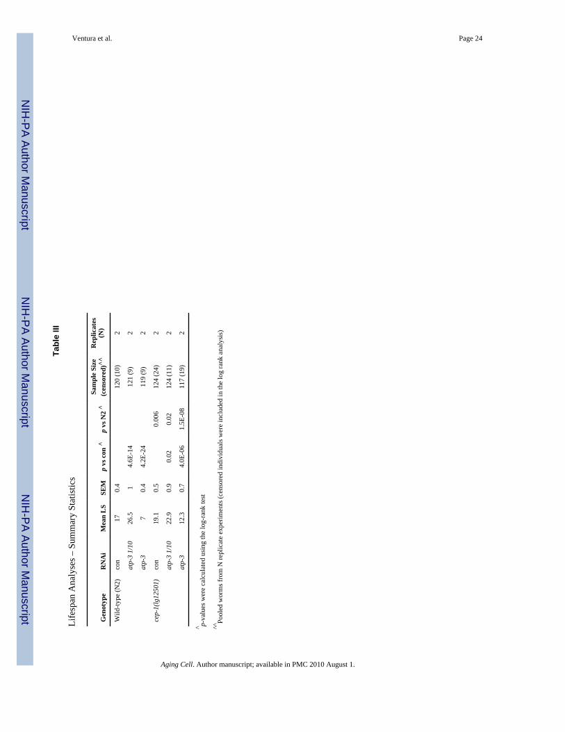
we obtained similar lifespan outcomes upon feeding different atp-3 RNAi dilutions to adifferent cep-1 allele, cep-1(lg12501) (Fig. 4B, Fig. S4C and Table III).
In summary, our results are consistent with a Janus-faced role of p53 in response to differentlevel of stress (Vousden & Lane 2007) and suggest that cep-1 may sense mitochondriaalterations and subsequently induce opposite effects on animal longevity depending on thelevel of mitochondrial stress.
cep-1 modulates Mit mutant developmentBesides changes in lifespan, Mit mutants display other phenotypic alterations that correlatewith degree of mitochondrial disruption (Rea et al. 2007). Severe reduction of either atp-3or isp-1 by feeding RNAi is sufficient to arrest development in wild-type animals (Rea et al.2007). Developmental arrest is also observed in animals fed for two consecutive generationswith RNAi against nuo-2 (which encodes the NDUFS3 subunit of NADH-ubiquinoneoxidoreductase) (Ventura & Rea 2007). cep-1(gk138) mutant strain had no effect on thedevelopmental rate of animals fed control vector but it clearly prevented the developmentalarrest induced by either undiluted atp-3 or isp-1 RNAi (Fig. 5A), although this effect wasnot fully penetrant. Similar results were obtained using the cep-1(lg12501) allele fedundiluted atp-3 (Fig. S6), and when undiluted nuo-2 RNAi was fed for two consecutivegenerations (Fig. 5B). These findings suggest cep-1 can modulate development underconditions of bioenergetic stress.
The DNA intercalating agent ethidium bromide (EtBr) inhibits mitochondrial (mt) DNAreplication and transcription at low concentrations without causing detectable effects onnuclear DNA (Nass 1972). For this reason it is widely used in mammalian cells to mimichuman mitochondrial diseases (Biswas et al. 1999). Previous studies have also shown thatEtBr induces both developmental arrest and increase lifespan in C. elegans (Tsang & Lemire2002). We found that the developmental arrest induced by 75 µg/ml of EtBr is partiallyreverted by loss of cep-1 (Fig. 5A), lending further support to a role for CEP-1 in sensingmitochondrial bioenergetic alterations and modulating the coordinate expression of multipleMit mutant phenotypes. This finding again underscores the notion that the effect of cep-1 inour above experiments is not due simply to subtle differences in RNAi efficacy.
Life shortening and developmental arrest induced by severe electron transport chaindisruption involves egl-1
Mitochondrial ETC disruption can lead to DNA damage, which is a major p53-activatingstressor in human cells and, depending on its severity, can in turn lead either to transientcell-cycle arrest or apoptosis. In worms, atm-1 (the C. elegans ortholog of the human AtaxiaTeleangectasia gene), and egl-1 (the C. elegans Bcl-2 homology domain-3 (BH3)-onlyprotein), act upstream and downstream, respectively, of cep-1 to induce germline apoptosisin response to DNA damage (Hofmann et al. 2002; Garcia-Muse & Boulton 2005). EGL-1is also required to induce apoptosis and mitochondrial fragmentation of the 131 out of 1090somatic cells that normally die during C. elegans development (Conradt & Horvitz 1998;Jagasia et al. 2005), and it is required in the embryo to induce autophagy in response tostarvation (Maiuri et al. 2007). We tested if either atm-1 or egl-1 were essential to the life-lengthening or the life-shortening effects of mitochondrial ETC disruption.
We found that atm-1(gk186) is long lived while egl-1(n487) is short lived compared to wild-type animals (Table IV). We observed that both atm-1 and egl-1 play no apparent role in thelife extension that occurs following mild frh-1 RNAi treatment (Fig. S5 A, B and Table IV),or following feeding with 1/10 diluted atp-3 RNAi (Fig. S5C, Fig. 6A and Table IV).Furthermore, atm-1 plays no obvious role in the life shortening that occurs following
Ventura et al. Page 5
Aging Cell. Author manuscript; available in PMC 2010 August 1.
NIH
-PA Author Manuscript
NIH
-PA Author Manuscript
NIH
-PA Author Manuscript

treatment with undiluted atp-3 RNAi (Fig. S5D and Table IV). Loss of egl-1, on the otherhand, shortens the lifespan of control fed animal but partially reverted the lifespanshortening effect caused by growth on undiluted atp-3 RNAi (Fig. 6B and Table IV). Thiseffect was never complete, illustrating that other factors await identification or thategl-1(n487) is not fully penetrant. Consistently, we observed that egl-1 knock-out animalssuppressed the developmental arrest induced by undiluted atp-3 RNAi, but this phenotypewas also not fully penetrant (data not shown). We conclude that mild mitochondrialdisruption extends lifespan in a cep-1 dependent manner but independently of the DNA-damage apoptotic pathway, while severe reduction of mitochondrial function requires, inpart, the cep-1/egl-1 pathway to decrease lifespan and arrest development in the Mitmutants.
In agreement with our above observations showing a functional requirement for egl-1 onlyat strong atp-3 RNAi concentrations, we observed that growth of wild type animals ontreatments that increased lifespan, namely mild frh-1 RNAi or a 1/10 dilution of atp-3RNAi, only marginally induced egl-1 mRNA (Fig. 6C). Neither treatment affected theexpression of an egl-1::gfp translational reporter strain (data not shown). On the contrary,growth of wild type animals on a treatment that shortens lifespan, namely undiluted atp-3RNAi, strongly induced egl-1 mRNA (Fig. 6C), although not at the classical levels shown inresponse to DNA-damage inducing agents (Hofmann et al. 2002;Garcia-Muse & Boulton2005). Induction of egl-1 by atp-3 RNAi at 1/10 and undiluted potency are significantlysuppressed by loss of cep-1. These findings reveal a new role for the cep-1/egl-1 pathwayacting during C. elegans development to regulate animal lifespan.
DISCUSSIONFriedreich’s Ataxia (FRDA), as well as many other disorders associated with mitochondrialdysfunction, is a chronic, degenerative disease that often becomes clinically apparent later inlife. It is probable that protective pathways are activated in the pre-symptomatic phase ofFRDA to adapt and cope with dysfunctional mitochondria, thus delaying its clinicalappearance. Similar pathways may be induced in C. elegans by a mild decrease of thefrataxin protein (Ventura et al. 2006). We have now shown that protective stress-responsegenes, such as heat shock proteins and antioxidants, are strongly induced in worms inresponse to mild frataxin silencing. In addition we found that the tumor suppressor proteinp53 has a key role in modulating the phenotypic effects resulting from mitochondrialdisruption. Our findings hence invoke new players in the etiology of human mitochondrialassociated diseases such as FRDA and Leigh Syndrome.
Protective Responses to Mitochondrial DysfunctionHormetic responses to mild heat shock or oxidative stress have been associated withprolonged longevity in C. elegans. When we looked at the contribution of daf-16 and skn-1(which encode transcription factors that modulate heat shock and oxidative stress responses,respectively), to the longevity increase induced by frh-1 RNAi, we found that loss of neithertranscription factor prevented life extension. Our results indicate that oxidative stressresponse, at least that controlled by these two transcription factors, is not the limiting factorregulating Mit mutant lifespan. Importantly if, as we have previously hypothesized (Venturaet al. 2006), similar pathways counteract mitochondrial dysfunction in both long-livedanimals and in the pre-symptomatic phase of FRDA, our result imply that oxidative stressresponses may also play a role in the human disease, but that it might not be the only playerin eliciting protective pathways to help delay the established pathology.
Caloric Restriction (CR) extends C. elegans longevity and increases respiratory rate in aSIR2.1 and SKN-1 dependent manner (Wang & Tissenbaum 2006; Bishop & Guarente
Ventura et al. Page 6
Aging Cell. Author manuscript; available in PMC 2010 August 1.
NIH
-PA Author Manuscript
NIH
-PA Author Manuscript
NIH
-PA Author Manuscript

2007). We found that frh-1 RNAi increased C. elegans lifespan independently of sir2.1 and,as mentioned, skn-1. Our findings do not, however, discount a role for increasedmitochondrial respiration resulting from a compensatory induction of mitochondrialbiogenesis in response to frh-1 RNAi. Indeed, induction of mitochondrial biogenesis inresponse to mild mitochondrial dysfunction in the pre-symptomatic phase of FRDA couldhelp sustain cellular viability and thus act to clinically delay disease onset. In humans, p53 isknown to regulate both mitochondrial biogenesis and mitochondrial respiration (Donahue etal. 2001; Matoba et al. 2006) and we have now found that p53/cep-1 is required for lifeextension in the Mit mutants. Derry and colleagues have reported that loss of cep-1, per se,causes significant transcriptional alterations (Derry et al. 2007), although these findingswere not confirmed in a subsequent study (Greiss et al. 2008). Nevertheless, among thepurportedly altered genes of the former analysis, there were many encoding formitochondrial proteins (listed in Table S1). We have previously shown that mitochondrialETC protein expression is pliant and responds uniquely in different Mit mutants (Ventura &Rea 2007). Additional work is warranted in order to understand the role played by p53 in theregulation of mitochondrial biogenesis, mitochondrial proteins composition and itsrelationship with longevity specification and disease prevention in response to mitochondrialdysfunction.
The Janus-faced role of p53Considerable evidence supports a dual role for p53 in eliciting different responses dependingon level of cellular stress (Vousden & Lane 2007). Mild or transient cellular damage, suchas mild increase in free radical production, minor DNA damage, and transitory glucose orATP depletion, induce protective p53 pathways that improve antioxidant defenses and DNArepair mechanisms, and fulfill energy requirements. In these instances p53 temporarilyarrests the cell-cycle until the stress has been resolved. More severe oxidative stress,irreparable DNA damage, or complete ATP deprivation leads to p53-dependent cell death orirreversible cell-cycle arrest (replicative cell senescence). Both p53-dependent responsesaccount for its tumor suppressor activity in humans. Consistent with the Janus-faced activityof p53 (Vousden & Lane 2007), and with p53’s role as a sensor and mediator ofmitochondrial metabolism (Jones et al. 2005; Mandal et al. 2005), we have shown thatcep-1, the C. elegans p53 ortholog, modulates Mit mutant lifespan in an opposite manner,depending on the level of mitochondrial stress experienced. cep-1 is required for increasedlongevity under mild mitochondrial disruption, and for mediating the detrimental effect onlifespan when mitochondrial damage is more severe.
Recently, two Mit mutations that increase lifespan, isp-1(qm150) and clk-1(qm30), wereshown to protect against tumor growth in the C. elegans gld-1 tumor-like mutant, in a cep-1-dependent fashion (Pinkston et al. 2006). In conjunction with our present findings, theseresults imply that modulating mitochondrial function, by way of p53, can both regulatelongevity and concurrently serve as an anti-tumor strategy. A similar situation seems to existin mice where increased, but otherwise normally-regulated, levels of p53 lead to both tumorsuppression and anti-aging effects (Matheu et al. 2007).
p53-activating stressors can lead to transient cell-cycle arrest or apoptosis. While we can notrule out the possibility that apoptosis via CEP-1 independent pathways play a role in theappearance of Mit mutant phenotypes, our results with egl-1 and atm-1 knock out strains,suggest that apoptosis is not the main mechanism through which CEP-1 is extendinglongevity in the Mit mutants. On the other hand, deregulated induction of a CEP-1dependent apoptotic pathway may explain the short lifespan induced by severemitochondrial stress (this study and (Senoo-Matsuda et al. 2003). Independent of its pro-apoptotic activity, EGL-1 is known to also induce mitochondrial fragmentation (Delivani et
Ventura et al. Page 7
Aging Cell. Author manuscript; available in PMC 2010 August 1.
NIH
-PA Author Manuscript
NIH
-PA Author Manuscript
NIH
-PA Author Manuscript

al. 2006), a process that may contributes to its lifespan shortening effect (Chan & Mattson1999).
In worms the pro-apoptotic and anti-proliferative effects of cep-1 can follow differentpathways (Derry et al. 2007). Moreover, in the gld-1 mutant, the two long-lived Mitmutants, isp-1 (qm150) and clk-1(qm30), reduce germ cell hyperproliferation withoutinducing germ cell apoptosis (Pinkston et al. 2006). Furthermore, aak-2 is an AMP kinaserecently shown to be required for isp-1 and clk-1 Mit mutant longevity (Curtis et al. 2006).This same kinase is also necessary for the longevity increase induced by mild treatment ofatp-3 RNAi (our unpublished observation). Two papers have shown that, in response tomitochondrial dysfunction and energy deprivation, p53 induces cell-cycle arrest via AMPkinase-dependent phosphorylation (Jones et al. 2005; Mandal et al. 2005). Theseobservation suggest that an AMPK/CEP-1 metabolic checkpoint may play an integral role inspecifying Mit mutant longevity.
Another role of p53, which is Janus-faced and therefore potentially relevant to mitochondrialstress, is the control of autophagy (Maiuri et al. 2007; Levine & Abrams 2008). Autophagyis a form of organelle and cellular digestion that can be either necessary or detrimental fortissue homeostasis and organismal survival. It has been associated both with prevention andcausation of diseases in human, including neurodegenerative disorders (Vellai et al. 2007;Levine & Kroemer 2008). Autophagic genes are required during normal development in C.elegans and also for the increased lifespan observed in several mutant backgrounds,including a p53/cep-1 knock out strain (Melendez et al. 2003; Tavernarakis et al. 2008; Tothet al. 2008). Moreover, gain-of function mutation of the sole C. elegans BH3-only proteinEGL-1, induces autophagy, while deletion of EGL-1 compromises starvation-inducedautophagy (Maiuri et al. 2007). Based on our results it will be interesting to determinewhether autophagy is associated and/or necessary to specify longevity resulting fromdifferent degrees of mitochondrial dysfunction.
A developmental checkpoint role for cep-1: the whole p53 family in one gene?In our studies we found that the absence of cep-1 partially rescued the developmental arrestinduced by severe mitochondrial damage. Importantly, we showed that cep-1 mutants alsoprevented the developmental arrest induced by treatment with EtBr, ruling out the possibilitythat lack of cep-1 is simply modulating RNAi potency rather then the response tomitochondrial stress. A developmental role of cep-1 in response to stress is consistent with acep-1-dependent metabolic checkpoint acting during C. elegans development (Rea et al.2007) and/or by cep-1 functions reminiscent of other p53 family members, such as those ofp63 and p73 in differentiation and development (D'Erchia et al. 2006; Ou et al. 2007).Under normal, well-fed conditions, cep-1 appears to be dispensable for C. elegansdevelopment. Only under conditions of increased energy and nucleotide demand, when itsfunction may be required for mitochondrial biogenesis, we observed a requirement forcep-1. These conditions may exist when cells are rapidly proliferating, such as duringembryonic C. elegans development or at the transition between L3–L4 stages, the point ofgonad formation. The regulation of animal lifespan through a mitochondrial checkpoint inproliferating cells during animal development is an interesting possibility.
Concluding RemarksOur data suggest a model in which mild disruption of ETC function in C. elegans inducestransient cellular damage (perhaps genotoxic stress, proliferation errors and/ or organelledysfunction), which is readily repaired or eliminated by sub-lethal activation of cep-1. Atemporary halt of the cell-cycle would allow for metabolic adaptation to mild mitochondrialstress and to elicit proper defenses against oxidative and other stressors; cep-1 activation
Ventura et al. Page 8
Aging Cell. Author manuscript; available in PMC 2010 August 1.
NIH
-PA Author Manuscript
NIH
-PA Author Manuscript
NIH
-PA Author Manuscript

also mediates increased animal lifespan. When mitochondrial dysfunction surpasses acritical threshold, cep-1 functionality is altered in an attempt to eliminate irreparablydamaged cell leading to the detrimental phenotypes observed in the Mit mutants (Fig. 5).Our current findings suggest that p53 has a previously unrecognized role in modulatinganimal lifespan and development in response to mitochondrial stress. p53 activity is now aprime target for investigations into the pathogenesis and treatment of human mitochondrialdisorders.
EXPERIMENTAL PROCEDURESNematode Strains and Maintenance
Standard nematode culturing techniques were employed (Wood 1988). The following strainswere utilized: N2 (wild-type), SJ4100 [hsp-6::gfp(zcIs13)], TJ375 [hsp-16.2::gfp(gpIs1)],CL2166 [dvIs19[pAF15(gst-4::gfp::NLS)] III], CL691 [dvIs19[pAF15(gst-4::gfp::NLS)III]; skn-1(zu67) IV/nT1[unc-?(n754);let-?](IV;V)], EU1 [skn-1(zu67)IV/nT1[unc-?(n754)let-?](IV;V)], EU31[skn-1(zu135)IV/nT1[unc-?(n754)let-?](IV;V)], GR1307[daf-16(mgDf50)], TJ1052 [age-1(hx546)], VC199 [sir-2.1(ok434)], RB654[sir-2.3(ok444)], VC172 [cep-1(gk138)], TJ1 [cep-1(gk138)], XY1054 [cep-1(lg12501)],VC381 [atm-1(gk186)], MT1082 [egl-1(n487)], MD166 [egl-1::gfp(bcIs1)].
Feeding RNAiRNAi feeding techniques were previously described (Ventura et al. 2005; Rea et al. 2007).The following RNAi feeding constructs were derived from the Ahringer C. elegans RNAilibrary (Kamath & Ahringer 2003): atp-3 (F27C1.7), cco-1 (F26E4.9), nuo-2 (T10E9.7)(Kamath & Ahringer 2003) while feeding RNAi constructs against frh-1 and isp-1 have beenpreviously described (Ventura et al. 2005; Rea et al. 2007).
Lifespan and Statistical AnalysisSurvival analyses began from hatching and were recorded at 20°C using synchronouspopulations of 60–100 animals per strain. We calculated mean and standard deviation of themean from pooled population of animals and used the log-rank test to assess differences insurvival curves between pooled populations as previously described (Rea et al. 2007).
Developmental AnalysisDevelopment was followed at 20°C on populations of synchronous animals from the time ofhatching after a 4 hour synchronized egg lay, which was carried out directly on RNAi platesor EtBr plates. Images were collected 4 and 5 days after the time of egg lay for the parentaland first generation animals, respectively. Live worm images, to record development, werecaptured with a digital camera connected to a dissecting microscope.
MicroscopyFor fluorescence images, individual worms were randomly picked using a regular dissectingscope from a population of 200 worms for each condition. Remaining worms were thencollected for western blotting or followed for lifespan analysis. Worms were arranged forimage analysis on 2% agarose pads and Nomarski and fluorescence images were capturedvia a Ziess Axioskop running SlideMaker 4.0 software retrofitted with deconvolutioncapabilities and a digital fluorescent camera containing a Sedat (Quad) filter set. GFPfluorescence was collected using a FITC 490/528nm absorption/emission filter pair (peaktransmission with a 20/38 nm bandpass). All images were collected using identical exposuresettings.
Ventura et al. Page 9
Aging Cell. Author manuscript; available in PMC 2010 August 1.
NIH
-PA Author Manuscript
NIH
-PA Author Manuscript
NIH
-PA Author Manuscript

Quantitative RT-PCRQuantitation of egl-1 and atp-3 mRNA in frh-1 and atp-3 RNAi-treated animals relative tovector-only treated animals was undertaken using primers specific for egl-1 (Schumacher etal. 2005) and atp-3 (Rea et al. 2007). Data was normalized to a non-variable control gene(F23B2.13). mRNA was extracted from approximately 5000 synchronized worms persample, using a Total RNA and Protein Isolation Kit (Macherey-Nagel). Oligo(dT) primedcDNA was synthesized using a SuperScript cDNA Synthesis Kit (Invitrogen). QuantitativePCR (qPCR) was undertaken on an ABI Prism® 7000 instrument with SYBR Green PCRMaster Mix kit (Applied Biosystems).
Western Blot AnalysisWestern Blot analysis of whole-worm protein extracts were prepared from ~200 adultworms. Animals were boiled in 5% SDS, 0,02% β-mercaptoethanol and 1mM proteaseinhibitor cocktail (SIGMA P2714). Following protein quantitation (BCA, Pierce), proteinlevels were measured by western analysis, quantified using densitometry, and normalizedagainst actin. Antibodies employed were anti-GFP (MSA02, MitoSciences, Oregon, 1:1000dilution) and anti-Actin (AC-15, Sigma, 1:5000).
Supplementary MaterialRefer to Web version on PubMed Central for supplementary material.
AcknowledgmentsWe thank A. Kell for technical assistance and Dr. D. Wu for help with statistical analysis. Most of the strains usedin this work were provided by the C. elegans Gene Knockout Consortium, which is funded by the National Instituteof Health and National Center for Research Resource. Dr. C. Link constructed strain CL2166 and CL691. StrainXY1054 was a kind gift of Dr. A. Gartner, SJ4100 of Dr. D. Ron, and MD166 of Dr. B. Conradt. This work wassupported by NIA grant RO1-AG16219 to T.E.J.; NIA grant R21-AG025207-01A1 to S.L.R.; Telethon grantGGP060059, ASI-MoMa, FARA and Ataxia UK to R.T.; NAF and FIRC post doctoral fellowship to N.V.
ABBREVIATIONS
CEP C. elegans p53
ETC Electron Transport Chain
FRDA Friedreich’s Ataxia
Mit mutants Mitochondrial mutants
NDUFS3 NADH Dehydrogenase [Ubiquinone] iron (Fe)-Sulfur protein 3
HMAD Human Mitochondrial-Associated Disease
FRH frataxin
gst Gluthathione-S-Transferase
hsp heat shock protein
SKN SkiNhead
Nrf Nuclear factor erythroid-2 Related Factor
ARE Antioxidant Responsive Element
Daf Abnormal Dauer Formation
SIRT sirtuins
Ventura et al. Page 10
Aging Cell. Author manuscript; available in PMC 2010 August 1.
NIH
-PA Author Manuscript
NIH
-PA Author Manuscript
NIH
-PA Author Manuscript
FOXO FOrkhead boX
ATM Ataxia Telangectasia Mutated
EGL EGg Laying defective
ATP adenosine triphosphate
CCO Cytochrome C Oxidase
ISP Iron-Sulfur Protein
EtBr ethidium bromide
GLD defective in Germ Line Development
AMPK adenosine triphosphate (AMP)-activated protein kinase.
REFERENCESAn JH, Blackwell TK. SKN-1 links C. elegans mesendodermal specification to a conserved oxidative
stress response. Genes Dev. 2003; 17:1882–1893. [PubMed: 12869585]Arum O, Johnson TE. Reduced expression of the Caenorhabditis elegans p53 ortholog cep-1 results in
increased longevity. J Gerontol A Biol Sci Med Sci. 2007; 62:951–959. [PubMed: 17895432]Bauer JH, Helfand SL. New tricks of an old molecule: lifespan regulation by p53. Aging Cell. 2006;
5:437–440. [PubMed: 16968311]Bensaad K, Vousden KH. p53: new roles in metabolism. Trends Cell Biol. 2007; 17:286–291.
[PubMed: 17481900]Bishop NA, Guarente L. Two neurons mediate diet-restriction-induced longevity in C. elegans. Nature.
2007; 447:545–549. [PubMed: 17538612]Biswas G, Adebanjo OA, Freedman BD, Anandatheerthavarada HK, Vijayasarathy C, Zaidi M,
Kotlikoff M, Avadhani NG. Retrograde Ca2+ signaling in C2C12 skeletal myocytes in response tomitochondrial genetic and metabolic stress: a novel mode of inter-organelle crosstalk. Embo J.1999; 18:522–533. [PubMed: 9927412]
Campuzano V, Montermini L, Molto MD, Pianese L, Cossee M, Cavalcanti F, Monros E, Rodius F,Duclos F, Monticelli A, Zara F, Canizares J, Koutnikova H, Bidichandani SI, Gellera C, Brice A,Trouillas P, De Michele G, Filla A, De Frutos R, Palau F, Patel PI, Di Donato S, Mandel JL,Cocozza S, Koenig M, Pandolfo M. Friedreich's ataxia: autosomal recessive disease caused by anintronic GAA triplet repeat expansion. Science. 1996; 271:1423–1427. [PubMed: 8596916]
Chan SL, Mattson MP. Caspase and calpain substrates: roles in synaptic plasticity and cell death. JNeurosci Res. 1999; 58:167–190. [PubMed: 10491581]
Conradt B, Horvitz HR. The C. elegans protein EGL-1 is required for programmed cell death andinteracts with the Bcl-2-like protein CED-9. Cell. 1998; 93:519–529. [PubMed: 9604928]
Curtis R, O'Connor G, DiStefano PS. Aging networks in Caenorhabditis elegans: AMP-activatedprotein kinase (aak-2) links multiple aging and metabolism pathways. Aging Cell. 2006; 5:119–126. [PubMed: 16626391]
Cypser JR, Tedesco P, Johnson TE. Hormesis and aging in Caenorhabditis elegans. Exp Gerontol.2006; 41:935–939. [PubMed: 17067771]
D'Erchia AM, Tullo A, Lefkimmiatis K, Saccone C, Sbisa E. The fatty acid synthase gene is aconserved p53 family target from worm to human. Cell Cycle. 2006; 5:750–758. [PubMed:16582625]
Delivani P, Adrain C, Taylor RC, Duriez PJ, Martin SJ. Role for CED-9 and Egl-1 as regulators ofmitochondrial fission and fusion dynamics. Mol Cell. 2006; 21:761–773. [PubMed: 16543146]
Derry WB, Bierings R, van Iersel M, Satkunendran T, Reinke V, Rothman JH. Regulation ofdevelopmental rate and germ cell proliferation in Caenorhabditis elegans by the p53 gene network.Cell Death Differ. 2007; 14:662–670. [PubMed: 17186023]
Ventura et al. Page 11
Aging Cell. Author manuscript; available in PMC 2010 August 1.
NIH
-PA Author Manuscript
NIH
-PA Author Manuscript
NIH
-PA Author Manuscript

Derry WB, Putzke AP, Rothman JH. Caenorhabditis elegans p53: role in apoptosis, meiosis, and stressresistance. Science. 2001; 294:591–595. [PubMed: 11557844]
Dillin A, Hsu AL, Arantes-Oliveira N, Lehrer-Graiwer J, Hsin H, Fraser AG, Kamath RS, Ahringer J,Kenyon C. Rates of behavior and aging specified by mitochondrial function during development.Science. 2002; 298:2398–2401. [PubMed: 12471266]
Donahue RJ, Razmara M, Hoek JB, Knudsen TB. Direct influence of the p53 tumor suppressor onmitochondrial biogenesis and function. Faseb J. 2001; 15:635–644. [PubMed: 11259382]
Garcia-Muse T, Boulton SJ. Distinct modes of ATR activation after replication stress and DNAdouble-strand breaks in Caenorhabditis elegans. Embo J. 2005; 24:4345–4355. [PubMed:16319925]
Greiss S, Schumacher B, Grandien K, Rothblatt J, Gartner A. Transcriptional profiling in C. eleganssuggests DNA damage dependent apoptosis as an ancient function of the p53 family. BMCGenomics. 2008; 9:334. [PubMed: 18627611]
Henderson, ST.; Rea, SL.; Johnson, TE. Dissecting the Processes of Aging Using the NematodeCaenorhabditis elegans. New York: Academic Press; 2005.
Hofmann ER, Milstein S, Boulton SJ, Ye M, Hofmann JJ, Stergiou L, Gartner A, Vidal M, HengartnerMO. Caenorhabditis elegans HUS-1 is a DNA damage checkpoint protein required for genomestability and EGL-1-mediated apoptosis. Curr Biol. 2002; 12:1908–1918. [PubMed: 12445383]
Jagasia R, Grote P, Westermann B, Conradt B. DRP-1-mediated mitochondrial fragmentation duringEGL-1-induced cell death in C. elegans. Nature. 2005; 433:754–760. [PubMed: 15716954]
Jones RG, Plas DR, Kubek S, Buzzai M, Mu J, Xu Y, Birnbaum MJ, Thompson CB. AMP-activatedprotein kinase induces a p53-dependent metabolic checkpoint. Mol Cell. 2005; 18:283–293.[PubMed: 15866171]
Kahn NW, Rea SL, Moyle S, Kell A, Johnson TE. Proteasomal dysfunction activates the transcriptionfactor SKN-1 and produces a selective oxidative-stress response in Caenorhabditis elegans.Biochem J. 2008; 409:205–213. [PubMed: 17714076]
Kamath RS, Ahringer J. Genome-wide RNAi screening in Caenorhabditis elegans. Methods. 2003;30:313–321. [PubMed: 12828945]
Levine B, Abrams J. p53: The Janus of autophagy? Nat Cell Biol. 2008; 10:637–639. [PubMed:18521069]
Levine B, Kroemer G. Autophagy in the pathogenesis of disease. Cell. 2008; 132:27–42. [PubMed:18191218]
Link CD, Cypser JR, Johnson CJ, Johnson TE. Direct observation of stress response in Caenorhabditiselegans using a reporter transgene. Cell Stress Chaperones. 1999; 4:235–242. [PubMed:10590837]
Maiuri MC, Le Toumelin G, Criollo A, Rain JC, Gautier F, Juin P, Tasdemir E, Pierron G, TroulinakiK, Tavernarakis N, Hickman JA, Geneste O, Kroemer G. Functional and physical interactionbetween Bcl-X(L) and a BH3-like domain in Beclin-1. Embo J. 2007; 26:2527–2539. [PubMed:17446862]
Mandal S, Guptan P, Owusu-Ansah E, Banerjee U. Mitochondrial regulation of cell cycle progressionduring development as revealed by the tenured mutation in Drosophila. Dev Cell. 2005; 9:843–854. [PubMed: 16326395]
Matheu A, Maraver A, Klatt P, Flores I, Garcia-Cao I, Borras C, Flores JM, Vina J, Blasco MA,Serrano M. Delayed ageing through damage protection by the Arf/p53 pathway. Nature. 2007;448:375–379. [PubMed: 17637672]
Matoba S, Kang JG, Patino WD, Wragg A, Boehm M, Gavrilova O, Hurley PJ, Bunz F, Hwang PM.p53 regulates mitochondrial respiration. Science. 2006; 312:1650–1653. [PubMed: 16728594]
Melendez A, Talloczy Z, Seaman M, Eskelinen EL, Hall DH, Levine B. Autophagy genes are essentialfor dauer development and life-span extension in C. elegans. Science. 2003; 301:1387–1391.[PubMed: 12958363]
Nass MM. Differential effects of ethidium bromide on mitochondrial and nuclear DNA synthesis invivo in cultured mammalian cells. Exp Cell Res. 1972; 72:211–222. [PubMed: 4337144]
Ou HD, Lohr F, Vogel V, Mantele W, Dotsch V. Structural evolution of C-terminal domains in thep53 family. Embo J. 2007; 26:3463–3473. [PubMed: 17581633]
Ventura et al. Page 12
Aging Cell. Author manuscript; available in PMC 2010 August 1.
NIH
-PA Author Manuscript
NIH
-PA Author Manuscript
NIH
-PA Author Manuscript

Pinkston JM, Garigan D, Hansen M, Kenyon C. Mutations that increase the life span of C. elegansinhibit tumor growth. Science. 2006; 313:971–975. [PubMed: 16917064]
Puccio H, Simon D, Cossee M, Criqui-Filipe P, Tiziano F, Melki J, Hindelang C, Matyas R, Rustin P,Koenig M. Mouse models for Friedreich ataxia exhibit cardiomyopathy, sensory nerve defect andFe-S enzyme deficiency followed by intramitochondrial iron deposits. Nat Genet. 2001; 27:181–186. [PubMed: 11175786]
Rea SL. Metabolism in the Caenorhabditis elegans Mit mutants. Exp Gerontol. 2005; 40:841–849.[PubMed: 16137850]
Rea SL, Ventura N, Johnson TE. Relationship between mitochondrial electron transport chaindysfunction, development, and life extension in Caenorhabditis elegans. PLoS Biol. 2007; 5:e259.[PubMed: 17914900]
Schumacher B, Hofmann K, Boulton S, Gartner A. The C. elegans homolog of the p53 tumorsuppressor is required for DNA damage-induced apoptosis. Curr Biol. 2001; 11:1722–1727.[PubMed: 11696333]
Schumacher B, Schertel C, Wittenburg N, Tuck S, Mitani S, Gartner A, Conradt B, Shaham S. C.elegans ced-13 can promote apoptosis and is induced in response to DNA damage. Cell DeathDiffer. 2005; 12:153–161. [PubMed: 15605074]
Senoo-Matsuda N, Hartman PS, Akatsuka A, Yoshimura S, Ishii N. A complex II defect affectsmitochondrial structure, leading to ced-3- and ced-4-dependent apoptosis and aging. J Biol Chem.2003; 278:22031–22036. [PubMed: 12672828]
Tavernarakis N, Pasparaki A, Tasdemir E, Maiuri MC, Kroemer G. The effects of p53 on wholeorganism longevity are mediated by autophagy. Autophagy. 2008; 4
Toth ML, Sigmond T, Borsos E, Barna J, Erdelyi P, Takacs-Vellai K, Orosz L, Kovacs AL, Csikos G,Sass M, Vellai T. Longevity pathways converge on autophagy genes to regulate life span incaenorhabditis elegans. Autophagy. 2008; 4:330–338. [PubMed: 18219227]
Tsang WY, Lemire BD. Mitochondrial genome content is regulated during nematode development.Biochem Biophys Res Commun. 2002; 291:8–16. [PubMed: 11829454]
Tullet JM, Hertweck M, An JH, Baker J, Hwang JY, Liu S, Oliveira RP, Baumeister R, Blackwell TK.Direct inhibition of the longevity-promoting factor SKN-1 by insulin-like signaling in C. elegans.Cell. 2008; 132:1025–1038. [PubMed: 18358814]
Vellai T, Toth ML, Kovacs AL. Janus-faced autophagy: a dual role of cellular self-eating inneurodegeneration? Autophagy. 2007; 3:461–463. [PubMed: 17471017]
Ventura N, Rea S, Henderson ST, Condo I, Johnson TE, Testi R. Reduced expression of frataxinextends the lifespan of Caenorhabditis elegans. Aging Cell. 2005; 4:109–112. [PubMed:15771615]
Ventura N, Rea SL. Caenorhabditis elegans mitochondrial mutants as an investigative tool to studyhuman neurodegenerative diseases associated with mitochondrial dysfunction. Biotechnol J. 2007;2:584–595. [PubMed: 17443764]
Ventura N, Rea SL, Testi R. Long-lived C. elegans mitochondrial mutants as a model for humanmitochondrial-associated diseases. Exp Gerontol. 2006; 41:974–991. [PubMed: 16945497]
Vousden KH, Lane DP. p53 in health and disease. Nat Rev Mol Cell Biol. 2007; 8:275–283. [PubMed:17380161]
Wallace DC. A mitochondrial paradigm of metabolic and degenerative diseases, aging, and cancer: adawn for evolutionary medicine. Annu Rev Genet. 2005; 39:359–407. [PubMed: 16285865]
Wang F, Nguyen M, Qin FX, Tong Q. SIRT2 deacetylates FOXO3a in response to oxidative stress andcaloric restriction. Aging Cell. 2007; 6:505–514. [PubMed: 17521387]
Wang Y, Tissenbaum HA. Overlapping and distinct functions for a Caenorhabditis elegans SIR2 andDAF-16/FOXO. Mech Ageing Dev. 2006; 127:48–56. [PubMed: 16280150]
Wood, WB. The Nematode Caenorhabditis elegansed^. New York: Cold Spring Harbor Laboratory;1988. p. 667
Yoneda T, Benedetti C, Urano F, Clark SG, Harding HP, Ron D. Compartment-specific perturbationof protein handling activates genes encoding mitochondrial chaperones. J Cell Sci. 2004;117:4055–4066. [PubMed: 15280428]
Ventura et al. Page 13
Aging Cell. Author manuscript; available in PMC 2010 August 1.
NIH
-PA Author Manuscript
NIH
-PA Author Manuscript
NIH
-PA Author Manuscript

Figure 1. Frataxin suppression induces stress response genes(A) Nomarski (DIC, top panels) and fluorescence (GFP, bottom panels) images of gst-4,hsp-6 and hsp-16.2 GFP reporter strains fed for three consecutive generations (F3) on vectorcontrol (con) or frataxin RNAi (frh-1). GFP expression reveals the induction of GST-4 andHSP-6 in the parental generation (P0) compare to the third generation (F3). For HSP16.2,GFP expression has been analyzed in animals cultured for three generations on frh-1 RNAi(untreated F3) or after 12 hours recovery from a 1.5h heatshock (35°C) (F3+HS). (B) GFPinduction of animals treated as in A was quantified by western blotting and normalizedagainst actin. Shown are results from one experiment out of three performed with similarresults.
Ventura et al. Page 14
Aging Cell. Author manuscript; available in PMC 2010 August 1.
NIH
-PA Author Manuscript
NIH
-PA Author Manuscript
NIH
-PA Author Manuscript

Figure 2. Loss of cep-1, but not skn-1, daf-16 or sir-2.1 KO, significantly shortened the increase inlifespan induced by frh-1 RNAiSurvival analysis of different knock-out strains, skn-1(zu67), daf-16(mgDf50), sir2.1(ok434)and cep-1(gk138) carried out, along with their wild-type (WT) control strains, after feedingfor three consecutive generations with vector alone (con) or RNAi targeting frataxin (frh-1).frh-1 RNAi increased lifespan on wild-type strains (closed vs open circles) as well as onskn-1(zu67) (A), daf-16(mgDf50) (B) and sir2.1(ok434) (C) (closed vs open triangles). (D)cep-1(gk138) significantly shortened the increase in lifespan induced by frh-1 RNAi (closedvs open triangles). Compare red arrows. Shown are cumulative data from at least 2experiments.
Ventura et al. Page 15
Aging Cell. Author manuscript; available in PMC 2010 August 1.
NIH
-PA Author Manuscript
NIH
-PA Author Manuscript
NIH
-PA Author Manuscript

Figure 3. cep-1 modulates lifespan in opposite directions depending on the level of mitochondrialprotein suppressionSurvival curves of wild-type (WT) and cep-1(gk138) animals fed bacteria containing RNAiagainst atp-3 (A), isp-1 (B) or cco-1 (C) diluted 1/10 with empty vector-containing bacteria(RNAi 1/10); or RNAi against atp-3 (D), isp-1 (E) or cco-1 (F) left undiluted (RNAiundiluted). Shown are cumulative data from at least 2 experiments.
Ventura et al. Page 16
Aging Cell. Author manuscript; available in PMC 2010 August 1.
NIH
-PA Author Manuscript
NIH
-PA Author Manuscript
NIH
-PA Author Manuscript
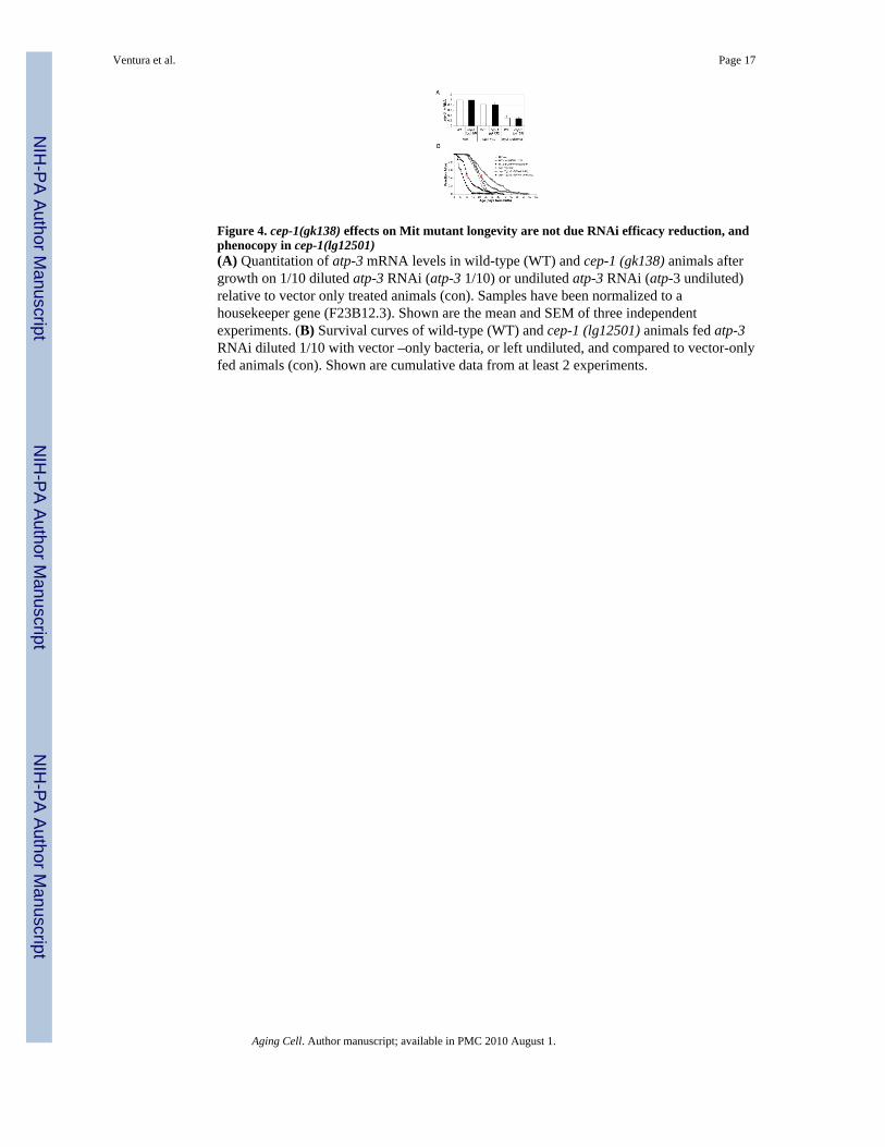
Figure 4. cep-1(gk138) effects on Mit mutant longevity are not due RNAi efficacy reduction, andphenocopy in cep-1(lg12501)(A) Quantitation of atp-3 mRNA levels in wild-type (WT) and cep-1 (gk138) animals aftergrowth on 1/10 diluted atp-3 RNAi (atp-3 1/10) or undiluted atp-3 RNAi (atp-3 undiluted)relative to vector only treated animals (con). Samples have been normalized to ahousekeeper gene (F23B12.3). Shown are the mean and SEM of three independentexperiments. (B) Survival curves of wild-type (WT) and cep-1 (lg12501) animals fed atp-3RNAi diluted 1/10 with vector –only bacteria, or left undiluted, and compared to vector-onlyfed animals (con). Shown are cumulative data from at least 2 experiments.
Ventura et al. Page 17
Aging Cell. Author manuscript; available in PMC 2010 August 1.
NIH
-PA Author Manuscript
NIH
-PA Author Manuscript
NIH
-PA Author Manuscript

Figure 5. cep-1 regulates animal development in response to severe mitochondrial disruption(A) Digital images of four day old wild-type (WT) and cep-1(gk138) animals fed from thetime of hatching with either empty vector (con), undiluted isp-1 or atp-3 RNAi, or EtBr(75µg/ml). (B) Digital images of five day old animals fed from the time of hatching withempty vector (con) or undiluted nuo-2 RNAi (‘P0’ indicates starting generation animals,‘F1’ specifies progeny animals derived from P0 hermaphrodites and maintained under thesame feeding RNAi conditions).
Ventura et al. Page 18
Aging Cell. Author manuscript; available in PMC 2010 August 1.
NIH
-PA Author Manuscript
NIH
-PA Author Manuscript
NIH
-PA Author Manuscript

Figure 6. egl-1 is a downstream target of cep-1 that contributes to lifespan modulation aftersevere atp-3 disruption(A, B) Survival analysis of wild-type (WT) and egl-1(n487) animals fed atp-3 RNAi diluted1/10 with vector –only bacteria, or left undiluted, and compared to vector-only fed animals(con). Shown are cumulative data from 2 experiments. (C) Quantitative measurement ofegl-1 mRNA in wild-type (WT) and cep-1 (gk138) animals following growth on 1/10 dilutedatp-3 RNAi (atp-3 1/10) or atp-3 RNAi left undiluted (atp-3 undiluted). Data is shownrelative to vector-control fed animals (con) and is normalized to a housekeeping gene(F23B12.3). Shown are mean and SEM of three different experiments.
Ventura et al. Page 19
Aging Cell. Author manuscript; available in PMC 2010 August 1.
NIH
-PA Author Manuscript
NIH
-PA Author Manuscript
NIH
-PA Author Manuscript

Figure 7. Evolutionary conserved functionality of Janus-faced p53/cep-1 in C. elegansMild or transient cellular and organismal damage induces p53 protective pathways leadingto damage repair, growth, survival and increased longevity. More severe or irreversibledamage induces p53 suppressive responses that eliminate cells and irreversibly compromisethe organsim. Such drastic actions lead to growth arrest and/or decreased survivalprobability.
Ventura et al. Page 20
Aging Cell. Author manuscript; available in PMC 2010 August 1.
NIH
-PA Author Manuscript
NIH
-PA Author Manuscript
NIH
-PA Author Manuscript

NIH
-PA Author Manuscript
NIH
-PA Author Manuscript
NIH
-PA Author Manuscript
Ventura et al. Page 21
Tabl
e I
Life
span
Ana
lyse
s – S
umm
ary
Stat
istic
s
Gen
otyp
eR
NA
i*M
ean
LS
SEM
p vs
con
^p
vs N
2 ^
Sam
ple
size
(cen
sore
d)^^
Rep
licat
es(N
)
Wild
-type
(N2)
con
15.9
0.2
393
(34)
5
frh-
120
.40.
34.
4E-2
540
6 (4
4)5
skn-
1(zu
67)
con
12.3
0.3
8.4E
-16
140
(20)
2
frh-
120
.20.
52.
5E-2
60.
644
136
(18)
2
gst-4
::gf
pco
n18
.10.
416
5 (1
8)2
con+
O2
20.7
0.4
0.00
3418
1 (8
)2
frh-
123
.30.
53.
5E-1
112
5 (1
3)2
skn-
1(zu
67);
gst-4
::gf
pco
n12
.30.
312
0 (2
0)2
con+
O2
12.4
0.3
0.8
146
(18)
2
frh-
120
.10.
59.
3E-2
713
6 (1
8)2
frh-
1+O
2#19
.50.
83.
9E-1
476
(2)
1
skn-
1(zu
135)
con
11.3
0.2
3.39
E-29
121
(2)
2
frh-
116
.80.
84.
9E-1
10.
0002
65 (3
)1
age-
1(hx
546)
con
22.2
1.1
5.5E
-09
97 (1
0)1
frh-
131
.30.
91.
0E-0
62.
8E-2
696
(11)
1
daf-1
6(m
gDf5
0)co
n11
.30.
24.
4E-2
112
1 (1
3)2
frh-
117
.80.
55.
8E-1
90.
003
124
(24)
2
sir2
.1(o
k434
)co
n14
.20.
29.
5E-1
317
8 (3
7)2
frh-
117
.10.
42.
7E-0
70.
005
191
(27)
2
sir2
.3(o
k444
)co
n12
.30.
39.
5E-0
981
(21)
1
frh-
116
.70.
42.
4E-1
10.
2580
(28)
1
cep-
1(gk
138)
BX10
con
17.6
0.3
0.00
134
3 (1
30)
5
frh-
119
.10.
32.
2E-0
70.
002
414
(45)
5
cep-
1(lg
1250
1)co
n17
.80.
45.
5E-0
518
4 (2
7)3
* Life
span
ana
lyse
s wer
e pe
rfor
med
on
3rd
gene
ratio
n w
orm
s cul
ture
d co
ntin
uous
ly o
n fr
h-1
RN
Ai.
Unl
ess s
peci
fied,
all
othe
r life
span
ana
lyse
s wer
e de
rived
usi
ng w
orm
s cul
ture
d fo
r a si
ngle
gen
erat
ion
onun
dilu
ted
isp-
1, a
tp-3
, cco
-1 a
nd n
uo-2
RN
Ai;
O2
is tr
eatm
ent w
ith H
yper
baric
Oxy
gen
Aging Cell. Author manuscript; available in PMC 2010 August 1.

NIH
-PA Author Manuscript
NIH
-PA Author Manuscript
NIH
-PA Author Manuscript
Ventura et al. Page 22^ p-
valu
es w
ere
calc
ulat
ed u
sing
the
log-
rank
test
^^Po
oled
wor
ms f
rom
N re
plic
ate
expe
rimen
ts (c
enso
red
indi
vidu
als w
ere
incl
uded
in th
e lo
g ra
nk a
naly
sis)
# p-va
lues
aga
inst
frh-
1 R
NA
i on
the
sam
e st
rain
is 0
.9
BX10
indi
cate
s out
-cro
ssed
ten
times
and
refe
rs to
the
cep-
1(gk
138)
alle
le u
sed
in th
is w
ork
Aging Cell. Author manuscript; available in PMC 2010 August 1.

NIH
-PA Author Manuscript
NIH
-PA Author Manuscript
NIH
-PA Author Manuscript
Ventura et al. Page 23
Tabl
e II
Life
span
Ana
lyse
s – S
umm
ary
Stat
istic
s
Gen
otyp
eR
NA
iM
ean
LS
SEM
p vs
con
^p
vs N
2 ^
Sam
ple
Size
(cen
sore
d)^^
Rep
licat
es(N
)
Wild
-type
(N2)
con
15.9
0.2
393
(34)
5
atp-
3 1/
1028
.50.
82.
8E-3
312
4 (1
4)2
atp-
3 1/
214
.20.
38.
6E-0
515
7 (3
4)2
atp-
38.
40.
24E
-254
160
(55)
2
isp-
1 1/
1029
.70.
44.
2E-9
137
5 (5
1)5
isp-
125
.30.
42E
-53
280
(29)
4
cco-
1 1/
1024
.40.
62.
9E-3
018
1 (1
4)3
cco-
124
.90.
72.
5E-2
411
8 (2
3)2
cep-
1(gk
138)
BX10
con
17.6
0.3
0.00
0434
3 (1
30)
5
atp-
3 1/
1024
.70.
85.
7E-1
30.
008
118
(19)
2
atp-
3 1/
220
.30.
86.
8E-0
51.
3E-0
916
0 (6
5)2
atp-
325
.70.
98.
6E-1
68E
-256
160
(21)
2
isp-
1 1/
1022
.70.
56.
4E-1
72.
0E-1
433
9 (5
8)5
isp-
129
0.6
1.1E
-41
3.2E
-10
282
(40)
4
cco-
1 1/
1018
.30.
60.
005
2.3E
-09
185
(23)
3
cco-
127
12.
5E-1
80.
009
119
(36)
2
^ p-va
lues
wer
e ca
lcul
ated
usi
ng th
e lo
g-ra
nk te
st
^^Po
oled
wor
ms f
rom
N re
plic
ate
expe
rimen
ts (c
enso
red
indi
vidu
als w
ere
incl
uded
in th
e lo
g ra
nk a
naly
sis)
BX10
indi
cate
s out
-cro
ssed
ten
times
and
refe
r to
the
cep-
1(gk
138)
alle
le u
sed
in th
is w
ork
Aging Cell. Author manuscript; available in PMC 2010 August 1.
NIH
-PA Author Manuscript
NIH
-PA Author Manuscript
NIH
-PA Author Manuscript
Ventura et al. Page 24
Tabl
e III
Life
span
Ana
lyse
s – S
umm
ary
Stat
istic
s
Gen
otyp
eR
NA
iM
ean
LS
SEM
p vs
con
^p
vs N
2 ^
Sam
ple
Size
(cen
sore
d)^^
Rep
licat
es(N
)
Wild
-type
(N2)
con
170.
412
0 (1
0)2
atp-
3 1/
1026
.51
4.6E
-14
121
(9)
2
atp-
37
0.4
4.2E
-24
119
(9)
2
cep-
1(lg
1250
1)co
n19
.10.
50.
006
124
(24)
2
atp-
3 1/
1022
.90.
90.
020.
0212
4 (1
1)2
atp-
312
.30.
74.
0E-0
61.
5E-0
811
7 (1
9)2
^ p-va
lues
wer
e ca
lcul
ated
usi
ng th
e lo
g-ra
nk te
st
^^Po
oled
wor
ms f
rom
N re
plic
ate
expe
rimen
ts (c
enso
red
indi
vidu
als w
ere
incl
uded
in th
e lo
g ra
nk a
naly
sis)
Aging Cell. Author manuscript; available in PMC 2010 August 1.

NIH
-PA Author Manuscript
NIH
-PA Author Manuscript
NIH
-PA Author Manuscript
Ventura et al. Page 25
Tabl
e IV
Life
span
Ana
lyse
s – S
umm
ary
Stat
istic
s
Gen
otyp
eR
NA
iM
ean
LS
SEM
p vs
con
^p
vs N
2 ^
Sam
ple
Size
(cen
sore
d)^^
Rep
licat
es(N
)
Wild
-type
(N2)
con
16.3
0.3
241
(29)
4
frh-
121
.30.
54.
2E-1
523
4 (6
6)4
atp-
3 1/
1020
.20.
64.
3E-0
911
9 (9
)2
atp-
35.
30.
31.
2E-6
912
3 (1
2)2
atm
-1(g
k186
)co
n19
.50.
49.
8E-1
223
7 (5
5)4
frh-
126
.20.
75.
7E-1
32.
6E-0
612
1 (2
5)2
atp-
3 1/
1025
.50.
92.
5E-0
91.
2E-0
612
1 (4
5)2
atp-
35.
10.
34.
4E-5
10.
612
0 (2
8)2
egl-1
(n48
7)co
n14
.6*
0.4
0.12
241
(74)
4
frh-
125
0.9
1.1E
-15
0.00
0712
0 (5
6)2
atp-
3 1/
1018
.80.
84.
2E-0
60.
911
5 (1
9)2
atp-
37.
70.
51.
1E-1
60.
0002
115
(21)
2
^ p-va
lues
wer
e ca
lcul
ated
usi
ng th
e lo
g-ra
nk te
st
^^Po
oled
wor
ms f
rom
N re
plic
ate
expe
rimen
ts (c
enso
red
indi
vidu
als w
ere
incl
uded
in th
e lo
g ra
nk a
naly
sis)
* Mea
n LS
is si
gnifi
cant
ly sh
orte
r com
pare
d to
N2,
t-te
st =
0.0
02
Aging Cell. Author manuscript; available in PMC 2010 August 1.