Embed Size (px)
Citation preview

1345
The male sex is one of the risk factors for coronary artery disease (CAD) and the average life expectancy for men
with CAD is ≈8 years less than that of women with CAD. Atherosclerosis is the primary cause of CAD, and it therefore represents the most common cause of morbidity and mortal-ity from CAD worldwide.1 Atherosclerosis is an inflammatory disease, characterized by lipid and macrophage depositions in the arterial wall.2 The atherosclerosis is initiated by the activation of endothelial cells (ECs), leading to high expres-sion of adhesion molecules for recruiting inflammatory cells.3 Macrophage colony-stimulating factor induces monocytes to enter the plaque to differentiate into macrophages and foam cells, which is a critical step for the atherosclerosis.4 In the plaque centers, foam cells and extracellular lipid droplets form a core region, which is surrounded by a cap of smooth muscle
cells (SMCs) and a collagen-rich matrix. T cells, macrophages, and mast cells infiltrate the lesion and are particularly abundant in the shoulder region of the plaque where the atheroma grows. Many of the immune cells exhibit signs of activation and pro-duce inflammatory cytokines.4
Almost all of the previous studies were focused on the androgen effect on atherosclerosis as androgen is considered to be the main factor for male sex risk in atherosclerosis.5 However, several recent results demonstrated a protective role of androgens.6,7
Compared with the effect of androgen, few studies have been done to reveal the androgen receptor (AR) effect on atherosclerosis. AR is activated by binding of androgens,8,9 but it can also exert its function without the binding of androgen.10 Therefore, it becomes obvious that the AR
Abstract—The male sex has a higher risk to develop coronary artery diseases, including atherosclerosis. The androgen receptor (AR) is expressed in several atherosclerosis-associated cell types, including monocytes/macrophages, endothelial cells (ECs), and smooth muscle cells (SMCs), but its pathophysiological role in each cell type during the development of atherosclerotic lesions remains unclear. Using the Cre-loxP system, we selectively knocked out AR in these 3 cell types and the resultant AR knockout (ARKO) mice, monocyte/macrophage ARKO, EC-ARKO, and SMC-ARKO, were then crossed with the low-density lipoprotein receptor (LDLR) deficient (LDLR−/−) mice to develop monocyte/macrophage ARKO-LDLR−/−, EC-ARKO-LDLR−/−, and SMC-ARKO-LDLR−/− mice for the study of atherosclerosis. The results showed that the monocyte/macrophage ARKO-LDLR−/− mice had reduced atherosclerosis compared with the wild-type-LDLR−/− control mice. However, no significant difference was detected in EC-ARKO-LDLR−/− and SMC-ARKO-LDLR−/− mice compared with wild-type-LDLR−/− mice, suggesting that the AR in monocytes/macrophages, and not in ECs and SMCs, plays a major role to promote atherosclerosis. Molecular mechanism dissection suggested that AR in monocytes/macrophages upregulated the tumor necrosis factor-α, integrin β2, and lectin-type oxidized LDL receptor 1 molecules that are involved in 3 major inflammation-related processes in atherosclerosis, including monocytes/macrophages migration and adhesion to human umbilical vein ECs, and subsequent foam cell formation. Targeting AR via the AR degradation enhancer, ASC-J9, in wild-type-LDLR−/− mice showed similar effects as seen in monocyte/macrophage ARKO-LDLR−/− mice with little influence on lipid profile. In conclusion, the AR in monocytes/macrophages plays key roles in atherosclerosis and targeting AR with ASC-J9 may represent a new potential therapeutic approach to battle atherosclerosis. (Hypertension. 2014;63:1345-1353.) • Online Data Supplement
Key Words: androgen ◼ atherosclerosis ◼ macrophages ◼ receptors, androgen
Received November 10, 2013; first decision December 4, 2013; revision accepted February 26, 2014.From the George Whipple Lab for Cancer Research, Departments of Pathology, Urology, Radiation Oncology, Wilmot Cancer Center (C.-K.H., H.P., J.L.,
J.S., S.O.L., C.C.) and Department of Pathology, Aab Cardiovascular Research Institute (E.C.), University of Rochester Medical Center, Rochester, NY; Chawnshang Chang Sex Hormone Research Center, The Kidney and Blood Purification Center, Tianjin Institute of Urology, Tianjin Medical University, Tianjin, China (H.P., L.W., Y.N.); and Sex Hormone Research Center, China Medical University/Hospital, Taichung, Taiwan (C.C.).
*These authors contributed equally to this work.The online-only Data Supplement is available with this article at http://hyper.ahajournals.org/lookup/suppl/doi:10.1161/HYPERTENSIONAHA.
113.02804/-/DC1.Correspondence to Chawnshang Chang, George Whipple Lab for Cancer Research, Departments of Pathology, Urology, Radiation Oncology, Wilmot
Cancer Center, University of Rochester Medical Center, 601 Elmwood Ave, Box 626, Rochester, NY 14642. E-mail [email protected] or Lin Wang, Chawnshang Chang Sex Hormone Research Center, The Kidney and Blood Purification Center, Tianjin Institute of Urology, Tianjin Medical University, 23 Pingjiang Road, Hexi District, Tianjin, China 300211. E-mail [email protected]
New Therapy via Targeting Androgen Receptor in Monocytes/Macrophages to Battle Atherosclerosis
Chiung-Kuei Huang,* Haiyan Pang,* Lin Wang, Yuanjie Niu,* Jie Luo, Eugene Chang, Janet D. Sparks, Soo Ok Lee, Chawnshang Chang
© 2014 American Heart Association, Inc.
Hypertension is available at http://hyper.ahajournals.org DOI: 10.1161/HYPERTENSIONAHA.113.02804
Androgen Receptor and Atherosclerosis
by guest on December 1, 2014http://hyper.ahajournals.org/Downloaded from by guest on December 1, 2014http://hyper.ahajournals.org/Downloaded from by guest on December 1, 2014http://hyper.ahajournals.org/Downloaded from by guest on December 1, 2014http://hyper.ahajournals.org/Downloaded from by guest on December 1, 2014http://hyper.ahajournals.org/Downloaded from by guest on December 1, 2014http://hyper.ahajournals.org/Downloaded from by guest on December 1, 2014http://hyper.ahajournals.org/Downloaded from by guest on December 1, 2014http://hyper.ahajournals.org/Downloaded from by guest on December 1, 2014http://hyper.ahajournals.org/Downloaded from by guest on December 1, 2014http://hyper.ahajournals.org/Downloaded from by guest on December 1, 2014http://hyper.ahajournals.org/Downloaded from

1346 Hypertension June 2014
effect is not always the same as the androgen effect. One of the recent findings from wound healing studies demon-strated that the AR knockout (ARKO) effect is critical in mediating the suppressive effect in wound healing, yet such AR effects could not be reversed by androgen treatment,11 suggesting that AR could function through androgen-inde-pendent pathways. Ikeda et al12 recently investigated the effect of androgen/AR on atherosclerosis using the apolipo-protein E–deficient (ApoE−/−) derivative of the total ARKO mice (GARKO-ApoE−/−); however, their studies failed to distinguish the effect of androgen versus AR because their mouse model lacked both AR and androgen (serum testos-terone dropped to almost an undetectable level).
Based on the inflammatory characteristic of atherosclero-sis and the role of AR in enhancing inflammatory responses, we hypothesized that AR would promote the progression of atherosclerosis and the AR in monocytes/macrophages might exert the primary function. In this study, cell-specific ARKO male mice were used to demonstrate that the AR in mono-cytes/macrophages is critical in atherosclerosis.
Materials and MethodsPlease see the online-only Data Supplement for materials and methods.
ResultsAtherosclerotic Plaques Were Increased in GARKO-Low-Density Lipoprotein Receptor–Deficient Mice But Decreased in Monocytes/Macrophage AR Knockout-Low-Density Lipoprotein Receptor–Deficient Mice Compared With the Wild-Type-Low-Density Lipoprotein Receptor–Deficient Littermate Control MiceUsing the Cre-loxP system, we either knocked out AR ubiq-uitously or selectively in 3 major cell types associated with atherosclerosis: monocytes/macrophages, ECs, and SMCs, and the resultant monocytes/macrophage ARKO (MARKO), ECs-ARKO (EARKO), and SMC-ARKO (SARKO) mice were then crossed with the low-density lipoprotein receptor (LDLR)–deficient (LDLR−/−) mice to develop GARKO-LDLR−/−, MARKO-LDLR−/−, EARKO-LDLR−/−, and SARKO-LDLR−/− mice. Figure S1A in the online-only Data Supplement shows the genotyping results of 4 types of the generated ARKO mice and Figure S1B shows the depletion of AR in the bone mar-row–derived macrophages obtained from MARKO-LDLR−/− mice. No significant change in body weight was observed in the MARKO-LDLR−/− mice, but we observed increases in body weights in the other 2 types of mice (EARKO-LDLR−/− and SARKO-LDLR−/−) (Figure S1C).
After atherosclerosis was developed by feeding mice with high-fat diet for 16 weeks, aortas were excised from each group of mice and stained with Oil red O to visualize atherosclerotic plaques. Compared with the wild-type (WT)-LDLR−/− littler-mate control mice, the GARKO-LDLR−/− showed increased atherosclerotic plaques and reduced androgen levels, which are consistent with previous findings (Figure 1A–1D).13 However, the MARKO-LDLR−/− mice showed significantly less atherosclerosis (25.9% reduction; P value<0.001; n=9) without an altered androgen level as compared with the WT-LDLR−/− littermate control mice (Figure 2A, 2B, and
2E). In contrast, we found little differences in plaque areas in the EARKO-LDLR−/− and SARKO-LDLR−/− mice as com-pared with their littermate controls. The results indicate that there are no significant differences in androgen levels among EARKO-LDLR−/−, SARKO-LDLR−/−, and their littermate controls (Figure 2A, 2B, and 2F). We then performed hema-toxylin and eosin staining of aortic tissues of these mice and detected reduced plaque area in the MARKO-LDLR−/− mice (38% reduction; P value <0.001; n=6) compared with the lit-termate control mice (n=5; Figure 2C; quantification of rela-tive plaque areas is shown in Figure 2D).
Together, the results of these studies using the LDLR−/− derivatives of the 4 different ARKO mice demonstrated that the AR in monocytes/macrophages, but not in ECs and SMCs, played an important role in the promotion of atherosclerosis.
Depletion of AR in Monocytes/Macrophages Reduces Monocytes Infiltration into the Aorta Plaque AreaA crucial step in atherosclerosis is an infiltration of monocytes into the subendothelium of the arteries where they differen-tiate into macrophages and become functionally active.14 We therefore investigated monocytes infiltration in aortic tissues of the MARKO-LDLR−/− and WT-LDLR−/− littermate control mice by staining tissues with the monocyte-specific anti-body, Mac-3. We found that the Mac-3 positively stained cell numbers (represented as positively stained cell percentage) were significantly decreased in the MARKO-LDLR−/− mice (5.28±2.11%; n=7) compared with their littermate control mice (20.57±4.42%; n=6; Figure 3A).
We also examined collagen deposition in aortas of MARKO-LDLR−/− and WT-LDLR−/− mice via Masson Trichrome staining, and results demonstrated significantly lower collagen deposition in the MARKO-LDLR−/− mice (10.14±1.08%; n=5) compared with the littermate control mice (14.48±1.28%; n=5; Figure 3B).
We further investigated whether invaded SMC numbers were modulated by the ARKO in monocytes/macrophages and found that the α-smooth muscle actin positively stained SMC numbers were increased in the MARKO-LDLR−/− mice (28.70±1.94%; n=5) compared with the control littermate mice (12.37±1.71%; n=6; Figure 3C). This suggested that higher numbers of the migrated SMCs may stabilize the plaques leav-ing them less susceptible to rupture.15,16 Therefore, we speculate that the ARKO in monocytes/macrophages might contribute to the stability of plaques as shown by the increased numbers of the invaded SMCs in MARKO-LDLR−/− mice, thereby protect-ing mice from developing into the acute coronary syndrome.
Together, results of Figure 3A to 3C demonstrate that the loss of AR in monocytes/macrophages resulted in decreased infiltrat-ing monocytes/macrophages in the subendothelium of arteries with decreased collagen deposition and increased SMCs, and all these combined effects led to the suppression of atherosclerosis.
AR in Monocyte THP-1 Cells Promoted Their Migration and Adhesion to Human Umbilical Vein ECs and Foam Cell FormationTo further confirm the above in vivo findings, we applied in vitro assays to show the AR role in monocytes/macrophages
by guest on December 1, 2014http://hyper.ahajournals.org/Downloaded from

Huang et al Macrophage AR Promotes Atherosclerosis 1347
in promoting atherosclerosis. We first investigated whether the AR expression in monocytes influences their migra-tion. Monocyte THP-1 cells were infected with lentivirus carrying either scramble control sequence or AR-siRNA and Figure 4A-a (upper) shows successful knockdown of AR in these cells. In the migration assay, the THP-1 cells, either the AR knocked down (THP-1siAR) or the scramble control (THP-1sc) cells, were placed in the upper chamber, whereas the conditioned media of human umbilical vein ECs (HUVECs) were placed in the lower chamber of transwell plates as shown in Figure 4A-a (lower) cartoon. We found that the migration of the THP-1siAR cells was suppressed, when compared with the THP-1sc cells (Figure 4A-b). However, when we manipulated AR expression in HUVECs and used the conditioned media of the AR expressing and depleted HUVECs in the tests, migration of THP-1 cells was not influenced significantly (data not shown), suggesting that AR expression in monocytes, and not in ECs, is critical in mediating this migration process.
We also tested adhesion of THP-1 cells onto HUVECs (a cartoon in Figure 4A-c, upper). Similarly, we used THP-1siAR and THP-1sc cells in the test and tagged THP-1 cells with green fluorescence dye before the reaction so that the adhered green fluorescence dye–labeled cells can easily be detected (middle). After incubation of 2 types of cells for 2 hours at 37°C, numbers
of the adhered fluorescent THP-1 cells were counted. Similar to the migration test, we found that adhesion of the THP-1siAR onto HUVECs was significantly reduced (60%) compared with the THP-1sc (Figure 4A-c, lower; quantification, right; representative images in Figure S2A). However, the manipula-tion of AR expression in HUVECs did not influence adhesion of THP-1 cells onto HUVECs significantly (data not shown). These results also suggest that AR expression in monocytes, and not in ECs, is critical in mediating this migration process and in promoting the adhesion process.
In addition to migration and adhesion, foam cell forma-tion is the third key step of atherosclerosis development.17 Therefore, we tested whether the AR expression in THP-1 cells can also affect this process. THP-1siAR and THP-1sc cells were treated with mouse-derived colony-stimulating fac-tor to stimulate differentiation of monocytes into macrophages and these resultant macrophages were subsequently treated with oxidized LDL to induce foam cell formation. After stain-ing of foam cells with Oil red O, the positively stained cells numbers were compared. As shown in Figure 4A-d, the THP-1siAR cells have significantly reduced foam cell formation (11.89±2.18%) as compared with the THP-1sc control cells (17.65±1.72%), indicating that the AR in THP-1 cells also plays a stimulatory role in this process.
Taken together, Figure 4A results suggest that the AR in monocytes/macrophages plays positive roles in promoting the 3 major inflammation-associated processes in atherosclerosis: (1) migration, (2) adhesion of monocytes to ECs, and (3) subse-quent foam cell formation.
Key AR-Modulated Molecules That Affect Migration of Monocytes to ECs in THP-1 CellsWe next investigated the molecular mechanisms by which the AR in monocytes/macrophages promote the 3 inflammation-associated processes via modulation of expres-sions of the target molecules of these processes.
Among the migration-related molecules, including tumor necrosis factor (TNF)-α, interleukin-6, chemokine (C-C motif) ligand 2 and C-C chemokine receptor type 2 we assayed, we found that the mRNA expressions of TNF-α and interleu-kin-6 were the most significantly inhibited in THP-1siAR cells compared with those in THP-1sc cells (Figure 4B-a). We then selected TNF-α for further investigation. As shown in Figure 4B-b, we found that the secreted TNF-α protein was decreased dramatically in THP-1siAR cells compared with that in the THP-1sc cells, confirming the positive regulation of AR in TNF-α expression at both protein and mRNA levels.
We then asked whether AR could modulate TNF-α at tran-scriptional levels via chromatin immunoprecipitation assay. The results showed that the AR could bind to the androgen response element (ARE; TNF promoter ARE1: +36≈+41; ARE2: +50≈+55) on the 5′ promoter region of TNF-α (Figure 4B-c), and the luciferase assay also confirmed that AR could induce TNF-α expression at the transcriptional level (Figure 4B-d).
We further applied functional assays to test whether block-ing TNF-α via a neutralizing antibody of TNF-α could sup-press the AR effect in promoting migration of THP-1 cells into ECs. As shown in Figure 4B-e, addition of TNF-α neutralizing
Figure 1. Plaque formation was accelerated in general androgen receptor knockout in low-density lipoprotein receptor–deficient (GARKO-LDLR−/−) mice compared with the wild-type (WT)-LDLR−/− littermate control mice. A, Plaque area was analyzed after staining with Oil red O stain. Graph on the right is quantification results of plaque area over total area. B, Masson Trichrome staining was used to analyze collagen deposition in WT-LDLR−/− and GARKO-LDLR−/− mice. Graph on right shows the quantification results of collagen positive area over total area. Arrows indicate collagen area (blue color). Magnification, ×400. Androgens, (C) testosterone, and (D) dihydrotestosterone (DHT) were measured using ELISA kit in WT-LDLR−/− and GARKO-LDLR−/− mice. *P<0.05, **P<0.01, and ***P<0.001.
by guest on December 1, 2014http://hyper.ahajournals.org/Downloaded from

1348 Hypertension June 2014
antibody reduces THP-1sc cells migration to the level of the THP-1siAR cells, indicating that TNF-α is indeed critical in mediating the promoter role of AR in this process.
We also examined whether the TNF-α expression is reduced in the aortic tissues of the MARKO-LDLR−/− mice compared with their WT-LDLR−/− littermate control mice and found significantly reduced expressions of TNF-α in aortic tissues of MARKO-LDLR−/− mice compared with the WT-LDLR−/− littermate control mice (Figure 4B-f), confirming the in vitro effect of AR modulation of TNF-α for promoting monocytes/macrophages migration to ECs.
Key AR-Modulated Molecules That Affect Adhesion of Monocytes to ECs in THP-1 CellsNext we surveyed molecules that have been documented to play important roles in the adhesion process,15,16 and found that the expression of integrin family members (integrin [ITG] αM, αV, β1, β2) and P-selectin glycoprotein ligand was suppressed in THP-1siAR cells compared with that in the THP-1sc control cells (Figure 4C-a). We further dissected the molecular mecha-nism how AR could modulate ITGβ2 in the adhesion process, and confirmed AR could modulate ITGβ2 expression at both mRNA and protein levels (Figure 4C-a and b).
Figure 2. Plaque area was reduced in the monocyte/macrophage androgen receptor knockout (ARKO) in low-density lipoprotein receptor–deficient (MARKO-LDLR−/−) mice compared with the wild-type (WT)-LDLR−/− control mice, but not reduced in the endothelial cell-ARKO (EARKO)-LDLR−/− and smooth muscle cell-ARKO (SARKO)-LDLR−/− mice. A, Oil red O staining of aortae obtained from the MARKO-LDLR−/−, EARKO-LDLR−/−, and SARKO-LDLR−/− mice and their WT-LDLR−/− littermate control mice. Aortae were stained with Oil red O and positive red stained area indicates plaques formed. B, Quantification results of positive plaque area over total area from A. C. Hematoxylin–eosin staining of aortic tissues of MARKO-LDLR−/−, EARKO-LDLR−/−, and SARKO-LDLR−/− mice and their WT-LDLR−/− littermate control mice. Magnification, ×100 (inset, ×400). Two arrowheads indicate the plaque area. D, Quantification of plaque area over total area from C. E, Testosterone levels were determined in MARKO-LDLR−/− mice and their WT-LDLR−/− littermate controls. F, Testosterone levels in WT-LDLR−/−, SARKO-LDLR−/−, and EARKO-LDLR−/− mice. **P<0.01.
by guest on December 1, 2014http://hyper.ahajournals.org/Downloaded from

Huang et al Macrophage AR Promotes Atherosclerosis 1349
We further examined AR modulation in ITGβ2 expression at the transcriptional level and found that AR could bind to ARE (AGACCAnnnTGATCA, location −4462 to −4447) on the 5′ promoter region of ITGβ2 via chromatin immunopre-cipitation assay (Figure 4C-c). The luciferase assay further confirmed that AR could induce ITGβ2 expression at the tran-scriptional level in the 293T cells (Figure 4C-d).
When we introduced the neutralizing antibody of ITGβ2 into the culture, we were able to inhibit adhesion of THP-1sc cells, but not the THP-1siAR cells (Figure 4C-e; representa-tive images in Figure S2B), indicating that the ITGβ2 mol-ecule is critical in this AR-mediated adhesion process.
Finally, we examined ITGβ2 expression in the aortic tis-sues of the MARKO-LDLR−/− mice and their WT-LDLR−/− lit-termate control mice and found significantly reduced ITGβ2 expression in aortic tissues of MARKO-LDLR−/− mice com-pared with their littermate control mice (Figure 4C-f), which also confirmed the AR-mediated reduction of ITGβ2 in vivo.
Key AR-Modulated Molecules That Affect Foam Cell Formation in THP-1 CellsWe then investigated the target molecules in the foam cell formation process, such as lectin-type oxidized LDL receptor
1 (LOX-1), lysosomal acid lipase, and acyl–coenzyme A: cholesterol acyltransferase, in THP-1siAR and THP-1sc cells and found that expressions of these 3 molecules were suppressed in the THP-1siAR cells (Figure 4D-a). Further analysis of LOX-1 expression in THP-1siAR and THP-1sc cells showed lower mRNA expression of LOX-1 in THP-1siAR cells compared with the THP-1sc cells (Figure 4D-b).
We also used the chromatin immunoprecipitation assay to determine whether AR could modulate LOX-1 expression at the transcriptional level, and the results showed that AR could bind to ARE (ATAAAAnnnTGTTTT, location −4209 to −4194) on the 5′ promoter region of LOX-1 (Figure 4D-c). As expected, the luciferase assay also confirmed AR could induce LOX-1 expression at the transcriptional level (Figure 4D-d).
We further tested whether the blocking of LOX-1 by treat-ing the THP-1siAR and THP-1sc cells with the LOX-1 neu-tralizing antibody could inhibit foam cell formation. As shown in Figure 4D-e, the foam cell formation was significantly inhibited on antibody treatment in THP-1sc cells, but not in THP-1siAR cells.
We then examined LOX-1 expression in the aortic tissues of the MARKO-LDLR−/− and their WT-LDLR−/− littermate control mice and found significantly reduced expressions of LOX-1 in aortic tissues of MARKO-LDLR−/− mice compared with their littermate control mice (Figure 4D-f), suggesting that AR could go through modulation of LOX-1 expression to influence form cell formation in vivo.
Together, results from Figure 4 show that the AR in monocytes/macrophages might play a stimulatory role to promote atherosclerosis via multiple modulations of various key molecules, which might explain why we could detect decreased plaque formation in the MARKO-LDLR−/− mice (see Figure 2).
Targeting Monocyte/Macrophage AR With ASC-J9 to Battle AtherosclerosisWe then asked how we can target monocytes/macrophages AR in the body to mimic the MARKO-LDLR−/− mice effect and use that strategy as a potential new therapeutic approach to battle atherosclerosis. ASC-J9 is an AR degradation enhancer, which functions through the proteasome machinery to selec-tively degrade AR. ASC-J9 could increase Akt-modulated AR and Mdm2 interaction and recruit the proteasome complex to enhance AR’s degradation.18 The earlier studies on wound healing demonstrated that ASC-J9 was able to selectively degrade AR in monocytes/macrophages and mimicked the MARKO mice effect with improved wound healing.11
We first tested the in vitro ASC-J9 effects in inhibiting 3 processes: migration and adhesion of monocytes onto ECs and subsequent foam cell formation. Our results showed that ASC-J9 could enhance AR protein degradation in THP-1 cells starting at 2.5 μmol/L concentration (Figure 5A). When we used 5-μmol/L ASC-J9 to test its effect on THP-1 cell migra-tion into HUVECs, we found significant reduction in THP-1 cells migration (Figure 5B). We then tested ASC-J9 effect on the adhesion of THP-1 cells onto HUVECs. As shown in Figure 5C, THP-1 cell adhesion was also significantly reduced on ASC-J9 treatment (representative images in Figure S2C). We further examined ASC-J9 effects on foam cell formation
Figure 3. Macrophage infiltration and collagen deposition were reduced, but smooth muscle cells (SMCs) invasion was increased in the monocyte/macrophage androgen receptor knockout in low-density lipoprotein receptor–deficient (MARKO-LDLR−/−) mice compared with the wild-type (WT)-LDLR−/− mice. A, Aortic tissues of the MARKO-LDLR−/− and WT-LDLR−/− mice were stained with Mac-3 antibody. Arrows indicate stained Mac-3 positively stained cells. Magnification, ×100. Quantification is shown below images **P<0.01. B, Aortic tissues of the MARKO-LDLR−/− and WT-LDLR−/− mice were used in the Masson Trichrome staining. Arrows indicate positively stained cells. Magnification, ×100. Quantification is shown below images. *P<0.05. C, Immunohistochemical staining of SMCs invasion. Aortic tissues of the MARKO-LDLR−/− and WT-LDLR−/− mice stained with α-smooth muscle actin (SMA). Arrows indicate positively stained cells. Magnification, ×100. Quantification is shown below images. **P<0.01.
by guest on December 1, 2014http://hyper.ahajournals.org/Downloaded from

1350 Hypertension June 2014
Figure 4. A, Androgen receptor (AR) in THP-1 cells play a stimulatory role to promote the migration and adhesion to human umbilical vein endothelial cells (HUVECs) and foam cell formation. a, THP-1 cells with AR knocked down (THP-1siAR) and scramble control (THP-1sc), and HUVECs conditioned media were placed in transwell plates as described in the cartoon (lower). b, The migrated cells were stained and the positively stained cells numbers were measured. **P<0.01. c, THP-1siAR and THP-1sc were tagged with green fluorescence dye and then incubated with HUVECs. The adhered THP-1 cells with green fluorescence were counted. Quantification is shown on the right. Magnification, ×100. **P<0.01. d, The THP-1siAR and THP-1sc cells were induced to differentiate into macrophages by mouse-derived colony-stimulating factor (M-CSF). The cells were then incubated with oxidized low-density lipoprotein (oxLDL) to evaluate the ability of cells to uptake oxLDL (cartoon at left). At the end of reaction, cells were stained with filtered Oil red O (middle). Magnification, ×100. Quantification is shown on the right. **P<0.01. B, The AR in THP-1 cells promotes migration to HUVECs. a, Quantitative polymerase chain reaction (qPCR) analysis testing mRNA expression levels of candidate molecules involved in migration process. **P<0.01. b, ELISA test showing tumor necrosis factor (TNF)-α secretion by the THP-1siAR and THP-1sc cells. ***P<0.001. c, Chromatin immunoprecipitation (ChIP) assay. AR antibody was used to pull down TNF-α promoter region, which contains androgen response elements (AREs; upper). d, TNF-α promoter transactivation was measured using luciferase constructs containing AR binding sites of the promoter region of TNF-α molecule in the presence of pBabe vector only or pBabe-AR. Human embryonic kidney (HEK)-293 cells were used in this assay. e, Blocking effect of migration of THP-1 cells to HUVECs on incubation with the TNF-α antibody. f, Immunohistochemical (IHC) staining of TNF-α in aortic tissues obtained from the monocyte/macrophage AR knockout in low-density lipoprotein receptor–deficient (MARKO-LDLR−/−) and wild-type (WT)-LDLR−/− mice. Arrowheads indicate positive stained areas. Magnification, ×100. Quantification is shown on the right. *P<0.05, **P<0.01, and ***P<0.001. C, The AR in THP-1 cells promotes adhesion to HUVECs. a, qPCR analysis testing mRNA expression levels of candidate molecules involved in adhesion process. b, Western blot analysis showing AR and integrin (ITG) β2 in the THP-1siAR and THP-1sc cells. GAPDH was used as control. c, ChiP assay. AR antibody was used to pull down ITGβ2 promoter region, which contains AREs. d, ITGβ2 promoter transactivation was measured using luciferase construct containing ITGβ2 promoter region in the presence or absence of AR with or without dihydrotestosterone (DHT). HEK-293 cells were used in this assay. e, Blocking effect of adhesion of THP-1siAR and THP-1sc cells onto the HUVECs on incubation with the ITGβ2 antibody. f, IHC staining of ITGβ2 in aortic tissues obtained from the MARKO-LDLR−/− and WT-LDLR−/− mice. Magnification, ×400. Arrowheads indicate positive stained area. Quantification is shown on the right. *P<0.05, **P<0.01, and ***P<0.001. D, The AR in macrophages promotes foam cell formation. a, qPCR analysis testing mRNA expression levels of candidate molecules involved in foam cell formation process. **P<0.01. b, AR and lectin-type oxidized LDL receptor 1 (LOX-1) expression levels were determined using Western blot in THP-1sc and THP-1siAR cells. GAPDH served as loading control. c, LOX-1 promoter region containing ARE (upper) was pulled down with AR antibody and amplified with primers specific targeting ARE region (lower). d, LOX-1 promoter region was constructed to pGL3 luciferase vector. The LOX-1 promoter transactivation was measured in the presence or absence of AR with or without DHT. e, Blocking effect on foam cell formation on incubation with the LOX-1 antibody. Quantification is shown on the right. f, IHC staining of LOX-1 in aortic tissues obtained from the MARKO-LDLR−/− and WT-LDLR−/− mice. Magnification, ×100. Quantification is shown on the right. *P<0.05, **P<0.01, and ***P<0.001.
by guest on December 1, 2014http://hyper.ahajournals.org/Downloaded from

Huang et al Macrophage AR Promotes Atherosclerosis 1351
and obtained similar results showing suppressive effects of ASC-J9 (2.5–10 μmol/L; Figure 5d).
We then extended our studies of ASC-J9 effects into in vivo mice. ASC-J9 was injected intraperitoneally every other day (75 mg/kg per day) into the 16-week-old WT-LDLR−/− mice, 8 weeks after starting high-fat diet feeding and continued for 8 weeks. In parallel, control littermates received vehicle only. After a total of 16 weeks of high-fat diet feeding, mice were euthanized and atherosclerosis parameters were analyzed. Interestingly, the results showed that the WT-LDLR−/− mice treated with ASC-J9 had significantly decreased plaque area (25.1±4.9%; n=11) compared with the vehicle control mice (19.2±5.7%; n=5; Figure 6A). We then examined whether the treatment with ASC-J9 leads to decreased AR expression in macrophages. The immunohistochemical staining revealed that expression of the monocyte/macrophage AR (Figure 6B) and macrophages infiltration (Figure 6C) were significantly decreased in these ASC-J9–treated mice. Importantly, we found that mice treated with ASC-J9 still had normal body weights with near normal serum testosterone and normal fer-tility, which is consistent with the previous results showing few side effects.11,19
Together, the results from in vitro cell lines (Figure 5) and in vivo mice studies (Figure 6) suggest that ASC-J9 may have a therapeutic potential to battle the atherosclerosis in the future.
DiscussionIn these studies we found that general ARKO in LDLR−/− (GARKO-LDLR−/−) mice significantly increased atherosclerotic plaques, whereas ARKO in monocytes/macrophages signifi-cantly reduced plaque area as shown in MARKO-LDLR−/− mice. However, the mice lacking AR in ECs and SMCs failed to show such an effect, suggesting that the AR in monocytes/macrophages may play critical roles in promoting atherosclero-sis. Interestingly, MARKO-LDLR−/− mice showed completely
opposite results to the GARKO-LDLR−/− mice. Actually, there may be multiple factors contributing to these contrasting effects observed in GARKO-LDLR−/− versus MARKO-LDLR−/− mice on atherosclerosis. First, this may be explained by the serum testosterone level difference because the serum testosterone levels in the GARKO-LDLR−/− mice dropped dramatically, whereas the serum testosterone levels in the MARKO-LDLR−/− mice remained almost unchanged as compared with the WT-LDLR−/− littermate control mice. Early studies documented that testosterone therapy could lead to reduced cholesterol and LDL levels,20–23 and higher frequency of atherosclerosis was also observed in prostate cancer patients after receiving andro-gen deprivation therapy,24 suggesting that androgens may play protective roles in atherosclerosis development. Our results showing GARKO-LDLR−/− mice with low level of serum tes-tosterone developed atherosclerosis, yet MARKO-LDLR−/− mice with normal serum testosterone level developed little atherosclerosis are in agreement with these clinical data.
Second, we may explain the opposite atherosclerosis phe-notypes of the GARKO-LDLR−/− and MARKO-LDLR−/− mice by lipid profile differences. When we investigated lipid lev-els in these 2 types of ARKO mice, it was shown that total cholesterol and triglyceride contents were significantly higher in GARKO-LDLR−/− mice than in the WT-LDLR−/− litter-mate control mice, whereas MARKO-LDLR−/− mice showed little change (Figure S3A and S3B). These results sug-gest that the higher levels of cholesterol and triglyceride in GARKO-LDLR−/− mice might contribute to the higher rate of plaque formation.
The high lipid levels in GARKO-LDLR−/− mice may be because of the low testosterone levels. However, our earlier studies in GARKO mice of metabolism and the current find-ings in SARKO-LDLR−/− mice indicate that the higher lipid levels may not be independently affected by testosterone lev-els.25 When we investigated whole lipid/glucose metabolism
Figure 5. ASC-J9 treatment blocked THP-1 cell migration and adhesion into human umbilical vein endothelial cells (HUVECs) and foam cell formation. A, Western blot analysis showing androgen receptor (AR) protein levels in THP-1 cells on ASC-J9 treatment. B, In vitro test of ASC-J9 effect on migration of THP-1 cells into HUVECs. THP-1 cells were treated with ASC-J9 (10 μmol/L) for 2 days and used for the migration experiment. *P<0.05. C, In vitro test of ASC-J9 effect on adhesion of THP-1 cells onto HUVECs. **P<0.01. D, In vitro test of ASC-J9 effect on foam cell formation. THP-1 cells were treated with various concentrations of ASC-J9 for 2 days and used for the experiment. Quantification is shown on the right. **P<0.01.
by guest on December 1, 2014http://hyper.ahajournals.org/Downloaded from

1352 Hypertension June 2014
difference in GARKO mice, we observed that GARKO mice had high lipid levels, and these high lipid levels were not reversed when we treated the GARKO mice with androgen.25 Next, we investigated in which tissues the ARKO is critical in elevating lipid levels. We found that ARKO in SMCs results in significant increases in triglyceride and cholesterol with-out altering androgen levels (Figure S3C). We also found that the ARKO in neurons in the neuronal ARKO mice resulted in high lipid levels and further revealed that the neuronal ARKO mice had induced hypothalamic insulin resistance, which, in turn, led to hepatic insulin resistance, lipid accumulation, and visceral obesity.26 Therefore, it can be speculated that the higher lipid levels observed in GARKO-LDLR−/− mice may be related to the smooth muscle cellular and neuronal ARKO in those mice. Although SARKO-LDLR−/− mice exhibit similar atherosclerotic plaques as the littermate controls, whether the atherosclerosis mouse model using the neuronal ARKO mice will lead to higher atherosclerosis similar to the GARKO-LDLR−/− mice needs to be investigated in the future to draw a solid conclusion.
No matter what the major factor is, it is obvious that tar-geting androgen/AR signaling in the whole body (as current androgen deprivation therapy does in prostate cancer patients using various antiandrogens to suppress androgen binding to AR in the whole body) may not be an effective way to sup-press atherosclerosis and therefore, we need to develop a way
to suppress monocytes/macrophages without affecting testos-terone and lipid levels.
We, therefore, suggest the use of the AR degradation enhancer ASC-J9 as a new potential therapeutic approach to battle ath-erosclerosis with rationales as follows. (1) The ASC-J9–treated WT-LDLR−/− mice mimicked the MARKO-LDLR−/− mice effect, similar to the early study demonstrating that ASC-J9 could degrade AR in macrophages to reach the therapeutic effect in macrophages-mediated wound healing.11 (2) The ASC-J9–treated WT-LDLR−/− mice showed reduced plaque formation to a similar extent as observed in MARKO-LDLR−/− mice. (3) Mice treated with ASC-J9 show little change in serum testoster-one11 and AR could be degraded in selective cell types, includ-ing macrophages, SMCs, prostate, and heart (Figure S4A). (4) In vivo data demonstrated that ASC-J9–treated mice have nor-mal libido and sexual activity with normal fertility.11,19,27–29
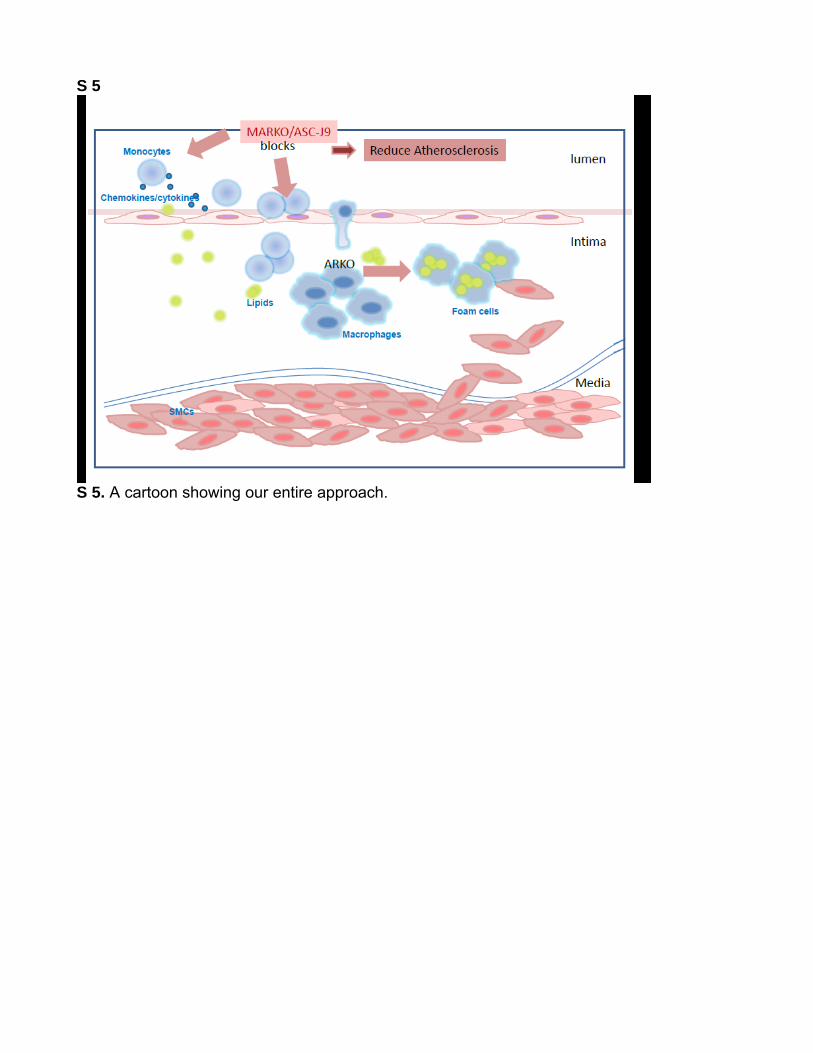
In summary, we conclude that targeting AR in monocytes/macrophages may be a new effective therapy to battle ath-erosclerosis that can be achieved by exploiting ASC-J9 as a therapeutic approach. Figure S5 cartoon explains our entire approach.11
PerspectivesAtherosclerosis is an inflammatory and metabolic disease. AR plays important roles in metabolism and inflammation. AR promotes inflammatory responses as demonstrated using cas-trated male mice and GARKO mice to show reduced inflam-matory responses. However, androgen and AR signaling are complicated in lipid and glucose metabolism and abnormal metabolism would increase lipid accumulation in the blood stream to cause atherosclerosis. In our previous studies, we have shown that neuron and hepatocyte-specific ARKO mice have elevated lipid profiles, whereas adipocyte-specific ARKO mice have decreased lipid profiles.30 In the current study, we showed that MARKO mice have slightly lower lipid profiles, EARKO mice have no change on lipid profiles, but SARKO mice have surprisingly elevated lipid profiles, even higher than GARKO mice. From these results of many cell-specific ARKO mice, it could be speculated that the changed lipid pro-files of GARKO mice is a summation of AR effects on dif-ferent kinds of cells that are involved in metabolism. It might not be easy to simply target AR in the whole body, because this would worsen the atherosclerosis progression. Luckily, we investigated an AR degradation enhancer, ASC-J9, and found that it has the potential to target AR in monocytes/mac-rophages to suppress atherosclerosis. Although ASC-J9 could suppress atherosclerosis progression, we actually do not know whether treatment with ASC-J9 could result in insulin resis-tance, which has been shown using GARKO and several cell-specific ARKO mice. Future studies are needed to evaluate the safety of ASC-J9 before initiating future clinical studies.
AcknowledgmentsWe thank K. Wolf for help in editing the article.
Source of FundingThis work was supported by National Institutes of Health grants (CA127300 and CA156700) and Taiwan Department of Health Clinical Trial and Research Center of Excellence grant DOH99-TD-B-111-004 (China Medical University, Taichung, Taiwan).
Figure 6. Therapeutic effect of ASC-J9 in reducing atherosclerosis by inhibiting monocytes infiltration. A, ASC-J9 effects on plaque formation in mice. Wild type low-density lipoprotein receptor–deficient (WT-LDLR−/−) mice fed with high-fat diet (HFD) for 8 weeks were injected with ASC-J9 (75 mg/kg body weight) every other day for another 8 weeks with continued HFD treatment. Quantification is shown below images, *P<0.05. B, Immunohistochemical (IHC) staining results of androgen receptor (AR) in aortic tissues of vehicle and ASC-J9–treated mice. C, Mac-3 IHC staining results in aortic tissues of vehicle and ASC-J9–treated mice. For both B and C, magnification, ×100; insets, ×400. Arrowheads indicate positive stained cells. Quantification is shown below images, **P<0.01.
by guest on December 1, 2014http://hyper.ahajournals.org/Downloaded from

Huang et al Macrophage AR Promotes Atherosclerosis 1353
DisclosuresASC-J9 was patented by the University of Rochester, the University of North Carolina, and AndroScience, and then licensed to AndroScience. Both the University of Rochester and C. Chang own royalties and eq-uity in AndroScience. The other authors report no conflicts.
References 1. Eckardstein Av, Wu FC. Testosterone and atherosclerosis. Growth Horm
IGF Res. 2003;13(Suppl A):S72–S84. 2. Ross R. Atherosclerosis–an inflammatory disease. N Engl J Med.
1999;340:115–126. 3. Out R, Hoekstra M, Habets K, Meurs I, de Waard V, Hildebrand RB, Wang
Y, Chimini G, Kuiper J, Van Berkel TJ, Van Eck M. Combined deletion of macrophage ABCA1 and ABCG1 leads to massive lipid accumulation in tissue macrophages and distinct atherosclerosis at relatively low plasma cholesterol levels. Arterioscler Thromb Vasc Biol. 2008;28:258–264.
4. Hansson GK. Inflammation, atherosclerosis, and coronary artery disease. N Engl J Med. 2005;352:1685–1695.
5. Kaufman JM, Vermeulen A. The decline of androgen levels in elderly men and its clinical and therapeutic implications. Endocr Rev. 2005;26:833–876.
6. Svartberg J. Epidemiology: testosterone and the metabolic syndrome. Int J Impot Res. 2007;19:124–128.
7. Qiu Y, Yanase T, Hu H, Tanaka T, Nishi Y, Liu M, Sueishi K, Sawamura T, Nawata H. Dihydrotestosterone suppresses foam cell formation and atten-uates atherosclerosis development. Endocrinology. 2010;151:3307–3316.
8. Chang CS, Kokontis J, Liao ST. Molecular cloning of human and rat comple-mentary DNA encoding androgen receptors. Science. 1988;240:324–326.
9. Heinlein CA, Chang C. Androgen receptor in prostate cancer. Endocr Rev. 2004;25:276–308.
10. Sugita S, Kawashima H, Tanaka T, Kurisu T, Sugimura K, Nakatani T. Effect of type I growth factor receptor tyrosine kinase inhibitors on phos-phorylation and transactivation activity of the androgen receptor in pros-tate cancer cells: ligand-independent activation of the N-terminal domain of the androgen receptor. Oncol Rep. 2004;11:1273–1279.
11. Lai JJ, Lai KP, Chuang KH, Chang P, Yu IC, Lin WJ, Chang C. Monocyte/mac-rophage androgen receptor suppresses cutaneous wound healing in mice by enhancing local TNF-alpha expression. J Clin Invest. 2009;119:3739–3751.
12. Ikeda Y, Aihara K, Yoshida S, Sato T, Yagi S, Iwase T, Sumitomo Y, Ise T, Ishikawa K, Azuma H, Akaike M, Kato S, Matsumoto T. Androgen-androgen receptor system protects against angiotensin II-induced vascular remodeling. Endocrinology. 2009;150:2857–2864.
13. Bourghardt J, Wilhelmson AS, Alexanderson C, De Gendt K, Verhoeven G, Krettek A, Ohlsson C, Tivesten A. Androgen receptor-dependent and independent atheroprotection by testosterone in male mice. Endocrinology. 2010;151:5428–5437.
14. Galkina E, Ley K. Immune and inflammatory mechanisms of atheroscle-rosis (*). Annu Rev Immunol. 2009;27:165–197.
15. Sukhova GK, Williams JK, Libby P. Statins reduce inflammation in ath-eroma of nonhuman primates independent of effects on serum cholesterol. Arterioscler Thromb Vasc Biol. 2002;22:1452–1458.
16. Libby P, Ridker PM, Maseri A. Inflammation and atherosclerosis. Circulation. 2002;105:1135–1143.
17. Glass CK, Witztum JL. Atherosclerosis. the road ahead. Cell. 2001;104:503–516.
18. Lai KP, Huang CK, Chang YJ, Chung CY, Yamashita S, Li L, Lee SO, Yeh S, Chang C. New therapeutic approach to suppress castration-resistant prostate cancer using ASC-J9 via targeting androgen receptor in selective prostate cells. Am J Pathol. 2013;182:460–473.
19. Yang Z, Chang YJ, Yu IC, Yeh S, Wu CC, Miyamoto H, Merry DE, Sobue G, Chen LM, Chang SS, Chang C. ASC-J9 ameliorates spinal and bul-bar muscular atrophy phenotype via degradation of androgen receptor. Nat Med. 2007;13:348–353.
20. Saad F, Gooren LJ, Haider A, Yassin A. A dose-response study of tes-tosterone on sexual dysfunction and features of the metabolic syndrome using testosterone gel and parenteral testosterone undecanoate. J Androl. 2008;29:102–105.
21. Saad F, Gooren L, Haider A, Yassin A. An exploratory study of the effects of 12 month administration of the novel long-acting testosterone undecanoate on measures of sexual function and the metabolic syndrome. Arch Androl. 2007;53:353–357.
22. Tenover JS. Effects of testosterone supplementation in the aging male. J Clin Endocrinol Metab. 1992;75:1092–1098.
23. Zgliczynski S, Ossowski M, Slowinska-Srzednicka J, Brzezinska A, Zgliczynski W, Soszynski P, Chotkowska E, Srzednicki M, Sadowski Z. Effect of testosterone replacement therapy on lipids and lipoproteins in hypogonadal and elderly men. Atherosclerosis. 1996;121:35–43.
24. Shahani S, Braga-Basaria M, Basaria S. Androgen deprivation therapy in prostate cancer and metabolic risk for atherosclerosis. J Clin Endocrinol Metab. 2008;93:2042–2049.
25. Lin HY, Xu Q, Yeh S, Wang RS, Sparks JD, Chang C. Insulin and leptin resistance with hyperleptinemia in mice lacking androgen receptor. Diabetes. 2005;54:1717–1725.
26. Yu IC, Lin HY, Liu NC, Sparks JD, Yeh S, Fang LY, Chen L, Chang C. Neuronal androgen receptor regulates insulin sensitivity via suppression of hypothalamic NF-κB-mediated PTP1B expression. Diabetes. 2013;62:411–423.
27. Yamashita S, Lai KP, Chuang KL, Xu D, Miyamoto H, Tochigi T, Pang ST, Li L, Arai Y, Kung HJ, Yeh S, Chang C. ASC-J9 sup-presses castration-resistant prostate cancer growth through degrada-tion of full-length and splice variant androgen receptors. Neoplasia. 2012;14:74–83.
28. Wu MH, Ma WL, Hsu CL, Chen YL, Ou JH, Ryan CK, Hung YC, Yeh S, Chang C. Androgen receptor promotes hepatitis B virus-induced hepato-carcinogenesis through modulation of hepatitis B virus RNA transcrip-tion. Sci Transl Med. 2010;2:32ra35.
29. Ma WL, Hsu CL, Wu MH, Wu CT, Wu CC, Lai JJ, Jou YS, Chen CW, Yeh S, Chang C. Androgen receptor is a new potential therapeutic target for the treatment of hepatocellular carcinoma. Gastroenterology. 2008;135:947–955, 955 e941-945.
30. Yu IC, Lin HY, Liu NC, Wang RS, Sparks JD, Yeh S, Chang C. Hyperleptinemia without obesity in male mice lacking androgen receptor in adipose tissue. Endocrinology. 2008;149:2361–2368.
What Is New?•This is the first study using cell-specific androgen receptor (AR) knockout
mice to further clarify AR roles in 3 major types of cells that are involved in atherosclerosis initiation and progression.
What Is Relevant?•AR in monocytes/macrophages is important for atherosclerosis de-
velopment but not AR in endothelial cells and smooth muscle cell. Although general AR knockout mice develop severe atherosclerosis, monocytes/macrophage AR knockout mice have reduced atheroscle-rotic plaques, indicating that targeting AR in monocytes/macrophages
might have therapeutic effects on atherosclerosis. Using an AR deg-radation enhancer, ASC-J9, to treat atherosclerotic mouse, we were able to show that ASC-J9 might be a potential therapeutic approach in atherosclerosis.
SummaryMonocytes/macrophages AR would promote atherosclerosis pro-gression. Targeting monocytes/macrophages AR with ASC-J9, an AR degradation enhancer could suppress atherosclerosis progres-sion and suggested targeting monocytes/macrophages AR as a novel therapeutic approach in atherosclerosis.
Novelty and Significance
by guest on December 1, 2014http://hyper.ahajournals.org/Downloaded from
ONLINE DATA SUPPLEMENT: New therapy via targeting androgen receptor in monocytes/macrophages to battle atherosclerosis
Chiung-Kuei Huanga,*, Haiyan Pangb,a,*, Yuanjie Niub,, Jie Luoa , Lin Wangb, Eugene Changc,
Janet Sparksa, Soo Ok Leea and Chawnshang Changa,d,#
aGeorge Whipple Lab for Cancer Research, Departments of Pathology, Urology, Radiation
Oncology, and the Wilmot Cancer Center, University of Rochester Medical Center, NY 14642,
USA bChawnshang Chang Sex Hormone Research Center and The Kidney and Blood Purification
Center, Tianjin Institute of Urology, Tianjin Medical University, Tianjin 300211, China cAab Cardiovascular Research Institute and Department of Pathology, University of Rochester
Medical Center, NY 14642, USA
dSex Hormone Research Center, China Medical University/Hospital, Taichung 404, Taiwan
(*Contributed equally)
#Corresponding Authors: Chawnshang Chang
E-mail: [email protected]
Phone: 1-585-273-4500
Fax: 1-585-756-4133
Running Title: Macrophage AR promotes atherosclerosis

Supplemental Material and Methods Animals All mice used in developing transgenic mice were of the C57BL/6 background. The floxed androgen receptor (fAR) mice were generated by inserting loxP sites to flank of exon 2 of AR gene. The LDLR-/-, β-actin promoter driven Cre (ACTB Cre) (for GARKO), lysozyme promoter driven Cre (Lyz-Cre) (for MARKO), vascular endothelial cadherin promoter driven Cre (VEcad-Cre) (for EARKO), and transgelin promoter drive (Tagln-Cre) (for SARKO) mice were purchased from the Jackson Laboratory (Bar Harbor, ME). The fAR/AR-LDLR-/- mice and the Cre+/- LDLR-/- mice were generated by crossing the fAR/AR or the Cre+/- mice with the LDLR-/- mice separately. Subsequent crossing of the resultant fAR/AR-LDLR-/- mice with the Cre+/- LDLR-/- mice led to the generation of double KO, cell specific ARKO-LDLR-/- mice (MARKO-LDLR-/-, EARKO-LDLR-/- AND SARKO-LDLR-/-). Accelerated atherosclerosis was induced by feeding mice with high fat diet (HFD, containing 1.25% cholesterol) for 16 weeks under a 12-hour light/12-hour dark cycle, starting at 8 weeks of age. All animal protocols were approved by the University of Rochester Committee on Animal Resources. Cell lines and reagents THP1 and HUVEC cells were purchased from ATCC TNFα ELISA kit was purchased from eBioscience. Manson Trichrome staining kit was purchased from Sigma. Oil-red-O powder was purchased from Sigma. High fat diets (HFD) were purchased from Teklad Lab Animal Diets-Harlan Laboratories. ASC-J9® treatment of mice ASC-J9® was provided by AndroScience Inc. (CA). ASC-J9® (75 mg/Kg of body weight) was suspended in dimethylacetamide (DMA), diluted with saline, and given to mice everyday (i.p) 8 wks after starting HFD feeding and continued for 8 wks. In parallel, control littermates received vehicle only. Atherosclerotic lesion analysis After 16 weeks of HFD feeding, mice were sacrificed with peritoneal overdose injection of pentobarbital. Whole aortas were excised and thoroughly cleaned by removing adventitial fat and connective tissue under microscope. Then the aortas were dissected and opened longitudinally, fixed in 4% paraformaldehyde for 1 hr, and stained with Oil-red-O solution for 1 hr. Aortas were then rinsed with distilled water and images acquired with microscope. The percentage of plaque area to total aortic area was quantified using Image Pro Plus 5.0 software. 10 µm paraffin sections were prepared for examining cross section plaque area (by H&E staining), and collagen deposition (by Masson’s Trichrome Staining). For immunohistostaining (IHC) and immunofluorescence (IF) staining, paraffin sections were incubated with primary antibodies against AR (N20, Santa Cruz), Mac-3 (dilution 1:250 BD Biosciences), alpha smooth muscle actin (SMA) (dilution 1:250 Sigma-Aldrich), integrin (ITG), and lectin-type oxidized LDL receptor 1 (LOX-1), followed by incubation with the secondary antibodies conjugated with either 3, 3'-diaminobenzidine (DAB) for IHC, or Alexa Fluor 488 or 596 for IF, and DAPI for nuclear staining. The sections were examined under microscope (BX41; Olympus) and the positive stained areas (cells) were quantified by Image Pro Plus 5.0 software. Analysis of levels of lipids and androgens in sera of mice obtained Mice blood samples were collected after 16 hrs of fasting. Sera were obtained by

centrifugation and stored at -80°C for further analysis. Serum concentrations of total cholesterol (Cayman), triglycerides (Thermo Scientific), HDL, LDL (Bioassay Systems), testosterone (Endocrine Technologies) and DHT (Alpha Diagnostic International) were determined according to the instructions given by the companies indicated.
Cell culture and transfection. HUVECs cells were cultured in HUVEC media (NIH) supplemented with EC growth kit including human recombinant VEGF, EGF, FGF, and IGF-1, L-glutamine, heparin sulfate, hydrocortisone hemisuccinate, fetal bovine serum, ascorbic acid (ATCC). THP-1 cells were cultured in RPMI-1640 with 10% serum, 1% L-glutamine, 1% penicillin- streptomycin. The 293T cells were transfected using pWPI lentiviral vectors carrying AR-siRNA or its scramble siRNA control using Lipofectamine 2000 (Invitrogen). After 4 hrs of transfection, media were replaced with the complete media and the supernatants were collected. THP-1 cells were then infected with the supernatants. Transwell migration assay Migration of THP-1 cells was tested using transwell plate with polycarbonate filters (5 µm pores). THP-1 cells, infected with lentivirus carrying either AR-siRNA or scrambled siRNA, or treated with ASC-J9®, were placed into the upper wells, whereas the conditioned medium (CM) of HUVECs were placed in lower chamber. The migration of THP-1 cells into the HUVECs CM was then determined after 24 hrs of incubation. The migrated cells were stained and counted under a microscope. Adhesion assay THP-1 cells were incubated with green fluorescence labeling dye (Molecular Probes, Eugene, OR) for half hour and used for reaction. The same numbers of HUVECs and the GFP-tagged THP-1 cells, THP-1siAR and THP-1sc, were placed in the culture dishes in the presence of amixture of the HUVEC and THP-1 cell culture medium (1:1) and incubated for 2 hrs at 37°C to allow adhesion of THP-1 cells to HUVECs. The unbound cells were washed away with PBS 3 times gently and the number of adhered THP-1 cells with green fluorescence was counted under microscope. Foam cell formation assay The THP-1 cells, THP-1siAR and THP-1sc, were induced to differentiate into macrophages by M-CSF (R & D Systems) for 3 days. Then the cells were incubated with oxLDL at 37°C for 24 hrs to evaluate the ability of cells to uptake oxLDL. Then the cells were fixed with 4% paraformaldehyde for 30 min and stained with filtered Oil-red-O. To test the effect of ASC-J9® on foam cell formation, cells were treated with ASC-J9® for 2 days, and then followed by incubation with M-CSF. The number of Oil-red-O positively stained foam cells was counted under a microscope. Real-time quantitative PCR analysis Total RNAs were obtained from THP-1 cells, infected with lentivirus carrying either AR-siRNA or scrambled RNA using Trizol reagent (Invitrogen). RNAs (5 µg) was used for reverse transcription reaction using SuperScript III Reverse Transcriptase (Invitrogen). The cDNAs obtained were subjected to real-time PCR for detecting mRNA level of AR, TNF-α, Il-6, ITG, and Lox-1 using SYBR Green (Invitrogen) and iCycler system (BioRad).

Masson’s Trichrome Staining Masson’s Trichrome staining was performed using Masson’s Trichrome kit (Sigma-Aldrich) according to the instruction manual. Statistical analysis Quantitative data are expressed as mean ± SD. 2-tailed Student’s t test or 1-way ANOVA were used for comparisons of parameters among two groups. One-way analysis of variance was used for three group parameters comparisons. The value of p < 0.05 was considered statistically significant. *p<0.05, **p<0.01, and ***p<0.001

S 1. The genotyping of mice models. a. The genotypes of fAR-LDLR and Cre in WT-LDLR-/-, GARKO-LDLR-/-, MARKO-LDLR-/-, EARKO-LDLR-/-, and SARKO-LDLR-/-. b. AR proteins were detected in bone marrow macrophages (BM-MSCs) of WT-LDLR-/- and MARKO-LDLR-/- mice. c. Body weights of 4 kinds of specific ARKO-LDLR-/- mice. *p<0.05 and ***p<0.001

S 2. THP1 cells adhesion studies. (a) Representative images of Fig. 4A-c. (b) Representative images of Fig. 4C-e. (c) Representative images of Fig. 5c.

S 3. Analysis of triglyceride, HDL, and LDL levels in sera of the GARKO-LDLR-/-, MARKO-LDLR-/-, EARKO-LDLR-/-, SARKO-LDLR-/-, and WT-LDLR-/- mice. a. Lipid profile analysis results of WT-LDLR-/- and GARKO-LDLR-/- mice. b. Lipid profiles of the MARKO-LDLR-/- together with their WT-LDLR-/- littermate control mice. c. Lipid profile analysis results of EARKO-LDLR-/- and SARKO-LDLR-/- together with their WT-LDLR-/- controls. *p<0.05 and *** p<0.0001

S 4. a. Western blot analysis demonstrating AR levels in several organs of the ASCJ-9® treated WT-LDLR-/- and the vehicle treated control mice. SV: Seminal Vesicle. b. IHC staining of AR in plaque areas of aortas from vehicle and ASC-J9® treated mice. Ellipse dotted lines indicate smooth muscle layers. Arrowheads indicate infiltrated cells.
S 5
S 5. A cartoon showing our entire approach.
Sparks, Soo Ok Lee and Chawnshang ChangChiung-Kuei Huang, Haiyan Pang, Lin Wang, Yuanjie Niu, Jie Luo, Eugene Chang, Janet D.
AtherosclerosisNew Therapy via Targeting Androgen Receptor in Monocytes/Macrophages to Battle
Print ISSN: 0194-911X. Online ISSN: 1524-4563 Copyright © 2014 American Heart Association, Inc. All rights reserved.
is published by the American Heart Association, 7272 Greenville Avenue, Dallas, TX 75231Hypertension doi: 10.1161/HYPERTENSIONAHA.113.02804
2014;63:1345-1353; originally published online March 31, 2014;Hypertension.
http://hyper.ahajournals.org/content/63/6/1345World Wide Web at:
The online version of this article, along with updated information and services, is located on the
http://hyper.ahajournals.org/content/suppl/2014/03/31/HYPERTENSIONAHA.113.02804.DC1.htmlData Supplement (unedited) at:
http://hyper.ahajournals.org//subscriptions/
is online at: Hypertension Information about subscribing to Subscriptions:
http://www.lww.com/reprints Information about reprints can be found online at: Reprints:
document. Permissions and Rights Question and Answer this process is available in the
click Request Permissions in the middle column of the Web page under Services. Further information aboutOffice. Once the online version of the published article for which permission is being requested is located,
can be obtained via RightsLink, a service of the Copyright Clearance Center, not the EditorialHypertensionin Requests for permissions to reproduce figures, tables, or portions of articles originally publishedPermissions:
by guest on December 1, 2014http://hyper.ahajournals.org/Downloaded from