Embed Size (px)
Citation preview
Proc. Nat. Acad. Sci. USAVol. 72, No. 2, pp. 474-478, February 1975
Demonstration That Monocytes Rather Than Lymphocytes Are the Insulin-Binding Cells in Preparations of Human Peripheral Blood MononuclearLeukocytes: Implications for Studies of Insulin-Resistant States in Man
(phagocytosis/adherence columns/autoradiography/receptors)
RONALD H. SCHWARTZ*t, A. RAFFAELE BIANCOT§, BARRY S. HANDWERGER*¶, AND C. RONALD KAHNT* The Immunology Branch, National Cancer Institute and $ The Diabetes Branch, National Institute of Arthritis, Metabolism andDigestive Diseases, National Institutes of Health, Bethesda, Maryland 20014
Communicated by Irving M. London, November 7, 1974
ABSTRACT Insulin receptors have been demonstratedon mononuclear leukocytes prepared by centrifugation ofbuffy coats from normal blood donors on Ficoll-Hypaquegradients. The cell type that specifically binds insulin inthis mixture of lymphocytes and monocytes has never beenclearly identified, although it was assumed to be thelymphocyte since this cell constitutes about 80%/c of thepopulation. In the present studies, insulin-binding assayswere performed on the mononuclear leukocyte preparationbefore and 'after selective depletion or enrichment formonocytes using glass wool or Sephadex G-10 adherencecolumns. The amount of 'l25-labeled insulin specificallybound correlated significantly with the number of mono-cytes but not with the number of B or T lymphocytes.Approximately 90% of the specific insulin binding of thispreparation could be accounted for by its content ofmonocytes. The amount of binding was unaffected byphagocytosis of latex particles or by metabolic inhibitorsadded to prevent endocytosis. Autoradiograms made onsmears of whole peripheral blood and mononuclearleukocytes demonstrated that all of the cells that bound'251-labeled insulin were large mononuclear cells, 85-90%7of which could be identified as monocytes by morpholog-ical criteria or by the functional criterion of latex particleingestion. Since insulin receptor concentration may bealtered in disease states in man, it is essential, when usingthis cell population for detecting such changes, to quan-titate the number of monocytes in the preparation so thatthe insulin-binding data can be appropriately interpreted.
Previous studies by Gavin, Archer, and their coworkers (1-4)demonstrated that bone marrow-derived (B) lymphoblastoidcell lines maintained in continuous culture and peripheralblood mononuclear leukocytes from normal blood donorspurified by Ficoll-Hypaque gradient centrifugation containreceptors for insulin. In the latter case, it was assumed that thebinding was to receptors on lymphocytes, which comprise themajority of this population. However, a subsequent study byKrug et al. (5) failed to detect any significant insulin binding toperipheral blood lymphocytes purified by passage over tightlypacked nylon wool. Since this separation procedure has re-cently been shown to remove B lymphocytes (6-9), it was
Abbreviations: B and T lymphocytes, bone marrow-derived andthymus-derived lymphocytes, respectively.t Address reprint requests to: Laboratory of Immunology, Bldg.10, Rm. 11N319, NIAID, NIH, Bethesda, MId. 20014.§ On leave from the Institute of Clinical Medicine, Departmentof Medicine, University of Naples Medical School II, Naples,Italy.
¶1 Present address: Department of Medicine, University Hos-pital, University of Minnesota School of Medicine, MinneapolisMinn. 55455.
suggested by Olefsky and Reaven (10) that the insulin bindingmeasured in preparations of mononuclear leukocytes mighthave been to receptors on B lymphocytes. On the other hand,monocytes can constitute a significant portion of peripheralblood mononuclear leukocytes (11), and these cells are alsodepleted in preparations made using nylon wool (5, 12). Thus,the identity of the mononuclear leukocyte that binds insulinremained an openl question.
In the present work we address ourselves directly to thisproblem. By various depletion and enrichment experimentswe show that specific insulin binding correlates with thenumber of phagocytic cells (monocytes) in peripheral bloodmononuclear leukocytes and not with the number of Blymphocytes or other cell types. In addition, direct visualiza-tion of the cells binding '251-labeled insulin by autoradiog-raphy demonstrated that these cells are predominantly mono-cytes both by morphological and functional criteria.
MATERIALS AND METHODS
Cell Preparations. Peripheral blood (300-500 ml) fromnormal human volunteers was drawn into acid-citrate-(lextrose solution and centrifuged at 1500 X g for 3 min at 200.The buffv coat was removed, diluted 1:2 with phosphate-buffered saline (pH 7.2), and fractionated on Ficoll (P'har-macia, Piscataway, N.J.)-Hypaque (Winthrop, New York,N.Y.) gradients (p = 1.077) according to the method of1Bdyum (13). The resulting interfaces were pooled and the cells(henceforth referred to as mononuclear leukocytes) were
washed and counted. Viability, as assessed by trypan blue dyeexclusion, was always greater than 95%. The 1B lymphdblas-toid cell line, II9, which has been shown to have a largenumber of insulin receptors, was kindly supplied by Dr.Donald Buell (14-16).
Identification of Mlonocytes and Lymphocytes. Monocyteswere identified by morphological criteria (17) in cytocentri-fuge smears stained with Giemsa's stain and by the functionalcriterion of latex particle ingestion (18). B lymphocytes were
identified by the use of fluorescein-conjugated, rabbit anti-human immunoglobulin (Cappel Laboratories, Downingtown,Pa., lot no. 6684) (19). Cells were stained after exposure tolatex particles and only nonphagocytic, fluorescent cells were
considered positive. Thymus-derived (T) lymphocytes were
identified by sheep erythrocyte rosette formation (20).
Depletion and Enrichment of Mlonocytes. AMonocytes were
depleted from mononuclear leukocytes by two adherencetechniques. The first involved passage of the cells over col-
474

Insulin Receptors on Human Monocytes 475
umns of glass wool (21). Two grams of washed (0.2 M HCl anddistilled water) Pyrex wool were packed tightly into a 20-mlplastic syringe and further washed with Eagle's minimal es-
sential medium containing 10% fetal calf serum. Cells (5 X10⁸ in 5 ml of medium) were loaded onto the column at roomtemperature. The cells were washed through the columnwithout incubation, using 50 ml of medium, and the nonadher-ent, monocyte-depleted population was collected (yield, 20-30%). The second technique involved passage of the mono-
nuclear leukocytes over columns of Sephadex G-10. Theprocedure described by Ly and Mishell (22) was used exceptthat the bed of glass beads was replaced with a small amount(<40 mg) of glass wool. The yield of cells in the nonadherent,monocyte-depleted population was 35-60%.A monocyte-enriched population was also obtained from the
Sephadex G-10 columns. This was accomplished after removalof the nonadherent cells by dispersing the Sephadex in excess
medium and mechanically agitating them with a Vortex mixerto remove the adherent cells (yield, 20-30% of the cells putonto the column).
Insulin-Binding Assay. Porcine insulin (Eli Lilly, Indian-apolis, Ind.) was iodinated to a specific activity of 100-200/uCi/,ug (2). Binding studies were performed as described (2, 3)with the exception that 0.1 M N-2-hydroxyethylpiperazine-N'-2-ethanesulfonic acid (Hepes), pH 8, replaced 25 mM Tris,pH 7.4. The cells were incubated with insulin for 3 hr at 220and then separated from the unbound insulin. The specific1251-labeled insulin in the cell pellets was determined as
described in Table 1.
Autoradiography. Cells were incubated for 3 hr at 150 in thepresence of 6-20 ng of 1251-labeled insulin, washed three timesthrough cold fetal calf serum adjusted to pH 8, smeared ontogelatin-coated slides, and fixed with 1% (v/v) glutaraldehyde
in phosphate-buffered saline. Autoradiograms were thenprepared by the method of Davie and Paul (23). Cells were
considered to have insulin receptors if four or niore silvergrains above background were observed over or closely sur-
rounding the cell. In several experiments the cells were
allowed to ingest latex particles prior to the binding of 1251-labeled insulin.
RESULTS
In 10 experiments the average composition of the mononuclearleukocytes prepared from peripheral blood buffy coats byFicoll-Hypaque gradient centrifugation was 22.2 2.6%(range 10-36) monocytes, 8.6 1.7% (range 2-16) 1B lympho-cytes, and 68 2.6% (range 58-79) nonphagocytic, non-imniunoglobulin-bearing cells. The last category was shown toconsist almost exclusively of T lymphocytes by the criterionof rosette formation with sheep erythrocytes. The mononuclearleukocytes also contained a few contaminating granulocytes(about 1%) and a variable number of platelets. Erythrocytecontamination never exceeded one erythrocyte per 10 leuko-cytes. The average specific insulin binding of this mixedpopulation was 5.1 0.7% (range, 3.8-7.9) of the 0.1-0.5 ng
of 1251-labeled insulin added to the cells.To ascertain the role of monocytes in this insulin binding,
mononuclear leukocytes were selectively depleted of thesecells by adherence techniques. When the percentage ofmonocytes was reduced from 27.5% to 1.3% by passage of themixed population over glass wool columns, the specific insulinbinding was reduced from 5.9% to 1.2% (Table 1). In con-
trast, the percentage of immunoglobulin-bearing cells (B3lymphocytes) remained unchanged and the percentage ofnonimmunoglobulin-bearing, nonphagocytic cells increased.Thus, depletion of 95% of the mrionocytes resulted in an 80%decrease in the specific insulin binding. If the remaining pha-
TABLE 1. Binding of '251-labeled insulin to mononuclear leukocytes separated on glass wool and Sephadex G-1O adherence columns
% Specific % Nonimmunoglobulin-bearing,Cell populations* insulin bindingt % Monocytest % B lymphocytes§ nonphagocytic cells
Glass wool columnsUnseparated cells 5.9 i 21 27.5 i 8.5 4 i 2 68.5 i 10.5
(3.8-7.9) (19-36) (2-6) (58-79)Nonadherent cells 1.2 + 0.3 1.3 ± 0.3 4 ± 3 94.7 ± 3.3
(0.3-1.5) (1-1.7) (1-7) (91-98)Sephadex G-10 columns
Unseparated cells 4.7 ± 0.5 23.2 ± 4.9 8.3 ± 3 65.5 ± 3.5(4.0-6.1) (16-32.5) (3-15) (60-72)
Nonadherent cells 0.79 ± 0.03 2.0 ± 1.5 6.3 ± 1.3 90.7 ± 2.3(0.72-0.88) (0.33-5) (3-9) (86-93)
Adherent cells 7.9 + 1.4 39.0 ± 3 8.7 ± 2 50.5 ± 1.5(5.4-11.7) (36-42) (1-12) (49-52)
* Unseparated cells are the mononuclear leukocytes derived from Ficoll-Hypaque gradient separation of human peripheral blood buffycoats. The nonadherent cells were those mononuclear leukocytes that were not retained by the glass wool or Sephadex G-10 matrix afterthe columns were washed with 50 ml of medium. The adherent cells from Sephadex G-10 were those cells that were retained after washingbut removed by subsequent mechanical agitation.
t Cells (4 to 8 X 10⁷) were incubated with 0. 1-0.5 ng of 125I-labeled insulin for 3 hr at 220, pH 8, in the presence or absence of 50,000ng/ml of unlabeled insulin. The percentage of 125I-labeled insulin specifically bound was calculated by subtracting the percentage of"51-labeled insulin bound in the presence of unlabeled insulin (nonspecific) from the percentage bound in the absence of unlabeled insulin(total).
t Phagocytic cells were quantitated by the criterion of latex particle ingestion. These cells were almost entirely monocytes, as most ofthe granulocytes had been removed during the Ficoll-Hypaque separation.
§ B lymphocytes were quantitated by staining with fluorescein-conjugated rabbit anti-human immunoglobulin. Only those stainedcells that had not ingested latex particles were considered positive.
¶ All values are expressed as mean percentages ± SEMI, with the range in parentheses.
Proc. Nat. Acad. Sci. USA 72 (1975)

476 Cell Biology: Schwartz et al.
z
z
aw-J
w
aLI
0 1 2 or 0.5 1 0 2 4 6
MONOCYTES B LYMPHOCYTES 1g- LYMPHOCYTES(10-7/ml) (1 0-7/ml) (1 0-7/ml)
FIG. 1. Correlation between the quantitative level of insulin binding and the number of monocytes. Monocytes were quantitated by
latex particle ingestion (A) and B lymphocytes by staining with fluorescein-conjugated rabbit anti-human immunoglobulin (B). The
remaining cells were classified as nonimmunoglobulin-bearing, nonphagocytic cells (Ig- lymphocytes in C). Insulin binding was carried
out as described in the footnotes to Table 1. Correlation coefficients (r) were calculated under the assumption that the paired data points
were a random sample from a bivariate normal distribution. In the case of the monocytes (A), the best-fitting straight line was drawn
through the data points using a linear regression analysis of Y (insulin binding) on X (cell number).
gocytic cells had been removed, the binding would have beenexpected to decrease another 4%.Mononuclear leukocytes were also depleted of monocytes
by passage over Sephadex G-10 columns (Table 1). Whenthe percentage of monocytes in the mixed population was
reduced from 23.2% to 2.0%, the specific insulin binding was
reduced from 4.7% to 0.79%. Again the percentage of Blymphocytes remained essentially unchanged and the per-
centage of nonimmunoglobulin-bearing, nonphagocytic cellswas increased. Based on these figures, the monocytes account
for 91% of the binding.A major advantage of the Sephadex G-10 separation proce-
dure is that it allows the recovery of a substantial portion ofthe cells that adhere to the column; this population is usuallyenriched for phagocytic cells. As can be seen in Table 1, thespecific insulin binding and the number of monocytes in theadherent population showed parallel increases relative to theunseparated population of cells. In contrast, the percentage ofB lymphocytes was the same for the two populations and thepercentage of nonimmunoglobulin-bearing, nonphagocyticcells declined. The demonstration that the adherent cellsrecovered from the column have functional insulin receptors
suggests that the lack of binding in the nonadherent popula-tion was not the result of blockage or disruption of the insulinreceptors by the columns. Further support for this point was
obtained by passage of the lymphoblastoid cell line (IM9)through a Sephadex G-10 column. The cells that emerged inthe nonadherent population showed as much specific insulinbinding (15.1%) as the unseparated cells (12.5%0).
Fig. 1 is a compilation of all the experiments in which bothinsulin binding and the number of monocytes or B lympho-cytes were determined. These include unseparated cell popula-tions as well as the separated populations from both glass wooland Sephadex G-10 experiments. The quantitative level ofinsulin binding correlated significantly (P < 0.001) with thenumber of monocytes (Fig. 1A). The slope (0.89) of the best-fitting straight line was not significantly different from theslope (1.0) of the line obtained on serial dilution of a popula-tion containing a fixed number of monocytes. There was no
correlation with the number of B lymphocytes (Fig. 1B) andan inverse correlation with the number of nonimmunoglobulin-
bearing, nonphagocytic cells (Fig. 1C). The latter merelyreflects the absence of monocytes in this population as a resultof the derived nature of the data (by subtraction). Thus, of thetwo major cell types in mononuclear leukocytes, monocytesrather than lymphocytes account for most of the insulin bind-ing.Although the rapid kinetics of insulin dissociation from
monocytes (A.R.B., manuscript in preparation) made itunlikely that the monocytes were simply ingesting insulin-receptor complexes shed from other cells, this possibility was
ruled out by studying insulin binding in the presence ofmetabolic inhibitors to block endocytosis (24, 25). Both 3 mMsodium azide and 0.1 mM iodoacetamide either alone or incombination failed to prevent binding of 12-I-labeled insulin.Cell viability in both treated and control groups was greaterthan 90% at the end of the incubation. Conversely, phago-cytosis of latex particles by monocytes did not affect insulinbinding. Both the degree of specific binding and the inhibitionof binding of 12I-labeled insulin by various amounts of un-
labeled insulin were identical for mononuclear leukocytespreincubated either with or without latex particles.
Visualization of the cells that bind 1251-.labeled insulin byautoradiography directly demonstrated insulin receptors on
monocytes (Fig. 2) in both mononuclear leukocytes and whole
peripheral blood. All of tho cells seen with silver grains over
them could be morphologically classified as large mononuclearcells. Most of these cells (>85%) could be positively identified
as monocytes; however, the remaining cells with silver grainscould not be distinguished from large lymphocytes. In addi-
tion, there appeared to be a small number of large mono-
nuclear cells that did not have silver grains associated with
them. Small lymphocytes, neutrophils, and eosinphils did not
show any binding by this technique. The study with whole
blood ruled out the possibility that the procedure used for
preparing mononuclear leukocytes had resulted in a selective
loss of subpopulations of lymphocytes or granulocytes that
might otherwise have bound insulin under these conditions.
Similar autoradiograms were observed when the binding of
insulin was carried out in the presence of metabolic inhibitors.
Slides made of cell preparations in which the binding of 125I-
labeled insulin was carried out in the presence of a large excess
r=0.323 BP >0.1
0 0
0
0 0
00 09 0
r=-0.487 C0.05 > P > 0.025
A
AA
A A
AAA A
AA
A
A AA AI I
Proc. Nat. Acad. Sci. USA 72 (1975)
Insulin Receptors on Human Monocytes 477
of unlabeled insulin showed no localization of grains aroundany cell type, at the exposure times examined (1-3 weeks),indicating that the binding was specific.To further characterize the cell type that bound insulin, the
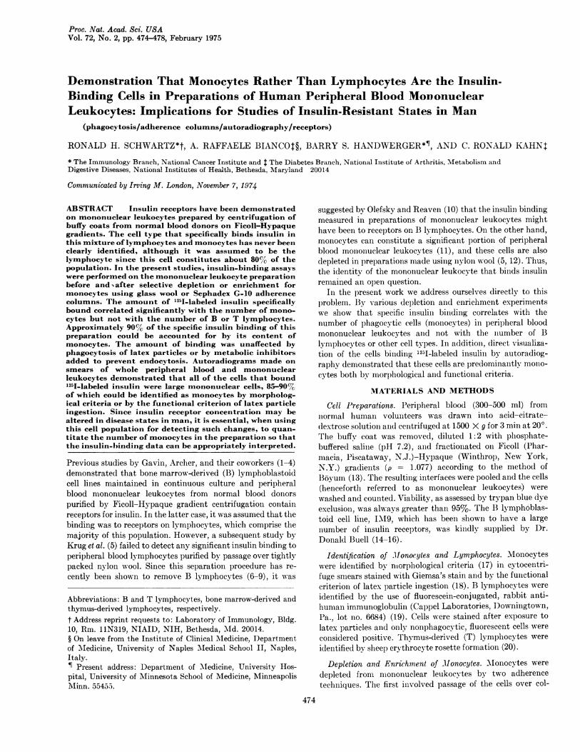
mononuclear leukocytes were allowed to phagocytize latexparticles prior to insulin binding and autoradiography. Asnoted above, the ingestion of latex particles did not decreasethe specific insulin binding, indicating that the insulin receptorwas apparently not modulated by phagocytosis. Silver grainswere localized over and around those cells that had ingestedthe latex particles (Fig. 2). Ninety percent of the cells bindinginsulin were latex-positive. The remaining 10% could bemorphologically characterized as large mononuclear cells.Only 14% of the latex-ingesting cells showed no insulin bind-ing. Again, competition experiments with unlabeled insulineliminated the localization of silver grains. Thus, the pre-dominant cell type in mononuclear leukocytes that bindsinsulin appears to be a phagocytic cell with the morphologiccharacteristics of a monocyte. In addition, there appears to bea small population of nonphagocytic, large mononuclear cellsthat also bind insulin.
DISCUSSIONThe purpose of this study was to identify the insulin-bindingcell present in Ficoll-Hypaque preparations of mononuclearleukocytes from human peripheral blood. Fractionation ofthis population by several techniques showed a highly signifi-cant positive correlation between insulin binding and thenumber of monocytes, but no positive correlation with thenumber of lymphocytes, red blood cells, or platelets. Depletionof monocytes from the mixed population by use of glass woolor Sephadex G-10 adherence columns reduced the insulinbinding by 80-90% with no consistent change in B or Tlymphocytes. Recovery of the cells adherent to the SephadexG-10 column gave a population of cells that was enriched formonocytes and that showed an increased amount of insulinbinding.
Since granulocytes are similar to monocytes with respect tophagocytic and adherence properties, it was possible that thesmall number of contaminating granulocytes (1%) in mono-nuclear leukocytes accounted for the majority of the insulinbinding. This possibility was ruled out by a direct examinationof the cells binding 15I-labeled insulin using autoradiography.Silver grains were found in significant numbers only over largemononuclear cells, greater than 85% of which could be iden-tified as monocytes by morphology and the functional criterionof latex particle ingestion. Silver grains were never seen insignificant numbers over small lymphocytes or granulocytes.Thus, the cell responsible for the major portion of the insulinbinding in mononuclear leukocytes appeared to be the mono-cyte.Receptors on monocytes could only account for 90% of the
specific insulin binding by mononuclear leukocytes. Theremaining 10% of the binding was seen by autoradiography tobe to nonphagocytic, large mononuclear cells. It is possiblethat this subpopulation of insulin-binding cells representsnonphagocytic, nonadherent monocytes. On the other hand,these cells might be circulating large lymphocytes. Such cellshave been shown in the rat (26) to be mainly dividing precur-sors of intestinal plasma cells, i.e., blast-transformed oractivated B lymphocytes. In this regard, Krug et al. (5) re-ported that lectin-activated lymphocytes acquire receptors forinsulin.
U
FIG. 2. Localization of 125I-labeled insulin binding to mono-nuclear phagocytes by autoradiography. Mononuclear leukocyteswere first incubated with latex particles for 30 min at 37'. Thecells were then exposed to 12 ng/ml of ln'I-labeled insulin for 3hr at 15', pH 8, after which they were washed several timesthrough fetal calf serum, pH 8, at 40 and smeared onto slides.Autoradiograms were prepared and developed after 3 weeks ofexposure. The latex particles appear in the photographs as smallwhite beads in the cytoplasm of the monocytes. The l2nI-inducedsilver grains appear as black dots. Magnification is 800 X. Thefigure at the top demonstrates a monocyte with more than 20grains and four lymphocytes with no grains; the lower figureillustrates the same distribution of 125I-labeled insulin in a largerfield of mononuclear cells.
About 15% of the monocytes appeared not to bind insulinby autoradiography, suggesting the existence of a subpopula-tion of monocytes. One possibility is that the two monocytepopulations represent cells in different parts of the cell cycle,since differential expression of surface antigens has been re-ported in various stages of the cycle (27). On the other hand,the lack of binding could simply be due to a technical artifact,such as thinning of the emulsion over some cells or loss of 1250-labeled insulin from receptors during the washing procedure.The major lesson to be learned from these studies is that in
any mixed population one should not assume that the pre-dominant cell type is necessarily responsible for hormonebinding or any other biochemical function under study. Whenpossible, it is important to quantitatively correlate the bindingwith changes in cell comlosition or with other specific markersof cell function and to localize the binding to the particularcell type by autoradiography or other techniques. With re-gard to these points, the presence of insulin receptors has beenreported on granulocytic leukemia cells and cultured lympho-blastoid cells as well as on normal granulocytes and thymo-cytes (1-4, 28). In contrast, our autoradiograms failed to de-
Proc. Nat. Acad. Sci. USA 72 (1975)

478 Cell Biology: Schwartz et al.
tect similar receptors in peripheral blood granulocytes and Band T lymphocytes. It is possible that normal blood cells otherthlan monocytes have receptors for insulin, but that these re-
cep)tors are present in smaller numbers or are of lower affinitythan those oln the monocyte, or require different assay condi-tions for detection. In any of these cases, binding to the receptormight have been missed in our autoradiograms. Alternatively,it is possible that contaminating monocytes or histiocytes are
the source of the insulin receptors in the preparations of normalgranulocytes and thymocytes reported by others. Finally,neoplastic and transformed cells may exhibit surface receptorsnot presellt on their normal counterparts. Until further experi-ments are done, it is impossible to resolve the discrepancies inthese various studies.
Finally, we should consider how these findings affect theusefulness of the human mononuclear leukocyte preparation inthe study of insulin receptors in man. Alterations in theinsulin-receptor interaction have been observed in liver (29)and adipose tissue (30) of animals with diseases in which thereis increased or decreased insulin sensitivity. The insulinreceptor on the monocyte is indistinguishable from those inliver and fat by multiple criteria (refs. 31 and 32; A.R.B.,manuscript in preparation) and provides a readily accessiblesource of material for studying hormone-receptor interactionsin man. Archer et al. (3, 4) and Olefsky and Reaven (33) havedemonstrated decreased insulin binding to mononuclear leuko-cytes from patients with some insulin-resistant states anddiabetes. In light of the present results, it becomes essentialwhen studying such patients to quantitate the number ofmonocytes in the preparation. In several patients with markedinsulin resistance, previously reported to have a decrease ininsulin receptors (4), our more recent studies demonstrate thatnormalization of insulin-binding data to monocyte numberactually increases the apparent magnitude of the insulinreceptor deficiency. Recognition that the monocyte is themajor insulin-binding cell in the mononuclear leukocytepreparation should allow one to better define the insulinreceptor deficit previously reported in obesity (3) and todetect even more subtle alterations in the insulin-receptorinteraction in other disease states by eliminating a previouslyunsuspected variable.
We thank Mrs. W. S. Chappell, Mrs. F. J. Shoup, and Mrs.V. B. Weber of the NIH Blood Bank for drawing the blood andpreparing the buffy coats used in these experiments. In addition,we thank Mr. John Williams for technical assistance and Drs.Jesse Roth and William Terry for many stimulating discussionsand advice. A.R.B. is a recipient of PHS Fogarty InternationalFellowship FOS TW 1964-01.
1. Gavin, J. R., III, Archer, J. A., Lesniak, M. A., Gorden, P.& Roth, J. (1972) J. Clin. Invest. 51, 35a.
2. Gavin, J. R., III, Roth, J., Jen, P. & Freychet, P. (1972)Proc. Nat. Acad. Sci. USA 69, 747-751.
3. Archer, J. A., Gorden, P., Gavin, J. R., III, Lesniak, M. A.& Roth, J. (1973) J. Clin. Endocrinol. Metab. 36, 627-633.
4. Archer, J. A., Gorden, P., Kahn, C. R., Gavin, J. R., III,Neville, D. M., Jr., Martin, M. M. & Roth, J. (1973) J.Clin. Invest. 21, 4a.
5. Krug, U., Krug, F. & Cuatrecasas, P. (1972) Proc. Nat.Acad. Sci. USA. 69, 2604-2608.
6. Eisen, S. A., Wedner, H. J. & Parker, C. W. (1972) Immunol.Commun. 1, 571-577.
7. Greaves, M. F. & Brown, G. (1974) J. Immunol. 112, 420-423.
8. Julius, M. H., Simpson, E. & Herzenberg, L. A. (1973) Eur.J. Immunol. 3, 645-649.
9. Handwerger, B. S. & Schwartz, R. H. Transplantation, inpress.
10. Olefsky, J. & Reaven, G. M. (1974) J. Clin. Endocrinol.Metab. 38, 554-560.
11. Zucker-Franklin, D. (1974) J. Immunol. 112, 234-240.12. Oppenheim, J. J., Leventhal, B. G. & Hersh, E. M. (1968)
J. Immuncl. 101, 262-270.13. Boyum, A. (1968) Scand. J. Clin. Invest. 21, Suppl. 97, 77-
89.14. Smith, R. W. & Woody, J. N. (1974) Transplantation 17,
503-507.15. Shevach, E. M., Herberman, R., Frank, M. & Green, I.
(1972) J. Clin. Invest. 51, 1933-1938.16. Gavin, J. R., III, Roth, J., Neville, D. M., Jr., DeMeyts, P.
& Buell, 1). N. (1974) Proc. Nat. Acad. Sci. USA 71, 84-88.17. Wintrobe, M. M. (1967) Clinical Hematology (Lea &
Febiger, Philadelphia), 6th Ed., p. 241.18. Cline, M. J. & Lehrer, R. I. (1968) Blood 32, 423-435.19. Dickler, H. B. & Kunkel, H. G. (1972) J. Exp. Med. 136,
191-196.20. Weiner, M. S., Bianco, C. & Nussenzweig, V. (1973) Blood
42, 939-946.21. Pattengale, P. K., Smith, R. W. & Gerber, P. (1974) J.
Nat. Cancer Inst. 52, 1081-1086.22. Ly, I. & Mishell, R. I. (1974) J. Immunol. Meth., 5, 239-248.23. Davie, J. M. & Paul, W. E. (1971) J. Exp. Med. 134, 495-
516.24. Byrt, P. & Ada, G. L. (1969) Immunology 17, 503-516.25. Cohn, Z. A. (1966) J. Exp. Med. 124, 557-571.26. Gowans, J. L. & Knight, E. J. (1964) Proc. Roy. Soc. Ser. B.
159, 257-282.27. Pasternak, C. A., Warmsley, A. M. H. & Thomas, D. B.
(1971) J. Cell Biol. 50, 562-564.28. Goldfine, I. D., Gardner, J. D. & Neville, D. M., Jr. (1972)
J. Biol. Chem. 247, 6919-6926.29. Kahn, C. R., Neville, D. M., Jr. & Roth, J. (1973) J. Biol.
Chem. 248, 244-250.30. Freychet, P., Laudat, M. H., Laudat, P., Rosselin, G.,
Kahn, C. R., Gorden, P. & Roth, J. (1972) FEBS Lett. 25,339-342.
31. Gavin, J. R., III, Gorden, P., Roth, J., Archer, J. A. &Buell, D. N. (1973) J. Bidl. Chem. 248, 2202-2207.
32. Kahn, C. R., Freychet, P., Neville, D. M., Jr. & Roth, J.(1974) J. Biol. Chem. 249, 2249-2257.
33. Olefsky, J & Reaven, G. (1974) Clin. Res. 22, 129A.
Proc. Nat. Acad. Sci. USA 72 (1975)