Embed Size (px)
DESCRIPTION
cirurgia
Citation preview

New roles for astrocytes: Redefiningthe functional architecture of the brainMaiken Nedergaard1, Bruce Ransom2 and Steven A. Goldman3
1Department of Cell Biology, New York Medical College, Valhalla, NY 10595, USA2Department of Neurology, University of Washington School of Medicine, Seattle, WA 98195-6465, USA3Department of Neurology and Neuroscience, Cornell University Medical Center, NY 10021, USA
Astrocytes have traditionally been considered ancillary,
satellite cells of the nervous system. However, work
over the past decade has revealed that they interact
with the vasculature to form a gliovascular network
that might organize not only the structural architecture
of the brain but also its communication pathways, acti-
vation, thresholds and plasticity. The net effect is that
astroglia demarcate gray matter regions, both cortical
and subcortical, into functional compartments whose
internal activation thresholds and external outputs are
regulated by single glial cells. The array of these astro-
cyte-delimited microdomains along the capillary micro-
vasculature allows the formation of higher-order
gliovascular units, which serve to match local neural
activity and blood flow while regulating neuronal firing
thresholds through coordinative glial signaling. By
these means, astrocytes might establish the functional
as well as the structural architecture of the adult brain.
To classical histologists, glial cells – including astrocytesand oligodendrocytes – were after-thoughts, given theovert complexity and beauty of the neuronal elements ofthe brain. The very word glia evinces the relative disdainwith which early microscopists must have viewed thissmall stubby cell, whose silver impregnation profile was somuch less impressive than that of its neuronal neighbors[1]. The word glia is derived from the Greek word gliokwhich, although commonly translated as glue, also meansslime. But astrocytes are highly fibrous cells of greatstructural complexity, uniquely characterized by a densearray of processes interposed between neuronal elements,some of which contact and envelop local vascular walls.The traditional concept of astrocytes has been one of aphenotype intended for service to others – to regulate andoptimize the environment within which neurons function.As such, astrocytes maintain tight control of local ion andpH homeostasis, deliver glucose and provide metabolicsubstrates. Astrocytes also clear neuronal waste, includ-ing not only metabolic byproducts but also neurotrans-mitters released during synaptic transmission, which aresequestered through active uptake. In short, astrocytesare multifunctional housekeeping cells that, by theirfunctions, have allowed neurons to become progressively
specialized for the tasks of information processing,encoding and transfer.
In this review, these traditional conceptions of astro-cytic form and function will be reconsidered, to make thepoint that astrocytes provide not so much the glue of theneuronal network of the brain as its dynamic, self-organizing and auto-regenerative scaffold. We can nowconsider astrocytes as dynamic regulators of neuronalproduction, network insertion, phenotype and functionalactivity. As such, astrocytes might serve neurons not somuch as servants but as parents – both literally, as adevelopmental source of new neurons, and figuratively, asthe regulators and judges of neuronal behavior andactivities. Simply stated, astrocytes tell neurons what todo, besides just cleaning up their mess. The addition ofthese functions to the known repertoire of glial activitiesover the past decade has expanded our conception of thisphenotype and our appreciation for its seminal importancein normal functioning of the adult brain.
The phylogenetic advance of the astrocyte
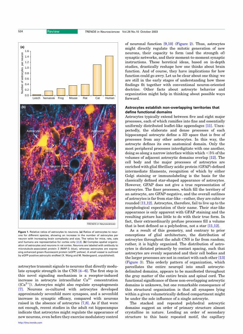
The relative number of astrocytes, expressed both as aproportion of total brain cell number and as a ratio toneuronal number, increases with dramatically withphylogeny and brain complexity. In the leech, a typicalganglion is composed of 25–30 neurons and only oneastrocyte (Figure 1). In Caenorhabditis elegans, neuronsoutnumber glia by 6:1 [2], whereas astrocytes and neuronsare represented in a ratio of 1:3 in the cortex of lowermammals such as rats and mice. In the human cortex,there are 1.4 astrocytes for every neuron [3]. The relativeexpansion of astrocytes compared to neurons with evol-ution and, more specifically, the relative predominance ofastrocytes in the human brain cannot be readily explainedon the basis of differences in glial metabolic support, whichappear little different across species and are certainlysimilar among higher vertebrates.
What evolutionary pressure (or pressures) led to thisexpansion of astrocytes relative to neurons? Setting asidethe ‘how’ question to focus on ‘why’, it is plausible that thegreater abundance of astrocytes with evolution could be afunction of network complexity: increasingly dense andsophisticated synaptic networks require greater degrees oflocal modulation and control. In fact, astrocytes arebeginning to seem very well suited for such duties.Compelling recent evidence supports the concept thatCorresponding author: Maiken Nedergaard ([email protected]).
Review TRENDS in Neurosciences Vol.26 No.10 October 2003 523
http://tins.trends.com 0166-2236/$ - see front matter q 2003 Elsevier Ltd. All rights reserved. doi:10.1016/j.tins.2003.08.008
astrocytes transmit signals to neurons that directly modu-late synaptic strength in the CNS [4–6]. The first step inthis novel signaling mechanism is a receptor-inducedincrease in astrocyte intracellular Ca2þ concentration([Ca2þ]). Astrocytes might also regulate synaptogenesis[7]. Neurons co-cultured with astrocytes developedapproximately sevenfold more synapses, and a sevenfoldincrease in synaptic efficacy, compared with neuronsraised in the absence of astrocytes [7,8]. As if that werenot enough, recent studies in the adult rat hippocampusindicate that astrocytes might regulate the appearance ofnew neurons, even before they exercise modulatory control
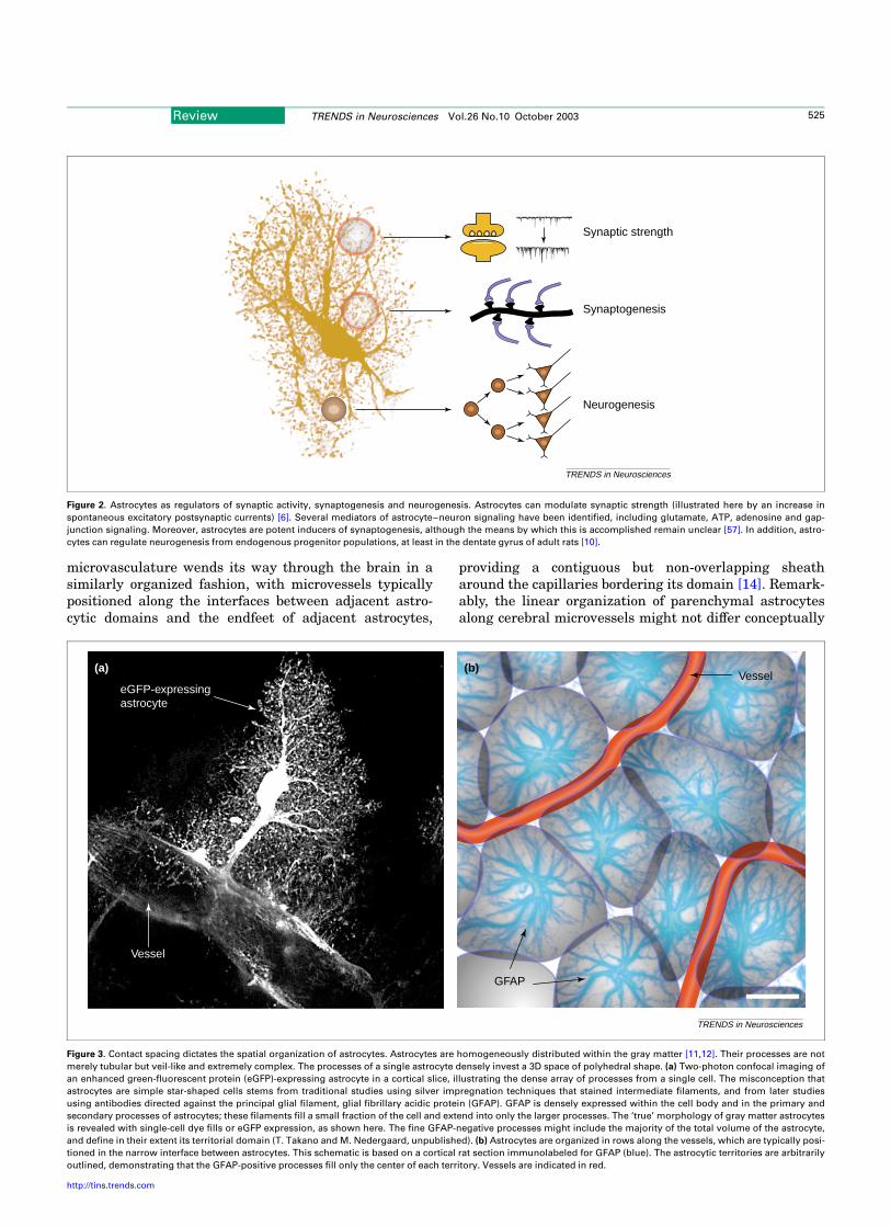
of neuronal function [9,10] (Figure 2). Thus, astrocytesmight directly regulate the mitotic generation of newneurons, their capacity to form (and the strength of)synaptic networks, and their moment-to-moment synapticinstructions. These heretical ideas, based on in-depthstudies, drastically reshape how one thinks about brainfunction. And of course, they have implications for howfunction could go awry. Let us be clear about one thing: weare still in the early stages of understanding how thesefindings fit together with conventional neuron-orienteddoctrine. Other facts about astrocyte behavior andorganization might help in thinking about possible waysforward.
Astrocytes establish non-overlapping territories that
define functional domains
Astrocytes typically extend between five and eight majorprocesses, each of which ramifies into fine and essentiallyuniformly distributed leaflet-like appendages [11]. Unex-pectedly, the elaborate and dense processes of eachhippocampal astrocyte define a 3D space that is free ofprocesses from any other astrocytes. In this way, theastrocyte defines its own anatomical domain. Only themost peripheral processes interdigitate with one another,doing so along a narrow interface within which ,5% of thevolumes of adjacent astrocytic domains overlap [12]. Thecell body and the major processes of astrocytes areenriched with glial fibrillary acidic protein (GFAP)-definedintermediate filaments, recognition of which by eitherGolgi staining or immunolabeling is the basis for theclassically defined star-shaped appearance of astrocytes.However, GFAP does not give a true representation ofastrocytes. The finer processes, which fill the territory ofan astrocyte, are GFAP-negative, and the overall outlinesof astrocytes is far from star-like –rather, they are cubic orrounded [11,12]. Astrocytes, therefore, fail to live up to themorphological expectation of their name. Their star-likeappearance is only apparent with GFAP staining and theresulting picture has little to do with their true form. Infact, their extraordinarily profuse processes fill a volumethat is best defined as a polyhedron, not a star [11,12].
As a result of this geometry, and contrary to priorconceptions of glial architecture, the distribution ofastrocytes throughout the adult CNS is far from random;rather, it is highly organized. The distribution of astro-cytes is dictated primarily by contact spacing, such thatastrocytes are evenly separated and their cell bodies andthe larger processes are not in contact with each other [13](Figure 3). This orderly pattern of organization, whichparcellates the entire neuropil into small astrocyte-delimited domains, appears to be manifested throughoutthe gray matter of the entire brain and spinal cord. Thefunctional significance of these non-overlapping astrocyticdomains is unknown, but one remarkable consequence ofthis structural organization is that all synapses lyingwithin a given volumetrically defined compartment mightbe under the sole influence of a single astrocyte.
The stacked and repeated polyhedral astrocyticdomains suggest an order of primary structure almostcrystalline in nature. Lending an order of secondarystructure to this basic repeated motif, the capillary
Figure 1. Relative ratios of astrocytes to neurons. (a) Ratios of astrocytes to neur-
ons for different species, showing an increase in the number of astrocytes per
neuron with increasing brain complexity and size. The ratios for mice, rats, cats
and humans are representative for cortex only [2,3]. (b) Complex spatial organiz-
ation of astrocytes and neurons in rat cortex. Neurons are labeled with antibody to
microtubule-associated protein 2 (MAP-2; blue), whereas astrocytes are expres-
sing enhanced green-fluorescent protein (eGFP; yellow). A small vessel is outlined
by eGFP-positive astrocytic endfeet (X. Wang and M. Nedergaard, unpublished).
Astrocyte
Vessel
Astrocyte
Neuron
TRENDS in Neurosciences
(a)
(b)
0.0
0.2
0.4
0.6
0.8
1.0
1.2
1.4
1.6
Leech Nematode Frog Mouse Rat Cat Human
Num
ber
of a
stro
cyte
s p
er n
euro
n
Review TRENDS in Neurosciences Vol.26 No.10 October 2003524
http://tins.trends.com
microvasculature wends its way through the brain in asimilarly organized fashion, with microvessels typicallypositioned along the interfaces between adjacent astro-cytic domains and the endfeet of adjacent astrocytes,
providing a contiguous but non-overlapping sheatharound the capillaries bordering its domain [14]. Remark-ably, the linear organization of parenchymal astrocytesalong cerebral microvessels might not differ conceptually
Figure 2. Astrocytes as regulators of synaptic activity, synaptogenesis and neurogenesis. Astrocytes can modulate synaptic strength (illustrated here by an increase in
spontaneous excitatory postsynaptic currents) [6]. Several mediators of astrocyte–neuron signaling have been identified, including glutamate, ATP, adenosine and gap-
junction signaling. Moreover, astrocytes are potent inducers of synaptogenesis, although the means by which this is accomplished remain unclear [57]. In addition, astro-
cytes can regulate neurogenesis from endogenous progenitor populations, at least in the dentate gyrus of adult rats [10].
TRENDS in Neurosciences
Neurogenesis
Synaptogenesis
Synaptic strength
Figure 3. Contact spacing dictates the spatial organization of astrocytes. Astrocytes are homogeneously distributed within the gray matter [11,12]. Their processes are not
merely tubular but veil-like and extremely complex. The processes of a single astrocyte densely invest a 3D space of polyhedral shape. (a) Two-photon confocal imaging of
an enhanced green-fluorescent protein (eGFP)-expressing astrocyte in a cortical slice, illustrating the dense array of processes from a single cell. The misconception that
astrocytes are simple star-shaped cells stems from traditional studies using silver impregnation techniques that stained intermediate filaments, and from later studies
using antibodies directed against the principal glial filament, glial fibrillary acidic protein (GFAP). GFAP is densely expressed within the cell body and in the primary and
secondary processes of astrocytes; these filaments fill a small fraction of the cell and extend into only the larger processes. The ‘true’ morphology of gray matter astrocytes
is revealed with single-cell dye fills or eGFP expression, as shown here. The fine GFAP-negative processes might include the majority of the total volume of the astrocyte,
and define in their extent its territorial domain (T. Takano and M. Nedergaard, unpublished). (b) Astrocytes are organized in rows along the vessels, which are typically posi-
tioned in the narrow interface between astrocytes. This schematic is based on a cortical rat section immunolabeled for GFAP (blue). The astrocytic territories are arbitrarily
outlined, demonstrating that the GFAP-positive processes fill only the center of each territory. Vessels are indicated in red.
(b)
GFAP
(a)
eGFP-expressingastrocyte
Vessel
TRENDS in Neurosciences
Vessel
Review TRENDS in Neurosciences Vol.26 No.10 October 2003 525
http://tins.trends.com
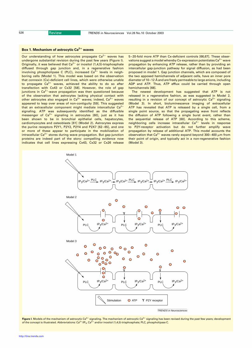
Box 1. Mechanism of astrocytic Ca21 waves
Our understanding of how astrocytes propagate Ca2þ waves has
undergone substantial revision during the past few years (Figure I).
Originally, it was believed that Ca2þ or inositol (1,4,5)-trisphosphate
diffused through gap junction and, in a regenerative fashion
involving phospholipase C (PLC), increased Ca2þ levels in neigh-
boring cells (Model 1). This model was based on the observation
that connexin (Cx)-deficient cell lines, which were otherwise unable
to propagate Ca2þ waves, achieved the ability to do so after
transfection with Cx43 or Cx32 [58]. However, the role of gap
junctions in Ca2þ-wave propagation was then questioned because
of the observation that astrocytes lacking physical contact with
other astrocytes also engaged in Ca2þ waves; indeed, Ca2þ waves
appeared to leap over areas of non-contiguity [59]. This suggested
that an extracellular component might mediate intercellular Ca2þ
signaling. ATP was subsequently identified as the diffusible
messenger of Ca2þ signaling in astrocytes [60], just as it has
been shown to be in bronchial epithelial cells, hepatocytes,
cardiomyocytes and osteoblasts [61] (Model 2). Astrocytes express
the purine receptors P2Y1, P2Y2, P2Y4 and P2X7 [62–65], and one
or more of these appear to participate in the mobilization of
intracellular Ca2þ stores during wave propagation. But gap-junction
proteins are indeed part of the story: compelling evidence now
indicates that cell lines expressing Cx43, Cx32 or Cx26 release
5–20-fold more ATP than Cx-deficient controls [66,67]. These obser-
vations suggest a model whereby Cx-expression potentiates Ca2þ wave
propagation by enhancing ATP release, rather than by providing an
intercellular gap-junction pathway for signal diffusion, as had been
proposed in model 1. Gap junction channels, which are composed of
the two apposed hemichannels of adjacent cells, have an inner pore
diameter of 10–12 A and are freely permeable to large anions, including
ADP and ATP. Thus, ATP efflux could be carried through open
hemichannels [68].
The newest development has suggested that ATP is not
released in a regenerative fashion, as was suggested in Model 2,
resulting in a revision of our concept of astrocytic Ca2þ signaling
(Model 3). In short, bioluminescence imaging of extracellular
ATP has revealed that ATP is released by a single cell, from a
single point source, so that the propagating wave front reflects
the diffusion of ATP following a single burst event, rather than
the sequential release of ATP [60]. According to this scheme,
neighboring cells increase intracellular Ca2þ levels in response
to P2Y-receptor activation but do not further amplify wave
propagation by release of additional ATP. This model accounts the
observation that Ca2þ waves rarely expand beyond 300–400 mm from
their point of origin, and typically act in a non-regenerative fashion
(Model 3).
Figure I. Models of the mechanism of astrocytic Ca2þ signaling. The mechanism of astrocytic Ca2þ signaling has been revised during the past few years; development
of the concept is illustrated. Abbreviations: Ca2þ/IP3, Ca2þ and/or inositol (1,4,5)-trisphosphate; PLC, phospholipase C.
TRENDS in Neurosciences
Model 1
Model 2
Model 3
IP3/Ca2+ IP3/Ca2+
IP3/Ca2+
IP3/Ca2+ IP3/Ca2+ IP3/Ca2+
IP3/Ca2+ IP3/Ca2+
PLC
PLC PLC PLC
IP3/Ca2+ IP3/Ca2+PLC IP3/Ca2+ IP3/Ca2+PLC
PLC PLC PLC
Stimulation ATP P2Y receptor
Review TRENDS in Neurosciences Vol.26 No.10 October 2003526
http://tins.trends.com

from that exhibited in exocrine tissues, in which cells aretypically arrayed lined in rows along the vasculature.Unlike exocrine tissues, however, another order ofstructural complexity has been imposed upon the glialsyncytium, in that neurons are dispersed among theastrocytic domains, with an almost unfathomable numberof fine neurites penetrating each astrocytic domain andbeing surrounded by its glial processes.
Communication within this multicellular syncytium israpid and coordinated, despite the electrical inexcitabilityof the cells. Gap junctions are evenly distributed along theastrocytic processes, often interconnecting adjacent astro-cytic processes derived from the same cell. Only in thenarrow interface of adjacent cells do gap junctions appearto couple processes from different cells [15]. The function ofgap junctions, which interconnect astrocytic processeswidely, is to minimize the differences between individualprocesses by mediating the indiscriminate sharing of allmolecules with molecular weights ,1000 kDa [16]. Thesignificance of this arrangement is unknown but gapjunctions are permeable to glucose and, among otherfunctions, might facilitate the diffusion of energy sub-strates from the nutritive vessels to metabolically activeneurons embedded within the astrocytic matrix. Thinprocesses of astrocytes are probably are still able tooperate in a manner that is independent of theirneighbors. Ca2þ-imaging experiments have revealed thatBergman glial cells displayed Ca2þ increments limited to‘microdomains’ in response to stimulation of parallelfibers, and that these local increases in [Ca2þ] remainlocalized with no propensity for spread to surroundingprocesses or the cell body [17].
Signal propagation among astrocytes
Having learned from neurons by classical electrophysio-logical methods, these same methods were eagerly appliedto astrocytes. The results were somewhat boring. Astro-cytes are electrically non-excitable and they respond tocurrent injection with only passive changes in membranepotential. Their resting membrane potential is maintainedat , 285 mV and displays little fluctuation in response toa wide variety of stimuli [4,18]. This stability and their lowinput resistance is likely to reflect the fact that gapjunctions effectively couple many of these cells into anelectrical syncytium. The predominance of Kþ channelsover other ion channels in these cells also contributes totheir inexcitability [19]. Having determined that astro-cytes were electrically non-excitable, they were immedi-ately presumed to have no involvement in signaltransmission. Although this view is no longer tenable, itmust also be said that we are still struggling to understandthe ‘rules’ of glia–neuron communication.
Cornell-Bell and colleagues reported in 1990 thatastrocytes reacted to glutamate with transient increasesin cytosolic [Ca2þ], followed by inter-astrocytic propa-gation of a wave of increased [Ca2þ] [20]. Further analysesrevealed that this process of astrocytic ‘Ca2þ signaling’ canbe divided into two separate forms: (i) Ca2þ oscillations,defined as repetitive increases in [Ca2þ] within single cells,and (ii) Ca2þ waves, defined as radially propagatingincreases in cytosolic [Ca2þ] that originate from a single
cell and sequentially engage neighboring cells [21]. Theprimary mechanism underlying both types of signaling isthe mobilization of intracellular Ca2þ stores. Receptoractivation is coupled to a membrane-associated G protein,which stimulates the release of Ca2þ from the endoplasmicreticulum via the activation of phospholipase C andconsequent production of inositol (1,4,5)-trisphosphate.Although the presence of both L-type and T-type voltage-gated Ca2þ channels has been demonstrated in vitro andby immunolabeling of brain sections [22], astrocytes in situdo not appear to use this mechanism as a pathway for Ca2þ
entry [23]. Likewise, ion channels gated by glutamate andACh receptors, which cause Ca2þ influx in culturedastrocytes, seem to play at best a minor role in astrocyteCa2þ signaling in situ [23,24] – although Bergmann gliado exhibit a response to glutamate that is modified byAMPA or the AMPA-receptor antagonist 6-nitro-7-sulfa-moyl-benz( f)quinoxaline-2,3-dione (NBQX) [23].
Astrocytic Ca2þ signaling was originally described incultured monolayers, in which Ca2þ increments can bepropagated from a point source to extend throughout anastrocytic syncytium [25]. Astrocytes in freshly preparedbrain slices also support propagation of Ca2þ waves [26](Box 1). Ca2þ oscillations of astrocytes in situ are triggeredby mechanical stimulation or exposure to neurotransmit-ters, including glutamate, GABA and ATP [27]. Spon-taneous Ca2þ oscillations in astrocytes in situ are seen inboth the thalamus [28] and hippocampus [29]. A subpopu-lation of astrocytes in these two regions displayedspontaneous Ca2þ oscillation in the absence of neuronalactivity. The oscillations were not blocked by antagonistsof metabotropic glutamate receptors or purine receptors,suggesting that astrocytes in situ might exhibit intrinsicsignaling.
The functional significance of these spontaneous,neuron-independent astrocytic Ca2þ signals is unclear.Ca2þ oscillations in immature neurons have been associ-ated with neurotransmitter differentiation, axonal growthand establishment of neuronal networks [30,31]. Similarly,different frequencies of Ca2þ activity are associated withacute and systematic differences in patterns of geneexpression by lymphoid cells [32]. Another importantaspect of frequency-coded information is the requirementfor specific cellular ‘decoders’ that can translate Ca2þ
signaling into a cellular response. The two most studiedCa2þ-sensitive proteins that are involved in decoding Ca2þ
signals are Ca2þ/calmodulin-dependent (CaM) kinase andCa2þ/phosphlipid-dependent protein kinase (PKC),although only the latter has been evaluated in astrocytes.Glutamate-induced astrocytic Ca2þ oscillations appear todepend on the periodic translocation and activation of PKCwhich, in turn, responds to the oscillating concentrationsof diacylglycerol and Ca2þ [33].
Both modalities of astrocytic signaling, Ca2þ oscil-lations and Ca2þ waves, are readily transmitted tosurrounding neurons, which display prolonged increasesin intracellular [Ca2þ]. The discovery that astrocytesactively signal to each other and surrounding neuronshas led to the realization that astrocytes are likely to play amore active role in information processing than previouslyrecognized [34,35]. The key observation that certifies this
Review TRENDS in Neurosciences Vol.26 No.10 October 2003 527
http://tins.trends.com

position is that astrocyte Ca2þ waves can either enhance ordiminish nearby neuronal activity. Astrocytic Ca2þ signal-ing has been associated with brief (seconds to minutes)modulation of synaptic strength [4]. Less commonly,activation of Ca2þ signaling in hippocampal astrocyteshas exerted longer-lasting potentiation of inhibitorysynaptic transmission, which was sustained for .20 minbeyond the cessation of glial Ca2þ signaling [4].
Normal and pathological manifestations of glial Ca21
signaling in vivo
Astrocytic Ca2þ waves are mediated primarily by releaseof ATP and activation of purine receptors (Box 1). In
cultured astrocytes, Ca2þ waves are routinely generatedby electrical or mechanical stimulation, but they can alsobe initiated by exposure to transmitters or by removal ofextracellular Ca2þ. In addition, recent studies have usedconfocal imaging to demonstrate that astrocytes in situ canpropagate long-distance Ca2þ waves. However, Ca2þ
waves in slices can be generated only by intense electricalstimulation ($100 Hz) and waves do not appear to beelicited by neuronal activity or transmitter challenge [26].Thus, it remains possible that long-distance intercellularCa2þ waves represent a pathophysiological manifestationof astrocytic Ca2þ signaling that might not contribute tonormal physiological signaling events.
Indeed, astrocytic Ca2þ waves share several propertieswith the phenomenon of spreading depression, a patho-logical suppression of cortical neuronal excitability thathas been implicated in the propagation of ischemic injury[36]. Pathologically, spreading depression is a generalizedresponse of gray matter to a variety of noxious influences[37], and has been associated not only with secondaryinjury after stroke and trauma but also with migraine andpost-seizure depression. Mechanistically, spreadingdepression is best described as a slowly moving wave oftissue depolarization; experimentally, it can be evokedeither by local electrical stimulation or by application of Kþ
or glutamate. Spontaneous waves of spreading depressionare generated in the setting of cerebral ischemia andtrauma, and it has been estimated that the infarct volumeincreases by 23% for each wave of spreading depressionthat is generated [38]. It now appears likely that astrocyticCa2þ waves underlie spreading depression. Ca2þ imagingexperiments have shown that astrocytic Ca2þ wavesconstitute the leading edge of a propagating spreadingdepression wave [39,40]. That said, surprisingly littleinformation exists with regard to astrocytic Ca2þ signalingin the setting of brain trauma or ischemia. Yet this is a fieldin marked flux: new imaging techniques, based uponmultiphoton imaging of live animals, can now visualizeastrocytic Ca2þ signaling in response to pathologicalinsults and are expected to increase rapidly our under-standing of the extent to which astrocytes contribute tosecondary injury.
Transmitter release by astrocytes
One of the principal functions of astrocytes is uptake ofneurotransmitters released from nerve terminals [41]. Butastrocytes can also release neuroactive agents, includingtransmitters, eicosanoids, steroids, neuropeptides andgrowth factors [42]. The regulation and mechanism (ormechanisms) of astrocyte-mediated release of neuroactivecompounds are for most agents poorly defined. They arealso hotly debated, given their theoretical importance inbrain function. Release of glutamate appears to be theprimary mechanism by which astrocytes modulate synap-tic transmission [4]. Astrocytic glutamate release inresponse to synaptic activity appears strictly dependenton mobilization of intracellular Ca2þ stores and isattenuated, as expected, when intracellular [Ca2þ] isstabilized by preloading with either the Ca2þ chelatorBAPTA or the Ca2þ-ATPase inhibitor thapsigargin.Despite intense effort, the pathway of glutamate release
Figure 4. Functional anatomy of the gliovascular interface. Many channels and
receptors essential for astrocyte function are densely localized to the vascular end-
feet facing the vessel wall. (a) Within the brain, the water channel protein aqua-
porin-4 (Aqp-4; red) is expressed by astrocytes exclusively. Aqp-4 is concentrated
at the luminal surfaces of the astrocytic endfeet, which clearly outline the vascular
bed to which they adhere. Glial fibrillary acidic protein (GFAP; white) is present in
the cell bodies and large processes of astrocytes. GFAP is also expressed by those
astrocytic processes that make contact with larger vessels. As a result, vascular
endfeet plastered along the walls of larger vessels typically coexpress Aqp-4 and
GFAP [14]. By contrast, astrocytic endfeet in contact with capillaries are Aqp-4-
positive but only occasionally GFAP-positive. Thus, astrocyte endfeet in contact
with capillaries are detectable only by labeling of Aqp-4. (b) A schematic cross sec-
tion of the blood–brain barrier. Receptors (e.g. P2Y) and channels (e.g. Kþ chan-
nels and Aqp-4) are concentrated at the luminal surface of the astrocytic endfeet,
apposing the perivascular basal lamina.
TRENDS in Neurosciences
(a)
(b)
GFAPAqp-4
Endothelial cell
Gap junction
Tight junction
Astrocyte
Basal lamina
Aqp-4P2Y receptorsK+ channels
Pericyte
Review TRENDS in Neurosciences Vol.26 No.10 October 2003528
http://tins.trends.com
in this context is not understood. One line of work hassuggested that glutamate is released by regulated exocy-tosis [43], whereas others have provided evidence forchannel-mediated release of glutamate [44]. The primaryarguments for vesicular release are the requirement forincreases in cytosolic Ca2þ and for sensitivity to tetanusneurotoxin and bafilomycin [45]. The major arguments forchannel-mediated release are that several Cl2 channelblockers reversibly inhibit release and that glutamate is notreleased in isolation but, rather, together with other aminoacids present in high concentration in astrocytes, includingtaurine and aspartate [46]. Recently, unopposed gap junc-tions, calledhemichannels, have been shown to release largeamounts of glutamate and other amino acids in response tomanipulation of extracellular [Ca2þ] [47]. Likewise, acti-vation of the P2X7 purine receptor results in glutamaterelease from cultured astrocytes [48]. Both of these path-ways are insensitive to preloading with BAPTA or thapsi-gargin, and might thereby not play a role in Ca2þ-dependentglutamate release [46,47]. The possibility that reversal ofglutamate transport has a significant role in this has beeneliminated: glutamate transport inhibitors do not affectCa2þ-dependent astrocytic glutamate release [45].
Interactions among astrocytes, neurons and endothelial
cells define the gliovascular unit
The blood–brain barrier is a diffusion barrier that impedesexchange of molecules between the two tissues. Theprimary seal of the blood–brain barrier is formed byendothelial tight junctions. Astrocytes enwrap the vascu-lature with a large number of endfeet plastered at thevessel wall (Figure 4), although their role in the blood–brain barrier is poorly defined and they are not believed tohave a barrier function in the mammalian brain [49].Several factors released by astrocytes might be importantfor the induction and maintenance of the blood–brainbarrier, as manifested by the appearance of endothelialtight junctions in cells ensheathed by astrocytic endfeet.Several investigators have studied this issue and haveidentified agents released by astrocytes, including trans-forming growth factor a (TGFa) and glial-derived neuro-trophic factor (GDNF), that support the formation of tightjunctions in cultured endothelial cells [50]. An inherentproblem in the field is that the cellular interactions of theimmature blood–brain barrier have thus far been studiedin 2D culture models lacking the structural complexity ofthe cerebral microvasculature. As a result, although thesestudies have shown that astrocytes promote maturation ofbrain endothelial cells in vitro, it has been difficult todetermine definitively the role of astrocytes in theinitiation and the consolidation of the tight-junctionseals of the blood–brain barrier.
The tight organization of astrocytes around the vascu-lature provides anatomical evidence for the necessity forglucose to enter astrocytes on its way to neurons.Astrocytes might not impede glucose uptake becausethey express a large number of glucose transporters. Avariation on this theme is that astrocytes take up theglucose by transport mechanisms, convert it to lactate, andthen deliver lactate to neurons on demand [51,52].Although there might be proof for this mechanism under
severe hypoglycemic conditions necessitating the break-down of astrocytic glycogen [52,53], it remains controversialin the context of normal brain function [54]. To the extentthat this does occur, the limited interdigitation of astrocyticprocesses suggests that individual astrocytes would beresponsible for glucose delivery to neurons within theirown territory – in essence, parceling the neuropil into smallmetabolically independent domains (Figure 3).
New lines of work have shown that receptors andchannels essential for the function of astrocytes aredensely concentrated at their vascular endfeet. Especiallyintriguing is the observation that the water channelaquaporin-4 and purine receptors – mediators of astro-cytic Ca2þ signaling (Box 1) – are expressed primarily atthe gliovascular interface [14] (Figure 4). A recent studyhas shown that astrocytes can convey signals fromneurons to the vasculature and that astrocytes therebymight play a central role in functional hyperemia. Themodel predicts that glutamate released during synaptictransmission, through activation of metabotropic gluta-mate receptors, triggers astrocytic Ca2þ signaling. In turn,astrocytic Ca2þ signaling is associated with release of avasoactive cyclooxygenase product resulting in arteriolardilation and thereby in activity dependent increases inlocal blood flow [55,56].
Concluding remarks
Ideas about glial function originally sprang from theanatomy of these cells. Modern researchers have gainedknowledge about glial cells by studying their physiologyand biochemistry in isolation from their normal cellularpartners. This was necessary to avoid the confoundingcomplexity of the intact CNS. The risk, however, is that welose sight of the in vivo anatomical relationships of thesecells, which define what their sphere of influence can be.Current evidence indicates that anatomy and functionconverge in practical ways around the concept of agliovascular unit. In fact, it is appealing to think of thisas a basic unit of functional organization, with great utilityin moving forward from our current state of under-standing. It also highlights the important limits ofproceeding to study astrocytes in the absence of theirnatural partners.
AcknowledgementsWe thank Marisa Cotrina and Greg Arcuino for discussions, and MarieSimard, Takahiro Takano and Xiaohai Wang for the data illustrated inFigures 1–4. Our studies are supported by NINDS/NIH, the Brain TumorAssociation, and New York State Spinal Cord Injury Research Program.
References
1 Virchow, R. (1846) Uber das granulierte Anschen der Waudungen derGerhirnventrikel. Allg. Z. Psychiatr. 3, 242
2 Sulston, J.E. et al. (1983) The embryonic cell lineage of the nematodeCaenorhabditis elegans. Dev. Biol. 100, 64–119
3 Bass, N.H. et al. (1971) Quantitative cytoarchitectonic distribution ofneurons, glia, and DNA in rat cerebral cortex. J. Comp. Neurol. 143,481–490
4 Kang, J. et al. (1998) Astrocyte-mediated potentiation of inhibitorysynaptic transmission. Nat. Neurosci. 1, 683–692
5 Haydon, P.G. (2001) Glia: listening and talking to the synapse. Nat.Rev. Neurosci. 2, 185–193
6 Newman, E.A. New roles for astrocytes: regulation of synapticfunction. Trends Neurosci. (in press)
Review TRENDS in Neurosciences Vol.26 No.10 October 2003 529
http://tins.trends.com

7 Pfrieger, F.W. and Barres, B.A. (1997) Synaptic efficacy enhanced byglial cells in vitro. Science 277, 1684–1687
8 Ullian, E.M. et al. (2001) Control of synapse number by glia. Science291, 657–661
9 Song, H. et al. (2002) Astroglia induce neurogenesis from adult neuralstem cells. Nature 417, 39–44
10 Horner, P. and Palmer, T. New roles for astrocytes: La vida loca! Thenightlife of an astrocyte. Trends Neurosci. (in press)
11 Bushong, E.A. et al. (2002) Protoplasmic astrocytes in CA1 stratumradiatum occupy separate anatomicaldomains. J.Neurosci. 22, 183–192
12 Ogata, K. and Kosaka, T. (2002) Structural and quantitative analysisof astrocytes in the mouse hippocampus. Neuroscience 113, 221–233
13 Chan-Ling, T. and Stone, J. (1991) Factors determining the shape,spacing and distribution of astrocytes in the cat retina: The contact-spacing model of astrocyte interaction. J. Comp. Neurol. 303, 387–399
14 Simard, M. et al. Signaling at the gliovascular interface. J. Neurosci.(in press)
15 Rohlmann, A. and Wolff, J. (1996) Subcellular Topography andPlasticity of Gap Junction Distribution on Astrocytes, R.G. LandesCompany
16 Rose, C. and Ransom, B.R. (1997) Gap junctions equalize intracellularNaþ concentrations in astrocytes. Glia 20, 299–307
17 Grosche, J. et al. (1999) Microdomains for neuron–glia interaction:parallel fiber signaling to Bergmann glial cells. Nat. Neurosci. 2,139–143
18 Kang, J. and Nedergaard, M. (1999) Imaging Astrocytes in Acute BrainSlices, Cold Spring Harbor Laboratory Press
19 Ransom, B.R. and Sontheimer, H. (1992) The neurophysiology of glialcells. J. Clin. Neurophysiol. 9, 224–251
20 Cornell-Bell, A.H. et al. (1990) Glutamate induces calcium waves incultured astrocytes: long-range glial signaling. Science 247, 470–473
21 Berridge, M.J. et al. (1998) Calcium – a life and death signal. Nature395, 645–648
22 Westenbroek, R.E. et al. (1998) Upregulation of L-type Ca2þ channelsin reactive astrocytes after brain injury, hypomyelination, andischemia. J. Neurosci. 18, 2321–2334
23 Verkhratsky, A. et al. (1998) Glial calcium: homeostasis and signalingfunction. Physiol. Rev. 78, 99–141
24 Bergles, D.E. and Jahr, C.E. (1998) Glial contribution to glutamateuptake at Schaffer collateral–commissural synapses in the hippo-campus. J. Neurosci. 18, 7709–7716
25 Cornell-Bell, A.H. et al. (1990) Glutamate induces calcium waves incultured astrocytes: long-range glial signaling. Science 247, 470–473
26 Schipke, C.G. et al. (2002) Astrocyte Ca2þ waves trigger responses inmicroglial cells in brain slices. FASEB J. 16, 255–257
27 Dani, J.W. and Smith, S.J. (1995) The triggering of astrocytic calciumwaves by NMDA-induced neuronal activation. Ciba Found. Symp. 188,195–205
28 Parri, H.R. et al. (2001) Spontaneous astrocytic Ca2þ oscillations insitu drive NMDAR-mediated neuronal excitation. Nat. Neurosci. 4,803–812
29 Nett, W.J. et al. (2002) Hippocampal astrocytes in situ exhibit calciumoscillations that occur independent of neuronal activity.J. Neurophysiol. 87, 528–537
30 Yuste, R. et al. (1992) Neuronal domains in developing neocortex.Science 257, 665–669
31 Gu, X. and Spitzer, N.C. (1995) Distinct aspects of neuronaldifferentiation encoded by frequency of spontaneous Ca2þ transients.Nature 375, 784–787
32 Li, W. et al. (1998) Cell-permeant caged InsP3 ester shows that Ca2þ
spike frequency can optimize gene expression. Nature 392, 936–94133 Codazzi, F. et al. (2001) Control of astrocyte Ca2þ oscillations and
waves by oscillating translocation and activation of protein kinase C.Curr. Biol. 11, 1089–1097
34 Nedergaard, M. (1994) Direct signaling from astrocytes to neurons incultures of mammalian brain cells. Science 263, 1768–1771
35 Parpura, V. et al. (1994) Glutamate-mediated astrocyte-neuronsignalling. Nature 369, 744–747
36 Nedergaard, M. (1996) Spreading depression as a contributor toischemic brain damage. Adv. Neurol. 71, 75–83
37 Hansen, A.J. and Nedergaard, M. (1988) Brain ion homeostasis incerebral ischemia. Neurochem. Pathol. 9, 195–209
38 Mies, G. et al. (1993) Correlation between peri-infarct DC shifts andischaemic neuronal damage in rat. NeuroReport 4, 709–711
39 Basarsky, T.A. et al. (1998) Imaging spreading depression andassociated intracellular calcium waves in brain slices. J. Neurosci.18, 7189–7199
40 Kunkler, P.E. and Kraig, R.P. (1998) Calcium waves precedeelectrophysiological changes of spreading depression in hippocampalorgan cultures. J. Neurosci. 18, 3416–3425
41 Anderson, C.M. and Swanson, R.A. (2000) Astrocyte glutamatetransport: review of properties, regulation, and physiological func-tions. Glia 32, 1–14
42 Martin, D.L. (1992) Synthesis and release of neuroactive substances byglial cells. Glia 5, 81–94
43 Araque, A. et al. (2000) SNARE protein-dependent glutamate releasefrom astrocytes. J. Neurosci. 20, 666–673
44 Jeremic, A. et al. (2001) ATP stimulates calcium-dependent glutamaterelease from cultured astrocytes. J. Neurochem. 77, 664–675
45 Bezzi, P. et al. (1998) Prostaglandins stimulate calcium-dependentglutamate release in astrocytes. Nature 391, 281–285
46 Nedergaard, M. et al. (2002) Beyond the role of glutamate as aneurotransmitter. Nat. Rev. Neurosci. 3, 748–755
47 Ye, Z.C. et al. (2003) Functional hemichannels in astrocytes: a novelmechanism of glutamate release. J. Neurosci. 23, 3588–3596
48 Duan, S. et al. (2003) P2X7 receptor-mediated release of excitatoryamino acids from astrocytes. J. Neurosci. 23, 1320–1328
49 del Zoppo, G.J. and Hallenbeck, J.M. (2000) Advances in the vascularpathophysiology of ischemic stroke. Thromb. Res. 98, 73–81
50 Abbott, N.J. (2002) Astrocyte–endothelial interactions and blood–brain barrier permeability. J. Anat. 200, 629–638
51 Tsacopoulos, M. and Magistretti, P.J. (1996) Metabolic couplingbetween glia and neurons. J. Neurosci. 16, 877–885
52 Wender, R. et al. (2000) Astrocytic glycogen influences axon functionand survival during glucose deprivation in central white matter.J. Neurosci. 20, 6804–6810
53 Hertz, L. and Dienel, G.A. (2002) Energy metabolism in the brain. Int.Rev. Neurobiol. 51, 1–102
54 Chih, C.P. et al. (2001) Do active cerebral neurons really use lactaterather than glucose? Trends Neurosci. 24, 573–578
55 Zonta, M. et al. (2003) Neuron-to-astrocyte signaling is central to thedynamic control of brain microcirculation. Nat. Neurosci. 6, 43–50
56 Anderson, C.M. and Nedergaard, M. (2003) Astrocyte-mediatedcontrol of cerebral microcirculation. Trends Neurosci. 26, 340–344
57 Pfrieger, F. New roles for astrocytes: regulation of synaptogenesis.Trends Neurosci. (in press)
58 Charles, A.C. et al. (1992) Intercellular calcium signaling via gapjunctions in glioma cells. J. Cell Biol. 118, 195–201
59 Hassinger, T.D. et al. (1996) An extracellular signaling component inpropagation of astrocytic calcium waves. Proc. Natl. Acad. Sci. U. S. A.93, 13268–13273
60 Arcuino, G. et al. (2002) Intercellular calcium signaling mediated bypoint-source burst release of ATP. Proc. Natl. Acad. Sci. U. S. A. 99,9840–9845
61 Burnstock, G. (2002) Potential therapeutic targets in the rapidlyexpanding field of purinergic signalling. Clin. Med. 2, 45–53
62 Franke, H. et al. (2001) P2X receptor expression on astrocytes in thenucleus accumbens of rats. Neuroscience 108, 421–429
63 John, G.R. et al. (2001) Extracellular nucleotides differentiallyregulate interleukin-1b signaling in primary human astrocytes:implications for inflammatory gene expression. J. Neurosci. 21,4134–4142
64 Moran-Jimenez, M.J. and Matute, C. (2000) Immunohistochemicallocalization of the P2Y(1) purinergic receptor in neurons and glial cellsof the central nervous system. Brain Res. Mol. Brain Res. 78, 50–58
65 Zhu, Y. and Kimelberg, H.K. (2001) Developmental expression ofmetabotropic P2Y(1) and P2Y(2) receptors in freshly isolated astro-cytes from rat hippocampus. J. Neurochem. 77, 530–541
66 Cotrina, M.L. et al. (1998) Connexins regulate calcium signaling bycontrolling ATP release. Proc. Natl. Acad. Sci. U. S. A. 95, 15735–15740
67 Cotrina, M.L. et al. (2000) ATP-mediated glia signaling. J. Neurosci.20, 2835–2844
68 Bennett, M.V. New roles for astrocytes: release of neuroactivesubstances by Cx-hemichannels. Trends Neurosci. (in press)
Review TRENDS in Neurosciences Vol.26 No.10 October 2003530
http://tins.trends.com