Embed Size (px)
Citation preview

Pak. J. Bot., 42(6): 4335-4343, 2010.
NEW RECORDS OF ECTOMYCORRHIZAE FROM PAKISTAN
A.R. NIAZI*, A.N. KHALID AND S.H. IQBAL
Department of Botany, University of the Punjab Quaid-e-Azam Campus, Lahore, 54590, Pakistan
Abstract
A contribution is made to the biodiversity of mushrooms and ectomycorrhizae from
Himalayan moist temperate forests of Pakistan with ectomycorrhizal morphotypes of Cantharellus cibarius and Tricholoma aurantium as new record from Pakistan. Ectomycorrhizae of these fungi are reported for the first time with Pinus wallichiana and Abies pindrow. Introduction
Research on ectomycorrhizae (ECM) has evolved greatly over the last 40 years. From the first morphological analyses to the latest ecological, physiological and genetic studies, the range of information now available is outstanding. Despite all the scientific advances, the identification of the fungi involved remains one of the first steps to be followed when studying ECM communities. Molecular techniques are efficient tools to achieve this aim, but the morphological and anatomical features of ECM are still an invaluable source of information for the better understanding of the fungal component (de-Roman et al., 2005). Many ECM morphotypes have been more or less accurately described since the beginning of ectomycorrhizal symbiosis research in the late 19th century. A few authors have tried to establish classification systems in order to develop identification keys paralleling those available for plants and animals, but this is a difficult task. Dominick (1969) made a first attempt. A few years later Zak (1973) stated that a detailed description of each ECM was essential for identification and that this description should be based on several collections in order to note possible variations in the ECM over different developmental stages. In order to characterize accurately the ectomycorrhizal communities in a given forest, both the above and below ground fungal components must be sampled. Based on root-tip and sporocarp surveys, ectomycorrhizal fungal species are known to co-exist in species-rich communities far more diverse than those of their host trees (Dahlberg, 2001). In case of mycorrhizal fungal diversity, it is important to know how it is associated with the dominant plant species and communities they form, and these communities are very diverse on tree root system (Gibson & Deacon, 1996).
The work in Pakistan regarding the biodiversity of mushrooms and their ectomycorrhizas from Himalayan moist temperate forests of Pakistan have been started in the recent years. Up till know, there were 22 ECM morphotypes published in identified and unidentified form from Pakistan (Afshan et al., 2003; Khalid & Niazi, 2003; Kazmi et al., 2004; Niazi et al., 2006, 2007, 2009). The present report gives an account of ectomycorrhizae with Pinus wallichiana and Abies pindrow form Pakistan. *E-mail: [email protected]

A.R. NIAZI ET AL.,
4336
Materials and Methods Study area: The sites of Murree hills, Khanspur-Ayubia, Kuzagali, Nathiagali, Dungagali and Sheran (Kaghan Valley) from Himalayan moist temperate forests were visited regularly for sampling at various intensities between July 2006 to August 2007. All these sites contain dominant stands of Abies pindrow Royle with varying stands of Pinus wallichiana A.B.Jacks., Cedrus deodara (Roxb. ex Lambert) G. Don, Picea smithiana Boiss, Taxus wallichiana Zucc. alongwith intermixture of deciduous or broad leave trees. The dominant understory vegetation consists of patches of Viburnum cotinifolium D. Don, Urtica dioica L., and Skimmia laureola (DC.) Sieb & Zucc. ex. Wall. etc. Sampling and identification: Each site was visited a number of times during rainy seasons. Sporocarps were photographed and their field notes were prepared. The maximum efforts were made to trace the connection of the sporocarp with its morphotypes exactly beneath it for identification. The other method was that the cap of the sporocarp was removed after noting important characters and a soil block of 10 x 5cm along with stipe exactly beneath it, containing roots of the plant was taken out with the help of a sharp digger and wrapped in polythene bags to avoid evaporation. The sporocarps were air dried for further analysis and kept in labelled boxes with necessary informations and brought back to the laboratory. The soil samples were stored in refrigerator at 4oC for future analyses. They were given special designation number for future identification. Sporocarp description: Field notes made on fresh material (sporocarps) were supplemented by a more careful examination in laboratory. Microscopic work was performed to determine spore size and other distinguishing characters like basidia, cystidia etc. Sporocarps were tried to identify up to genus or species level by using literature and consulting experts. CABI Bioscience on-line database was used for their nomenclature. Cleaning of ECM morphotypes: The soil blocks were soaked in tap water overnight to loosen the adhering soil particles. The attached soil particles were removed with camel hair brush under stereomicroscope. The washed roots were kept in McCartney bottles with a few drops of Max-liquid and shaked gently for 2-5 min., to wash the roots more clearly. Two to three washings were given for more clearing. Microscopic characterization: Morphotypes were characterized (morphologically and anatomically) with the help of Zeiss stereomicroscope and hyphal connections (rhizomorphs) leading from sporocarps to fungal mantles were established after Agerer (1991). The characters were carried out on freshly isolated material, using day light quality and black background. Separations made under the dissecting microscope were confirmed by examining fungal structures in more detail under a compound microscope and drawn on the paper with the help of Camera Lucida at 400 to 1000 x. Color reactions: Color reactions of fungal tissues were noted with Meltzer's reagent, 10% KOH and Lactic acid following Agerer (1991). Results Cantharellus cibarius Fr., Syst. mycol. (Lundae) 1: 318 (1821). (Fig. 1A)
Pileus 3-10 cm broad, at first flattened with irregular incurved margins later becoming wavy, lobed, and depressed at the centre, looks funnel like or infundibuliform,
NEW RECORDS OF ECTOMYCORRHIZAE FROM PAKISTAN
4337
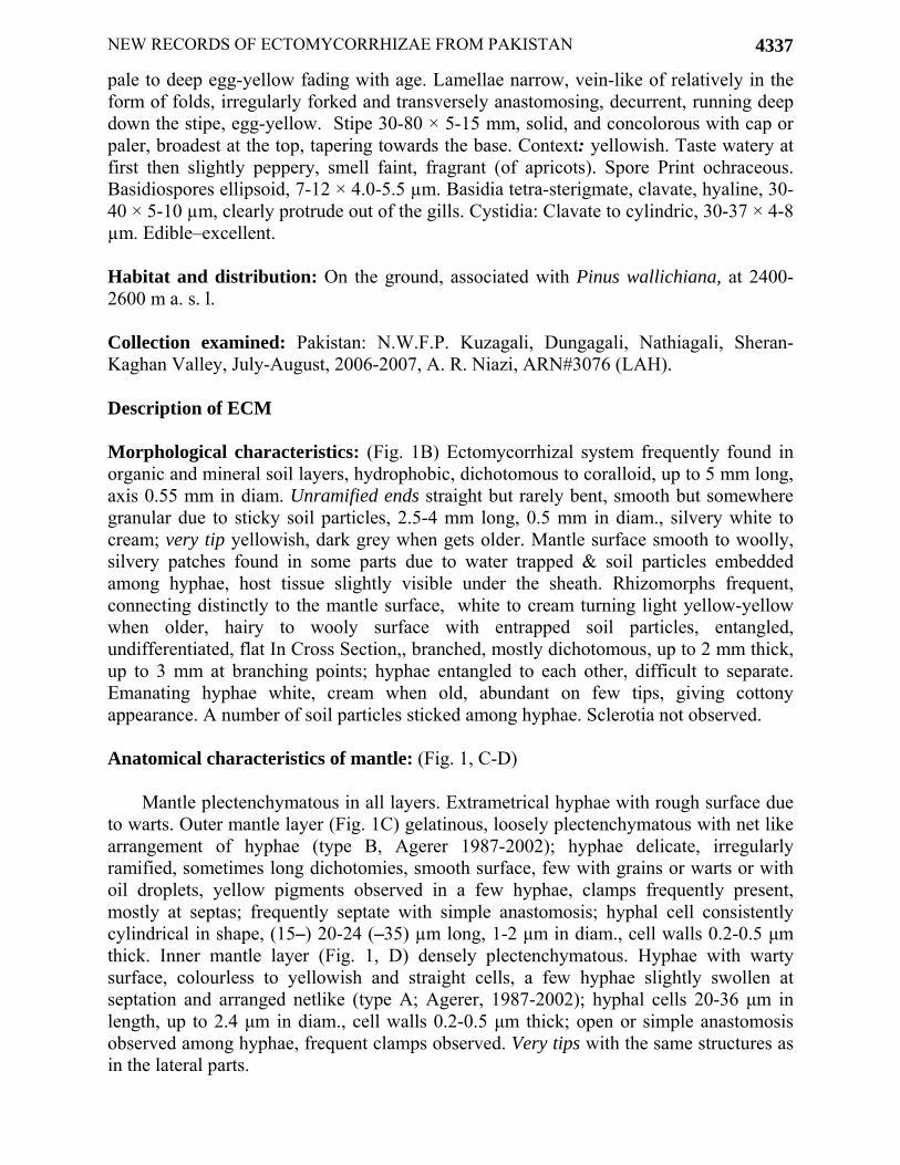
pale to deep egg-yellow fading with age. Lamellae narrow, vein-like of relatively in the form of folds, irregularly forked and transversely anastomosing, decurrent, running deep down the stipe, egg-yellow. Stipe 30-80 × 5-15 mm, solid, and concolorous with cap or paler, broadest at the top, tapering towards the base. Context: yellowish. Taste watery at first then slightly peppery, smell faint, fragrant (of apricots). Spore Print ochraceous. Basidiospores ellipsoid, 7-12 × 4.0-5.5 µm. Basidia tetra-sterigmate, clavate, hyaline, 30-40 × 5-10 µm, clearly protrude out of the gills. Cystidia: Clavate to cylindric, 30-37 × 4-8 µm. Edible–excellent. Habitat and distribution: On the ground, associated with Pinus wallichiana, at 2400-2600 m a. s. l. Collection examined: Pakistan: N.W.F.P. Kuzagali, Dungagali, Nathiagali, Sheran-Kaghan Valley, July-August, 2006-2007, A. R. Niazi, ARN#3076 (LAH). Description of ECM Morphological characteristics: (Fig. 1B) Ectomycorrhizal system frequently found in organic and mineral soil layers, hydrophobic, dichotomous to coralloid, up to 5 mm long, axis 0.55 mm in diam. Unramified ends straight but rarely bent, smooth but somewhere granular due to sticky soil particles, 2.5-4 mm long, 0.5 mm in diam., silvery white to cream; very tip yellowish, dark grey when gets older. Mantle surface smooth to woolly, silvery patches found in some parts due to water trapped & soil particles embedded among hyphae, host tissue slightly visible under the sheath. Rhizomorphs frequent, connecting distinctly to the mantle surface, white to cream turning light yellow-yellow when older, hairy to wooly surface with entrapped soil particles, entangled, undifferentiated, flat In Cross Section,, branched, mostly dichotomous, up to 2 mm thick, up to 3 mm at branching points; hyphae entangled to each other, difficult to separate. Emanating hyphae white, cream when old, abundant on few tips, giving cottony appearance. A number of soil particles sticked among hyphae. Sclerotia not observed. Anatomical characteristics of mantle: (Fig. 1, C-D)
Mantle plectenchymatous in all layers. Extrametrical hyphae with rough surface due to warts. Outer mantle layer (Fig. 1C) gelatinous, loosely plectenchymatous with net like arrangement of hyphae (type B, Agerer 1987-2002); hyphae delicate, irregularly ramified, sometimes long dichotomies, smooth surface, few with grains or warts or with oil droplets, yellow pigments observed in a few hyphae, clamps frequently present, mostly at septas; frequently septate with simple anastomosis; hyphal cell consistently cylindrical in shape, (15–) 20-24 (–35) µm long, 1-2 μm in diam., cell walls 0.2-0.5 μm thick. Inner mantle layer (Fig. 1, D) densely plectenchymatous. Hyphae with warty surface, colourless to yellowish and straight cells, a few hyphae slightly swollen at septation and arranged netlike (type A; Agerer, 1987-2002); hyphal cells 20-36 μm in length, up to 2.4 μm in diam., cell walls 0.2-0.5 μm thick; open or simple anastomosis observed among hyphae, frequent clamps observed. Very tips with the same structures as in the lateral parts.

A.R. NIAZI ET AL.,
4338
Fig. 1, A-F. (A) Cantharellus cibarius (sporocarps) (B) ECM system (C) outer mantle layer (D) inner mantle layer (E) Internal view of rhizomorph (F) Emanating hyphae. Scale bar: 2 mm for A and B, 20 µm for C–F.
A
C
B
D
E
F

NEW RECORDS OF ECTOMYCORRHIZAE FROM PAKISTAN
4339
Anatomical characteristics of emanating elements (Fig. 1E-F)
Rhizomorphs (Fig. 1E) undifferentiated internally (type B, Agerer, 1991); margins rather hairy; hyphae compactly arranged and of uniform diam., branched, smooth to rough with warts on surface, entangled, frequently septate, clamps common, compactly woven hyphae and sometime fused with each other with clamped anastomosis, simple anastomosis also observed; hyphal cells 20-38 μm length, 2.3-4 μm in diam., walls <0.5 μm thick. Emanating hyphae (Fig. 1F) smooth, 3-6 μm thick in diam., ramified, ramification Y-shaped, cell walls thick, up to 1.0 µm, septa with well-developed clamps, at septa formation or somewhere else. Cystidia lacking. Chlamydospores not observed. Anatomy of mantle in cross section; mantle appears thick as continuous hyphal mat, plectenchymatous throughout forming a distinct sheath, (40–) 45-50 (–55) µm thick, different layers discernible, cortical cells with Hartig net in 2-3 rows, mostly 2 rows reaching endodermis, frequently ramified. Anatomy of mantle in longitudinal section; plectenchymatous hyphae entangled, running parallel towards root length, different layers discernible. Cortical cells with Hartig net in 2-3 rows, Hartig net in section around cortical cell in 1–2 rows, roundish to cylindrical, 2-3 µm thick, frequently ramified. Color reactions: 15% KOH, Meltzer’s Reagent & Lactic acid-no reaction. Voucher specimen: Ectomycorrhizae under Pinus wallichiana, in Herbarium, Department of Botany, University of the Punjab, Lahore, Pakistan. A. R. Niazi # ARN-12086E (LAH). Comments
In Cantharelloid clade, Cantharellaceae has been reported as ectomycorrhizal (Agerer, 1987-2006). Mleczko (2002) has described the ectomycorrhizal morphotypes of Cantharellus cibarius with Pinus sylvestris. This is the first report of ectomycorrhizal association of C. cibarius with P. wallichiana from Pakistan. This species is already collected and reported as ectomycorrhizal under coniferous forests from Pakistan (Ahmed et al., 1997; Khalid, 1998). The field ectomycorrhizae of this fungus with Pinus wallichiana are new record for Pakistan. Tricholoma aurantium (Schaeff.) Ricken, Die Blätterpilze: 332 (1914). (Fig. 2A)
Pileus 30-100 mm broad, Fleshy, convex or nearly flat with an obtuse umbo, margins inrolled at first, then expanded; umbelicate, surface viscid when wet, smooth or with scattered, appressed fibers and scales, color varies from yellow-orange to tawny to rusty orange or orange-brown or orange-red. Context: 5-9 mm thick at disc, white. Lamellae adnate or notched, close, narrow; white staining rusty brown, developing brown to reddish brown discolorations. Stipe 4-10 cm long; 1.0-2.5 cm thick; more or less equal, or tapering to the base; covered with dense, orangish scales that terminate in a line near the apex; white above; often hollow; Context thick; white. Basidiospores ellipsoid to subglobose, smooth, 4-6 × 3-5 µm, inamyloid, spore print white. Basidia tetra sterigmate, 46-53 × 11-12 µm, clavate to cylindrical. Cystidia: absent. Habitat and Distribution: On the ground, associated with Abies pindrow, at 2400-2580 m a. s. l.

A.R. NIAZI ET AL.,
4340
Fig. 2. A-F. (A) Tricholoma aurantium (sporocarps). (B) ECM system (C) Loosely plectenchymatous outer mantle layer (D) Densely plectenchymatous inner mantle layer (E) Internal view of rhizomorph. (F) Emanating hyphae, Scale bar: 2 mm for A and B, 20 µm for C – F.
A
C
B
D
E
F

NEW RECORDS OF ECTOMYCORRHIZAE FROM PAKISTAN
4341
Collection examined: Pakistan: N.W.F.P., Ayubia, August, 2007, A. R. Niazi, ARN # 12086T (LAH). Description of ECM Morphological characteristics: (Fig. 2B)
Ectomycorrhizal System frequently found forming short mycelial strands; white mats in mineral soil, hydrophobic, monopodial-pinnate to monopodial-pyramidal, up to 17 mm long, axis 0.9 mm diam. Unramified ends straight to bent, 2-5 mm long, 0.5 mm diam., white to light yellowish to light brown; smooth or grainy; shiny or reflective; host cells visible through transparent mantle except in white areas of cottony emanating hyphae and adhering soil, very tip white to brown, dark brown when gets older. Mantle surface smooth but somewhere granular, sometimes cottony, host tissue slightly visible under the sheath at few points. Rhizomorphs very frequent, occurring every where in the form of hyphal fans connecting to the axis and mycorrhizal ends at flat angles, mostly originating from restricted points, connected over longer distance to mantle surface, white, up to 0.25 mm in diam., thicker near fruit bodies up to 0.5 mm, frequently ramified, entangled, rounded to flat in cross section, margins not smooth, several emanating hyphae emanating out from the distal end. Emanating Hyphae white, tortuous, frequent, abundant on mycorrhizal system, giving cottony appearance. Often enmeshing soil. Sclerotia not observed. Anatomical characteristics of mantle: (Fig. 2C-D) Outer mantle layers (Fig. 2C) loosely plectenchymatous, gelatinous, with net like arrangement of hyphae, no hyphal pattern discernable (type C, Agerer 1987-2002), few hyphae run parallel to each other, other forming ring like structures, hyphae delicate, irregularly ramified, frequently septate with clear to granular contents, consistently cylindrical in shape; hyphal cells 25 (-40) -90 (-150) µm, 1.5-3 µm diam., thin cell walls, up to 0.3 µm, smooth surface, a few ampulate cells observed, inflation 5-7 µm; clamps not observed, H-type anastomosis observed. Inner mantle layer (Fig. 2D) densely plectenchymatous. Hyphae with smooth surface, colourless, straight with clear to granular contents; hyphal bundles form ring like structure (type A; Agerer, 1987-2002); hyphal cells 42-76 µm in length, up to 2.8 µm in diam., cell walls 0.2-0.5 µm thick; clamps and anastomosis not observed among hyphae, slightly constricted at septum. Very tips with the same structures as in the lateral parts. Anatomical characteristics of emanating elements: (Fig. 2E-F)
Rhizomorphs (Fig. 2E) differentiated with central random or isolated wider hyphae, margins rather smooth; hyphae compactly arranged and of uniform diam., branched, smooth or with crystalline ornamentation, entangled and jointed, cells 20-150 µm in length, 2-3 µm in diam., walls 0.5-0.7 µm thick, frequently septate; septa complete or sometimes enlarged, clamps lacking, compactly woven hyphae and sometime fused with each other by ‘H’ type of anastomosis, a few hyphae with inflated cells near septum, inflations at one side of the septa, up to 8 µm in diam.; central wider hypha up to 6 µm in diam. Emanating hyphae (Fig. 2F) common, smooth with normal ends, slightly swellings
A.R. NIAZI ET AL.,
4342
and constriction at septa, thick 3-4 µm in diam., cell walls thick, up to 0.45 µm, hyphae ramified, frequently septate with without clamps, cells 25-125 µm long and up to 2 µm in diam. In cross section; mantle appears thick as hyphal aggregations, plectenchymatous throughout forming a distinct sheath, up to 45 µm thick, different layers discernible, cortical cells with Hartig net in 2-3 rows, frequently ramified.
Longitudinal section; plectenchymatous hyphae entangled, running parallel towards root length, different layers discernible, sheath in the form of heaps. Cortical cells with Hartig net in 2-3 rows. Color reactions: 15% KOH- light yellow; Lactic acid- & Meltzer’s Reagent - no reaction. Voucher specimen: Ectomycorrhizae under Abies pindrow, in Herbarium, Department of Botany, University of the Punjab, Lahore, Pakistan, A. R. Niazi, # ARN-12086E (LAH). Comments: The ECM of Tricholoma aurantium are reported as a new record from Pakistan. The ECM of this species have already been thoroughly described (Uhl, 1988) with Picea sp. Here A. pindrow is reported as a new host for this fungus. Acknowledgments
Authors are very grateful to Higher Education (HEC) of Pakistan for providing financial support to explore the biodiversity of mushrooms and ectomycorrhizas from Himalayan moist forests of Pakistan. References Afshan, N., A.N. Khalid and A.R. Niazi. 2003. New ectomycorrhizas from Sakesar Hills.
Mycopath, 1(2): 100-104. Agerer, R. 1987-2002. Color Atlas of Ectomycorrhizae. Einhorn Verlag, Schwäbisch Gmünd Agerer, R. 1991. Characterization of ectomycorrhizae. In: Techniques for the study of mycorrhiza.
Methods in microbiology, (Eds.): J.R. Norris, D.J. Read and A.K. Varma. 23: 25-73. Academic Press London
Dahlberg, A. 2001.Community ecology of ectomycorrhizal fungi: an advancing Interdisciplinary field. New Phytol., 150: 555-562.
De-Roman, M., V. Claveria and M.D. Miguel. 2005. A revision of the descriptions of ectomycorrhizas published since 1961. Mycol.Res., 109(10): 1063-1104.
Dominik, T. 1969. Key to ectotrophic mycorrhizae. Folia Forestalia Polonica, 15: 309-328. Gibson, F. and J.W. Deacon. 1996. Experimental study of establishment of ectomycorrhizas in
different regions of birch roots systems. Trans. Br. Mycol. Soc., 91: 239-251. Iqbal, S.H., M. Yousaf and M. Younas. 1980. A field survey of mycorrhizal association in ferns of
Pakistan. New Phytol., 87: 69-79. Kazmi, S.A.R., A.N. Khalid and A.R. Niazi. 2004. Ectomycorrhizal diversity with Himalayan
Poplar (Populus ciliata Wall ex Royle). Mycopath., 2(2): 75-78. Khalid, A.N and A.R. Niazi. 2003. New ectomycorrhizas in association with Poplar from
Himalayan moist temperate forests of Pakistan. Mycopath., 1(1): 95-98. Khalid, A.N. 1998. Taxonomic affinities of ectomycorrhizal macromycetes of pines of Pakistan.
Ph.D. Disser. Pb. Univ. Lahore. Mleczko, P. 2002. In: Color Atlas of Ectomycorrhizae, (Ed.): R. Agerer. Plate 159. Einhorn Verlag
+ Druck GmbH, Schwabisch Gmund

NEW RECORDS OF ECTOMYCORRHIZAE FROM PAKISTAN
4343
Niazi, A.R., A.N. Khalid and S.H. Iqbal. 2007. Descolea flavoannulata and its ectomycorrhiza from Pakistan's Himalayan moist temperate forests. Mycotaxon, 101: 375-383.
Niazi, A.R., S.H. Iqbal and A.N. Khalid. 2006. Biodiversity of mushrooms and ectomycorrhiza. 1. Russula brevipes Peck and its ectomycorrhiza, a new record from Himalayan moist temperate forests of Pakistan. Pak. J. Bot., 38(4): 1271-1277.
Niazi, A.R., S.H. Iqbal and A.N. Khalid. 2009. Ectomycorrhizae between Amanita rubescens and Himalayan Spruce (Picea smithiana) from Pakistan. Mycotaxon, 107: 73-80.
Uhl, M. 1988. Identifizierung und Charakterisierung von Ektomykorrhizen en Pinus silvestris und von Ektomykorrhizen aus der Gattung Tricholoma. Diss. Univ. München.
Zak, J.C. 1973. Classification of Ectomycorrhizae. In: Ectomycorrhizae, their ecology and physiology; (Eds.): G.C. Marks and T.T. Kozlowaski. 43-73. Academic Press, New York.
(Received for publication 1 June 2009)