Embed Size (px)
Citation preview
3
New Insight on Entangled DNA Repair Pathways: Stable Silenced Human Cells for
Unraveling the DDR Jigsaw
Biard Denis S.F. Commissariat à l’Energie Atomique,
DSV-iRCM / INSERM U935, Villejuif France
1. Introduction
1.1 Need for outstanding cell models for studying interwoven DNA repair pathways During normal human cell growth, each cell is exposed to numerous DNA-damaging
events. DNA lesions are mainly inflicted by endogenous insults, such as normal biochemical
activities, by-product synthesis and the in situ production of reactive oxygen species (ROS).
DNA is also subject to genotoxic injuries resulting from diverse exogenous sources. It is not
surprising that living organisms have evolved numerous intricate strategies to counteract
these environmental pressures and to allow living cells to thrive in aerobic conditions.
Through evolution various highly sophisticated pathways for protecting the genetic
information have been retained. The first lines of defense include detoxification metabolisms
and defense against oxidative stress. When these caretaker processes fail to insure a correct
protection of biological molecules, such as genomic and mitochondrial DNA, DNA repair
pathways become the ultimate bulwark against DNA damage. However, when DNA
damage is not dealt with properly, it can adversely threaten the fidelity of the genetic
information and ultimately lead to hereditary diseases or neoplastic processes.
Amongst DNA lesions, DNA double-strand breaks (DSBs) are likely to be the ultimate lethal
ones because unrepaired they can lead to chromosomal rearrangements, malignant
transformation or apoptosis (Roos & Kaina, 2006). Endogenous DSBs mainly arise from the
processing of single-strand breaks (SSBs) when they are converted to DSBs by DNA
replication and/or transcription mechanisms (Mladenov & Iliakis, 2011). Given the chemical
variety of DNA lesions encountered, evolution has retained a large diversity of DNA repair
pathways and a tight interplay between DNA replication and DNA repair. While numerous
DNA repair mechanisms exist, the major pathways include mismatch, excision and
recombinational repair (Hoeijmakers, 2001) and some factors can participate in divergent
processes. This is the case of the structure-specific endonuclease ERCC1 / XPF, which is
required in two distinct mechanisms: excision (nucleotide excision repair or NER) and
recombinational (single-strand annealing or SSA) repair pathways. ERCC1 / XPF
endonuclease plays a critical role in NER by being recruited at the site of damaged DNA in
order to cleave one strand of the damaged DNA. It is also involved in SSA, which appears to
www.intechopen.com

DNA Repair − On the Pathways to Fixing DNA Damage and Errors
44
be an alternative pathway to homologous recombination (HR) or nonhomologous end
joining (NHEJ) (Al-Minawi et al., 2008). Moreover, albeit belonging to the same complex, it
is hypothesized that ERCC1 and XPF have distinct functions in vivo because their deficiency
can lead to different phenotypes in humans. For instance, the only patient carrying a
mutated ERCC1 gene exhibits a cerebro-oculo-facio-skeletal syndrome with severe
neurological defects but a moderate sensitivity to UV light and mitomycin C, the hallmark
of XPF patients (Jaspers et al., 2007). Furthermore, there is a large body of evidence that
raises the notion that the failure of one DNA repair pathway could modify the efficiency
and/or fidelity of another one. An interesting example is the cross-talk between the
mismatch repair (MMR) and recombinational pathways. MMR appears to be an essential
mechanism for guaranteeing the fidelity of DNA replication because misincorporated
nucleotides have to be excised immediately after DNA synthesis. Inherited defects in the
MMR trigger a spontaneous mutation rate 50- to 1000-fold higher than that observed in
MMR proficient cells, with a tremendous increase of spontaneous base substitution and
frameshift mutations (for review (Iyer et al., 2006)). These mutational events could facilitate
illegitimate recombination between nearly-homologous sequences, contributing to the onset
of hereditary nonpolyposis colon cancer (HNPCC) (for review (Iyer et al., 2006)). Human
MutS┙ ((Msh2-Msh6 heterodimer), and MutL┙ (Mlh1-Pms2 heterodimer) participate in the
fidelity of genetic recombination and the suppression of gene amplification (Chen et al.,
2001).
Another recurring theme in the DNA repair of complex genomes, such as the mammalian
genome, is the existence of proteins with partly overlapping activities. This genetic
redundancy appears essential for maintaining the stability of a complex genome but this
represents a major drawback for experimental approaches designed to unravel the specific
functions of a particular DNA repair protein. A classic example arises from the repair of
uracil by BER. BER includes a recognition step which is performed by specific uracil-DNA
glycosylases, following by a synthesis step conducted by the DNA polymerase beta (Pol ┚).
Different DNA glycosylases travel down the DNA molecule scanning for potential lesions
(Sartori et al., 2002), and gene redundancy might make it difficult to generate uracil
glycosylase–deficient cells because there are several genes in the mammalian genome that
encode enzymes able to excise uracil from DNA (Pearl, 2000). Another example is the PARP
family where PARP1 and PARP2 possess partially redundant functions as well as divergent
activities (Menissier de Murcia et al., 2003, Schreiber et al., 2002). This functional partial
overlap explains the survival of human cells when either the PARP1 or PARP2 gene is
silenced (see below); in contrast double PARP1 and PARP2 knockdown leads to cell death
(unpublished data). Fortunately, the genetic redundancy and the overlap between DNA
repair pathways support the notion that compensating repair activities can take place over
time. This is essential to understanding of DNA damage response (DDR)–deficient human
cells, but also cells handled in vitro, such as knockdown cells.
An additional point in the complex study of DNA repair factors is that several of them are involved in other physiological pathways, even in the absence of DNA damage. This is the case for certain of the NER factors and their tight relationship with the transcriptional machinery (Le May et al., 2010a, Le May et al., 2010b). This is also observed when different DNA repair pathways are key building blocks in the primary and secondary antibody diversification processes taking place in B lymphocytes (for review (Durandy, 2009)).
www.intechopen.com
New Insight on Entangled DNA Repair Pathways: Stable Silenced Human Cells for Unraveling the DDR Jigsaw
45
Actually, BER, D-NHEJ (classic DNA PKcs-dependent NHEJ), b-NHEJ (backup NHEJ), MMR and DNA damage signaling factors actively contribute to immunoglobulin diversification. These compelling data explain why a mutation of one DNA repair gene could trigger fetal
or embryonic death or lead to a dramatic hereditary disorder. Human syndromes where one
DNA repair gene is mutated have been collectively classified as “DDR-defective syndromes”.
The range of clinical features associated with these disorders attests to the complexity of the
DDR, its redundancy and its connection with other essential processes. That explains the
diversity of the phenotypes observed in patients with DNA repair disorders (Table 1). It
turned out that growing evidence demonstrates that ubiquitylation of key proteins is
critically involved in the emergence of DDR-defective syndromes as observed for NER
(DDB2), HR (FancD2) or TLS (PCNA). (for review (Huang & D'Andrea, 2006)). The pivotal
role of DNA repair pathways during normal human development explains one hallmark of
numerous DDR-defective syndromes. Actually, the main clinical features observed in
numerous DDR-defective syndromes are hematopoietic defects (e.g. anemia or
immunodeficiency) and neurological deficits (e.g. microcephaly), in parallel with genomic
instability and specific DNA damage-induced sensitivities. This highlights crosslinks
between DNA repair mechanisms and either neuronal development (O'Driscoll & Jeggo,
2008) or immunoglobulin diversification processes (for review (Durandy, 2009)). Hence,
numerous DDR-defective disorders exhibit microcephaly, such as LIG4 syndrome (DNA
ligase IV gene) but also XLF-Cernunnos-SCID (XLF-Cernunnos gene), Seckel syndrome (ATR
gene), Nijmegen breakage syndrome (NBS1 gene), Fanconi anemia (FancD1/BRCA2 gene),
Bloom syndrome (BLM gene), Cockayne syndrome (CSA, CSB, XPB, XPD and XPG genes),
Xeroderma pigmentosum (XPA to XPG genes), and cerebro-oculo-facio-skeletal syndrome
(ERCC1 gene) ((Jaspers et al., 2007); for review (McKinnon, 2009, O'Driscoll & Jeggo, 2008)).
LIG4 syndrome and XLF-Cernunnos-SCID also exhibit multiple immune abnormalities
because both LigIV and XLF/Cernunnos are involved in the V(D)J and NHEJ pathways
which are required during the primary repertoire of antibodies and the secondary
diversification processes (Yan et al., 2007).
1.2 Long term silenced human cells A better understanding of these hereditary disorders requires detailed insight into each
DNA repair pathway that can operate on the damaged genome. Furthermore, the
importance of the DDR during the multistage process leading to tumorigenesis emphasizes
the need for outstanding biological tools to study DNA repair genes. Altogether this
compelling evidence points to the need of outstanding cell models for unraveling the DDR
jigsaw both for fundamental research and for the development of novel therapeutic
strategies.
Over the last seven years we have developed a rational strategy to silence the main DNA repair factors so as to unveil their functions. Since the emergence of the RNA interference technology, many studies have developed transient or middle-term gene silencing experiments targeting DDR genes, but few of them have characterized stable clones. Our project is based on the exceptional efficiency of pEBVsiRNA vectors in ensuring stable gene silencing. Our approach has been extensively described previously (Biard, 2007, Biard & Angulo, 2007).
www.intechopen.com

DNA Repair − On the Pathways to Fixing DNA Damage and Errors
46
Pathways Diseases or syndromes
Mutated genes(targeted genes*)
Main symptoms and/or remarks
Cell cycle control
Li-Fraumeni syndrome
p53 Sarcoma, breast, brain, leukemia
familial
retinoblastomaRb Retinoblastoma, osteosarcoma
familia melanoma p16 Melanoma, pancreas cancers
Sporadic cancers Chk1 Colorectal, stomach, lung, endometrial, melanoma, mesothelioma cancers (for
review (Solyom et al., 2010))
Li-Fraumeni
syndromeChk2
Breast, lung, colon, urinary, bladder, testis cancers, melanoma
Signaling pathways
Ataxia telangiectasia ATM Neurodegeneration, sterility,
telangiectasia, dysarthria, immunological defects, sensitivity to IR, lymphomas
ATR-Seckel syndrome
ATR Microcephaly and mental retardation,
growth defects
Ataxia telangiectasia-
like disorder MRE11
Ataxia, neurodegeneration, dysarthria and oculomotor apraxia, mild
immunological defects, lymphomas
Familial breast cancer
1 & 2BRCA1, BRCA2
Chromosome instability, sensitivity to DNA damage, HR deficiency, cancer
Nijmegen breakage
syndromeNBS1
Microcephaly, immunological defects and lymphoid malignacy, lymphomas
NBS-like disorder
(NBSlD)Rad50
DNA DSB repair
LIG4 syndrome LIG4 Microcephaly, developmental/growth
delay, immunodeficiency and lymphomas
Human
immunodeficiency with microcephaly
XLF/Cernunnos Microcephaly, immunodeficiency
glioblastoma (M059J
cells)DNAPKcs (Allalunis-Turner et al., 1993)
Fanconi anaemia BCRA2 Microcephaly and medulloblastoma, Bone
marrow and congenital defects
DNA SSB repair
Spinocerebellar ataxia with axonal
neuropathyTDP1
Ataxia, neurodegeneration, peripheral axonal motor, and sensory neuropathy,
and muscle weakness…
Ataxia with
oculomotor apraxia 1APTX
Ataxia, neurodegeneration, oculomotor apraxia and peripheral neuropathy,
hypercholesterolaemia and hypoalbuminaemia
cerebro-oculo-facio-skeletal syndrome
ERCC1 Microcephaly, moderate sensitivity to UV
and mitomycin C (Jaspers et al., 2007)
NER Xeroderma
pigmentosum (XP)XPA to XPG
Neurodegeneration and microcephaly, UV sensitivity and skin cancer
Cockayne syndrome
(CS) CSA, CSB, XPB,
XPD, XPG
Microcephaly and dysmyelination , TCR-specific disorder. Segmental progeria, no
increase in cancer incidence
Trichothiodystrophy
(TDD) XPD, XPB, TTD-
A
Neurodevelopmental defects and dysmyelination, brittle hair, nails and
scaly skin. Segmental progeria without an increase in cancer incidence (Giglia-Mari
et al., 2004)
www.intechopen.com

New Insight on Entangled DNA Repair Pathways: Stable Silenced Human Cells for Unraveling the DDR Jigsaw
47
Pathways Diseases or syndromes
Mutated genes(targeted genes*)
Main symptoms and/or remarks
TLS XP variant (XP-V) XPV (pol iota) UV-induced skin cancer
DNA cross link repair
& Ubiquitin
ligase
Fanconi anaemia FancA, B, C, D1 (BRCA2), D2, E, F, G, I, J, L, M, N
Microcephaly and medulloblastoma, cervical cancer, brain tumours i(FANCD2
and FANCN), anaemia, developmental defects, ovarian carcinomas, head and neck squamous cell carcinomas, bone
marrow failure, and myeloid leukemias.
BER Multiple colorectal
adenoma and carcinoma
MutYH DNA glycosylase involved in the repair of oxidative damage (Al-Tassan et al., 2002)
Ligase I Ligase I Iimmunodeficiencies and cellular
hypersensitivity to DNA-damaging agents (Barnes et al., 1992)
MMR HNPCC MLH1, MLH3, MSH2, MSH6,
PMS2
Hereditary nonpolyposis colon cancer, rectum, gastric, endometrium, ovarian, urinary organ cancers (Peltomaki, 2003)
Helicase Werner syndrome WRN Severe progeria, various cancers
Rothmund Thomson
syndromeRTS Osteosarcomas
Bloom syndrome BLM Proportional dwarfism, leukemias,
lymphomas and others cancer
Ataxia with
oculomotor apraxia 2SETX
Ataxia, neurodegeneration and oculomotor apraxia
(*: genes targeted with pEBVsiRNA plasmids)
Table 1. Main DDR defective Syndromes. (Adaptated from (Hoeijmakers, 2001) and (McKinnon, 2009)).
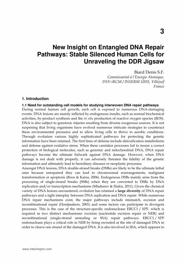
Briefly, for each gene, three pEBVsiRNA vectors are constructed and validated through both short-term (several days) and long-term (several weeks) experiments. Afterwards, we used only one “validated” vector to establish stable clones (Fig. 1). Four years ago we adopted the DSIR program developed by Vandenbrouck and collaborators (Vert et al., 2006) to design shRNA sequences. This program includes an exact similarity search algorithm for potential off-target detection. In a recent comparison of methods for a rational siRNA design, DSIR is among the three best predictive programs (Matveeva et al., 2007). Our siRNA sequences mainly target the open reading frame of the targeted genes, but when necessary we also use siRNA sequences stretching to the 3’-UTR (e.g. for rescue experiments). Among the targeted genes and in using our approach we have always obtained at least one vector able to impose long-term shut down greater than 80% as compared with control cells (as evidenced by real-time RT-PCR). Using this technology, more than 160 human genes in different human cell models such as HeLa (Ame et al., 2009, Amine et al., 2009, Aressy et al., 2008, Betous et al., 2009, Biard, 2007, Biard et al., 2005, Biard & Angulo, 2007, Boehler et al., 2011, Bouley et al., 2010, Britton et al., 2009a, Despras et al., 2007, Godon et al., 2008, Le May et al., 2010b, Ousset et al., 2010, Pennarun et al., 2008, Pennarun et al., 2010, Wu et al., 2007), U2OS (Betous et al., 2009, Rey et al., 2009) and MRC5-V1 (Bouquet et al., 2011, Britton et al., 2009b, Schmutz et al., 2010) cells have been silenced. Our approach has also been successfully tested in other human tumor-derived cell lines, such as RKO (Biard & Angulo, 2007), HCT-116 (Aressy et al., 2008), Caco2 (Coant et al., 2010), SH-SY5Y cells (Schulte et al., 2008), MCF7, MDA-MB 231, K562, UT7
www.intechopen.com

DNA Repair − On the Pathways to Fixing DNA Damage and Errors
48
07bd0077 07bd0077
07bd0067 07bd0067
DAPIXRCC1
07bd0071 07bd0071
07bd0073 07bd0073
XRCC1KD cells (pBD1063; day 4)
XRCC1KD cells (pBD1064; day 4)
CTL cells (day 4)
XRCC1KD cells (pBD1065; day 4)
A) Short term validation
of 3 vectors per gene
B) Selection of clones C) Phenotypic analyses
(XRCC1 and LigIII are partners)
DAPIXRCC1
07BD289 07bd289
07bd293 07bd293
07BD295 07bd295
XRCC1KD clone 3 (day 125)
Lig3KD clone 11 (day 145)
CTL cells (day 197)
07bd200 07bd200
07bd202 07bd202
07bd206 07bd206
DAPIXRCC1
XRCC1KD clone 3
XRCC1KD clone 11
CTL cells
DAPILig3
07BD298 07bd298
07bd307 07bd307
07bd303 07bd303
XRCC1KD clone 3 (day 125)
CTL cells (day 197)
Lig3KD clone 11 (day 145)
Validation by real time PCR in comparison with CTL cells using GAPDH and Actin as internal controls
Lig3KD : 94%XRCC1KD : 87%
Fig. 1. Establishment of stable clones.
(papers in preparation), and even in mouse NIH-3T3 cells (Meulle et al., 2008). Some authors
have previously suggested the importance of “position-specific” criteria for efficient gene
silencing. With the benefit of hindsight, we have never observed such a positioning effect in
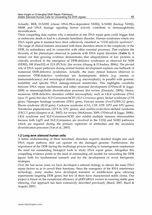
either short-term (few days) or long term (several months) experiments. In Figure 2 we show
the position of different siRNA sequences able to impose a very efficient long-term gene
silencing along a representative mRNA and we demonstrate no positioning effect.
10% 20% 30% 40% 50% 60% 70% 80% 90% 100%
mRNA
5’ 3’
Fig. 2. Position of validated siRNA sequences along a representative mRNA.
www.intechopen.com
New Insight on Entangled DNA Repair Pathways: Stable Silenced Human Cells for Unraveling the DDR Jigsaw
49
The maintenance of stable gene silencing for several months affords the opportunity to validate different siRNA sequences for an unfailing and specific gene silencing. Importantly, transient assays may mask the real effects of gene silencing, due to the saturation of the RNAi (and miRNA) machinery and by side-effects resulting from the high siRNA concentrations currently used. In the long-term experiments, we do not exclude the possibility of skews, and the suppression of gene expression over a long period may provoke compensatory cellular responses during an “adaptive period”. During this period, cellular metabolism may compensate for the decrease in protein concentration, particularly if the protein plays an important role in the cell. These compensating activities are also observed during the multistage process leading to tumorigenesis, where a normal cell undergoes serial genetic changes, including initiation, clonal expansion, pre-malignant lesions, and malignant progression, before acquiring a tumor phenotype. These properties acquired by cells to escape DDR defects are essential to our understanding of tumor cell behavior following chemo- or radiotherapy. We can now assess the usability of the numerous stable clones affecting all branches of the DDR that have been created. This unique cell model appears relevant for studying DNA repair, DNA replication, DNA recombination and cross-talk between them. To date, we have established numerous clones, creating a library of stable isogenic cells which no longer express a specific DNA repair gene. This approach has helped us to untangle the interwoven DNA repair pathways and represents a powerful tool for research, drug screening and for preclinical testing of new therapies. This review will concentrate on two fields of research investigated using these knockdown clones.
2. Example of stable DNA repair gene silencing studies
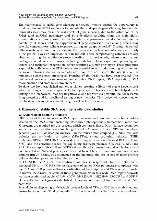
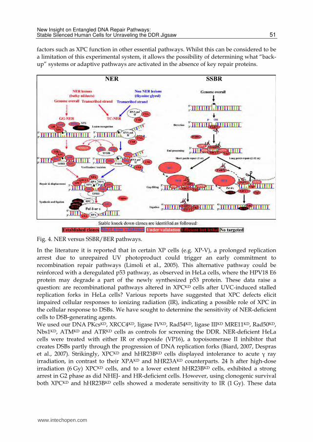
2.1 Dual roles of some NER factors NER is one of the more versatile DNA repair processes and removes diverse bulky lesions located on one DNA strand, including UV-induced photoproducts. In mammals, more than 30 proteins are required for this process, which comprises first a DNA damage recognition and structure distortion step involving XPC-hHR23B-centrin 2 and XPE in the global genome (GG)-NER or RNA polymerase II in the transcription-coupled (TC)-NER. NER also includes the verification of lesions (XPA-RPA), strand-separating helicases (TFIIH containing XPB and XPD DNA helicases), structure-specific endonucleases (ERCC1-XPF and XPG), and the enzymes needed for gap filling (DNA polymerase ├/┝, PCNA, RFC, and RPA). For example, ERCC1KD and XPFKD cells exhibited a tremendous and stable decrease of both targeted mRNA and protein, as evidenced by real time PCR and immunofluorescence staining (fig. 3). Beside, as documented in the literature, the loss of one of these proteins induces the disappearance of the other partner. In GG-NER, the XPC-hHR23B-centrin 2 complex is responsible for the detection of damaged DNA. In TC-NER the displacement of stalled RNA polymerase complexes with the CSA and CSB proteins allows coordination of transcription and DNA repair. In order to unravel new roles for some of these gene products in this wide DNA repair network, we have established stable XPAKD, XPCKD, hHR23AKD, hHR23BKD, ERCC1KD and XPF KD HeLa cells. In the figure 4 established clones are represented for the NER and SSBR pathways. Several clones displaying undetectable protein levels of XPA or XPC were established and grown for more than 300 days in culture with a tremendous stability of the gene-silenced
www.intechopen.com

DNA Repair − On the Pathways to Fixing DNA Damage and Errors
50
07bd342 07bd342
DAPIXPF
07bd339 07bd339
07bd344 07bd344
ERCC1KD clone 11 (day 111)
CTL cells (day 197)
XPFKD clone 3 (day 62)
07bd336 07bd336
DAPIERCC1
07bd330 07bd330
07bd332 07bd332
XPFKD clone 3 (day 62)
CTL cells (day 197)
ERCC1KD clone 7 (day 111)
Validation by real time PCR in comparison with CTL cells using GAPDH and Actin as internal controls
XPFKD : 84%ERCC1KD : 83%
Fig. 3. Analysis of ERCC1KD and XPFKD cells by immunofluorescence.
and expected phenotypes (Biard et al., 2005). As expected, XPAKD and XPCKD HeLa cells
were highly UVC sensitive and exhibited cell cycle arrest in early and middle S phase after
UVC irradiation, showing that the persistence of UVC lesions blocks DNA replication. Both
clones also show an impaired unscheduled DNA synthesis (UDS) after UVC irradiation.
However, unlike XPA, the silencing of the XPC gene dramatically impeded HeLa cell
growth. Furthermore, XPCKD HeLa clones were more sensitive to UVC than their XPAKD
counterparts. In parallel we have analyzed the behavior of our hHR23BKD and hHR23AKD
cells. hHR23BKD cells displayed a significant sensitivity to UVC, in contrast to their
hHR23AKD counterparts which strongly tolerated UVC irradiation (Biard, 2007). While
hHR23AKD cells were not blocked in S phase after UVC irradiation, the exit from the S-phase
of hHR23BKD cells was hindered, suggesting the presence of unrepaired (or unrepairable)
UVC-induced DNA damage. These data clearly demonstrate that hHR23A and hHR23B
have diverse biological functions in human cells and that hHR23BKD cells have a phenotype
closely resembling that of XPCKD cells. To understand why the silencing of the XPC gene can
trigger major changes in cell behavior, we have performed hygromycin B withdrawal
experiments.
After about 200 days of culture, hygromycin B was removed from the culture medium in order to reverse the gene-silencing phenotype by the slow and progressive disappearance of pEBV episomes. Under these experimental conditions, XPA or XPC protein levels returned to “control” levels after 15 to 20 days in culture. Unexpectedly, reverted XPCKD cells (XPC re-expressing cells) did not recover a normal resistance to UVC, unlike XPAKD cells. This striking result suggests that irreversible genetic changes have been fixed in the genome during the long-term XPC gene silencing and that, beside their canonical roles, some NER
www.intechopen.com
New Insight on Entangled DNA Repair Pathways: Stable Silenced Human Cells for Unraveling the DDR Jigsaw
51
factors such as XPC function in other essential pathways. Whilst this can be considered to be a limitation of this experimental system, it allows the possibility of determining what “back-up” systems or adaptive pathways are activated in the absence of key repair proteins.
Fig. 4. NER versus SSBR/BER pathways.
In the literature it is reported that in certain XP cells (e.g. XP-V), a prolonged replication
arrest due to unrepaired UV photoproduct could trigger an early commitment to
recombination repair pathways (Limoli et al., 2005). This alternative pathway could be
reinforced with a deregulated p53 pathway, as observed in HeLa cells, where the HPV18 E6
protein may degrade a part of the newly synthesized p53 protein. These data raise a
question: are recombinational pathways altered in XPCKD cells after UVC-induced stalled
replication forks in HeLa cells? Various reports have suggested that XPC defects elicit
impaired cellular responses to ionizing radiation (IR), indicating a possible role of XPC in
the cellular response to DSBs. We have sought to determine the sensitivity of NER-deficient
cells to DSB-generating agents. We used our DNA PKcsKD, XRCC4KD, ligase IVKD, Rad54KD, ligase IIIKD MRE11KD, Rad50KD, Nbs1KD, ATMKD and ATRKD cells as controls for screening the DDR. NER-deficient HeLa cells were treated with either IR or etoposide (VP16), a topoisomerase II inhibitor that creates DSBs partly through the progression of DNA replication forks (Biard, 2007, Despras et al., 2007). Strikingly, XPCKD and hHR23BKD cells displayed intolerance to acute ┛ ray irradiation, in contrast to their XPAKD and hHR23AKD counterparts. 24 h after high-dose irradiation (6 Gy) XPCKD cells, and to a lower extent hHR23BKD cells, exhibited a strong arrest in G2 phase as did NHEJ- and HR-deficient cells. However, using clonogenic survival both XPCKD and hHR23BKD cells showed a moderate sensitivity to IR (1 Gy). These data
www.intechopen.com
DNA Repair − On the Pathways to Fixing DNA Damage and Errors
52
suggest that beside its canonical function in the early steps of the NER, the XPC protein could be essential in the coordination of other recovery pathways, such as those involved in the repair of IR- and etoposide-induced DNA damage. In mock treated cells, the persistence of XPC on chromatin structures was shown by experiments in which the XPC protein remained tightly anchored to detergent-insoluble nuclear structures (Despras et al., 2007). Interestingly, XPC was released from these structures after induction of DSBs by calicheamicin or neocarzinostatin, two potent specific DSB inducers. The reduction of chromatin-fixed XPC correlated with the increase of H2AX phosphorylation and presumably with the recruitment of DNA repair factors at sites of damaged DNA. This sequence of events was partly confirmed by the subsequent recruitment of phosphorylated-XRCC4 and LigIV into the less extractable nuclear fraction after DSB induction, as previously described (Drouet et al., 2005). Therefore, XPC should be considered as a genome caretaker protein, which is (i) recruited for initiating the GG-NER in the presence of bulky DNA damage, but which (ii) also displays other functions in the presence of DSBs. Using the HeLa isogenic KD model we have also focused our attention on the efficiency of NER-deficient cells in performing NHEJ, using an in vitro assay making use of DNA PKcsKD and XRCC4KD cells. The DNA PKcsKD cells used displayed an undetectable protein level and a nearly total loss of the endogenous kinase activity (Despras et al., 2007), and the isolated XRCC4KD clones all displayed a residual XRCC4 protein level corresponding to about 15% of the control (CTL); this residual level might reflect the essential role played by XRCC4 in cell survival. These XRCC4KD cells are particularly interesting experimentally too as there are no human cell lines lacking the XRCC4 protein. In ligase IVKD, DNA PKcsKD and XRCC4KD cells, NHEJ efficiencies dropped to 50, 30 and 20%, respectively, as compared with control (personal data and (Despras et al., 2007)). This also correlated with a markedly increased sensitivity towards IR. Our results also argue for XRCC4 being a limiting factor in the NHEJ process, at least in vitro. Strikingly, while the expression of NHEJ factors was not altered in XPCKD cells, XPC deficiency led to a decrease of in vitro NHEJ efficiency. In both XPCKD and DNA PKcsKD cells, XRCC4 and ligase IV proteins were mobilized to damaged nuclear structures at lower doses of chemical DSB inducer in comparison with proficient cells. In contrast, XPA gene silencing did not modify HeLa cell response to DSBs. Our results reinforce the notion that XPCKD cells display an unexpected behavior towards DSBs, presumably due to an intrinsic characteristic of XPC, rather than being a consequence of NHEJ deficiency. We can also rule out a direct role of XPC in the NHEJ process per se. Presumably XPC deficiency could locally change the chromatin structure and interfere with other pathways. It is notable that in our experiments we have always observed that XPA gene silencing could lead to an enhanced cell growth several weeks after transfection of HeLa cells and in the absence of genotoxic injuries. In contrast, knocking down of XPC triggered major growth defects and tremendous cellular stress as well as elevated sensitivity to genotoxic agents. Presumably XPA and XPC can participate in major pathways required for normal growth, but with opposite effects. Because relationships between some NER factors and transcription have been extensively related in the literature (for review (Le May et al., 2010a)), we have questioned whether XPA and XPC factors could be involved in the regulation of transcription in the absence of exogenous DNA damage. The transcription / repair factor TFIIH is organized into a core complex (XPB, XPD, p62, p52, p44, p34, and p8/TTDA) that associates with the Cdk-activating kinase (CAK) complex (Cdk7, cyclin H,
www.intechopen.com
New Insight on Entangled DNA Repair Pathways: Stable Silenced Human Cells for Unraveling the DDR Jigsaw
53
and MAT1). In response to DNA damage, XPA catalyzes the detachment of the CAK from the core TFIIH, changing this transcription factor into a repair factor (Coin et al., 2008). Thereafter, new NER proteins are recruited around the TFIIH factor such as XPC / hHR23B. After repair, resumption of CAK activity is required for continuation of transcription. By using our XPAKD, XPCKD and ERCC1KD clones, we have determined the role of these NER proteins during the transcriptional regulation of active promoters. Interestingly, we observed that the recruitment of NER factors at promoters of inducible nuclear receptor genes (including the retinoic acid receptors ┙ and ┛) occurred in a sequential order and required XPC, CSB, XPA / RPA, the two endonucleases, XPG and ERCC1 / XPF and XPE with the RNA pol II machinery (Le May et al., 2010b). This transcriptional complex containing NER factors is formed in the absence of any genotoxic injury, at the site of the promoter. Contrary to the coordinated recruitment observed in control cells, none of the NER factors were recruited to the promoter in XPCKD HeLa cells. XPC association is thus a pre-requisite step and abnormal XPC protein levels could affect normal transcription. This XPC-dependent transcriptional complex is distinct from a repair complex. In contrast, in XPAKD cells, only XPC and CSB were detected at the promoter, and in ERCC1KD cells we detected XPC, XPA, and XPG together with RAR, RXR, RNA pol II, and TFIIH. Furthermore, during the transcriptional initiation step, XPC is required to achieve optimal DNA demethylation and histone posttranscriptional modifications. In control cells, transcription initiation and recruitment of NER factors are accompanied by a global DNA demethylation. A local DNA demethylation at sites of 5’-CpG-3’ islands was also detected around the proximal RAR┚2 promoter region. In contrast, in XPCKD, XPAKD, and ERCC1KD HeLa cells the global methylation levels were lowered as compared with control cells. More importantly, XPCKD and XPAKD cells, but not ERCC1KD cells, failed to demethylate the RAR┚2 promoter. Afterwards, during the transcription elongation in distal regions of the gene, NER factors escort the RNA-Pol and form a complex which now excludes XPC but needs CSB. This latter complex could appear as a pre-TC-NER complex. In all of these studies, the phenotype of the knockdown HeLa cells was compared with that of deficient XP and CS fibroblasts from patients. Altogether these data demonstrate that NER factors could actively contribute to transcription of particular promoters in the absence of DNA damage and then interfere with cellular homeostasis. These results help us to explain the striking phenotype observed in our XPCKD and hHR23BKD cells in comparison with control cells or their XPAKD counterparts. Recently, in an effort to silence other genes belonging to the NER, we have observed that DDB1 gene silencing strongly disrupts HeLa cell growth a few weeks after transfection (unpublished data). This raises the question whether XPE (DDB2-DDB1 heterodimer) also participates in transcription regulation in the absence of exogenous DNA damage, as has been seen for XPC.
2.2 Parp1, between inhibition and gene silencing We have also employed our cell model to shed light on the poly(ADP-ribose) polymerase (PARP) family. New developments of mono- and combined therapeutic approaches based on PARP inhibitors reinforce the crucial role played by these proteins in the DDR. The PARP family contains 17 members and its founding member, PARP1, carries out the majority of poly(ADP-ribose) (PAR) synthesis in mammalian cells (Ame et al., 2004, D'Amours et al., 2001). Poly(ADP-ribosyl)ation is an immediate DNA damage–dependent posttranslational modification of numerous nuclear proteins indispensable for an accurate
www.intechopen.com
DNA Repair − On the Pathways to Fixing DNA Damage and Errors
54
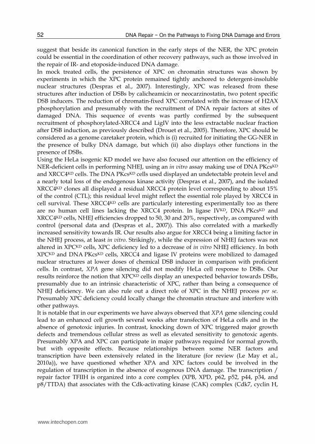
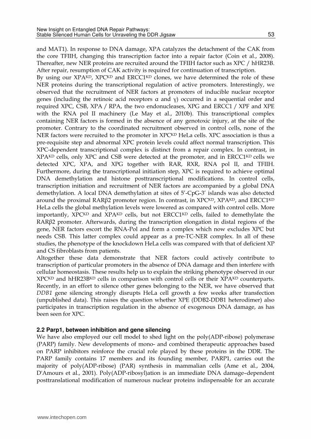
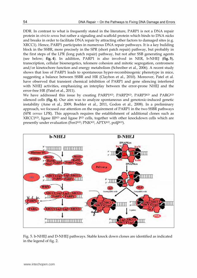
DDR. In contrast to what is frequently stated in the literature, PARP1 is not a DNA repair protein in stricto sensu but rather a signaling and scaffold protein which binds to DNA nicks and breaks in order to facilitate DNA repair by attracting other factors to damaged sites (e.g. XRCC1). Hence, PARP1 participates in numerous DNA repair pathways. It is a key building block in the SSBR, more precisely in the SPR (short patch repair) pathway, but probably in the first steps of the LPR (long patch repair) pathway, but not after SSB generating agents (see below; fig. 4). In addition, PARP1 is also involved in NER, b-NHEJ (fig. 5), transcription, cellular bioenergetics, telomere cohesion and mitotic segregation, centromere and/or kinetochore function and energy metabolism (Schreiber et al., 2006). A recent study shows that loss of PARP1 leads to spontaneous hyper-recombinogenic phenotype in mice, suggesting a balance between SSBR and HR (Claybon et al., 2010). Moreover, Patel et al. have observed that transient chemical inhibition of PARP1 and gene silencing interfered with NHEJ activities, emphasizing an interplay between the error-prone NHEJ and the error-free HR (Patel et al., 2011). We have addressed this issue by creating PARP1KD, PARP2KD, PARP3KD and PARGKD silenced cells (fig. 6). Our aim was to analyze spontaneous and genotoxic-induced genetic instability (Ame et al., 2009, Boehler et al., 2011, Godon et al., 2008). In a preliminary approach, we focused our attention on the requirement of PARP1 in the two SSBR pathways (SPR versus LPR). This approach requires the establishment of additional clones such as XRCC1KD, ligase IIIKD and ligase IKD cells, together with other knockdown cells which are presently under evaluation (Fen1KD; PNKKD, APTXKD, pol┚KD).
Fig. 5. b-NHEJ and D-NHEJ pathways. Stable knock down clones are identified as indicated in the legend of fig. 2.
www.intechopen.com
New Insight on Entangled DNA Repair Pathways: Stable Silenced Human Cells for Unraveling the DDR Jigsaw
55
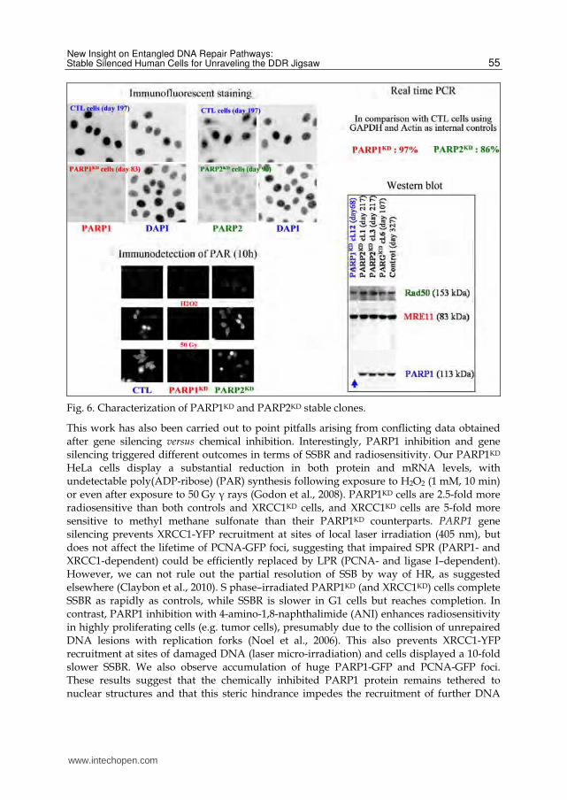
Fig. 6. Characterization of PARP1KD and PARP2KD stable clones.
This work has also been carried out to point pitfalls arising from conflicting data obtained after gene silencing versus chemical inhibition. Interestingly, PARP1 inhibition and gene silencing triggered different outcomes in terms of SSBR and radiosensitivity. Our PARP1KD HeLa cells display a substantial reduction in both protein and mRNA levels, with undetectable poly(ADP-ribose) (PAR) synthesis following exposure to H2O2 (1 mM, 10 min) or even after exposure to 50 Gy ┛ rays (Godon et al., 2008). PARP1KD cells are 2.5-fold more radiosensitive than both controls and XRCC1KD cells, and XRCC1KD cells are 5-fold more sensitive to methyl methane sulfonate than their PARP1KD counterparts. PARP1 gene silencing prevents XRCC1-YFP recruitment at sites of local laser irradiation (405 nm), but does not affect the lifetime of PCNA-GFP foci, suggesting that impaired SPR (PARP1- and XRCC1-dependent) could be efficiently replaced by LPR (PCNA- and ligase I–dependent). However, we can not rule out the partial resolution of SSB by way of HR, as suggested elsewhere (Claybon et al., 2010). S phase–irradiated PARP1KD (and XRCC1KD) cells complete SSBR as rapidly as controls, while SSBR is slower in G1 cells but reaches completion. In contrast, PARP1 inhibition with 4-amino-1,8-naphthalimide (ANI) enhances radiosensitivity in highly proliferating cells (e.g. tumor cells), presumably due to the collision of unrepaired DNA lesions with replication forks (Noel et al., 2006). This also prevents XRCC1-YFP recruitment at sites of damaged DNA (laser micro-irradiation) and cells displayed a 10-fold slower SSBR. We also observe accumulation of huge PARP1-GFP and PCNA-GFP foci. These results suggest that the chemically inhibited PARP1 protein remains tethered to nuclear structures and that this steric hindrance impedes the recruitment of further DNA
www.intechopen.com
DNA Repair − On the Pathways to Fixing DNA Damage and Errors
56
repair proteins. These data emphasize that the need for careful interpretation of results from the use of chemical inhibitors which could be riddled with pitfalls. Moreover, it is noteworthy that PARP inhibitors not only inhibit PARP1, but also PARP2 and PARP3 (Loseva et al., 2010). After a genotoxic injury, PARP1 activation leads to a tremendous but transient synthesis of PAR, in order to label DNA-damaged sites, open the chromatin structure and recruit repair factors, such as the scaffold protein XRCC1 (Dantzer et al., 2006). Because this reaction is transient, PAR polymers have to be rapidly degraded by PARG. PARP1 and PARG display opposite enzymatic activities which govern the balance between life and death after DNA injuries. Our knockdown clones clearly demonstrate that PARP1, PARP2, PARP3 and PARG activities contribute to this homeostasis, even in the absence of exogenous genotoxic attack (Ame et al., 2009, Boehler et al., 2011). PARGKD HeLa cells exhibit a stable loss of the three PARG isoforms (nuclear, cytoplasmic and mitochondrial) and a spectacular loss of function. Surprisingly, constitutive PARG depletion and subsequent PAR accumulation are rather beneficial in that they protect cells from spontaneous SSBs and telomeric abnormalities. In contrast, irradiation of PARGKD cells triggers PAR accumulation, delayed SSB and DSB repair, centrosome amplification and mitotic defects, all of which contribute to cell death by mitotic catastrophe (Ame et al., 2009). The complexity and the redundancy of the PARP family members toward the DDR are reinforced by our recent data demonstrating that PARP3 is a newcomer in the cellular response to DNA damage and mitotic progression (Boehler et al., 2011). PARP3 is closely related to PARP1 and PARP2, but unlike these two counterparts PARP3 is a mono(ADP-ribose) polymerase. It has been proposed that PARP3 could be involved in transcriptional silencing in association with Polycomb group proteins. Moreover, PARP3 could also be a component of the DDR because it is found in complexes mainly containing Ku70 and Ku80, but also PARP1, DNA ligase III, DNA PKcs and DNA ligase IV (Rouleau et al., 2007). This raises the question whether PARP3 participates in SSBR (when PARP1 is deficient?), D-NHEJ (with DNA PKcs, DNA ligase IV, Ku70, and Ku80), b-NHEJ (with DNA ligase III) and telomere maintenance (with Ku70 and Ku80). This was partly confirmed by a recent study which shows that PARP3 might be a novel DSB sensor which functions in the same pathway as APLF (aprataxin- and PNK-like factor) in order to accelerate chromosomal DSB repair (Rulten et al., 2011). APLF is a poly(ADP-ribose)-binding protein which interacts directly with Ku80 and XRCC4 at sites of DSBs (Macrae et al., 2008). To gain further insight into PARP3 function in the DDR we have validated pEBVsiPARP3 plasmids targeting the two known PARP3 isoforms. Stable clones exhibiting an almost complete depletion of PARP3 were carefully characterized (Boehler et al., 2011). PARP3KD cells displayed spontaneous DSBs and genome instability, delayed repair after irradiation, but no significant radiosensitivity as compared with control cells. Our results reinforce recent data showing that PARP3-deficient cells were as sensitive to a topoisomerase I poison (camptothecin) as control cells (Loseva et al., 2010). These unexpected results could be explained by partly compensating activities between PARP3 and PARP1. These data strongly suggest a functional synergistic cross-talk between PARP1 and PARP3. Interestingly, PARP3 interacts directly and strongly with PARP1 and PARP3 is able to activate PARP1 in the absence of DNA (Loseva et al., 2010). Another significant event observed in PARP3KD cells is an elevated frequency of sister telomere fusions and sister telomere loss. This is explained by the functional association between PARP3, tankyrase I and NuMa (microtubule-associated protein involved in spindle dynamics). Altogether, these three proteins appear to be key
www.intechopen.com
New Insight on Entangled DNA Repair Pathways: Stable Silenced Human Cells for Unraveling the DDR Jigsaw
57
regulators of mitotic progression. This study will now continue by establishing new cell lines silenced for other members of the PARP family such as PARP9, PARP14, tankyrase 1 (PARP5a) and tankyrase 2 (PARP5b).
3. Conclusions
In the field of cancer research, numerous questions remain unanswered, such as how do different pathways cooperate to repair DNA damage in tumor cells? How can we explain the chemo- and radioresistance of tumor cells? Can we target DDR to enhance chemotherapy? How do genetic compensation events take place? How can we detect the combinations of genes leading to synthetic lethality? Are DNA repair factors involved in other processes? All of these questions have to be carefully analyzed in order to design specific and less toxic therapies for cancer. Currently, chemotherapeutic approaches are based on the fact that highly proliferating (tumor and unfortunately hair, bone marrow and colon) cells are more sensitive to DNA damage than their slowly proliferating (normal) counterparts. Alterations in DNA repair pathways in tumor cells can make some cancer cells dependent on a reduced set of DNA repair pathways for their survival. These adaptive but potentially error-prone bypasses could render DNA damage–based cancer therapies less efficient and allow tumor cells to escape specific treatments. Recently substantial progress has been made through studies of genes involved in the DDR in order to circumvent rescue pathways. A breakthrough has emerged with the concept of synthetic lethality, which is defined as a genetic interaction where the minimal combination of two nonlethal mutations leads to cell death. Because naturally occurring synthetic lethal mutants are unviable we have to develop outstanding cell models in order to unravel the DDR and subsequently to detect these combinations that give rise to synthetic lethality. In light of these concerns, an emerging strategy has been to use PARP inhibitors (e.g. iniparib, olaparib or veliparib) combined or not with DNA-damaging chemotherapeutic agents in the treatment of breast and ovarian cancers exhibiting germ-line mutations in BRCA genes (Bryant et al., 2005, Farmer et al., 2005, Mullan et al., 2006). Because of the partial redundancy between BRCA
functions, PARP inhibitors have to be administered to patients displaying loss of copies of both the BRCA1 and BRCA2 (FancD1) genes. This approach is based on compelling evidence demonstrating why BRCA1 and 2 act as molecular determinants in the response to chemotherapeutic agents (Quinn et al., 2003). Amongst prominent defects observed in BRCA1/2-deficient tumor cells, aberrant G2/M checkpoint control and impaired DNA repair (HR) modulate sensitivity to genotoxic agents (Hartman & Ford, 2002, Moynahan et al., 1999). Interestingly, BRCA1 also participates in GG-NER (but not TC-NER) in a p53-independent manner by inducing the expression of XPC, DDB2 (XPE), and GADD45 (Hartman & Ford, 2003). In tumor cells, compensating repair activities taking place during clonal expansion could compensate HR (and GG-NER) deficiencies with other DNA repair pathways, such as those dependent on PARP1 (SSBR or b-NHEJ). In these conditions, PARP inhibition might lead to the persistence of DNA lesions normally repaired by HR and trigger tumor cell death without affecting normal cells (Farmer et al., 2005). Other genetic defects could lead to synthetic lethality associated with PARP inhibition, such as impaired PTEN (phosphatase and tensin homolog) (Mendes-Pereira et al., 2009), Fanconi anemia genes (D'Andrea, 2010) or ATM (Williamson et al., 2010) genes. Now, this approach has been enlarged to metastatic triple-negative breast cancers having inherent defects in DNA repair (O'Shaughnessy et al., 2011). Interestingly, a recent paper shows that PARP inhibition could
www.intechopen.com
DNA Repair − On the Pathways to Fixing DNA Damage and Errors
58
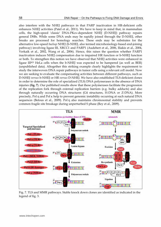
also interfere with the NHEJ pathways in that PARP inactivation in HR-deficient cells enhances NHEJ activities (Patel et al., 2011). We have to keep in mind that, in mammalian cells, the high-speed 'classic' DNA-PKcs–dependent NHEJ (D-NHEJ) pathway repairs general DSBs. While some DNA ends may be rapidly joined through the D-NHEJ, other breaks are processed for homology searches. These ends may be substrates for the alternative low-speed backup NHEJ (b-NHEJ, also termed microhomology-based end-joining pathway) involving ligase III, XRCC1 and PARP1 (Audebert et al., 2006, Iliakis et al., 2004, Verkaik et al., 2002, Wang et al., 2006). Hence, this raises the question whether PARP1 inactivation induces NHEJ compensation due to impaired HR function or b-NHEJ function or both. To strengthen this notion we have observed that NHEJ activities were enhanced in ligase IIIKD HeLa cells when the b-NHEJ was expected to be hampered (as well as BER) (unpublished data). Altogether this striking example clearly highlights the requirement to study the interwoven DNA repair pathways in tumor cells using a relevant cell model. Now we are seeking to evaluate the compensating activities between different pathways, such as D-NHEJ versus b-NHEJ or HR versus D-NHEJ. We have also established TLS-deficient clones in order to determine the role of specialized (TLS) DNA polymerases in the absence of DNA injuries (fig. 7). Our published results show that these polymerases facilitate the progression of the replication fork through external replication barriers (e.g. bulky adducts) and also through naturally occurring DNA structures (G4 structures, H-DNA or Z-DNA). More precisely, Pol η and Pol κ help to prevent genomic instability occurring at such natural DNA sequences (Betous et al., 2009). Pol η also maintains chromosomal stability and prevents common fragile site breakage during unperturbed S phase (Rey et al., 2009).
Fig. 7. TLS and MMR pathways. Stable knock down clones are identified as indicated in the legend of fig. 3.
www.intechopen.com

New Insight on Entangled DNA Repair Pathways: Stable Silenced Human Cells for Unraveling the DDR Jigsaw
59
To conclude, the major advantage of this strategy is the rapid establishment of new stable knockdown clones in various tumor-derived cells, which display stable gene silencing. A recent development has been to create dual pEBVsiRNA plasmids allowing efficient knockdown of two or more genes. For instance, double knockdown cells have been created where both DNA PKcs and ligase III were efficiently silenced with a single pEBVsiRNA vector. These cells, which grow normally, are expected to be deficient for both D-NHEJ and b-NHEJ. We have also developed plasmids targeting an endogenous gene and re-expressing an exogenous transcript carrying functional mutations. The latter approach allows mutant cells to be generated when the loss of the targeted gene is lethal. Hence, because we can easily and efficiently create DDR-deficient cells where one or more genes are silenced, we are now able to unravel the spectacular network of DNA repair pathways.
4. Acknowledgements
The author addresses special thanks to J. Hall for the critical reading of this manuscript and their kindly help. I am indebted to my collaborators who have greatly contributed to this work: Drs V. Favaudon, J. Hall, C. Godon and F. Mégnin for PARP1 and SSBR; Drs V. Schreiber, F. Dantzer, J.C. Amé and Boehler, C. for the PARP family; Drs F. Coin, N. Le May, and J.M. Egly for transcription; and Dr A. Sarasin and D. Despras for NER.
5. References
Allalunis-Turner, M. J.; Barron, G. M.; Day, R. S., 3rd; Dobler, K. D. & Mirzayans, R. (1993).
Isolation of two cell lines from a human malignant glioma specimen differing in
sensitivity to radiation and chemotherapeutic drugs. Radiat Res, Vol. 134, pp 349-
54.
Al-Minawi, A. Z.; Saleh-Gohari, N. & Helleday, T. (2008). The ERCC1/XPF endonuclease is
required for efficient single-strand annealing and gene conversion in mammalian
cells. Nucleic Acids Res, Vol. 36, pp 1-9.
Al-Tassan, N.; Chmiel, N. H.; Maynard, J.; Fleming, N.; Livingston, A. L.; Williams, G. T.;
Hodges, A. K.; Davies, D. R.; David, S. S.; Sampson, J. R. & Cheadle, J. P. (2002).
Inherited variants of MYH associated with somatic G:C-->T:A mutations in
colorectal tumors. Nat Genet, Vol. 30, pp 227-32.
Ame, J. C.; Fouquerel, E.; Gauthier, L. R.; Biard, D.; Boussin, F. D.; Dantzer, F.; de Murcia, G.
& Schreiber, V. (2009). Radiation-induced mitotic catastrophe in PARG-deficient
cells. J Cell Sci, Vol. 122, pp 1990-2002.
Ame, J. C.; Spenlehauer, C. & de Murcia, G. (2004). The PARP superfamily. Bioessays, Vol.
26, pp 882-93.
Amine, A.; Rivera, S.; Opolon, P.; Dekkal, M.; Biard, D. S.; Bouamar, H.; Louache, F.; McKay,
M. J.; Bourhis, J.; Deutsch, E. & Vozenin-Brotons, M. C. (2009). Novel anti-
metastatic action of cidofovir mediated by inhibition of E6/E7, CXCR4 and
Rho/ROCK signaling in HPV tumor cells. PLoS One, Vol. 4, pp e5018.
Aressy, B.; Bugler, B.; Valette, A.; Biard, D. & Ducommun, B. (2008). Moderate variations in
CDC25B protein levels modulate the response to DNA damaging agents. Cell
Cycle, Vol. 7, pp 2234-40.
www.intechopen.com
DNA Repair − On the Pathways to Fixing DNA Damage and Errors
60
Audebert, M.; Salles, B.; Weinfeld, M. & Calsou, P. (2006). Involvement of polynucleotide
kinase in a poly(ADP-ribose) polymerase-1-dependent DNA double-strand breaks
rejoining pathway. J Mol Biol, Vol. 356, pp 257-65.
Barnes, D. E.; Tomkinson, A. E.; Lehmann, A. R.; Webster, A. D. & Lindahl, T. (1992).
Mutations in the DNA ligase I gene of an individual with immunodeficiencies and
cellular hypersensitivity to DNA-damaging agents. Cell, Vol. 69, pp 495-503.
Betous, R.; Rey, L.; Wang, G.; Pillaire, M. J.; Puget, N.; Selves, J.; Biard, D. S.; Shin-ya, K.; Vasquez, K. M.; Cazaux, C. & Hoffmann, J. S. (2009). Role of TLS DNA polymerases eta and kappa in processing naturally occurring structured DNA in human cells. Mol Carcinog, Vol. 48, pp 369-78.
Biard, D. S. (2007). Untangling the relationships between DNA repair pathways by silencing more than 20 DNA repair genes in human stable clones. Nucleic Acids Res, Vol. 35, pp 3535-50.
Biard, D. S.; Despras, E.; Sarasin, A. & Angulo, J. F. (2005). Development of new EBV-based vectors for stable expression of small interfering RNA to mimick human syndromes: application to NER gene silencing. Mol Cancer Res, Vol. 3, pp 519-29.
Boehler, C.; Gauthier, L. R.; Mortusewicz, O.; Biard, D. S.; Saliou, J. M.; Bresson, A.; Sanglier-Cianferani, S.; Smith, S.; Schreiber, V.; Boussin, F. & Dantzer, F. (2011). Poly(ADP-ribose) polymerase 3 (PARP3), a newcomer in cellular response to DNA damage and mitotic progression. Proc Natl Acad Sci U S A, Vol., pp
Bouley, J.; Pionneau, C.; Varinot, J.; Biard, D.; Genestie, C.; Antoine, M.; Coulet, F.; Henri Stern, M.; Stoppa-Lyonnet, D. & Soubrier F. (2010). Proteomic analysis of BRCA1-depleted cell line reveals a putative role for RPA2 up-regulation in BRCA1 breast tumor development. Proteomics Clin. Appl, Vol. 4, pp 1-10.
Bouquet, F.; Ousset, M.; Biard, D.; Salles, B. & Muller, C. (2011). A DNA-dependent stress response involving DNA-PK occurs in hypoxic cells and contributes to cellular adaptation to hypoxia. J Cell Sci, Vol. In press, pp in press.
Britton, S.; Frit, P.; Biard, D.; Salles, B. & Calsou, P. (2009a). ARTEMIS nuclease facilitates apoptotic chromatin cleavage. Cancer Res, Vol. 69, pp 8120-6.
Britton, S.; Froment, C.; Frit, P.; Monsarrat, B.; Salles, B. & Calsou, P. (2009b). Cell nonhomologous end joining capacity controls SAF-A phosphorylation by DNA-PK in response to DNA double-strand breaks inducers. Cell Cycle, Vol. 8, pp 3717-22.
Bryant, H. E.; Schultz, N.; Thomas, H. D.; Parker, K. M.; Flower, D.; Lopez, E.; Kyle, S.; Meuth, M.; Curtin, N. J. & Helleday, T. (2005). Specific killing of BRCA2-deficient tumours with inhibitors of poly(ADP-ribose) polymerase. Nature, Vol. 434, pp 913-7.
Chen, S.; Bigner, S. H. & Modrich, P. (2001). High rate of CAD gene amplification in human cells deficient in MLH1 or MSH6. Proc Natl Acad Sci U S A, Vol. 98, pp 13802-7.
Claybon, A.; Karia, B.; Bruce, C. & Bishop, A. J. (2010). PARP1 suppresses homologous recombination events in mice in vivo. Nucleic Acids Res, Vol. 38, pp 7538-45.
Coant, N.; Ben Mkaddem, S.; Pedruzzi, E.; Guichard, C.; Treton, X.; Ducroc, R.; Freund, J. N.; Cazals-Hatem, D.; Bouhnik, Y.; Woerther, P. L.; Skurnik, D.; Grodet, A.; Fay, M.; Biard, D.; Lesuffleur, T.; Deffert, C.; Moreau, R.; Groyer, A.; Krause, K. H.; Daniel, F. & Ogier-Denis, E. (2010). NADPH oxidase 1 modulates WNT and NOTCH1 signaling to control the fate of proliferative progenitor cells in the colon. Mol Cell Biol, Vol. 30, pp 2636-50.
www.intechopen.com
New Insight on Entangled DNA Repair Pathways: Stable Silenced Human Cells for Unraveling the DDR Jigsaw
61
Coin, F.; Oksenych, V.; Mocquet, V.; Groh, S.; Blattner, C. & Egly, J. M. (2008). Nucleotide excision repair driven by the dissociation of CAK from TFIIH. Mol Cell, Vol. 31, pp 9-20.
D'Amours, D.; Sallmann, F. R.; Dixit, V. M. & Poirier, G. G. (2001). Gain-of-function of poly(ADP-ribose) polymerase-1 upon cleavage by apoptotic proteases: implications for apoptosis. J Cell Sci, Vol. 114, pp 3771-8.
D'Andrea, A. D. (2010). Susceptibility pathways in Fanconi's anemia and breast cancer. N Engl J Med, Vol. 362, pp 1909-19.
Dantzer, F.; Ame, J. C.; Schreiber, V.; Nakamura, J.; Menissier-de Murcia, J. & de Murcia, G. (2006). Poly(ADP-ribose) polymerase-1 activation during DNA damage and repair. Methods Enzymol, Vol. 409, pp 493-510.
Despras, E.; Pfeiffer, P.; Salles, B.; Calsou, P.; Kuhfittig-Kulle, S.; Angulo, J. F. & Biard, D. S. (2007). Long-term XPC silencing reduces DNA double-strand break repair. Cancer Res, Vol. 67, pp 2526-34.
Drouet, J.; Delteil, C.; Lefrancois, J.; Concannon, P.; Salles, B. & Calsou, P. (2005). DNA-dependent protein kinase and XRCC4-DNA ligase IV mobilization in the cell in response to DNA double strand breaks. J Biol Chem, Vol. 280, pp 7060-9.
Durandy, A. (2009). Immunoglobulin class switch recombination: study through human natural mutants. Philos Trans R Soc Lond B Biol Sci, Vol. 364, pp 577-82.
Farmer, H.; McCabe, N.; Lord, C. J.; Tutt, A. N.; Johnson, D. A.; Richardson, T. B.; Santarosa, M.; Dillon, K. J.; Hickson, I.; Knights, C.; Martin, N. M.; Jackson, S. P.; Smith, G. C. & Ashworth, A. (2005). Targeting the DNA repair defect in BRCA mutant cells as a therapeutic strategy. Nature, Vol. 434, pp 917-21.
Giglia-Mari, G.; Coin, F.; Ranish, J. A.; Hoogstraten, D.; Theil, A.; Wijgers, N.; Jaspers, N. G.; Raams, A.; Argentini, M.; van der Spek, P. J.; Botta, E.; Stefanini, M.; Egly, J. M.; Aebersold, R.; Hoeijmakers, J. H. & Vermeulen, W. (2004). A new, tenth subunit of TFIIH is responsible for the DNA repair syndrome trichothiodystrophy group A. Nat Genet, Vol. 36, pp 714-9.
Godon, C.; Cordelieres, F. P.; Biard, D.; Giocanti, N.; Megnin-Chanet, F.; Hall, J. & Favaudon, V. (2008). PARP inhibition versus PARP-1 silencing: different outcomes in terms of single-strand break repair and radiation susceptibility. Nucleic Acids Res, Vol. 36, pp 4454-64.
Hartman, A. R. & Ford, J. M. (2002). BRCA1 induces DNA damage recognition factors and enhances nucleotide excision repair. Nat Genet, Vol. 32, pp 180-4.
Hartman, A. R. & Ford, J. M. (2003). BRCA1 and p53: compensatory roles in DNA repair. J Mol Med, Vol. 81, pp 700-7.
Hoeijmakers, J. H. (2001). Genome maintenance mechanisms for preventing cancer. Nature, Vol. 411, pp 366-74.
Huang, T. T. & D'Andrea, A. D. (2006). Regulation of DNA repair by ubiquitylation. Nat Rev Mol Cell Biol, Vol. 7, pp 323-34.
Iliakis, G.; Wang, H.; Perrault, A. R.; Boecker, W.; Rosidi, B.; Windhofer, F.; Wu, W.; Guan, J.; Terzoudi, G. & Pantelias, G. (2004). Mechanisms of DNA double strand break repair and chromosome aberration formation. Cytogenet Genome Res, Vol. 104, pp 14-20.
Iyer, R. R.; Pluciennik, A.; Burdett, V. & Modrich, P. L. (2006). DNA mismatch repair: functions and mechanisms. Chem Rev, Vol. 106, pp 302-23.
www.intechopen.com

DNA Repair − On the Pathways to Fixing DNA Damage and Errors
62
Jaspers, N. G.; Raams, A.; Silengo, M. C.; Wijgers, N.; Niedernhofer, L. J.; Robinson, A. R.; Giglia-Mari, G.; Hoogstraten, D.; Kleijer, W. J.; Hoeijmakers, J. H. & Vermeulen, W. (2007). First reported patient with human ERCC1 deficiency has cerebro-oculo-facio-skeletal syndrome with a mild defect in nucleotide excision repair and severe developmental failure. Am J Hum Genet, Vol. 80, pp 457-66.
Le May, N.; Egly, J. M. & Coin, F. (2010a). True lies: the double life of the nucleotide excision repair factors in transcription and DNA repair. J Nucleic Acids, Vol. 2010, pp
Le May, N.; Mota-Fernandes, D.; Velez-Cruz, R.; Iltis, I.; Biard, D. & Egly, J. M. (2010b). NER factors are recruited to active promoters and facilitate chromatin modification for transcription in the absence of exogenous genotoxic attack. Mol Cell, Vol. 38, pp 54-66.
Limoli, C. L.; Giedzinski, E. & Cleaver, J. E. (2005). Alternative recombination pathways in UV-irradiated XP variant cells. Oncogene, Vol. 24, pp 3708-14.
Loseva, O.; Jemth, A. S.; Bryant, H. E.; Schuler, H.; Lehtio, L.; Karlberg, T. & Helleday, T. (2010). PARP-3 is a mono-ADP-ribosylase that activates PARP-1 in the absence of DNA. J Biol Chem, Vol. 285, pp 8054-60.
Macrae, C. J.; McCulloch, R. D.; Ylanko, J.; Durocher, D. & Koch, C. A. (2008). APLF (C2orf13) facilitates nonhomologous end-joining and undergoes ATM-dependent hyperphosphorylation following ionizing radiation. DNA Repair (Amst), Vol. 7, pp 292-302.
Matveeva, O.; Nechipurenko, Y.; Rossi, L.; Moore, B.; Saetrom, P.; Ogurtsov, A. Y.; Atkins, J. F. & Shabalina, S. A. (2007). Comparison of approaches for rational siRNA design leading to a new efficient and transparent method. Nucleic Acids Res, Vol. 35, pp e63.
McKinnon, P. J. (2009). DNA repair deficiency and neurological disease. Nat Rev Neurosci, Vol. 10, pp 100-12.
Mendes-Pereira, A. M.; Martin, S. A.; Brough, R.; McCarthy, A.; Taylor, J. R.; Kim, J. S.; Waldman, T.; Lord, C. J. & Ashworth, A. (2009). Synthetic lethal targeting of PTEN mutant cells with PARP inhibitors. EMBO Mol Med, Vol. 1, pp 315-22.
Menissier de Murcia, J.; Ricoul, M.; Tartier, L.; Niedergang, C.; Huber, A.; Dantzer, F.; Schreiber, V.; Ame, J. C.; Dierich, A.; LeMeur, M.; Sabatier, L.; Chambon, P. & de Murcia, G. (2003). Functional interaction between PARP-1 and PARP-2 in chromosome stability and embryonic development in mouse. Embo J, Vol. 22, pp 2255-63.
Meulle, A.; Salles, B.; Daviaud, D.; Valet, P. & Muller, C. (2008). Positive regulation of DNA double strand break repair activity during differentiation of long life span cells: the example of adipogenesis. PLoS One, Vol. 3, pp e3345.
Mladenov, E. & Iliakis, G. (2011). Induction and repair of DNA double strand breaks: The increasing spectrum of non-homologous end joining pathways. Mutat Res, Vol., pp in press.
Moynahan, M. E.; Chiu, J. W.; Koller, B. H. & Jasin, M. (1999). Brca1 controls homology-directed DNA repair. Mol Cell, Vol. 4, pp 511-8.
Mullan, P. B.; Gorski, J. J. & Harkin, D. P. (2006). BRCA1--a good predictive marker of drug sensitivity in breast cancer treatment? Biochim Biophys Acta, Vol. 1766, pp 205-16.
Noel, G.; Godon, C.; Fernet, M.; Giocanti, N.; Megnin-Chanet, F. & Favaudon, V. (2006). Radiosensitization by the poly(ADP-ribose) polymerase inhibitor 4-amino-1,8-
www.intechopen.com
New Insight on Entangled DNA Repair Pathways: Stable Silenced Human Cells for Unraveling the DDR Jigsaw
63
naphthalimide is specific of the S phase of the cell cycle and involves arrest of DNA synthesis. Mol Cancer Ther, Vol. 5, pp 564-74.
O'Driscoll, M. & Jeggo, P. A. (2008). The role of the DNA damage response pathways in brain development and microcephaly: insight from human disorders. DNA Repair (Amst), Vol. 7, pp 1039-50.
O'Shaughnessy, J.; Osborne, C.; Pippen, J. E.; Yoffe, M.; Patt, D.; Rocha, C.; Koo, I. C.; Sherman, B. M. & Bradley, C. (2011). Iniparib plus chemotherapy in metastatic triple-negative breast cancer. N Engl J Med, Vol. 364, pp 205-14.
Ousset, M.; Bouquet, F.; Fallone, F.; Biard, D.; Dray, C.; Valet, P.; Salles, B. & Muller, C. (2010). Loss of ATM positively regulates the expression of hypoxia inducible factor 1 (HIF-1) through oxidative stress: Role in the physiopathology of the disease. Cell Cycle, Vol. 9, pp 2814-22.
Patel, A. G.; Sarkaria, J. N. & Kaufmann, S. H. (2011). Nonhomologous end joining drives poly(ADP-ribose) polymerase (PARP) inhibitor lethality in homologous recombination-deficient cells. Proc Natl Acad Sci U S A, Vol. 108, pp 3406-11.
Pearl, L. H. (2000). Structure and function in the uracil-DNA glycosylase superfamily. Mutat Res, Vol. 460, pp 165-81.
Peltomaki, P. (2003). Role of DNA mismatch repair defects in the pathogenesis of human cancer. J Clin Oncol, Vol. 21, pp 1174-9.
Pennarun, G.; Granotier, C.; Hoffschir, F.; Mandine, E.; Biard, D.; Gauthier, L. R. & Boussin, F. D. (2008). Role of ATM in the telomere response to the G-quadruplex ligand 360A. Nucleic Acids Res, Vol., pp 1741-54.
Pennarun, G.; Hoffschir, F.; Revaud, D.; Granotier, C.; Gauthier, L. R.; Mailliet, P.; Biard, D. S. & Boussin, F. D. (2010). ATR contributes to telomere maintenance in human cells. Nucleic Acids Res, Vol. 38, pp 2955-63.
Quinn, J. E.; Kennedy, R. D.; Mullan, P. B.; Gilmore, P. M.; Carty, M.; Johnston, P. G. & Harkin, D. P. (2003). BRCA1 functions as a differential modulator of chemotherapy-induced apoptosis. Cancer Res, Vol. 63, pp 6221-8.
Rey, L.; Sidorova, J. M.; Puget, N.; Boudsocq, F.; Biard, D. S.; Monnat, R. J., Jr.; Cazaux, C. & Hoffmann, J. S. (2009). Human DNA polymerase eta is required for common fragile site stability during unperturbed DNA replication. Mol Cell Biol, Vol. 29, pp 3344-54.
Roos, W. P. & Kaina, B. (2006). DNA damage-induced cell death by apoptosis. Trends Mol Med, Vol. 12, pp 440-50.
Rouleau, M.; McDonald, D.; Gagne, P.; Ouellet, M. E.; Droit, A.; Hunter, J. M.; Dutertre, S.; Prigent, C.; Hendzel, M. J. & Poirier, G. G. (2007). PARP-3 associates with polycomb group bodies and with components of the DNA damage repair machinery. J Cell Biochem, Vol. 100, pp 385-401.
Rulten, S. L.; Fisher, A. E.; Robert, I.; Zuma, M. C.; Rouleau, M.; Ju, L.; Poirier, G.; Reina-San-Martin, B. & Caldecott, K. W. (2011). PARP-3 and APLF function together to accelerate nonhomologous end-joining. Mol Cell, Vol. 41, pp 33-45.
Sartori, A. A.; Fitz-Gibbon, S.; Yang, H.; Miller, J. H. & Jiricny, J. (2002). A novel uracil-DNA glycosylase with broad substrate specificity and an unusual active site. Embo J, Vol. 21, pp 3182-91.
Schmutz, V.; Janel-Bintz, R.; Wagner, J.; Biard, D.; Shiomi, N.; Fuchs, R. P. & Cordonnier, A. M. (2010). Role of the ubiquitin-binding domain of Pol{eta} in Rad18-independent
www.intechopen.com
DNA Repair − On the Pathways to Fixing DNA Damage and Errors
64
translesion DNA synthesis in human cell extracts. Nucleic Acids Res, Vol. 38, pp 6456-65.
Schreiber, V.; Ame, J. C.; Dolle, P.; Schultz, I.; Rinaldi, B.; Fraulob, V.; Menissier-de Murcia, J. & de Murcia, G. (2002). Poly(ADP-ribose) polymerase-2 (PARP-2) is required for efficient base excision DNA repair in association with PARP-1 and XRCC1. J Biol Chem, Vol. 277, pp 23028-36.
Schreiber, V.; Dantzer, F.; Ame, J. C. & de Murcia, G. (2006). Poly(ADP-ribose): novel functions for an old molecule. Nat Rev Mol Cell Biol, Vol. 7, pp 517-28.
Schulte, J. H.; Kuhfittig-Kulle, S.; Klein-Hitpass, L.; Schramm, A.; Biard, D. S.; Pfeiffer, P. & Eggert, A. (2008). Expression of the TrkA or TrkB receptor tyrosine kinase alters the double-strand break (DSB) repair capacity of SY5Y neuroblastoma cells. DNA Repair (Amst), Vol., pp
Solyom, S.; Pylkas, K. & Winqvist, R. (2010). Screening for large genomic rearrangements of the BRIP1 and CHK1 genes in Finnish breast cancer families. Fam Cancer, Vol. 9, pp 537-40.
Verkaik, N. S.; Esveldt-van Lange, R. E.; van Heemst, D.; Bruggenwirth, H. T.; Hoeijmakers, J. H.; Zdzienicka, M. Z. & van Gent, D. C. (2002). Different types of V(D)J recombination and end-joining defects in DNA double-strand break repair mutant mammalian cells. Eur J Immunol, Vol. 32, pp 701-9.
Vert, J. P.; Foveau, N.; Lajaunie, C. & Vandenbrouck, Y. (2006). An accurate and interpretable model for siRNA efficacy prediction. BMC Bioinformatics, Vol. 7, pp 520.
Wang, M.; Wu, W.; Rosidi, B.; Zhang, L.; Wang, H. & Iliakis, G. (2006). PARP-1 and Ku compete for repair of DNA double strand breaks by distinct NHEJ pathways. Nucleic Acids Res, Vol. 34, pp 6170-82.
Williamson, C. T.; Muzik, H.; Turhan, A. G.; Zamo, A.; O'Connor, M. J.; Bebb, D. G. & Lees-Miller, S. P. (2010). ATM deficiency sensitizes mantle cell lymphoma cells to poly(ADP-ribose) polymerase-1 inhibitors. Mol Cancer Ther, Vol. 9, pp 347-57.
Wu, P. Y.; Frit, P.; Malivert, L.; Revy, P.; Biard, D.; Salles, B. & Calsou, P. (2007). Interplay between Cernunnos-XLF and Nonhomologous End-joining Proteins at DNA Ends in the Cell. J Biol Chem, Vol. 282, pp 31937-31943.
Yan, C. T.; Boboila, C.; Souza, E. K.; Franco, S.; Hickernell, T. R.; Murphy, M.; Gumaste, S.; Geyer, M.; Zarrin, A. A.; Manis, J. P.; Rajewsky, K. & Alt, F. W. (2007). IgH class switching and translocations use a robust non-classical end-joining pathway. Nature, Vol. 449, pp 478-82.
www.intechopen.com
DNA Repair - On the Pathways to Fixing DNA Damage and ErrorsEdited by Dr. Francesca Storici
ISBN 978-953-307-649-2Hard cover, 380 pagesPublisher InTechPublished online 09, September, 2011Published in print edition September, 2011
InTech EuropeUniversity Campus STeP Ri Slavka Krautzeka 83/A 51000 Rijeka, Croatia Phone: +385 (51) 770 447 Fax: +385 (51) 686 166www.intechopen.com
InTech ChinaUnit 405, Office Block, Hotel Equatorial Shanghai No.65, Yan An Road (West), Shanghai, 200040, China
Phone: +86-21-62489820 Fax: +86-21-62489821
DNA repair is fundamental to all cell types to maintain genomic stability. A collection of cutting-edge reviews,DNA Repair - On the pathways to fixing DNA damage and errors covers major aspects of the DNA repairprocesses in a large variety of organisms, emphasizing foremost developments, questions to be solved andnew directions in this rapidly evolving area of modern biology. Written by researchers at the vanguard of theDNA repair field, the chapters highlight the importance of the DNA repair mechanisms and their linkage to DNAreplication, cell-cycle progression and DNA recombination. Major topics include: base excision repair,nucleotide excision repair, mismatch repair, double-strand break repair, with focus on specific inhibitors andkey players of DNA repair such as nucleases, ubiquitin-proteasome enzymes, poly ADP-ribose polymeraseand factors relevant for DNA repair in mitochondria and embryonic stem cells. This book is a journey into thecosmos of DNA repair and its frontiers.
How to referenceIn order to correctly reference this scholarly work, feel free to copy and paste the following:
Biard Denis S.F. (2011). New Insight on Entangled DNA Repair Pathways: Stable Silenced Human Cells forUnraveling the DDR Jigsaw, DNA Repair - On the Pathways to Fixing DNA Damage and Errors, Dr. FrancescaStorici (Ed.), ISBN: 978-953-307-649-2, InTech, Available from: http://www.intechopen.com/books/dna-repair-on-the-pathways-to-fixing-dna-damage-and-errors/new-insight-on-entangled-dna-repair-pathways-stable-silenced-human-cells-for-unraveling-the-ddr-jigs

© 2011 The Author(s). Licensee IntechOpen. This chapter is distributedunder the terms of the Creative Commons Attribution-NonCommercial-ShareAlike-3.0 License, which permits use, distribution and reproduction fornon-commercial purposes, provided the original is properly cited andderivative works building on this content are distributed under the samelicense.